Cardiogenic Shock: Complications of Acute...
47
1 Cardiogenic Shock: Complications of Acute MI Michael H. Picard MD Massachusetts General Hospital Harvard Medical School No disclosures
Transcript of Cardiogenic Shock: Complications of Acute...

1
Cardiogenic Shock:
Complications of Acute MI
Michael H. Picard MD
Massachusetts General Hospital
Harvard Medical School
No disclosures

2
MOC credits for attending Echo Hawaii
• Make sure you enter your ABIM number in your
account with ASE
3
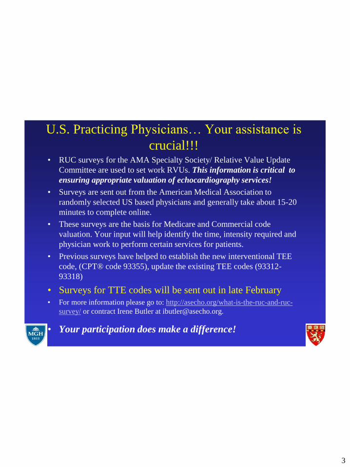
• RUC surveys for the AMA Specialty Society/ Relative Value Update
Committee are used to set work RVUs. This information is critical to
ensuring appropriate valuation of echocardiography services!
• Surveys are sent out from the American Medical Association to
randomly selected US based physicians and generally take about 15-20
minutes to complete online.
• These surveys are the basis for Medicare and Commercial code
valuation. Your input will help identify the time, intensity required and
physician work to perform certain services for patients.
• Previous surveys have helped to establish the new interventional TEE
code, (CPT® code 93355), update the existing TEE codes (93312-
93318)
• Surveys for TTE codes will be sent out in late February
• For more information please go to: http://asecho.org/what-is-the-ruc-and-ruc-
survey/ or contract Irene Butler at [email protected].
• Your participation does make a difference!
U.S. Practicing Physicians… Your assistance is
crucial!!!

4
Complications of MI: role of TTE & TEE
• Ongoing chest pain– ischemia
– pericarditis
– pulmonary embolism
– pseudoaneurysm, subacute free wall rupture
• shock, hemodynamic compromise w/ or w/o new murmur– mitral regurgitation
– ventricular septal rupture
– Primary LV failure
– RV MI
• stroke– LV thrombus
• VT– aneurysms

5
Cardiogenic shockdual role for echo
• Early – differential diagnosis
• confirm or establish the etiology of the hypotension
– more rapid triage to appropriate therapy• Including mechanical circulatory support
– assessing prognosis (LVEF, MR, RV function)
• Later– response to treatment
– prognosis
6
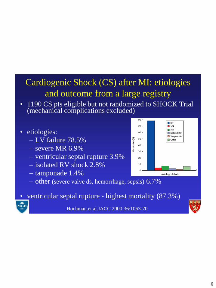
Cardiogenic Shock (CS) after MI: etiologies
and outcome from a large registry
Hochman et al JACC 2000;36:1063-70
• 1190 CS pts eligible but not randomized to SHOCK Trial (mechanical complications excluded)
• etiologies:– LV failure 78.5%
– severe MR 6.9%– ventricular septal rupture 3.9%
– isolated RV shock 2.8%– tamponade 1.4%
– other (severe valve ds, hemorrhage, sepsis) 6.7%
• ventricular septal rupture - highest mortality (87.3%)

7
Cardiogenic shock: Same clinical status, different
echoes, different etiologies
53 yo M hypotension within
12 hours of admission for MI
81 yo M with 3 vessel CAD
including left main ds admitted
with wheezing, hypotension and
pulmonary edema

8
M.R. - 64 yo F with shock 15 days after
first anterior MIHx of HTN, elevated chol; 2 weeks prior to transfer
to MGH she presented to suburban hospital with
evolving MI (saw PCP for GI upset, SOB).
No prior hx of angina
R/I for large anterior MI, recurrent CHF treated
medically. Echo there - dilated LV, EF < 20%,
moderate MR
on morning of d/c (day 15), in shower experienced
chest pain and lightheadedness.
9
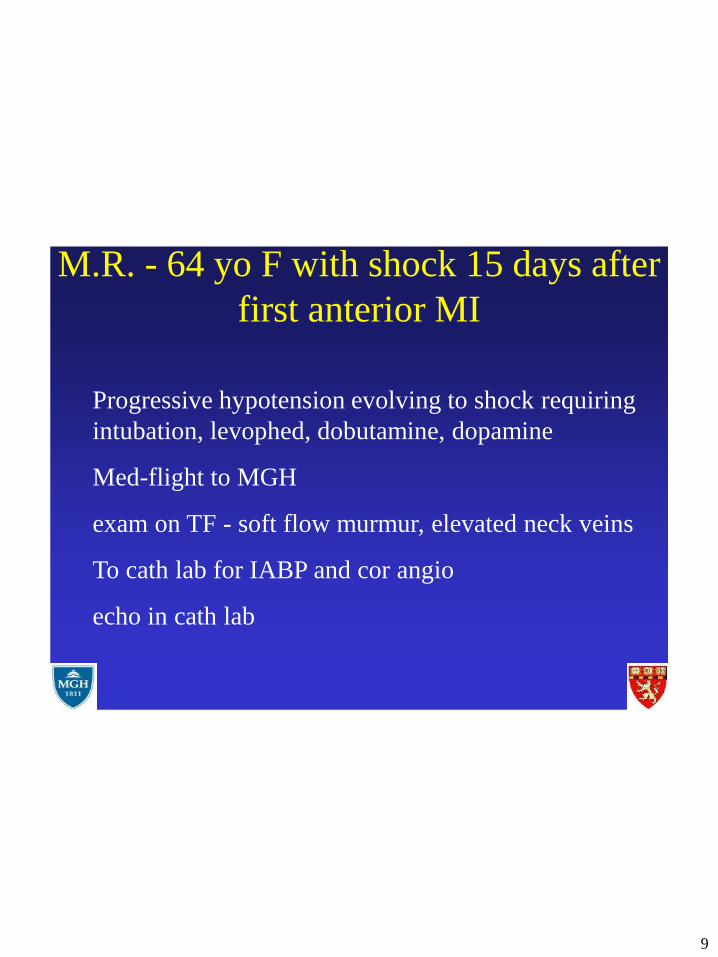
M.R. - 64 yo F with shock 15 days after
first anterior MI
Progressive hypotension evolving to shock requiring
intubation, levophed, dobutamine, dopamine
Med-flight to MGH
exam on TF - soft flow murmur, elevated neck veins
To cath lab for IABP and cor angio
echo in cath lab

10

11
M.R. - 64 yo F with shock 15 days after
first anterior MI

12
M.R. - 64 yo F with shock 15 days after first
anterior MI – ventricular septal rupture
Treatment
IABP
urgent surgery
saphenous vein graft to RCA (80% mid
RCA, clean circ, proximal LAD recanalized)
LV opened through infarct, 1-1.5 defect in
mid to apical septum, re-endothelialization with
bovine pericardium patch, infarct trimmed, primary
closure of VSD

13
Myocardial Perforationseptal, free wall, papillary muscle
• Typically occurs Day 2 – 8 post-MI
• Risk factors
– 1° - Q waves, first MI
– 2° - women > male, elderly, hypertensives
– 3o - delayed recognition, continued physical activity
• 90 % fatal
• Pathophysiology
– total or near-total occlusion of coronary artery with inadequate collateral circulation

14
rupture of the ventricular septum
• 0.5 - 2% of MIs; 1 - 5% of MI deaths
• exam -
– new holosystolic murmur. +/- thrill, biventricular CHF, CO,
cardiogenic shock (exam may mimic pap mus rupture)
• anterior MI (60%) - apical septum
• inferior MI - basal septum (worse prognosis)
• pathophysiology
– L to R shunt, RV volume overload, PA flow, systemic
flow and CO, BP
• RV function - important determinant of survival

15
Ventricular septal rupture - role of TEE
pre-op planning• Location
– antero-apex
• involvement of free wall?
– infero-basal
• involvement of subvalvular apparatus (MV + TV)
• complex (multiple) vs. simple
• RV size and function
• severity of MR
16
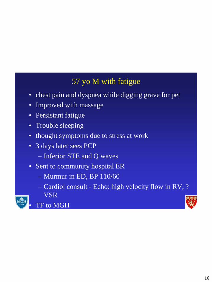
57 yo M with fatigue
• chest pain and dyspnea while digging grave for pet
• Improved with massage
• Persistant fatigue
• Trouble sleeping
• thought symptoms due to stress at work
• 3 days later sees PCP
– Inferior STE and Q waves
• Sent to community hospital ER
– Murmur in ED, BP 110/60
– Cardiol consult - Echo: high velocity flow in RV, ?
VSR
• TF to MGH
17
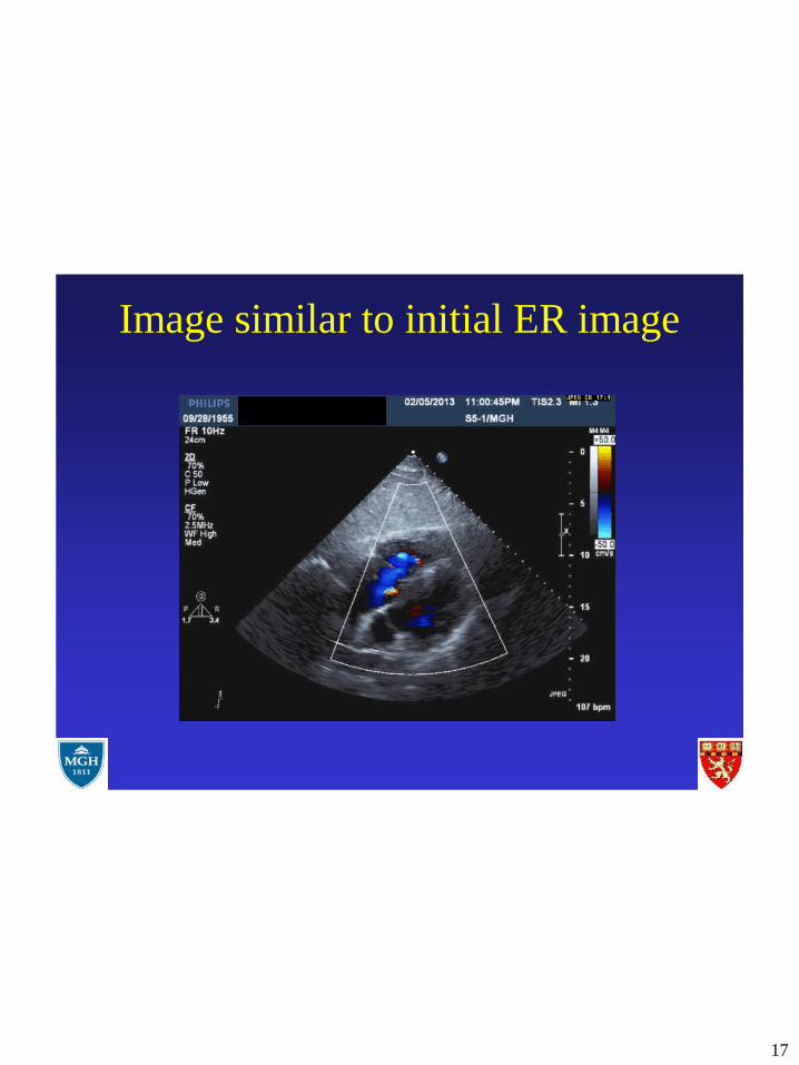
Image similar to initial ER image

18
TTE by fellow on call

19

20
21
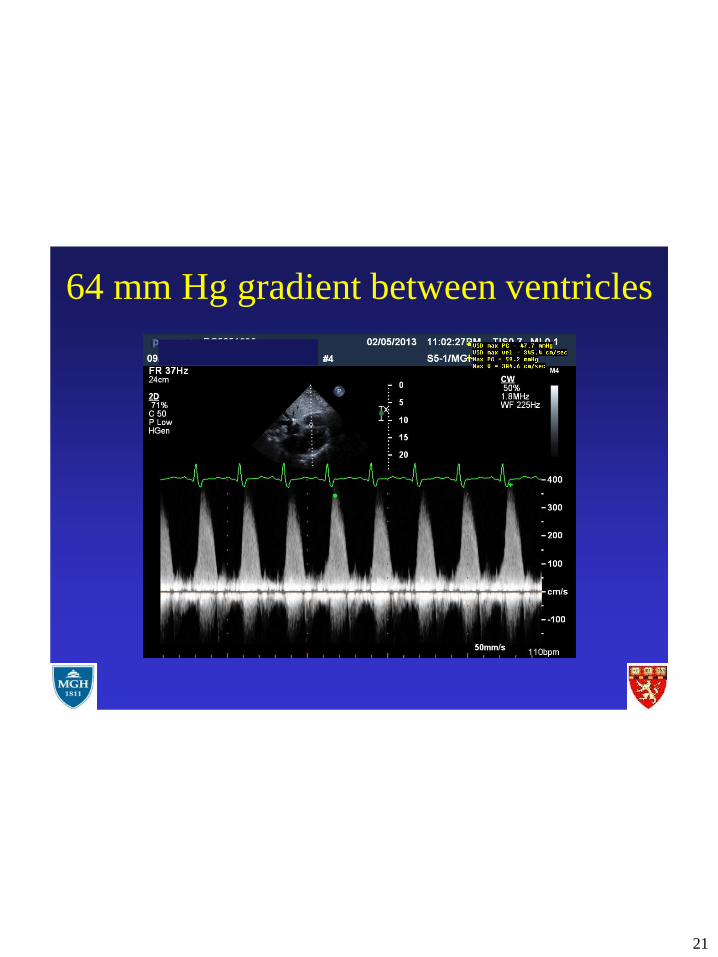
64 mm Hg gradient between ventricles

22
• Percutaneous device closure vs. surgical repair ?
23
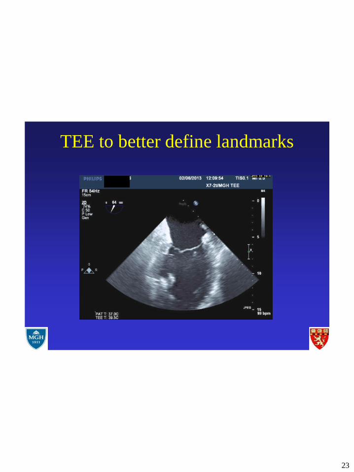
TEE to better define landmarks

24

25
Transesophageal Echocardiogram
• Overall normal LV systolic function with inferior
WMA
• Large VSD from base of inferior septum (near
insertion of AV valves) to inferior edge of
posteromedial papillary muscle (LV orifice
24x16 mm by 3D imaging)
• Trace-mild MR, mild TR
• RV diffusely hypokinetic
• No PFO
• No pericardial effusion25

26
VSR Morphology• Simple:
– Direct, “through-and-
through”
– Anteroapical septum
• Complex:
– Hemorrhage with irregular,
serpiginous tracts
– Basal inferoposterior
septum
– May have concomitant
ventricular free wall or
papillary muscle tears26
(Bimbaum Y, et al. N Engl J Med. 2002; Edwards BS, et al. Am J Cardiol. 1984; Mann JM,
Roberts WC. Am J Cardiol. 1988; Reynolds HR. Eur Heart J. 2010)

27
• Good surgical repair (defect and free wall)
including CABG
• Long recovery due to slow recovery of RV
function

28
Echo in Cardiogenic Shock:
Is TTE sufficient ?
• TEE when TTE does not reveal the answer
– inadequate TTE
– unexplained hemodynamic instability
• small MI but cardiogenic shock
• inadequate response to treatment
– high index of suspicion of mechanical
complication

29
76 yo F pulmonary edema
and shock 48 hours after RCA
stent for acute MI

30

31
Day 4 s/p IMI – collapses during cardiac rehab

32
Papillary muscle rupture: flail mitral leaflet
• 1% of MIs; 5% of MI deaths
• total rupture - rare (shock, fatal)
partial rupture - more common
chest pain, hypotension, shock, initial period of
stabilization followed by rapid unpredictable
deterioration, pulmonary edema (asymmetric)
Exam - new, loud holosystolic murmur at apex
(murmur decreases as BP falls); severe CHF

33
Papillary muscle rupture: flail mitral leaflet
• Important to distinguish etiology of MR as this will not improve with relief of ischemia
• inferior MI > anterior MI
– posteromedial PM > anterolateral PM
(PDA) (LAD/Diag + Circ)
• Echo
– small inferior WMA, whipping motion of tip of MV leaflet, complete transection rare - rupture of tip more common
– imaging - sweep through entire valve
– MR may be underestimated! - eccentric nature of jet, large regurgitant orifice
34
67 yo M with acute MI – hypotension at presentation; primary
PTCA of culprit circ lesion. Return to cath lab 12 hours later
for re-look due to persistant hypotension – circ open.
TTE in CCU

35
36
Subacute rupture
• Same risk factors, presentation different
• moderate pericardial effusion
– with tamponade - modest progressive hypotension
– without tamponade
• management
– surgery vs. medical therapy
• Difficult TTE/TEE diagnosis
– Role of contrast
37
TEE in subacute free wall rupture
• Rare, case reports
• TEE findings
– discontinuity of wall or marked thinning of wall
– thinned akinetic wall
– pericardial effusion +/- thrombus in pericardial space
– low velocity Doppler flow in myocardium
• LV contrast
– delineate serpiginous channels, opacify pericardial
space

38
Elderly diabetic, hypertensive women with 3 days of chest
pain and dyspnea found unresponsive at home
revived and then transferred from community hospital for R/O
aortic dissection
TEE at MGH ER negative for dissection, however……..

39

40

41
Surgical findings
• Thinned inferior wall
• Pericardial hematoma
• Myocardial hemorrhage
• Multiple comlex serpiginous tracts in the inferior
wall
42
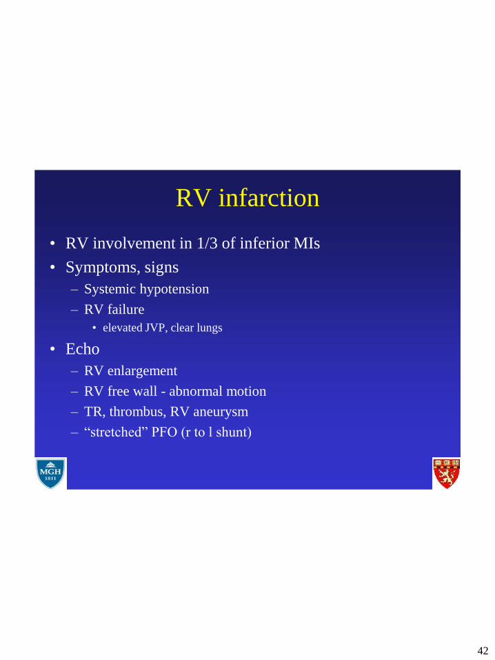
RV infarction
• RV involvement in 1/3 of inferior MIs
• Symptoms, signs
– Systemic hypotension
– RV failure
• elevated JVP, clear lungs
• Echo
– RV enlargement
– RV free wall - abnormal motion
– TR, thrombus, RV aneurysm
– “stretched” PFO (r to l shunt)

43
RV MI

44
2015 SCAI/ACC/HFSA/STS Clinical Expert Consensus
Statement on the Use of Percutaneous Mechanical Circulatory
Support Devices in CV Care
• suggested indications
– Short term support (or stabilization for transfer)
– High risk interventions
• PCI, perc valve, VT ablation
– Post-cardiotomy
– Cardiogenic shock anticipated to be quickly reversible
• Revascularized acute MI
– Decompensated heart failure
– Complications of acute MI, Acute valve failure (pre-op)
– Impaired oxygenation
48
Rihal et al, JACC 2015;65:e7-e26
45
ASE LVAD guidelines Aug 2015

46
types of mechanical support
Remove blood from: Inject blood into:
IABP Aorta aorta
ECMO (VA) RA Descending aorta
Tandem Heart LA Descending aorta
Impella LVOT Ascending aorta
LVAD
RVAD
BiVAD
LV apex or LA
RV
Ascending Ao
Pulm artery
ECMO (VV) Venous or RA Pulm Artery

47
Mechanical complications of MIconclusions
• May mimic other problems
• prompt recognition and intervention can reduce
the high mortality
• key to early diagnosis:
– high index of suspicion
– use of bedside noninvasive imaging
• TTE, TEE, contrast