Cardiacarrhythmiasinacutecoronarysyndromes: position …€¦ ·...
19
EHRA/ACCA/EAPCI POSITION PAPER Cardiac arrhythmias in acute coronary syndromes: position paper from the joint EHRA, ACCA, and EAPCI task force Bulent Gorenek * † (Chairperson, Turkey), Carina Blomstro ¨ m Lundqvist † (Sweden), Josep Brugada Terradellas † (Spain), A. John Camm † (UK), Gerhard Hindricks † (Germany), Kurt Huber ‡ (Austria), Paulus Kirchhof † (UK), Karl-Heinz Kuck † (Germany), Gulmira Kudaiberdieva † (Turkey), Tina Lin † (Germany), Antonio Raviele † (Italy), Massimo Santini † (Italy), Roland Richard Tilz † (Germany), Marco Valgimigli } (The Netherlands), Marc A. Vos † (The Netherlands), Christian Vrints ‡ (Belgium), and Uwe Zeymer ‡ (Germany) Document Reviewers: Gregory Y.H. Lip (Review Coordinator) (UK), Tatjania Potpara (Serbia), Laurent Fauchier (France), Christian Sticherling (Switzerland), Marco Roffi (Switzerland), Petr Widimsky (Czech Republic), Julinda Mehilli (Germany), Maddalena Lettino (Italy), Francois Schiele (France), Peter Sinnaeve (Belgium), Giueseppe Boriani (Italy), Deirdre Lane (UK), and Irene Savelieva (on behalf of EP-Europace, UK) Introduction It is known that myocardial ischaemia and infarction leads to severe metabolic and electrophysiological changes that induce silent or symptomatic life-threatening arrhythmias. Sudden cardiac death is most often attributed to this pathophysiology, but many patients survive the early stage of an acute coronarysyndrome (ACS) reaching a medical facility where the management of ischaemia and infarction must include continuous electrocardiographic (ECG) and hemo- dynamic monitoring, and a prompt therapeutic response to incident sustained arrhythmias. During the last decade, the hospital locations in which arrhythmias are most relevant have changed to include the cardiac catheterization laboratory, since the preferred management of early acute ACS is generally interventional in nature. However, a large proportion of patients are still managed medically. Both atrial and ventricular arrhythmias may occur in the setting of ACS and sustained ventricular tachyarrhythmias (VAs) may be asso- ciated with circulatory collapse and require immediate treatment. Atrial fibrillation (AF) may also warrant urgent treatment when a fast ventricular rate is associated with hemodynamic deterioration. The management of other arrhythmias is also based largely on symptoms rather than to avert progression to more serious arrhythmias. Prophylactic antiarrhythmic management strategies have largely been discouraged. Although the mainstay of antiarrhythmic therapy used to rely on antiarrhythmic drugs (AADs), particularly sodium channel blockers and amiodarone, their use has now declined, since clinical evidence to support such treatment has never been convincing. Therapy for acute coronary syndrome and arrhythmia management are now based increasingly on invasive approaches. The changes in the clinical approach to arrhythmia management in ACS have been so substantial that the European Heart Rhythm Association, the Acute Cardiovas- cular Care Association and the European Association of Percutan- eous Cardiovascular Interventions established a task force to define the current position. Mechanisms of ischaemia-related ventricular arrhythmias Generation of the ventricular action potential by voltage and sub- strate dependent ion currents is the basis for the contraction of * Corresponding author. Prof. Bulent Gorenek MD FACC FESC, Eskisehir Osmangazi University Cardiology Department, Eskisehir, Turkey. Tel: +90 222 2292266; Fax: +90 222 2292266. E-mail address: [email protected] † EHRA. ‡ ACCA. } EAPCI. Developed in partnership with the European Heart Rhythm Association (EHRA), the Acute Cardiovascular Care Association (ACCA), and the European Association of Percutaneous Cardiovascular Interventions (EAPCI). The article has been co-published with permission in EP-Europace, European Heart Journal - Acute Cardiovascular and Eurointervention. All rights reserved in respect of European Heart Journal - Acute Cardiovascular and Eurointervention. & The Authors 2014. For EP-Europace, & The Author 2014. Europace doi:10.1093/europace/euu208 Europace Advance Access published August 29, 2014 at ESC Member (Europace) on September 9, 2014 http://europace.oxfordjournals.org/ Downloaded from
Transcript of Cardiacarrhythmiasinacutecoronarysyndromes: position …€¦ ·...

EHRA/ACCA/EAPCI POSITION PAPER
Cardiacarrhythmias inacutecoronarysyndromes:position paper from the joint EHRA, ACCA,and EAPCI task forceBulent Gorenek*†(Chairperson, Turkey), Carina Blomstrom Lundqvist†(Sweden),Josep Brugada Terradellas†(Spain), A. John Camm†(UK), Gerhard Hindricks†
(Germany), Kurt Huber‡(Austria), Paulus Kirchhof†(UK), Karl-Heinz Kuck†
(Germany), Gulmira Kudaiberdieva†(Turkey), Tina Lin†(Germany), Antonio Raviele†
(Italy), Massimo Santini†(Italy), Roland Richard Tilz†(Germany), Marco Valgimigli}
(The Netherlands), Marc A. Vos†(The Netherlands), Christian Vrints‡(Belgium), andUwe Zeymer‡(Germany)Document Reviewers: Gregory Y.H. Lip (Review Coordinator) (UK), Tatjania Potpara (Serbia), Laurent Fauchier(France), Christian Sticherling (Switzerland), Marco Roffi (Switzerland), Petr Widimsky (Czech Republic),Julinda Mehilli (Germany), Maddalena Lettino (Italy), Francois Schiele (France), Peter Sinnaeve (Belgium),Giueseppe Boriani (Italy), Deirdre Lane (UK), and Irene Savelieva (on behalf of EP-Europace, UK)
IntroductionIt is known that myocardial ischaemia and infarction leads to severemetabolic and electrophysiological changes that induce silent orsymptomatic life-threatening arrhythmias. Sudden cardiac death ismost often attributed to this pathophysiology, but many patientssurvive the early stage of anacutecoronarysyndrome(ACS) reachinga medical facility where the management of ischaemia and infarctionmust include continuous electrocardiographic (ECG) and hemo-dynamic monitoring, and a prompt therapeutic response to incidentsustained arrhythmias. During the last decade, the hospital locationsin which arrhythmias are most relevant have changed to include thecardiac catheterization laboratory, since the preferred managementof early acute ACS is generally interventional in nature. However, alarge proportion of patients are still managed medically.
Both atrial and ventricular arrhythmias may occur in the setting ofACS and sustained ventricular tachyarrhythmias (VAs) may be asso-ciated with circulatory collapse and require immediate treatment.Atrial fibrillation (AF) may also warrant urgent treatment when afast ventricular rate is associated with hemodynamic deterioration.The management of other arrhythmias is also based largely onsymptoms rather than to avert progression to more serious
arrhythmias. Prophylactic antiarrhythmic management strategieshave largely been discouraged.
Although the mainstay of antiarrhythmic therapy used to rely onantiarrhythmic drugs (AADs), particularly sodium channel blockersand amiodarone, their use has now declined, since clinical evidenceto support such treatment has never been convincing. Therapy foracute coronary syndrome and arrhythmia management are nowbased increasingly on invasive approaches. The changes in the clinicalapproach to arrhythmia management in ACS havebeen so substantialthat the European Heart Rhythm Association, the Acute Cardiovas-cular Care Association and the European Association of Percutan-eous Cardiovascular Interventions established a task force todefine the current position.
Mechanisms of ischaemia-relatedventricular arrhythmiasGeneration of the ventricular action potential by voltage and sub-strate dependent ion currents is the basis for the contraction of
* Corresponding author. Prof. Bulent Gorenek MD FACC FESC, Eskisehir Osmangazi University Cardiology Department, Eskisehir, Turkey. Tel: +90 222 2292266;Fax: +90 222 2292266. E-mail address: [email protected]† EHRA.‡ ACCA.}EAPCI.
Developed in partnership with the European Heart Rhythm Association (EHRA), the Acute Cardiovascular Care Association (ACCA), and the European Association of PercutaneousCardiovascular Interventions (EAPCI).
The article has been co-published with permission in EP-Europace, European Heart Journal - Acute Cardiovascular and Eurointervention. All rights reserved in respect of European HeartJournal - Acute Cardiovascular and Eurointervention. & The Authors 2014. For EP-Europace, & The Author 2014.
Europacedoi:10.1093/europace/euu208
Europace Advance Access published August 29, 2014 at ESC M
ember (Europace) on Septem
ber 9, 2014http://europace.oxfordjournals.org/
Dow
nloaded from

each individual myocardial cell. This ionic balance can be disturbed byinsults, such as ischaemia/reperfusion.
Arrhythmogenesis early in the course of an ACS, manifested asoften polymorphic ventricular tachycardia (VT) or ventricular fibril-lation (VF) is observed in a minority of patients with acute ischaemiaand is often associated with genetic predisposition.1 Incidence ofin-hospital mortality due to acute heart failure or VT/VF has declinedmarkedly with the widespread use of reperfusion strategies.
Acute myocardial ischaemia leads to adenosine tri-phosphate de-ficiency, anaerobic glycolysis causing acidosis, elevation of extracellu-lar potassium (K+), and lysophosphatidylcholine accumulation. Thismultifactorial sequence of events results electrophysiologically in(i) ionic imbalance: (a) shorter duration of the action potential by ac-tivation of the substrate related potassium current: IKatp and (b) lessreduced resting membrane potential through inhibition of the inwardrectifying potassium current: IK1; (ii) less contractile force by eventsthat culminate in the mishandling of intracellular calcium (Ca2+);and (iii) a reduced conduction velocity because of less functionalgap junctions.2
Myocardial reperfusion may cause profound electrophysiologicalalterations, dependent on the prior duration of ischaemia. A VToccurs more frequently with increasing duration of ischaemia, butlater in the course of an ACS, and with extensive myocardialdamage, the incidence of VT declines. Important contributorsinvolved in arrhythmogenesis are the Na+/Ca2+ exchange pump,the slowly activating delayed rectifier K+ current and phosphory-lation of sarcoplasmic reticulum proteins by CAMKII (calcium andcalmodulin-dependent protein kinase II).3 The intracellular Ca2+
overload (among others caused by reactive oxidative stress) willresult in spontaneous Ca2+ oscillations (calcium overload paradox)that trigger early and delayed after depolarizations induced ectopicbeats. In addition, they will contribute to spatial and temporal disper-sion of repolarization (variation in the action potential duration) andto re-entrant arrhythmias based on unidirectional conduction block,fractionation of cellular electrograms, and short action potentialdurations. For arrhythmogenic mechanisms, the currents flowingfrom the ischaemic/reperfused to the non-ischaemic zones aremost important.
The mechanisms responsible for the initiation of these VA maydiffer based on the underlying disease (Figure 1)4: (intramural)re-entry in ischaemia, whereas triggered activity appears to be thedominant mechanism in reperfusion.5 Also when looking at the re-sponsible mechanism for VT perpetuation, their role may differ butthey contribute both to the severity of the arrhythmia and tosudden cardiac death.
Incidence, predictors, andoutcomes of sustained ventriculararrhythmias in patients with acutecoronary syndromeSudden death due to sustained VA is common in patients sufferingfrom an untreated myocardial infarction (MI). In fact, electrical cardio-version (CV)/defibrillation and management of acute volume over-load were the principal life-saving measures in the coronary care
units before the advent of reperfusion therapies, beta-blockers, anti-thrombotic therapy, and statins.6 The electrical changes in acutely is-chaemic myocardium, and especially in the border zone of anevolving MI, initiate and maintain these arrhythmias (see above).Prompt and adequate revascularization therapy, usually by interven-tional reopening of occluded vessels and stabilization of the culpritlesion with a stent, combined with initiation of adequate secondaryprevention therapies (statin, dual antiplatelet therapy, beta-blockers,angiotensin-converting enzyme inhibitors, angiotensin receptorblockers) aimed at preventing subsequent acute coronary events,4
have markedly reduced these life-threatening events in modern cor-onary care units. Patients who present with sustained VA in the firstfew days after a MI can hence be characterized by the following fea-tures, summarized in Figure 1:
(1) Late presenters (i.e. patients in whom reperfusion therapy wasdelayed due to an insufficient chain of care from first onset ofsymptoms to transfer to an acute coronary care centre, orlong patient-related delay, severe acute ischaemia in Figure 1).
(2) Patients in whom revascularization was not or only partially suc-cessful due to technical difficulties (severe acute ischaemia inFigure 1).
(3) Patients who carry an arrhythmogenic substrate prior to theacuteevent, eitherdue toaprior infarctionordue toapredispos-ition to electrophysiological instability (‘pre-existing myocardialdamage’ in Figure 1).
Late presentersEven in highly developed healthcare systems, only 70–80% of patientswho present with ST-elevation myocardial infarction (STEMI) receivereperfusion therapy.7 Some patients never reach out for help to thehealthcare system, while the diagnosis is delayed in others. Yetothers will present with VF or syncope as the initial sign of the acuteevent, necessitating adequate resuscitation. High maximal creatinekinase (CK) values, Q waves on the ECG, markedly reduced leftventricular (LV) function in the sub-acute phase after a MI and a longhistory of symptoms attributable to the infarct prior to first treatmentare good markers for these patients once they have reached thehospital. Patients at high risk in the setting of an ACS, easily assessedby clinical risk scores such as GRACE or TIMI,8–12 are likely to sufferfrom larger myocardial damage, especially when the ECG showsprominent ischaemic changes, the patient presents with persistentsymptoms after initiation of therapy, or CK/troponin releasepatterns suggest extensive myocardial damage. These patients are atincreased risk of sustained VA.13
Incomplete revascularizationRapid revascularization of the culprit lesion is often feasible, but maysometimes pose technical challenges, e.g. in highly calcified orbifurcated lesions. Furthermore, about 30–40% of STEMI patientsand 70–80% of those with STEMI and cardiogenic shock presentwith a relevant stenosis in the non-culprit vessel.14 These patientsremain at risk for recurrent ischaemic events even after the acuterevascularization procedure and seem to be at increased risk forsustained VA.14 Acareful reviewof the coronaryanatomy can identifythese patients. Emerging data suggest that early completion ofrevascularization may be beneficial, but further data are needed.15
B. Gorenek et al.Page 2 of 19
at ESC Mem
ber (Europace) on September 9, 2014
http://europace.oxfordjournals.org/D
ownloaded from

Arrhythmogenic substrate priorto the index eventMost patients survive their first ACS. Owing to progression of ar-teriosclerosis, a second acute event will often occur despitemaximal preventive therapy. Patients who suffer from an acute cor-onary event with pre-existing reduced LV function and myocardial
scars are at risk for sustained VA in the acute and sub-acute phaseof a MI. Echocardiographic signs of markedly reduced LV functionor ECG signs of an old MI can identify such patients. Furthermore,patients with increased sympathetic activity, or taken to theextreme of cardiogenic shock, are at increased risk of sustained VAin the setting of a ‘recurrent’ acute coronary event. Recent evidence
Inherited cardiomyopathies:
Long QT,short QT,WPW,Brugada,ARVC,HCM,DCM
Common genetic variants
Prior infarct
Pre-existing myocardial damage
Acuteischaemia
Autonomicimbalance
Acute strain
VT
VF
Cellular and tissueproarrhythmia
-scar-focal fibrosis-hypertrophy
-altered ion homeostasis-loss of intercellular connection
Leading to focal electrical activityconduction disturbance
II
V1
Figure1 Scheme ofdrivers forarrhythmias in acute coronary syndromes.Apre-existing substrate for ventriculararrhythmias, either secondary toan old myocardial infarction, due to a cardiomyopathy, or secondary to a genetic predisposition to ventricular arrhythmias, interacts with acute is-chaemia, autonomic tone, and acute left ventricular strain to create triggered activity and ventricular arrhythmias.4 ARVC, arrhythmogenic right ven-tricular cardiomyopathy; DCM, dilated cardiomyopathy; HCM, hypertrophic cardiomyopathy; VF, ventricular fibrillation; VT, ventriculartachycardia; WPW, Wolf-Parkinson-White syndrome (Adapted from Heart, Kirchhof P, Breithardt G, Eckardt L. Primary prevention of suddencardiac death, 92, 1873–8, Copyright 2006, with permission from BMJ Publishing Group Ltd and from J Cardiovasc Pathol, 19, Basso C, Rizzo S,Thiene G. The metamorphosis of myocardial infarction following coronary recanalization, 22–8, Copyright 2010, with permission from Elsevier).
Cardiac arrhythmias in acute coronary syndromes Page 3 of 19
at ESC Mem
ber (Europace) on September 9, 2014
http://europace.oxfordjournals.org/D
ownloaded from

. . . . . . . . . . . . . . . . . . . . . . . . . . . . . . . . . . . . . . . . . . . . . . . . . . . . . . . . . . . . . . . . . . . . . . . . . . . . . . . . . .
supports the assumption that sustained VA in the setting of an ACS isalso dependent on an individual genetic predisposition to suchevents. This is most obvious in patients with inherited cardiomyo-pathies that confer electrical instability (Figure 1),4 but may also bemediated by common genetic variants.1,16
Recommendations for evaluation of patients with acutecoronary syndrome at risk for ventricular tachyarrhythmia
Patients with acute coronary syndrome who present with either of thefollowing conditions
† late from the onset of the symptoms,† incomplete revascularization,† presence of substrate prior to the event,† or those patients with complications,
should be considered at increased risk for arrhythmiadevelopment duringinitial evaluation.
Antiarrhythmic therapy in patientswith sustained ventriculararrhythmias and acute coronarysyndromeThe incidence of sustained VT and VF occurring within 48 h of theonset of an ACS seems to have decreased over the pastdecades.17 –19
This is most likely due to the widespread availability of revascular-ization therapy, limiting the size of infarction and to an increased useof beta-blockers.8 However, in a recent retrospective analysis of tworandomized trials sustained VA occurred in almost 6% of patients inthe very early phase of acute MI indicating the significance of VT/VF inthis situation.20 The role of AAD therapy for the treatment of sus-tained VT/VF in ACS has been strongly debated and questioned.17
Only limited data exist from clinical trials concerning the use ofAADs in ACS. Most of the reports available are limited by (i) uncon-trolled patient selection, (ii) multiple-pragmatic treatment regimens,(iii) small sample size, (iv) variable endpoints, and (v) at least partiallyby some degree of selection and outcome bias. Controlled rando-mized trials comparing different AADs in ACS are completely lacking.
The nature of VT/VF in the setting of ACS is complex, dynamic, anddifferent from the chronic phase of MI (see above).21,22 Variabledegrees of ischaemia and reperfusion may be present which signifi-cantly affect both arrhythmia mechanisms and—even more import-antly—the effects of AADs. Almost all AADs act either in a voltage orrate-dependent manner, and some drugs have both characteristics.Thus, AAD action may be significantly altered in ischaemic/reper-fused myocardium as compared with non-affected myocardium.This may result in significant electrophysiological heterogeneity andgradients in conduction and refractoriness.
If ischaemia is suspected tobe responsible for thearrhythmia, imme-diate reperfusion is of utmost importance.23 Early use of beta-blockersin the setting of ACS reduces mortality and the incidence of VA and istherefore recommended.17,24 Similarly, correction of hypomagnes-aemia andhypokalaemia isencouragedbecauseof thepotential contri-bution of electrolyte disturbances to VA. Statin therapy reduces
mortality in patients with coronary heart disease and is thereforepart of the recommended routine medication. Early and intensivestatin therapy hasbeen reported to reduce the incidence ofprematureventricular complexes (PVCs) and non-sustained VA.25
Antiarrhythmic drugs are widely used, but are of modest efficacyand have important side effects. Only sparse data exist to guideAAD therapy for sustained VA related to ACS. However, over-whelming data exist that have shown the potential harm of mostAAD therapy for patients with ischaemic heart disease.26 Neverthe-less, even with the advance of non-pharmacological treatmentoptions as catheter ablation, temporary or chronic AAD therapymay still be considered for the treatment of refractory VA. Initialattempts to tackle VT/VF in ACS, even when recurrent, should beelectrical CV/defibrillation. Antiarrhythmic drug treatment shouldbe considered only if episodes of VT/VF are frequent and can nolonger be controlled by successive CV/defibrillation. Especially inpatients with recurrent VT/VF triggered by PVCs arising from partiallyinjured Purkinje fibres, catheter ablation has been shown veryeffective and should be considered (see catheter ablation in ACS).27
Lidocaine is a class I AAD that may reduce the incidence of VArelated to myocardial ischaemia, although no beneficial effect onearly mortality has been demonstrated.28 Because of potential proar-rhythmia, with a trend towards excess mortality, prophylactic treat-ment with lidocaine has largely been discouraged.17 On the otherhand, prophylactic treatment with lidocaine after cardiac arrest andsuccessful resuscitation has shown significant beneficial effects forboth, recurrence of VA and survival.29 However, others reported alower risk of death at 24 h after thrombolysis and prophylactic lido-caine and a neutral effect on overall mortality.30 Recent analysis ofGUSTO IIB and III studies has shown reduced early mortality inpatients with VF/VFafter acute MI and a neutral effect on 30-day mor-tality.20 Considering the overall efficacy and side effects, lidocaineshould be considered as an AAD for the acute intravenous manage-ment of recurrent VT/VF in ACS.
Amiodarone is a class III AAD with multiple additional electro-physiological properties. When AAD therapy is necessary in patientswith severe structural heart disease, amiodarone may have the mostbalanced efficacy-to-risk profile. In a pooled database from twosimilar randomized clinical trials (the European AmiodaroneMyocar-dial Infarction Trial and the Canadian Amiodarone Myocardial Infarc-tion Trial), that evaluated use of amiodarone in primary prevention inpatients recovering from MI, cardiac death and arrhythmic death orresuscitated cardiac arrest were significantly lower in patients receiv-ing amiodarone, compared with placebo, if they were also receivingbeta-blockers. There appeared to be no benefit of amiodaroneover placebo in patients not receiving beta-blockers.31 However,even amiodarone treatment resulted in excess mortality whengiven over long periods to patients with advanced heart failure.32 Itbelongs therefore to the few AADs that are considered to haveno or little effect on long-term prognosis when given to patientswith severe structural heart disease and/or extensive MI. In thecontext of ACS, amiodarone therapy—compared with lidocaine—retrospectively has shown an increased short- and long-term mortal-ity.20 On the other hand, in the setting of out-of-hospital cardiacarrest, amiodarone was associated with better survival to admissionrate as compared with lidocaine in patients with refractory toshock therapy VF.33 Amiodarone should be considered for the
B. Gorenek et al.Page 4 of 19
at ESC Mem
ber (Europace) on September 9, 2014
http://europace.oxfordjournals.org/D
ownloaded from

. . . . . . . . . . . . . . . . . . . . . . . . . . . . . . . . . . . . . . . . . . . . . . . . . . . . . . . . . . . . . . . . . . . . . . . . . . . . . . . . . .
suppression (intravenous or oral) and prevention (oral) of recurrentarrhythmias along with beta-blockers.
Flecainide, propafenone, and ajmaline exert their antiarrhythmicpotential by significant slowing of conduction. In the setting of ACS,these effects may result in aggravation rather than termination ofVT/VF. The antiarrhythmic potential of these AADs for the suppres-sion and termination of VA has been mainly studied until the1990s.34–37 However, in the setting of ACS, these drugs may causean aggravation of VT/VF. After the publication of the CAST trialthat has shown an increased mortality in patients after MI treatedwith encainide, flecainide, or moricizine as compared with placebo,further research to class I AAD and VA has largely been abandoned.26
Thus, these drugs should not be used in ACS.Dofetilide and azimilide are class III AADs that prolong cardiac
repolarization and refractory period with proven efficacy on the sup-pression of VA.38– 40 The treatment with azimilide did not affect themortality of patients with a recent MI and an LV ejection fraction of15% to 35%.41 Only few data exist on the use of dronedarone forthe treatment of VA; however, an increased mortality has beenreported in patients with AF and heart failure treated with dronedar-one.42 All class III AADs increase the QT interval with a risk fortorsade de pointes tachycardias. However, none of these drugs hadbeen specifically investigated for the treatment of VA related toACS and can thus not be recommended for this indication.
Ranolazine is a piperazine derivative with a chemical structuresimilar to that of lidocaine. The MERLIN-TIMI 36 study randomized6560 patients with ACS to ranolazine or placebo.43 Although therewas no significant difference in the combined primary endpoint ofcardiovascular death, MI, or recurrent ischaemia, the incidence ofnon-sustained VT (,30 s) was significantly reduced by ranolazinecompared with placebo.43,44 The role of ranolazine as an AAD isnevertheless still investigational.45
Recommendations for antiarrhythmic therapy in patients withacute coronary syndrome and ventricular tachyarrhythmia
For patients with acute coronary syndrome (ACS) without ventriculararrhythmias, prophylactic antiarrhythmic drug treatment should notbe administered.
† If life-threatening ventricular arrhythmias related to ACS occurdespite optimal revascularization, early treatment withbeta-blockers, balancing electrolytes, and sedation to reducesympathetic drive and/or overdrive stimulation, repetitive electricalcardioversion/defibrillation should be considered first.
† If antiarrhythmic drug therapy is necessary on top of these measuresfor the acute treatment of recurrent ventricular arrhythmias relatedto ACS after failure or non-availability of other treatmentcapabilities, administration of intravenous amiodarone is reasonable,followed by intravenous lidocaine, if necessary (Figure 2).
† When experience is available, early catheter ablation should beconsidered when other treatments fail (see below).
† Ventricular tachyarrhythmia in the first minutes after successfulreperfusion therapy can be transient without need for treatment,known as reperfusion arrhythmias.
† If frequent premature ventricular complexes and non-sustainedventricular tachycardia continue despite successful reperfusiontherapy under sufficient beta-blocker therapy, they should only betreated if hemodynamically important. This treatment should followthe same principles as the treatment of sustained VA.
How to manage electrical stormand inappropriate implantablecardioverter-defibrillator shocksin patients with acute coronarysyndromeIn patients with ACS, electrical storm and inappropriate implantablecardioverter-defibrillator (ICD) shocks are associated with poorprognosis.46–49
Electrical storm is defined as the three or more episodes of VT orVF in any 24-hour period.46,50 It results from interplay betweenpre-existing vulnerable substrate and acute triggers. It is a rare butvery serious event and is associated with a poor prognosis.51 Mostinvestigations failed to reveal any clear cause for the developmentof electrical storm. Acute ischaemia is more likely to induce VF orpolymorphic VT than monomorphic VT.50
The majority of inappropriate shocks were related to supraventri-cular tachycardia.48,49 Both appropriate and inappropriate implanta-ble ICD shocks were significant predictors of death, whereas nochange was noted in mortality among those with anti-tachycardiapacing (ATP)-treated arrhythmias.52
Acute treatment and methods to reduceincidence of implantable cardioverter-defibrillator shockElectrical storm constitutes a critical situation both on managementof hemodynamically unstable arrhythmias andbecause it is associatedwith significant elevated sympathetic tone, which facilitate furtherarrhythmias.
The patient has to be hospitalized and monitored in an intensivecare unit. The most effective treatment is to reverse the ischaemiawith emergency coronary revascularization or with anti-ischaemic,antithrombotic (antiplatelet, anticoagulant) agents. The intraven-ous administration of magnesium and potassium may be under-taken in patients with QT prolongation or hypokalemia. Thesuppression of malignant arrhythmias is an accepted indicationfor placement of a percutaneous ventricular assist device53 (seebelow).
The key intervention in electrical storm is blocking the sympatheticsystem through the intravenous administration of beta-blockers,especially propranolol;54 combined with sedatives, such as ben-zodiazepine. Intravenous analgesics and sedatives should also begiven. Amiodarone has moderate negative inotropic effects, is rela-tively safe in patients who haveheart failure and is effective as adjunct-ive therapy to prevent recurrent ICD shocks.55 Trials have notconfirmed lidocaine is superior to other antiarrhythmics.56 If anICD fails to convert a life-threatening rhythm, external defibrillationpads should be ready for use.
Implantable cardioverter-defibrillator programming is probablya key issue to prevent electrical storm.57 Although all devices arepre-programmed, it is important to review the device settingsbecause these settings may be inappropriate for the individualpatient.50 Patients should receive an ICD with ATP programmingeven if the clinical presentation was VF, or VT was not inducedat the electrophysiological study (EPS).58 Programming ICD to
Cardiac arrhythmias in acute coronary syndromes Page 5 of 19
at ESC Mem
ber (Europace) on September 9, 2014
http://europace.oxfordjournals.org/D
ownloaded from

. . . . . . . . . . . . . . . . . . . . . . . . . . . . . . . . . . . . . . . . . . . . . . . . . . . . . . . . . . . . . . . . . . . . . . . . . . . . . . . . . .
deliver ATP for VT faster than 200/min is safe, reduces the need forshocks and improves mortality compared with conventionaltherapy.59 The number of VT cycles needed for detection can beincreased from nominal values to allow spontaneous termination,and sustained rate duration should be prolonged or disabled.Redetection can carefully be prolonged to reduce the risk of in-appropriate detection of non-sustained VT in many patients.50,60
In some cases, it may be advisable to turn off the ICD in orderto avoid multiple ineffective shocks provided that the patientis strictly monitored in an intensive care unit and that CV/defibrillation might be provided in a timely fashion in case of life-threatening arrhythmias.
On the basis of the observation that VT tends to be abrupt in onsetand somewhat regular (high stability), sinus tachycardia tends to ac-celerate gradually, and AF is irregularly irregular (low stability), itwas found that programming sudden onset and stability criteriahelped to discriminate supraventricular arrhythmias and reducedthe risk of inappropriate shock.61 In addition, just as the surfaceQRS complex during VT generally differs from that during sinusrhythm, discrimination algorithms can be used.
Radiofrequency ablation of PVC originating from the Purkinje fibrenetworkholds greatpromise in the treatmentofelectrical storm.Thetypical indication for ablation is VT refractory to medical therapy inpatients who also receive multiple ICD shocks.52 In patients withelectrical storm, catheter ablation should be considered as a treat-ment option at an early stage.27,62
Recommendations for management of electrical storm andinappropriate shocks in patients with acute coronary syndrome
† Initial management of patients with electrical storm requires identifyingand correcting underlying ischaemia.
† Amiodarone, beta-blockers and electrolyte correction as needed formthe cornerstone of antiarrhythmic therapy.
† Patients who have implantable cardioverter-defibrillator may requiredevice reprogramming.
† A focal rescue ablation approach targeting the trigger for ventricularfibrillation early after acute myocardial infarction seems promising(Figure 2).
Catheter ablation of sustainedventricular arrhythmias in acutecoronary syndromeIncessant sustainedVAthatoccurs inACSisa life-threateningcondition,carrying a highmorbidity and mortality rate. It can be difficult to controlwith AADs, as class I AADs and amiodarone efficacy in reduction ofmortality is controversial and time is required for amiodarone toreach therapeutic levels to provide effective rhythm control. CatheterablationofVAduring theacutephaseof anACS is rarelyperformed, andmost ablations for incessant VA occur post-MI. However, in patientswith intractable, drug-refractory VA, catheter ablation after treating
Reccurent VT/VF and Electrical Storm in ACS
Cardioversion/defibrillation
Overdrive pacing
Attempt complete revascularizationTreat ischaemia
Correct electrolyte imbalanceβ-blocker therapy
Deep sedation
Recurrent VT/VF
AmiodaroneLidocaine
Consider catheter ablation
Electrical Storm
AmiodaroneConsider ICD reprogramming
Consider catheter ablationConsider LVAD implantation
Figure 2 Treatment recommendations for recurrent VT/VF and electrical storm in ACS. ACS, acute coronary syndrome; ICD, implantablecardioverter-defibrillator; LVAD, left ventricular assist device; VF, ventricular fibrillation; VT, ventricular tachycardia.
B. Gorenek et al.Page 6 of 19
at ESC Mem
ber (Europace) on September 9, 2014
http://europace.oxfordjournals.org/D
ownloaded from

the underlying ischaemia can play an important role and these patientsshould be referred to a specialized ablation centre.
The two main mechanisms implicated in the induction of sustainedVA in ACS are macro-re-entry from heterogeneous substrate, andafter-depolarizations and triggered activity from impaired butischaemia-resistant Purkinje fibres within areas of myocardial ischae-mia leading to PVC,27 and both can be targeted for ablation (Figure 3).
Radiofrequency ablation using an irrigated-tip ablation catheter, inconjunction with a 3D-electroanatomical mapping system is mostcommonly undertaken. This is done via either an antegrade transsep-tal, retrogradeaortic oracombinedapproach. In the majorityof casesdue the sub-endocardial location of the Purkinje fibres and the is-chaemic myocardial substrate, endocardial mapping, and ablation suf-fices. Mapping of PVCs requires that they occur frequently during theelectrophysiology procedure and ideally should be performed duringan electrical storm, as usual pacing maneuvers and medicationsare often unable to initiate ischaemia-induced PVCs. Activationmapping can then be performed, while assessing for Purkinje poten-tials. These low-amplitude, high-frequency signals precede the ven-tricular extrasystole signal by up to 160 ms at the site where theculprit PVC originates, usually in the region of the ischaemic border-zone of the MI territory.27
When spontaneous PVCs and VA are not seen, pace-mapping canbe performed against PVCs recorded prior to the procedure;however, only when they are monomorphic in nature. Ablation end-point is the suppression of the triggering PVC, as well as the loss of thePurkinje potential. Substrate-guided ablation using bipolar amplitudemapping, particularly in patients with un-mappable VA, has also beensuccessfully reported, although the best ablation approach has yet tobe defined. Ablation lines and clusters through areas of ischaemia andslow conduction to homogenize substrate, as well as circumferen-tial ablation around substrate and substrate isolation have beendescribed.63– 69
Reported acute success rates are up to 72%, with only 6% experi-encing recurrence of VA storms. Recurrence occurred more fre-quently in patients with monomorphic VT electrical storm, ascompared with those with polymorphic VT or VF. Recurrence wasalso significantly reduced only in patients where all VAs were sup-pressed during the ablation procedure, which has been shown tobe a good marker of long-term success and survival.62
Complications associated with catheter ablation of VA includecardiac tamponade, ischaemic stroke, atrioventricular (AV) block,valvular injury, cardiac decompensation, and death. Peri-proceduralmortality is reported to be up to 3% in this sub-group of unstable
Figure3 Ventricular tachycardia in a patient with an acute coronary syndrome: (A) repetitive monomorphic ventricular tachycardia two days afterpercutaneous coronary intervention of the circumflex artery in the setting of an acute coronary syndrome; (B) recurrent ventricular tachycardiainduced by a monomorphic premature ventricular complex.
Cardiac arrhythmias in acute coronary syndromes Page 7 of 19
at ESC Mem
ber (Europace) on September 9, 2014
http://europace.oxfordjournals.org/D
ownloaded from
. . . . . . . . . . . . . . . . . . . . . . . . . . . . . . . . . . . . . . . . . . . . . . . . . . . . . . . . . . . . . . . . . . . . . . . . . . . . . . . . . .
patients, and is mostly associated with uncontrolled, intractable ar-rhythmia. The long-term mortality post ablation is up to 18%,mainly due to VA and acute decompensated heart failure.70
In addition, ICDs have reduced mortality from VA due to myocar-dial ischaemia. However, it is also clear that both appropriate and in-appropriate ICD shocks confer an increased mortality rate andreduced quality of life. Thus, early ablation of patients with electricalstorm, which leads to significantly decreased recurrence rates forrecurrent VA, should decrease long-term morbidity and mortalityassociated with ICD shocks.
Catheter ablation of VA in ACS should be recommended, parti-cularly in electrical storm; however, due to the often hemodynamicallyunstable nature of these patients, it is a highly complex procedure withsignificant associated morbidity and mortality. Therefore, it requireshighly trained electrophysiologists with an excellent knowledge baseand experience in VT ablation and a high volume electrophysiologicallaboratory to perform such procedures. To facilitate this, a referralnetwork similar to that for ACS should be established to improvepatient access to such adequately equipped hospitals.
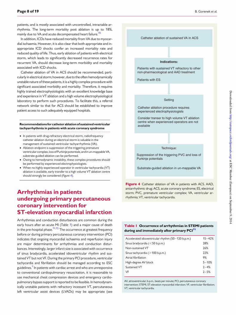
Recommendations forcatheterablationof sustainedventriculartachyarrhythmia in patients with acute coronary syndrome
† In patients with drug-refractory electrical storm, radiofrequencycatheter ablation during an electrical storm is valuable in themanagement of sustained ventricular tachyarrhythmia (VA).
† Ablation endpoint is suppression of the triggering prematureventricularcomplex, lossofPurkinjepotentials, and in un-mappableVA,substrate-guided ablation can be performed.
† Owing to hemodynamic instability, these complex procedures shouldbe performed by experienced electrophysiologists.
† When no highly experienced operator in ventricular tachycardia (VT)ablation is available, early transfer to a high volume VT ablation centreshould strongly be considered (Figure 4).
Arrhythmias in patientsundergoing primary percutaneouscoronary intervention forST-elevation myocardial infarctionArrhythmias and conduction disturbances are common during theearly hours after an acute MI (Table 1) and a major cause of deathin the pre-hospital phase.71,72 The occurrence at greatest frequencybefore or during primary percutaneous coronary intervention (PCI)indicates that ongoing myocardial ischaemia and reperfusion injuryare major determinants for arrhythmias and conduction distur-bances. Interestingly, larger infarct size is associated with occurrenceof sinus bradycardia, accelerated idioventricular rhythm and sus-tained VT but not VF. During the primary PCI procedure, ventriculartachycardia and fibrillation should be managed according to ESCguidelines.7 In patients with cardiac arrest and who are unresponsiveto conventional cardiopulmonary resuscitation, it is reasonable touse mechanical chest compression devices and emergency cardio-pulmonary bypass support is reported to be feasible. In hemodynam-ically unstable patients with refractory incessant VT, percutaneousleft ventricular assist devices (LVADs) may be appropriate (see
Catheter ablation of sustained VA in ACS
Indications:
Patients with sustained VT refractory to othernon-pharmacological and AAD treatment
Patients with ES
Setting
Catheter ablation procedure requiresexperienced electrophysiologists
Consider transer to high volume VT ablationcentre when experienced operators are notavailable
Technique:
Suppression of the triggering PVC and loss ofPurkinje potentials
Substrate-guided ablation in un-mappable VA
Figure 4 Catheter ablation of VA in patients with ACS. AAD,antiarrhythmic drug; ACS, acute coronary syndrome; ES, electricalstorm; PVC, premature ventricular complex; VA, ventricular ar-rhythmia; VT, ventricular tachycardia.
Table 1 Occurrence of arrhythmias in STEMI patientsduring and immediately after primary PCI71
Accelerated idioventricular rhythm (50–120 b.p.m.) 15–42%
Sinus bradycardia (,50 b.p.m.) 28%
Non-sustained VT 26%
Sinus tachycardia (.100 b.p.m.) 22%
Atrial fibrillation 9%
High-degree AV block 5–10%
Sustained VT 2–4%
VF 2–5%
AV, atrioventricular; b.p.m., beats per minute; PCI, percutaneous coronaryintervention; STEMI, ST-elevation myocardial infarction; VF, ventricular fibrillation;VT, ventricular tachycardia.
B. Gorenek et al.Page 8 of 19
at ESC Mem
ber (Europace) on September 9, 2014
http://europace.oxfordjournals.org/D
ownloaded from

. . . . . . . . . . . . . . . . . . . . . . . . . . . . . . . . . . . . . . . . . . . . . . . . . . . . . . . . . . . . . . . . . . . . . . . . . . . . . . . . . .
below). Following intervention, the incidence of arrhythmias rangesfrom 6% to 28% for new-onset AF, 7–13% for non-sustained VT,5–10% for high-degree AV block (≤30 beats/min lasting for ≥8 s),7%–16% for sinus bradycardia (≤30 beats/min lasting for ≥8 s),5% for sinus arrest (≥5 s), 3–6% for sustained VT, and 3–6% forVFaccording to retrospective registrydata orprospective recordingsfrom cardiac monitors implanted soon during an acute MI.73
The occurrence of AF is frequently associated with severe LVdamage and heart failure. Episodes may last from minutes to hoursand are often repetitive. The arrhythmia is most often well toleratedand no specific treatment is required, other than anticoagulation. Insome instances, the fast ventricular rate contributes to heartfailure, requiring prompt treatment using direct current cardiover-sion (DCCV) with further management as indicated below. Severalstudies have suggested that development of AF in the setting ofacute MI is an independent predictor of all-cause mortality, irrespect-ive of the treatment given.74,75 Atrial fibrillation notonly increases therisk for ischaemic stroke duringhospitalizationbut also during follow-up, and this includes patients with paroxysmal AF that has reverted tosinus rhythm at the time of discharge. Accordingly, patients with AFand risk factors for thromboembolism should be adequately treatedwith oral anticoagulation. Because AF will generally require anticoa-gulation, when choosing a stent in these patients, the benefits ofdrug-eluting stents on restenosis should be weighed carefullyagainst the substantial bleeding risks that are associated with the pro-longed combination of triple antithrombotic therapy (see below).Other supraventricular tachycardias are uncommon and are usuallyself-limiting.
Ventricular premature beats are almost universal on the first dayofthe acute phase and more complex arrhythmias (multiform com-plexes, short runs, or the R-on-T phenomenon) are common.Their value as predictors of VF is questionable. No specific therapyis required.
The long-term prognostic significance of early (,48 h) VF or sus-tained VT in patients with acute MI is still controversial.
In the APEX-AMI trial, VT/VF occurred in #6% of patients withSTEMI presenting for primary PCI.15 Two-thirds of these events oc-curred before the end of cardiac catheterization (defined as earlyevents) and 90% within 48 h. Ventricular tachycardia/VF was notbenign and was associated with substantially increased morbidityand mortality. Some of this association was related to older age,greater prevalence of comorbid conditions, and adverse presentingand post-cardiac catheterization features (ST resolution and TIMIflow) as shown by the attenuation of the risk with adjustment ofthese factors in a multivariate model. However, evenafter accountingfor these variables, any VT/VF remained associated with a more than3-fold higher risk of 90-day mortality in patients undergoing primaryPCI. The prognostic significance of late VT/VF appeared to be greaterthan early VT/VF with more than 5- and 2-fold higher risks of 90-daymortality, respectively. Thus, these data support the prognosticimportance of (any, early, or late) VT/VF as an independent andincremental risk marker, although this does not prove a cause-and-effect relationship. However, sustained VT/VF after primary PCI inthe HORIZONS-AMI trial was not significantly associated with3-year mortality or major adverse clinical events.76 Clinical manage-ment of sustained VT and VF after primary PCI is according to ESCguidelines.7
Recommendations for management of arrhythmias in patientsundergoing primary percutaneous coronary intervention forST-elevation myocardial infarction
† Ventricular tachyarrhythmia (VA) developed during primarypercutaneous coronary intervention (PCI) should be treated usingdirect current cardioversion (DCCV), overdrive pacing, beta-blockersand amiodarone for sustained ventricular tachycardia (VT)/ventricularfibrillation.7
† Electrolyte balance correction, beta-blockers, and amiodarone shouldbe considered for treatment of polymorphic VT.
† For refractory VA in hemodynamically unstable patients, percutaneousventricular assist device may be considered.
† Atrial fibrillation developed during PCI with high ventricular rateleading to hemodynamic compromise should be treated using DCCVwith further management as indicated below.
Arrhythmias in cardiogenic shockcomplicating acute myocardialinfarctionCardiac arrhythmias including VT/VF, AF, and conduction distur-bances are common in patients with acute MI complicated by cardio-genic shock and are associated with high short-term mortality.77–81
Cardiogenic shock, acute ischaemia, and the use of inotropes aremain risk factors for the development of arrhythmic events.81 There-fore, correcting factors underlying cardiogenic shock, substrate, andtriggers for arrhythmia, play main role in their management.17,82,83
Ventricular arrhythmiasSustained VT occurs in 17–21% and VF is seen slightly more often(24–29%) in selected patients with cardiogenic shock and acuteMI, undergoing thrombolysis or primary PCI.77,78 In cardiogenicshock, sustained VT/VF might deteriorate hemodynamic status andLV dysfunction. The goal of therapy should be to restore therhythm immediately to avoid hypoperfusion, which leads toend-organ damage. Regardless of the cause of cardiogenic shock, sus-tained VT/VF must be treated promptly with DCCV without hesita-tion. Catheter ablation may be indicated as a salvage procedure if VTrecurs (see above).27 Antiarrhythmic therapy (usually intravenousamiodarone, lidocaine) and electrolyte balance correction could beconsidered if necessary for acute management.17 Care should betaken with intravenous amiodarone to avoid hypotension. Forselected patients with refractory VT/VF and rapidly deterioratinghemodynamic and clinical status, implantation of percutaneousLVAD and extracorporeal membrane oxygenation (ECMO)-assistedprimary PCI have been found promising as a bridge to recovery, ingaining time to implement and assist appropriate therapies andprolonging survival.53,84 –95 Implantation of percutaneous LVAD(Impella 2.5 assist device) in patients with MI and cardiogenicshock, as well as presenting with cardiac arrest was accompaniedby reduction of tissue hypoxia, hemodynamic stabilization, and im-provement of neurological outcome.84,85,93,94 Extracorporeal mem-brane oxygenation -assisted PCI significantly improved recovery andsurvival of patients with cardiogenic shock and refractory VT/VF ascompared with intra-aortic balloon pump (IABP) with 69.23% of
Cardiac arrhythmias in acute coronary syndromes Page 9 of 19
at ESC Mem
ber (Europace) on September 9, 2014
http://europace.oxfordjournals.org/D
ownloaded from

. . . . . . . . . . . . . . . . . . . . . . . . . . . . . . . . . . . . . . . . . . . . . . . . . . . . . . . . . . . . . . . . . . . . . . . . . . . . . . . . . .
patients successfully weaning off the support in the ECMO group andonly 12.5% in the IABP group (P ¼ 0.02).92 The latter is no longerrecommended.96 –99 The IABP-Shock II study demonstrated nobeneficial effect of IABP on tissue hypoxia, time to hemodynamicstabilization, short- and long-term mortality in patients with cardio-genic shock as compared with medical treatment.98,99 Recentmeta-analysis on the effect of IABP according to the type of reperfu-sion in patients with MI and cardiogenic shock showed overall noeffect on the risk of in-hospital and long-term mortality and trendto higher risk of in-hospital death with IABP support in patientsundergoing primary PCI [relative risk 1.18, 95% confidence interval(CI) 1.04–1.34].96 Intra-aortic balloon pump is not expected towork in case of VF, as there is no circulation to augment.
Proper hemodynamic support with inotropes and vasopressors isrequired, though caution must be taken for dopamine use, since itincreases risk of arrhythmias in patients with shock,100 which areless seen with other inotropes. Coronary reperfusion is key to im-proving survival. Recent pooled analysis of both the SMASH andSHOCK trials82 showed that in cardiogenic shock patients, earlyrevascularization reduced risk of death by 18% (relative risk 0.82,95% CI 0.70–0.96) compared with the initial medical stabilization.Therefore, early hemodynamic stabilization and arrhythmia manage-ment should not delay revascularization.
Atrial fibrillationAbout 11–20% of patients with acute MI presenting with cardiogenicshock develop AF.77,78 Several mechanisms underlying cardiogenicshock can predispose to AF by creating either a substrate or atrigger for this arrhythmia. Hemodynamic changes as an increase inpulmonary capillary wedge pressure and left atrial pressure, canlead to AF. The adverse hemodynamic consequences of AF canquickly lead to a worsening of symptoms, which may be difficult tomanage. Immediate DCCV should be performed to restore sinusrhythm and hemodynamic stability when cardiac output is compro-mised.7 Amiodarone intravenous can be used for acute ratecontrol with caution of hypotensive effect.101 Though evidence onthe efficacy of amiodarone in the setting of cardiogenic shock islimited, in one study of single bolus intravenous amiodarone usefor acute rate control it also resulted in (delayed) pharmacologicalconversion to sinus rhythm.101 In patients with pre-existing AF onoral amiodarone therapy, it may also enhance success of DCCV.102
Other rate controlling agents must be avoided due to negative ino-tropic effects. In refractory cases, AV node ablation with biventricularor LV stimulation could be considered for rate control, as they havebeen found feasible in correction of hemodynamic derangement ofpatients with cardiogenic shock and severe heart failure.103,104
Conduction disturbancesHigh-grade AV block and asystole develop in about 23–35% of acuteMI patients with cardiogenic shock,77,78 especially in inferior infarc-tions with proximal right coronary artery (RCA) occlusion. Therate of complete heart block is much lower in adequately revascular-ized infarct patients, and prompt revascularization of the infarctartery should be attempted. Bradyarrhythmias are induced byeither autonomic imbalanceor ischaemia and necrosisof the conduc-tion system (see section below). It is important to recognize which ofthem are transient and which are likely to progress to irreversible and
symptomatic high-degree AV block, which has been associated with amortality rate approaching 80% due in large part to greater loss offunctioning myocardium. Temporary transvenouspacing is necessaryfor patients with severe and symptomaticbradyarrhythmias if theydonot resolve within few minutes after reperfusion.7,105
Recommendations for management of arrhythmias incardiogenic shock complicating acute myocardial infarction
Regardless of the type of arrhythmia, treatment of the underlyingcardiogenic shockwithprompt revascularization shouldbe doneas theprimary procedure and should not be delayed by arrhythmiatreatment.
† Acute management of ventricular tachycardia (VT)/ventricularfibrillation (VF) in the setting of cardiogenic shock includesimmediate direct current cardioversion (DCCV), amiodarone,lidocaine if necessary.
† Percutaneous left ventricular assist device and extracorporealmembrane oxygenation-assisted percutaneous coronaryintervention can be used in case of refractory VT/VF.
† Catheter ablation may be considered as a salvage procedure ifarrhythmia persists (see above).
† Atrial fibrillation shouldbemanagedbyDCCVif highventricular ratecompromises cardiac output; amiodarone can be used for ratecontrol and assisting cardioversion.
† Atrioventricular node ablation with biventricular or left ventricularstimulation could be considered if above rate control measures fail.
† Severe and symptomatic bradyarrhythmias accompanied byhemodynamic instability require placement of temporarypacemaker if they do not resolve within few minutes afterreperfusion.
Incidence, prognostic implications,and treatment of atrial fibrillationin patients with an acute coronarysyndromeAtrial fibrillation, the most commonly encountered clinical arrhyth-mia, often coexists with acute MI. Atrial fibrillation has been reportedto complicate the course of acute MI in 2.3–21% of hospitalizedpatients.106 In recent years, the widespread use of early reperfusiontherapy (thrombolysis and PCI) as well as the use of beta-blockers,angiotensin-converting enzyme inhibitors and angiotensin II inhibi-tors has led to a substantial decline in the incidence of post-MI AF.However, as the population ages and as AF increases with age, wecan expect that AF will still remain a frequent and worrisome compli-cation of acute MI.
Atrial fibrillation may already be present at the time of hospital ad-mission for acute MI, or may develop during the hospital stay. Pre-existing AF accounts for approximately one third of all cases of AFobserved in patients with acute MI, and new-onset AF for the remain-ing two-thirds.107
Possible underlying mechanisms of AF in the setting of acute MI areatrial ischaemia or infarction, acute hypoxia or hypokalaemia, peri-cardial inflammation, increased LV diastolic pressure and left atrialpressure, hemodynamic impairment secondary to LV dysfunctionand abnormalities of autonomic regulation.108 These mechanisms
B. Gorenek et al.Page 10 of 19
at ESC Mem
ber (Europace) on September 9, 2014
http://europace.oxfordjournals.org/D
ownloaded from

. . . . . . . . . . . . . . . . . . . . . . . . . . . . . . . . . . . . . . . . . . . . . . . . . . . . . . . . . . . . . . . . . . . . . . . . . . . . . . . . . .
may be found alone or in combination, and may superimpose onpredisposing diseases, such as previous cardiomyopathy, valvulardisease, or chronic lung disease.
Once AF develops, usually there is a significant worsening ofhemodynamics due to the high ventricular rate, irregular ventricularfilling, and/or loss of atrial contribution to cardiac output.
Independent predictors of the occurrence of AF in acute MIinclude older age, elevated heart rates at admission, pre-existingAF, LV hypertrophy, presence of heart failure symptoms, and LV dys-function. These risk factors have been described regardless of thetype of reperfusion therapy (i.e. none, thrombolysis, PCI). Moreover,patients who develop AF more often have hypertension, diabetes,previous MI, multi-vessel coronary artery disease, higher levels ofbiomarkers of myocardial damage, and low TIMI 3 flow grade afterreperfusion therapy.106,107
The prognostic impact of AF that occurs in the setting of acute MIis still controversial, with some studies describing an independentadverse effect on mortality and others failing to detect such an asso-ciation.106 However, a recent systematic review and meta-analysis,including 43 studies and 278 854 patients, showed that AF carriesan excess risk of in-hospital, short-term (,30 days), mid-term(.30 days to 1 year), and long-term (.1 year) mortality amongpatients with acute MI, with at least a 40% increase, regardless ofthe type of AF (pre-existing or new-onset). Increased risk includedboth sudden and non-sudden cardiac death. This worse prognosispersisted, in patients with new-onset AF, even after adjustment forage, diabetes mellitus, hypertension, prior MI, heart failure, and cor-onary revascularization.109 Moreover, according to a recent largecommunity study, the mortality risk, in patients with new-onsetAF, seems to be greatest when the arrhythmia develops later than30 days post-acute MI [hazard ratio (95% CI) 2.58 (2.21 to 3.00) vs.1.81 (0.45 to 2.27) for AF between 3 and 30 days, and 1.63 (1.37 to1.93) for AF within 2 days].107
In addition to increased mortality, patients with acute MI and AFhave also a higher incidence of re-infarction, cardiogenic shock,heart failure, and asystole, probably as an expression of the moresevere impairment of coronary circulation and hemodynamicstatus when AF develops.106
Finally, AF complicating acute MI is also associated with anincreased risk of subsequent ‘spontaneous’ AF, and also with anincreased risk of ischaemic stroke both during hospitalization andduring follow-up; even if the AF is transient and reverses back tosinus rhythm before hospital discharge.106
Literature evidence for the management of AF in the setting ofacute MI stemming from controlled clinical trials is lacking, so thetreatment is essentially founded on practical basis.
In many cases, the arrhythmia is well tolerated and no specifictreatment is required.
In other instances, the high ventricular rate associated with AF maycontribute to hemodynamic impairment and heart failure, requiringprompt therapeutic intervention. Adequate rate control representsthe most important first therapeutic approach in this setting, and maybe accomplished either by administration of beta-blockers or pos-sibly non-dihydropyridine calcium antagonists, either orally or intra-venously. In patients with acute MI and AF associated with severe LVdysfunction or heart failure, the negative inotropic effect of beta-blockers or calcium antagonists may result in further impairment of
pump function. In such circumstances, rate control may be achievedby intravenous administration of amiodarone and/or digitalis.7
Urgent DCCV is recommended for patients with severe hemo-dynamic instability or intractable ischaemia, or when adequate ratecontrol cannot be achieved with pharmacologic agents. In thislatter case, transesophageal echocardiography should be consideredto exclude the presence of thrombi in left atrium. In addition to elec-trical CV, amiodarone can be used for restoration of sinus rhythmwhen the hemodynamic situation is stable. This drug is usually pre-ferred to other AADs because of its limited negative inotropiceffect. The administration of class IC AADs, instead, is consideredharmful in patientswith acuteMI and hence is not recommended.7,110
In the post hoc analysis in patients with coronary heart disease andatrial fibrillation from the ATHENA (A placebo-controlled, double-blind, parallel arm Trial to assess the efficacy of dronedarone400 mg b.i.d. for the prevention of cardiovascular Hospitalizationor death from any cause in patiENts with Atrial fibrillation/atrialflutter) trial, dronedarone reduced all-cause mortality, cardiovascu-lar hospitalization, and the first ACS event. However, experiencewith dronedarone in the setting of ACS is limited, and the drug isnot recommended for thus indication.111
Finally, patients with AFand moderate-to-severe thromboembolicrisk (CHA2DS2-VASc score ≥2) should be adequately treated withoral anticoagulants (either vitamin K antagonists or new oral anticoa-gulants) to reduce the risk of stroke or systemic embolisms (seebelow).
Recommendations for the management of atrial fibrillation inacute coronary syndrome
† For patients with acute coronary syndrome and pre-existing or newlydeveloped atrial fibrillation (AF), rate control using beta-blockers orpossibly calcium antagonists is recommended, whereas in patients withsevere LV dysfunction using amiodarone and/or digitalis isrecommended.
† Urgent direct current cardioversion is required if AF is accompanied byhemodynamic instability.
† For restoration of sinus rhythm in addition to electrical cardioversionamiodarone can be considered. Other antiarrhythmic drugs might beharmful in the setting of acute myocardial infarction.
† Adequate anticoagulation according to the individual risk of stroke andembolism is recommended, usually requiring a period of combinationtherapy of antiplatelet agents and anticoagulants.
Risk assessment forthromboembolic events, bleeding,and choice of antithrombotictherapy in patients with atrialfibrillation and acute coronarysyndromePatients with ACS and permanent, paroxysmal or persistent non-valvular AFarea special subgroup with an increased risk for ischaemicand embolic events, as well as bleeding complications. Since theseevents are closely related to clinical outcomes including mortality,
Cardiac arrhythmias in acute coronary syndromes Page 11 of 19
at ESC Mem
ber (Europace) on September 9, 2014
http://europace.oxfordjournals.org/D
ownloaded from

. . . . . . . . . . . . . . . . . . . . . . . . . . . . . . . . . . . . . . . . . . . . . . . . . . . . . . . . . . . . . . . . . . . . . . . . . . . . . . . . . .
decision making in the individual patient should be made by takinginto account the risk for thromboembolic and bleeding hazards alike.
Stroke risk stratificationBasedon the recentESCguidelines strokerisk stratification shouldbemade by the CHA2DS2-VASc score, which means that all patientswith ACS will have a score of at least 1 and will therefore be candi-dates for oral anticoagulation.112 – 114 Patients with STEMI usuallyare treated with primary PCI and stenting in over 90% of cases,while PCI will be performed in about 50–60% of patients presentingwith non-ST elevation ACS. Both groups of patients have an indica-tion for dual antiplatelet therapy (DAPT). Hence, ‘triple’ therapy(DAPT and oral anticoagulation) for a defined period after theevent is usually required.7,8 One controlled trial suggested thattherapy with clopidogrel and an oral anticoagulant could be slightlysafer than ‘triple therapy’,115 but more data are needed to confirmthis.
Bleeding risk stratificationPatients with an increased thromboembolic risk usually have clinicalcharacteristics that are also associated with increase bleeding ten-dency, i.e. advanced age, female gender, renal dysfunction, femoralaccess, and history of prior bleeding hazards, especially of pepticulcer. Accordingly, the use of specific risk scores (HAS-BLEDscore) to stratify for bleeding tendency in patients with AF116
seems practicable. In AF patients with and without anticoagulationtherapy, it has higher accuracy in prediction of bleeding events andeasier to use as compared with other scores.117,118 In patients withACS, receiving triple antithrombotic therapy HAS-BLED score wasfound to have diagnostic value in prediction of bleeding.119
HAS-BLED score is currently recommended for risk stratificationof bleeding in patients with AF and ACS.112,116,120,121
Choice of antithrombotic therapybased onthrombosis and bleeding risksWhen AF affords anticoagulation in addition to DAPT, ‘triple’ antith-rombotic therapy [DAPT plus an anticoagulant, usually a vitamin Kantagonist (VKA) with an INR goal of 2.0–2.5] is recommended forthe shortest time necessary,122 which is usually 1 month after baremetal stents and 3(–6) months after drug-eluting stent in stable/elective patients as well as 6 months after ACS independent of thestent type and treatment strategy used, followed by one antiplateletagent (clopidogrel or aspirin) plus an anticoagulant (‘dual’ therapy)for up to 12 months, and then by anticoagulant monotherapy life-long.122 VKAs were shown to be at least as effective as aspirin in sec-ondary prevention after acute MI.123 Ongoing research andexpert-based position papers might widen the indication fornon-VKA oral anticoagulants (NOACs) as replacement for VKAs inthis clinical setting in the near future.124
Against the existing guidelines, in clinical practice ‘triple’ therapy isless often used than recommended.125 In weighting the risk ofembolic stroke and bleeding, some extrapolations from trials com-paring antiplatelet therapies and anticoagulation have been includedin clinical decision making. In the ACTIVE-A trial,126 DAPT with
aspirin and clopidogrel has been more effective than aspirin alonein preventing stroke and systemic embolism in patients with AF.However, DAPT was inferior to warfarin in the ACTIVE-Wstudy.127 Table 2 summarizes the embolic, ischaemic and bleedingevents in the ACTIVE-trials.
From these data and the relatively low stroke rate in the first3 months after ACS, it seems reasonable to recommend DAPTalone or VKA plus clopidogrel for 12 months for stable/electivepatients with non-valvular AF with a CHA2DS2-VASc score of 1when the bleeding risk is high.120 In patients with non-valvular AFplus ACS, independent of the stent type and treatment strategyused, ‘triple’ therapy for 4 weeks, followed by ‘dual’ therapy for12 months should be used and in selected cases with low risk ofstent thrombosis/ischaemic events ‘dual’ therapy alone may be con-sidered when the CHA2DS2-VASc score is 1 and the bleeding risk ishigh.120 In all instances, this therapy might be followed by oral antic-oagulation alone. However, in patients with a higher CHA2DS2-VAScscore ‘triple’ therapy, followed by ‘dual’ therapy and finally oral antic-oagulation alone should be considered as indicated above.
There exist two subgroups of patients with stent implantation and/or ACS and AF, (i) the subgroup of patients with a history of AFalready under oral anticoagulation, and (ii) a subgroup of patientswith AF occurring under chronic DAPT. While recent recommenda-tions allow the further use of NOACs in a reduced dosage in casepatients were already pre-treated with a NOAC, little is knownabout the treatment strategy in the latter group and the updated pos-ition paper, a joint effort of the Working Group on Thrombosis, aswell as EHRA, EAPCI, and ACCA, are non-specific by offeringboth, a start with VKAs or NOACs.120
Recommendations for risk assessment for thromboembolicevents, bleeding and choice of antithrombotic therapy inpatients with atrial fibrillation and acute coronary syndrome
† In patients with acute coronary syndrome and atrial fibrillation,prevention of thromboembolic events should be based onindividualized risk stratification of thromboembolic events (risk ofstroke-CHA2DS2-VASc score) to define the need for anticoagulation,risk stratification of bleeding (HAS-BLED score) to define the level ofanticoagulation or choice of non-vitamin K antagonist oralanticoagulant strategy, and measures to minimize bleedingcomplications (stent choice, radial approach, and consequent short-and long-term dual antiplatelet therapy).120
New bundle branch block andatrioventricular conductiondisorders as complication ofmyocardial infarctionAtrioventricular conduction disturbances are well-known complica-tions of acute MI.128,129 During the first few hours of myocardial is-chaemia, they may be related to an autonomic imbalance with avagal hyperactivity leading to a transient slowing of AV conduction.
B. Gorenek et al.Page 12 of 19
at ESC Mem
ber (Europace) on September 9, 2014
http://europace.oxfordjournals.org/D
ownloaded from

. . . . . . . . . . . . . . . . . . . . . . . . . . . . . . . . . . . . . . . . . . . . . . . . . . . . . . . . . . . . . . . . . . . . . . . . . . . . . . . . . .
Myocardial ischaemia and necrosis may induce transient dysfunctionor irreversible damage of the AV conduction system that may lead tothe development of a new bundle branch block (BBB) or worsen asymptomatic high degree AV block necessitating temporary or per-manent cardiac pacing.
In order to fully understand the relationship between myocardialischaemia-infarction and the occurrence of new AV conduction dis-turbances, it is important to review the vascular supply of the differ-ent parts of the conduction system. The AV node and also the Hisbundle are supplied by the AV nodal artery that takes origin fromthe RCA in a 90% of the cases and from the left circumflex arteryin 10% of the patients. A post mortem study has shown that proximalleft anterior descending coronary artery (LAD) septal perforatorsperfuse the right bundle branch and the anterior fascicle of the leftbundle branch in 90% of cases, whereas the posterior fascicle ofthe left BBB is perfused by the conus branch of the RCA in the major-ity of the cases.130 There is dual blood supply to each of the fasciclesin half of the patients.123 An occlusion of the LAD therefore willinduce a right BBB or an anterior left fascicular block. The develop-ment of a new left BBB will occur only in the presence of a proximalocclusion of both the RCA and the LAD.130
Asecondor thirddegreeAVblock inpatientswith anacute inferiorwall MI is almost always (90% of the patients) located above the Hisbundle. Most often, this only results in a moderate bradycardia with ajunctional escape rhythm with narrow QRS complexes. This conduc-tion disorder most frequently is transient and is associated with a lowmortality risk.
In patients with an anterior wall MI, a high degree AV block is mostoften located below the AV node. Total AV block generally occursabruptly during the first 24 h after MI and is frequently preceded bythe development of a new right BBB with the right or left axis devi-ation and QR pattern in lead V1. Most often, it is associated with anextensive myocardial necrosis of the septum and anterior wall inthe presence of severe multivessel disease involving both the LADand the RCA or a dominant left circumflex artery. Usually it causescatastrophic hemodynamic problems frequently leading to lethalpump failure.
Patients with a BBB have more comorbidities and are less likely toreceive timely reperfusion therapy.131 It remains uncertain if the pres-ence of a BBB at presentation has a prognostic importance. After ad-justment for confounding factors, the presence of a BBB was nolonger associated with an increased mortality risk. However, patients
who develop a new left BBB within 60 min after admission have athree times higher 30-day mortality.132
Recommendations for management of new bundle branch blockand atrioventricular conduction disorders in acute coronarysyndrome
† Prompt opening of the infarct vessel is often sufficient to reversenew-onset ischaemic conduction disturbances. This is especiallytrue for atrioventricular (AV) block in the setting of inferiorinfarctions.
† Temporary pacing is indicated for symptomatic life-threateningbradycardia not resolving after successful reperfusion and after medicaltreatment in the presence of high-degree AV block and intraventricularconduction defects.
† Permanent pacing is considered for disturbances that persist beyondthe acute phase after the myocardial infarction.
Temporary pacing in intensivecardiac care unit in patientspresenting with acute coronarysyndrome: indications andtechniques
Temporary pacing in cardiac intensivecare unitTemporary endocardial pacing has been used since the early 1960s tomaintain cardiac output during episodes of extreme bradycardia, AVblock, and asystole. Before the advent of cardiac pacemakers (PMs),the combination of acute MI and complete AV block was usuallyfatal, but now pacing electrodes can be safely placed percutaneouslyinto the right ventricle under local anaesthesia. Prognosis is influencednot only by complications of the procedure, but also by the underlyingmyocardial damage, which originally lead to the conduction defect. Inpatients,whohavenot sufferedanacuteMI andare admitted tocardiacintensive care unit, temporary pacing may be required for treatment ofasystole or bradycardia due to primitive cardiac conduction diseases,drug toxicity, electrolyte disturbances, or surgery.133
. . . . . . . . . . . . . . . . . . . . . . . . . . . . . . . . . . . . . . . . . . . . . . . . . . . . . . . . . . . . . . . . . . . . . . . . . . . . . . . . . . . . . . . . . . . . . . . . . . . . . . . . . . . . . . . . . . . . . . . . . . . .
. . . . . . . . . . . . . . . . . . . . . . . . . . . . . . . . . . . . . . . . . . . . . . . . . . . . . . . . . . . . . . . . . . . . . . . . . . . . . . . . . . . . . . . . . . . . . . . . . . . . . . . . . . . . . . . . . . . . . . . . . . . . . . . . . . . . . . . . . . . . . . . . . . . . . . . . . . . . . . . . . . . . . . . . . . . . . . .
Table 2 Embolic, ischaemic, and bleeding events in the ACTIVE trials126,127
ACTIVE-A ACTIVE-W
Aspirin (%) Aspirin 1 clopidogrel (%) Aspirin 1 clopidogrel (%) Warfarin (%)
Stroke 3.3 2.4 2.4 1.4
Myocardial infarction 0.9 0.7 0.86 0.55
Major bleeding 1.3 2.0 1.7 1.6
Cardiac arrhythmias in acute coronary syndromes Page 13 of 19
at ESC Mem
ber (Europace) on September 9, 2014
http://europace.oxfordjournals.org/D
ownloaded from

. . . . . . . . . . . . . . . . . . . . . . . . . . . . . . . . . . . . . . . . . . . . . . . . . . . . . . . . . . . . . . . . . . . . . . . . . . . . . . . . . .
Techniques for temporary pacingEndocardial temporary pacing leads are introduced via a central vein:right interior jugular, right subclavian vein or femoral veins. Pacingwires are commonly 5F or 6F. Under fluoroscopy, the lead isadvanced into the right atrium and then, crossing the tricuspidvalve, placed at the right ventricle apex. A check for positional stabil-ity, asking the patient to take some deep breaths or cough forcefullyand watch for failure of capture, is suggested. A chest X-ray toconfirm satisfactorypositioningof thewireand toexcludeapneumo-thorax, is recommended as well.
Following cardiac surgery, patients may have temporary epicardialelectrodes in case of postoperative bradyarrhythmia. These are usedin the samewayas transvenouspacing leads, but the threshold may behigher.
In emergency situations, transthoracic pacing is a better alterna-tive, although it is not a substitute for temporary transvenous PM,and represents only a bridge to maintain life.
ComplicationsA temporary PM is a foreign body that may remain in the circulationfor days and has a high propensity for infections, thus requiring surgi-cal asepsis and antibiotic prophylaxis.134,135
Moreover, in case of a femoral approach for temporary pacing,restricted patient mobility may be cause of asymptomatic deepvein thrombosis, therefore antithrombotic prophylaxis as well isrecommended.
The main frightening complication of temporary pacing in an ACSsetting is the cardiac perforation causing cardiac tamponade.
The incidence of complications and malfunctions may be signifi-cant, occurring within 24 h in about 50% of patients and within48 h in 86% of patients. Failure to capture or sense, VT during cath-eter insertion, fever and phlebitis are the most common complica-tions. Other possible, more rare ones include phrenic capture,vascular complications for femoral approach (hematoma, pseudoa-neurysm, arteriovenous fistula), hemothorax, and pneumothoraxfor subclavian and jugular accesses, cardiac perforation and tampon-ade. Sepsis, phlebitis, and pulmonary embolus are more commonwith temporary PMs in place for 72 h or longer.136
In many patients, particularly after acute MI, placing a pacing leadwithin the right ventricle will promote ventricular ectopic activityand occasionally prolonged VA.134 These will usually resolve oncemanipulation of the lead has ceased but will occasionally requireremoval of the lead or repositioning.137
An eventual cardiac perforation is usually manifest by raised pacingthresholds and occasionally by pericarditic pain and a pericardial fric-tion rub. At worst, this will result in cardiac tamponade and willrequire appropriate urgent treatment. The increased risk of cardiacperforation and tamponade may be related to several factors such as
(1) the emergency setting;(2) not-specialist doctors inserting the wire;(3) higher stiffness of the leads;(4) concomitant use of potent antithrombotics (GP IIb/IIIa inhibi-
tors);(5) inferior MI (with frail wall).
These and other considerations led to the drafting of new detailedguidelines by ESC and EHRA.105
Recommendations for temporary pacing in intensive cardiaccare unit in patients presenting with acute coronary syndrome:indications and techniques
† Temporary transvenous pacing should not be used routinely and onlyas a last resort when positive chronotropic drugs (e.g. isoproterenol,atropine, etc.) are insufficient or contraindicated.
† Temporary transvenous pacing should be limited to cases of(i) high-degree atrioventricular block without adequate escape rhythm;(ii) life-threatening bradyarrhythmias, such as those that occur duringinterventional procedures (e.g. during percutaneous coronaryintervention) or, rarely, in acute settings such as acute myocardialinfarction, drug toxicity, electrolyte disturbances, or concomitantsystemic infection.
† If the indications for permanent pacing are established, every effortshould be made to implant a permanent pacemaker as soon as possible.
† An external stimulator (transcutaneous) does not provide reliableventricular stimulation and therefore should only be used, under stricthaemodynamic and electrocardiographic monitoring, when no otheroption is available. As soon as possible, an alternative action should beundertaken (administration of chronotropic drugs or temporary orpermanent pacing).
Electrical cardioversion forarrhythmias in acute coronarysyndrome patients with implantedrhythm devicesThe different techniques for electrical CV depend on the varioustypes and positioning of paddles, energy strategies and waveformsused. When performed in patients with an implanted PM and ICDsystems, the choice between the different techniques can affect theoutcome and safety of a CV.
Whenusing biphasic defibrillators, self-adhesive defibrillation padsare safe and effective and an acceptable alternative to standard defib-rillation paddles for both defibrillation and CV. The majority ofstudies comparing paddle size orientations and positioning (anterior-apex, anterior-posterior, anterior-left infrascapular, anterior-rightinfrascapular) have shown equal effectiveness in defibrillation (forVAs) or elective AF CV success, although an anterior-lateral elec-trode position was reported to increase efficacy and require fewershocks for common atrial flutter.138 In recent resuscitation guide-lines, an anterior-posterior position is generally accepted as an alter-native position to an anterior-lateral position.139
Thenumerouscase reports ondevice and lead failure following ex-ternal CV in patients with implanted PM systems, has led to certainprecautions including paddle positions to increase the safety of theprocedure.
The risk of damage to the device is minimized by diodes that shuntcurrent away from device and may be further reduced by keepingmaximal distance between shock electrodes and the device. Leadsmay not be protected by these measures and current density may in-crease at the lead tip, particularly with unipolar leads, leading to myo-cardial damage at the electrode–endocardial surface. Such damage
B. Gorenek et al.Page 14 of 19
at ESC Mem
ber (Europace) on September 9, 2014
http://europace.oxfordjournals.org/D
ownloaded from

. . . . . . . . . . . . . . . . . . . . . . . . . . . . . . . . . . . . . . . . . . . . . . . . . . . . . . . . . . . . . . . . . . . . . . . . . . . . . . . . . .
may result in local myocardial oedema with a transient increase ofpacing threshold and fibrosis with permanent progressive increasedpacing threshold and a deterioration of sensing function. Theseobservations thus further argue for an anterior-posterior positionfor CV and also for defibrillation in patients with devices.
Very fewstudies have analysed the safety of external CV in patientswith implanted PM systems.140 Transient loss of capture (up to30 min) occurred in 50% of patients, a six-fold increase of ventricularpacing thresholds, sensing failure in 41%ofpatients, and threecasesofPM malfunction were reported in a study evaluating patients with PMdevices implanted in the right pectoral region and with unipolar elec-trodes.141 It was concluded that CV represents a serious hazard topatients with an implanted PM. In that study, however, only mono-phasic shocks were used and placed in an anterior-lateral orientation,which may create an electrical field in the same direction as the ven-tricular lead. An anterior-posterior positioning of CV electrodes issuperior as it prevents this ‘antenna-like effect’ of the ventricularlead and result in lower energy requirements for termination ofAF.141 Since biphasic shocks are superior to monophasic shocks forCV of AF142 and in terminating VF, all new defibrillators delivershocks using biphasic waveforms. Mono- and biphasic shock wave-forms for external CV were recently compared with regard tosafety and efficacy in a prospective study of patients with implanteddevices.143 Lower energy levels and at least .8 cm distance to theimplanted device were adopted to minimize risk for damage to thedevices and leads. Systems were tested immediately before andafter CV, and 1 h and 1 week later with respect to device and lead in-tegrity. Pacing impedances were reduced immediately after CV andreturned to baseline values within 1 week. Ventricular sensing wasreduced immediately after CV. There was no device or lead dysfunc-tion in any patient. Cardioversion was successful in 95% of patients.The lower cumulative energy requirement for biphasic as comparedwith monophasic shocks indicated a theoretical advantage for bi-phasic shocks.
Electrical CV can thus be safely and effectively performed inpatients with implanted rhythm devices, if the precautions outlinedare considered. These recommendations also apply to emergencysituations of VA in patients with implanted devices.
Recommended external electrical cardioversion technique inpatients with implanted rhythm devices
† The anterior-posterior paddle/pad position on the chest isrecommended
† The placement of paddles/pads should not delay defibrillation† The paddle/pad should be placed on the chest wall ideally at least 8 cm
from the generator position† Biphasic shocks are preferred for electrical cardioversion† Check for changes in pacing or sensing parameters 1 h after
cardioversion
ConclusionThis document summarizes the current position, contemporaryapproaches, and offers recommendations on how to identify ACSpatients at risk for arrhythmias; manage tachyarrhythmias and
bradyarrhythmias using pharmacological and non-pharmacological,device- and catheter-based approaches including electrical CV,implantation of temporary PMs, programming of ICD, catheterablation; and implement clinical and referral strategies. Theserecommendations are provided for acute care and management ofVA, AF, bradyarrhythmias, and prevention of stroke and embolismin patients with ACS, and arrhythmias developing during coronaryinterventions, in patients with implanted devices and cardiogenicshock. Management of intractable arrhythmias requires the team-based approach including specialists in acute cardiac care, interven-tional cardiology, and electrophysiology.
Further supportive to the current knowledge evidence on thespecific issues of arrhythmias management in the setting of ACSand answers to the gaps in knowledge emphasized in the currentdocument should be pursued in future studies.
Supplementary materialSupplementary material is available at Europace online.
AcknowledgementsEHRA Scientific Documents Committee, Prof. Gregory YH Lip(EHRA Scientific Documents Committee Chair), Prof. BulentGorenek (EHRA Scientific Documents Committee Co-Chair),Prof. Christian Sticherling, Prof. Laurent Fauchier, Prof. Hein Heidbu-chel, Dr Angel Moya Mitjans, Prof. Marc A Vos, Dr Michele Brignole,Prof. Gheorghe-Andrei Dan, Dr Michele Gulizia, Dr Francisco Marin,Prof. Giuseppe Boriani, Dr Deirdre Lane, and Dr Irene Savelieva.
Conflict of interest: Detailed conflict of interest statement is avail-able as Supplementary material online.
References1. Bezzina CR, Pazoki R, Bardai A, Marsman RF, de Jong JS, Blom MT et al. Genome-
wide association study identifies a susceptibility locus at 21q21 for ventricular fib-rillation in acute myocardial infarction. Nat Genet 2010;42:688–91.
2. Janse MJ, Wit AL. Electrophysiological mechanisms of ventricular arrhythmiasresulting from myocardial ischemia and infarction. Physiol Rev 1989;69:1049–69.
3. Said M, Becerra R, Valverde CA, Kaetzel MA, Dedman JR, Mundina C et al. CAMKII:a main signal responsible for early reperfusion arrhythmias. JMCC 2011;51:936–44.
4. Kirchhof P, Breithardt G, Eckardt L. Primary prevention of sudden cardiac death.Heart 2006;92:1873–8.
5. Pogwizd SM, Corr PB. Electrophysiologic mechanisms underlying arrhythmias dueto reperfusion of ischemic myocardium. Circulation 1987;76:404–26.
6. Nabel EG, Braunwald E. A tale of coronaryartery disease and myocardial infarction.N Engl J Med 2012;366:54–63.
7. Steg PG, James SK, Atar D, Badano LP, Blomstrom-Lundqvist C, Borger MA et al.ESC Guidelines for the management of acute myocardial infarction in patients pre-senting with ST-segment elevation. Eur Heart J 2012;33:2569–619.
8. Hamm CW, Bassand JP, Agewall S, Bax J, Boersma E, Bueno H et al. ESC Guidelinesfor the management of acute coronary syndromes in patients presenting withoutpersistent ST-segment elevation: the Task Force for the management of acute cor-onary syndromes (ACS) in patients presenting without persistent ST-segment ele-vation of the European Society of Cardiology (ESC). Eur Heart J 2011;32:2999–3054.
9. Granger CB, Goldberg RJ, Dabbous O, Pieper KS, Eagle KA, Cannon CP et al.Global Registry of Acute Coronary Events Investigators. Predictors of hospitalmortality in the global registry of acute coronary events. Arch Intern Med 2003;163:2345–53.
10. Antman EM, Cohen M, Bernink PJ, McCabe CH, Horacek T, Papuchis G et al. TheTIMI risk score for unstable angina/non-ST elevation MI: a method for prognostica-tion and therapeutic decision-making. JAMA 2000;284:835–42.
11. Lev EI, Kornowski R, Vaknin-Assa H, Porter A, Teplitsky I, Ben-Dor I et al. Compari-son of the predictive value of four different risk scores for outcomes of patients
Cardiac arrhythmias in acute coronary syndromes Page 15 of 19
at ESC Mem
ber (Europace) on September 9, 2014
http://europace.oxfordjournals.org/D
ownloaded from
with ST-elevation acute myocardial infarction undergoing primary percutaneouscoronary intervention. Am J Cardiol 2008;102:6–11.
12. Scruth EA, Cheng E, Worrall-Carter L. Risk score comparison of outcomes inpatients presenting with ST-elevation myocardial infarction treated with percutan-eous coronary intervention. Eur J Cardiovasc Nurs 2013;12:330–6.
13. Pride YB, Appelbaum E, Lord EE, Sloan S, Cannon CP, Sabatine MS et al. for the TIMIStudy Group. Relation between myocardial infarct size and ventricular tachyar-rhythmia among patients with preserved left ventricular ejection fraction followingfibrinolytic therapy for ST-segment elevation myocardial infarction. Am J Cardiol2009;104:475–9.
14. Wald DS, Morris JK, Wald NJ, Chase AJ, Edwards RJ, Hughes LO et al. PRAMI Inves-tigators. Randomized trial of preventive angioplasty in myocardial infarction. N EnglJ Med 2013;369:1115–23.
15. Mehta RH, Starr AZ, Lopes RD, Hochman JS, Widimsky P, Pieper KS et al. Incidenceof and outcomes associated with ventricular tachycardia or fibrillation in patientsundergoing primary percutaneous coronary intervention. JAMA 2009;301:1779–89.
16. Crotti L, Hu D, Barajas-Martinez H, De Ferrari GM, Oliva A, Insolia R et al. Torsadesde pointes following acute myocardial infarction: evidence for a deadly link with acommon genetic variant. Heart Rhythm 2012;9:1104–12.
17. Zipes DP, Camm AJ, Borggrefe M, Buxton AE, Chaitman B, Fromer M et al. ACC/AHA/ESC 2006 guidelines for management of patients with ventricular arrhyth-mias and the prevention of sudden cardiac death-executive summary: a report ofthe American College of Cardiology/American Heart Association Task Forceand the European Society of Cardiology Committee for Practice Guidelines(Writing Committee to Develop Guidelines for Management of Patients with Ven-tricular Arrhythmias and the Prevention of Sudden Cardiac Death) Developed incollaboration with the European Heart Rhythm Association and the HeartRhythm Society. Europace 2006;8:746–837.
18. Askari AT, Shishehbor MH, Kaminski MA, Riley MJ, Hsu A, Lincoff AM; on behalf ofthe GUSTO-V Investigators. The association between early ventricular arrhyth-mias, renin-angiotensin-aldosterone system antagonism, and mortality in patientswith ST-segment-elevation myocardial infarction: insights from global use of strat-egies to open coronary arteries (GUSTO) V. Am Heart J 2009;158:238–43.
19. Demidova MM, Smith JG, Hoijer CJ, Holmqvist F, Erlinge D, Platonov PG. Prognos-tic impact of early ventricular fibrillation in patients with ST-elevation myocardialinfarction treated with primary PCI. Eur Heart J 2012;1:302–11.
20. Piccini JP, Schulte PJ, Pieper KS, Mehta RH, White HD, Van de Werf F et al. Antiar-rhythmic drug therapy for sustained ventricular arrhythmias complicating acutemyocardial infarction. Crit Care Med 2011;39:78–83.
21. Carmeliet E. Cardiac ionic currents and acute ischemia: from channels to arrhyth-mias. Physiol Rev 1999;79:1–102.
22. Vrana M, Pokorny J, Marcian P, Fejfar Z. Class I and III antiarrhythmic drugs for pre-vention of sudden cardiac death and management of postmyocardial infarctionarrhythmias. A review. Biomed Pap Med Fac Univ Palacky Olomouc Czech Repub2013;157:114–24.
23. Nalliah CJ, Zaman S, Narayan A, Sullivan J, Kovoor P. Coronary artery reperfusionfor ST elevation myocardial infarction is associated with shorter cycle length ven-tricular tachycardia and fewer spontaneous arrhythmias. Europace 2014;16:1053–60.
24. Chatterjee S, Chaudhuri D, Vedanthan R, Fuster V, Ibanez B, Bangalore S et al. Earlyintravenous beta-blockers in patients with acute coronary syndrome-Ameta-analysis of randomized trials. Int J Cardiol 2013;168:915–21.
25. He XZ, Zhou SH, Wan XH, Wang HY, Zhong QH, Xue JF. The effect of early andintensive statin therapy on ventricular premature beat or nonsustained ventriculartachycardia in patients with acute coronary syndrome. Clin Cardiol 2011;34:59–63.
26. The Cardiac Arrhythmia Suppression Trial (CAST) Investigators. Preliminaryreport: effect of encainide and flecainide on mortality in a randomized trial of ar-rhythmia suppression after myocardial infarction. N Engl J Med 1989;321:406–12.
27. Bansch D, Oyang F, Antz M, Arentz T, Weber R, Val-Mejias JE et al. Successful cath-eter ablation of electrical storm after myocardial infarction. Circulation 2003;108:3011–6.
28. Wyman MG, Wyman RM, Cannom DS, Criley JM. Prevention of primary ventricu-lar fibrillation in acutemyocardial infarctionwith prophylactic lidocaine. Am JCardiol2004;94:545–51.
29. Kudenchuk PJ, Newell C, White L, Fahrenbruch C, ReaT, Eisenberg M. Prophylacticlidocaine for post resuscitation care of patients with out-of-hospital ventricular fib-rillation cardiac arrest. Resuscitation 2013;84:1512–8.
30. Alexander JH, Granger CB, Sadowski Z, Aylward PE, White HD, Thompson TDet al.; The GUSTO-I and GUSTO-IIb Investigators. Prophylactic lidocaine use inacute myocardial infarction: incidence and outcomes from two internationaltrials. Am Heart J 1999;137:799–805.
31. Boutitie F, Boissel JP, Connolly SJ, Camm AJ, Cairns JA, Julian DG et al. Amiodaroneinteraction with beta-blockers: analysis of the merged EMIAT (European
Myocardial Infarct Amiodarone Trial) and CAMIAT (Canadian Amiodarone Myo-cardial Infarction Trial) databases. Circulation 1999;99:2268–75.
32. Bardy GH, Lee KL, Mark DB, Poole JE, Packer DL, Boineau R et al. Amiodarone orimplantable cardioverter-defibrillator for congestive heart failure. N Engl J Med2005;352:225–37.
33. Dorian P, Cass D, Schwartz B, Cooper R, Gelaznikas R, Barr A. Amiodarone ascompared with lidocaine for shock-resistant ventricular fibrillation. N Engl J Med2002;346:884–90.
34. Apostolakis S, Oeff M, Tebbe U, Fabritz L, Breithardt G, Kirchhof P. Flecainideacetate for the treatment of atrial and ventricular arrhythmias. Expert Opin Pharmac-other 2013;14:347–57.
35. Singh BN, Kaplinsky E, Kirsten E, Guerrero J. Effects of propafenone on ventriculararrhythmias: double-blind, parallel, randomized, placebo-controlled dose-rangingstudy. Am Heart J 1988;116:1542–51.
36. Koppel C, Wagemann A, Martens F. Pharmacokinetics and antiarrhythmic efficacyof intravenous ajmaline in ventricular arrhythmia of acute onset. Eur J Drug MetabPharmacokinet 1989;14:161–7.
37. Manz M, Mletzko R, Jung W, Luderitz B. Electrophysiological and haemodynamiceffects of lidocaine and ajmaline in the management of sustained ventricular tachy-cardia. Eur Heart J 1992;13:1123–8.
38. Dorian P, Borggrefe M, Al-Khalidi HR, Hohnloser SH, Brum JM, Tatla DS et al.SHock inhibition Evaluation with azimiLiDe (SHIELD) Investigators. Placebo-controlled, randomized clinical trial of azimilide for prevention of ventriculartachyarrhythmias in patients with an implantable cardioverter defibrillator. Circula-tion 2004;110:3646–54.
39. Baquero GA, Banchs JE, Depalma S, Young SK, Penny-Peterson ED, Samii SM et al.Dofetilide reduces the frequency of ventricular arrhythmias and implantable cardi-overter defibrillator therapies. J Cardiovasc Electrophysiol 2012;23:296–301.
40. Bollmann A, Husser D, Cannom DS. Antiarrhythmic drugs in patients with implan-table cardioverter-defibrillators. Am J Cardiovasc Drugs 2005;5:371–8.
41. Camm AJ, Pratt CM, Schwartz PJ, Al-Khalidi HR, Spyt MJ, Holroyde MJ et al. Azimi-Lide post Infarct surVival Evaluation (ALIVE) Investigators. Mortality in patientsafter a recent myocardial infarction: a randomized, placebo-controlled trial of azi-milide using heart rate variability for risk stratification. Circulation 2004;109:990–6.
42. Køber L, Torp-Pedersen C, McMurray JJ, Gøtzsche O, Levy S, Crijns H et al.Dronedarone Study Group. Increased mortality after dronedarone therapy forsevere heart failure. N Engl J Med 2008;358:2678–87.
43. Morrow DA, Scirica BM, Karwatowska-Prokopczuk E, Murphy SA, Budaj A,Varshavsky S et al. Effects of ranolazine on recurrent cardiovascular events inpatients with non-ST-elevation acute coronary syndromes: the MERLIN-TIMI 36randomized trial. JAMA 2007;297:1775–83.
44. Karwatowska-Prokopczuk E, Wang W, Cheng ML, Zeng D, Schwartz PJ,Belardinelli L. The risk of sudden cardiac death in patients with non-ST elevationacute coronary syndrome and prolonged QTc interval: effect of ranolazine. Euro-pace 2013;15:429–36.
45. Verrier RL, Kumar K, Nieminen T, Belardinelli L. Mechanisms of ranolazine’s dualprotection against atrial and ventricular fibrillation. Europace 2013;15:317–24.
46. Sesselberg HW, Moss AJ, McNitt S, Zareba W, Daubert JP, Andrews ML et al.MADIT-II Research Group. Ventricular arrhythmia storms in postinfarctionpatients with implantable defibrillators for primary prevention indications: aMADIT-II substudy. Heart Rhythm 2007;4:1395–402.
47. Brigadeau F, KouakamC,KlugD, MarquieC,Duhamel A,Mizon-GerardFet al. Clin-ical predictors and prognostic significance of electrical storm in patients withimplantable cardioverter defibrillators. Eur Heart J 2006;27:700–7.
48. Daubert JP, Zareba W, Cannom DS, McNitt S, Rosero SZ, Wang P et al. MADIT IIInvestigators. Inappropriate implantable cardioverter-defibrillator shocks inMADIT II: frequency, mechanisms, predictors, and survival impact. J Am CollCardiol 2008;51:1357–65.
49. van Rees JB, Borleffs CJ, de Bie MK, Stijnen T, van Erven L, Bax JJ et al. Inappropriateimplantable cardioverter-defibrillator shocks: incidence, predictors, and impact onmortality. J Am Coll Cardiol 2011;57:556–62.
50. Israel C, Barold S. Electrical storm in patients with an implanted defibrillator:a matter of definition. Ann Noninvasive Electrocardiol 2007;12:375–82.
51. Guerra F, Shkoza M, Scappini L, Flori M, Capucci A. Role of electrical storm asa mortality and morbidity risk factor and its clinical predictors: a meta-analysis.Europace 2014;16:347–53.
52. Borne R, Varosy P, Masoudi F. Implantable cardioverter-defibrillator shocks epi-demiology, outcomes, and therapeutic approaches. JAMA Intern Med 2013;173:859–65.
53. Bunch TJ, Mahapatra S, Reddy YM, Lakkireddy D. The role of percutaneous left ven-tricular assist devices during ventricular tachycardia ablation. Europace 2012;14:ii26–32.
54. Nademanee K, Taylor R, Bailey WE, Rieders DE, Kosar EM. Treating electricalstorm: sympathetic blockade versus advanced cardiac life support-guidedtherapy. Circulation 2000;102:742–7.
B. Gorenek et al.Page 16 of 19
at ESC Mem
ber (Europace) on September 9, 2014
http://europace.oxfordjournals.org/D
ownloaded from

55. Connolly SJ, Dorian P, Roberts RS, Gent M, Bailin S, Fain ES et al. Optimal Pharma-cological Therapy in Cardioverter Defibrillator Patients (OPTIC) Investigators.Comparison of beta-blockers, amiodarone plus beta-blockers, or sotalol for pre-vention of shocks from implantable cardioverter defibrillators: the OPTIC Study:a randomized trial. JAMA 2006;295:165–71.
56. Eifling M, Razavi M, Massumi A. The evaluation and management of electrical storm.Tex Heart Inst J 2011;38:111–21.
57. Braunschweig F, Boriani G, Bauer A, Hatala R, Herrmann-Lingen C, Kautzner J et al.Management of patients receiving implantable cardiac defibrillator shocks: recom-mendations for acute and long-term patient management. Europace 2010;12:1673–90.
58. Mont L, Valentino M, Sambola A, Matas M, Aguinaga L, Brugada J. Arrhythmia recur-rence in patients with a healed myocardial infarction who received an implantabledefibrillator: analysis according to the clinical presentation. J Am Coll Cardiol 1999;34:351–7.
59. Moss AJ, Schuger C, Beck CA, Brown MW, Cannom DS, Daubert JP et al.MADIT-RIT Trial Investigators. Reduction in inappropriate therapy and mortalitythrough ICD programming. N Engl J Med 2012;367:2275–83.
60. Gasparini M, Proclemer A, Klersy C, Kloppe A, Lunati M, Ferrer JB et al. Effect oflong-detection interval vs standard-detection interval for implantablecardioverter-defibrillators on antitachycardia pacing and shock delivery: theADVANCE III randomized clinical trial. JAMA 2013;309:1903–11.
61. Brugada J, Mont L, Figueiredo M, Valentino M, MatasM, Navarro-Lopez F. Enhanceddetection criteria in implantable defibrillators. J Cardiovasc Electrophysiol 1998;9:261–8.
62. Carbucicchio C, Santamaria M, Trevisi N, Maccabelli G, Giraldi F, Fassini G et al.Catheter ablation for the treatment of electrical storm in patients with implantablecardioverter-defibrillators: short- and long-term outcomes in a prospective single-centre study. Circulation 2008;117:462–9.
63. Aliot EM, Stevenson WG, Almendral-Garrote JM, Bogun F, Calkins CH,Delacretaz E et al. European Heart Rhythm Association, European Society of Car-diology, Heart Rhythm Society. EHRA/HRS Expert Consensus on Catheter Abla-tion of Ventricular Arrhythmias: developed in a partnership with the EuropeanHeart Rhythm Association (EHRA), a Registered Branch of the European Societyof Cardiology (ESC), and the Heart Rhythm Society (HRS); in collaboration withthe American College of Cardiology (ACC) and the American Heart Association(AHA). Europace 2009;11:771–817.
64. Reddy VY, Neuzil P, Taborsky M, Ruskin JN. Short-term results of substratemapping and radiofrequency ablation of ischemic ventricular tachycardia using asaline-irrigated catheter. J Am Coll Cardiol 2003;41:2228–36.
65. Tilz RR, Makimoto H, Lin T, Rillig A, Deiss S, Wissner E et al. Electrical isolation of asubstrate after myocardial infarction: a novel ablation strategy for un-mappableventricular tachycardias—Feasibility and clinical outcome. Europace 2014;16:1040–52.
66. Stevenson WG, Friedman PL, Kocovic D, Sager PT, Saxon LA, Pavri B. Radiofre-quency catheter ablation of ventricular tachycardia after myocardial infarction.Circulation 1998;98:308–14.
67. Marchlinski FE, Callans DJ, Gottlieb CD, Zado E. Linear ablation lesions for controlof unmappable ventricular tachycardia in patients with ischemic and nonischemiccardiomyopathy. Circulation 2000;101:1288–96.
68. Soejima K, Suzuki M, Maisel WH, Brunckhorst CB, Delacretaz E, Blier L et al.Catheter ablation in patients with multiple and unstable ventricular tachycardiasafter myocardial infarction: short ablation lines guided by re-entry circuit isthmusesand sinus rhythm mapping. Circulation 2001;104:664–9.
69. Tan VH, Yap J, Hsu LF, Liew R. Catheter ablation of ventricular fibrillation triggersand electrical storm. Europace 2012;14:1687–95.
70. Stevenson WG, Wilber DJ, Natale A, Jackman WM, Marchlinski FE, Talbert T et al.Multicenter Thermocool VT Ablation Trial Investigators. Irrigated radiofrequencycatheter ablation guided by electroanatomic mapping for recurrent ventriculartachycardia after myocardial infarction: the Multicenter Thermocool VentricularTachycardia Ablation Trial. Circulation 2008;118:2773–82.
71. Terkelsen CJ, Sorensen JT, Kaltoft AK, Nielsen SS, Thuesen L, Botker HE et al.Prevalence and significance of accelerated idioventricular rhythm in patients withST-elevation myocardial infarction treated with primary percutaneous coronaryintervention. Am J Cardiol 2009;104:1641–6.
72. Lowel H, Lewis M, Hormann A. [Prognostic significance of prehospital phase inacute myocardial infarct. Results of the Augsburg Myocardial Infarct Registry,1985–1988]. Dtsch Med Wochenschr 1991;116:729–33.
73. Bloch Thomsen PE, Jons C, Raatikainen MJ, Moerch Joergensen R, Hartikainen J,Virtanen V et al. Long-term recording of cardiac arrhythmias with an implantablecardiac monitor in patients with reduced ejection fraction after acute myocardialinfarction: the Cardiac Arrhythmias and Risk Stratification After Acute MyocardialInfarction (CARISMA) study. Circulation 2010;122:1258–64.
74. Rene AG, Genereux P, Ezekowitz M, Kirtane AJ, Xu K, Mehran R et al. Impact ofatrial fibrillation in patients with ST-elevation myocardial infarction treated with
percutaneous coronary intervention (from the HORIZONS-AMI [HarmonizingOutcomes With Revascularization and Stents in Acute Myocardial Infarction]trial). Am J Cardiol 2014;113:236–42.
75. Lopes RD, Elliott LE, White HD, Hochman JS, Van de Werf F, Ardissino D et al.Antithrombotic therapy and outcomes of patients with atrial fibrillation followingprimary percutaneous coronary intervention: results from the APEX-AMI trial. EurHeart J 2009;30:2019–28.
76. Mehta RH, Yu J, Piccini JP, Tcheng JE, Farkouh ME, Reiffel J et al. Prognostic signi-ficance of postprocedural sustained ventricular tachycardia or fibrillation inpatients undergoing primary percutaneous coronary intervention (from theHORIZONS-AMI Trial). Am J Cardiol 2012;109:805–12.
77. Mehta RH, Califf RM, Yang Q, Pieper KS, White HD, Ohman EM et al. Impact ofinitial heart rate and systolic blood pressure on relation of age and mortalityamong fibrinolytic-treated patients with acute ST-elevation myocardial infarctionpresenting with cardiogenic shock. Am J Cardiol 2007;99:793–6.
78. Lindholm MG, Boesgaard S, Thune JJ, Kelbaek H, Andersen HR, Kober L;DANAMI-2 Investigators. Percutaneous coronary intervention for acute MI doesnot prevent in-hospital development of cardiogenic shock compared to fibrinoly-sis. Eur J Heart Fail 2008;10:668–74.
79. French JK, Armstrong PW, CohenE, Kleiman NS, O’Connor CM, Hellkamp AS et al.Cardiogenic shock and heart failure post–percutaneous coronary intervention inST-elevation myocardial infarction: observations from “Assessment of Pexelizu-mab in Acute Myocardial Infarction. Am Heart J 2011;162:89–97.
80. Ruiz -Bailen M, Rucabado-Aguilar L, Castillo-Rivera AM, Exposito-Ruiz M,Morante-Valle A, Rodrıguez-Garcıa JJ et al. Cardiogenic shock in acute coronarysyndrome in the Spanish population. Med Sci Monit 2008;14:PH46–57.
81. Jeger RV, Assmann SF, Yehudai L, Ramanathan K, Farkouh ME, Hochman JS. Causesof death and re-hospitalization in cardiogenic shock. Acute Card Care 2007;9:25–33.
82. Jeger RV, Urban P, Harkness SM, Tseng CH, Stauffer JC, Lejemtel TH et al. Earlyrevascularization is beneficial across all ages and a wide spectrum of cardiogenicshock severity: a pooled analysis of trials. Acute Card Care 2011;13:14–20.
83. Zeymer U, Vogt A, Zahn R, Weber MA, Tebbe U, Gottwik M et al. Predictors ofin-hospital mortality in 1333 patients with acute myocardial infarction complicatedby cardiogenic shock treated with primary percutaneous coronary intervention(PCI). Eur Heart J 2004;25:322–8.
84. Lauten A, Engstrom AM, Jung C, Empen K, Erne P, Cook S et al. Percutaneous left-ventricular support with the Impella-2.5-assist device in acute cardiogenic shock:results of the Impella-EUROSHOCK-Registry. Circ Heart Fail 2013;6:23–30.
85. O’Neill WW, Schreiber T, Wohns DH, Rihal C, Naidu SS, Civitello AB et al. Thecurrentuse of Impella 2.5 in acutemyocardial infarctioncomplicated bycardiogenicshock: results from the USpella registry. J Interv Cardiol 2014;27:1–11.
86. Tsao NW, Shih CM, Yeh JS, Kao YT, Hsieh MH, Ou KL et al. Extracorporeal mem-brane oxygenation-assisted primary percutaneous coronary intervention mayimprove survival of patients with acute myocardial infarction complicated by pro-found cardiogenic shock. Crit Care 2012;27:530.e1–11.
87. Beurtheret S, Mordant P, Paoletti X, Marijon E, Celermajer DS, Leger P et al. Emer-gency circulatory support in refractory cardiogenic shock patients in remote insti-tutions: a pilot study (the cardiac-RESCUE program). Eur Heart J 2013;34:112–20.
88. Kagawa E, Inoue I, Kawagoe T, Ishihara M, Shimatani Y, Kurisu S et al. Assessment ofoutcomes and differences between in- and out-of-hospital cardiac arrest patientstreated with cardiopulmonary resuscitation using extracorporeal life support.Resuscitation 2010;81:968–73.
89. Belle L, Mangin L, Bonnet H, Fol S, Santre C, Delavenat L et al. Emergency extracor-poreal membrane oxygenation in a hospital without on-site cardiac surgical facil-ities. EuroIntervention 2012;8:375–82.
90. Wu MY, Tseng YH, Chang YS, Tsai FC, Lin PY. Using extracorporeal membraneoxygenation to rescue acute myocardial infarction with cardiopulmonary collapse:the impact of early coronary revascularization. Resuscitation 2013;84:940–5.
91. Chung ES, Lim C, Lee HY, Choi JH, Lee JS, Park KH. Results of extracorporeal mem-brane oxygenation (ECMO) support before coronary reperfusion in cardiogenicshock with acute myocardial infarction. Korean J Thorac Cardiovasc Surg 2011;44:273–8.
92. Kim H, Lim SH, Hong J, Hong YS, Lee CJ, Jung JH et al. Efficacy of veno-arterial extra-corporeal membrane oxygenation in acute myocardial infarction with cardiogenicshock. Resuscitation 2012;83:971–5.
93. Manzo-Silberman S, Fichet J, Mathonnet A, Varennea O, Ricome S, Chaib A et al.Percutaneous left ventricular assistance in post cardiac arrest shock: comparisonof intraaortic blood pump and IMPELLA Recover LP2.5. Resuscitation 2013;14:609–15.
94. Seyfarth M, Sibbing D, Bauer I, Frohlich G, Bott-Flugel L, Byrne Ret al. A randomizedclinical trial to evaluate the safety and efficacy of a percutaneous left ventricularassist device versus intra-aortic balloon pumping for treatment of cardiogenicshock caused by myocardial infarction. J Am Coll Cardiol 2008;52:1584–8.
95. Wijns W, Kolh P, Danchin N, Di Mario C, Falk V, Folliguet T et al. Guidelines onmyocardial revascularization. Eur Heart J 2010;31:2501–55.
Cardiac arrhythmias in acute coronary syndromes Page 17 of 19
at ESC Mem
ber (Europace) on September 9, 2014
http://europace.oxfordjournals.org/D
ownloaded from

96. Romeo F, Acconcia MC, Domenico S, Romeo A, Muscoli S, Valente S et al. Theoutcome of intra-aortic balloon pump support in acute myocardial infarction com-plicated by cardiogenic shock according to the type of revascularization: a compre-hensive meta-analysis. Am Heart J 2013;165:679–92.
97. Werdan K, Ruß M, Buerke M, Delle-Karth G, Geppert A, Schondube FA. Cardio-genic shock due to myocardial infarction: diagnosis, monitoring and treatment.A German-Austrian S3 guideline. Dtsch Arztebl Int 2012;109:343–51.
98. Thiele H, Zeymer U, Neumann FJ, Ferenc M, Olbrich HG, Hausleiter J et al. for theIABP-SHOCK II Trial Investigators. Intra-aortic balloon support for myocardial in-farction with cardiogenic shock. N Engl J Med 2012;367:1287–96.
99. ThieleH,ZeymerU,Neumann FJ, FerencM,OlbrichHG, Hausleiter Jet al. Intraaor-tic Balloon Pump in cardiogenic shock II (IABP-SHOCK II) Trial Investigators.Intra-aortic balloon counterpulsation in acute myocardial infarction complicatedby cardiogenic shock (IABP-SHOCK II): Final 12-month results of a randomised,open-label trial. Lancet 2013;382:1638–45.
100. De Backer D, Biston P, Devriendt J, Madl C, Chochrad D, Aldecoa C et al. for theSOAP II Investigators. Comparison of dopamine and norepinephrine in the treat-ment of shock. N Engl J Med 2010;362:779–89.
101. Hofmann R, Steinwender C, Kammler J, Kypta A, Wimmer G, Leisch F. Intravenousamiodarone bolus for treatment of atrial fibrillation in patients with advanced con-gestive heart failure or cardiogenic shock. Wien Klin Wochenschr 2004;116:744–9.
102. Singh SN, Tang XC, Reda D, Singh BN. Systematic electrocardioversion for atrialfibrillation and role of antiarrhythmic drugs: a substudy of the SAFE-T trial. HeartRhythm 2009;6:152–5.
103. Eitel C, Gaspar T, Bode K, Andrea B, Sommer P, Stoepel C et al. Temporary left ven-tricular stimulation in patients with refractory cardiogenic shock and asynchronousleft ventricular contraction: a safety and feasibility study. Heart Rhythm 2013;10:46–52.
104. Hokimoto S, Usuku H, Tsuchiya T, Oshima S, Kugimiya H, Ogawa H. Atrioventricu-lar nodal ablation and biventricular pacing therapy with coronary venoplasty forsevere heart failure with drug refractory atrial tachycardia. Inter Med 2008;47:1219–23.
105. Brignole M, Auricchio A, Baron-Esquivias G, Bordachar P, Boriani G, Breithardt OAet al. 2013 ESC guidelines on cardiac pacing and cardiac resynchronization therapy.The Task Force on cardiac pacing and cardiac resynchronization therapy of theEuropean Society of Cardiology (ESC). Developed in collaboration with theEuropean Heart Rhythm Association (EHRA). Europace 2013;15:1070–118.
106. Schmitt J, Duray G, Gersh BJ, Hohnloser SH. Atrial fibrillation in acute myocardialinfarction: a systematic review of the incidence, clinical features and prognosticimplications. Eur Heart J 2009;30:1038–45.
107. Jabre P, Jouven X, Adnet F, Thabut G, Bielinski SJ, Weston SA et al. Atrial fibrillationand death after myocardial infarction: a community study. Circulation 2011;123:2094–100.
108. Gorenek B, Kudaiberdieva G. Arrhythmic emergencies in ICCU. Minerva Medica2013;104:383–90.
109. Jabre P, Roger VL, Murad MH, Chamberlain AM, Prokop L, Adnet F et al. Mortalityassociated with atrial fibrillation in patients with myocardial infarction: a systematicreview and meta-analysis. Circulation 2011;123:1587–93.
110. Teo KK, Yusuf S, Furberg CD. Effects of prophylactic antiarrhythmic drug therapy inacute myocardial infarction. An overview of results from randomized controlledtrials. JAMA 1993;270:1589–95.
111. Pisters R, Hohnloser SH, Connolly SJ, Torp-Pedersen C, Naditch-Brule L, Page RLet al. Effect of dronedarone on clinical end points in patients with atrial fibrillationand coronary heart disease: insights from the ATHENA trial. Europace 2014;16:174–81.
112. Camm AJ, Lip GY, De Caterina R, Savelieva I, Atar D, Hohnloser SH et al. 2012focused update of the ESC Guidelines for the management of atrial fibrillation:an update of the 2010 ESC Guidelines for the management of atrial fibrillation-developed with the special contribution of the European Heart Rhythm Associ-ation. Europace 2012;14:1385–413.
113. Lip GYH, Nieuwlaat R, Pisters R, Lane DA, Crijns HJGM. Refining clinical risk strati-fication for predicting stroke and thromboembolism in atrial fibrillation using anovel risk factor-based approach. The Euro Heart Survey on Atrial Fibrillation.Chest 2010;137:263–72.
114. Olesen JB, Lip GYH, Hansen ML, Hansen PR, Tolstrup JS, Lindhardsen J et al. Valid-ation of risk stratification schemes for predicting stroke and thromboembolism inpatients with atrial fibrillation: nationwide cohort study. BMJ 2011;342:d124.
115. Dewilde WJ, Oirbans T, Verheugt FW, Kelder JC, De Smet BJ, Herrman JP et al. Useof clopidogrel with or without aspirin in patients taking oral anticoagulant therapyand undergoing percutaneous coronary intervention: an open-label, randomised,controlled trial. Lancet 2013;381:1107–15.
116. Lip GYH, Andreotti F, Fauchier L, Huber K, Hylek E, Knight E et al. Bleeding risk as-sessment and management in atrial fibrillation patients: a position document fromthe European Heart Rhythm Association, endorsed by the European Society ofCardiology Working Group on Thrombosis. Europace 2011;13:723–46.
117. Roldan V, Marın F, Fernandez H, Manzano-Fernandez S, Gallego P, Valdes M et al.Predictive value of the HAS-BLED and ATRIA bleeding scores for the risk ofserious bleeding in a “real-world” population with atrial fibrillation receiving anti-coagulant therapy. Chest 2013;143:179–84.
118. Olesen JB,LipGYH,HansenPR,Lindhardsen J,AhlehoffO,AnderssonCetal.Bleedingrisk in real world patients with atrial fibrillation: comparison of two established bleed-ing prediction schemes in a nationwide cohort. J Thromb Haemost 2011;9:1460–7.
119. Smith JG, Wieloch M, Koul S, Braun OO, Lumsden J, Rydell E et al. Triple antithrom-botic therapy following an acute coronary syndrome: prevalence, outcomes andprognostic utility of the HAS-BLED score. EuroIntervention 2012;8:672–8.
120. Lip GYH,Windecker S,HuberK, KirchhofP, Marin F,Ten Berg JM et al. Managementof antithrombotic therapy in atrial fibrillation patients presenting with acute coron-ary syndrome and/or undergoing percutaneous coronary or valve interventions: ajoint consensus document of the European Society of Cardiology. Working Groupon Thrombosis, European Heart Rhythm Association [EHRA], European Associ-ation of Percutaneous Cardiovascular Interventions [EAPCI] and European Asso-ciation of Acute Cardiac Care [ACCA]. Eur Heart J 2014; in press.
121. De Caterina R, Husted S, Wallentin L, Andreotti F, Arnesen H, Bachmann F et al.Vitamin K antagonists in heart disease: current status and perspectives (SectionIII). Position paper of the ESC Working Group on Thrombosis-Task Force onAnticoagulants in Heart Disease. Thromb Haemost 2013;110:1087–107.
122. Lip G, Huber K, Andreotti F, Arnesen H, Airaksinen K, Cuisset T et al. ConsensusDocument of European Society of Cardiology Working Group on Thrombosis.Antithrombotic management of atrial fibrillation patients presenting with acutecoronary syndrome and/or undergoing coronary stenting: executive summary—a Consensus Document of the European Society of Cardiology Working Groupon Thrombosis, endorsed by the European Heart Rhythm Association (EHRA)and the European Association of Percutaneous Cardiovascular Interventions(EAPCI). Eur Heart J 2010;31:1311–8.
123. Hurlen M, Abdelnoor M, Smith P, Erikssen J, Arnesen H. Warfarin, aspirin, or bothafter myocardial infarction. N Engl J Med 2002;347:969–74.
124. Heidbuchel H, Verhamme P, Alings M, Antz M, Hacke W, Oldgren J et al. EuropeanHeart Rhythm Association Practical Guide on the use of new oral anticoagulants inpatients with non-valvular atrial fibrillation. Europace 2013;15:625–51.
125. Potpara TS, Lip GY, DagresN, EstnerHL, LarsenTB, Blomstrom-LundqvistC;Con-ducted by the Scientific Initiatives Committee, European Heart Rhythm Associ-ation. Management of acute coronary syndrome in patients with non-valvularatrial fibrillation: results of the European Heart Rhythm Association Survey. Euro-pace 2014;16:293–8.
126. The ACTIVE Investigators. Effect of clopidogrel added to aspirin in patients withatrial fibrillation. N Engl J Med 2009;360:2066–78.
127. The ACTIVE-Writing Group. Clopidogrel plus aspirin versus oral anticoagulationfor atrial fibrillation in the Atrial fibrillation Clopidogrel Trial with Irbesartan forprevention of Vascular Events (ACTIVE W): a randomised controlled trial.Lancet 2006;367:1903–12.
128. Hreybe H, Saba S. Location of acute myocardial infarction and associated arrhyth-mias and outcome. Clin Cardiol 2009;32:274–7.
129. Gang UJO, Hvelplund A, Pedersen S, Iversen A, Jøns C, Abildstrøm SZ et al. High-degree atrioventricular block complicating ST-segment elevation myocardial in-farction in the era of primary percutaneous coronary intervention. Europace2012;14:1639–45.
130. Frink RJ, James TN. Normal blood supply to the human his bundle and proximalbundle branches. Circulation 1973;47:8–18.
131. Sgarbossa EB, Pinski SL, Topol EJ, Califf RM, Barbagelata A, Goodman SG et al.Acute myocardial infarction and complete bundle branch block at hospital admis-sion: clinical characteristics and outcome in the thrombolytic era. J Am Coll Cardiol1998;31:105–10.
132. Wong C-K, Stewart RAH, Gao W, French JK, Raffel C, White HD. Prognosticdifferences between different types of bundle branch block during the earlyphase of acute myocardial infarction: insights from the Hirulog and Early Reperfu-sion or Occlusion (HERO)-2 trial. Eur Heart J 2006;27:21–8.
133. Jowett NI, Thompson DR, Pohl JE. Temporary transvenous cardiac pacing: 6-yearexperience in one coronary care unit. Postgrad Med J 1989;65:211–5.
134. Fitzpatrick A, Sutton R. A guide to temporary pacing. BMJ 1992;304:365–9.135. McCann P. A review of temporary cardiac pacing wires. Indian Pacing Electrophysiol
2007;7:40–9.136. Austin JL, Preis LK, Crampton RS, Beller GA, Martin RP. Analysis of pacemaker mal-
function and complications of temporary pacing in the coronary care unit. Am JCardiol 1982;49:301–6.
137. Gammage MD. Temporary cardiac pacing. Heart 2000;83:715–20.138. Risius T, Mortensen K, Schwemer TF, Aydin MA, Klemm HU, Ventura R et al. Com-
parison of antero-lateral versus antero-posterior electrode position for biphasicexternal cardioversion of atrial flutter. Am J Cardiol 2009;104:1547–50.
139. Jacobs I, Sunde K, Deakin CD, Hazinski MF, Kerber RE, Koster RW et al. on behalf ofDefibrillation Chapter Collaborators. Part 6: defibrillation: 2010 International
B. Gorenek et al.Page 18 of 19
at ESC Mem
ber (Europace) on September 9, 2014
http://europace.oxfordjournals.org/D
ownloaded from

Consensus on Cardiopulmonary Resuscitation and Emergency CardiovascularCare Science with Treatment Recommendations. Circulation 2010;122(suppl 2):S325–37.
140. Altamura G, Bianconi L, Lo Bianco F, Toscano L, Ammirati F, Pandozi C et al. Trans-thoracic DC shock may represent a serious hazard in pacemaker dependentpatients. Pacing Clin Electrophysiol 1995;18:194–8.
141. Kirchhof P, Eckhardt L, Loh P, Weber K, Fischer RJ, Seidl KH et al. Anterior–posterior versus anterior–lateral electrode positions for external cardioversionof atrial fibrillation: a randomised trial. Lancet 2002;360:1275–9.
142. Page RL, Kerber RE, Russell JK, Trouton T, Waktare J, Gallik D et al. Biphasic versusmonophasic shock waveform for conversion of atrial fibrillation: the results of aninternational randomized, double-blind multicenter trial. J Am Coll Cardiol 2002;39:1956–63.
143. Manegold JC, Israel CW, Ehrlich JR, Duray G, Pajitnev D, Wegener FT et al.External cardioversion of atrial fibrillation in patients with implantedpacemaker or cardioverter-defibrillator systems: a randomized comparisonof monophasic and biphasic shock energy application. Eur Heart J 2007;28:1731–8.
Cardiac arrhythmias in acute coronary syndromes Page 19 of 19
at ESC Mem
ber (Europace) on September 9, 2014
http://europace.oxfordjournals.org/D
ownloaded from