1 Arrhythmias and Devices Module 1. 2 Objectives Recognize typical rhythms and rhythm disorders.

T 16
Case Example: You have just admitted to the nursery a term infant who was delivered by emergency cesarean section. The obstetrician stated that the infant had expe-rienced intrauterine arrhythmias. The infant’s vital signs are stable, he is breathing room air, the chest x-ray is normal, there is no murmur, and the physical assessment examination is normal. The ECG rhythm strip shows a rare beat that is different in appearance from the infant’s sinus beats. Could you identify the beat? Would you know if it were something to be concerned about?
Would you be able to determine if this infant needed to be kept in the NICU based on his ECG status?
Many nurses believe they need to recognize only the life-threatening arrhythmias and the common ones. However, as the case just cited exemplifies, it is impor-tant that NICU nurses be familiar with nonthreatening and less common rhythm disturbances so that they can make thorough assessments. This chapter classifies cardiac rhythms and arrhythmias by the location of the stimulus.
cardiac rhythms aNd arrhythmias
3

Cardiac rhythms can be classified as five types:
Sinus: Sinus rhythms originate in the SA node. They may be normal, fast, slow, or irregular.
Atrial: Atrial rhythms originate from somewhere in the atria, usually from a focus other than the SA node. They indicate an abnormality in atrial conduction.
Junctional: Also known as nodal rhythms, junctional rhythms originate from a focus in the AV node.
Ventricular: Ventricular rhythms originate from a focus in the ventricles and indicate a problem with ven-tricular conduction. Any sustained ventricular rhythm can decrease cardiac output, resulting in severely com-promised hemodynamics.
Heart blocks: Heart blocks result from an actual blockage to impulse formation or propagation anywhere along the conduction pathway.
Cardiac arrhythmias can also be classified as bradyar-rhythmias or tachyarrhythmias. Bradycardia may be caused by depressed function of the SA node or disor-ders of conduction. Tachycardia may be the result of cel-lular mechanisms such as re-entry or automaticity. This book discusses bradycardias and tachycardias under each of the pertinent rhythm types.
SINUS RHYTHMSSinus rhythms are so named because they are electri-
cally stimulated by the sinoatrial (SA) node. The SA node is considered to be the heart’s pacemaker. Other areas of the heart have the ability to set the pace when normal pacemaking mechanisms fail. However, in sinus rhythms, the SA node normally sets the pace.
Neonatal ECG Interpretation fl 3 Cardiac Rhythms and Arrhythmias 16

T 17
normAl sInus rhythm
The rhythm normally seen in neonates and infants is called normal sinus rhythm (Figure 3-1).
ParameTers
♥♥ Normal P, QRS, and T waves♥♥ Normal PR interval and QRS interval♥♥ Heart rate of 90–180 bpm♥♥ P wave before every QRS complex♥♥ PP and RR intervals the same throughout the rhythm strip
♥♥ No pauses♥♥ No early beats
sInus BrAdycArdIA
Brady- refers to a slower than normal rhythm. In sinus bradycardia (SB) (Figure 3-2), the rhythm origi-nates in the SA node.
Parameters♥♥ Normal P, QRS, and T waves♥♥ Normal PR, QRS, and QT interval measurements♥♥ Heart rate less than 90 bpm
When reviewing the ECG, look at the RR intervals. In bradycardia, the intervals lengthen (the R waves occur farther and farther apart) as the heart rate becomes slower and slower (Figure 3-3). Do not rely solely on the digital number on the ECG monitor to diagnose brady-cardia. Artifact, such as movement, can give a false reading. Always look at the RR intervals and at the patient before initiating treatment. If the patient is pink and breathing, you may wish to monitor the infant’s ability to resume a normal rate.
IncidenceSinus bradycardia is the most frequently observed
rhythm disturbance in premature infants.9 It may occur in up to 90 percent of preterm infants on the first day of

Neonatal ECG Interpretation fl 3 Cardiac Rhythms and Arrhythmias 17
Figure 3-1 fl Normal sinus rhythm.
Figure 3-2 fl Sinus bradycardia.
Figure 3-3 fl Lengthening of RR interval with slowing heart rate.

T 18
life.1 It is responsible for up to 35 percent of the arrhyth-mias in infants.10
Etiology♥♥ Apnea (the most common cause in neonates)♥♥ Blood pressure elevation♥♥ Congenital heart disease♥♥ Electrolyte imbalances♥♥ Hypothermia♥♥ Hypothyroidism♥♥ Immaturity of the central nervous system in the pre-mature infant
♥♥ Increased vagal tone, such as with spitting, yawning, distended abdomen, or increased intracranial pressure
♥♥ Medications♥♥ Oversedation♥♥ Sinus node disease♥♥ Vagal stimulation
TreatmentSinus bradycardia may be transient and asymptom-
atic. It is important to evaluate the infant’s color and respiratory status during any bradycardic episode. If the infant is symptomatic, stimulate or provide respiratory support as indicated. If the slow heart rate persists, begin resuscitation using Neonatal Resuscitation protocol.11 Treat the cause if the infant is symptomatic or brady-cardia recurs.
ComplicationsHypotension and decreased cardiac output can result
from sinus bradycardia. If not treated, they can lead to asystole and death.
sInus tAchycArdIA
Tachy- refers to a faster than normal rhythm. In sinus tachycardia (also called sinus tach or ST), the SA node stimulates conduction at a faster than normal rate (Figure 3-4).
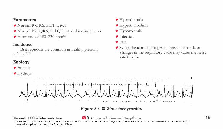
Parameters♥♥ Normal P, QRS, and T waves♥♥ Normal PR, QRS, and QT interval measurements♥♥ Heart rate of 180–230 bpm12
IncidenceBrief episodes are common in healthy preterm
infants.12,13
Etiology♥♥ Anemia♥♥ Hydrops
♥♥ Hyperthermia♥♥ Hyperthyroidism♥♥ Hypovolemia♥♥ Infection♥♥ Pain♥♥ Sympathetic tone changes, increased demands, or changes in the respiratory cycle may cause the heart rate to vary
Neonatal ECG Interpretation fl 3 Cardiac Rhythms and Arrhythmias 18
Figure 3-4 fl Sinus tachycardia.
T 19
TreatmentNo treatment is indicated if the condition is tran-
sient. Remove or treat the cause. With hydrops, ST can be associated with cardiovascular collapse; the infant should be assessed closely and treated as for shock.
ComplicationsWith any tachycardia, the ventricles do not have suf-
ficient time to fill with blood prior to contraction. This may lead to hypotension and decreased cardiac output.
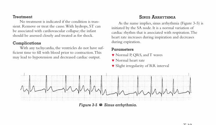
sInus ArrhythmIA
As the name implies, sinus arrhythmia (Figure 3-5) is initiated by the SA node. It is a normal variation of cardiac rhythm that is associated with respiration. The heart rate increases during inspiration and decreases during expiration.
Parameters♥♥ Normal P, QRS, and T waves♥♥ Normal heart rate♥♥ Slight irregularity of RR interval
Figure 3-5 fl Sinus arrhythmia.
IncidenceThis arrhythmia is common in the healthy
newborn.13 It usually occurs at lower heart rates, such as when the infant is resting or sleeping.14
EtiologySinus arrhythmia is probably the result of intratho-
racic pressure changes that occur with respiration.
TreatmentThis is a normal variation and does not require
intervention or special monitoring.14
ComplicationsThere are no complications.
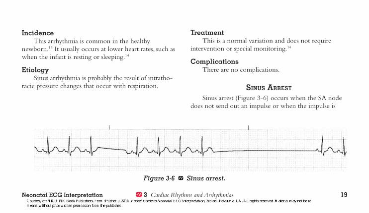
sInus Arrest
Sinus arrest (Figure 3-6) occurs when the SA node does not send out an impulse or when the impulse is
Neonatal ECG Interpretation fl 3 Cardiac Rhythms and Arrhythmias 19
Figure 3-6 fl Sinus arrest.

T 20
blocked. After a pause, either the SA node recovers or another pacemaking area takes over. Sinus node dysfunc-tion can be further complicated after cardiac surgery with coexistent atrial arrhythmias. This combination is termed sick sinus syndrome (SSS) or tachy-brady syndrome.15–17
Parameters♥♥ Normal QRS and T waves♥♥ P waves may change in appearance♥♥ Absent or delayed beat, followed by normal conduction
♥♥ Heart rate slows during arrest, then may accelerate to normal or tachycardiac arrhythmias
IncidenceSick sinus syndrome is common after surgery for
congenital heart disease, especially if the surgery is near the sinus node.15–18
Etiology♥♥ Autonomic heart disease associated with sympathetic and parasympathetic responses
♥♥ Cardiac surgery♥♥ Digitalis toxicity♥♥ Hyperkalemia♥♥ Increased sensitivity to vagal stimulation♥♥ Increased vagal tone♥♥ Inflammation of the SA node♥♥ Injury to the SA node♥♥ Myopathies
TreatmentIdentify and treat the cause. If bradycardia persists,
the infant may require treatment with epinephrine. Isoproterenol has also been used to increase the SA node rate but may result in accelerated rhythms.15 Atrial pacing may be indicated for known SA node injury and can eliminate the need for drug therapy in some infants.10

Pacing may also alleviate signs of fatigue and improve hemodynamic status in SSS.19
ComplicationsEscape rhythms may follow sinus arrest and compro-
mise cardiac output. Escape rhythms are nonsinus rhythms that occur during or after recovery from another arrhythmia.
ATRIAL RHYTHMSAtrial rhythms originate in the atria or the SA node.
Because the P wave reflects what is happening in the atria, focusing on what is happening with the P wave helps the clinician determine if atrial rhythm distur-bances are present.
PremAture AtrIAl contrActIons
A premature atrial contraction (PAC) (Figure 3-7) is an early beat that originates in a pacemaker
Neonatal ECG Interpretation fl 3 Cardiac Rhythms and Arrhythmias 20
Figure 3-7 fl Premature atrial contractions.

T 21
cell of the atria, other than the SA node. The impulse then travels its normal path through the remainder of the conduction system, often resulting in ventricular contraction with a normal QRS complex and T wave. PACs are also known as atrial premature beats (APBs).
Parameters♥♥ Variation in shape and size of P waves♥♥ Variation in PP interval♥♥ Early beats that otherwise look normal♥♥ Usually normal QRS complexes and T waves
IncidencePACs can occur frequently in neonates, yet are rare
by one month of age.12 PACs have been documented in 5 to 30 percent of premature infants.9
EtiologyPACs are almost always benign in the neonate.
However, they may not be benign when associated with these factors:
♥♥ Atropine, caffeine, theophylline, or inotropic agent administration
♥♥ Cardiac tumors, myopathies, or myocarditis♥♥ Digitalis toxicity♥♥ Electrolyte abnormalities♥♥ Hyperthyroidism♥♥ Irritation from a central line♥♥ Sepsis♥♥ Severe respiratory distress or hypoxia♥♥ Structural heart disease

TreatmentNo treatment is necessary for isolated cases. If PACs
are associated with symptoms such as hypotension or altered consciousness, treat the cause, and obtain a 12-lead ECG to rule out underlying heart disease.
ComplicationsPACs are almost always benign in the asymptomatic
infant. Some infants may develop supraventricular tachy-cardias later. Otherwise, complications are related to the cause.
AtrIAl tAchycArdIA
Atrial tachycardia (atrial tach) (Figure 3-8), is a type of supraventricular tachycardia (SVT) discussed in more detail later in this chapter. There are several forms of atrial tachycardia, including atrial ectopic tachycardia (AET), multifocal atrial tachycardia (MAT), and parox-ysmal atrial tachycardia (PAT). In the case of AET, the focus and cause are known. AET and MAT are caused by increased atrial automaticity and can result from repeated firing of a single ectopic focus in the atria, AET, or from multiple ectopic foci in the atria, MAT. Paroxysmal atrial
Neonatal ECG Interpretation fl 3 Cardiac Rhythms and Arrhythmias 21
Figure 3-8 fl Example of atrial tachycardia (simulation).

T 22
tachycardia is a type of atrial tachycardia that starts and stops suddenly and is characterized by extremely rapid rates (230–300 bpm).
Atrial tachycardia generally has a gradual onset with rates greater than 180 bpm.6,20 It can be seen in infants with normal hearts, in those with congenital heart disease, and after cardiac surgery because of high levels of circulating catecholamines.6,15
Parameters♥♥ Variation in size and pattern of P waves♥♥ Usually slow to develop (except for PAT)♥♥ Sustained or intermittent rhythm♥♥ Variation in atrial rate♥♥ With AET and MAT, ventricles may respond normally or may require multiple atrial stimuli to evoke a ven-tricular response13 (see Heart Blocks)
IncidenceAtrial tachycardia is uncommon but may sometimes
be seen after cardiac surgery.21 AET is probably respon-sible for 10 percent of all supraventricular tachycardias (SVTs).18
Etiology♥♥ May occur postoperatively because of elevated circu-lating catecholamines
♥♥ May be idiopathic♥♥ Occurs primarily after surgery performed at or near the pulmonary veins22
♥♥ PAT is associated with cor pulmonale or ischemic heart disease23
TreatmentAdenosine is not effective in treating most forms of
atrial tachycardia. Intravenous esmolol can be used acutely.14 Digoxin may reduce the heart rate by blocking

the AV node.10,15 Phenytoin may suppress the ectopic focus.15 Medications such as flecainide, propafenone, atenolol, sotalol, and amiodarone may be used for a refractory rhythm.14,16,20 Transcatheter or surgical ablation of the focus may be required. Because of the fast rates, PAT is treated with vagal stimulation, adenosine, pro-pranolol, procainamide,6 cardioversion (Chapter 4), or pacing.
ComplicationsDecreased cardiac output.
WAnderIng AtrIAl PAcemAker
The SA node is not always the origin of atrial stim-ulation. In some instances, the focus (or pacemaker) changes from one area of the atria to another and then another. As the focus changes, or “wanders,” the shape of the P wave also changes. This variation in the P wave characterizes the wandering atrial pacemaker rhythm, also known as WAP (Figure 3-9).
Parameters♥♥ Variation in shape and size of P wave
Neonatal ECG Interpretation fl 3 Cardiac Rhythms and Arrhythmias 22
Figure 3-9 fl Wandering atrial pacemaker.

T 23
♥♥ Possible variation in PR interval♥♥ Variation in PP and RR intervals♥♥ QRS and T waves are normal in shape and measurement
IncidenceWandering atrial pacemaker is seen frequently in the
newborn period.22
EtiologyImpulses originate from varying atrial foci.
Wandering atrial pacemaker is a benign arrhythmia asso-ciated with increased vagal tone.12
TreatmentNo treatment is necessary for this condition.
ComplicationsWandering atrial pacemaker causes no
complications.
AtrIAl flutter
Atrial flutter (Figure 3-10) originates in an ectopic atrial focus. P waves occur in rapid succession. The P waves are all identical, but not all are conducted to the ventricles. The identical, rapidly repeating P waves
Figure 3-10 fl Atrial flutter.

produce a “sawtooth” pattern on the ECG. Atrial flutter is thought to originate in the right atrium and is some-times categorized as a type of SVT.12,24 Atrial flutter can also occur in conjunction with heart block, with a resul-tant slow ventricular rate.
Parameters♥♥ Multiple P waves (flutter waves) prior to each QRS complex
♥♥ P waves usually have a sawtooth appearance♥♥ Atrial rate of 200–600 bpm10,15,20,24
♥♥ Slow, normal, or rapid ventricular rate♥♥ A specific number of P waves may occur prior to each QRS complex (ratios such as 2:1, 3:1, or 4:1).
IncidenceAtrial flutter comprises 9 to 14 percent of neonatal
tachyarrhythmias.25 Arterial catheters are thought to be responsible for three in eight of the episodes noted in
infants.26 Atrial flutter may occur in up to 50 percent of infants after cardiac surgery and is related to atrial scar-ring and right atrial enlargement.18,24
Etiology♥♥ Atrial septal aneurysms♥♥ Cardiac catheterization♥♥ Congenital heart disease associated with atrial dilation or hydrops
♥♥ Costello syndrome♥♥ Damage to the sinus node♥♥ Digoxin toxicity♥♥ Restrictive cardiomyopathy
TreatmentTreatment goals include slowing the fast rate, pre-
venting recurrence, and decreasing complications and symptoms. Digoxin, diltiazem, and amiodarone may be helpful in controlling rate.24 Intravenous procainamide
Neonatal ECG Interpretation fl 3 Cardiac Rhythms and Arrhythmias 23
T 24
may provide acute conversion, yet should be preceded by digitalization to prevent accelerated AV conduction. Adenosine has been used with varied results.12 For the hemodynamically unstable infant, cardioversion (Chapter 4) may be indicated. However, cardioversion is contrain-dicated if the arrhythmia results from digoxin toxicity because electrical stimulation can augment the myocar-dial excitability in these cases. Radiofrequency catheter ablation can be used for persistent atrial flutter. Digoxin is recommended for long-term suppression.24 Anticoagulation is indicated for chronic atrial flutter.
Complications24
♥♥ Brain and other end organ injury♥♥ Heart failure♥♥ Low cardiac output♥♥ Thrombosis♥♥ Sudden death
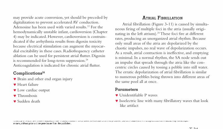
AtrIAl fIBrIllAtIon
Atrial fibrillation (Figure 3-11) is caused by simulta-neous firing of multiple foci in the atria (usually origi-nating in the left atrium).24 These foci fire at different rates, producing an unorganized atrial rhythm. Because only small areas of the atria are depolarized by the chaotic impulses, no real wave of depolarization occurs. As a result, atrial contraction is ineffective, and emptying is minimal. In a normal rhythm, the SA node sends out an impulse that spreads through the atria like the con-centric circles caused by tossing a pebble into still water. The erratic depolarization of atrial fibrillation is similar to numerous pebbles being thrown into different areas of the same pool all at once.
Parameters♥♥ Unidentifiable P waves♥♥ Isoelectric line with many fibrillatory waves that look like artifact

♥♥ Normal appearing QRS complexes♥♥ Irregular conduction with variations in RR interval♥♥ Variable ventricular rate of 120–250 bpm
IncidenceThis rhythm is rare in infants; the exact incidence in
the newborn remains unknown.27 Of infants presenting with atrial fibrillation, 77 percent have had intracardiac surgery.28 Infants with markedly distended atria may also occasionally develop this rhythm.15
Etiology♥♥ Atrial dilation♥♥ Large atrial septal defect (ASD)♥♥ Mitral valve abnormalities
TreatmentTreatment is similar to that for atrial flutter.29 If the
infant is unstable, cardioversion (Chapter 4) is the primary treatment. If the ventricular rate is fast, digoxin or β-blockers may slow the AV conduction rate. Procainamide,6 amiodarone, flecainide, propafenone, or
Neonatal ECG Interpretation fl 3 Cardiac Rhythms and Arrhythmias 24
Figure 3-11 fl Atrial fibrillation.

T 25
sotalol15 may be used to prevent recurrence. The long-term treatment is surgical repair of the underlying cardiac defects.
ComplicationsDecreased ventricular filling and decreased cardiac
output may occur. Inadequate emptying of the atria may cause stagnation and thrombus formation, potentially contributing to cerebral or other organ embolisms.
suPrAventrIculAr tAchycArdIA
Supraventricular tachycardia (SVT) (Figure 3-12) is a very fast rhythm that originates somewhere above the ventricles. SVT usually originates from an ectopic focus in the atria or from the area around the AV node. SVT is named based on the location of the stimulus if it is known, or based on the cause. Some of the names include atrioventricular tachycardia, re-entrant tachy-cardia, multifocal atrial tachycardia, pre-excitation tachycardia, paroxysmal atrial tachycardia, and junctional ectopic tachycardia.
Figure 3-12 fl Supraventricular tachycardia.

Parameters♥♥ Consistent RR interval♥♥ Heart rate of 230–400 bpm♥♥ Normal or narrow QRS complex♥♥ Relatively fixed heart rate despite changes in activity♥♥ Variation in PR interval29
♥♥ Visible or invisible P wave (may be lost in previous T wave)
IncidenceSVT is the most common symptomatic tachydys-
rhythmia in newborns.13 It occurs in 1.5–4 of every 1,000 neonates and represents 13 percent of all child-hood arrhythmias.27,30
Etiology♥♥ Cardiac defects such as:• Ebstein’s anomaly
• Transposition of the great vessels• Tricuspid atresia
♥♥ Conduction abnormalities such as Wolff-Parkinson-White (WPW) syndrome
♥♥ Myocarditis♥♥ Systemic infections♥♥ Thyrotoxicosis
TreatmentTreatment of SVT is accomplished in three phases:
termination, initial therapy, and maintenance.12 The severity of the symptoms also influences treatment deci-sions. If the infant does not demonstrate congestive heart failure or has only mild symptoms such as tachypnea, the dysrhythmia may be terminated by placing ice over the infant’s face; yet this must be done cautiously to ensure proper ventilation.9,15,29 Also, it is important to monitor for reflex bradycardia.
Neonatal ECG Interpretation fl 3 Cardiac Rhythms and Arrhythmias 25

T 26
Symptomatic infants (shock or congestive heart failure) warrant rapid treatment, including vagal maneu-vers and IV adenosine, followed by cardioversion (Chapter 4) or transesophageal pacing.12,15,16,29,31
It is important to note that repeated doses of ade-nosine should be avoided, as they may result in perpetua-tion of the arrhythmia. As a result, initial therapy may also include intravenous infusion of esmolol, procain-amide, or amiodarone.12 SVT tends to be a recurring problem requiring maintenance (oral) therapy. First-line maintenance therapy may include digoxin or β-blockers such as propranolol or sotalol.32 Second-line medications for refractory SVT may include amiodarone, flecainide, propranolol, or sotalol. Note: Digoxin and calcium channel blockers should be avoided when SVT is caused by WPW syndrome, as they can increase the conduction rate and lead to ventricular fibrillation or sudden death. Surgical ablation, a method used to destroy the arrhyth-mia’s focus, may be necessary in cases where medical therapy is not successful.15
ComplicationsWith sustained SVT, decreased cardiac output and
congestive heart failure can occur.9
JUNCTIONAL RHYTHMSJunctional rhythms, also known as nodal rhythms,
are stimulated by the AV node, rather than the SA node. The wave of depolarization travels backward to stimulate the atria and forward to stimulate the ventricles. This backward, or retrograde, atrial depolarization may result in an inverted P wave (P is below the isoelectric line), absent P waves that are hidden in the QRS complex, retrograde P waves (a P wave occurring after the QRS when ventricular conduction occurs before atrial depo-larization), or a very short PR interval.
nodAl rhythm
A nodal rhythm (Figure 3-13) may be missed because all the waveforms appear normal except for an

absent or abnormal P wave. The bradycardia that occurs with hypoxic episodes is often a junctional bradycardia (no P waves) rather than a sinus bradycardia.
Parameters♥♥ Absent or inverted P waves, possible retrograde P waves (following QRS complexes), or short PR interval
♥♥ Normal, bradycardic, or tachycardic rate♥♥ Normal QRS interval♥♥ Normal PP and RR intervals
IncidenceNodal rhythms may be present in 18–70 percent of
premature infants, and up to 28 percent of normal newborns.16
Etiology♥♥ May be normal in the newborn♥♥ May be associated with congenital heart disease♥♥ Hypoxia
Neonatal ECG Interpretation fl 3 Cardiac Rhythms and Arrhythmias 26
Figure 3-13 fl Nodal rhythm.

T 27
TreatmentMonitor asymptomatic patients. Treat the cause if
condition is the result of hypoxia. In some cases of symp-tomatic junctional bradycardia, atropine or isoproterenol may increase the heart rate and encourage the SA node to take over.33
ComplicationsThere may be decreased cardiac output if the
arrhythmia is associated with high or low blood pressure.
JunctIonAl ectoPIc tAchycArdIA
Junctional ectopic tachycardia (JET) (Figure 3-14) is a type of supraventricular tachycardia that occurs almost exclusively after surgery for congenital heart disease. There is also a rare congenital form of JET. JET is caused by increased automaticity of the AV node and bundle of His, resulting in an increased ventricular rate. The atria are frequently unaffected and continue to function
Figure 3-14 fl Junctional ectopic tachycardia.

normally; thus, JET may be the only form of SVT in which the ventricular rate can exceed the atrial rate.
Parameters♥♥ Gradual increase in heart rate over several hours♥♥ Possibility of hidden P waves or P waves after QRS complexes
♥♥ Possibility of inverted waves (when visible)♥♥ Usually transient (lasting a few hours to a few days)♥♥ Variable ventricular rate, with periods where it speeds up and slows down
♥♥ Ventricular rate of 100 bpm or greater (up to 370 bpm)
IncidenceJET is rare, occurring primarily after surgery near
the AV junction.15 The highest postoperative incidence is in the very young neonate who has had long or difficult surgeries and who requires inotropic support after
bypass.21 With congenital JET, 50 percent of patients have a positive family history, and the mortality rate is 35 percent.21
Etiology♥♥ Enhanced automaticity of the AV node or Purkinje system
♥♥ Related to surgical irritation ♥♥ A rare congenital form may be noted prenatally or in the first few weeks of life and is probably hereditary
TreatmentIf the patient is asymptomatic and the heart rate is
less than 170 bpm, no treatment may be required. However, with faster rates, there may be acute hemody-namic consequences requiring treatment. Initial treat-ment of postoperative JET includes correcting contributing factors such as electrolyte abnormalities and anemia, optimizing sedation, and correcting fever.
Neonatal ECG Interpretation fl 3 Cardiac Rhythms and Arrhythmias 27

T 28
Amiodarone is the primary treatment. If the infant does not respond to amiodarone, additional pharmacologic treatment can include dexmedetomidine, magnesium, procainamide, or esmolol. Temporary pacing, cooling, or ablation may also be indicated. The congenital form is treated with amiodarone or β-blockers. Ablation is indi-cated for refractory JET or heart failure.34
ComplicationsComplications include severe hemodynamic com-
promise as a result of the accelerated rate. Congestive heart failure and acute cardiovascular collapse may occur.
VENTRICULAR RHYTHMSVentricular rhythms are associated with problems
originating in the ventricles. Because the QRS complex reflects ventricular depolarization and contraction, the QRS complex is abnormal in these dysrhythmias.
PremAture ventrIculAr contrActIons
Premature ventricular contractions (PVCs) (Figure 3-15 a & b) originate from an ectopic focus or foci in the ventricles. They occur early in the cycle, before the SA node can fire. The impulse does not follow the usual conduction system. Thus conduction is slow, resulting in a wide QRS complex.2,9 PVCs can have the same shape and appearance each time they are present, or they can have different shapes resulting from different morphologies. If they are all the same shape, they are termed unifocal PVCs.6,17 If they have different shapes, they are known as multifocal PVCs. PVCs may also be labeled on the basis of their frequency: For example, bigeminy denotes a rhythm in which a PVC occurs every other beat; trigeminy, every third beat.
Parameters♥♥ Normal baseline rhythm♥♥ Periodic early beats

Neonatal ECG Interpretation fl 3 Cardiac Rhythms and Arrhythmias 28
Figure 3-15 a & b fl Premature ventricular contractions.a. In the first rhythm strip, the PVCs are all the same in appearance; therefore, they probably are being elicited
from the same focus (unifocal).b. The PVCs in the second strip are not alike in appearance and are elicited from more than one focus (multifocal).
PVC PVC PVC PVC ↓ ↓ ↓ ↓
↑ ↑ ↑♥ PVC PVC PVC
a
b

T 29
♥♥ Early beats characterized by wide QRS complexes♥♥ QRS complexes are usually taller and deeper than normal
♥♥ Compensatory pause before the next normal beat♥♥ Early beat may not perfuse; thus arterial blood pres-sure (BP) tracings may be dampened during the PVC
IncidencePVCs are reported to occur in as many as 33 percent
of newborns, but they disappear within the first four to eight weeks of life.35
Etiology♥♥ May be normal♥♥ Acidosis♥♥ Cardiac tumors16
♥♥ Decreased cardiac output♥♥ Electrolyte or metabolic disturbances♥♥ Irritation from invasive catheters
♥♥ May be associated with caffeine, digoxin, or anti-arrhythmic agent administration14
♥♥ Myocarditis26
♥♥ Prolonged QT syndrome♥♥ Systemic infection36
♥♥ Underlying cardiac disease
TreatmentIf PVCs are infrequent or the infant is asymptomatic,
treatment may not be required. If the infant is symptom-atic, treat the cause and give lidocaine as needed. Lidocaine may also be given if the PVCs are frequent (more than ten per minute or in runs of three or more in a row) or if the R wave occurs very close to or at the same time as the T wave (this R-on-T phenomenon can result in ventricular tachycardia).
ComplicationsIf frequent, PVCs may compromise cardiac output.
They may progress to ventricular tachycardia or ventric-ular fibrillation.

long Qt syndrome
Prolonged QT intervals, also known as Long QT syndrome (LQTS) have a QT interval of >0.44 seconds as the key identifying factor (Figure 3-16).7 Although this is a syndrome that may be genetic or acquired, it is included in the VenTriCular rhyThms section because of its likelihood to progress to more serious ventricular arrhythmias and sudden death. The genetic form is asso-ciated with alterations in potassium and sodium involved in the control of ventricular repolarization.37 The acquired form may be associated with reactions to certain medications.
The QT interval is commonly measured from the onset of the QRS to the end of the T wave. The dura-tion of the QT interval varies as the heart rate changes. As a result, Bazett’s formula is commonly used to measure the corrected QT interval:12
QTc = QT (in seconds)
√RR (preceding RR interval in seconds)
Parameters♥♥ Normal P and QRS waveforms♥♥ Normal rate♥♥ QT interval exceeds the normal range♥♥ T wave may be notched, biphasic, or inverted
IncidenceA mild, benign prolongation may be present after a
stressful birth, and usually resolves within 72 hours.12
Neonatal ECG Interpretation fl 3 Cardiac Rhythms and Arrhythmias 29
Figure 3-16 fl Prolonged QT interval with hypocalcemia.

T 30
Physiologic prolongation of the QT interval may also be present in 2.5 percent of healthy newborns until six months of age. LQTS is reported to be an uncommon rhythm disturbance in infants;7 the European Society of Cardiology Task Force estimates an incidence of 1:3,000–5,000.37 Congenital LQTS is associated with sudden infant death syndrome (SIDS), with gene variants found in 9.5 percent of SIDS victims.12
Etiology7
♥♥ Antiarrhythmic medications such as quinidine and procainamide
♥♥ Jervell and Lange-Nielson syndrome♥♥ Romano-Ward syndrome
TreatmentTypical presenting symptoms in neonates include
syncope, seizures, and sudden death, thus early diagnosis is essential, especially in cases with a known family
history of prolonged QT syndrome.14 β-blockers are the initial treatment of choice. Propranolol and nadolol are most commonly used and are effective in eradicating the arrhythmia in the majority of cases. Compliance must be stressed and dosages adjusted with weight throughout childhood. Patients with persistent syncopal episodes may require internal defibrillator placement. A perma-nent pacemaker may be indicated in patients who have a prolonged QT interval along with a second degree AV block.7,19
ComplicationsComplications include seizures, ventricular arrhyth-
mias, and heart blocks. In particular, LQTS is character-ized by delayed ventricular repolarization and an increased risk of ventricular arrhythmias, particularly a polymorphic type of ventricular tachycardia known as torsades de pointes (discussed in the next section), which can lead to ventricular fibrillation and sudden death.12

ventrIculAr tAchycArdIA
Ventricular tachycardia—also known as V tach or VT—originates from a focus in the ventricle (rather than from the SA node). When the ventricles do not receive an impulse from above, they will fire at their own intrinsic rate (50–60 bpm). As the name implies, V tach is a rhythm that is faster than the normal ventricular rate (Figure 3-17). It is a dangerous rhythm because rapid ventricular contractions prevent the ventricles from completely filling and emptying with each contraction. This rhythm can greatly reduce cardiac output.
Ventricular tachycardia may rapidly progress to more lethal arrhythmias. Always assume that a wide QRS tachycardia is V tach until proven otherwise.
Torsades de pointes is a type of V tach. It’s name lit-erally means “twisting of the points.” In this type of VT, the QRS complexes gradually change from one ventric-ular focus to another. There may be a normal-width QRS complex between two episodes of VT marked by wide QRS complexes. The QRS complex may change completely every five to ten beats.
Neonatal ECG Interpretation fl 3 Cardiac Rhythms and Arrhythmias 30
Figure 3-17 fl Ventricular tachycardia.

T 31
Parameters♥♥ Altered or widened QRS complexes without P waves (P waves are hidden in QRS complexes.)
♥♥ QRS complex has a sawtooth or flutter appearance♥♥ May look like several PVCs in a row (Three or more PVCs in a row are called a short run of ventricular tachycardia.)
♥♥ Heart rate of 120–240 bpm♥♥ RR intervals may be regular♥♥ Inadequate ventricular contraction and cardiac output
Distinguishing VT from SVT Those who are just learning to interpret ECGs can
sometimes find it difficult to distinguish ventricular tachycardia from supraventricular tachycardia (Figures 3-18 and 3-19). Table 3-1 compares the two dysrhyth-mias. Remember: Always assume that a tachycardia with a wide QRS is ventricular tachycardia. Treat it as an emergency until proven otherwise!
Figure 3-19 fl Ventricular tachycardia.
Figure 3-18 fl Supraventricular tachycardia.

IncidenceVentricular tachycardia is rare in neonates.15 Of
infants who have episodes of ventricular tachycardia, 85 percent have an abnormal heart. The majority have cardiac tumors, particularly rhabdomyomas, cardiac fibromas, or Purkinje cell tumors.12 Another 10 percent have myocarditis.35 After cardiac surgery, ventricular
tachycardia may occur in 10 to 15 percent of infants, and 5 to 10 percent of those may experience sudden death.18
Etiology♥♥ Acidosis♥♥ Electrolyte imbalance♥♥ Cardiac anomalies
Neonatal ECG Interpretation fl 3 Cardiac Rhythms and Arrhythmias 31
Table 3-1 fl Comparison of Supraventricular Tachycardia and Ventricular Tachycardia��������������������������������������������������������������������������������
Characteristic SVT VTOccurrenceininfants
Pwaves
QRScomplexdeflection*
QRScomplexwidth
Hemodynamics
Vagalmaneuversastreatment
��������������������������������������������������������������������������������
*DeflectionreferstolocationoftheQRScomplexaboveor belowtheisoelectricline.Normaldeflectionwilldependonwhichleadonthecardiacmonitorisselected.
Frequent
Maybeabnormal
Usuallynormal
Usuallynormalornarrow
ToleratedbetterinitiallythanVT
MaysloworstopSVT
Uncommon
Nonepresent
Maybeoppositenormal
Wideordifferent
Altered
Noteffective

T 32
♥♥ Cardiac tumors♥♥ Cardiomyopathy♥♥ Drug toxicity♥♥ Exposure to some antiarrhythmic medications♥♥ Hypoxia♥♥ Mitral valve prolapse♥♥ Myocardial fibrosis♥♥ Myocarditis♥♥ “Tet” spells, which occur with tetralogy of Fallot, may include cyanosis, irritability, pallor, flaccidity, tachy-pnea, and/or loss of consciousness13
♥♥ Tumors of the ventricle
TreatmentTreatment is based on the presence or absence of a
pulse: If the infant is symptomatic and has a pulse, lido-caine therapy, sedation, and cardioversion (Chapter 4) are indicated. Ventricular tachycardia without a pulse is treated the same as ventricular fibrillation: with CPR
and defibrillation (Chapter 4).31 Antiarrhythmic drugs such as lidocaine (usually the first drug of choice),10 flecainide, or amiodarone may be used.12 Treat the cause. For infants with recurrent torsades de pointes, magne-sium sulfate infusion is the drug of choice. Overdrive pacing is also effective.14
ComplicationsComplications include severely compromised
cardiac output and progression to ventricular fibrillation.
ventrIculAr fIBrIllAtIon
Ventricular fibrillation—also known as V fib or VF—is created by rapid stimuli from many ventricular ectopic foci (Figure 3-20). This stimulation causes a chaotic twitching or quivering of the ventricles. There is no effective cardiac output because the heart muscle is “quivering” rather than pumping.

Parameters♥♥ Irregular, coarse ECG pattern without identifiable waves
♥♥ No pulse or audible heartbeat
IncidenceVentricular fibrillation is rare in infants.15 Most
infants with ventricular fibrillation have abnormal hearts.25
Etiology14
♥♥ Acidosis♥♥ Cardiac anomalies♥♥ Drug toxicity♥♥ Electrolyte imbalance♥♥ Hypoxia♥♥ Myocarditis
Neonatal ECG Interpretation fl 3 Cardiac Rhythms and Arrhythmias 32
Figure 3-20 fl Ventricular fibrillation.

T 33
TreatmentImmediate resuscitation, including CPR and
defibrillation (Chapter 4), is indicated. Lidocaine drug therapy may be indicated. Treat the cause. An implantable or external automated defibrillator can be used for ven-tricular fibrillation.38
ComplicationsVentricular fibrillation is lethal.
HEART BLOCKSIn a heart block, the electrical impulse is blocked at
some point in the cardiac cycle. This may simply prolong the time required for the impulse to pass through the cardiac cycle, or it may prevent the impulse from trav-eling from the atria to the ventricles.
Bundle BrAnch Block
Under normal circumstances, the right and left ven-tricles are depolarized simultaneously. This results in a single QRS complex. Bundle branch block (BBB) is caused by a slowing of the impulse in either the right or left bundle branch, prolonging conduction through the ventricles. Most situations of BBB cause depolarization of one ventricle before the other. This results in two QRS complexes that are often merged into one QRS with a characteristic “rabbit ear” appearance (Figure 3-21).
Bundle branch blocks may occur in either branch (right or left) and in specific divisions of each bundle. A 12-lead ECG printout (Chapter 5) is needed to deter-mine whether the block is occurring in the left or right bundle branch. Left bundle branch block (LBBB) is diag-nosed by assessing the R wave in leads V
5 and V
6; right
bundle branch block (RBBB) is diagnosed by assessing leads V
1 and V
2.2,29 Identification of the specific location

of the block is an advanced interpretation technique not discussed in this text because it does not necessarily affect treatment or outcome.
Parameters♥♥ Notched R waves or R waves with a “rabbit ear” appearance
♥♥ Widened QRS complexes
IncidenceBundle branch block is relatively common in
the pediatric population. It is estimated to occur in
0.2 percent of normal children.6 LBBB is infrequent and usually occurs only after cardiac surgery of the aortic valve or subvalvular area. RBBB occurs in 32 to 44 percent of children after ventricular septal defect (VSD) repair,39 and in 59 to 100 percent of children after repair of tetralogy of Fallot.18
Etiology♥♥ Possible association with Ebstein’s anomaly, Kearns-Sayre syndrome, and neuromuscular disorders
♥♥ Electrolyte imbalance
Neonatal ECG Interpretation fl 3 Cardiac Rhythms and Arrhythmias 33
Figure 3-21 fl Bundle branch block.

T 34
♥♥ Heredity♥♥ Myocarditis♥♥ Post-cardiac surgery or transplantation♥♥ Volume overload
TreatmentFollow BBB conservatively.
ComplicationsBundle branch block may progress to a higher
degree block or to complete heart block.29
fIrst degree heArt Block
First degree heart block (Figure 3-22) occurs at the AV node and causes a longer than normal delay between atrial and ventricular stimulation. Normally, a short pause follows atrial depolarization to allow the ventricles time to fill with blood before contracting. This pause is reflected in the PR interval. A prolonged PR interval
indicates first degree heart block. Diagnosis requires a printout of the rhythm strip and measurement of the PR interval.
Parameters♥♥ Prolonged PR interval, generally >0.11–0.14 seconds, based on age and HR6
♥♥ Otherwise normal ECG appearance and measurements
IncidenceFirst degree heart block may occur in up to 6
percent of neonates.40
Etiology♥♥ May be normal in the neonate♥♥ Large ASD♥♥ AV canal10
♥♥ Digitalis effect♥♥ Ebstein’s anomaly6

♥♥ Electrolyte abnormalities♥♥ Hypothermia♥♥ Increased vagal tone♥♥ Infectious or inflammatory conditions♥♥ Patent ductus arteriosus23
♥♥ Pulmonary stenosis23
♥♥ Surgical injury to the AV node or Purkinje system♥♥ Valve disorders
TreatmentEvaluate the hemodynamic status of the infant and
treat the cause. Follow first degree heart block conservatively.
ComplicationsThere usually are no complications for this condi-
tion. Complications may be associated with the cause.
Neonatal ECG Interpretation fl 3 Cardiac Rhythms and Arrhythmias 34
Figure 3-22 fl First degree heart block.

T 35
second degree heArt Block
There are two types of second degree heart block: Mobitz I and Mobitz II.
Mobitz IAlso known as Wenckebach block, Mobitz I (Figure
3-23) may be normal in infants. It is marked by a pro-gressive lengthening of the PR interval until one P wave is not conducted.
ParameTers
♥♥ Progressive lengthening of PR interval, culminating in nonconduction of the beat (P wave is not followed by a QRS complex.)
♥♥ Normal P wave and QRS complex in preceding and following cycles
♥♥ Otherwise normal rate
inCidenCe
Mobitz I occurs rarely in infants.40
Figure 3-23 fl Second degree heart block: Mobitz I.
ShortPR
LongPR
LongerPR
DroppedQRS

eTiology
♥♥ May be normal in infants, especially during sleep or with increased vagal tone
♥♥ Disease or damage to the AV node or Purkinje system♥♥ Infectious or inflammatory conditions♥♥ Structural heart disease
TreaTmenT
Evaluate the hemodynamic status of the infant, and treat the cause. Follow Mobitz I second degree heart block conservatively.
ComPliCaTions
There usually are no complications of Mobitz I.
Mobitz IIMobitz II (Figure 3-24) is the more serious form of
second degree heart block because it can abruptly
Neonatal ECG Interpretation fl 3 Cardiac Rhythms and Arrhythmias 35
Figure 3-24 fl Second degree heart block: Mobitz II.
↓

T 36
progress to complete (third degree) heart block. In Mobitz II, the impulse that triggers ventricular depolar-ization (shown by the QRS complex) is not conducted for one or two beats after a normal P wave.
ParameTers
♥♥ Normal P waves♥♥ Normal PR intervals♥♥ “Dropped” QRS complexes (complex does not occur after all P waves); usually occur in a cyclic fashion (2:1, 3:1, 4:1)
♥♥ Varied RR intervals (due to dropped QRS)
inCidenCe
Mobitz II is uncommon in neonates.10,27
eTiology14
♥♥ Associated with prolonged QT syndrome♥♥ Congenital heart disease♥♥ Diffuse disease of the conduction system♥♥ Hypocalcemia
♥♥ Infection or inflammation♥♥ Surgical injury♥♥ Systemic disorders♥♥ Dropped QRS
TreaTmenT
Treat the cause. Mobitz II may require pacing if it is caused by surgical damage or if the infant is having symptoms such as bradycardia, hypotension, or an altered state of consciousness. Symptomatic bradycardias are usually responsive to atropine, epinephrine, or isoproter-enol until temporary pacing is available.12 Treatment should be aggressive if associated with prolonged QT syndrome.14
ComPliCaTions
Mobitz II often progresses to third degree (com-plete) heart block.
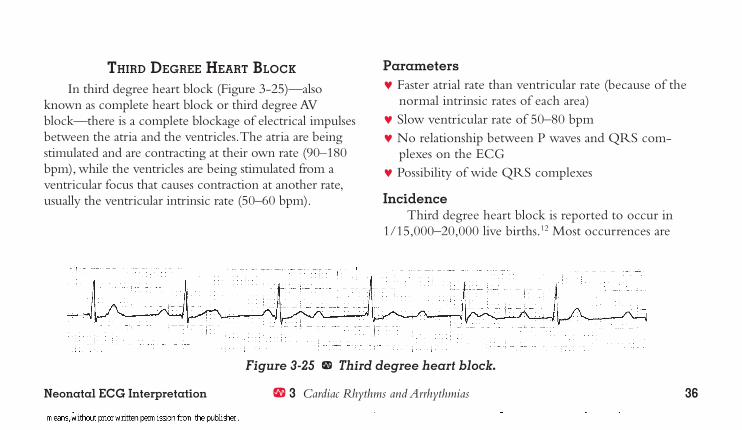
thIrd degree heArt Block
In third degree heart block (Figure 3-25)—also known as complete heart block or third degree AV block—there is a complete blockage of electrical impulses between the atria and the ventricles. The atria are being stimulated and are contracting at their own rate (90–180 bpm), while the ventricles are being stimulated from a ventricular focus that causes contraction at another rate, usually the ventricular intrinsic rate (50–60 bpm).
Parameters♥♥ Faster atrial rate than ventricular rate (because of the normal intrinsic rates of each area)
♥♥ Slow ventricular rate of 50–80 bpm♥♥ No relationship between P waves and QRS com-plexes on the ECG
♥♥ Possibility of wide QRS complexes
IncidenceThird degree heart block is reported to occur in
1/15,000–20,000 live births.12 Most occurrences are
Neonatal ECG Interpretation fl 3 Cardiac Rhythms and Arrhythmias 36
Figure 3-25 fl Third degree heart block.

T 37
associated with maternal antibodies that cross the pla-centa during the critical stage of fetal cardiac tissue development and disrupt normal formation of the con-duction system. Other infants (25–35 percent) with complete heart block have structural cardiac anomalies.27 Third degree heart block is also reported to occur in 1–2 percent of patients after cardiac surgery, especially VSD and tetralogy repairs.18
Etiology♥♥ Acute increase in vagal tone♥♥ Acute rheumatic fever♥♥ Congenital heart defects, such as atrial or ven tricular septal defects, transposition, or AV canal10
♥♥ Digoxin toxicity♥♥ Heredity♥♥ Infections
♥♥ Maternal collagen vascular diseases such as lupus ery-thematosus6,10 or Sjögren syndrome12,14
♥♥ Myocarditis♥♥ Myopathies♥♥ Post-surgical manipulation♥♥ Trauma
TreatmentIn symptomatic patients, such as those who are
hypotensive or have an altered level of consciousness, cardiac compressions may be required to maintain cardiac output if the ventricular response is very slow. Temporary/transesophageal or permanent pacing is the treatment of choice for symptomatic cases.19 Isoproterenol may be indicated in some situations and will help to increase cardiac output.29 Atropine may be useful if the cause is associated with increased vagal tone

rather than with AV node dysfunction.29 Permanent pacemaker treatment is indicated if the block is persistent and due to surgical causes.
Infants with congenital forms, such as those associ-ated with lupus erythematosus, are usually asymptomatic and may not require immediate treatment.14 However, prophylactic pacing decreases the incidence of complica-tions in infants with congenital heart block.19 Most will require a pacemaker sometime during childhood or ado-lescence.12 Pacing is indicated for a heart rate of less than 55 bpm. If congestive heart failure is present, pacing is indicated with a heart rate less than 70 bpm.19
ComplicationsComplications include inadequate cardiac output,
congestive heart failure, and possible death. The slow rate creates a potential for escape rhythms.
PACED RHYTHMSAs mentioned in previous sections, pacemakers are
often indicated in the treatment of dysrhythmias. Pacemakers deliver electrical impulses to the myocar-dium in an effort to stimulate depolarization when it does not occur independently. Pacemaker impulses are seen on the ECG rhythm strip as very narrow, spiked waveforms. The location of these waveforms is based on the type of pacemaker in use. There are a variety of pacemakers:
Temporary pacemakers are utilized for dysrhythmias believed to be short term, or as a bridge until permanent pacemaker placement can be done. The transesophageal method is the primary method for temporary pacing and is usually limited to atrial stimulation, because there are difficulties associated with ventricular lead placement.19
Permanent pacemakers are indicated when there is irreversible dysfunction or damage to the conduction
Neonatal ECG Interpretation fl 3 Cardiac Rhythms and Arrhythmias 37

T 38
system. Permanent pacemakers are available for even the smallest of infants.
Asynchronous pacemakers provide an electrical stimulus to the heart at a set rate, regardless of sponta-neous cardiac activity. These pacers are seldom used because of their potential to produce lethal dysrhythmias with an improperly timed electrical stimulus.
Demand pacemakers sense the electrical activity of the heart and deliver an electrical stimulus only when one is not sensed from the myocardium in a specified time frame.
Atrial pacemakers provide impulses to stimulate atrial depolarization only. They can be useful in treating
sinus bradycardias in which the AV conduction system is intact; sick sinus syndrome; and some atrial arrhythmias. With an atrial pacemaker, the pacemaker spike is visible just prior to the P wave. (Figure 3-26).
Ventricular pacemakers provide impulses that stimu-late only ventricular depolarization. This type of pacing may be useful in an infant with a heart block who has normal sinus node function. With a ventricular pace-maker, the ECG may have a normal sinus P wave fol-lowed by a pause, a pacemaker spike, and then a QRS complex. Or the P wave may be absent, with only a pacemaker spike followed by a QRS complex (Figure 3-27).

Neonatal ECG Interpretation fl 3 Cardiac Rhythms and Arrhythmias 38
Figure 3-27 fl Ventricular pacemaker spikes.
Figure 3-26 fl Atrial pacemaker spikes.

T 39
Dual-chamber pacemakers have the ability to provide impulses that can stimulate both the atria and the ventricles. These pacemakers may be used in the treatment of bradycardia, heart block, SA node disease, and atrial arrhythmias. With this type of pacemaker, a pacer spike is seen prior to the P wave and a second spike seen prior to the QRS complex (Figure 3-28).
Figure 3-28 fl Dual-chamber pacemaker spikes.

ECG CHANGES AND ELECTROLYTE ABNORMALITIES
Consistency of wave contour is an important factor in evaluating an ECG strip. Wave contours are
particularly important in helping to determine hyperka-lemia and hypokalemia.
In hyperkalemia, the most common ECG change is a peaked T wave (Figure 3-29a). Additional findings may include an elevated ST segment (Figure 3-29b), a
Neonatal ECG Interpretation fl 3 Cardiac Rhythms and Arrhythmias 39
Figure 3-29a fl Hyperkalemia, marked by peaked T waves.
Figure 3-29b fl Hyperkalemia, marked by elevated ST segments.

T 40
widened or flattened P wave, a widened QRS complex, and possible PVCs and other ventricular arrhythmias.
In hypokalemia, the T wave is generally flat or inverted (Figure 3-30). There may also be another wave after the T wave, known as a U wave, which is wide and prominent (Figure 3-31). The PR interval may be pro-longed, and the QRS may be wide. Untreated hypoka-lemia can lead to more serious arrhythmias.
Hypercalcemia and hypocalcemia may also cause ECG changes. The QT interval may be shorter than normal with hypercalcemia (Figure 3-32a), or it may be prolonged with hypocalcemia (Figure 3-32b).
Figure 3-30 fl Hypokalemia marked by flat T waves.

Neonatal ECG Interpretation fl 3 Cardiac Rhythms and Arrhythmias 40
Figure 3-32a fl Changes in QT interval with hypercalcemia.
Figure 3-32b fl Changes in QT interval with hypocalcemia.
Figure 3-31 fl Hypokalemia marked by U waves.
U U U U