Cardiac Emergencies in the First Year of...
28
Cardiac Emergencies in the First Year of Life Linton Yee, MD a,b, * a Department of Pediatrics, Division of Hospital and Emergency Medicine, Duke University School of Medicine, Durham, NC 27710, USA b Department of Surgery, Division of Emergency Medicine, Duke University School of Medicine, Durham, NC 27710, USA The presence of a distressed or obtunded infant in any adult or pediatric emergency department can prove to be a challenging process in airway man- agement, vascular access, and decision making. Cardiac emergencies, as well as a number of other diseases, can present in this manner. It is essential to ac- curately diagnose and expeditiously care for these potentially complicated car- diac patients. Diagnosis can be difficult because of a number of nonspecific elements in the history and physical exam. However, by developing an effective strategy in dealing with these patients, the emergency department manage- ment of these individuals can be completed in an efficient and prompt manner. The most challenging scenarios of cardiac emergencies in the first year of life include cyanotic episodes, congestive heart failure, cardiogenic shock or collapse, and arrhythmias. All of these emergent presentations can be the re- sult of either the initial presentation of disease or as a known complication of an already diagnosed cardiac lesion. In approaching cardiac emergencies, cardiac disease can be divided into structural disease, conduction abnormalities, and acquired illnesses. While recognizing that many lesions can be a combination of many defects, struc- tural congenital heart disease can be divided into cyanotic and acyanotic categories. The cyanotic category can be further subdivided into increased and decreased pulmonary blood flow. Division of the acyanotic category is based on left-to-right shunting and left ventricular outflow obstruction. Conduction abnormalities can be congenital or the result from a new-onset illness. Acquired heart disease includes cardiomyopathies, myocarditis, pericarditis, endocarditis, and Kawasaki’s disease. * Department of Pediatrics, Division of Hospital and Emergency Medicine, Duke University School of Medicine, Durham, NC 27710. E-mail address: [email protected] 0733-8627/07/$ - see front matter Ó 2007 Elsevier Inc. All rights reserved. doi:10.1016/j.emc.2007.08.001 emed.theclinics.com Emerg Med Clin N Am 25 (2007) 981–1008
Transcript of Cardiac Emergencies in the First Year of...

Emerg Med Clin N Am
25 (2007) 981–1008
Cardiac Emergenciesin the First Year of Life
Linton Yee, MDa,b,*aDepartment of Pediatrics, Division of Hospital and Emergency Medicine,
Duke University School of Medicine, Durham, NC 27710, USAbDepartment of Surgery, Division of Emergency Medicine,
Duke University School of Medicine, Durham, NC 27710, USA
The presence of a distressed or obtunded infant in any adult or pediatricemergency department can prove to be a challenging process in airway man-agement, vascular access, and decision making. Cardiac emergencies, as wellas a number of other diseases, can present in this manner. It is essential to ac-curately diagnose and expeditiously care for these potentially complicated car-diac patients. Diagnosis can be difficult because of a number of nonspecificelements in the history andphysical exam.However, by developing an effectivestrategy in dealing with these patients, the emergency department manage-ment of these individuals can be completed in an efficient and promptmanner.
The most challenging scenarios of cardiac emergencies in the first year oflife include cyanotic episodes, congestive heart failure, cardiogenic shock orcollapse, and arrhythmias. All of these emergent presentations can be the re-sult of either the initial presentation of disease or as a known complicationof an already diagnosed cardiac lesion.
In approaching cardiac emergencies, cardiac disease can be divided intostructural disease, conduction abnormalities, and acquired illnesses. Whilerecognizing that many lesions can be a combination of many defects, struc-tural congenital heart disease can be divided into cyanotic and acyanoticcategories. The cyanotic category can be further subdivided into increasedand decreased pulmonary blood flow. Division of the acyanotic categoryis based on left-to-right shunting and left ventricular outflow obstruction.Conduction abnormalities can be congenital or the result from a new-onsetillness. Acquired heart disease includes cardiomyopathies, myocarditis,pericarditis, endocarditis, and Kawasaki’s disease.
* Department of Pediatrics, Division of Hospital and Emergency Medicine, Duke
University School of Medicine, Durham, NC 27710.
E-mail address: [email protected]
0733-8627/07/$ - see front matter � 2007 Elsevier Inc. All rights reserved.
doi:10.1016/j.emc.2007.08.001 emed.theclinics.com

982 YEE
A cyanotic patient suggests that there is cyanotic congenital heart diseasewith shunting from the right to the left. In a patient with cardiogenic shockor collapse (the result of outflow obstruction and pump failure), the infantmay appear mottled, ashen, and gray. A patient with left-to-right shuntingand congestive heart failure can appear to be normal in color [1–7]. Thisarticle will discuss the cardiac emergencies that may present within the firstyear of life.
Basic pathophysiology
There are a number of changes that occur within the cardiovascular sys-tem in the transition from a fetus to a newborn. The placenta functions asthe pulmonary system for the fetus, as oxygenated blood is transferredfrom the placenta to the fetus via the umbilical vein. At birth, blood thentravels through a now lower resistance pulmonary system for oxygenationwith closure of the shunts that were used between the pulmonary andsystemic circulations (foramen ovale, ductus arteriosus, ductus venosus).Expansion of the lungs and the elimination of fluid from the lungs causedilatation of the pulmonary vasculature, which then leads to a decrease inpulmonary resistance and increased pulmonary blood flow. Oxygenationof the blood through the pulmonary system leads to the closure of theumbilical vessels, the ductus arteriosus, and the ductus venosus. Decreasedpulmonary artery resistance and subsequent increased systemic resistancechanges the flow though the atria, with pressures now higher in the left atriathan the right, resulting in the closure of the foramen ovale [8,9].
Cyanosis
Cyanosis is seen when desaturated blood is present in the capillary beds.Deoxygenated hemoglobin is blue and the presence of cyanosis means thatthere is 3 to5 mg/dL of deoxyhemoglobin in the blood. This correspondswith a room air oxygen saturation of 70% to 85% [10,11]. Because theoxygen carrying capacity is based on the amount of hemoglobin availableto carry oxygen, an infant who is polycythemic and cyanotic is still ableto deliver oxygen to tissues as opposed to an anemic infant who may notappear cyanotic but is not able to deliver oxygen to tissues.
It is important to differentiate between central and peripheral cyanosis asthe evaluation and treatment differ based on the underlying cause. Thereare a number of different causes for central cyanosis. These include central ner-vous system (CNS) depression, pulmonary disease, and cardiac disease as wellas sepsis andmetabolic disease and toxic ingestions. Peripheral cyanosis is theresult of acrocyanosis, exposure to cold, and decreased peripheral perfusion.
Factors to keep in mind when assessing cyanosis are the arterial oxygensaturation, the oxygen binding capacity (hemoglobin), and the arteriove-nous oxygen difference [10].
983CARDIAC EMERGENCIES
Cyanotic heart disease
There are five well-known cyanotic congenital heart lesionsdalso knownas the ‘‘Terrible Ts.’’ They are Tetralogy of Fallot (TOF), Transposition ofthe Great Arteries (TGA), Tricuspid Atresia (TA), Total AnomalousVenous Return (TAPVR), and Truncus Arteriosus.
Tetralogy of Fallot
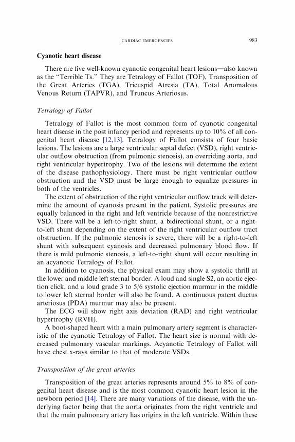
Tetralogy of Fallot is the most common form of cyanotic congenitalheart disease in the post infancy period and represents up to 10% of all con-genital heart disease [12,13]. Tetralogy of Fallot consists of four basiclesions. The lesions are a large ventricular septal defect (VSD), right ventric-ular outflow obstruction (from pulmonic stenosis), an overriding aorta, andright ventricular hypertrophy. Two of the lesions will determine the extentof the disease pathophysiology. There must be right ventricular outflowobstruction and the VSD must be large enough to equalize pressures inboth of the ventricles.
The extent of obstruction of the right ventricular outflow track will deter-mine the amount of cyanosis present in the patient. Systolic pressures areequally balanced in the right and left ventricle because of the nonrestrictiveVSD. There will be a left-to-right shunt, a bidirectional shunt, or a right-to-left shunt depending on the extent of the right ventricular outflow tractobstruction. If the pulmonic stenosis is severe, there will be a right-to-leftshunt with subsequent cyanosis and decreased pulmonary blood flow. Ifthere is mild pulmonic stenosis, a left-to-right shunt will occur resulting inan acyanotic Tetralogy of Fallot.
In addition to cyanosis, the physical exam may show a systolic thrill atthe lower and middle left sternal border. A loud and single S2, an aortic ejec-tion click, and a loud grade 3 to 5/6 systolic ejection murmur in the middleto lower left sternal border will also be found. A continuous patent ductusarteriosus (PDA) murmur may also be present.
The ECG will show right axis deviation (RAD) and right ventricularhypertrophy (RVH).
A boot-shaped heart with a main pulmonary artery segment is character-istic of the cyanotic Tetralogy of Fallot. The heart size is normal with de-creased pulmonary vascular markings. Acyanotic Tetralogy of Fallot willhave chest x-rays similar to that of moderate VSDs.
Transposition of the great arteries
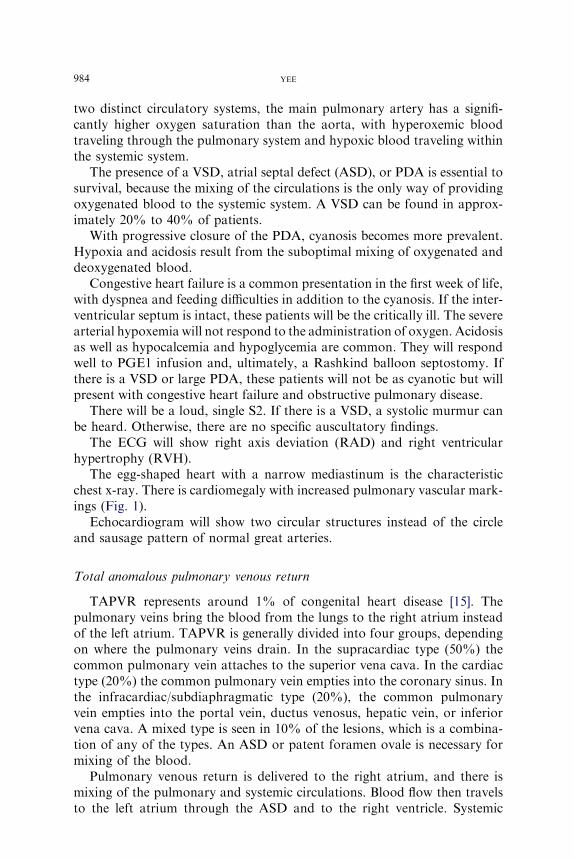
Transposition of the great arteries represents around 5% to 8% of con-genital heart disease and is the most common cyanotic heart lesion in thenewborn period [14]. There are many variations of the disease, with the un-derlying factor being that the aorta originates from the right ventricle andthat the main pulmonary artery has origins in the left ventricle. Within these
984 YEE
two distinct circulatory systems, the main pulmonary artery has a signifi-cantly higher oxygen saturation than the aorta, with hyperoxemic bloodtraveling through the pulmonary system and hypoxic blood traveling withinthe systemic system.
The presence of a VSD, atrial septal defect (ASD), or PDA is essential tosurvival, because the mixing of the circulations is the only way of providingoxygenated blood to the systemic system. A VSD can be found in approx-imately 20% to 40% of patients.
With progressive closure of the PDA, cyanosis becomes more prevalent.Hypoxia and acidosis result from the suboptimal mixing of oxygenated anddeoxygenated blood.
Congestive heart failure is a common presentation in the first week of life,with dyspnea and feeding difficulties in addition to the cyanosis. If the inter-ventricular septum is intact, these patients will be the critically ill. The severearterial hypoxemia will not respond to the administration of oxygen. Acidosisas well as hypocalcemia and hypoglycemia are common. They will respondwell to PGE1 infusion and, ultimately, a Rashkind balloon septostomy. Ifthere is a VSD or large PDA, these patients will not be as cyanotic but willpresent with congestive heart failure and obstructive pulmonary disease.
There will be a loud, single S2. If there is a VSD, a systolic murmur canbe heard. Otherwise, there are no specific auscultatory findings.
The ECG will show right axis deviation (RAD) and right ventricularhypertrophy (RVH).
The egg-shaped heart with a narrow mediastinum is the characteristicchest x-ray. There is cardiomegaly with increased pulmonary vascular mark-ings (Fig. 1).
Echocardiogram will show two circular structures instead of the circleand sausage pattern of normal great arteries.
Total anomalous pulmonary venous return
TAPVR represents around 1% of congenital heart disease [15]. Thepulmonary veins bring the blood from the lungs to the right atrium insteadof the left atrium. TAPVR is generally divided into four groups, dependingon where the pulmonary veins drain. In the supracardiac type (50%) thecommon pulmonary vein attaches to the superior vena cava. In the cardiactype (20%) the common pulmonary vein empties into the coronary sinus. Inthe infracardiac/subdiaphragmatic type (20%), the common pulmonaryvein empties into the portal vein, ductus venosus, hepatic vein, or inferiorvena cava. A mixed type is seen in 10% of the lesions, which is a combina-tion of any of the types. An ASD or patent foramen ovale is necessary formixing of the blood.
Pulmonary venous return is delivered to the right atrium, and there ismixing of the pulmonary and systemic circulations. Blood flow then travelsto the left atrium through the ASD and to the right ventricle. Systemic

985CARDIAC EMERGENCIES
arterial desaturation occurs as the result of mixing of pulmonary and sys-temic blood. Pulmonary blood flow determines the amount of desaturationof systemic arterial blood. If there is no obstruction to pulmonary venousreturn, there is minimal desaturation of the systemic blood. If there isobstruction to pulmonary venous return, there is significant cyanosis.With the blood from both the pulmonary and systemic circulations pumpedby the right ventricle, there can be volume overload, with subsequent rightventricular and atrial enlargement.
In a patient without pulmonary venous obstruction, there can be a historyof frequent pneumonias and growth difficulties. Patients will frequentlypresent with a congestive heart failure presentation with tachypnea, tachy-cardia, and hepatomegaly, in addition to slight cyanosis. There will bea hyperactive right ventricular impulse, with a split and fixed S2. A grade2 to 3/6 systolic ejection murmur is at the upper left sternal border, witha mid diastolic rumble at the left lower sternal border.
The ECG will show right axis deviation, right ventricular hypertrophy,and right atrial enlargement (Fig. 2).
Chest x-ray will exhibit significant cardiomegaly with increased pulmo-nary vascular markings (Fig. 3). The characteristic ‘‘snowman sign’’ is foundin infants older than 4 months.
In those patients with TAPVR and pulmonary venous obstruction,cyanosis and respiratory distress dominate the presentation. There can beminimal cardiac exam findings aside from a loud and single S2 and galloprhythm. A murmur is usually not found.
The ECG will also show right axis deviation and right ventricular hyper-trophy and the chest radiograph will have a normal heart silhouette withlung fields consistent with pulmonary edema.
Fig. 1. Chest radiograph of TGA with cardiomegaly and increased vascular markings.

986 YEE
Tricuspid atresia
Tricsupid atresia represents 1% to 2% of congenital heart disease in in-fancy [16]. There is no tricuspid valve and there is underdevelopment of theright ventricle and pulmonary artery. Therefore, pulmonary blood flow isdecreased. With no flow across the right atrium to the right ventricle, theright atrium needs a right-to-left shunt to empty, making an ASD, VSD,or PDA essential for survival. The great arteries are transposed in 30% ofthe cases, with a VSD and no pulmonic stenosis. In 50% of cases there isnormal artery anatomy, with a small VSD and pulmonic stenosis.
Fig. 2. ECG of TAPVR with right atrial enlargement, right ventricular hypertrophy.
Fig. 3. Chest radiograph of TAPVR with cardiomegaly and increased vascular markings.

987CARDIAC EMERGENCIES
There will be right atrial dilatation and hypertrophy because all systemicvenous return is shunted from the right atrium to the left atrium. Enlarge-ment of the left atrium and ventricle occurs because of the work of handlingboth systemic and pulmonary returns.
The amount of cyanosis is inversely related to the amount of pulmonaryblood flow.
Severe cyanosis, tachypnea, and poor feeding are common presentations.There is a single S2. The murmur is a grade 2 to 3/6 systolic regurgitantmurmur from the VSD and is heard best at the left lower sternal border.There can also be a continuous murmur of a PDA. Hepatomegaly can befound with congestive heart failure.
The ECG has a superior QRS axis, along with right atrial hypertrophy(RAH), left atrial hypertrophy (LAH) and left ventricular hypertrophy.The chest radiograph will show a normal to slight increase in heart sizealong with decreased pulmonary vascular markings.
Truncus arteriosus
Truncus arteriosus is seen in less than 1% of all congenital heart disease[17]. All of the pulmonary, systemic, and coronary circulations result froma single arterial trunk. A large VSD is associated with this, as well as abnor-malities of the coronary arteries.
DiGeorge syndrome (hypocalcemia, hypoparathyroidism, absence orhypoplasia of the thymus, chromosomal abnormalities) is often seen withtruncus arteriosus. Pulmonary blood flow can be normal, increased, ordecreased, depending on the type of truncus arteriosus.
There is a direct relationship between the amount of pulmonary bloodflow and the degree of systemic arterial oxygen saturation. Cyanosis is prev-alent with decreased pulmonary blood flow, and is minimal with increasedpulmonary blood flow. Congestive heart failure can be seen with increasedpulmonary blood flow. The left ventricle has to deal with significant volumeoverloads.
Usually within the first weeks of life, the patient will present with conges-tive heart failure and cyanosis. There will be a loud regurgitant 2 to 4/6systolic murmur at the left sternal border, sometimes associated witha high-pitched diastolic decrescendo murmur or a diastolic rumble. TheS2 will be single and accentuated.
The ECG will usually show bilateral ventricular hypertrophy and thechest radiograph will have cardiomegaly with increased pulmonary vascularmarkings.
Acyanotic heart disease
Left-to-right shunt lesions include ventricular septal defects, atrial septaldefects, patent ductus arteriosus, and endocardial cushion defects. This

988 YEE
group comprises almost 50% of all congenital heart disease [18]. Left-to-right shunt lesions have blood shunted from the systemic system intothe pulmonary system. The high pulmonary vascular resistance in the neo-nate controls the amounts shunted but once pulmonary vascular resistancestarts to drop in the first few weeks of life, pulmonary blood flow and pres-sures will increase. The extent of the lesion is directly related to the degree ofpulmonary vascular blood flow. More blood flow will lead to chamberenlargement, and increased pulmonary vascular pressures and subsequentsigns of congestive heart failure.
Atrial septal defects
Atrial septal defects comprise up to 10% of all congenital heart disease[19]. In infancy this connection from the left to right atria has the potentialfor causing problems in about 10% of patients [14]. If there is a large defect,or if there are associated defects, there will be considerable left-to-rightshunting and subsequent overload of the pulmonary circulation. Somedefects will close spontaneously but larger defects will require surgicalintervention.
Difficulty feeding and difficulty gaining weight are common complaints.The cardiac exam will have a widely split and fixed S2, with a grade 2 to
3/6 systolic ejection murmur at the upper left sternal border, sometimesassociated with a mid-diastolic rumble.
ECG findings include right axis deviation and right ventricular hypertro-phy or right bundle branch block.
Chest radiograph will have cardiomegaly with increased pulmonaryvascular markings.
Ventricular septal defects
Ventricular septal defects are the most common type of congenital heartdisease. Seen in approximately 25% of all congenital heart disease cases [20],ventricular septal defects allow for mixing of blood in the ventricles. Theextent of the defect determines the degree of disease. Small defects willhave minimal impact, as compared with large defects, which will cause pul-monary hypertension and congestive heart failure. Large VSDs have volumeand pressure overload in the right ventricle as well as volume overload in theleft atrium and left ventricle.
In larger VSDs, poor weight gain along with delayed development arecommon. Congestive heart failure and cyanosis are frequent presentations.
The exam will have a grade 2 to 5/6 systolic murmur (holosystolic) heardbest at the left lower sternal border. A systolic thrill or diastolic rumble canalso be present with a narrowly split S2.
ECG findings in a moderate VSD will show left atrial hypertrophy andleft ventricular hypertrophy. In a larger VSD there will be left and right

989CARDIAC EMERGENCIES
ventricular hypertrophy and left atrial hypertrophy. The chestradiograph can show cardiomegaly as well as increased pulmonary vascularmarkings.
Patent ductus arteriosus
Seen in 10% of all congenital heart disease, the ductus arteriosus remainspatent and does not close as it ordinarily would [18]. The degree of theleft-to-right shunting is dependent on the lesion length and diameter andpulmonary vascular resistance. The larger the left-to-right shunt, the moresymptomatic the patient will be. Ordinarily, in healthy patients the ductusarteriosus will close within 15 hours after birth and then will completelyseal around 3 weeks of age, becoming the ligamentum arteriosum. Hypoxiaand prematurity have a tendency to keep the ductus arteriosus patent.
If the defect is large, as with all left-to-right shunts, signs of congestiveheart failure will be present.
Physical exam will be remarkable for a grade 1 to 4/6 continuous machin-ery like murmur heard best at the left upper sternal border. A diastolicrumble can also be present as well as bounding peripheral pulses.
ECG findings can show left and right ventricular hypertrophy in largePDAs.
Chest radiograph will have cardiomegaly and increased pulmonaryvascular markings.
Endocardial cushion defect
When the endocardial cushion does not develop properly, there will bedefects to the atrial septum, the ventricular septum, and the atrioventricularvalves. Complete defects involve the entire endocardial cushion and willhave atrial and ventricular septal lesions and a common atrioventricularvalve. Incomplete or partial defects have atrial involvement with an intactventricular septum. There can also be variations of both complete andincomplete lesions. A history of failure to thrive, and multiple respiratorytract infections are common. Endocardial cushion defects represent around3% of congenital heart disease and almost two thirds have the completeform [18]. Down’s syndrome is strongly associated with the completeform of endocardial cushion defects.
Left-to-right shunting is directly dependent on the extent of the defects,with complete lesions presenting with congestive heart failure early fromvolume overload in both the left and right ventricles.
Cardiac exam will be remarkable for a hyperactive precordium, a sys-tolic thrill, a loud holosystolic regurgitant murmur, and a loud and splitS2.
The ECG will show a superior QRS axis with RVH, right bundlebranch block (RBBB), and left ventricular hypertrophy, along with a pro-longed PR interval (Fig. 4).

990 YEE
Coarctation of the aorta
Coarctation of the aorta represents 8% to 10% of congenital heart dis-ease and is seen in males in a 2:1 ratio [21]. There is congenital narrowingof the aorta, in the upper thoracic aorta in the region of the ductus arte-riosus. The extent of illness is a factor of the degree of narrowing, thelength of the narrowing and the presence of other cardiac defects. If theright ventricle supplies the descending aorta via the PDA in fetal life, in-fants will be symptomatic early. Many other cardiac defects are presentsuch as a VSD, PDA, and aortic hypoplasia and collateral circulation isunderdeveloped.
The PDA is able to temporarily negate the obstructive effects of the co-arctation obstruction. Additionally, the PDA can maintain blood flow toareas distal to the obstruction. When the PDA eventually closes, the devel-opment of pulmonary hypertension and subsequent pulmonary venous con-gestion leads to congestive heart failure.
Tachypnea, feeding difficulties, and minimal urine output along withshock and metabolic acidosis are common presentations. When presentingin congestive heart failure, there will be a loud gallop, a murmur may ornot be present, and pulses will be weak.
The ECG will show RVH or RBBB. There will be significant cardiome-galy as well as pulmonary edema on chest radiograph (Fig. 5). In older chil-dren, the appearance of notching of the first rib, also known as the ‘‘3 Sign’’may be present.
The presence of decreased pulses in the lower extremities is key in thediagnosis of a coarctation. Comparison of the right upper extremity bloodpressures and pulse oximeter readings with the lower extremity aids in the
Fig. 4. ECG of CAVC (common AV canal) or endocardial cushion defect with superior QRS
axis.

991CARDIAC EMERGENCIES
diagnosis. If the patient is in significant shock, however, pressures can bedecreased everywhere.
Hypoplastic left heart syndrome
Hypoplastic left heart syndrome (HLHS) includes hypoplasia of the leftventricle and hypoplasia of the ascending aorta and aortic arch. There canbe atresia or marked stenosis of the mitral and aortic valves. The left atriumis also underdeveloped. The ultimate result is that of minimal left ventricularoutflow [22].
In utero, the pulmonary vascular resistance is higher than the systemicvascular resistance. The right ventricle (through the right-to-left shunt ofthe ductus arteriosus) and the elevated pulmonary vascular resistance areable to keep a normal perfusion pressure to the descending aorta and sys-temic fetal system. The hypoplastic left ventricle does not contribute. AnASD allows the left atrium to decompress. All systemic blood flow is depen-dent on the ductus arteriosus. After birth, significant problems occur.Systemic vascular resistance is now greater than pulmonary vascular resis-tance, reversing the pressure system. The patent ductus arteriosus now be-gins to gradually close. With the nonfunctioning left side and increasedsystemic vascular resistance, cardiac output falls and aortic pressure drops.This leads to circulatory shock and metabolic acidosis. Increased pulmonaryblood flow leads to an increase in left atrial pressure and subsequent pulmo-nary edema.
These patients appear listless, dusky with tachypnea. There is a singleheart sound with a systolic ejection murmur and diminished pulses. The
Fig. 5. Chest radiograph of coarctation with cardiomegaly and pulmonary edema.

992 YEE
ECG will show right atrial enlargement, right ventricular hypertrophy, andpeaked P waves. The chest radiograph will show cardiomegaly.
Aortic stenosis
Aortic stenosis is seen in 6% of congenital heart disease, with a 4:1 ratioin males [23]. The stenosis will be at the valvular, supravalvular, or subvalv-ular level, with the degree of obstruction determining the severity of diseasein the patient. Those with severe obstruction (approximately 10% to 15%)will present with congestive heart failure in infancy [24]. Left ventricularhypertrophy will develop with severe stenosis. The most common type ofaortic stenosis is a bicuspid aortic valve. William Syndrome has supravalv-ular stenosis in addition to elfin facies, mental retardation, and pulmonaryartery stenosis.
The physical exam will be remarkable for a systolic thrill in the region ofthe upper right sternal border, suprasternal notch, or carotid arteries. Therecan be an ejection click. The murmur will be a rough or harsh systolic mur-mur grade 2 to 4/6 at the right intercostal space or left intercostal space withtransmission to the neck.
In cases of severe aortic stenosis, the ECG will show left ventricular hy-pertrophy. If there is resultant congestive heart failure, the chest radiographwill show cardiomegaly.
Anomalous origin of the left coronary artery (ALCAPA Syndrome,
Bland-White-Garland Syndrome)
In anomalous origin of the left coronary artery (also known as ALCAPAor Bland-White-Garland Syndrome), the left coronary artery has origins inthe pulmonary artery instead of the aorta. When pulmonary artery pressurediminishes in the second to third month of life, there will be decreased per-fusion of the left ventricle, resulting in a distressed patient with cardiome-galy and congestive heart failure. There may or may not be a murmurconsistent with mitral regurgitation [25,26].
The ECG will show myocardial infarction with abnormally deep andwide Q waves, inverted T waves, and ST segment changes in the precordialleads (Fig. 6). The chest radiograph will be most likely show cardiomegaly.An echocardiogram will help in the diagnosis, with an aortogram ifnecessary.
Acquired disease
Inflammatory diseases of the heart are grouped under carditis. Includedin this group are myocarditis, pericarditis, and endocarditis (along withvalvulitis).

993CARDIAC EMERGENCIES
Myocarditis
There are a number of different etiologies in myocarditis. Infectious andautoimmune, as well as toxin-mediated processes can contribute to the in-flammatory response in the myocardium [27,28].
Viruses, such as adenovirus, coxsackievirus, echovirus, mumps, and ru-bella, are the most commonly associated infectious agents. Nonviral causessuch as protozoans (Chaga’s Disease seen in South America) also causemyocarditis. Less frequently, bacteria, rickettsia, fungal, mycobacteria,and other parasites can be etiologic agents.
Kawasaki’s disease and acute rheumatic fever as well as collagen vasculardisease can also be seen with myocarditis. Toxic myocarditis is the result ofdrug ingestion.
Infants may present with vomiting, decreased activity, poor feeding, andcongestive heart failure, with tachycardia, tachypnea, a gallop rhythm, anddecreased heart tones.
There are no specific lab tests for myocarditis. Erythrocyte sedimentationrate, white blood cell count, myocardial enzymes, and cardiac troponin willbe normal or elevated. Troponin levels are thought to be more sensitive thancardiac enzymes [29]. Chest radiograph will show cardiomegaly and, de-pending on the extent of the disease, pulmonary venous congestion.
ECG abnormalities are common but are nonspecific. There will be tachy-cardia, low QRS voltages, flattened or inverted T waves with ST-T wavechanges, and prolongation of the QT interval. Arrhythmias such as prema-ture contractions are also seen.
Fig. 6. ECG of anomalous origin of the left coronary artery (ALCAPA) with deep and wide Q
waves, inverted T waves, and ST segment changes.

994 YEE
Echocardiogram studies will show dilatation of the heart chambers anddecreased left ventricular function. The echocardiogram will also help toevaluate myocardial contractility and the presence of a pericardial effusion.Radionuclide scanning and endomyocardial biopsies can help in confirmingthe disease.
The mortality rate in symptomatic neonates with acute viral myocarditiscan be significant. Management of myocarditis revolves around identifyingan etiologic agent and, if identified, treating that suspected agent, treatingthe congestive heart failure, and controlling the arrhythmias. Rest, supple-mental oxygen, rapid-acting diuretics like furosemide, and rapid-acting ino-tropic agents such as dopamine and dobutamine are mainstays in treatmentalong with the use of angiotensin-converting enzyme inhibitors like capto-pril. Digoxin is used cautiously because of its potential to inducearrhythmias. In Kawasaki’s disease, high-dose immunoglobulins havebeen beneficial. Other treatment modalities, such as immunosuppressiveagents and corticosteroids (except in severe rheumatic carditis) are not uni-versally accepted.
Pericarditis
Inflammation of the pericardium is the hallmark of pericarditis. The mostcommon cause in infancy is a viral etiology such as coxsackie, echovirus, ad-enovirus, or influenza. Viral pericarditis is usually associated with a viralmyocarditis, with the myocarditis being the more prominent entity. Bacterialcauses include Staphylococcus aureus, Streptococcus pnuemoniae, Haemophi-lus influenzae, Neisseria meningitides, and streptococci as well as tuberculo-sis. Acute rheumatic fever, collagen vascular disease, and uremia can alsocause a pericarditis. Postpericardiotomy syndrome is seen in patients whohave had cardiac surgery involving interruption of the pericardium.
Since the pericardium is a fixed space, the extent of symptoms and signsof disease will be determined by the rate of accumulation of fluid and by thehealth of the myocardium.
If the myocardium is normal and fluid accumulation is slow, then the pa-tient will tolerate the pericarditis better than if there was underlying myocar-dial injury with a slow collection of fluid or if there was a rapid collection ofa large amount of fluid.
If pericardial tamponade were to occur, the heart, to improve hemody-namics would increase heart rate (improves cardiac output), increase sys-temic vascular resistance (offset hypotension), and improve diastolic fillingby systemic and pulmonary venous constriction.
There is usually a predisposing illness in the history, with an upper respi-ratory infection or, in the case of a bacterial pericarditis, a pneumonia,empyema, osteomyelitis, pyelonephritis, or tonsillitis.
A pericardial friction rub is diagnostic. A murmur may not be found andthe heart will be hypodynamic.

995CARDIAC EMERGENCIES
On ECG there will be a low-voltage QRS complex. Early in the disease,ST segments will be elevated everywhere except in V1 and aVR. Later in thedisease, ST segments will return to normal and the T waves will flatten orinvert. A chest radiograph will show cardiomegaly, with the heart in a wa-ter-bottle shape.
Echocardiogram is the key to establishing the presence of an effusion.Additionally, the echo can also evaluate for cardiac tamponade, as it willshow the collapse of the right atrial wall or the right ventricular wall indiastole.
To treat pericarditis or pericardiocentesis, surgical intervention is essen-tial, especially if an infectious etiology is suspected. Multiple blood culturesare also indicated as well as standard fluid studies. In milder cases notrequiring drainage or antibiotics, antivirals, or antifungals, nonsteroidalanti-inflammatory drugs can be used to treat the discomfort.
In postpericardiotomy syndrome, which can affect as many as 30% of pe-diatric patients who undergo cardiovascular surgery involving the pericar-dium, the patients will present with fever, irritability, and a pericardialfriction rub anywhere from a month to a few months postoperatively. Theetiology is thought to be autoimmune [30].
In cardiac tamponade with signs of tachycardia, tachypnea is an immedi-ate concern.
Chest radiograph will have cardiomegaly and pleural effusion. ECG willhave ST segment elevation and flat or inverted T waves. The most helpfultest is an echocardiogram because this will assess the amount of pericardialeffusion as well as the presence of cardiac tamponade.
Endocarditis
Congenital heart disease is a significant risk factor in infective endocardi-tis. It is thought that turbulent flow from pressure gradients leads to endo-thelial damage and thrombus formation. Transient bacteremia then seedsthe damaged areas. With the exception of a secundum ASD, all congenitalheart diseases and valvular heart diseases are prone to endocarditis, espe-cially if there is any artificial material within the heart (prosthetic heart valveor graft). Common bacterial causes include S viridans, enterococci, andS aureus as well as fungal and bacteria such as Eikenella, Cardiobacterium[31].
In infancy, endocarditis is rare and is associated with open-heart surgery.The usual presentation is with fulminant disease and a septic appearance.A heart murmur and fever are always present. Embolic phenomena tendto be seen more in the adult population.
Using the Duke Criteria for Infective Endocarditis, a patient must havetwo major criteria or one major criterion with three minor criteria or fiveminor criteria. Major criteria include two separately obtained positive bloodcultures growing the typical microorganisms and an echocardiogram with
996 YEE
endocardial involvement such as an intracardiac mass on a valve, abscess,partial dehiscence of a prosthetic valve, or new valvular regurgitation.Minor criteria include predisposing conditions, fever, vascular phenomena(emboli, hemorrhages, Janeway lesions), and immunologic phenomena (glo-merulonephritis, Osler’s nodes, Roth spots, rheumatoid factor), microbio-logical evidence (positive blood culture not meeting major criteria), andechocardiographic findings (not meeting major criteria).
While an echocardiogram identifying valvular vegetation is helpful in theevaluation, the echocardiogram is not 100% sensitive or specific. Because ofthis, a negative echocardiogram does not exclude endocarditis. A moredefinitive diagnosis is made by obtaining a positive blood culture. Theisolation of a specific microorganism is key to determining antibiotictherapy. Treatment regimens may take place for weeks to be certain thatthe microorganism has been eliminated.
Kawasaki’s disease
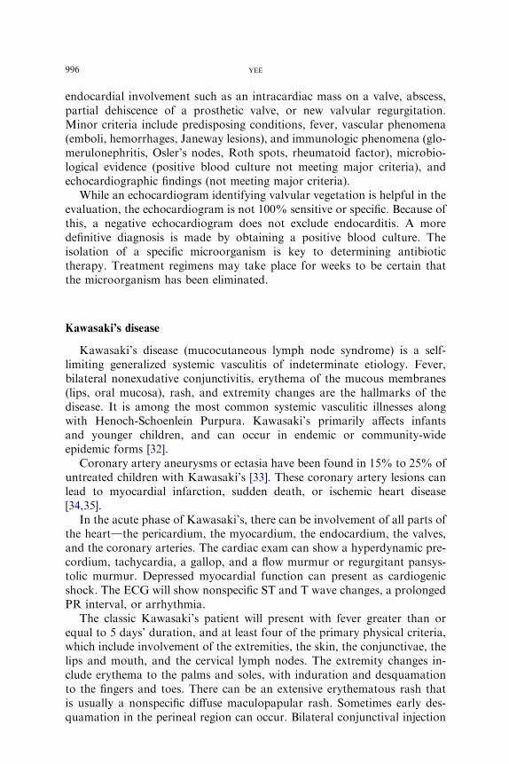
Kawasaki’s disease (mucocutaneous lymph node syndrome) is a self-limiting generalized systemic vasculitis of indeterminate etiology. Fever,bilateral nonexudative conjunctivitis, erythema of the mucous membranes(lips, oral mucosa), rash, and extremity changes are the hallmarks of thedisease. It is among the most common systemic vasculitic illnesses alongwith Henoch-Schoenlein Purpura. Kawasaki’s primarily affects infantsand younger children, and can occur in endemic or community-wideepidemic forms [32].
Coronary artery aneurysms or ectasia have been found in 15% to 25% ofuntreated children with Kawasaki’s [33]. These coronary artery lesions canlead to myocardial infarction, sudden death, or ischemic heart disease[34,35].
In the acute phase of Kawasaki’s, there can be involvement of all parts ofthe heartdthe pericardium, the myocardium, the endocardium, the valves,and the coronary arteries. The cardiac exam can show a hyperdynamic pre-cordium, tachycardia, a gallop, and a flow murmur or regurgitant pansys-tolic murmur. Depressed myocardial function can present as cardiogenicshock. The ECG will show nonspecific ST and T wave changes, a prolongedPR interval, or arrhythmia.
The classic Kawasaki’s patient will present with fever greater than orequal to 5 days’ duration, and at least four of the primary physical criteria,which include involvement of the extremities, the skin, the conjunctivae, thelips and mouth, and the cervical lymph nodes. The extremity changes in-clude erythema to the palms and soles, with induration and desquamationto the fingers and toes. There can be an extensive erythematous rash thatis usually a nonspecific diffuse maculopapular rash. Sometimes early des-quamation in the perineal region can occur. Bilateral conjunctival injection

997CARDIAC EMERGENCIES
involving the bulbar conjunctivae is seen around the time of the fever. Therecan be erythema; peeling, cracking, or bleeding from the lips and mouth;a strawberry tongue; and diffuse erythema of the mucosa of the oropharynx.The cervical lymphadenopathy is generally unilateral, and usually one nodeis greater than 1.5 cm in diameter.
Lab findings include thombocytosis (appears in second week, peaking inthird week), leukocytosis, and anemia. Thrombocytopenia in active diseaseis a risk factor for coronary aneurysms. There is elevation of the C-reactiveprotein (CRP) and erythrocyte sedimentation rate (ESR). Serum transami-nases can be moderately elevated. Gammaglutamyl transpeptidase (GGT) iselevated in a majority of patients.
In the younger patient, an incomplete or atypical presentation is common[36]. Diagnosis is often made by echocardiogram findings of coronary arteryabnormalities [37].
Pharmacologic management of the acute phase of Kawasaki’s includesaspirin and intravenous immunoglobulin (IVIG). High-dose aspirin at80 to 100 mg/kg per day dosed four times a day along with IVIG have anadditive anti-inflammatory effect [32]. Length of treatment with aspirin isvariable. IVIG is thought to have a generalized anti-inflammatory effectand is dosed at 2 g/kg in a single infusion. Best results are seen whenIVIG is started within the first 7 to 10 days of illness.
Cardiomyopathies
Cardiomyopathies affect the heart muscle and are divided into threecategories. They are hypertrophic, dilated, or congestive and restrictive(Fig. 7).
In hypertrophic cardiomyopathies, there is significant ventricular muscu-lar hypertrophy and increased ventricular contractility but these factorslimit or reduce ventricular filling.
An autosomal dominant link has been documented [38]. The left ventricleis relatively stiff and affects diastolic ventricular filling. The physical exam isnotable for a sharp upstroke of the arterial pulse [39]. There can be a systolicejection murmur or holosystolic murmur.
The ECG will show left ventricular hypertrophy, ST and T wave changes,deep Q waves, and decreased R waves. The chest radiograph may showa globular heart or cardiomegaly.
Dilated or congestive cardiomyopathies have ventricular dilatation withdiminished contractility. This is the most common form of cardiomyopa-thies and results from infectious or toxic etiologies. They will present withevidence of congestive heart failure. A significant S3 will be found on exam.
Restrictive cardiomyopathies limit diastolic filling of the ventricles. Thisis the least common form and results from noncompliant ventricular wallsthat have been subject to an infiltrative process such as a glycogen storagedisease.

998 YEE
Arrhythmias
Damage, from either congenital or acquired causes, to cardiac structurewill predispose the patient to arrhythmias. There can be congenital abnor-malities to the conduction system, injured conduction pathways from sur-gery or postinflammatory changes, or irritation to the conduction systemfrom injured myocardium. Arrhythmias have their origins in the atrial orventricular conduction systems.
The most common arrhythmia is paroxysmal SVT [40,41]. The usualcause is idiopathic. The majority of patients with SVT have normal hearts,with 23% having congenital heart disease and 22% with Wolff-Parkinson-White (WPW) syndrome [42].
WPW is associated with congenital heart disease, such as transposition ofthe great arteries. WPW is a preexcitation syndrome with an accessory path-way between the atria and ventricles.
SVT is a narrow complex tachycardia with a rate ranging from 220 to 280beats per minute in the 1-year age group. The determination of sinus tachy-cardia and a reentrant tachycardia must be made before the initiation oftherapy. In this age group, pulse rate will linearly increase with body tem-perature, at a ratio of 10 beats per minute per �C increase in body temper-ature [43].
The ECG in SVT will show a regular rhythm with no beat-to-beat vari-ability and a heart rate greater than 220 beats per minute in the infant. Pwaves can be present but are usually not. In most cases, the QRS complexis narrow. In a hemodynamically unstable SVT, immediate synchronizedcardioversion with 0.5 to 1.0 J per kilogram should be done. In a hemody-namically stable SVT, vagal maneuvers can be initiated. Applying a bag ofice water to the face for 15 to 30 seconds can be used. Adenosine is the drug
Fig. 7. ECG of hypertrophic cardiomyopathy with increased voltages throughout.

999CARDIAC EMERGENCIES
of choice. Adenosine acts by temporarily blocking conduction at the AVnode, thereby interrupting the reentrant circuit. Because the drug is rapidlymetabolized, IV access as close to the heart is ideal, with the drug deliveredvia a rapid intravenous injection. Constant cardiorespiratory monitoringshould be in place. Initial dosing of adenosine is 0.1 mg/kg. If there is noresponse, the next dose should be doubled. The maximum dosing is 0.25to 0.35 mg/kg (Fig. 8A, B). Verapamil should not be used in the patientyounger than 1 year because of the potential for hypotension and cardiovas-cular collapse [44].
In WPW there is a ventricular preexcitation pathway because of an acces-sory pathway between the atria and ventricles [3]. There is a short PR inter-val, a prolonged QRS duration, and delta waves (Fig. 9). Slowing theconduction through the atrioventricular node can allow another pathwayto become dominant.
In a WPW-induced SVT, adenosine can cause atrial fibrillation, whichcan then lead to ventricular fibrillation. This underscores the need foralways having resuscitation material at the bedside whenever dealing witharrhythmias.
Sick Sinus Syndrome is usually the result of cardiac surgery involving theatria or can be from myocarditis. The sinus node no longer acts as theprimary pacemaker of the heart or functions at a significantly slower rate.This leads to marked sinus bradycardia, sinus arrest with a junctionalescape, atrial flutter, fibrillation, or SVT.
Fig. 8. (A) ECG of supraventricular tachycardia (SVT) in a 19-day-old. (B) Rhythm changes
after adenosine.

1000 YEE
Fig. 8 (continued).

1001CARDIAC EMERGENCIES
AV block is found when there is an interruption of the conduction of thenormal sinus impulse and the subsequent ventricular response. There arefirst-degree, second-degree, and third-degree blocks.
The first-degree block has a prolonged PR interval because of delayedconduction through the AV node. This is the result of a cardiomyopathy,congenital heart disease, postcardiac surgery, or digitalis toxicity or canbe found in healthy patients.
In a second-degree block, not all of the P waves are followed by QRScomplexes. The Mobitz Type I Wenckebach phenomenon has a PR intervalthat gets progressively longer until the QRS complex is completely dropped.The block is at the AV node level and can be attributed to myocarditis, car-diomyopathy, surgery, congenital heart disease, or digitalis toxicity. TheMobitz Type II block has similar etiologies but the block is at the Bundleof His. AV conduction is either all or none. There is potential for a completeblock to develop. In two-to-one or three-to-one blocks, the block is at thelevel of the AV node, but can also be at the Bundle of His.
Third-degree or complete heart blocks have independent atrial and ven-tricular activity. There are regular P waves at a normal heart rate for age.The QRS complexes are also regular but at a slower rate than the P waves.The usual presentation in infancy is congestive heart failure. Congenitalcomplete heart blocks have a normal QRS complex duration and can befound in patients with a structurally normal heart. A history of maternal lu-pus or connective tissue disease such as Sjogen’s Syndrome predispose apatient to complete heart block (Fig. 10). It is thought that there is transpla-cental passage of autoimmune antibodies affecting the atrioventricular node[45]. Acquired complete heart blocks are the result of cardiac surgery butcan also be attributed to cardiomyopathies and myocarditis and have a pro-longed QRS duration.
Fig. 9. ECG of WPW with delta waves.

1002 YEE
If asymptomatic, no intervention is indicated. If symptomatic, atropine,isoproterenol or temporary transvenous ventricular pacing are sometimesrequired.
Surgical repairs
The surgical repair of congenital heart disease continues to progress, withsome lesions now repaired in the neonatal period, and most lesions repairedin the first couple of months of life. There are still patients, however, whomay appear in the emergency department with no prior surgery, palliativesurgery, or corrective surgery. These patients may have a less than optimalnutritional status, can be on multiple medications, or can be exhibiting post-operative complications such as a dysrhythmia or post pericardiotomy syn-drome. Also a shunt could develop stenosis.
A Blalock-Taussig shunt is used in the Tetralogy of Fallot. This shuntjoins the subclavian artery to the ipsilateral pulmonary artery. The modifiedBlalock-Taussig shunt uses a Gore-Tex shunt and requires less dissection, isnot dependent on the vessel length, and has decreased shunt failure [46].
The Rastelli procedure is done in older patients, and is used in severe Te-tralogy of Fallot with significant right ventricular outflow tract obstruction.There is patch closure of the VSD, with the placement of a conduit from theright ventricle to the pulmonary artery.
The Mustard and Senning operations were used in the Transposition ofthe Great Arteries and functioned at the atrial level. The Mustard opera-tion was an atrial switch using prosthetic material for an intra-atrial baffle,while the Senning operation used native material for an intra-atrial baffle.
Fig. 10. ECG of complete heart block, patient’s mother with lupus.

1003CARDIAC EMERGENCIES
Because of atrial dysrhythmias and the inability of the right ventricle tofunction as a normal left ventricle in later life, these procedures were dis-continued. The Arterial Switch, which has now replaced the Mustard andSenning, corrects the TGA at the great artery level. The aortic trunk is at-tached to the left ventricle and the pulmonic trunk is attached to the rightventricle.
The Fontan operation is done in HLHS, tricuspid atresia, and HRHS.This shunt is a cavocaval baffle to pulmonary artery anastomosis. Systemicvenous return is redirected to the pulmonary artery.
The bidirectional Glenn (cavopulmonary shunt) or hemi-Fontan opera-tion anastomoses the superior vena cava to the right pulmonary arteryand is performed in patients with HLHS and HRHS. The bidirectionalGlenn operation is usually done at 6 months of age, and the hemi-Fontanat 1.5 years of age.
The Norwood operation, performed in the neonatal period, is a palliativeprocedure in HLHS [47]. The hypoplastic aorta is reconstructed using anaortic or pulmonary artery allograft, the main pulmonary artery is divided,a Gore-Tex shunt is placed on the right to establish pulmonary blood flow,and the atrial septum is excised to provide interatrial mixing [48].
Complications that may be seen in the postoperative patient includedysrhythmias, obstruction of the surgical grafts or conduits, endocarditis,myocardial ischemia or postpericardiotomy syndrome.
Management of acute issues
Cardiac emergencies in the first couple of weeks of life will involve cya-nosis and shock. The ductal-dependent lesions dominate this group and pre-serving ductal patency is crucial in managing these patients. While many ofthese patients will be diagnosed in the newborn nursery, the advent of earliernewborn discharges increases the chances that the patient will present to theemergency department for the initial diagnosis.
Cyanotic or hypoxemic episodes are seen in patients with congenitalheart disease (usually Tetralogy of Fallot). They will present with hyper-pnea, irritability, and increasing cyanosis along with a decreased intensityof the underlying heart murmur. A decrease in systemic vascular resistanceor increased resistance to the right ventricular outflow tract increases right-to-left shunting, causing hyperpnea and, then, increased systemic venousreturn. This causes increased right-to-left shunting through the VSD.
To manage a ‘‘tet spell’’ the patient should be placed in a knee-chest po-sition. Morphine sulfate (0.1 to 0.2 mg/kg subcutaneously [SC] or intramus-cularly [IM]) will stop the hyperpnea. Oxygen may or may not help becausethe issue is to improve pulmonary blood flow. Sodium bicarbonate (1 mEq/kg IV) can treat the acidosis. Propanolol (0.01 to 0.2 mg/kg IV over 5 min-utes) can be beneficial. Phenylephrine (0.02 mg/kg IV) can help to increase

1004 YEE
systemic vascular resistance. Ketamine (1 to 3 mg/kg IV) can also increasesystemic vascular resistance and provide sedation.
Tricuspid Atresia, Transposition of the Great Arteries, Total AnomalousPulmonary Venous Return, Truncus Arteriosus, Hypoplastic Right HeartSyndrome, and Pulmonary Atresia can all present with cyanosis or shockin the first couple of weeks of life. Cyanosis or congestive heart failurewill be the usual presentation of Tetralogy of Fallot. Shock will be the initialpresentation for Hypoplastic Left Heart Syndrome, Aortic Stenosis, andCoarctation of the Aorta.
The key to dealing with the ductal-dependent lesions is to start intrave-nous prostaglandin E1 (PGE1). Decreasing pulmonary vascular resistancewill help in left-to-right shunting and increasing pulmonary blood flow.The initial dose of PGE1 is 0.05 mg/kg/min. If at all possible, consultationwith pediatric cardiology as well as the critical (neonatal or pediatric) carestaff is beneficial. Apnea and hypotension are potential complicating side ef-fects of PGE1 so management of the airway is essential as well as determin-ing that the patient is not possibly septic. Additionally, the side effect offever can cloud the potential sepsis picture. In certain variants of TAPVR,PGE1 can actually exacerbate the symptoms. Supplemental oxygen can has-ten the closure of the ductus arteriosus, so this must be used with caution.
Acyanotic lesions that are dependent on ductal flow will present with car-diogenic shock.
Those lesions with critical left heart obstruction such as HLHS, aorticstenosis, and coarctation of the aorta depend on the ductus to maintain sys-temic perfusion. Poor perfusion, diminished pulses, and pallor are common,and the presentation can mimic sepsis. If central cyanosis is present, a re-sponse to oxygen may not take place or the patient may become worse.
Airway management is paramount, as mechanical ventilation can in-crease pulmonary vascular resistance [49]. Increasing right-to-left shuntingover the PDA will improve systemic perfusion. Volume assists in treatingthe acidosis and fluid deficits. Vasopressors can be initiated if decreased ven-tricular function is evident.
Patients with critical right heart obstruction such as Tetralogy of Fallotand pulmonic stenosis are also ductal dependent. Airway management isa primary concern. IV prostaglandins are also key in the management, espe-cially with oxygen saturations less than 70%. Decreasing pulmonary vascu-lar resistance will help in left-to-right shunting and increasing pulmonaryblood flow.
Congestive heart failure in the first year of life is generally associated withcongenital heart disease but can also be the result of acquired disease such asmyocarditis, arrhythmias, sepsis, and respiratory and metabolic diseases.Pressure overload, volume overload, decreased inotropic function, andrhythm abnormalities can all be factors in causing congestive heart failure.Cardiac congenital abnormalities that have predisposition to presentingwith congestive heart failure include left ventricular outflow obstruction

1005CARDIAC EMERGENCIES
(such as coarctation of the aorta and aortic stenosis) and volume overload(left-to-right shunts, VSDs, TAPVR). Endocardial cushion defects withcomplete involvement and AV valve insufficiency will present acutely ill inthe first couple of months of life.
Difficulty feeding, tachypnea, tachycardia, cardiomegaly, hepatomegaly,and rales are all common findings. Prolonged feeding times with diaphoresiscan function as a stress test for the infant. Pulmonary diseases can also pres-ent in the same fashion as cardiac disease. Supplemental oxygen may not helpin differentiating between the two. Echocardiogram is much more definitive.
To treat congestive heart failure, inotropic assistance is important. Mod-ification of preload (end diastolic volume roughly equivalent to the intravas-cular volume), afterload, contractility, and heart rate all play roles. Cardiacoutput is determined by heart rate multiplied by stroke volume. In the under1-year-old, heart rate is the primary method of increasing cardiac output.
Airway management is important and should take precedence, as a stabi-lized airway and mechanical ventilation can prevent respiratory decompen-sation. Elevation of the head of the patient can help to decrease pulmonaryblood volume. Morphine sulfate assists in treating agitation. Bicarbonatecan be used in severe acidosis.
If immediate intervention is needed, dopamine and dobutamine areappropriate choices.
Dopamine is started at a continuous infusion at 5 to10 mg/kg/min. Thereshould be a rapid response to the chronotropic effects with increases in heartrate and blood pressure and urine output. Dobutamine is also started asa continuous infusion at the same dosing. Dobutamine has less of an ar-rhythmic potential and chronotropic effect than dopamine and because ofits vasodilatory effect, reduces afterload. Dobutamine should be used withcaution in the less than 1 year of age population. Dobutamine will improvecardiac output without increasing blood pressure so if there is severe hypo-tension, dobutamine may be a better choice as an adjunct rather thanprimary agent [4,50].
Amrinone (0.5 mg/kg IV over 3 minutes) and milrinone (loading dose of10 to 50 mg/kg IV over 10 minutes) can also be considered as potential aidsin treating congestive heart failure. They do not increase the heart rate buthave inotropic and vasodilator properties.
Digoxin is the inotrope of choice in the nonacute setting. Digoxin im-proves cardiac contractility and subsequently increases cardiac output.Care must be taken with dosing regimens. Diuretics such as furosemide pro-mote diuresis.
Summary
The diagnosis and management of cardiac emergencies in the first year oflife can be challenging and complicated. By reviewing the pathophysiology

1006 YEE
of the heart and circulation, one can be more prepared for these difficultscenarios.
Early presentations will usually be the result of ductal-dependent lesionsand will appear with cyanosis and shock. Later presentations will be the re-sult of volume overload or pump failure and will present with signs of con-gestive heart failure. Acquired diseases will also present as congestive heartfailure or arrhythmias.
References
[1] Burton DA, Cabalka AK. Cardiac evaluation of infants. The first year of life. Pediatr Clin
North Am 1994;41(5):991–1015.
[2] Flynn PA, Engle MA, Ehlers KH. Cardiac issues in the pediatric emergency department.
Pediatr Clin North Am 1992;39(4):955–68.
[3] Woods WA, McCulloch MA. Cardiovascular emergencies in the pediatric patient. Emerg
Med Clin North Am 2005;23(4):1233–49.
[4] GewitzMH,Woolf PK. Cardiac emergencies. In: Fleisher GR, Ludwig S, editors. Textbook
of pediatric emergency medicine. 5th edition. Philadelphia: Lippincott Williams and Wil-
kins; 2006. p. 717–58.
[5] WoolridgeDP. Congenital heart disease in the pediatric emergency department. Part I: path-
ophysiology and clinical characteristics. Pediatric Emergency Medicine Reports 2002;7(7):
69–80.
[6] Woolridge DP. Congenital heart disease in the pediatric emergency medicine department.
Part II: managing acute and chronic complications. Pediatric Emergency Medicine Reports
2002;7(8):81–92.
[7] Hoffman JI, Kaplan S. The incidence of congenital heart disease. J Am Coll Cardiol 2002;
39(12):1890–900.
[8] Lees MH, King DH. Cyanosis in the newborn. Pediatr Rev 1987;9(2):36–42.
[9] FriedmanAH, Fahey JT. The transition from fetal to neonatal circulation: normal responses
and implications for infants with heart disease. Semin Perinatol 1993;17(2):106–21.
[10] Nadas AS, Fyler DC. Hypoxemia. In: Keane JF, Lock JE, Fyler DC, editors. Nada’s pedi-
atric cardiology. 2nd edition. Philadelphia: Saunders Elsevier; 2006. p. 97–101.
[11] Martin L,Khalil H.Howmuch reduced hemoglobin is necessary to generate cyanosis? Chest
1990;97(1):182–5.
[12] Waldman JD, Wernly JA. Cyanotic congenital heart disease with decreased pulmonary
blood flow in children. Pediatr Clin North Am 1999;46(2):385–404.
[13] Breitbart RE, Fyler DC. Tetralogy of Fallot. In: Keane JF, Lock JE, Fyler DC, editors.
Nada’s pediatric cardiology. 2nd edition. Philadelphia: Saunders Elsevier; 2006. p. 559–79.
[14] Studer M, Blackstone E, Kirklin J, et al. Determinants of early and late results of repair of
atrioventricular septal (conal) defects. J Thorac Cardiovasc Surg 1982;84(4):523–42.
[15] Keane JF, Fyler DC. Total anomalous pulmonary venous return. In: Keane JF, Lock JE,
FylerDC, editors.Nada’s pediatric cardiology. 2nd edition. Philadelphia: Saunders Elsevier;
2006. p. 773–81.
[16] Keane JF, Fyler DC. Tricuspid atresia. In: Keane JF, Lock JE, Fyler DC, editors. Nada’s
pediatric cardiology. 2nd edition. Philadelphia: Saunders Elsevier; 2006. p. 753–9.
[17] Williams JM, de LeeuwM, BlackMD, et al. Factors associated with outcomes of persistent
truncus arteriosus. J Am Coll Cardiol 1999;34(2):545–53.
[18] Driscoll DJ. Left to right shunt lesions. Pediatr Clin North Am 1999;46(2):355–68.
[19] Mahoney LT, Truesdell SC, Krzmarzick TR, et al. Atrial septal defects that present in
infancy. Am J Dis Child 1986;140(11):1115–8.

1007CARDIAC EMERGENCIES
[20] Kidd L, Driscoll D, Gersony W, et al. Second natural history study of congenital heart
defects: results of treatment of patients with ventricular septal defects. Circulation 1993;
87(Suppl 2):I38–51.
[21] Demircin M, Arsan S, Pasaoglu I, et al. Coarctation of the aorta in infants and neonates:
results and assessments of prognostic variables. J Cardiovasc Surg 1995;36(5):459–64.
[22] Bailey LL, Gundry SR. Hypoplastic left heart syndrome. Pediatr ClinNorthAm 1990;37(1):
137–50.
[23] Fedderly RT. Left ventricular outflow obstruction. Pediatr Clin North Am 1999;46(2):
369–84.
[24] Bando K, Turrentine MW, Sun K, et al. Surgical management of hypoplastic left heart syn-
drome. Ann Thorac Surg 1996;62(1):70–7.
[25] Chang RKR, Allada V. Electrocardiographic and echocardiographic features that distin-
guish anomalous origin of the left coronary artery from pulmonary artery from idiopathic
dilated cardiomyopathy. Pediatr Cardiol 2001;22(1):3–10.
[26] DeWolf D, Vercruysse T, Suys B, et al. Major coronary anomalies in childhood. Eur J
Pediatr 2002;161(12):637–42.
[27] Towbin JA, et al. Myocarditis. In: Allen HD, Gutgesell HP, Clark FB, editors. Moss and
Adam’s heart disease in infants, children and adolescents: including the fetus and young
adult. 6th edition. Baltimore (MD): Lippincott, Williams & Wilkins; 2001. p. 1197–215.
[28] Wheeler DS, Kooy NW. A formidable challenge: the diagnosis and treatment of viral myo-
carditis in children. Crit Care Clin 2003;19(3):365–91.
[29] Smith SC, Ladenson JH, Mason JW, et al. Elevations of cardiac troponin I associated with
myocarditis. Experimental and clinical correlates. Circulation 1997;95(1):163–8.
[30] Cabalka AK, Rosenblatt HM, Towbin JA, et al. Postpericardiotomy syndrome in pediatric
heart transplant recipients. Immunologic characteristics. Tex Heart Inst J 1995;22(2):170–6.
[31] Danilowicz D. Infective endocarditis. Pediatr Rev 1995;16(4):148–54.
[32] Newberger JW, Takahashi M, GerberMA, et al. Diagnosis, treatment, and long-term man-
agement of Kawasaki disease. AHAScientific Statement. Circulation 2004;110(17):2747–71.
[33] Genizi J, Miron D, Spiegel R, et al. Kawasaki disease in very young infants: high prevalence
of atypical presentation and coronary arteritis. Clin Pediatr 2003;42(3):263–7.
[34] Dajani AS, Taubert KA, Gerber MA, et al. Diagnosis and therapy of Kawasaki disease in
children. Circulation 1993;87(5):1776–80.
[35] Kato K, Koike S, Yokoyama T. Kawasaki disease. Effect of treatment on coronary artery
involvement. Pediatrics 1979;63(2):175–9.
[36] Rosenfeld EA, Corydon KE, Shulman ST. Kawasaki disease in infants less than one year of
age. J Pediatr 1995;126(4):524–9.
[37] Baer AZ, Rubin LG, Shapiro CA, et al. Prevalence of coronary artery lesions on the initial
echocardiogram in Kawasaki syndrome. Arch Pediatr Adolesc Med 2006;160(7):686–90.
[38] Burch M, Blair E. The inheritance of hypertrophic cardiomyopathy. Pediatr Cardiol 1999;
20(5):313–6.
[39] DeLucaM, Tak T. Hypertrophic cardiomyopathy. Tools for identifying risk and alleviating
symptoms. Postgrad Med 2000;107(7):127–40.
[40] Sachetti A, Moyer V, Baricella R, et al. Primary cardiac arrhythmias in children. Pediatr
Emerg Care 1999;15(2):95–8.
[41] Losek J, Endom E, Dietrich A, et al. Adenosine and pediatric supraventricular tachycardia
in the emergency department. Ann Emerg Med 1999;33(2):185–91.
[42] Saul PJ, Scott WA, Brown S, et al. Intravenous amiodarone for incessant tachyarrhythmias
in children. A randomized, double-blind, antiarrhythmic drug trial. Circulation 2005;
112(22):3470–7.
[43] Hanna CM, Greenes DS. How much tachycardia in infants can be attributed to fever? Ann
Emerg Med 2004;43(6):699–705.
[44] Epstein ML, Kiel EA, Victorica BE. Cardiac decompensation following verapamil therapy
in infants with supraventricular tachycardia. Pediatrics 1985;75(4):737–40.

1008 YEE
[45] BoutjdirM,ChenL, ZhangZH, et al. Serumand immunoglobinG from themother of a child
with congenital heart block induce conduction abnormalities and inhibit L-type calcium
channels in a rat heart model. Pediatr Res 1998;44(1):354–62.
[46] Ullom RL, Sade RM, Crawford FJ Jr, et al. The Blalock-Taussig shunt in infants: standard
versus modified. Ann Thorac Surg 1987;44(5):539–43.
[47] NorwoodWI, Lang P, Hansen DD. Physiologic repair of aortic atresia with hypoplastic left
heart syndrome. N Engl J Med 1983;308(1):23–36.
[48] Bove EL, Lloyd TR. Stage reconstruction for hypoplastic left heart syndrome. Ann Surg
1996;224(3):387–94.
[49] AtzAM,Feinstein JA, JonasRA. Preoperativemanagement of pulmonary venous hyperten-
sion in hypoplastic left heart syndrome with restrictive atrial septal defect. Am J Cardiol
1999;83(8):224–8.
[50] Lee C, Mason LJ. Pediatric cardiac emergencies. Anesthesiol Clin North Am 2001;19(2):
287–308.