Carcinomas of the Oral Cavity Histopathology Reporting Guide
21
Carcinomas of the Oral Cavity Histopathology Reporting Guide Sponsored by American Academy of Oral & Maxillofacial Pathology Version 1.1 Published September 2018 ISBN: 978-1-925687-19-4 Page 1 of 4 International Collaboration on Cancer Reporting (ICCR) Family/Last name Given name(s) Patient identifiers Date of request Accession/Laboratory number Elements in black text are CORE. Elements in grey text are NON-CORE. SCOPE OF THIS DATASET Date of birth DD – MM – YYYY NEOADJUVANT THERAPY (Note 1) Information not provided Not administered Administered, specify type Chemotherapy Radiotherapy Targeted therapy, specify if available Immunotherapy, specify if available OPERATIVE PROCEDURE (select all that apply) (Note 2) Biopsy (excisional, incisional), specify Resection, specify Glossectomy, specify Buccal mucosa, specify Lip, specify Mandibulectomy, specify Maxillectomy, specify Palatectomy, specify Not specified Vermilion border upper lip Vermilion border lower lip Mucosa of upper lip Mucosa of lower lip Commissure of lip SPECIMENS SUBMITTED (select all that apply) (Note 3) Not specified Neck (lymph node) dissection*, specify Other, specify Left Midline Right Laterality not specified Left Midline Right Laterality not specified Left Midline Right Laterality not specified Left Midline Right Laterality not specified Left Midline Right Laterality not specified Left Midline Right Laterality not specified Left Midline Right Laterality not specified Left Right Laterality not specified Left Right Laterality not specified * If a neck dissection is submitted, then a separate dataset is used to record the information. TUMOUR SITE (select all that apply) (Note 3) Not specified Lip Tongue Gingiva Floor of mouth Hard palate Other, specify Buccal mucosa Buccal vestibule Retromolar trigone Alveolar process Mandible Maxilla Left Midline Right Laterality not specified Left Midline Right Laterality not specified Lip Lateral border of tongue Ventral surface of tongue, not otherwise specified (NOS) Dorsal surface of tongue, NOS Anterior two-thirds of tongue, NOS Upper gingiva (gum) Lower gingiva (gum) Oral cavity DD – MM – YYYY
Transcript of Carcinomas of the Oral Cavity Histopathology Reporting Guide

Carcinomas of the Oral CavityHistopathology Reporting Guide
Sponsored by
American Academy of Oral & Maxillofacial Pathology
Version 1.1 Published September 2018 ISBN: 978-1-925687-19-4 Page 1 of 4International Collaboration on Cancer Reporting (ICCR)
Family/Last name
Given name(s)
Patient identifiers Date of request Accession/Laboratory number
Elements in black text are CORE. Elements in grey text are NON-CORE. SCOPE OF THIS DATASET
Date of birth DD – MM – YYYY
NEOADJUVANT THERAPY (Note 1)Information not providedNot administeredAdministered, specify type
ChemotherapyRadiotherapyTargeted therapy, specify if available
Immunotherapy, specify if available
OPERATIVE PROCEDURE (select all that apply) (Note 2)
Biopsy (excisional, incisional), specify
Resection, specifyGlossectomy, specify
Buccal mucosa, specify
Lip, specify
Mandibulectomy, specify
Maxillectomy, specify
Palatectomy, specify
Not specified
Vermilion border upper lip
Vermilion border lower lip
Mucosa of upper lip
Mucosa of lower lip
Commissure of lip
SPECIMENS SUBMITTED (select all that apply) (Note 3)
Not specified
Neck (lymph node) dissection*, specify
Other, specify
LeftMidline
RightLaterality not specified
LeftMidline
RightLaterality not specified
LeftMidline
RightLaterality not specified
LeftMidline
RightLaterality not specified
LeftMidline
RightLaterality not specified
LeftMidline
RightLaterality not specified
LeftMidline
RightLaterality not specified
Left Right Laterality not specified
Left Right Laterality not specified
* If a neck dissection is submitted, then a separate dataset is used to record the information.
TUMOUR SITE (select all that apply) (Note 3)
Not specifiedLipTongueGingivaFloor of mouthHard palateOther, specify
Buccal mucosaBuccal vestibule Retromolar trigoneAlveolar processMandibleMaxilla
LeftMidline
RightLaterality not specified
LeftMidline
RightLaterality not specified
Lip
Lateral border of tongue
Ventral surface of tongue, not otherwise specified (NOS)
Dorsal surface of tongue, NOS
Anterior two-thirds of tongue, NOS
Upper gingiva (gum)
Lower gingiva (gum)
Oral cavity
DD – MM – YYYY

Version 1.1 Published September 2018 ISBN: 978-1-925687-19-4 Page 2 of 4International Collaboration on Cancer Reporting (ICCR)
Vestibule of mouth
Alveolar process
Mandible
Maxilla
Maxillary
Mandibular
Maxillary
Mandibular
TUMOUR FOCALITY (Note 4)
UnifocalBilateralMultifocal, specify number of tumours in specimen
Cannot be assessed, specify
LeftMidline
RightLaterality not specified
LeftMidline
RightLaterality not specified
LeftMidline
RightLaterality not specified
LeftMidline
RightLaterality not specified
LeftMidline
RightLaterality not specified
LeftMidline
RightLaterality not specified
Other, specify including laterality
Floor of mouth, NOS
Hard palate
Buccal mucosa (inner cheek)
Left Right Laterality not specified
LeftMidline
RightLaterality not specified
LeftMidline
RightLaterality not specified
Retromolar trigone
Left Right Laterality not specified
Anterior floor of mouthLeftMidline
RightLaterality not specified
HISTOLOGICAL TUMOUR TYPE (select all that apply) (Note 6)(Value list from the World Health Organization Classification of Head and Neck Tumours (2017))
Squamous cell carcinoma
Minor salivary gland tumour, specify type
Neuroendocrine carcinoma, specify type
Other, specify type
Cannot be assessed, specify
Squamous cell carcinoma, conventional typeBasaloid squamous cell carcinomaPapillary squamous cell carcinomaVerrucous carcinomaSpindle (sarcomatoid) squamous cell carcinomaAdenosquamous cell carcinomaAcantholytic squamous cell carcinomaCarcinoma cuniculatumLymphoepithelial squamous cell carcinomaOther, specify
HISTOLOGICAL TUMOUR GRADE (Note 7)(Required for conventional squamous cell carcinoma only)
Not applicable GX: Cannot be assessedG1: Well differentiatedG2: Moderately differentiatedG3: Poorly differentiatedCannot be assessed, specify
TUMOUR DIMENSIONS (select all that apply) (Note 5)
Cannot be determined/surgical resection margins involved Maximum tumour dimension (largest tumour)
Additional dimensions (largest tumour)
Maximum depth of invasion(to reconstructed basement membrane)
x mm mm
mm
mm
DEPTH OF INVASION (Note 8)(Resection specimens and excisional biopsies only, not applicable to incisional biopsies)
≤5 mm depth of invasion>5 mm and ≤10 mm depth of invasion>10 mm depth of invasionCannot be assessed, specify
Version 1.1 Published September 2018 ISBN: 978-1-925687-19-4 Page 3 of 4International Collaboration on Cancer Reporting (ICCR)
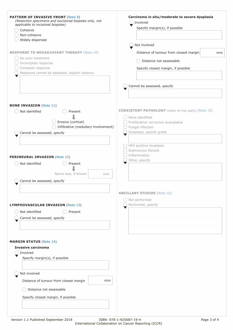
RESPONSE TO NEOADJUVANT THERAPY (Note 10)
No prior treatmentIncomplete responseComplete responseResponse cannot be assessed, explain reasons
PATTERN OF INVASIVE FRONT (Note 9)(Resection specimens and excisional biopsies only, not applicable to incisional biopsies)
CohesiveNon-cohesiveWidely dispersed
PERINEURAL INVASION (Note 12)
Cannot be assessed, specify
Nerve size, if known
Erosive (cortical)Infiltrative (medullary involvement)
BONE INVASION (Note 11)
Cannot be assessed, specify
mm
Not identified Present
Not identified Present
Carcinoma in situ/moderate to severe dysplasia
Distance of tumour from closest margin mm
Involved
Not involved
Specify margin(s), if possible
Cannot be assessed, specify
Distance not assessable
Specify closest margin, if possible
LYMPHOVASCULAR INVASION (Note 13)
Cannot be assessed, specify
Not identified Present
COEXISTENT PATHOLOGY (select all that apply) (Note 15)
None identifiedProliferative verrucous leukoplakiaFungal infectionDysplasia, specify grade
HPV positive dysplasiaSubmucous fibrosisInflammationOther, specify
Invasive carcinoma
MARGIN STATUS (Note 14)
Involved
Distance of tumour from closest margin
Specify margin(s), if possible
mm
Not involved
Distance not assessable
Specify closest margin, if possible
Not performedPerformed, specify
ANCILLARY STUDIES (Note 16)
Version 1.1 Published September 2018 ISBN: 978-1-925687-19-4 Page 4 of 4International Collaboration on Cancer Reporting (ICCR)
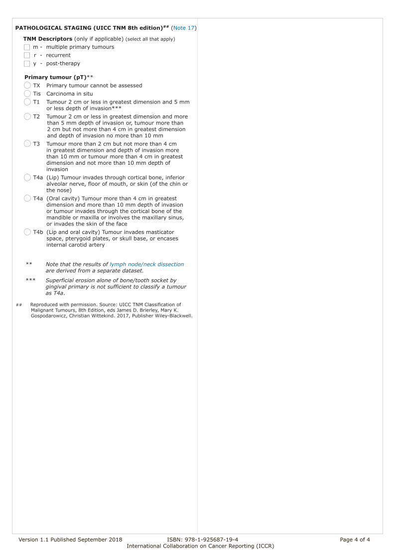
PATHOLOGICAL STAGING (UICC TNM 8th edition)## (Note 17)
m - multiple primary tumoursr - recurrenty - post-therapy
TX Primary tumour cannot be assessedTis Carcinoma in situT1 Tumour 2 cm or less in greatest dimension and 5 mm
or less depth of invasion***T2 Tumour 2 cm or less in greatest dimension and more
than 5 mm depth of invasion or, tumour more than 2 cm but not more than 4 cm in greatest dimension and depth of invasion no more than 10 mm
T3 Tumour more than 2 cm but not more than 4 cm in greatest dimension and depth of invasion more than 10 mm or tumour more than 4 cm in greatest dimension and not more than 10 mm depth of invasion
T4a (Lip) Tumour invades through cortical bone, inferior alveolar nerve, floor of mouth, or skin (of the chin or the nose)
T4a (Oral cavity) Tumour more than 4 cm in greatest dimension and more than 10 mm depth of invasion
or tumour invades through the cortical bone of the mandible or maxilla or involves the maxillary sinus, or invades the skin of the face
T4b (Lip and oral cavity) Tumour invades masticator space, pterygoid plates, or skull base, or encases internal carotid artery
TNM Descriptors (only if applicable) (select all that apply)
** Note that the results of lymph node/neck dissection are derived from a separate dataset.
*** Superficial erosion alone of bone/tooth socket by gingival primary is not sufficient to classify a tumour as T4a.
Primary tumour (pT)**
## Reproduced with permission. Source: UICC TNM Classification of Malignant Tumours, 8th Edition, eds James D. Brierley, Mary K. Gospodarowicz, Christian Wittekind. 2017, Publisher Wiley-Blackwell.
1
Scope
The dataset has been developed for the reporting of resection and biopsy specimens of invasive carcinomas of the oral cavity, including lip and tongue. Mucosal melanoma, lymphomas and sarcomas are not included. In addition, neck dissections and nodal excisions are dealt with in a separate dataset, and this dataset should be used in conjunction, where applicable.
Note 1 – Neoadjuvant therapy (Non-core)
Reason/Evidentiary Support
There is no agreed upon system for grading tumour regression in oral squamous cell carcinoma that has been treated with neoadjuvant therapy. However, a history of previous radiotherapy and/or chemotherapy should be included as histologic changes related to the therapy such as necrosis may affect interpretation of the tumour.
Back
Note 2 – Operative procedure (Core)
Reason/Evidentiary Support
Important to correlate the type of procedure (excisional biopsy or resection) with the material received for patient safety. Site-specific designations are required for accurate staging and for cancer registration. Modification of the resection, e.g. partial, total should be described. For example: hemi-glossectomy, partial glossectomy; hemi-mandibulectomy, segmental (partial) mandibulectomy; partial maxillectomy, total maxillectomy; selective neck dissection, modified neck dissection.1,2
Back
Note 3 – Specimens submitted (Core) and Tumour site (Core)
Reason/Evidentiary Support
The anatomy and surgical interventions of the oral cavity are complex and it is important to ensure accurate and precise communication between the pathologists and the treating and diagnostic team with respect to exact anatomic site of involvement, tumour laterality and specific operative procedures.3-5
The protocol applies to all carcinomas arising at these sites (see Figure 1). For large cancers that involve more than one site, the primary site of involvement should be recorded.
2
Mucosal Lip. The lip begins at the junction of the vermilion border with the skin and includes only the vermilion surface or that portion of the lip that meets the opposing lip. Buccal Mucosa (Inner Cheek). Refers to the mucous membrane lining of the inner surface of the cheeks and lips of contact of the opposing lips to the line of attachment of mucosa of the upper and lower alveolar ridge and pterygomandibular raphe.
Lower Alveolar Ridge. This refers to the mucosa overlying the alveolar process of the mandible, which extends from the line of attachment of mucosa in the buccal vestibule to the line of free mucosa of the floor of the mouth. Posteriorly it extends to the ascending ramus of the mandible.
Upper Alveolar Ridge. This refers to the mucosa overlying the alveolar process of the maxilla, which extends from the line of attachment of mucosa in the upper gingival buccal vestibule to the junction of the hard palate. The posterior margin is the upper end of the pterygopalatine arch.
Floor of the Mouth. This is a semilunar space over the mylohyoid and hypoglossus muscles, extending from the inner surface of the lower alveolar ridge to the undersurface of the tongue. The posterior boundary is the base of the anterior pillar of the tonsil. It is divided into two sides of the submaxillary and sublingual salivary glands.
Hard Palate. This is the semilunar area between the upper alveolar ridge and the mucous membrane covering the palatine process of the maxillary palatine bones. It extends from the inner surface of the superior alveolar ridge to the posterior edge of the palatine bone.
Anterior Two-Thirds of the Tongue (Oral Tongue). This is the freely mobile portion of the tongue that extends anteriorly from the line of circumvallate papillae to the undersurface (ventral) of the tongue at the junction of the floor of the mouth. It includes the tip of tongue, lateral borders, dorsal surface and ventral tongue.
Retromolar trigone. A triangular shaped region extending distal from the mandibular third molar as the base and attaches to the hamulus of the medial pterygoid process of the sphenoid bone as the apex.
‘Not specified’ should be used rarely and only after good effort has been employed to obtain the requisite information.

3
Figure 1. Anatomic sites and subsites for lip and oral cavity
Back
Note 4 – Tumour focality (Non-core)
Reason/Evidentiary Support
True multifocal or synchronous oral cavity carcinomas are rare. Patients with oral squamous cell carcinomas have a high incidence (2-3%) of developing a second primary lesion however these are usually metachronous lesions. The theory of field cancerization whereby contiguous genetically altered areas of mucosa lead to the development of neoplasms have been supported by studies evaluating clonality and other molecular markers. Proliferative verrucous leukoplakia has the propensity of developing multifocal tumours. It is rare to have multiple tumours disconnected but not uncommon to have more than one squamous cell carcinoma connected via dysplasia. The location, proximity to dysplastic epithelium, depth and nodal status remain important. Tumour focality seems to be a standard not just for staging and pathology but for clinical trials and treatment considerations 6-9
Back
4
Note 5 – Tumour dimensions (Core)
Reason/Evidentiary Support
Tumour dimension is an important component in pathologic staging.10 The macroscopic diameter (in millimetres) should be used unless the histological extent is greater than macroscopically apparent, in which case the microscopic dimension is used. At times only microscopic evaluation actually differentiates what clinically (phenotypically) appears to be tumour from what is actual invasion (not dysplasia or inflammation). The maximum depth of invasion should be recorded as core and the discussion should include how/why depth of invasion is different than tumour thickness.11-19 As for other tissues, measurements are made pragmatically, acknowledging distortion of tissues by fixation and processing.11
Back
Note 6 – Histological tumour type (Core)
Reason/Evidentiary Support
The major histologic tumour types of squamous cell carcinoma as recognized by the World Health Organization (WHO) classification are squamous cell carcinoma, conventional type, basaloid, papillary, spindle, adenosquamous, acantholytic, lymphoepithelial, verrucous carcinoma and carcinoma cuniculatum. Hybrid lesions such as verrucous carcinoma and squamous cell carcinoma exist and should be recognized as it may affect prognosis.20 Subtypes should be assigned for both prognosis and cancer registry.21-23
Salivary carcinoma histologic type essentially defines its biologic behaviour and thus influences prognosis, patterns of recurrence and thus clinical management.24,25 Some carcinoma types (i.e. basal cell adenocarcinoma, conventional acinic cell carcinoma) are more indolent with locoregional recurrence but low nodal and distant metastatic rates.26
The major histologic salivary gland carcinomas of minor salivary glands as recognized by the WHO classification are acinic cell carcinoma, adenoid cystic carcinoma, adenocarcinoma NOS, (mammary analogue) secretory carcinoma, cystadenocarcinoma, epithelial-myoepithelial carcinoma, mucoepidermoid carcinoma (low, intermediate and high grade), polymorphous adenocarcinoma (low, intermediate and high grade), (hyalinizing) clear cell carcinoma, intraductal carcinoma, carcinosarcoma, myoepithelial carcinoma, oncocytic carcinoma.
Carcinoma ex pleomorphic adenoma is subclassifed by type and extent of invasion, the latter including minimally invasive, invasive and intracapsular (non-invasive) cancers. The definition for minimally invasive carcinomas varies, ranging from 1.5 mm to 6 mm. Invasive carcinomas extend beyond 6 mm; non-invasive cancers are completely confined to within the capsule without evidence of penetration into extracapsular tissue. Prior to diagnosing a non-invasive carcinoma ex pleomorphic adenoma, sectioning of the entire lesion for histologic evaluation is recommended to exclude the presence of invasive growth. Prognosis has been linked to degree of invasion with non-
5
invasive and minimally invasive cancers apparently having a better prognosis than invasive cancers.27,28
WHO classification of tumours of the oral cavity and mobile tongue29
Descriptor ICD-O codes
Epithelial tumours and lesions Squamous cell carcinoma 8070/3 Oral epithelial dysplasia
Low grade 8077/0 High grade 8077/2
a The morphology codes are from the International Classification of Diseases for Oncology (ICD-O). Behaviour is coded /0 for benign tumours; /1 for unspecified, borderline, or uncertain behaviour; /2 for carcinoma in situ and grade III intraepithelial neoplasia; and /3 for malignant tumours © WHO/International Agency for Research on Cancer (IARC). Reproduced with permission
Back
Note 7 – Histological tumour grade (Core)
Reason/Evidentiary Support
Based on the WHO classifications, three histologic grades of squamous cell carcinoma, conventional type are used: well, moderately or poorly differentiated.29 The most aggressive or highest grade should be recorded if the tumour has a varied histology. Grading requires the assessment of keratinization, mitotic activity, cellular and nuclear pleomorphism, pattern of invasion and host response.1,30-33 Squamous cell carcinoma subtypes such as verrucous carcinoma, basaloid squamous cell carcinoma, papillary squamous cell carcinoma are not graded.
Grading of minor salivary gland tumours follows the criteria for major salivary gland tumours.26,27
Back
Note 8 – Depth of invasion (Core)
Reason/Evidentiary Support
Depth of invasion (DOI) in oral cavity squamous cell carcinoma, particularly of the tongue, has been identified as an important prognostic indicator. DOI is not synonymous with tumour thickness. In the recent American Joint Committee on Cancer (AJCC) the tumour stage (T) has been changed to reflect the importance of DOI.10 DOI increases T by 1 step for every 5 mm, whereby T1 is tumour ≤2 cm and DOI ≤5 mm, T2 is tumour ≤2 cm and DOI >5 mm and ≤10 mm or tumour >2 cm but ≤4 cm and ≤10
6
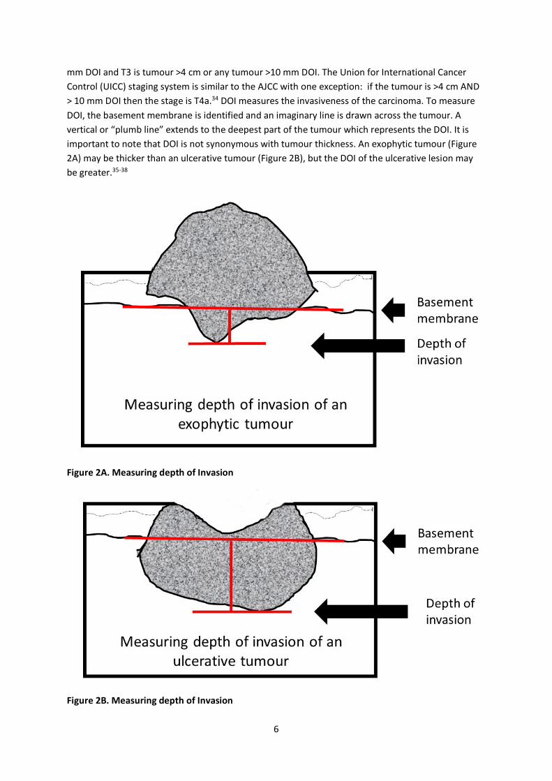
mm DOI and T3 is tumour >4 cm or any tumour >10 mm DOI. The Union for International Cancer Control (UICC) staging system is similar to the AJCC with one exception: if the tumour is >4 cm AND > 10 mm DOI then the stage is T4a.34 DOI measures the invasiveness of the carcinoma. To measure DOI, the basement membrane is identified and an imaginary line is drawn across the tumour. A vertical or “plumb line” extends to the deepest part of the tumour which represents the DOI. It is important to note that DOI is not synonymous with tumour thickness. An exophytic tumour (Figure 2A) may be thicker than an ulcerative tumour (Figure 2B), but the DOI of the ulcerative lesion may be greater.35-38
Figure 2A. Measuring depth of Invasion
Figure 2B. Measuring depth of Invasion

7
Note 9 – Pattern of Invasive front (Core)
Reason/Evidentiary Support
The pattern of invasion in oral squamous cell carcinoma has proven prognostic value and should be reported as cohesive or non-cohesive (Figure 3). It is important to evaluate the most complex area of tumour-stroma interface (“worst” area) and ideally assessment should only be made on resection specimens or excisional biopsies. Acknowledgement is made that at times non-surgical treatment decisions are made on incisional biopsy only specimens and consequently the best assessment of pattern of invasion should be noted. Cohesive invasion is defined as broad sheets of cancer cells and/or tumour nests >15 cells across. Non-cohesive invasion shows a spectrum of appearances that includes narrow strands, small groups of <15 tumour cells and single infiltrating tumour cells.35-38 For stage T1/T2 oral squamous cell carcinoma, particularly those arising in the tongue there is evidence that tumour satellites localized ≥ 1 mm away from the main tumour or nearest satellite (worst pattern of invasion WPOI-5) is a valid adverse prognostic factor.37,39
Figure 3. Pattern of Invasive front
Back
Note 10 – Response to neoadjuvant therapy (Non-core)
Reason/Evidentiary Support
There is no agreed system for grading tumour regression in oral squamous cell carcinoma that has been treated with neoadjuvant therapy. Specific pathologic changes in response to neoadjuvant chemotherapy, although well described in other organ systems is lacking in oral cavity cancer. These changes include necrosis, fibrosis, cytologic atypia and inflammation. As the field of neoadjuvant
8
therapy for oral cavity cancer evolves histologic changes important for treating clinicians may be better elucidated.
Back
Note 11 – Bone invasion (Core)
Reason/Evidentiary Support
Infiltrative bone involvement by squamous cell carcinoma correlates with a worse prognosis. Bone invasion may be a macroscopic feature, however sampling through the involved bone for histologic examination should be performed to obtain histologic evidence. The presence of bone invasion affects tumour staging and patients with bone invasion often have a worse prognosis. It is important to distinguish superficial cortical bone erosion from infiltrative invasion to the medullary bone as this is critical in accurate tumour staging. If bone is resected, then bone margins should be recorded.13
Back
Note 12 – Perineural invasion (Core and Non-core)
Reason/Evidentiary Support
Perineural invasion is associated with a worse prognosis, regardless of nerve size and should be recorded. The presence or absence of perineural and/or endoneural/intraneural invasion may impact subsequent therapy and prognosis.1,31,40-42
Back
Note 13 – Lymphovascular invasion (Core)
Reason/Evidentiary Support
There is a need to distinguish between intravascular tumour embolization and retraction artefact. Positive vascular invasion should be reported only when tumour emboli are identified within endothelial lined spaces. No distinction between venous channels and small lymphatics is required.33,43
Back

9
Note 14 – Margin status (Core)
Reason/Evidentiary Support
All surgical margins should be measured in millimetres histologically for both mucosal and deep margins. In the comments section, acknowledgement should be made how the surgical margin was measured, for example if the margin was submitted from the tumour bed margin at the time of the operative procedure rather than from the surgical specimen.44-47The presence of high grade dysplasia/carcinoma in situ at the margin is associated with an increased risk of local recurrence and this should be recorded. The definition of a “close” margin is not standardized but in the oral cavity from a surgical point of view >5 mm is clear and 1-5 mm is close while <1 mm is involved. Acknowledgement is made of fixation and processing distortion on measurements which may cause tissue shrinkage including the surgical margin.48 Acknowledgement is also made of any laser or electrocautery associated tissue distortion such as cellular and nuclear polymorphism, nuclear hyperchromatism, epithelial cell separation, collagen denaturation, etc. on measurements including the surgical margin.49-51 Any bone resection margins should be identified and comment on the presence or absence of carcinoma at these margins should be provided.13 Dysplastic changes include abnormal cellular organization, increased mitotic activity, and nuclear enlargement with pleomorphism.1,30,31,38,44-47,52 Although terminology varies, using the 2017 WHO criteria for oral dysplasia, dysplasia limited to the lower one-third of the epithelium is generally referred to as mild dysplasia.30 Moderate dysplasia is defined as cytological atypia extending to the middle third of the epithelium and severe dysplasia extends to the upper third of the epithelium. Carcinoma in situ is considered synonymous with severe dysplasia. Currently the use of a binary grading system similar to laryngeal dysplasia has been proposed but to date lacks validation in the oral cavity. In a binary system low-grade dysplasia includes mild dysplasia and mild-moderate dysplasia. The term high grade dysplasia includes moderate dysplasia, severe dysplasia and carcinoma in situ.30
Reporting of surgical margins for carcinomas of the minor salivary glands should follow those used for squamous cell carcinoma of oral cavity.
Back
Note 15 – Coexistent pathology (Non-core)
Reason/Evidentiary Support
The most common sites of dysplasia with the highest risk of malignant transformation are lateral and ventral tongue, floor of mouth, and lower lip. Dysplastic changes include abnormal cellular organization, increased mitotic activity including abnormal forms, and nuclear enlargement with pleomorphism. Although terminology varies, dysplasia limited to the lower one-third of the epithelium is generally referred to as mild dysplasia (low-grade dysplasia), dysplasia limited to the lower two-thirds as moderate dysplasia and dysplasia involving the full thickness as severe dysplasia/carcinoma in situ.52 However, when moderate dysplasia has marked cytologic atypia, then often the lesion will be upgraded to severe dysplasia. The term high-grade dysplasia includes moderate and severe dysplasia and carcinoma in situ. A recently described subset of oral dysplasia is
10
positive for high-risk HPV. The epithelium exhibits full-thickness dysplastic changes with karyorrhexis and apoptosis and the cells are strongly positive for p16 by Immunohistochemistry.53
Proliferative verrucous leukoplakia (PVL) is a distinct form of oral precancer of unknown etiology with a multifocal presentation and a progressive course with high recurrence rates and malignant transformation in as many as 70% of cases.54,55 This diagnosis requires adequate clinical information. Subepithelial fibrosis is a characteristic of oral submucous fibrosis and increased fibrosis is associated with an increased risk of epithelial dysplasia.56 Some inherited genetic mutations are associated with a higher risk of oral cancer development including Fanconi anemia, Li-Fraumeni syndrome and dyskeratosis congenita.29 Care must be taken to rule out reactive atypia which can be seen in epithelium adjacent to ulcers and with fungal infections.
Back
Note 16 – Ancillary studies (Non-core)
Reason/Evidentiary Support
In most cases, further studies are not required for the diagnosis. Epithelial immunohistochemical markers may be required for poorly differentiated or spindle cell carcinoma including AE1/AE3, CK5/6, p63 and p40.57 Lymphoepithelial squamous cell carcinoma in the oral cavity is rare and although not all cases are EBV-positive, Epstein-Barr virus (EBV)-encoded small RNAs (EBERs) studies are indicated.58 There is currently no role for routine HPV high risk type testing in oral squamous cell carcinoma.57-61
Back
Note 17 – Pathological staging (Core)
Reason/Evidentiary Support
By AJCC/UICC convention, the designation “T” refers to a primary tumour that has not been previously treated. Both staging systems integrate DOI into the T categories. Similar to skin malignancies, DOI is significantly associated with disease-free survival.62 Per the AJCC 8th edition, specific instructions are given to measure DOI.10 To measure DOI, the basement membrane is identified and an imaginary line is drawn across the tumour. A vertical or “plumb line” extends to the deepest part of the tumour which represents the DOI. It is important to note that DOI is not synonymous with tumour thickness. An exophytic tumour may be thicker than an ulcerative tumour, but the DOI of the ulcerative lesion may be greater. An important point to highlight is that the UICC 8th edition does not specify how DOI should be measured.34In addition as outlined in Note 8, UICC staging system is similar to the AJCC with one exception: if the tumour is >4 cm AND >10 mm DOI then the stage is T4a.34 Superficial erosion alone of bone/tooth socket by primary gingival tumour is not sufficient to classify a tumour as T4a which requires invasion into medullary bone.
11
Extranodal extension (ENE) is associated with adverse outcomes. ENE is defined as microscopic or macroscopic. Microscopic ENE is defined as ≤0.2 cm whereas macroscopic ENE is defined as ENE clinically apparent at time of dissection or microscopic tumour extension >0.2 cm beyond the lymph node capsule.
Back
References
1 Jerjes W, Upile T, Petrie A, Riskalla A, Hamdoon Z, Vourvachis M, Karavidas K, Jay A, Sandison A, Thomas GJ, Kalavrezos N and Hopper C (2010). Clinicopathological parameters, recurrence, locoregional and distant metastasis in 115 T1-T2 oral squamous cell carcinoma patients. Head Neck Oncol 2:9.
2 Rapidis AD, Gullane P, Langdon JD, Lefebvre JL, Scully C and Shah JP (2009). Major advances
in the knowledge and understanding of the epidemiology, aetiopathogenesis, diagnosis, management and prognosis of oral cancer. Oral Oncol 45(4-5):299-300.
3 Shah JP and Gil Z (2009). Current concepts in management of oral cancer--surgery. Oral
Oncol 45(4-5):394-401.
4 Nakhleh RE (2011). Quality in surgical pathology communication and reporting. Arch Pathol
Lab Med 135(11):1394-1397.
5 Nakhleh RE, Myers JL, Allen TC, DeYoung BR, Fitzgibbons PL, Funkhouser WK, Mody DR, Lynn
A, Fatheree LA, Smith AT, Lal A and Silverman JF (2012). Consensus statement on effective communication of urgent diagnoses and significant, unexpected diagnoses in surgical pathology and cytopathology from the College of American Pathologists and Association of Directors of Anatomic and Surgical Pathology. Arch Pathol Lab Med 136(2):148-154.
6 Dissanayaka WL, Jayasooriya PR, Kumarasiri PV and Tilakaratne WM (2010). A
histopathologic comparison between synchronous and single primary oral squamous cell carcinomas. Oral Surg Oral Med Oral Pathol Oral Radiol Endod 109(5):732-738.
7 Bedi GC, Westra WH, Gabrielson E, Koch W and Sidransky D (1996). Multiple head and neck
tumors: evidence for a common clonal origin. Cancer Res 56(11):2484-2487.
8 Feng Z, Xu QS, Niu QF, Qin LZ, Li JZ, Su M, Li H and Han Z (2016). Risk factors for patients with
multiple synchronous primary cancers involving oral and oropharyngeal subsites. Oral Surg Oral Med Oral Pathol Oral Radiol 121(4):360-366.
12
9 Bagan JV, Murillo J, Poveda R, Gavalda C, Jimenez Y and Scully C (2004). Proliferative verrucous leukoplakia: unusual locations of oral squamous cell carcinomas, and field cancerization as shown by the appearance of multiple OSCCs. Oral Oncol 40(4):440-443.
10 Amin MB, Edge S, Greene FL, Byrd DR, Brookland RK, Washington MK, Gershenwald JE,
Compton CC, Hess KR, Sullivan DC, Jessup JM, Brierley JD, Gaspar LE, Schilsky RL, Balch CM, Winchester DP, Asare EA, Madera M, Gress DM, Meyer LR (eds) (2017). AJCC Cancer Staging Manual 8th ed. Springer, New York.
11 Spiro RH, Huvos AG, Wong GY, Spiro JD, Gnecco CA and Strong EW (1986). Predictive value
of tumor thickness in squamous carcinoma confined to the tongue and floor of the mouth. Am J Surg 152(4):345-350.
12 Dhanda J, Uppal N, Chowlia H, Opie N, Al-Qamachi L, Shelat D, Aslam A, Yuffa A, Martin T,
Risk J, Triantafyllou A, Shaw R, Parmar S and Mehanna H (2016). Features and prognostic utility of biopsy in oral squamous cell carcinoma. Head Neck 38 Suppl 1:E1857-1862.
13 Ebrahimi A, Murali R, Gao K, Elliott MS and Clark JR (2011). The prognostic and staging
implications of bone invasion in oral squamous cell carcinoma. Cancer 117(19):4460-4467.
14 Shim SJ, Cha J, Koom WS, Kim GE, Lee CG, Choi EC and Keum KC (2010). Clinical outcomes for
T1-2N0-1 oral tongue cancer patients underwent surgery with and without postoperative radiotherapy. Radiat Oncol 5:43.
15 D'Cruz AK, Vaish R, Kapre N, Dandekar M, Gupta S, Hawaldar R, Agarwal JP, Pantvaidya G,
Chaukar D, Deshmukh A, Kane S, Arya S, Ghosh-Laskar S, Chaturvedi P, Pai P, Nair S, Nair D and Badwe R (2015). Elective versus Therapeutic Neck Dissection in Node-Negative Oral Cancer. N Engl J Med 373(6):521-529.
16 Byers RM, El-Naggar AK, Lee YY, Rao B, Fornage B, Terry NH, Sample D, Hankins P, Smith TL
and Wolf PJ (1998). Can we detect or predict the presence of occult nodal metastases in patients with squamous carcinoma of the oral tongue? Head Neck 20(2):138-144.
17 Tan WJ, Chia CS, Tan HK, Soo KC and Iyer NG (2012). Prognostic significance of invasion
depth in oral tongue squamous cell carcinoma. ORL J Otorhinolaryngol Relat Spec 74(5):264-270.
18 Kang CJ, Lin CY, Wang HM, Fan KH, Ng SH, Lee LY, Chen IH, Huang SF, Liao CT and Yen TC
(2011). The number of pathologically positive lymph nodes and pathological tumor depth predicts prognosis in patients with poorly differentiated squamous cell carcinoma of the oral cavity. Int J Radiat Oncol Biol Phys 81(4):e223-230.

13
19 Pentenero M, Gandolfo S and Carrozzo M (2005). Importance of tumor thickness and depth of invasion in nodal involvement and prognosis of oral squamous cell carcinoma: a review of the literature. Head Neck 27(12):1080-1091.
20 Patel KR, Chernock RD, Sinha P, Muller S, El-Mofty SK and Lewis JS, Jr. (2015). Verrucous
carcinoma with dysplasia or minimal invasion: a variant of verrucous carcinoma with extremely favorable prognosis. Head Neck Pathol 9(1):65-73.
21 Jayasooriya PR, Tilakaratne WM, Mendis BR and Lombardi T (2013). A literature review on
oral basaloid squamous cell carcinomas, with special emphasis on etiology. Ann Diagn Pathol 17(6):547-551.
22 Schick U, Pusztaszeri M, Betz M, Ghadjar P, Demiroz C, Kaanders JH and Ozsahin M (2013).
Adenosquamous carcinoma of the head and neck: report of 20 cases and review of the literature. Oral Surg Oral Med Oral Pathol Oral Radiol 116(3):313-320.
23 Thavaraj S, Cobb A, Kalavrezos N, Beale T, Walker DM and Jay A (2012). Carcinoma
cuniculatum arising in the tongue. Head Neck Pathol 6(1):130-134.
24 Baddour HM, Jr., Fedewa SA and Chen AY (2016). Five- and 10-Year Cause-Specific Survival
Rates in Carcinoma of the Minor Salivary Gland. JAMA Otolaryngol Head Neck Surg 142(1):67-73.
25 Olarte LS and Megwalu UC (2014). The Impact of Demographic and Socioeconomic Factors
on Major Salivary Gland Cancer Survival. Otolaryngol Head Neck Surg 150(6):991-998.
26 Seethala RR (2009). An update on grading of salivary gland carcinomas. Head Neck Pathol
3(1):69-77.
27 Seethala RR (2011). Histologic grading and prognostic biomarkers in salivary gland
carcinomas. Adv Anat Pathol 18(1):29-45.
28 Brandwein M, Huvos AG, Dardick I, Thomas MJ and Theise ND (1996). Noninvasive and
minimally invasive carcinoma ex mixed tumor: a clinicopathologic and ploidy study of 12 patients with major salivary tumors of low (or no?) malignant potential. Oral Surg Oral Med Oral Pathol Oral Radiol Endod 81(6):655-664.
29 El-Naggar AK, Chan JKC, Grandis JR, Takata T, Slootweg PJ Eds. (2017). WHO Classification of
Head and Neck Tumours (4th Edition). IARC, Lyon, France.
30 Woolgar JA and Triantafyllou A (2009). Pitfalls and procedures in the histopathological
diagnosis of oral and oropharyngeal squamous cell carcinoma and a review of the role of pathology in prognosis. Oral Oncol 45(4-5):361-385.
14
31 Woolgar JA (2006). Histopathological prognosticators in oral and oropharyngeal squamous cell carcinoma. Oral Oncol 42(3):229-239.
32 Kademani D, Bell RB, Bagheri S, Holmgren E, Dierks E, Potter B and Homer L (2005).
Prognostic factors in intraoral squamous cell carcinoma: the influence of histologic grade. J Oral Maxillofac Surg 63(11):1599-1605.
33 Adel M, Kao HK, Hsu CL, Huang JJ, Lee LY, Huang Y, Browne T, Tsang NM, Chang YL and
Chang KP (2015). Evaluation of Lymphatic and Vascular Invasion in Relation to Clinicopathological Factors and Treatment Outcome in Oral Cavity Squamous Cell Carcinoma. Medicine (Baltimore) 94(43):e1510.
34 International Union against Cancer (UICC) (2016). TNM Classification of Malignant Tumours
(8th Edition) [Incorporating corrections see https://www.uicc.org/sites/main/files/atoms/files/UICC%208th%20Edition%20Errata_25May2018%20final.pdf]. Brierley JD, Gospodarowicz MK, Wittekind C (eds). New York: Wiley-Blackwell.
35 Almangush A, Bello IO, Coletta RD, Makitie AA, Makinen LK, Kauppila JH, Pukkila M,
Hagstrom J, Laranne J, Soini Y, Kosma VM, Koivunen P, Kelner N, Kowalski LP, Grenman R, Leivo I, Laara E and Salo T (2015). For early-stage oral tongue cancer, depth of invasion and worst pattern of invasion are the strongest pathological predictors for locoregional recurrence and mortality. Virchows Arch 467(1):39-46.
36 Almangush A, Bello IO, Keski-Santti H, Makinen LK, Kauppila JH, Pukkila M, Hagstrom J,
Laranne J, Tommola S, Nieminen O, Soini Y, Kosma VM, Koivunen P, Grenman R, Leivo I and Salo T (2014). Depth of invasion, tumor budding, and worst pattern of invasion: prognostic indicators in early-stage oral tongue cancer. Head Neck 36(6):811-818.
37 Li Y, Bai S, Carroll W, Dayan D, Dort JC, Heller K, Jour G, Lau H, Penner C, Prystowsky M,
Rosenthal E, Schlecht NF, Smith RV, Urken M, Vered M, Wang B, Wenig B, Negassa A and Brandwein-Gensler M (2013). Validation of the risk model: high-risk classification and tumor pattern of invasion predict outcome for patients with low-stage oral cavity squamous cell carcinoma. Head Neck Pathol 7(3):211-223.
38 Kuan EC, Mallen-St Clair J, Badran KW and St John MA (2016). How does depth of invasion
influence the decision to do a neck dissection in clinically N0 oral cavity cancer? Laryngoscope 126(3):547-548.
39 Brandwein-Gensler M, Teixeira MS, Lewis CM, Lee B, Rolnitzky L, Hille JJ, Genden E, Urken
ML and Wang BY (2005). Oral squamous cell carcinoma: histologic risk assessment, but not margin status, is strongly predictive of local disease-free and overall survival. Am J Surg Pathol 29(2):167-178.
15
40 Chatzistefanou I, Lubek J, Markou K and Ord RA (2014). The role of neck dissection and postoperative adjuvant radiotherapy in cN0 patients with PNI-positive squamous cell carcinoma of the oral cavity. Oral Oncol 50(8):753-758.
41 Chinn SB, Spector ME, Bellile EL, McHugh JB, Gernon TJ, Bradford CR, Wolf GT, Eisbruch A
and Chepeha DB (2013). Impact of perineural invasion in the pathologically N0 neck in oral cavity squamous cell carcinoma. Otolaryngol Head Neck Surg 149(6):893-899.
42 Miller ME, Palla B, Chen Q, Elashoff DA, Abemayor E, St John MA and Lai CK (2012). A novel
classification system for perineural invasion in noncutaneous head and neck squamous cell carcinoma: histologic subcategories and patient outcomes. Am J Otolaryngol 33(2):212-215.
43 Fives C, Feeley L, O'Leary G and Sheahan P (2016). Importance of lymphovascular invasion
and invasive front on survival in floor of mouth cancer. Head Neck 38 Suppl 1:E1528-1534.
44 Hinni ML, Ferlito A, Brandwein-Gensler MS, Takes RP, Silver CE, Westra WH, Seethala RR,
Rodrigo JP, Corry J, Bradford CR, Hunt JL, Strojan P, Devaney KO, Gnepp DR, Hartl DM, Kowalski LP, Rinaldo A and Barnes L (2013). Surgical margins in head and neck cancer: a contemporary review. Head Neck 35(9):1362-1370.
45 Meier JD, Oliver DA and Varvares MA (2005). Surgical margin determination in head and
neck oncology: current clinical practice. The results of an International American Head and Neck Society Member Survey. Head Neck 27(11):952-958.
46 Maxwell JH, Thompson LD, Brandwein-Gensler MS, Weiss BG, Canis M, Purgina B, Prabhu
AV, Lai C, Shuai Y, Carroll WR, Morlandt A, Duvvuri U, Kim S, Johnson JT, Ferris RL, Seethala R and Chiosea SI (2015). Early Oral Tongue Squamous Cell Carcinoma: Sampling of Margins From Tumor Bed and Worse Local Control. JAMA Otolaryngol Head Neck Surg 141(12):1104-1110.
47 Chang AM, Kim SW, Duvvuri U, Johnson JT, Myers EN, Ferris RL, Gooding WE, Seethala RR
and Chiosea SI (2013). Early squamous cell carcinoma of the oral tongue: comparing margins obtained from the glossectomy specimen to margins from the tumor bed. Oral Oncol 49(11):1077-1082.
48 Chen CH, Hsu MY, Jiang RS, Wu SH, Chen FJ and Liu SA (2012). Shrinkage of head and neck
cancer specimens after formalin fixation. J Chin Med Assoc 75(3):109-113.
49 Seoane J, Caballero TG, Urizar JM, Almagro M, Mosquera AG and Varela-Centelles P (2010).
Pseudodysplastic epithelial artefacts associated with oral mucosa CO2 laser excision: an assessment of margin status. Int J Oral Maxillofac Surg 39(8):783-787.
16
50 Gonzalez-Mosquera A, Seoane J, Garcia-Caballero L, Lopez-Jornet P, Garcia-Caballero T and Varela-Centelles P (2012). Er,CR:YSGG lasers induce fewer dysplastic-like epithelial artefacts than CO2 lasers: an in vivo experimental study on oral mucosa. Br J Oral Maxillofac Surg 50(6):508-512.
51 Buchanan MA, Coleman HG, Daley J, Digges J, Sandler M, Riffat F and Palme CE (2016).
Relationship between CO2 laser-induced artifact and glottic cancer surgical margins at variable power doses. Head Neck 38 Suppl 1:E712-716.
52 Speight PM, Abram TJ, Floriano PN, James R, Vick J, Thornhill MH, Murdoch C, Freeman C,
Hegarty AM, D'Apice K, Kerr AR, Phelan J, Corby P, Khouly I, Vigneswaran N, Bouquot J, Demian NM, Weinstock YE, Redding SW, Rowan S, Yeh CK, McGuff HS, Miller FR and McDevitt JT (2015). Interobserver agreement in dysplasia grading: toward an enhanced gold standard for clinical pathology trials. Oral Surg Oral Med Oral Pathol Oral Radiol 120(4):474-482.e472.
53 Stojanov IJ and Woo SB (2015). Human papillomavirus and Epstein-Barr virus associated
conditions of the oral mucosa. Semin Diagn Pathol 32(1):3-11.
54 Akrish S, Ben-Izhak O, Sabo E and Rachmiel A (2015). Oral squamous cell carcinoma
associated with proliferative verrucous leukoplakia compared with conventional squamous cell carcinoma--a clinical, histologic and immunohistochemical study. Oral Surg Oral Med Oral Pathol Oral Radiol 119(3):318-325.
55 Gillenwater AM, Vigneswaran N, Fatani H, Saintigny P and El-Naggar AK (2013). Proliferative
verrucous leukoplakia (PVL): a review of an elusive pathologic entity! Adv Anat Pathol 20(6):416-423.
56 Jayasooriya PR, Nadeeka Jayasinghe KA and Mudiyanselage Tilakaratne W (2011).
Relationship between thickness of fibrosis and epithelial dysplasia in oral submucous fibrosis. J Investig Clin Dent 2(3):171-175.
57 Bice TC, Tran V, Merkley MA, Newlands SD, van der Sloot PG, Wu S and Miller MC (2015).
Disease-Specific Survival with Spindle Cell Carcinoma of the Head and Neck. Otolaryngol Head Neck Surg 153(6):973-980.
58 Rytkonen AE, Hirvikoski PP and Salo TA (2011). Lymphoepithelial carcinoma: two case
reports and a systematic review of oral and sinonasal cases. Head Neck Pathol 5(4):327-334.
59 Sgaramella N, Coates PJ, Strindlund K, Loljung L, Colella G, Laurell G, Rossiello R, Muzio LL,
Loizou C, Tartaro G, Olofsson K, Danielsson K, Fahraeus R and Nylander K (2015). Expression of p16 in squamous cell carcinoma of the mobile tongue is independent of HPV infection despite presence of the HPV-receptor syndecan-1. Br J Cancer 113(2):321-326.

17
60 Reuschenbach M, Kansy K, Garbe K, Vinokurova S, Flechtenmacher C, Toth C, Prigge ES, Thiele OC, Reinert S, Hoffmann J, von Knebel Doeberitz M and Freier K (2013). Lack of evidence of human papillomavirus-induced squamous cell carcinomas of the oral cavity in southern Germany. Oral Oncol 49(9):937-942.
61 Zafereo ME, Xu L, Dahlstrom KR, Viamonte CA, El-Naggar AK, Wei Q, Li G and Sturgis EM
(2016). Squamous cell carcinoma of the oral cavity often overexpresses p16 but is rarely driven by human papillomavirus. Oral Oncol 56:47-53.
62 Ebrahimi A, Gil Z, Amit M, Yen TC, Liao CT, Chaturvedi P, Agarwal JP, Kowalski LP, Kreppel M,
Cernea CR, Brandao J, Bachar G, Bolzoni Villaret A, Fliss D, Fridman E, Robbins KT, Shah JP, Patel SG and Clark JR (2014). Primary tumor staging for oral cancer and a proposed modification incorporating depth of invasion: an international multicenter retrospective study. JAMA Otolaryngol Head Neck Surg 140(12):1138-1148.