
CARCINOMA BREAST - SRM University BREAST Dr... · • Each breast has 15 to 20 sections (lobes)...
92
CARCINOMA BREAST DR.S.FLORET
Transcript of CARCINOMA BREAST - SRM University BREAST Dr... · • Each breast has 15 to 20 sections (lobes)...

CARCINOMA BREAST
DR.S.FLORET

Female Breast Anatomy
3
• Breasts consist mainly of fatty tissue interspersed with connective tissue
• There are also less conspicuous parts
– lobes– ducts– lymph nodes

Normal Breast
4
Breast profileA ductsB lobulesC dilated section of duct to hold milkD nippleE fatF pectoralis major muscleG chest wall/rib cage
EnlargementA normal duct cells
B basement membrane (duct wall)
C lumen (center of duct)
Illustration © Mary K. Bryson

Breast Gland
5
• Each breast has 15 to 20 sections (lobes) arranged like the petals of daisy
• Inside each lobe are many smaller structures called lobules
• At the end of each lobule are tiny sacs (bulbs) that can produce milk

Ducts
6
• Lobes, Lobules and bulbs, are linked by a network of thin tubes (ducts)
• Ducts carry milk from bulbs toward dark area of skin in the center of the breast (areola)
Ducts join together into larger ducts ending at the nipple, where milk is delivered
Duct
Areola

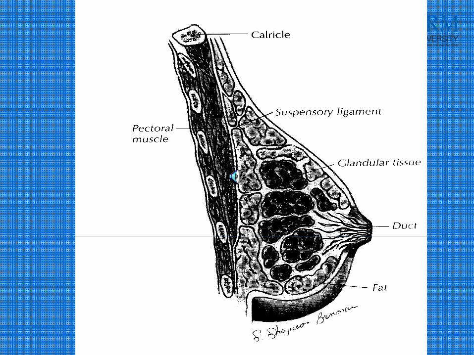
Cooper’s Suspensory Ligaments

Fixed to skin & underlying fascia by fibrous bands
a. Cooper’s (Suspensory) Ligaments
b. Ligaments may retract when breast tumors are present

Anatomy, continued …
STRUCTURE:
1. Outer surface convex, skin covered
2. Nipple:
a. At fourth intercostal space
b. Small conical/cylindrical prominence below center

Nipple location
4th intercostal space

“Tail of Spence”
Axillary Tail
Structure, continued …
BREAST is a Fatty Tissue: surrounds surface, fills spaces between lobes
a. Determines form & size of breast
b. No fatty deposit under nipple & areola

Breast: Fatty Tissue

Normal breast physiology and anatomy
• Symmetry and balance• Size
–weight–menstrual cycle– pregnancy and lactation
• Texture• Shape
– age

VESSELS & NERVES:
1. Arteries: derived from thoracic branches of three pairs of arteries
a. Axillary arteries
1) continuous with subclavian a. 2) gives rise to external mammary ( = lateral thoracic) artery

b. Internal mammary (thoracic) arteries
1) first descending branch of subclavian artery 2)
supply intercostal spaces & breast3) used for coronary bypass surgery
c. Intercostal arteries:
1) numerous branches from internal & external mammary arteries
2) supply intercostal spaces & breast

Subclavian a.
Axillary a.
External mammary (thoracic) a.
Internal mammary (thoracic) a.
Arterial Supply to the Breast

Vessels & Nerves, continued …
2. Veins: a. form a ring around the base of the nipple (“circulus venosus”)
b. Large veins pass from circulusvenosus to circumference of mammary gland, then to
c. External mammary vein to axillary veinor
d. Internal mammary vein to subclavian vein

Veins draining the Breast
Subclavian vein
External mammary vein

3. Innervation: derived from:
a. anterior & lateral cutaneousnerves of thorax
b. spinal segments T3 – T6

Lymphatic System
21
• Lymph ducts: Drain fluid that carries white blood cells (that fight disease) from the breast tissues into lymph nodes under the armpit and behind the breastbone
• Lymph nodes: Filter harmful bacteria and play a key role in fighting off infection
A network of vessels
Lymph ductLymph node

Structure, continued …
4. Lymphatics: clinically significant!a. Glandular lymphatics drain into
anterior axillary (pectoral) nodes central axillary nodes apical nodes
deep cervical nodes subclavicular (subclavian) nodes
b. Medial quadrants drain into parasternal nodes

Subclavian nodes
Axillary nodes
Lateral pectoral nodes
Parasternal nodes
Lymph Nodes of the Breast

Lymphatics, continued …
‐ Superficial regions of skin, areola, nipples:form large channels & drain into pectoral nodes .
NOTE: axillary nodes also drain lymph from arm.

Lymph Nodes and Lymph Drainage
Axillary Nodes

Pathology
• Types – from epithelium
Atrophic schirrous
Schirrous
Medullary
Mucinous
Inflammatory
• Sarcoma – from connective tissue

Histology Grading
• Nottingham Modification of Bloom & Richardson Grading System ‐ Good Prognostic Indicator
• Consists Of :‐Tubular FormationNucleus Size/ degree of pleomorphismMitotic Count
• Score Consists of :‐ I to IIIThe sum of the score is used to classify tumor grade
Grade I = 3 – 5Grade II = 6 – 7 Grade III = 8 – 9 Histological Grade is a strong predictor
• Women with Grade I – 10 Year survival 85 %• Women with Grade III – 10 Year survival < 45 %

Routes of Metastasis
• From medial lymphatics to parasternal nodes– Then to mediastinal nodes
• Across the sternum in lymphatics to
opposite side via cross‐mammary pathways– Then to contralateral breast
• From subdiaphragmatic lymphatics to nodes in abdomen– Then to liver, ovaries, peritoneum
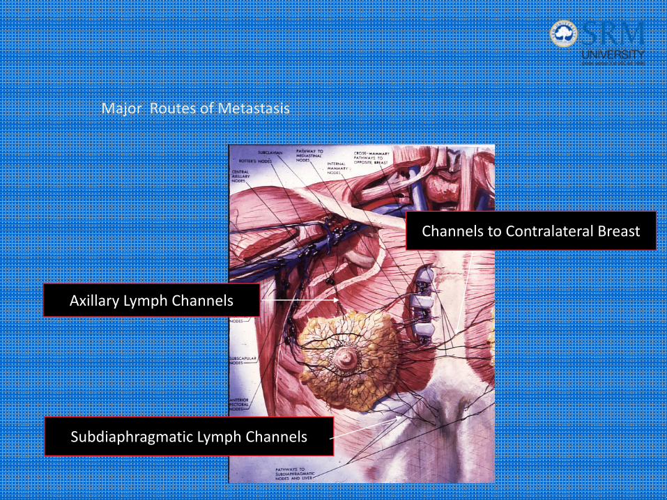
Subdiaphragmatic Lymph Channels
Channels to Contralateral Breast
Axillary Lymph Channels
Major Routes of Metastasis

Breast Cancer• The most common form of cancer among women
• The second most common cause of cancer related mortality
• 1 of 8 women (12.2%)• One third of women with breast cancer die from breast cancer

Breast carcinoma
• most frequent malignant tumor in females (followed by cervix and colon)
• highest incidence – developed countries
(USA 84,8/100 000F/Y, Western Europe 64,7/100 000F/Y)
• 2nd killer among cancers (1st = lung ca)
• risk factors: genetic predisposition (breast ca in close (1st
degree) relatives), proliferative changes, early menarche, late menopause, history of ca (breast, ovary, endometrium)
• importance of preventive controls! – early diagnosisbetter prognosis

Risk Factors for Breast Cancer
• Female (1% male)• Aging• Relative (mother or sister)
• Menstrual history– early on set– late menopause
• Child birth– After the age of 30

Risk Factors for Breast Cancer• Radiation exposure• Breast disease
– Atpyical Hyperplasia– Intraductal carcinoma in situ– Intralobular carcinoma in situ
• Obesity• Diet
– Fat
– Alcohol

Exogenous Estrogen
• Hormonal replacement therapy(HRT)–30% increased risk with long term use
• Oral Contraceptives(OC)– risk slight– risk returns to normal once the use of OC’s has been discontinued

Genetics• BRCA‐1
• BRCA‐2
• P53, Rb‐1
• Her‐2/neu, c‐erB2, c‐myc

RISK FACTORS FOR CARCINOMA BREAST
• MAJOR FACTORS(three fold increase in risk)
• Female sex
• Age
• Previous history of breast cancer
• Parity
• Multiple papillomatosis
• Faamily history

• INTERMEDIATE RISK FACTORS:• Menstrual history(early menarche,latemenopause)
• Radiation exposure• History of cancer of ovary,uterus,colon• Body weight• Atypical hyperplasia
• MINOR FACTORS:• OCP’s• Alcohol

Abnormal signs and symptoms
• Puckering
• Dimpling
• Retraction
• Nipple discharge
• Thickening of skin or lump or “knot”
• Retracted nipple

Abnormal signs and symptoms
• Change in breast size
• Pain or tenderness
• Redness
• Change in nipple position
• Scaling around nipples
• Sore on breast that does not heal

‐ Retraction sign: “dimpling” involving skin, nipple or areola
‐Mobility of massa. Benign = movable
1) not attached 2) not invasive
b. Malignant = attached 1)May grow into bone

‐ Consistency of mass
a. Cysts = fluctuant; compressible
b. Fibroadenoma = rubbery
c. Carcinoma = firm, hard (like gravel)
‐ Axillary area lymph node enlargement

Physical signs:a. Slowly growing, painless mass
b. May demonstrate retracted nipplec. May be bleeding from nippled. May be distorted areola, or breast contoue. Skin dimpling in more advanced stages with retraction of Cooper’s
ligaments


Physical signs, continued …
f. Attachment of mass
g. Edema of skin 1)with “orange skin” appearance
(peau d’orange) 2) due to blocked lymphatics
h. Enlarged axillary or deep cervical lympnodes

Signs and Symptoms
45
Most common: lump or thickening in breast. Often painless
Change in color or appearance of areola
Redness or pitting of skin over the breast, like the skin of an orange
Discharge or bleeding
Change in size or contours of breast

Staging of Breast Cancer
• The American Joint Committee on Cancer (AJCC) has designated staging by TNM
• T= tumor size
• N = lymph node involvement
• M = metastasis

Stage 1
• Tumor < 2.0 cm in greatest dimension
• No nodal involvement (N0)
• No metastases (M0)

Breast ‐‐ CS Size and Extension T1 Examples
Adapted from: TNM Atlas, 3rd ed. 2nd rev., by B. Spiessl et al. Springer Verlag 1992.
TS 011 + Ext 30
TS 990 + Ext 10
TS 006 + Ext 20
TS 018 + Ext 10
TS 008 + Ext 10

Stage II
• Tumor > 2.0 < 5 cm
or
• Ipsilateral axillary lymph node (N1)
• No Metastasis (M0)

Adapted from: TNM Atlas, 3rd ed. 2nd rev., by B. Spiessl et al. Springer Verlag 1992.
Breast ‐‐ CS ExtensionT2 Example
TS 031 + Ext 10

Stage III
• Tumor > 5 cm (T3)
• or ipsilateral axillary lymph nodes fixed to each other or other structures (N2)
• involvement of ipsilateral internal mammary nodes (N3)
• Inflammatory carcinoma (T4d)

Adapted from: TNM Atlas, 3rd ed. 2nd rev., by B. Spiessl et al. Springer Verlag 1992.
Breast ‐‐ CS ExtensionT3 Example
TS 55 + Ext 20

Stage IV (Metastatic breast cancer)
• Any T
• Any N
• Metastasis (M1)

Adapted from: TNM Atlas, 3rd ed. 2nd rev., by B. Spiessl et al. Springer Verlag 1992.
Breast ‐‐ CS Extension 40Extension to chest wall (T4a)
Chest wall includesRibsIntercostal musclesSerratus anterior muscle
Does NOT includePectoral muscle (Ext 30)

Adapted from: TNM Atlas, 3rd ed. 2nd rev., by B. Spiessl et al. Springer Verlag 1992.
Breast ‐‐ CS Extension 51‐52Extensive skin involvement (T4b)
Skin ulceration
Satellite skin nodule

Adapted from: TNM Atlas, 3rd ed. 2nd rev., by B. Spiessl et al. Springer Verlag 1992.
Breast ‐‐ CS Extension 61‐62Chest wall and skin involvement
(T4c)
61 Chest wall plus skin involve‐ment < 50% of breast or NOS(codes 40 + 51)
62 Chest wall plus skin involve‐ ment> 50% of breast
(codes 40 + 52)
code 40
code
51 or 52

Progression to Breast Cancer

Types of breast cancer
• In situ– Intraductal (DCIS)
– Intralobular (LCIS)
• Invasive– Infiltrating ductal carcinoma
– Tubular carcinoma
– Medullary carcinoma
– Mucinous carcinoma

Breast carcinoma - classification
• IN SITU
•INVASIVE
• DUCTAL
•LOBULAR
Ductal in situ (intraductal)
Lobular in situ
Ductal invasive
Lobular invasive
+ other types (12)

Carcinoma in situ
• preinvasive ‐ does not form a palpable tumor
• not detected clinically (only X‐ray – screening !!!)
• multicentricity and bilaterality (namely LCIS)
• continuum: bland hyperplasia ‐ increasing atypism ‐carcinoma in situ
• no metastatic spread (basement membrane)
• risk of invasion depending on grade

Ductal Carcinoma in situ (DCIS)
61 Illustration © Mary K. Bryson
Ductalcancer cells
Normal ductalcell

Range of
Ductal Carcinoma in situ
62
Illus
tratio
n ©
Mar
y K
. Bry
son
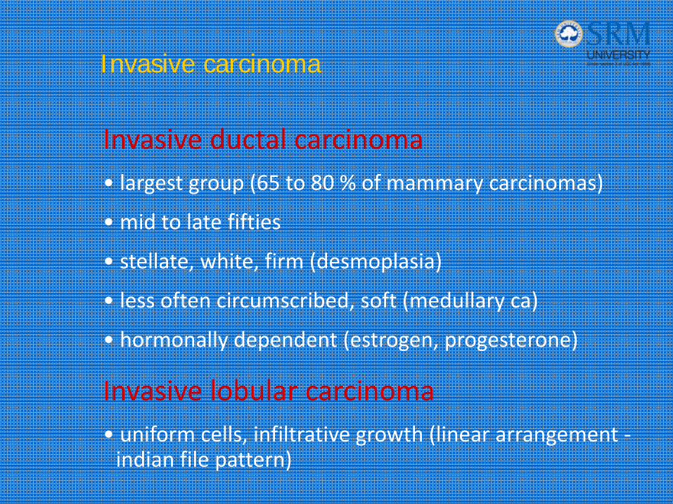
Invasive carcinoma
Invasive ductal carcinoma• largest group (65 to 80 % of mammary carcinomas)
• mid to late fifties
• stellate, white, firm (desmoplasia)
• less often circumscribed, soft (medullary ca)
• hormonally dependent (estrogen, progesterone)
Invasive lobular carcinoma• uniform cells, infiltrative growth (linear arrangement ‐indian file pattern)

Invasive Ductal Carcinoma
(IDC – 80% of breast cancer)
64
• The cancer has spread to the surrounding tissues
• Carcinoma refers to any cancer that begins in the skin or other tissues that cover internal organs
Illustration © Mary K. Bryson
Ductal cancer cells breaking through the wall

Invasive Lobular Carcinoma (ILC)
65
Illustration © Mary K. Bryson
Lobular cancer cells breaking through the wall

Cancer Can also Invade Lymph or Blood Vessels
66
Illustration © Mary K. Bryson
Cancer cells invade lymph duct
Cancer cells invade blood vessel

• other types: tubular, mucinous, medullary, inflammatory – together about 10 % of breast ca
• metastases: regional lymph nodes (axillary, parasternal), lungs, liver, bone marrow, brain
• treatment: surgery (radical – mastectomy, breast conserving surgery – lumpectomy),
radiotherapy
antihormonal therapy (Tamoxifen)
chemotherapy
Invasive carcinoma

Adapted from: TNM Atlas, 3rd ed. 2nd rev., by B. Spiessl et al. Springer Verlag 1992.
Breast ‐‐ CS Extension 71‐73Inflammatory carcinoma
“Diffuse dermal lymphatic involvement causing edema and reddening of the skin”

Breast ‐‐ CS Lymph NodesExamples
Adapted from: TNM Atlas, 3rd ed. 2nd rev., by B. Spiessl et al. Springer Verlag 1992.
Code 25‐‐ 1‐3 movable axillary LN only (N1a)
Code 71‐‐Microscopic int. mam. nodes; no pos axillary LN (N1b)
Code 72‐‐Microscopic int. mam. nodes;
1‐3 pos axillary LN (N1c)

Adapted from: TNM Atlas, 3rd ed. 2nd rev., by B. Spiessl et al. Springer Verlag 1992.
Breast ‐‐ CS Lymph NodesExamples
Code 50 4‐9 fixed/matted
axillary LN only (N2a)
Code 74Clin pos int. mam. nodes; no pos axillary LN (N2b)

Adapted from: TNM Atlas, 3rd ed. 2nd rev., by B. Spiessl et al. Springer Verlag 1992.
with/without axillary nodes
Breast ‐‐ CS Lymph NodesExamples
80
76
Code 75‐‐ Infraclavicular nodes (N3a)Code 76 ‐‐ Internal mammary and 4+ axillary nodes (N3b)
Code 80 ‐‐ Supraclavicular nodes (N3c)
75

Paget‘s disease of the nipple
• result of intraepithelial spread of intraductalcarcinoma
• large pale‐staining cells within the epidermis of the nipple
• limited to the nipple or extend to the areola
• pain or itching, scaling and redness, mistaken for eczema
• ulceration, crusting, and serous or bloody discharge

Cystosarcoma phyllodes(phyllodes tumor)
• initial description ‐ over 150 years ago ‐ fleshy tumor, leaf‐like pattern and cysts on cut surface
• circumscribed, connective tissue and epithelial elements (× fibroadenomas = greater connective tissue cellularity), 1‐15 cm
• less than 1 % of breast tumors
• benign, malignant
• metastases are hematogenous
low grade
high grade

Proliferative changes
• ductal and lobular hyperplasia
• atypical ductal and lobular hyperplasia
• higher risk for the cancer than "normal" population
• associated w. microcalcifications (!mammography!)
• incidental histological finding
• atypical hyperplasia = precancerous lesion

Methods of Detection
• Clinical exam by MD or nurse
• Mammography
• Monthly breast self‐exam (BSE)

Clinical examination• Performed by doctor or trained nurse practitioner
• Annually for women over 40
• At least every 3 years for women between 20 and 40
• More frequent examination for high risk patients

Breast Self Examination
• Opportunity for woman to become familiar with her breasts
• Monthly exam of the breasts and underarm area
• May discover any changes early
• Begin at age 20, continue monthly

When to do BSE
• Menstruating women‐ 5 to 7 days after the beginning of their period
• Menopausal women ‐same date each month
• Pregnant women –same date each month
• Takes about 20 minutes• Perform BSE at least once a month
• Examine all breast tissue

Why don’t more women practice BSE?
• Fear
• Embarrassment
• Youth
• Lack of knowledge
• Too busy, forgetfulness

Mammography• X‐ray of the breast• Has been shown to save lives in patients 50‐69
• Data mixed on usefulness for patients 40‐49
• Normal mammogram does not rule out possibility of cancer completely

Mammography‐more guidelines
• Mammogram facility guidelines
• Avoid mammogram week before period
• Don’t wear deodorant powder or cream
• Bring a list of the places and dates of other mammograms,biopsies you’ve had before
• If you don’t hear from the MD within 10 days, call the facility

Mammography Equipment
82

Computer-Aided Diagnosis
83
• Mammography allows for efficient diagnosis of breast cancers at an earlier stage
• Radiologists misdiagnose 10-30% of the malignant cases
• Of the cases sent for surgical biopsy, only 10-20% are actually malignant
• CAD systems can assist radiologists to reduce the above problems
National Cancer Institute

What Mammograms Show
84
Two of the most important mammographic indicators of breat cancers
– Masses– Microcalcifications: Tiny flecks of calcium – like
grains of salt – in the soft tissue of the breast that can sometimes indicate an early cancer.

Detection of Malignant Masses
85
Malignant masses have a more spiculated appearance
malignant
benign

Mammogram – Difficult Case
86
• Heterogeneously dense breast• Cancer can be difficult to
detect with this type of breast tissue
• The fibroglandular tissue (white areas) may hide the tumor
• The breasts of younger women contain more glands and ligaments resulting in dense breast tissue

Mammogram – Easier Case
87
• With age, breast tissue becomes fattier and has fewer glands
• Cancer is relatively easy to detect in this type of breast tissue

Different Views
88
Top-to-Bottom
Side-to-Side
MRI - Cancer can have a uniqueappearance – many small irregularwhite areas that turned out to becancer (used for diagnosis)

Treatment
Natural History• Tumor Doubling time – 100 days• Nodule of 1 cm to form – 8 years• Metastasis can occur if the tumor size exceed 0.5 cm
• HALSTED CONCEPT – It is a locoregional disease involving lymph nodes, & then systemic spread occurs. So treatment is Radical Mastectomy
• FISSURE CONCEPT – Does not spread in an orderly stepwise manner. Systemic spread occurs early even before lymphatic spread. So treatment is Multi Disciplinary

Treatment for Stage I & II
• Surgery – MRM
‐ QUART ( Quadrantectomy, for AxillaRT)
• Structures to be preserved – Biopsy specimen

Multimodality Treatment For Advanced
• Radiotherapy
• Chemotherapy
• Hormone therapy
Adjuvant therapy
Neo‐Adjuvant therapy

Thank You