Capture-based Next-Generation Sequencing Improves the ...
23
Cancers 2020, 12, 1505; doi:10.3390/cancers12061505 www.mdpi.com/journal/cancers Article Capture-based Next-Generation Sequencing Improves the Identification of Immunoglobulin/T-Cell Receptor Clonal Markers and Gene Mutations in Adult Acute Lymphoblastic Leukemia Patients Lacking Molecular Probes Roberta Cavagna 1,2,† , Marie L. Guinea Montalvo 1,† , Manuela Tosi 1 , Michela Paris 1 , Chiara Pavoni 1 , Tamara Intermesoli 1 , Renato Bassan 3 , Andrea Mosca 2 , Alessandro Rambaldi 1,4, * and Orietta Spinelli 1 1 Hematology and Bone Marrow Transplant Unit, Azienda Socio-Sanitaria Territoriale (ASST) Papa Giovanni XXIII, 24127 Bergamo, Italy; [email protected] (R.C.); [email protected] (M.L.G.M.); [email protected] (M.T.); [email protected] (M.P.); [email protected] (C.P.); [email protected] (T.I.); [email protected] (O.S.) 2 Department of Pathophysiology and Transplantation, Università degli Studi di Milano, 20122 Milano, Italy; [email protected] 3 Hematology Unit, dell’Angelo Hospital and SS. Giovanni and Paolo Hospital, 30174 Venezia Mestre, Italy; [email protected] 4 Department of Oncology and Hematology, Università degli Studi di Milano, 20122 Milano, Italy * Correspondence: [email protected]; Tel.: +39 035 2673684. † These authors contributed equally to this work. Received: 28 April 2020; Accepted: 5 June 2020; Published: 9 June 2020 Abstract: The monitoring of minimal residual disease (MRD) in Philadelphia-negative acute lymphoblastic leukemia (ALL) requires the identification at diagnosis of immunoglobulin/T-cell receptor (Ig/TCR) rearrangements as clonality markers. Aiming to simplify and possibly improve the patients’ initial screening, we designed a capture-based next-generation sequencing (NGS) panel combining the Ig/TCR rearrangement detection with the profiling of relevant leukemia-related genes. The validation of the assay on well-characterized samples allowed us to identify all the known Ig/TCR rearrangements as well as additional clonalities, including rare rearrangements characterized by uncommon combinations of variable, diversity, and joining (V-D-J) gene segments, oligoclonal rearrangements, and low represented clones. Upon validation, the capture NGS approach allowed us to identify Ig/TCR clonal markers in 87% of a retrospective cohort (MRD-unknown within the Northern Italy Leukemia Group (NILG)-ALL 09/00 clinical trial) and in 83% of newly-diagnosed ALL cases in which conventional method failed, thus proving its prospective applicability. Finally, we identified gene variants in 94.7% of patients analyzed for mutational status with the same implemented capture assay. The prospective application of this technology could simplify clonality assessment and improve standard assay development for leukemia monitoring, as well as provide information about the mutational status of selected leukemia-related genes, potentially representing new prognostic elements, MRD markers, and targets for specific therapies. Keywords: acute lymphoblastic leukemia; next-generation sequencing; minimal residual disease
Transcript of Capture-based Next-Generation Sequencing Improves the ...

Cancers 2020, 12, 1505; doi:10.3390/cancers12061505 www.mdpi.com/journal/cancers
Article
Capture-based Next-Generation Sequencing Improves the Identification of Immunoglobulin/T-Cell Receptor Clonal Markers and Gene Mutations in Adult Acute Lymphoblastic Leukemia Patients Lacking Molecular Probes
Roberta Cavagna 1,2,†, Marie L. Guinea Montalvo 1,†, Manuela Tosi 1, Michela Paris 1,
Chiara Pavoni 1, Tamara Intermesoli 1, Renato Bassan 3, Andrea Mosca 2, Alessandro Rambaldi 1,4,*
and Orietta Spinelli 1
1 Hematology and Bone Marrow Transplant Unit, Azienda Socio-Sanitaria Territoriale (ASST) Papa Giovanni
XXIII, 24127 Bergamo, Italy; [email protected] (R.C.); [email protected] (M.L.G.M.);
[email protected] (M.T.); [email protected] (M.P.); [email protected] (C.P.);
[email protected] (T.I.); [email protected] (O.S.) 2 Department of Pathophysiology and Transplantation, Università degli Studi di Milano, 20122 Milano, Italy;
[email protected] 3 Hematology Unit, dell’Angelo Hospital and SS. Giovanni and Paolo Hospital, 30174 Venezia Mestre, Italy;
[email protected] 4 Department of Oncology and Hematology, Università degli Studi di Milano, 20122 Milano, Italy
* Correspondence: [email protected]; Tel.: +39 035 2673684. † These authors contributed equally to this work.
Received: 28 April 2020; Accepted: 5 June 2020; Published: 9 June 2020
Abstract: The monitoring of minimal residual disease (MRD) in Philadelphia-negative acute
lymphoblastic leukemia (ALL) requires the identification at diagnosis of immunoglobulin/T-cell
receptor (Ig/TCR) rearrangements as clonality markers. Aiming to simplify and possibly improve
the patients’ initial screening, we designed a capture-based next-generation sequencing (NGS)
panel combining the Ig/TCR rearrangement detection with the profiling of relevant
leukemia-related genes. The validation of the assay on well-characterized samples allowed us to
identify all the known Ig/TCR rearrangements as well as additional clonalities, including rare
rearrangements characterized by uncommon combinations of variable, diversity, and joining
(V-D-J) gene segments, oligoclonal rearrangements, and low represented clones. Upon validation,
the capture NGS approach allowed us to identify Ig/TCR clonal markers in 87% of a retrospective
cohort (MRD-unknown within the Northern Italy Leukemia Group (NILG)-ALL 09/00 clinical trial)
and in 83% of newly-diagnosed ALL cases in which conventional method failed, thus proving its
prospective applicability. Finally, we identified gene variants in 94.7% of patients analyzed for
mutational status with the same implemented capture assay. The prospective application of this
technology could simplify clonality assessment and improve standard assay development for
leukemia monitoring, as well as provide information about the mutational status of selected
leukemia-related genes, potentially representing new prognostic elements, MRD markers, and
targets for specific therapies.
Keywords: acute lymphoblastic leukemia; next-generation sequencing; minimal residual disease

Cancers 2020, 12, 1505 2 of 23
1. Introduction
In the last decade, the monitoring of molecular minimal residual disease (MRD) has become the
gold standard in routine clinical practice both in pediatric and adult acute lymphoblastic leukemia
(ALL) to improve risk stratification and define therapeutic strategies [1–5]. Indeed, the evaluation of
MRD during induction and maintenance chemotherapy, or after hematopoietic stem cell
transplantation (HSCT), is nowadays considered the strongest predictive factor for ALL prognosis
[6,7]. The availability of MRD data relies on a standardized, time-consuming, and intense procedure
applying PCR amplification to detect the most frequent immunoglobulin/T-cell receptor (Ig/TCR)
gene rearrangements and leukemic fusion transcripts at diagnosis [8–11]. However, in about 5-10%
of cases, the use of this method fails to identify suitable markers for MRD evaluation [12], precluding
patients’ allocation in the most adequate risk class which is crucial to define the most suitable
therapy and to promptly prevent relapse. This failure can be attributed to the PCR technique
limitation concerning both the design and resolution of the assay. As a matter of fact, the large
number of possible combinations of variable, diversity, and joining (V-D-J) gene segments is faced
by PCR with a restricted number of primer combinations that can be used to affordably assess clonal
rearrangement identification due to diagnostic DNA availability, time to assay set-up, and costs.
Therefore, a PCR-based panel designed for clonality assessment at disease presentation is ineffective
in identifying rare rearrangements and in solving oligoclonality/low clonality issues.
New high-throughput technologies have become available and are being explored in order to
overcome the limits of the conventional and standardized MRD assessment [13]. A next-generation
sequencing (NGS) approach, similarly based on rearrangement amplification by PCR, has been
described to identify clonal markers at diagnosis [14–16] and to monitor MRD in lymphoid
malignancies [17–21]. All in all, NGS-based Ig/TCR marker screening greatly improved MRD
monitoring, being less time consuming, and highlighted the polyclonal false-positive scenario. An
alternative to amplicon-based NGS for Ig/TCR marker screening is a capture enrichment NGS
procedure based on the hybridization of probes selecting all the known V, D, and J regions
irrespective of their recombination status [22]. The capture method also allows for the inclusion of
highly complex (i.e., TCRA) or preferentially amplified Ig/TCR regions, which can hinder multiplex
PCR technique [14]. Therefore, this approach can potentially identify all the possible rearrangements
also offering, by a single test and using the same amount of specimen, the possibility to include other
probes recognizing leukemia-related DNA regions such as chromosomal translocation partners [22]
or genes with prognostic significance in hematologic malignancies [23]. Recently, many molecular
alterations have been suggested as possible candidates for predicting ALL outcome [24], or defining
precise features such as Philadelphia chromosome-like leukemia [25].
In this context, we developed a custom capture-based NGS approach for Ig/TCR clonal marker
identification at disease onset, also including the analysis of the mutational status of 25 genes
relevant in diagnostic workup and prognosis of ALL [24]. We aimed to demonstrate its value in
identifying common and uncommon Ig/TCR rearrangements as well as gene mutational status and
its prospective applicability to better define risk class.
2. Results
2.1. Validation of the Capture-Based NGS Panel
We applied a newly designed capture-based NGS assay (panel v1, see Materials and Methods)
to 10 previously characterized diagnostic samples. By comparing both Ig/TCR annotation and
complementarity-determining region 3 (CDR3) sequence, we identified 51 rearrangements already
found by standard PCR-based clonality assessment and Sanger sequencing. This novel approach
also detected 24 additional clonal rearrangements involving the following loci: IGH (n = 4), IGK (n =
2), IGL (n = 1), TCRB (n = 4), TCRG (n = 2), and TCRA and TCRD (n = 11). These clonal
rearrangements were mainly characterized by uncommon V-D-J combinations, low clonal
representation, and oligoclonality that could not be solved by the conventional homo-heteroduplex
clonality assessment (Table 1).

Cancers 2020, 12, 1505 3 of 23
Table 1. Immunoglobulin/T-cell receptor (Ig/TCR) rearrangements identified by next-generation sequencing (NGS) in the cohort of adult acute lymphoblastic leukemia
(ALL) patients enrolled into the Northern Italy Leukemia Group (NILG)-ALL 09/00 clinical trial, formerly evaluated for clonality assessment. These rearrangements have
been (Yes) or not (No) previously detailed by standard clonality assessment and Sanger sequencing following the conventional EuroMRD guidelines.
Patient ID ALL Lineage Locus V Gene del V n del D D Gene del D n del J J Gene Known Rearrangement Feature
BG_371 B
IGH IGHV3-9*01 −2 15 −2 IGHD6-19*01 −4 7 0 IGHJ3*02 Yes /
TRD+ TRDV2*02 −4 10 −28 TRDD3*01 NA NA NA NA Yes /
TRG TRGV2*01 0 20 NA NA NA NA −3 TRGJ1*01 Yes /
TRG TRGV3*02 −4 15 NA NA NA NA −6 TRGJ1*02 Yes /
BG_4502 T
TRB TRBV11-2*03 0 9 −3 TRBD2*01 −5 1 −4 TRBJ2-1*01 Yes /
TRB TRBV14*02 −3 4 −1 TRBD2*01 NA NA NA TRBJ2-6*01 Yes /
TRD TRDV1*01 0 15 0 TRDD3*01 −2 1 −3 TRDJ1*01 Yes /
TRG TRGV3*02 −1 10 NA NA NA NA −4 TRGJ1*02 Yes /
TRG TRGV9*01 −1 13 NA NA NA NA −6 TRGJ1*02 Yes /
BG_5038 B
IGH IGHV3-49*02 −15 8 −2 IGHD2-8*01 0 2 −2 IGHJ6*02 Yes /
IGK IGKV1-16*02 −17 9 −6 KDE NA NA NA NA Yes /
TRB TRBV20-1*05 0 7 −3 TRBD2*01 −5 3 −2 TRBJ2-3*01 Yes /
TRD+ TRDD2*01 −6 5 NA NA NA NA 0 TRDD3*01 Yes /
TRG TRGV9*01 0 5 NA NA NA NA −1 TRGJP1*01 Yes /
TRG TRGV3*02 0 4 NA NA NA NA 0 TRGJ1*02 Yes /
BG_5418 B
IGH IGHV4-30-2*01 −14 34 0 IGHD3-3*01 −7 8 −11 IGHJ6*02 Yes /
IGH IGHV3-7*02 −1 2 −10 IGHD2-2*01 −5 6 −4 IGHJ6*02 Yes /
IGK IGKV3-20*01 −3 3 NA NA NA NA 0 KDE Yes /
IGL IGLV2-8*01 −9 8 NA NA NA NA 0 IGLJ2*01 Yes /
TRG TRGV9*01 −10 10 NA NA NA NA −5 TRGJ1*02 Yes /
TRG TRGV11*01 −7 8 NA NA NA NA −3 TRGJ1*02 Yes /
BG_5452 T
IGH IGHV3-11*01 0 19 −2 IGHD6-19*01 −4 4 −9 IGHJ4*02 Yes /
IGH IGHV3-64D*06 −2 33 2 IGHD6-19*01 −4 4 9 IGHJ4*02 Yes /
IGH IGHV4-34*01 0 6 1 IGHD6-6*01 −7 1 0 IGHJ6*02 Yes /
IGH+ NA NA NA NA IGHD7-27*01 −2 3 −3 IGHJ2*01 Yes /
TRB NA NA NA NA TRBD1 33 0 0 TRBJ2-3 Yes /
TRB TRBV9*01 −3 10 NA TRBD2*01 NA NA 0 TRBJ2-1*01 Yes /
TRG TRGV10*02 −2 11 NA NA NA NA −6 TRGJ1*02 Yes /
TRG TRGV2*01 0 3 NA NA NA NA −1 TRGJ1*02 Yes /

Cancers 2020, 12, 1505 4 of 23
BG_9574 T
TRB TRBV14*01 −1 11 −4 NA NA NA NA TRBJ2-7*01 Yes /
TRB TRBV5-3*01 −2 3 0 TRBD1*01 −3 27 −4 TRBJ2-7*01 Yes /
TRD TRDV1*01 −5 5 0 TRDD2*01 0 11 0 TRDJ1*01 Yes /
TRG TRGV11*01 −11 3 −2 NA NA NA NA TRGJ2*01 Yes /
TRG TRGV4*02 −6 2 −1 NA NA NA NA TRGJ1*02 Yes /
TRG TRGV8*01 −1 3 −3 NA NA NA NA TRGJP2*01 Yes /
BG_9813 T TRB+ NA NA NA NA TRBD2*01 −3 0 −5 TRBJ2-5*01 Yes /
BG_9813 T
TRD+ TRDD2*01 −10 22 NA NA NA NA 0 TRDJ1*01 Yes /
TRG TRGV10*02 −1 3 NA NA NA NA −6 TRGJ1*02 Yes /
TRG TRGV9*01 −3 3 NA NA NA NA 0 TRGJ2*01 Yes /
BG_11360 B
IGH IGHV4-4*07 −2 9 −1 IGHD2-15*01 −6 12 −30 IGHJ6*04 Yes /
IGH+ NA NA NA NA IGHD7-27*01 0 12 −7 IGHJ6*02 Yes /
TRD+ TRDD2*01 0 5 NA NA NA NA −1 TRDD3*01 Yes /
TRD+ TRDV2*02 0 13 NA NA NA NA −3 TRDD3*01 Yes /
BG_11720 B
IGH IGHV6-1*01 0 5 −3 IGHD3-3*01 −2 2 −6 IGHJ6*02 Yes /
TRA+D TRDV2*03 0 5 −2 TRDD3*01 0 3 −4 TRAJ9*01 Yes /
TRG TRGV9*01 −6 13 NA NA NA NA −1 TRGJ1*02 Yes /
BG_11806 B
IGH IGHV1-2*02 −1 3 −3 IGHD2-21*02 −3 24 −1 IGHJ5*02 Yes /
IGK IGKV2-28*01 0 6 NA NA NA NA −1 IGKJ4*02 Yes /
IGL IGLV3-19*01 −5 14 NA NA NA NA −4 IGLJ3*02 Yes /
TRD+ TRDD2*01 0 2 NA NA NA NA 0 TRDD3*01 Yes /
TRG TRGV5*01 0 3 NA NA NA NA −1 TRGJ1*02 Yes /
BG_11806 B TRD+ TRDD2*01 0 13 NA NA NA NA 0 TRDJ3*01 No Uncommon V/DJ combinations
TRA+D TRDD2*01 −2 3 NA NA NA NA −7 TRAJ30*01 No Uncommon V/DJ combinations
BG_5038 B TRA+D TRDD2*01 −3 53 NA NA NA NA −15 TRAJ48*01 No Uncommon V/DJ combinations
BG_5418 B
TRA+D TRDD2*01 −1 14 NA NA NA NA −5 TRAJ29*01 No Uncommon V/DJ combinations
TRA+D TRDD2*01 −11 4 NA NA NA NA −1 TRAJ23*01 No Uncommon V/DJ combinations
TRA TRAV26-1*01 0 4 NA NA NA NA −2 TRAJ33*01 No Uncommon V/DJ combinations
TRA TRAV8-3*01 0 8 NA NA NA NA −7 TRAJ34*01 No Uncommon V/DJ combinations
BG_5452 T TRA TRAV21*01 0 1 NA NA NA NA 0 TRAJ48*01 No Uncommon V/DJ combinations
BG_9574 T TRA+D TRAV29/DV5*01 0 38 0 TRDD3*01 −4 3 0 TRDJ1*01 No Uncommon V/DJ combinations
TRA TRAV21*02 −7 0 −5 NA NA NA NA TRAJ24*02 No Uncommon V/DJ combinations
BG_9813 T TRA TRAV19*01 −2 10 NA NA NA NA −4 TRAJ36*01 No Uncommon V/DJ combinations
BG_11360 B IGH IGHV4-34*01 0 9 −18 IGHD2-2*01 0 2 −4 IGHJ6*03 No Oligoclonality

Cancers 2020, 12, 1505 5 of 23
BG_11720 B IGH IGHV4-34*01 0 6 −1 IGHD6-6*01 −7 1 0 IGHJ6*02 No Oligoclonality
BG_371 B
IGK IGKV1-33*01 −1 2 NA NA NA NA −2 IGKJ4*01 No Oligoclonality
IGK IGKV1-39*01 −4 8 NA NA NA NA −9 IGKJ2*02 No Oligoclonality
TRG TRGV4*02 −4 5 NA NA NA NA −3 TRGJ1*01 No Oligoclonality
BG_5418 B
TRB TRBV23-1*01 0 1 0 TRBD2*01 −7 14 −6 TRBJ2-7*01 No Oligoclonality
TRB TRBV10-3*01 −8 18 −7 TRBD2*01 NA NA NA TRBJ2-3*01 No Oligoclonality
TRB TRBV24-1*01 −13 5 −2 TRBD1*01 −3 5 2 TRBJ2-7*01 No Oligoclonality
BG_5452 T IGH IGHV6-1*01 0 5 −3 IGHD3-3*01 −2 2 −6 IGHJ6*02 No Oligoclonality
BG_9574 T TRG TRGV11*01 0 1 −25 NA NA NA NA TRGJ1*02 No Oligoclonality
BG_11360 B IGL IGLV3-10*01 −2 7 NA NA NA NA 0 IGLJ3*02 No Low represented clone
BG_371 B IGH IGHV1-3*02 −2 4 −17 IGHD3-16*01 −15 0 −18 IGHJ4*02 No Low represented clone
BG_9813 T TRB TRBV4-3*01 −1 21 −6 TRBD2*02 NA NA NA TRBJ1-1*01 No Low represented clone
IGK: IGκ rearrangements, IGL: IGλ rearrangements, TRB: TCRβ rearrangements, TRG: TCRγ rearrangements, TRD: TCRδ rearrangements, and TRA: TCRα
rearrangements. Symbol “+” refers to incomplete rearrangements. del V: nucleotides deletion in V gene segment, n: number of inserted nucleotides, del D: nucleotides
deletion in D gene segment, del J: nucleotides deletion in J gene segment, NA: not applicable, Yes: rearrangement already identified by standard procedure, No:
rearrangement identified by NGS and not previously identified by standard procedure.

Cancers 2020, 12, 1505 6 of 23
The validation of these NGS-recognized rearrangements using standard methods required
further efforts compared to routine procedures—11 uncommon V-D-J combinations were amplified
by rearrangement-specific primers designed based on NGS data (n = 5 in TCRA, n = 1 in TCRD, and n
= 5 in αδ (TCRA+D) loci) and sequenced by Sanger method; three low-represented clonalities (n = 1
IGH, n = 1 TCRB, and n = 1 IGL) that were missed by low sensitive regular PCR were finally revealed
by standard method only after re-amplification of faint heteroduplex PCR products and Sanger
sequencing; 10 oligoclonal rearrangements (n = 2 IGK, n = 3 IGH, n = 3 TCRB, and n = 2 TCRG) were
amplified and recognized within the typical, overlapped Sanger electropherograms. Notably, the
analysis of NGS data derived from human umbilical vein endothelial cells (HUVECs) and
mesenchymal cord blood cells samples did not identify any rearrangement in Ig/TCR genes,
testifying that no false rearrangements were produced during library preparation, sequence
generation, or data analysis.
2.2. Retrospective Application of the Capture-Based NGS Panel to MRD-Unknown ALL Patients
The capture-based NGS assay (panel v1) was then applied to a retrospective group of 23 ALL
patients (13 B-lineage ALL and 10 T-lineage ALL) enrolled into the Northern Italy Leukemia Group
(NILG)-ALL 09/00 clinical trial for whom it had been impossible to isolate clonal rearrangements or
to obtain a sensitive patient-specific molecular probe (MRD-unknown). These patients could not
have benefited from an MRD driven treatment and were treated according to the clinical risk score.
In this cohort, the capture-based NGS allowed for the identification of at least 1 rearrangement in 20
out of 23 patients (87%), recognizing overall 92 clonal rearrangements (IGH (n = 30), IGK (n = 6),
TCRB (n = 6), TCRG (n = 32), and TCRA and TCRD (n = 18)) detailed in Table 2.
Cancers 2020, 12, 1505 7 of 23
Table 2. Ig/TCR rearrangements identified by NGS in the minimal residual disease (MRD)-unknown cohort of adult ALL patients enrolled into the NILG-ALL 09/00
clinical trial. These rearrangements have not been previously identified by standard clonality assessment and Sanger sequencing following the conventional EuroMRD
guidelines, or they were not adequate to generate a suitable patient-specific molecular probe for the MRD evaluation.
Patient ID ALL Lineage Locus V Gene del V n del D D Gene del D n del J J Gene Validation Rearrangement Feature
BG_11584 B IGH IGHV6-1*01 −2 8 −6 IGHD6-6*01 0 3 −10 IGHJ4*02 Yes /
BG_2097 B IGH NA NA NA NA IGHD4-23*01 −2 1 −4 IGHJ2*01 Yes /
BG_855 B IGH IGHV6-1*01 −3 2 −4 IGHD2-2*01 −1 5 0 IGHJ6*03 Yes /
IGH IGHV3-23*01 0 19 −2 IGHD3-9*01 −8 13 −7 IGHJ4*02 Yes /
BG_10112 B IGH+ NA NA NA NA IGHD6-6*01 −2 0 −7 IGHJ4*02 Yes /
IGH+ NA NA NA NA IGHD1-7*01 −6 2 −4 IGHJ4*02 Yes /
BG_11053 B IGH+ NA NA NA NA IGHD2-2*01 −6 3 −4 IGHJ4*02 Yes /
BG_1125 B IGH+ NA NA NA NA IGHD6-25*01 0 −5 11 IGHJ4*02 Yes /
BG_11584 B IGH+ NA NA NA NA IGHD1-26*01 0 3 −3 IGHJ3*02 Yes /
BG_4254 B IGH+ NA NA NA NA IGHD3-9*01 −5 4 −11 IGHJ6*03 Yes /
IGH+ NA NA NA NA IGHD6-6*01 −4 3 −15 IGHJ5*02 Yes /
BG_5702 B IGH+ NA NA NA NA IGHD2-2*02 −6 7 −5 IGHJ5*02 Yes /
BG_8345 B IGH+ NA NA NA NA IGHD3-22*01 −2 7 1 IGHJ6*02 Yes /
IGH+ NA NA NA NA IGHD1-26*01 −2 0 7 IGHJ4*02 Yes /
BG_9445 B IGH+ NA NA NA NA IGHD2-2*02 −3 19 5 IGHJ6*03 Yes /
BG_11269 T TRB TRBV7-8*01 −3 14 NA NA NA NA −7 TRBJ1-4*01 Yes /
BG_6037 T TRB TRBV4-2*01 0 25 NA NA NA NA −3 TRBJ2-3*01 Yes /
TRB TRBV20-1*02 −3 15 −2 TRBD2*02 −2 19 −6 TRBJ2-1*01 Yes /
BG_10112 B TRG TRGV11*01 −4 7 NA NA NA NA −8 TRGJP1*01 Yes /
TRG TRGV11*01 −3 10 NA NA NA NA −1 TRGJ2*01 Yes /
BG_11269 T TRG TRGV4*02 −4 5 NA NA NA NA 0 TRGJ2*01 Yes /
TRG TRGV10*02 −6 0 NA NA NA NA −8 TRGJ1*02 Yes /
BG_11584 B
TRG TRGV4*02 −6 2 NA NA NA NA 0 TRGJ2*01 Yes /
TRG TRGV10*02 0 NA NA NA NA 4 −9 TRGJP1*01 Yes /
TRG TRGV11*01 0 0 NA NA NA NA −7 TRGJP1*01 Yes /
BG_855 B TRG TRGV9*01 −10 3 NA NA NA NA −2 TRGJ1*02 Yes /
BG_11053 B TRD+ NA NA NA NA TRDD2*01 −1 22 −1 TRDJ1*01 Yes /
TRD+ NA NA NA NA TRDD2*01 0 19 0 TRDJ4*01 Yes /
BG_4254 B TRD+ NA NA NA NA TRDD2*01 0 5 −2 TRDD3*01 Yes /
BG_8345 B TRD+ NA NA NA NA TRDD2*01 −3 15 0 TRDD3*01 Yes /
BG_11053 B
IGH+ NA NA NA NA IGHD3-9*01 −5 3 −4 IGHJ4*02 Yes Oligoclonality
IGH+ NA NA NA NA IGHD3-9*01 −5 0 −4 IGHJ6*02 Yes Oligoclonality
IGH+ NA NA NA NA IGHD3-9*01 −3 13 −6 IGHJ6*02 Yes Oligoclonality
BG_11345 T TRD+ NA NA NA NA TRDD2*01 −8 0 −14 TRDD3*01 Yes Oligoclonality
Cancers 2020, 12, 1505 8 of 23
BG_1125 B IGK IGKV1-27*01 −1 0 NA NA NA NA 2 IGKJ1*01 YesŦ /
BG_11269 T
IGK IGKV4-1*01 −1 0 NA NA NA NA −4 IGKJ4*01 YesŦ /
IGK IGKV2D-29*01 −2 6 NA NA NA NA 0 IGKJ2*01 YesŦ /
IGK IGKV1-6*01 −3 0 NA NA NA NA 0 IGKJ1*01 YesŦ /
BG_6037 T TRG TRGV3*01 0 0 NA NA NA NA 0 TRGJ1*02 YesŦ /
TRG TRGV2*01 0 4 NA NA NA NA −2 TRGJ1*02 YesŦ /
BG_2097 B TRA+D TRDV1*01 −2 8 NA NA NA NA −8 TRAJ29*01 YesŦ /
BG_855 B TRA+D NA NA NA NA TRDD2*01 −4 24 −5 TRAJ29*01 YesŦ /
TRA+D TRDV2*01 0 6 0 TRDD3*01 0 5 −4 TRAJ58*01 YesŦ /
BG_11269 T TRA TRAV26-1*01 −3 3 NA NA NA NA −4 TRAJ4*01 YesŦ /
BG_12438 T TRA TRAV19*01 −8 4 NA NA NA NA 0 TRAJ47*02 YesŦ /
BG_2097 B TRA TRAV13-1*01 −3 0 NA NA NA NA −3 TRAJ35*01 YesŦ /
BG_5702 B TRA TRAV16*01 −6 2 NA NA NA NA −4 TRAJ9*01 YesŦ /
BG_10487 B IGH+ NA NA NA NA IGHD3-16*02 −5 8 0 IGHJ4*02 YesŦ Biclonal sequence
IGH+ NA NA NA NA IGHD3-3*01 0 11 −3 IGHJ5*02 YesŦ Biclonal sequence
BG_6490 B IGH+ NA NA NA NA IGHD2-21*02 −1 2 0 IGHJ6*03 YesŦ Biclonal sequence
IGH+ NA NA NA NA IGHD2-8*01 −7 8 −2 IGHJ3*02 YesŦ Biclonal sequence
BG_8646 T TRD+ NA NA NA NA TRDD2*01 −8 0 −14 TRDD3*01 YesŦ Biclonal sequence
TRD+ NA NA NA NA TRDD2*01 −8 0 −16 TRDD3*01 YesŦ Biclonal sequence
BG_1125 B IGH IGHV4-34*02 0 10 NA NA NA NA −4 IGHJ5*01 No Low represented clone
BG_5702 B IGK IGKV2-28*01 −3 2 −7 KDE NA NA NA NA No Low represented clone
BG_12438 T TRB TRBV4-1*02 −3 2 −1 TRBD1*01 −3 8 −2 TRBJ2-7*01 No Low represented clone
BG_5702 B TRB TRBV4-1*02 0 2 0 TRBD1*01 −4 10 −8 TRBJ1-1*01 No Low represented clone
BG_11053 B TRG TRGV9*01 −1 6 NA NA NA NA 0 TRGJ1*02 No Low represented clone
BG_3895 T TRG TRGV2*01 0 4 NA NA NA NA −10 TRGJP2*01 No Low represented clone
BG_11053 B TRD+ NA NA NA NA TRDD2*01 0 14 −10 TRDJ1*01 No Low represented clone
BG_12438 T TRA TRAV21*01 −4 7 NA NA NA NA 0 TRAJ27*01 No Low represented clone
BG_2097 B TRA TRAV19*01 −10 5 NA NA NA NA 0 TRAJ47*01 No Low represented clone
BG_6490 B IGH IGHV4-34*12 0 9 1 IGHD2-8*02 −7 8 −2 IGHJ3*02 No Oligoclonality
BG_5702 B IGK IGKV3-20*01 −4 6 NA NA NA NA −2 IGKJ2*01 No Oligoclonality
BG_2097 B TRB TRBV6-5*01 0 2 −4 TRBD2*01 −7 0 −4 TRBJ1-5*01 No Oligoclonality
BG_11053 B TRG TRGV4*02 −3 0 NA NA NA NA −4 TRGJ2*01 No Oligoclonality
TRG TRGV11*01 −7 7 NA NA NA NA −1 TRGJ2*01 No Oligoclonality
BG_2097 B TRG TRGV3*01 0 15 NA NA NA NA −1 TRGJ2*01 No Oligoclonality
BG_9445 B TRG TRGV10*02 0 3 NA NA NA NA 4 TRGJ1*01 No Oligoclonality
BG_11584 B IGH IGHV4-31*02 −1 1 −3 IGHD3-10*01 −6 0 0 IGHJ4*02 ND <5%
BG_2481 T IGH NA NA NA NA IGHD7-27*01 −11 0 0 IGHJ1*01 ND <5%
BG_11054 B IGH+ NA NA NA NA IGHD6-13*01 3 0 21 IGHJ1*01 ND <5%
IGH+ NA NA NA NA IGHD7-27*01 0 2 6 IGHJ4*02 ND <5%

Cancers 2020, 12, 1505 9 of 23
BG_10112 B
TRG TRGV5*01 0 6 NA NA NA NA −1 TRGJ1*02 ND <5%
TRG TRGV9*01 0 9 NA NA NA NA 0 TRGJ1*02 ND <5%
TRG TRGV11*01 −9 12 NA NA NA NA −5 TRGJ1*02 ND <5%
TRG TRGV9*01 −2 4 NA NA NA NA 0 TRGJ2*01 ND <5%
TRG TRGV9*01 0 4 NA NA NA NA −2 TRGJ2*01 ND <5%
BG_11053 B
TRG TRGV10*02 −2 0 NA NA NA NA −1 TRGJ2*01 ND <5%
TRG TRGV11*01 −2 15 NA NA NA NA 0 TRGJ1*02 ND <5%
TRG TRGV11*01 0 10 NA NA NA NA −8 TRGJ1*01 ND <5%
TRG TRGV5*01 0 3 NA NA NA NA −5 TRGJ1*02 ND <5%
BG_11584 B
TRG TRGV11*01 0 NA NA NA NA 5 −2 TRGJ1*02 ND <5%
TRG TRGV11*01 −7 NA NA NA NA 3 −7 TRGJP1*01 ND <5%
TRG TRGV9*01 −2 NA NA NA NA 11 −8 TRGJ1*01 ND <5%
TRG TRGV11*01 −3 NA NA NA NA 0 −16 TRGJ1*02 ND <5%
BG_2097 B TRG TRGV4*02 0 6 NA NA NA NA −5 TRGJP1*01 ND <5%
TRG TRGV4*01 −5 14 NA NA NA NA 0 TRGJ1*02 ND <5%
BG_11053 B TRD+ NA NA NA NA TRDD2*01 0 16 0 TRDJ1*01 ND <5%
BG_12438 T TRG TRGV8*01 1 0 NA NA NA NA −7 TRGJP2*01 ND ND
BG_10640 B IGH+ NA NA NA NA IGHD3-3*01 −4 6 −5 IGHJ6*02 ND• ND
IGH+ NA NA NA NA IGHD2-2*02 −3 4 −10 IGHJ6*02 ND• ND
BG_4005 T Na Na Na Na Na Na Na Na Na Na ND /
BG_4255 T Na Na Na Na Na Na Na Na Na Na ND /
BG_4379 T Na Na Na Na Na Na Na Na Na Na ND /
For patients BG_4005, BG_4255, and BG_4379, we detected no Ig/TCR rearrangements. IGK: IGκ rearrangements, IGL: IGλ rearrangements, TRB: TCRβ rearrangements, TRG: TCRγ
rearrangements, TRD: TCRδ rearrangements, and TRA: TCRα rearrangements. Symbol “+” refers to incomplete rearrangements. del V: nucleotides deletion in V gene segment, n:
number of inserted nucleotides, del D: nucleotides deletion in D gene segment, del J: nucleotides deletion in J gene segment, NA: not applicable, Na: not available, Yes:
rearrangement confirmed by standard procedure, Ŧ: use of specific primers for conventional PCR set-up required, No: rearrangement identified by NGS not validated by standard
procedure, ND: not done, ND•: not done/exhausted diagnostic material.

Cancers 2020, 12, 1505 10 of 23
In all 13 adult B-lineage ALL cases, we recognized by NGS at least one D-J or V-D-J clonal
rearrangement, identifying a total of 72 rearrangements. On the contrary, our approach identified
D-J or V-D-J clonal rearrangements in 7 out of 10 adult T-lineage ALL cases. Among the complete
cohort of MRD unknown patients, we found 21 out of 92 rearrangements at level < 5% that were not
subjected to validation due to the 5% detection limit of standard procedure. Conversely, 69
rearrangements defined by NGS with a level ≥ 5% underwent the validation process by the
conventional method. Among these 69 cases, 53 were successfully amplified and Sanger sequenced
using implemented standard PCR methods based on the updated EuroMRD guidelines or specific
PCR appropriately designed for uncommon rearrangements based on NGS sequences. For 16 out of
69 rearrangements, we could not confirm the NGS sequences by conventional Sanger sequencing
because it does not allow for the discrimination of clones sharing the same V-J genes but with
different junctional regions. Confirmation was not possible for two rearrangements identified in one
sample due to diagnostic DNA exhaustion.
2.3. Prospective Application of the Capture-Based NGS Panel and Patient-Specific Probe Design
The prospective application of our capture-based NGS panel allowed for the identification of at
least one rearrangement in 10 out of 12 patients (83%), with a total of 40 clonal rearrangements
identified, located in the following loci: IGH (n = 4), IGK (n = 6), IGL (n = 3), TCRB (n = 5), TCRG (n =
14), and TCRA and TCRD (n = 8) (Table 3).

Cancers 2020, 12, 1505 11 of 23
Table 3. Ig/TCR rearrangements identified by NGS in the MRD-unknown cohort of newly diagnosed adult ALL patients. These rearrangements have not been previously
identified by standard clonality assessment and Sanger sequencing following the conventional EuroMRD guidelines, or they were not adequate to generate a suitable
patient-specific molecular probe for the MRD evaluation.
Patient ID ALL lineage Locus V Gene del V n del D D Gene del D n del J J gene Known Rearrangement feature
BG_41182 B TRG TRGV9*01 −1 0 NA NA NA NA −8 TRGJ1*02 Yes /
TRG TRGV9*01 −2 3 NA NA NA NA 0 TRGJ2*01 Yes /
BG_41408 T
TRB TRBV6-1*01 −10 26 NA NA NA NA 0 TRBJ2-1*01 Yes /
TRG TRGV11*01 −2 8 NA NA NA NA 0 TRGJP2*01 Yes /
TRG TRGV2*01 −25 0 NA NA NA NA 0 TRGJP2*01 Yes /
TRD TRDV2*02 −4 31 NA NA NA NA 0 TRDJ1*01 Yes /
BG_41985 B
TRG TRGV2*02 −4 5 NA NA NA NA −7 TRGJ1*02 Yes /
TRG TRGV3*01 −1 5 NA NA NA NA 0 TRGJP2*01 Yes /
TRG TRGV10*02 −1 6 NA NA NA NA −8 TRGJ1*02 Yes /
BG_42228 T
TRB+ NA NA NA NA TRBD2*01 −2 1 −5 TRBJ2-7*01 Yes /
TRG TRGV2*02 0 4 NA NA NA NA −2 TRGJ1*02 Yes /
TRG TRGV2*01 0 6 NA NA NA NA −9 TRGJ1*02 Yes /
BG_42309 B IGH IGHV3-23*01 −4 19 −9
IGHD3/OR1
5-3a*01 −9 8 −11 IGHJ6*02 Yes /
IGK+ Intron 0 2 NA NA NA NA −5 KDE Yes /
BG_41985 B IGH+ NA NA NA NA IGHD1-7*01 0 1 −2 IGHJ6*02 ND• /
IGH+ NA NA NA NA IGHD1-7*01 −3 11 −17 IGHJ6*02 ND• /
BG_41165 T
TRB TRBV30*02 −2 10 NA NA NA NA 0 TRBJ1-4*01 Na
Uncommon V/DJ
combinations and low
represented clone
TRA TRAV19*01 −2 4 NA NA NA NA −3 TRAJ50*01 Na
Uncommon V/DJ
combinations and low
represented clone
BG_41408 T TRD TRDV1*01 −8 20 0 TRDD3*01 −3 5 −3 TRDJ4*01 Na Uncommon V/DJ
combinations
BG_42228 T
TRD TRDV3*01 −1 9 0 TRDD3*01 −1 0 −3 TRDJ1*01 Na Uncommon V/DJ
combinations
TRA TRAV21*01 −4 1 NA NA NA NA −1 TRAJ29*01 Na Uncommon V/DJ
combinations
BG_42309 B
IGK GKV3-15*01 0 3 NA NA NA NA −1 IGKJ2*03 Na Uncommon V/DJ
combinations
IGL IGLV2-23*01 0 1 NA NA NA NA 0 IGLJ3*02 Na Uncommon V/DJ
combinations

Cancers 2020, 12, 1505 12 of 23
BG_37265 T-LL
IGK+ IGKV3-15*01 −2 5 NA NA NA NA 6 KDE Na Uncommon and NT in
T-lineage
IGK+ IGKV2-28*01 −3 0 NA NA NA NA 0 IGKJ4*01 Na Uncommon and NT in
T-lineage
IGK+ IGKV1-33*01 −3 0 NA NA NA NA 0 IGKJ4*01 Na Uncommon and NT in
T-lineage
BG_41182 B
TRA+D NA NA NA NA TRDD2*01 −3 7 −3 TRAJ48*01 No-Na Uncommon V/DJ
combinations
IGL IGLV4-3*01 0 2 NA NA NA NA −2 IGLJ3*02 No Uncommon V/DJ
combinations
IGL IGLV3-1*01 −4 3 NA NA NA NA −5 IGLJ2*01 No Uncommon V/DJ
combinations
TRG TRGV10*02 −19 8 NA NA NA NA −17 TRGJP1*01 No Uncommon V/DJ
combinations
BG_41408 T TRB TRBV9*01 −4 15 NA NA NA NA 0 TRBJ2-1*01 No Oligoclonality
BG_40129 T TRD+ TRDV2*01 0 2 NA NA NA NA 0 TRDD3*01 No Missed by heteroduplex
BG_41182 B IGH IGHV3-38-3*0
1 11 9 NA NA NA NA 2 IGHJ6*03 No Missed by heteroduplex
BG_41209 T TRD+ NA NA NA NA TRDD2*01 −7 4 −13 TRDD3*01 No Missed by heteroduplex
BG_42228 T TRB TRBV4-1*02 0 4 0 TRBD2*01 −2 0 −5 TRBJ2-1*01 No Missed by heteroduplex
BG_42309 B IGK+ IGKV1-17*01 −1 0 NA NA NA NA 8 KDE No Missed by heteroduplex
BG_39652 T-LL TRG TRGV4*01 −2 0 NA NA NA NA −5 TRGJ1*01 No Low represented clone
BG_41165 T TRG TRGV4*01 0 4 NA NA NA NA −11 TRGJ1*02 No Low represented clone
TRG TRGV11*01 −11 17 NA NA NA NA 6 TRGJP*01 No Low represented clone
BG_41182 B TRG TRGV2*01 −3 2 NA NA NA NA −8 TRGJ1*02 No Low represented clone
BG_41733 T Na Na Na Na Na Na Na Na Na Na ND /
BG_9541 T Na Na Na Na Na Na Na Na Na Na ND /
For patients BG_41733 and BG_9541, no Ig/TCR rearrangements were detected. T-LL: T-lymphoblastic lymphoma, IGK: IGκ rearrangements, IGL: IGλ rearrangements,
TRB: TCRβ rearrangements, TRG: TCRγ rearrangements, TRD: TCRδ rearrangements, and TRA: TCRα rearrangements. Symbol “+” refers to incomplete rearrangements.
del V: nucleotides deletion in V gene segment, n: number of inserted nucleotides, del D: nucleotides deletion in D gene segment, del J: nucleotides deletion in J gene
segment, NA: not applicable, Na: not available, Yes: rearrangement already identified by standard procedure, No: rearrangement identified by NGS and not previously
identified by standard procedure, ND: not done, ND•: not done/exhausted diagnostic material.

Cancers 2020, 12, 1505 13 of 23
In all the three adult B-lineage ALL studied cases, we recognized D-J or V-D-J clonal
rearrangements by NGS capture approach, identifying overall 18 rearrangements, including seven
clonal rearrangements already isolated by conventional method, but not adequate to patient-specific
probe generation. Among the nine remaining adult T-lineage cases, we identified D-J or V-D-J clonal
rearrangements in seven patients. Overall, the newly-identified rearrangements were again mainly
characterized by an uncommon V-D-J combination. In addition, an IGK rearrangement, routinely
tested exclusively in B-lineage ALL, was recognized as clonal in a T-lymphoblastic lymphoma
(T-LL) case, and one TCRG clonal rearrangement was not previously identified because it was a very
low represented clone (below the sensitivity of the homo/heteroduplex assay). Interestingly, the
standard heteroduplex assessment failed to recognize two newly identified clonal rearrangements (n
= 1 IGH and n = 1 IGK), despite their high clone abundance and the monoclonal feature.
Additionally, two newly identified IGH bi-clonal rearrangements were not tested by the standard
procedure because the paucity of the diagnostic bone marrow material did not allow for the
performance of the complete conventional PCR panel. In the high-throughput approach, 600 ng of
DNA was sufficient to perform the experiment and study the entire Ig/TCR region. In one of the two
cases still lacking a clonal marker, the blasts percentage in the diagnostic bone marrow sample was
around 5% (BG_39541).
On the basis of sequences provided by NGS, we were able to design allele-specific
oligonucleotide (ASO)-qPCR assays for the MRD assessment. In cases for whom enough material
was available (9 out of 10), we validated the assays on standard curve dilutions of diagnostic
specimen in normal buffycoat. All the Ig/TCR rearrangements, including uncommon V-D-J
combinations, proved useful as MRD markers. In detail, in five cases, we obtained at least one
patient-specific assay with the required sensitivity of 10−5 and quantitative range of 10−4, confirming
that the found rearrangements represented major leukemia clones missed by the conventional
approach at diagnosis. For two patients, assays reached the sensitivity of 10−4 and the quantitative
range of 10−3, while in the other two cases, the sensitivity was less than 10−4. Although low-sensitive
assays are not useful to asses MRD negativity, they can identify MRD-positive patients, allowing
appropriate medical intervention to avoid hematologic relapse. MRD assessment based on
developed assays allows for the prompt allocation of one MRD-positive patient to stem cell
transplant.
2.4. Gene Variant Analysis by NGS Approach
The single nucleotide variant (SNV) and insertion/deletion (indel) analysis of the genes of
interest allowed for the identification of 51 gene variants in 18 out of 19 (94.7%) analyzed patients (5
B- and 14 T-lineage), involving the following genes: CREB Binding Protein (CREBBP), Enhancer Of
Zeste 2 Polycomb Repressive Complex 2 Subunit (EZH2), F-Box And WD Repeat Domain
Containing 7 (FBXW7), Fms Related Receptor Tyrosine Kinase 3 (FLT3), Isocitrate Dehydrogenase
(NADP(+)) 2 (IDH2), IKAROS Family Zinc Finger 1 (IKZF1), Janus Kinase 1 (JAK1), Janus Kinase 2
(JAK2), Janus Kinase 3 (JAK3), KRAS Proto-Oncogene, GTPase (KRAS), Notch Receptor 1 (NOTCH1),
NRAS Proto-Oncogene, GTPase (NRAS), Paired Box 5 (PAX5), Phosphatase And Tensin Homolog
(PTEN), SH2B Adaptor Protein 3 (SH2B3), Tet Methylcytosine Dioxygenase 2 (TET2), and Tumor
Protein P53 (TP53) . The variant allele fraction (VAF) of the identified variants ranged from 5% to
79% (Table 4).

Cancers 2020, 12, 1505 14 of 23
Table 4. List of gene variants identified in 18 out of 19 adult ALL patients of which 7 were formerly evaluated for Ig/TCR rearrangements and 12 were newly-diagnosed
ALL patients.
Patient ID Gene HGVSc HGVSp VAF RD ARD Consequence
BG_42309 CREBBP NM_004380.2:c.4427C>T NP_004371.2:p.Pro1476Leu 78.6 641 504 missense_variant
BG_40129 EZH2 NM_004456.4:c.1613C>T NP_004447.2:p.Ser538Leu 51.7 410 212 missense_variant
BG_40129 EZH2 NM_004456.4:c.347T>C NP_004447.2:p.Leu116Pro 43.3 282 122 missense_variant
BG_41733 EZH2 NM_004456.4:c.1987T>A NP_004447.2:p.Tyr663Asn 18.2 292 53 missense_variant
BG_40129 FBXW7 NM_033632.3:c.1513C>T NP_361014.1:p.Arg505Cys 7.4 542 40 missense_variant
BG_4379 FBXW7 NM_033632.3:c.62G>A NP_361014.1:p.Gly21Asp 47.7 2514 1200 missense_variant
BG_41165 FLT3 NM_004119.2:c.1779_1793dupTTTCAGAGAATATGA NP_004110.2:p.Asp593_Tyr597dup 11.8 330 39 inframe_insertion
BG_41985 FLT3 NM_004119.2:c.2503G>A NP_004110.2:p.Asp835Asn 27.6 181 50 missense_variant
BG_4379 FLT3 NM_004119.2:c.2864A>G NP_004110.2:p.Tyr955Cys 51.7 1412 730 missense_variant
BG_37265 IDH2 NM_002168.2:c.419G>A NP_002159.2:p.Arg140Gln 22.7 1122 255 missense_variant
BG_39652 IDH2 NM_002168.2:c.547delGACinsAAG NP_002159.2:p.Asp183Lys 5.6 144 8 missense_variant
BG_41733 IDH2 NM_002168.2:c.419G>A NP_002159.2:p.Arg140Gln 45.8 690 316 missense_variant
BG_4379 IDH2 NM_002168.2:c.419G>A NP_002159.2:p.Arg140Gln 42.9 1559 668 missense_variant
BG_41165 IKZF1 NM_006060.4_dupl12.1:c.849G>T NP_006051.1_dupl12.1:p.Arg284Leu 43.6 165 72 missense_variant
BG_4379 IKZF1 NM_006060.4_dupl12.1:c.396G>T NP_006051.1_dupl12.1:p.Gly133Val 49.0 514 252 missense_variant
BG_40129 JAK1 NM_002227.2:c.1954T>C NP_002218.2:p.Tyr652His 52.5 179 94 missense_variant
BG_42228 JAK1 NM_002227.2:c.2107A>T NP_002218.2:p.Ser703Cys 42.8 566 242 missense_variant
BG_4255 JAK1 NM_002227.2:c.2170C>T NP_002218.2:p.Arg724Cys 12.2 1199 146 missense_variant
BG_41182 JAK2 NM_004972.3:c.2171T>C NP_004963.1:p.Ile724Thr 12.0 465 56 missense_variant
BG_11269 JAK3 NM_000215.3:c.1370G>A NP_000206.2:p.Cys457Tyr 49.2 177 87 missense_variant
BG_40129 JAK3 NM_000215.3:c.2570T>C NP_000206.2:p.Leu857Pro 47.2 301 142 missense_variant
BG_40129 JAK3 NM_000215.3:c.2536G>A NP_000206.2:p.Asp846Asn 13.4 307 41 missense_variant
BG_42228 JAK3 NM_000215.3:c.1533G>A NP_000206.2:p.Met511Ile 44.0 650 286 missense_variant
BG_4255 JAK3 NM_000215.3:c.1533G>A NP_000206.2:p.Met511Ile 29.1 955 278 missense_variant
BG_11269 KRAS NM_033360.2:c.182A>T NP_203524.1:p.Gln61Leu 6.6 802 53 missense_variant
BG_10442 NOTCH1 NM_017617.3:c.7324_7325insTC NP_060087.3:p.Asp2442ValfsTer36 31.3 412 129 frameshift_variant
BG_37265 NOTCH1 NM_017617.3:c.5033T>C NP_060087.3:p.Leu1678Pro 5.8 291 17 missense_variant
BG_40129 NOTCH1 NM_017617.3:c.4799T>A NP_060087.3:p.Leu1600Gln 43.8 105 46 missense_variant
BG_40129 NOTCH1 NM_017617.3:c.7541C>A NP_060087.3:p.Pro2514His 28.6 276 79 missense_variant
BG_40129 NOTCH1 NM_017617.3:c.7387delG NP_060087.3:p.Ala2463ProfsTer14 5.8 223 13 frameshift_variant

Cancers 2020, 12, 1505 15 of 23
BG_41209 NOTCH1 NM_017617.3:c.5165A>C NP_060087.3:p.Gln1722Pro 24.7 174 43 missense_variant,
splice_region_variant
BG_41209 NOTCH1 NM_017617.3:c.4787T>C NP_060087.3:p.Leu1596Pro 6.0 117 7 missense_variant
BG_41408 NOTCH1 NM_017617.3:c.3394C>T NP_060087.3:p.Arg1132Cys 20.6 194 40 missense_variant
BG_42228 NOTCH1 NM_017617.3:c.4778T>C NP_060087.3:p.Leu1593Pro 48.7 189 92 missense_variant
BG_4255 NOTCH1 NM_017617.3:c.4776_4777insAGAACC NP_060087.3:p.Phe1592_Leu1593insA
rgThr 19.6 168 33 inframe_insertion
BG_4255 NOTCH1 NM_017617.3:c.141-4A>G 5.2 155 8 splice_region_variant,
intron_variant
BG_37265 NRAS NM_002524.4:c.37G>C NP_002515.1:p.Gly13Arg 19.3 1464 283 missense_variant
BG_41408 NRAS NM_002524.4:c.35G>A NP_002515.1:p.Gly12Asp 26.8 1131 303 missense_variant
BG_4379 NRAS NM_002524.4:c.35G>A NP_002515.1:p.Gly12Asp 43.4 2130 924 missense_variant
BG_5038 PAX5 NM_016734.2:c.780+5G>T 45.5 321 146 splice_region_variant,
intron_variant
BG_21292 PTEN NM_000314.4:c.493G>T NP_000305.3:p.Gly165Ter 32.9 347 114 stop_gained,
splice_region_variant
BG_21292 PTEN NM_000314.4:c.736_737insAG NP_000305.3:p.Pro246GlnfsTer11 34.3 1130 388 frameshift_variant
BG_37265 SH2B3 NM_005475.2:c.1345G>A NP_005466.1:p.Glu449Lys 5.8 924 54 missense_variant
BG_39652 SH2B3 NM_005475.2:c.927-2delAGinsCT 5.4 112 6 splice_acceptor_varian
t
BG_4255 SH2B3 NM_005475.2:c.1038dupG NP_005466.1:p.Leu347AlafsTer38 50.2 944 474 frameshift_variant
BG_39652 TET2 NM_001127208.2:c.1588delCAinsTG NP_001120680.1:p.Gln530Trp 6.2 242 15 stop_gained
BG_41165 TET2 NM_001127208.2:c.5733delA NP_001120680.1:p.Lys1911AsnfsTer39 35.7 493 176 frameshift_variant
BG_10442 TP53 NM_000546.5:c.684_685insGGGGTTTGACC NP_000537.3:p.Cys229GlyfsTer3 5.9 236 14 stop_gained,frameshift
_variant
BG_10442 TP53 NM_000546.5:c.651_654dupGGTG NP_000537.3:p.Pro219GlyfsTer4 33.7 943 318 frameshift_variant
BG_11584 TP53 NM_000546.5:c.844C>G NP_000537.3:p.Arg282Gly 4.6 1034 47 missense_variant
BG_41209 TP53 NM_000546.5:c.390_426delCAACAAGATGTTTTGCCA
ACTGGCCAAGACCTGCCCT NP_000537.3:p.Asn131CysfsTer27 35.0 117 41 frameshift_variant
VAF: variant allele fraction; RD: read depth; ARD: alteration read depth.
Cancers 2020, 12, 1505 16 of 23
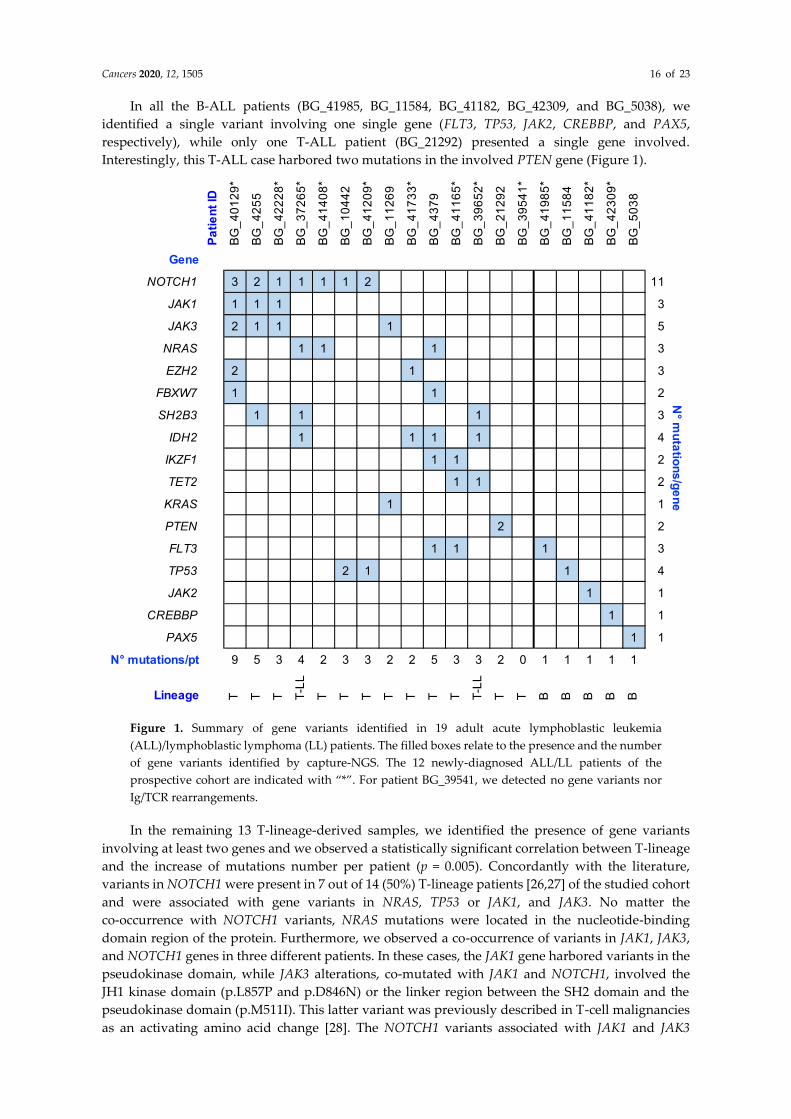
In all the B-ALL patients (BG_41985, BG_11584, BG_41182, BG_42309, and BG_5038), we
identified a single variant involving one single gene (FLT3, TP53, JAK2, CREBBP, and PAX5,
respectively), while only one T-ALL patient (BG_21292) presented a single gene involved.
Interestingly, this T-ALL case harbored two mutations in the involved PTEN gene (Figure 1).
Figure 1. Summary of gene variants identified in 19 adult acute lymphoblastic leukemia
(ALL)/lymphoblastic lymphoma (LL) patients. The filled boxes relate to the presence and the number
of gene variants identified by capture-NGS. The 12 newly-diagnosed ALL/LL patients of the
prospective cohort are indicated with “*”. For patient BG_39541, we detected no gene variants nor
Ig/TCR rearrangements.
In the remaining 13 T-lineage-derived samples, we identified the presence of gene variants
involving at least two genes and we observed a statistically significant correlation between T-lineage
and the increase of mutations number per patient (p = 0.005). Concordantly with the literature,
variants in NOTCH1 were present in 7 out of 14 (50%) T-lineage patients [26,27] of the studied cohort
and were associated with gene variants in NRAS, TP53 or JAK1, and JAK3. No matter the
co-occurrence with NOTCH1 variants, NRAS mutations were located in the nucleotide-binding
domain region of the protein. Furthermore, we observed a co-occurrence of variants in JAK1, JAK3,
and NOTCH1 genes in three different patients. In these cases, the JAK1 gene harbored variants in the
pseudokinase domain, while JAK3 alterations, co-mutated with JAK1 and NOTCH1, involved the
JH1 kinase domain (p.L857P and p.D846N) or the linker region between the SH2 domain and the
pseudokinase domain (p.M511I). This latter variant was previously described in T-cell malignancies
as an activating amino acid change [28]. The NOTCH1 variants associated with JAK1 and JAK3
Pa
tie
nt
ID
BG_40129*
BG_4255
BG_42228*
BG_37265*
BG_41408*
BG_10442
BG_41209*
BG_11269
BG_41733*
BG_4379
BG_41165*
BG_39652*
BG_21292
BG_39541*
BG_41985*
BG_11584
BG_41182*
BG_42309*
BG_5038
Gene
NOTCH1 3 2 1 1 1 1 2 11
JAK1 1 1 1 3
JAK3 2 1 1 1 5
NRAS 1 1 1 3
EZH2 2 1 3
FBXW7 1 1 2
SH2B3 1 1 1 3
IDH2 1 1 1 1 4
IKZF1 1 1 2
TET2 1 1 2
KRAS 1 1
PTEN 2 2
FLT3 1 1 1 3
TP53 2 1 1 4
JAK2 1 1
CREBBP 1 1
PAX5 1 1
N° mutations/pt 9 5 3 4 2 3 3 2 2 5 3 3 2 0 1 1 1 1 1
Lineage T T T T-LL
T T T T T T T T-LL
T T B B B B B
N° m
uta
tion
s/g
en
e

Cancers 2020, 12, 1505 17 of 23
mutations were located in the juxtamembrane heterodimerization (HD) N-terminal domain, but in
one of these cases (BG_40129) we identified two additional variants of NOTCH1 (p.P2514H and
p.A2463PfsTer14) involving the domains PEST (a peptide sequence rich in proline (P), glutamic acid
(E), serine (S), and threonine (T)) and TAD (transcriptional activation domain), respectively.
A statistical analysis performed on the co-occurrence of gene variants, although a limited
number of patients were molecularly profiled, demonstrated a statistically significant association
between JAK1 and JAK3 (p = 0.004) and JAK1 and NOTCH1 (p = 0.04). Moreover, in the T-LL studied
samples, we identified the association of IDH2 mutations and the presence of variants involving
SH2B3 (p = 0.09), while the co-occurrence of FLT3 and IKZF1 variants was observed in two out of
three T-ALL cases. We did not observe any association between gene variants and specific Ig/TCR
rearrangements. Of note, several variants (ranging from two to five) were identified in three patients
in whom no Ig/TCR rearrangements were recognized.
3. Discussion
In this work, we developed and validated a novel capture-based NGS panel useful for Ig/TCR
clonal marker screening and gene variant identification at diagnosis of ALL.
Although the identification process of leukemia-associated clonal Ig/TCR has been developed
and ruled during the last 20 years within European collaborative networks (EuroClonality and
EuroMRD, part of ESHLO foundation), this procedure remains laborious, time-consuming, and, in
some cases, ineffective. Therefore, MRD stratification using the gold standard ASO-qPCR approach
is nowadays feasible in about 95% of cases [12]. NGS technology has been demonstrated to be a
powerful tool for the identification of clonal rearrangements at disease presentation, resulting in
being less laborious and easier to perform than standard techniques [14–16]. Indeed, NGS proved
faster compared to multistep standard workflow, allowing an efficient patient-specific probe design
for timely MRD assessment, based on NGS-derived sequences.
Our capture-based NGS assay identified all clonal rearrangements previously found by
standard PCR and allowed the additional identification of clonal markers that are difficult to detect
by multiplex PCR [14]. It also provided supplementary information about bi-clonalities,
oligoclonalities, and uncommon rearrangements. This latter aspect is particularly important in
patients in whom no Ig/TCR clonalities can be identified by standard procedure. In our clinical
study, which started in the year 2000, this MRD-unknown cohort represented 16% of the enrolled
patients [5]. These patients could not have been followed during the treatment with an MRD
patient-specific probe, allowing a treatment based only on clinical features that often proved not
completely adequate to define the relapse risk of the patient. Therefore, we verified this new power
of the assay on patients prospectively enrolled into the active trial in which the standard method
failed. In this cohort for 9 out of 10 (90%) patients, a useful MRD assay has been designed on clonal
sequences provided by capture-based NGS assay, thus allowing a prospective risk-class assignment
(50%) or an MRD persistence identification. Moreover, NGS offers an important improvement in
terms of increased sensitivity, resulting in the possibility to identify at diagnosis minor leukemia
clones that could eventually be responsible for unexpected relapse. Indeed, the MRD monitoring by
ASO-qPCR requires the choice of one or two clonal rearrangements for follow-up measurements
[8,29], usually selected for better sensitivity and reflecting, in most cases, the abundance of the
leukemic clone among the cell population. Therefore, the ASO-qPCR approach necessarily leads to
losing track of minor subclones that might resist therapy and be responsible for disease resistance or
reappearance [8]. Given this and considering our results, it could be desirable to explore all the
possible rearrangements, including the uncommon ones not included into the standard PCR panel
or amplicon-based NGS panel recently published [14]. Nowadays, some but not all lineage-specific
rearrangements are investigated either in T- and B-lineage ALL with current methods. Indeed, it
could be suitable to test all possible rearrangements in any ALL in order to avoid the missing of
unexpected clonal markers.
A desirable approach to MRD assessment is also NGS-based. Some companies are offering a
full MRD evaluation by receiving diagnostic and follow-up samples. The development of a
Cancers 2020, 12, 1505 18 of 23
standardized amplicon-based, open access method is still ongoing within collaborative groups [21]
in which assay sensitivity and proper quantitation are the main issues. Upon resolving these issues,
one point of weakness still should be faced, namely, the search of only a few, highly represented
clones identified at diagnosis. This latter point would not be a problem for a capture-based method
in which all the rearrangements are entirely studied in each experiment.
The capture-based NGS approach also proved useful for saving the diagnostic material
collected at disease onset. Indeed, the amount of 1 μg of genomic DNA required for the complete
NGS panel for clonal marker screening, including the study of rare rearrangements and gene
profiling, is much lower than the amount of DNA needed for the amplicon-based techniques and the
genetic profiling performed separately. This consideration is pivotal, especially from the perspective
in which nowadays the MRD evaluation is still performed by ASO-qPCR, using serial dilutions of
the diagnostic specimen, with MRD quantification being expressed as logarithmic reduction
compared to the leukemia burden at disease onset. On the other hand, promising, newly described
techniques such as amplicon NGS or droplet digital PCR (ddPCR), aiming to achieve absolute
quantification of the residual leukemia level, are still under investigation and are not yet
standardized [30]. Additionally, the saving of diagnostic material is essential in hypocellular
specimens or when punctio sicca occurs and follow-up sample analysis is required for a long period
of time.
In our work, all B-ALL MRD-unknown patients, tested retrospectively as well prospectively,
showed at least one clonal rearrangement, while all patients still lacking a molecular marker in our
cohort belonged to the T-lineage group. For two of these patients, we can speculate that the tested
diagnostic sample was not adequate for the appropriate analysis. In fact, in one case (BG_4005), the
blasts percent content in the diagnostic sample was not available and we cannot exclude that the
specimen was not leukemia-representative. Likewise, the blasts percentage in the bone marrow
diagnostic sample for patient BG_39541 was around 5%, not sufficient to the proper leukemia
diagnosis according to the World Health Organization criteria. Even then, we can suppose that the
available diagnostic specimen was not correctly evaluable. However, the results thus far obtained
could suggest the definition of a small subgroup of very immature T-ALL in which Ig/TCR
rearrangements had not yet occurred, although further analysis is required to speculate the
maturation stage of leukemic lymphoblasts and to exclude technical limitations. Nevertheless, our
NGS approach allowed us to identify very small leukemia clones, representing less than 1% of the
global leukemia population; however, lacking a spike-in control to normalize clonal rearrangement
size compared to physiological rearrangements in normal lymphocytes, we could not exclude a
technical bias. Notably, the not-yet reported use as negative controls of HUVECs and mesenchymal
cells revealed no generation of artificial rearrangements during library generation or bioinformatic
data interpretation. In any case, it is important to note that the group of prospectively analyzed
MRD-unknown patients is characterized by a strong prevalence of T-lineage cases, while globally
the ratio between B- and T-ALL cases is unbalanced towards B-lineage occurrence.
The demanding process to obtain a mutational analysis of several genes that are prognostically
significant [23] or defining leukemia cases with specific features (Philadelphia-like ALL) [25]
prompted us to include a number of relevant genes into the capture design. This resulted in the
identification of several mutations in the studied ALL cases. In particular, all the B-ALL cases
harbored single gene variants involving CREBBP, FLT3, JAK2, PAX5, and TP53. On the contrary, in
all the T-ALL-derived samples, we observed the presence of at least two gene variants, with a
statistically significant increase of the mutations number in T-lineage patients compared to B-ALL
cases. On the basis of the data thus far obtained, we can conclude that for T-ALL patients the
inclusion of mutational status analysis of leukemia-associated genes could improve not only the
general knowledge about leukemogenesis, but it could have important consequences in clinical
practice. Indeed, we identified gene variants in three out of five T-ALL patients in which no
informative Ig/TCR rearrangements were recognized. Two of these variants were druggable
mutations (IDH2 p.R140Q) that could be considered for therapeutic intervention.
Cancers 2020, 12, 1505 19 of 23
Some limitations must be acknowledged. Our approach increases the complexity and the costs
to identify clonal markers. Capture-based technology is not yet standardized and it should be
externally validated before being routinely applied in clinical practice. Finally, this was a single
center study in which only a limited number of patients were evaluated. A clinical multicenter
validation was undertaken within the Euro-clonality NGS consortium [22].
4. Materials and Methods
4.1. Capture-Based NGS Panel Design
Two different capture-based NGS panels were designed using gene coordinates mapped on
GRCh38/hg38 gene assembly, targeting coding V, D, and J genes in the Ig/TCR loci for the
identification of D-J and V-D-J rearrangements (panel v1, 180 kb) and both Ig/TCR related regions, as
well as a selected group of genes of interest listed in Table S1 (panel v2, 350 kb). No differences were
detected between the two panels when applied to the same diagnostic sample (see Table S2, patient
BG_5038) and no reduction in our ability to identify V-D-J rearrangements was observed. The mean
of targeted aligned reads was 149,256 reads (min 228, max 276,500) for panel v1 and 2,391,017 reads
(min 1893, max 4,392,496) for panel v2. The minimum analyzed (i.e., rearranged) sequences per
sample should have been at least 1000.
4.2. Clinical Samples
Panel v1 was applied for the identification of clonal rearrangements on diagnostic bone marrow
material of 10 adult ALL patients (6 B- and 4 T-lineage, median age 33 years), including 2 human
umbilical vein endothelial cell (HUVEC) samples and 2 mesenchymal cord blood cell samples as
negative controls for Ig/TCR rearrangements. These patients have been formerly studied for
clonality assessment following the conventional EuroMRD guidelines [9,10], within the NILG-ALL
09/00 clinical trial (ClinicalTrials.gov identifier: NCT00358072) [5]. The same capture-based NGS
approach for clonality assessment was applied on 23 adult ALL patients (13 B- and 10 T-lineage,
median age 43 years) enrolled into the NILG-ALL 09/00 trial, in which the identification of a suitable
MRD molecular marker with standard procedure failed (MRD-unknown). The NGS panel v2 was
validated on diagnostic bone marrow samples of 7 adult ALL patients formerly evaluated for
Ig/TCR rearrangements and analyzed for TP53 mutational status by an independent NGS protocol (6
out of 7) [23]. The same panel was prospectively used on 12 newly diagnosed ALL patients (3 B-, 7
T-lineage, and 2 T-lymphoblastic lymphoma (T-LL); median age 40 years) missing suitable MRD
assays by standard approach. Patients’ clinical and biological characteristics are reported in Tables
S2 and S3. Since no oligoclonalities emerged from the analysis of capture-NGS performed on highly
diluted diagnostic samples in normal polyclonal mononuclear cells (serial dilutions according to
EuroMRD guidelines), we did not perform any specific tests to evaluate oligoclonalities in normal,
polyclonal lymphocytes.
4.3. DNA Extraction, Library Preparation, and Sequencing
Genomic DNA was isolated by Gentra Puregene Cell Kit (Qiagen, Hilden, Germany) or
Maxwell 16 Cell LEV DNA Purification Kit (Promega, Madison, WI, USA) from bone marrow
mononuclear cells obtained by Ficoll gradient (Cedarlane Laboratories Ltd., Burlington, ON,
Canada). For library preparation, we fragmented 600 ng–1 µg of genomic DNA to an average size of
600 bp by Covaris S220 Focused-Ultrasonicator (Covaris, Woburn, MA, USA) or Kapa enzymes
(Kapa Biosystems, Wilmington, MA, USA). Libraries in the pre-hybridization steps were prepared
with the KAPA HyperPlus Kit (Kapa Biosystems, Wilmington, MA, USA) and were then hybridized
to our custom-designed EZ SeqCap gene panel (Roche NimbleGen, Madison, WI, USA). The pool of
enriched libraries was sequenced on a MiSeq platform, using 300 bp paired-end reads (v3 chemistry,
Illumina, San Diego, CA, USA).

Cancers 2020, 12, 1505 20 of 23
4.4. NGS Data Analysis and Statistical Analysis
Bioinformatics data were processed separately. The identification of Ig/TCR rearrangements
was performed through the open-source application Vidjil (www.vidjil.org) [31,32]. Uncommon
V-D-J combinations were considered according to previously published literature [33–36]. Low
clonal representation was stated when a V-(D)-J annotation with unique CDR3 region sequence was
present with a clone size between 5% and 10% in the locus. Oligoclonality was defined when the
same V-(D)-J annotation was present with 3 or more different CDR3 regions each having a clonal
size ≥ 5%. The variant calling for gene alterations was performed by MiSeq Reporter Software, and
visualization of results was achieved by Illumina VariantStudio Data Analysis Software (Illumina,
San Diego, CA, USA) and the Integrative Genomics Viewer tool
(http://software.broadinstitute.org/software/igv/) [37,38]. For gene variant analysis, we considered
only variants with a read depth of at least 100 reads and VAF of 5%. Associations between different
gene variants and between gene variants and other characteristics were assessed using the Fisher
exact test for categorical variables and Wilcoxon rank-sum test for continuous variables. Two-sided
p-values <0.1 were considered significant. Statistical analysis was performed with R software
(version 3.6.2).
4.5. Clonality Assessment Validation
In all samples, we determined the Ig/TCR gene rearrangements at diagnosis by PCR,
homo-heteroduplex clonality assay, and Sanger sequencing according to BIOMED protocols [9,10].
The finger-print CDR3 regions of V-D-J gene rearrangements were detailed through the IMGT
V-Quest web application (www.IMGT.org). Additional rearrangements found by NGS were
validated by BIOMED-1/2 primers, if available, or by custom-made PCR primers. The design of the
latter was based on the Ig/TCR annotation derived from NGS sequences studied by IMGT; V-, D-,
and J-specific primers were designed by Primer Express Software v2.0 (Applied Biosystems, Foster
City, CA, USA) following BIOMED-1/2 recommendations. After PCR amplification, we performed
homo-heteroduplex clonality assay and Sanger sequencing. In case of weak PCR product in
heteroduplex analysis [10], we applied band elution and a second amplification before Sanger
sequencing.
5. Conclusions
In conclusion, our results indicated that the prospective application of NGS remarkably
simplified the assessment of clonality in adult ALL, providing additional information about the
mutational status of some ALL-related genes, with it now being the new standard to identify the
most informative Ig and TCR markers for the prospective evaluation of MRD.
Despite the long and extraordinarily successful history of Ig/TCR-based MRD evaluation for
childhood [1,2,39,40] and adult ALL patients [3–5,41], we have to keep in mind that Ig/TCR are
clonality markers and MRD evaluation reflects the leukemia chemo-sensitivity. No insight into
leukemia development or targeted therapy can be drawn. This approach was developed in times in
which the main information on ALL gene mutations was the broad gene involvement with low
frequency and there was no technical support for the study of such a large number of genes.
Nowadays, many new technologies have been developed that offer the possibility to analyze huge
genome sections.
Our NGS approach including Ig/TCR rearrangements as well as mutations in an increasable
number of genes could be a valid bridge toward a targeted MRD evaluation, allowing the
accumulation of a solid, comparative, and prospective experience.
Supplementary Materials: The following are available online at www.mdpi.com/2072-6694/12/6/1505/s1, Table
S1: List of genes included in the enhanced version of the capture-NGS panel (panel v2), Table S2: Clinical and
biological characteristics of adult ALL patients evaluated for the validation of the capture-NGS panel v1, Table
S3: Clinical and biological characteristics of adult ALL patients lacking an informative Ig/TCR molecular marker
for MRD evaluation (MRD-unknown).

Cancers 2020, 12, 1505 21 of 23
Author Contributions: R.C. and M.L.G.M. performed experiments, analyzed and interpreted data, and wrote
the manuscript. M.T. supervised experiments and collaborated in data interpretation. M.P. performed
experiments and collaborated in data interpretation. C.P. analyzed data and performed the statistical analysis.
T.I. and R.B. performed research and collected data. A.M. supervised the study, revised the manuscript, and
approved the final version. A.R. and O.S. designed the research, analyzed and interpreted data, supervised the
study, and wrote the manuscript. All authors revised the manuscript and approved the final version before
submission.
Funding: This work was supported by Associazione Italiana per la Ricerca sul Cancro (grant IG 2014 no. 16105),
Italy, and “Associazione PAOLO BELLI Onlus” - lotta alla leucemia e altre patologie, Bergamo, Italy.
Conflicts of Interest: The authors declare no conflict of interest.
References
1. Cavé, H.; van der Werff ten Bosch, J.; Suciu, S.; Guidal, C.; Waterkeyn, C.; Otten, J.; Bakkus, M.;
Thielemans, K.; Grandchamp, B.; Vilmer, E. Clinical significance of minimal residual disease in childhood
acute lymphoblastic leukemia. European Organization for Research and Treatment of Cancer--Childhood
Leukemia Cooperative Group. N. Engl. J. Med. 1998, 339, 591–598, doi:10.1056/NEJM199808273390904.
2. van Dongen, J.J.; Seriu, T.; Panzer-Grümayer, E.R.; Biondi, A.; Pongers-Willemse, M.J.; Corral, L.; Stolz, F.;
Schrappe, M.; Masera, G.; Kamps, W.A.; et al. Prognostic value of minimal residual disease in acute
lymphoblastic leukaemia in childhood. Lancet Lond. Engl. 1998, 352, 1731–1738,
doi:10.1016/S0140-6736(98)04058-6.
3. Szczepański, T.; Orfão, A.; van der Velden, V.H.; San Miguel, J.F.; van Dongen, J.J. Minimal residual
disease in leukaemia patients. Lancet Oncol. 2001, 2, 409–417.
4. Brüggemann, M.; Raff, T.; Flohr, T.; Gökbuget, N.; Nakao, M.; Droese, J.; Lüschen, S.; Pott, C.; Ritgen, M.;
Scheuring, U.; et al. Clinical significance of minimal residual disease quantification in adult patients with
standard-risk acute lymphoblastic leukemia. Blood 2006, 107, 1116–1123, doi:10.1182/blood-2005-07-2708.
5. Bassan, R.; Spinelli, O.; Oldani, E.; Intermesoli, T.; Tosi, M.; Peruta, B.; Rossi, G.; Borlenghi, E.; Pogliani,
E.M.; Terruzzi, E.; et al. Improved risk classification for risk-specific therapy based on the molecular study
of minimal residual disease (MRD) in adult acute lymphoblastic leukemia (ALL). Blood 2009, 113, 4153–
4162, doi:10.1182/blood-2008-11-185132.
6. Bassan, R.; Hoelzer, D. Modern therapy of acute lymphoblastic leukemia. J. Clin. Oncol. Off. J. Am. Soc. Clin.
Oncol. 2011, 29, 532–543, doi:10.1200/JCO.2010.30.1382.
7. Lussana, F.; Intermesoli, T.; Gianni, F.; Boschini, C.; Masciulli, A.; Spinelli, O.; Oldani, E.; Tosi, M.; Grassi,
A.; Parolini, M.; et al. Achieving Molecular Remission before Allogeneic Stem Cell Transplantation in
Adult Patients with Philadelphia Chromosome–Positive Acute Lymphoblastic Leukemia: Impact on
Relapse and Long-Term Outcome. Biol. Blood Marrow Transplant. 2016, 22, 1983–1987,
doi:10.1016/j.bbmt.2016.07.021.
8. van der Velden, V.H.J.; Hochhaus, A.; Cazzaniga, G.; Szczepanski, T.; Gabert, J.; van Dongen, J.J.M.
Detection of minimal residual disease in hematologic malignancies by real-time quantitative PCR:
Principles, approaches, and laboratory aspects. Leukemia 2003, 17, 1013–1034, doi:10.1038/sj.leu.2402922.
9. Pongers-Willemse, M.J.; Seriu, T.; Stolz, F.; d’Aniello, E.; Gameiro, P.; Pisa, P.; Gonzalez, M.; Bartram, C.R.;
Panzer-Grümayer, E.R.; Biondi, A.; et al. Primers and protocols for standardized detection of minimal
residual disease in acute lymphoblastic leukemia using immunoglobulin and T cell receptor gene
rearrangements and TAL1 deletions as PCR targets: Report of the BIOMED-1 CONCERTED ACTION:
Investigation of minimal residual disease in acute leukemia. Leukemia 1999, 13, 110–118.
10. van Dongen, J.J.M.; Langerak, A.W.; Brüggemann, M.; Evans, P. a. S.; Hummel, M.; Lavender, F.L.;
Delabesse, E.; Davi, F.; Schuuring, E.; García-Sanz, R.; et al. Design and standardization of PCR primers
and protocols for detection of clonal immunoglobulin and T-cell receptor gene recombinations in suspect
lymphoproliferations: Report of the BIOMED-2 Concerted Action BMH4-CT98-3936. Leukemia 2003, 17,
2257–2317, doi:10.1038/sj.leu.2403202.
11. van Dongen, J.J.M.; van der Velden, V.H.J.; Brüggemann, M.; Orfao, A. Minimal residual disease
diagnostics in acute lymphoblastic leukemia: Need for sensitive, fast, and standardized technologies. Blood
2015, 125, 3996–4009, doi:10.1182/blood-2015-03-580027.
12. Brüggemann, M.; Kotrova, M. Minimal residual disease in adult ALL: Technical aspects and implications
for correct clinical interpretation. Blood Adv. 2017, 1, 2456–2466, doi:10.1182/bloodadvances.2017009845.
Cancers 2020, 12, 1505 22 of 23
13. Langerak, A.W.; Brüggemann, M.; Davi, F.; Darzentas, N.; Dongen, J.J.M. van; Gonzalez, D.; Cazzaniga,
G.; Giudicelli, V.; Lefranc, M.-P.; Giraud, M.; et al. High-Throughput Immunogenetics for Clinical and
Research Applications in Immunohematology: Potential and Challenges. J. Immunol. 2017, 198, 3765–3774,
doi:10.4049/jimmunol.1602050.
14. Brüggemann, M.; Kotrová, M.; Knecht, H.; Bartram, J.; Boudjogrha, M.; Bystry, V.; Fazio, G.; Froňková, E.;
Giraud, M.; Grioni, A.; et al. Standardized next-generation sequencing of immunoglobulin and T-cell
receptor gene recombinations for MRD marker identification in acute lymphoblastic leukaemia; a
EuroClonality-NGS validation study. Leukemia 2019, doi:10.1038/s41375-019-0496-7.
15. Knecht, H.; Reigl, T.; Kotrová, M.; Appelt, F.; Stewart, P.; Bystry, V.; Krejci, A.; Grioni, A.; Pal, K.; Stranska,
K.; et al. Quality control and quantification in IG/TR next-generation sequencing marker identification:
Protocols and bioinformatic functionalities by EuroClonality-NGS. Leukemia 2019,
doi:10.1038/s41375-019-0499-4.
16. Scheijen, B.; Meijers, R.W.J.; Rijntjes, J.; van der Klift, M.Y.; Möbs, M.; Steinhilber, J.; Reigl, T.; van den
Brand, M.; Kotrová, M.; Ritter, J.-M.; et al. Next-generation sequencing of immunoglobulin gene
rearrangements for clonality assessment: A technical feasibility study by EuroClonality-NGS. Leukemia
2019, 33, 2227–2240, doi:10.1038/s41375-019-0508-7.
17. Faham, M.; Zheng, J.; Moorhead, M.; Carlton, V.E.H.; Stow, P.; Coustan-Smith, E.; Pui, C.-H.; Campana, D.
Deep-sequencing approach for minimal residual disease detection in acute lymphoblastic leukemia. Blood
2012, 120, 5173–5180, doi:10.1182/blood-2012-07-444042.
18. Ladetto, M.; Brüggemann, M.; Monitillo, L.; Ferrero, S.; Pepin, F.; Drandi, D.; Barbero, D.; Palumbo, A.;
Passera, R.; Boccadoro, M.; et al. Next-generation sequencing and real-time quantitative PCR for minimal
residual disease detection in B-cell disorders. Leukemia 2014, 28, 1299–1307, doi:10.1038/leu.2013.375.
19. Pulsipher, M.A.; Carlson, C.; Langholz, B.; Wall, D.A.; Schultz, K.R.; Bunin, N.; Kirsch, I.; Gastier-Foster,
J.M.; Borowitz, M.; Desmarais, C.; et al. IgH-V(D)J NGS-MRD measurement pre- and early
post-allotransplant defines very low- and very high-risk ALL patients. Blood 2015, 125, 3501–3508,
doi:10.1182/blood-2014-12-615757.
20. Wood, B.; Wu, D.; Crossley, B.; Dai, Y.; Williamson, D.; Gawad, C.; Borowitz, M.J.; Devidas, M.; Maloney,
K.W.; Larsen, E.; et al. Measurable residual disease detection by high-throughput sequencing improves
risk stratification for pediatric B-ALL. Blood 2018, 131, 1350–1359, doi:10.1182/blood-2017-09-806521.
21. Sánchez, R.; Ayala, R.; Martínez-López, J. Minimal Residual Disease Monitoring with Next-Generation
Sequencing Methodologies in Hematological Malignancies. Int. J. Mol. Sci. 2019, 20, 2832,
doi:10.3390/ijms20112832.
22. Wren, D.; Walker, B.A.; Brüggemann, M.; Catherwood, M.A.; Pott, C.; Stamatopoulos, K.; Langerak, A.W.;
Gonzalez, D.; EuroClonality-NGS consortium Comprehensive translocation and clonality detection in
lymphoproliferative disorders by next-generation sequencing. Haematologica 2017, 102, e57–e60,
doi:10.3324/haematol.2016.155424.
23. Salmoiraghi, S.; Montalvo, M.L.G.; Ubiali, G.; Tosi, M.; Peruta, B.; Zanghi, P.; Oldani, E.; Boschini, C.;
Kohlmann, A.; Bungaro, S.; et al. Mutations of TP53 gene in adult acute lymphoblastic leukemia at
diagnosis do not affect the achievement of hematologic response but correlate with early relapse and very
poor survival. Haematologica 2016, 101, e245–e248, doi:10.3324/haematol.2015.137059.
24. Mullighan, C.G. The molecular genetic makeup of acute lymphoblastic leukemia. Hematol. Am. Soc.
Hematol. Educ. Program 2012, 2012, 389–396, doi:10.1182/asheducation-2012.1.389.
25. Roberts, K.G.; Gu, Z.; Payne-Turner, D.; McCastlain, K.; Harvey, R.C.; Chen, I.-M.; Pei, D.; Iacobucci, I.;
Valentine, M.; Pounds, S.B.; et al. High Frequency and Poor Outcome of Philadelphia Chromosome-Like
Acute Lymphoblastic Leukemia in Adults. J. Clin. Oncol. Off. J. Am. Soc. Clin. Oncol. 2017, 35, 394–401,
doi:10.1200/JCO.2016.69.0073.
26. Mansour, M.R.; Sulis, M.L.; Duke, V.; Foroni, L.; Jenkinson, S.; Koo, K.; Allen, C.G.; Gale, R.E.; Buck, G.;
Richards, S.; et al. Prognostic implications of NOTCH1 and FBXW7 mutations in adults with T-cell acute
lymphoblastic leukemia treated on the MRC UKALLXII/ECOG E2993 protocol. J. Clin. Oncol. Off. J. Am.
Soc. Clin. Oncol. 2009, 27, 4352–4356, doi:10.1200/JCO.2009.22.0996.
27. Mansour, M.R.; Duke, V.; Foroni, L.; Patel, B.; Allen, C.G.; Ancliff, P.J.; Gale, R.E.; Linch, D.C. Notch-1
mutations are secondary events in some patients with T-cell acute lymphoblastic leukemia. Clin. Cancer
Res. Off. J. Am. Assoc. Cancer Res. 2007, 13, 6964–6969, doi:10.1158/1078-0432.CCR-07-1474.
Cancers 2020, 12, 1505 23 of 23
28. Degryse, S.; Bornschein, S.; de Bock, C.E.; Leroy, E.; Vanden Bempt, M.; Demeyer, S.; Jacobs, K.; Geerdens,
E.; Gielen, O.; Soulier, J.; et al. Mutant JAK3 signaling is increased by loss of wild-type JAK3 or by
acquisition of secondary JAK3 mutations in T-ALL. Blood 2018, 131, 421–425,
doi:10.1182/blood-2017-07-797597.
29. van der Velden, V.H.J.; Cazzaniga, G.; Schrauder, A.; Hancock, J.; Bader, P.; Panzer-Grumayer, E.R.; Flohr,
T.; Sutton, R.; Cave, H.; Madsen, H.O.; et al. Analysis of minimal residual disease by Ig/TCR gene
rearrangements: Guidelines for interpretation of real-time quantitative PCR data. Leukemia 2007, 21, 604–
611, doi:10.1038/sj.leu.2404586.
30. Kotrova, M.; Trka, J.; Kneba, M.; Brüggemann, M. Is Next-Generation Sequencing the way to go for
Residual Disease Monitoring in Acute Lymphoblastic Leukemia? Mol. Diagn. Ther. 2017, 21, 481–492,
doi:10.1007/s40291-017-0277-9.
31. Giraud, M.; Salson, M.; Duez, M.; Villenet, C.; Quief, S.; Caillault, A.; Grardel, N.; Roumier, C.;
Preudhomme, C.; Figeac, M. Fast multiclonal clusterization of V(D)J recombinations from
high-throughput sequencing. BMC Genom. 2014, 15, 409, doi:10.1186/1471-2164-15-409.
32. Duez, M.; Giraud, M.; Herbert, R.; Rocher, T.; Salson, M.; Thonier, F. Vidjil: A Web Platform for Analysis of
High-Throughput Repertoire Sequencing. PLoS ONE 2016, 11, e0166126, doi:10.1371/journal.pone.0166126.
33. Szczepański, T.; Beishuizen, A.; Pongers-Willemse, M.J.; Hählen, K.; Van Wering, E.R.; Wijkhuijs, A.J.;
Tibbe, G.J.; De Bruijn, M.A.; Van Dongen, J.J. Cross-lineage T cell receptor gene rearrangements occur in
more than ninety percent of childhood precursor-B acute lymphoblastic leukemias: Alternative PCR
targets for detection of minimal residual disease. Leukemia 1999, 13, 196–205, doi:10.1038/sj.leu.2401277.
34. Szczepański, T.; Pongers-Willemse, M.J.; Langerak, A.W.; van Dongen, J.J. Unusual immunoglobulin and
T-cell receptor gene rearrangement patterns in acute lymphoblastic leukemias. Curr. Top. Microbiol.
Immunol. 1999, 246, 205–213; discussion 214-215, doi:10.1007/978-3-642-60162-0_26.
35. Szczepański, T.; Flohr, T.; van der Velden, V.H.J.; Bartram, C.R.; van Dongen, J.J.M. Molecular monitoring
of residual disease using antigen receptor genes in childhood acute lymphoblastic leukaemia. Best Pract.
Res. Clin. Haematol. 2002, 15, 37–57, doi:10.1053/beha.2002.0184.
36. Szczepański, T.; van der Velden, V.H.J.; Hoogeveen, P.G.; de Bie, M.; Jacobs, D.C.H.; van Wering, E.R.; van
Dongen, J.J.M. Vδ2-Jα rearrangements are frequent in precursor-B–acute lymphoblastic leukemia but rare
in normal lymphoid cells. Blood 2004, 103, 3798–3804, doi:10.1182/blood-2003-08-2952.
37. Robinson, J.T.; Thorvaldsdóttir, H.; Winckler, W.; Guttman, M.; Lander, E.S.; Getz, G.; Mesirov, J.P.
Integrative genomics viewer. Nat. Biotechnol. 2011, 29, 24–26, doi:10.1038/nbt.1754.
38. Thorvaldsdóttir, H.; Robinson, J.T.; Mesirov, J.P. Integrative Genomics Viewer (IGV): High-performance
genomics data visualization and exploration. Brief. Bioinform. 2013, 14, 178–192, doi:10.1093/bib/bbs017.
39. Flohr, T.; Schrauder, A.; Cazzaniga, G.; Panzer-Grümayer, R.; van der Velden, V.; Fischer, S.; Stanulla, M.;
Basso, G.; Niggli, F.K.; Schäfer, B.W.; et al. Minimal residual disease-directed risk stratification using
real-time quantitative PCR analysis of immunoglobulin and T-cell receptor gene rearrangements in the
international multicenter trial AIEOP-BFM ALL 2000 for childhood acute lymphoblastic leukemia.
Leukemia 2008, 22, 771–782, doi:10.1038/leu.2008.5.
40. Basso, G.; Veltroni, M.; Valsecchi, M.G.; Dworzak, M.N.; Ratei, R.; Silvestri, D.; Benetello, A.; Buldini, B.;
Maglia, O.; Masera, G.; et al. Risk of Relapse of Childhood Acute Lymphoblastic Leukemia Is Predicted By
Flow Cytometric Measurement of Residual Disease on Day 15 Bone Marrow. J. Clin. Oncol. 2009, 27, 5168–
5174, doi:10.1200/JCO.2008.20.8934.
41. Ribera, J.-M.; Oriol, A.; Morgades, M.; Montesinos, P.; Sarrà, J.; González-Campos, J.; Brunet, S.; Tormo,
M.; Fernández-Abellán, P.; Guàrdia, R.; et al. Treatment of High-Risk Philadelphia Chromosome–
Negative Acute Lymphoblastic Leukemia in Adolescents and Adults According to Early Cytologic
Response and Minimal Residual Disease after Consolidation Assessed by Flow Cytometry: Final Results
of the PETHEMA ALL-AR-03 Trial. J. Clin. Oncol. 2014, doi:10.1200/JCO.2013.52.2425.
© 2020 by the authors. Licensee MDPI, Basel, Switzerland. This article is an open access
article distributed under the terms and conditions of the Creative Commons Attribution
(CC BY) license (http://creativecommons.org/licenses/by/4.0/).