Captopril and telmisartan ameliorate cisplatin … Journal of Scientific and Research Publications,...
13
International Journal of Scientific and Research Publications, Volume 6, Issue 1, January 2016 408 ISSN 2250-3153 www.ijsrp.org Captopril and telmisartan ameliorate cisplatin-induced testicular damage in rats via anti-inflammatory and antioxidant pathways Ahmed H. Eid * , Noha F. Abdelkader ** , Ola M. Abd El-Raouf * , Hala M. Fawzy * , Ezz-El-Din S. El-Denshary ** * Department of Pharmacology, National Organization for Drug Control and Research (NODCAR), Giza, Egypt ** Department of Pharmacology and Toxicology, Faculty of Pharmacy, Cairo University, Cairo, Egypt. Abstract- Despite the prevalent clinical applications of cisplatin, serious toxic side effects comprising reproductive toxicity confine its therapeutic efficacy. Thus, the current study explored the possible protective effect of captopril and telmisartan against cisplatin-induced testicular damage in rats. Captopril (100 mg/kg) and telmisartan (10 mg/kg) were orally administered for 15 days, whereas cisplatin (10 mg/kg; i.p.) was injected as a single dose at the 12 th day to induce testicular damage in adult male Sprague-Dawley rats. Cisplatin prominently decreased reproductive organs weights, sperm count, sperm motility, and increased sperm abnormalities, along with histopathological damage of testicular tissues. In addition, it resulted in a significant decline in serum testosterone as well as testicular enzymatic and non-enzymatic antioxidants levels (superoxide dismutase, catalase, glutathione peroxides, and reduced glutathione), parallel to a remarkable elevation in testicular content of malondialdehyde, nitric oxide, tumor necrosis factor-α, and nuclear factor-kappa B. Treatment with captopril or telmisartan markedly attenuated cisplatin-induced injury by suppression of oxidative/nitrosative stress and inflammation, amendment of antioxidant defenses, as well as improvement of steroidogenesis, spermatogenesis and testicular histological features. This study suggests a novel therapeutic application for captopril and telmisartan as protective agents against cisplatin-induced testicular toxicity through their promising anti-inflammatory and antioxidant capacities. Index Terms- Captopril, cisplatin, oxidative stress, telmisartan, testicular damage. I. INTRODUCTION isplatin is an extremely effective anticancer drug for the treatment of diverse types of solid tumors (Amin and Buratovich, 2009). Despite the improvement in quality of life of cancer patients; the use of cisplatin was restricted clinically by its major side effects including testicular toxicity (Beytur et al., 2012, Fung and Vaughn, 2011). In adult men, fertility can be preserved by spermatozoa cryopreservation and intracytoplasmic sperm injection, however, these methods are not feasible options for pre-pubertal patients (Rezvanfar et al., 2013). Besides, in adults, the freezing and thawing of semen can reduce the sperm quality (Maines et al., 1990). Interestingly, growing evidences revealed that combination therapy can be beneficial to overcome this special reproductive toxicity (Narayana et al., 2012, Ahmed et al., 2011). Male reproductive toxicity rises from targeting rapidly growing testicular cell types such as leydig, sertoli, and germ cells (Boekelheide, 2005). Cisplatin adversely impairs non target tissues via increased free radical generation and depletion of antioxidants; which results in inhibition of protein synthesis and DNA damage (Fung and Vaughn, 2011, Ilbey et al., 2009a). Growing evidence verifies that activation of the renin angiotensin system (RAS) is largely implicated in cisplatin-induced tissue injury (El-Sayed et al., 2008, Saleh et al., 2009) where cisplatin amplifies RAS components level both experimentally (El-Sayed et al., 2008, Okui et al., 2012) and clinically (Kurt et al., 1999). Indeed, angiotensin II, the chief component of RAS, induces tissue oxidative stress and apoptosis (Cai et al., 2003, Wolf, 2000). Besides, it triggers inflammation via upregulation of pro- inflammatory transcription factors, which results in a vast array of proinflammatory cytokines that further injures the tissues (Hayashi et al., 2010). Therefore, the current study was conducted to investigate the efficacy of RAS blockade against cisplatin-induced testicular damage. Captopril is an angiotensin converting enzyme inhibitor that reduces circulating and tissue levels of angiotensin II. Moreover, it is a powerful thiol-containing antioxidant (Liu et al., 2007) that possesses anti-inflammatory potential (Amirshahrokhi et al., 2010). Captopril is widely used for the treatment of hypertension, congestive heart failure, and diabetic nephropathy (Cohen et al., 1996). In context, telmisartan is a highly selective angiotensin II type-1 receptor blocker which is approved for the treatment of hypertension. It is endowed with potent anti-inflammatory, antioxidant and anti- apoptotic effects (Fouad et al., 2010). Interestingly, recent studies revealed that both drugs ameliorated chemotherapy-induced cardiotoxicity and nephrotoxicity (Abd-Ellah, 2012, Cadeddu et al., 2010, Ibrahim et al., 2009) as well as cadmium-induced testicular toxicity (Fouad and Jresat, 2013). Herein, the potential protective effects of captopril and telmisartan against testicular toxicity induced by cisplatin in male rats were investigated by the assessment of hormonal and spermatological changes, oxidative stress parameters, inflammatory markers, and the testicular histopathological alterations. II. MATERIALS AND METHODS Animals Adult male Sprague-Dawley rats; weighing 240-280 g, were obtained from the breeding colony of the National Organization for Drug Control and Research (NODCAR), Giza, Egypt. C
Transcript of Captopril and telmisartan ameliorate cisplatin … Journal of Scientific and Research Publications,...

International Journal of Scientific and Research Publications, Volume 6, Issue 1, January 2016 408 ISSN 2250-3153
www.ijsrp.org
Captopril and telmisartan ameliorate cisplatin-induced testicular damage in rats via anti-inflammatory and
antioxidant pathways Ahmed H. Eid*, Noha F. Abdelkader**, Ola M. Abd El-Raouf*, Hala M. Fawzy*, Ezz-El-Din S. El-Denshary**
*Department of Pharmacology, National Organization for Drug Control and Research (NODCAR), Giza, Egypt
**Department of Pharmacology and Toxicology, Faculty of Pharmacy, Cairo University, Cairo, Egypt. Abstract- Despite the prevalent clinical applications of cisplatin, serious toxic side effects comprising reproductive toxicity confine its therapeutic efficacy. Thus, the current study explored the possible protective effect of captopril and telmisartan against cisplatin-induced testicular damage in rats. Captopril (100 mg/kg) and telmisartan (10 mg/kg) were orally administered for 15 days, whereas cisplatin (10 mg/kg; i.p.) was injected as a single dose at the 12th day to induce testicular damage in adult male Sprague-Dawley rats. Cisplatin prominently decreased reproductive organs weights, sperm count, sperm motility, and increased sperm abnormalities, along with histopathological damage of testicular tissues. In addition, it resulted in a significant decline in serum testosterone as well as testicular enzymatic and non-enzymatic antioxidants levels (superoxide dismutase, catalase, glutathione peroxides, and reduced glutathione), parallel to a remarkable elevation in testicular content of malondialdehyde, nitric oxide, tumor necrosis factor-α, and nuclear factor-kappa B. Treatment with captopril or telmisartan markedly attenuated cisplatin-induced injury by suppression of oxidative/nitrosative stress and inflammation, amendment of antioxidant defenses, as well as improvement of steroidogenesis, spermatogenesis and testicular histological features. This study suggests a novel therapeutic application for captopril and telmisartan as protective agents against cisplatin-induced testicular toxicity through their promising anti-inflammatory and antioxidant capacities. Index Terms- Captopril, cisplatin, oxidative stress, telmisartan, testicular damage.
I. INTRODUCTION isplatin is an extremely effective anticancer drug for the treatment of diverse types of solid tumors (Amin and
Buratovich, 2009). Despite the improvement in quality of life of cancer patients; the use of cisplatin was restricted clinically by its major side effects including testicular toxicity (Beytur et al., 2012, Fung and Vaughn, 2011). In adult men, fertility can be preserved by spermatozoa cryopreservation and intracytoplasmic sperm injection, however, these methods are not feasible options for pre-pubertal patients (Rezvanfar et al., 2013). Besides, in adults, the freezing and thawing of semen can reduce the sperm quality (Maines et al., 1990). Interestingly, growing evidences revealed that combination therapy can be beneficial to overcome this special reproductive toxicity (Narayana et al., 2012, Ahmed et al., 2011).
Male reproductive toxicity rises from targeting rapidly growing testicular cell types such as leydig, sertoli, and germ cells (Boekelheide, 2005). Cisplatin adversely impairs non target tissues via increased free radical generation and depletion of antioxidants; which results in inhibition of protein synthesis and DNA damage (Fung and Vaughn, 2011, Ilbey et al., 2009a). Growing evidence verifies that activation of the renin angiotensin system (RAS) is largely implicated in cisplatin-induced tissue injury (El-Sayed et al., 2008, Saleh et al., 2009) where cisplatin amplifies RAS components level both experimentally (El-Sayed et al., 2008, Okui et al., 2012) and clinically (Kurt et al., 1999). Indeed, angiotensin II, the chief component of RAS, induces tissue oxidative stress and apoptosis (Cai et al., 2003, Wolf, 2000). Besides, it triggers inflammation via upregulation of pro-inflammatory transcription factors, which results in a vast array of proinflammatory cytokines that further injures the tissues (Hayashi et al., 2010). Therefore, the current study was conducted to investigate the efficacy of RAS blockade against cisplatin-induced testicular damage. Captopril is an angiotensin converting enzyme inhibitor that reduces circulating and tissue levels of angiotensin II. Moreover, it is a powerful thiol-containing antioxidant (Liu et al., 2007) that possesses anti-inflammatory potential (Amirshahrokhi et al., 2010). Captopril is widely used for the treatment of hypertension, congestive heart failure, and diabetic nephropathy (Cohen et al., 1996). In context, telmisartan is a highly selective angiotensin II type-1 receptor blocker which is approved for the treatment of hypertension. It is endowed with potent anti-inflammatory, antioxidant and anti-apoptotic effects (Fouad et al., 2010). Interestingly, recent studies revealed that both drugs ameliorated chemotherapy-induced cardiotoxicity and nephrotoxicity (Abd-Ellah, 2012, Cadeddu et al., 2010, Ibrahim et al., 2009) as well as cadmium-induced testicular toxicity (Fouad and Jresat, 2013). Herein, the potential protective effects of captopril and telmisartan against testicular toxicity induced by cisplatin in male rats were investigated by the assessment of hormonal and spermatological changes, oxidative stress parameters, inflammatory markers, and the testicular histopathological alterations.
II. MATERIALS AND METHODS Animals Adult male Sprague-Dawley rats; weighing 240-280 g, were obtained from the breeding colony of the National Organization for Drug Control and Research (NODCAR), Giza, Egypt.
C

International Journal of Scientific and Research Publications, Volume 6, Issue 1, January 2016 409 ISSN 2250-3153
www.ijsrp.org
Animals were accommodated under controlled environmental conditions (23±2°C temperature, 60±10% humidity, 12/12 h light/dark cycle) and were allowed standard chow diet and water ad libitum. The investigation complies with the Guide for the Care and Use of Laboratory Animals (NIH Publication No. 85-23, revised 1996) and was approved by the Ethics Committee of Faculty of Pharmacy, Cairo University, Cairo, Egypt (Permit Number: PT 922). Chemicals and drugs Cisplatin was purchased from Mylan (Saint-Priest, France). Captopril and telmisartan were obtained from Multipharma pharmaceutical and Sigma pharmaceutical industries; respectively (Cairo, Egypt). All other chemicals were of the highest purity and analytical grade. Experimental design Seventy rats were randomly divided into seven groups (10 rats each). Group I served as a control. Rats of group II received orally 0.5 % carboxymethyl cellulose (10 ml/kg) for 15 days. Animals of groups III & IV received orally captopril dissolved in water (100 mg/kg) (Fasihi et al., 2012) and telmisartan suspended in 0.5 % carboxymethyl cellulose (10 mg/kg) (Fouad and Jresat, 2013) for 15 days; respectively. Those of groups V, VI, and VII received a single intraperitoneal injection of cisplatin (10 mg/kg) on the 12th day to induce testicular toxicity (Longo et al., 2011); whereas, rats of groups VI and VII were treated with captopril and telmisartan; respectively, as previously described. Four days after cisplatin injection, rats were weighed, anesthetized, and blood samples were collected and centrifuged at 3000 xg for 10 minutes for serum separation to estimate testosterone level. Thereafter, animals were euthanized and the reproductive organs were immediately removed, cleaned from the adhering tissue, and weighed. The epididymal content for each rat was instantly collected to examine the spermatological parameters. Furthermore, the left testis was fixed in 10% formalin for histological examination. Whereas, the right testis was decapsulated, homogenized in 0.05 M potassium phosphate buffer (pH 7.4), and processed for the estimation of oxidative and inflammatory biomarkers. All samples were stored at -80°C until analysis. Spermatological Examination The epididimal content of each rat was promptly collected for the estimation of sperm count, motility as well as abnormalities as previously described (Bearden and Fluquary, 1980). Estimation of serum testosterone level Serum testosterone level was estimated according to manufacturer’s instructions using rat Testosterone EIA ELISA Kit (Enzo life sciences, San Diego, CA, USA). Estimation of oxidative/nitrosative and inflammatory biomarkers in testicular homogenate Testicular homogenate was divided into 3 aliquots, where the first one was used for estimation of malondialdehyde (MDA) (Deniz et al., 1997). The second aliquot was deproteinized with 5% sulfosalicylic acid, centrifuged at 1000 xg for 15 minutes and the obtained supernatant was used for the estimation of reduced glutathione (GSH) (Beutler et al., 1963). The third one was centrifuged at 4000 rpm for 15 minutes at 4°C and the resultant supernatant was used for the assay of superoxide dismutase (SOD), catalase (CAT), glutathione peroxidase (GPx), and nitric
oxide (NO) according to manufacturer’s prescripts using the corresponding Biodiagnostic colorimetric kits (Cairo, Egypt), while tumor necrosis factor (TNF)-α was assayed using rat TNF-α Elisa Kit (Enzo life sciences, San Diego, CA, USA). Histological examination Light microscopic evaluation The fixed testis was processed for paraffin embedding and 4 μm sections were prepared. Testicular sections were stained with haematoxylin and eosin (H&E) and examined using a light microscope. Qualitative histopathological damage in the seminiferous tubules was graded according to the severity of degenerative findings: (−) no obvious damage, (+) fewer than 25% of seminiferous tubules affected (mild), (++) 25–50% of seminiferous tubules affected (moderate), and (+++) over 50% of seminiferous tubules affected (severe) (Rezvanfar et al., 2013). Histopathological examination of testes was interpreted by an experienced observer who was blind to the sample identity to avoid any bias. Immunohistochemical analysis of testicular nuclear factor-kappa B (NF-κB) Paraffinized testicular sections were rehydrated in xylene and graded ethanol solutions. Samples were heated in citrate buffer (pH 6) for 20 minutes, cooled, and thereafter incubated with primary polyclonal rabbit anti-NF-κB antibody (1:200; Invitrogen, Carlsbad, CA, USA) overnight at 4°C. Sections were washed with phosphate buffered saline, and incubated for 30 min at 37°C with biotinylated secondary antibody, and then with Avidin DH and biotinylated horseradish peroxidase H complex according to Elite ABC kit instructions (Vector Laboratories Inc., Burlingame, CA, USA). After another wash with phosphate buffered saline, the reaction was revealed by diaminobenzidine tetrahydrochloride (DAB Substrate Kit, Vector Laboratories Inc., Burlingame, CA, USA) and the sections were counterstained with hematoxylin, dehydrated, and cleared in xylene then cover slipped for light microscopic examination. Statistical analysis Data were expressed as means ± S.E.M. Results were analyzed using one way analysis of variance test (One-way ANOVA) followed by Tukey-Kramer multiple comparison test. Statistical analysis was performed using GraphPad Prism software version 5 (San Diego, CA, USA); a probability level of less than 0.05 was accepted as statistically significant.
III. RESULTS
There was no change in any of the tested parameters between the control rats and those which received carboxymethyl cellulose, captopril, and telmisartan alone (at the selected doses). Alterations in body and reproductive organs weight (Table 1): A significant decrease in body as well as relative reproductive organs weight was observed as a result of cisplatin administration compared to control group. Cisplatin decreased body weight to 87.7% of control rats, an effect that was not alleviated by captopril nor telmisartan treatment. The weight of right and left testis, cauda epididymis, prostate and seminal vesicle were significantly decreased after cisplatin exposure to 82.9%, 85.2%, 76.6%, 73.7% and 80.7 % of the control group; respectively, while all of them were normalized by captopril

International Journal of Scientific and Research Publications, Volume 6, Issue 1, January 2016 410 ISSN 2250-3153
www.ijsrp.org
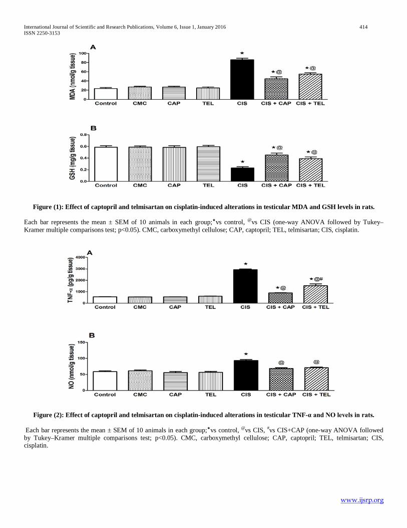
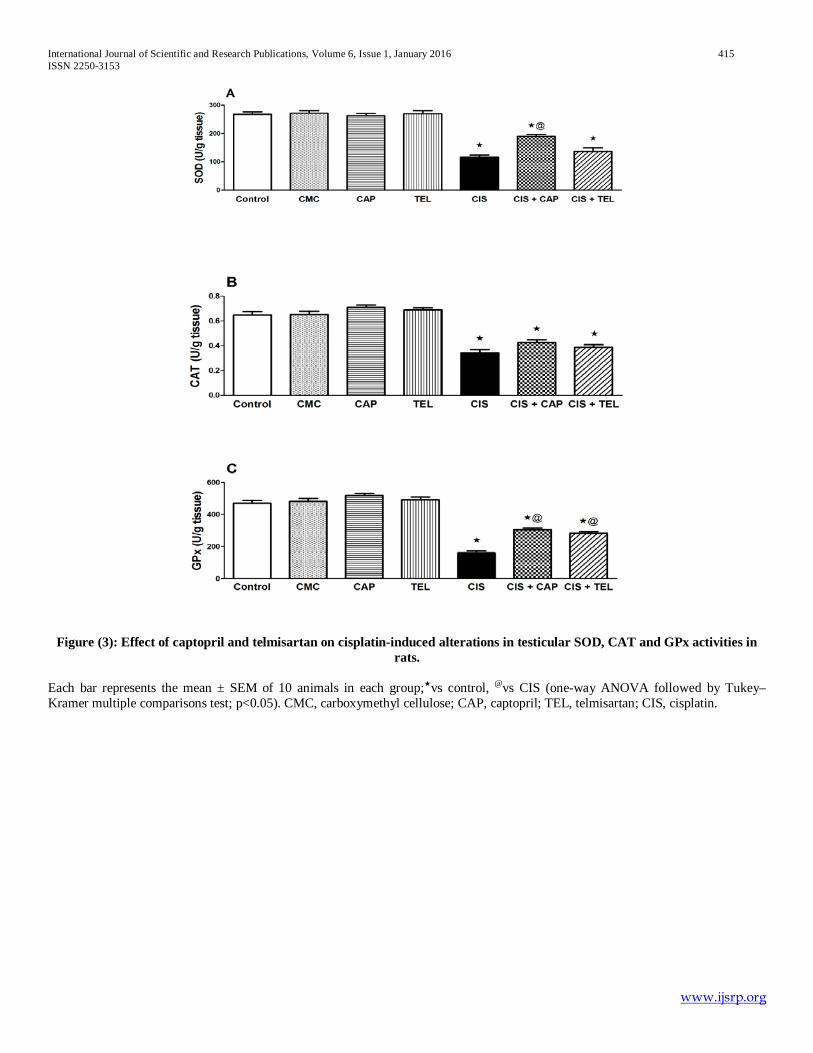
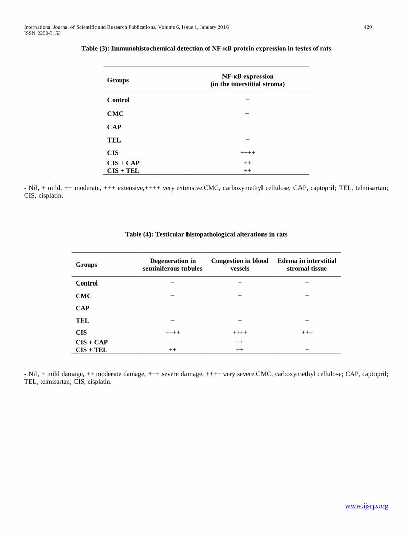
administration. Similarly, telmisartan restored reproductive organs weight except for cauda epididymis. Effects on spermatological parameters and serum testosterone level (Table 2): Cisplatin-treated rats showed a substantial decrease in sperm count and motility % almost by half and an increase in sperm abnormalities % to 2.7 folds compared to the corresponding values of the control group. Furthermore, serum testosterone level was markedly declined to 73.3% of the control group. Treatment with captopril alleviated the changes in motility %, sperm abnormalities %, and serum testosterone level by 32.5%, 35.6%, and 19.7%, respectively as related to the cisplatin-treated group together with a normalization of the sperm count. Meanwhile, telmisartan administration substantially raised sperm count, motility %, and serum testosterone level to 1.54, 1.25 and 1.20 folds; correspondingly, and reduced sperm abnormalities by 33.86% versus the cisplatin group. Effects on testicular oxidative/nitrosative stress and inflammatory biomarkers (Fig. 1-3): Cisplatin injection markedly amplified testicular MDA, NO, and TNF-α contents by 3.6, 1.6 and 5.2 folds, respectively; whereas it resulted in a decline in testicular GSH content along with SOD, CAT and GPx activities by 60.47%, 56.41%, 47.10% and 65.90%, respectively as compared to the control group. However, captopril and telmisartan treatment noticeably reversed such alterations except for CAT activity which was not affected by both drugs and SOD activity that was amended by captopril treatment only. In addition, the effect of captopril was more pronounced than telmisartan in terms of TNF-α decline. Effects on testicular NF-κB protein expression (Fig. 4 & Table 3): The immunohistochemical examination of testicular NF-κB protein expression displayed an enhanced expression confined within the interstitial stroma adjacent to the basement membrane of degenerated tubules in testes of cisplatin-treated rats (Fig. 4E), an effect that alleviated by management with captopril and telmisartan (Fig. 4F & G). Effects on histopathological damage in testicular tissues (Fig. 5 & Table 4): Testicular sections of the cisplatin group revealed atypical morphological features manifested as massive degeneration in the seminiferous tubules, shrinkage in germ cell layers and disruption of spermatogenesis, interstitial edema, congestion in blood vessels and replacement of the interstitial stroma with homogeneous eosinophilic material (Fig. 5E). Captopril treatment normalized these histological abnormalities and amended spermatogenesis, though a slight congestion in blood vessels of the interstitisal stroma was still observed (Fig. 5F). Likewise, telmisartan attenuated the histopathological changes and protected the seminiferous tubules albeit less pronounced than captopril (Fig. 5G).
IV. DISCUSSION
Testicular dysfunction and infertility are serious complications that may impact the clinical effectiveness of cisplatin. Thus, it seems imperative to search for agents that can protect against testicular toxicity whenever cisplatin chemotherapy is employed. Interestingly, preceding clinical studies revealed that addition of angiotensin converting enzyme
inhibitors and angiotensin receptor blockers to platinum-based first-line chemotherapy may contribute to prolonged survival in patients with advanced lung and gastric cancer (Kim et al., 2012, Wilop et al., 2009). Moreover, the combination of these drugs with chemotherapy markedly delayed tumor growth (Williams et al., 2005, Hosseinimehr, 2014), thus much attention has been directed to their protective effects against chemotherapy-induced cardiotoxicity and nephrotoxicity (Abd-Ellah, 2012, Arozal et al., 2010, El-Sayed et al., 2008, Hrenak et al., 2013, Malik et al., 2015). However, to the best of our knowledge, their protective effects against cisplatin-induced testicular toxicity have not been investigated. The current study highlights the protective actions of captopril and telmisartan against cisplatin-induced testicular toxicity in rats as verified by the restoration of testicular architecture, enhancement of steroidogenesis, preservation of spermatogenesis, modulation of the inflammatory reaction along with suppression of oxidative/nitrosative stress. In the current study, the observed reduction in relative reproductive organs weights in cisplatin-treated rats establishes the toxic potential of cisplatin on the reproductive system as previously documented by (Atessahin et al., 2006, Ilbey et al., 2009a). It was declared that this weight reduction is related to the oxidative damage induced by cisplatin (Adejuwon et al., 2015). Such effect was further accompanied by degeneration of seminiferous tubules with disruption of spermatogenesis, sperm dysfunction, besides a decline in the testicular function biomarker, i.e. testosterone. Many investigations confirmed our results (Adejuwon et al., 2015, Amin et al., 2012, Rezvanfar et al., 2013, Salem et al., 2012). Herein, captopril and telmisartan treatment almost restored the reduction in reproductive organs weights induced by cisplatin, which is consistent with a previous study (Kushwaha and Jena, 2013). Moreover, cisplatin-induced reduction in testosterone level was significantly reverted by captopril and telmisartan administration, which is in accordance with preceding investigations (Fouad and Jresat, 2013, Kushwaha and Jena, 2013). This remarkable reduction in the hormonal level might be explained by cisplatin-induced severe damages on leydig and sertoli cells resulting from increased generation of free radicals as one of the possible mechanisms (Tousson et al., 2014). Actually, testosterone is essential for the normal spermatogenesis as well as for the maintenance of normal structure of seminiferous tumbles (Sharpe et al., 1992). Noteworthy, captopril and telmisartan not only preserved the sperm characteristics but also maintained the testicular architecture by increasing the spermatogenic mass and decreasing the seminiferous tubules damage and necrosis. Such constructive effects may be attributed to the increased hormonal level, decreased apoptotic cell death and suppressed oxidative stress as well as lipid peroxidation (Fouad and Jresat, 2013, Kushwaha and Jena, 2013). It is worthy to mention that free radicals mediate reactions that are responsible for a wide range of cisplatin-persuaded side effects. Cisplatin disrupts the redox balance of testicular tissue via an increase in lipid peroxidation, mitochondrial dysfunction and oxidative stress which result in inhibition of protein synthesis and DNA damage (Fung and Vaughn, 2011). Such effects were revealed in this work as a reduction in testicular GSH content as well as SOD, CAT and GPx activities and an elevation in MDA

International Journal of Scientific and Research Publications, Volume 6, Issue 1, January 2016 411 ISSN 2250-3153
www.ijsrp.org
level. These findings are in coherence with previous experimental studies (Adejuwon et al., 2015, Amin et al., 2012, Salem et al., 2012). It is well known that oxidative stress stimulate transcription factors, including NF-κB (Kundu and Surh, 2005), which is a functional link between oxidative damage and inflammation (Hamza et al., 2015). Recent studies revealed that cisplatin-induced oxidative stress upregulated the expression of NF-κB, which consecutively increased the expression of numerous genes such as inducible nitric oxide synthase (iNOS) resulting in apoptotic cell death in the testes (Hamza et al., 2015, Sherif et al., 2014). This is in accordance with the present results which revealed that cisplatin promoted the expression of NF-κB and the inflammatory mediators TNF-α and NO. Indeed, inflammation is considered as one of the important mechanisms by which cisplatin mediates testicular injury through downstream signaling pathways (Ilbey et al., 2009b). In the present study, captopril and telmisartan administration tempered cisplatin-induced lipid peroxidation through decreased MDA level. They also amended the antioxidant status in testicular tissues via augmented GSH level of both reduced and peroxidase forms. In addition, only captopril significantly increased SOD activity in testicular tissues. Our results were in harmony with earlier reports showing that captopril and telmisartan attenuated lipid peroxidation and enhanced the antioxidant balance in testicular tissues in cadmium and STZ-induced testicular toxicity in rats (Fouad and Jresat, 2013, Kushwaha and Jena, 2013). Moreover, suppression of NF-κB instigated by both drugs in this work reduced the transcription of downstream genes as evident by the decline in testicular TNF-α and NO levels. In support of our results, former preceding studies reported that captopril and telmisartan can block the activation of NF-κB signaling pathway which promotes the transcription of NADPH oxidase, TNF-α, and iNOS genes (Fouad and Jresat, 2013, He et al., 2007, Ilieva et al., 2006, Kushwaha and Jena, 2013, Morishima et al., 2009, Takaya et al., 2006). These actions highlight the potential antioxidant and anti-inflammatory abilities of both drugs as protective agents against cisplatin-induced testicular damage in rats. Actually, angiotensin II; the dynamic component of RAS, induces a vicious cycle of oxidative stress and inflammation by activating the angiotensin II type-1 receptor (Wolf, 2000). Angiotensin II is a prominent oxidative stress initiator through activating the NADPH oxidase enzyme, hence upsurges the production of superoxide anion, hydrogen peroxide, and hydroxyl radicals (Cai et al., 2003). These oxidative mediators sequentially trigger inflammatory cascades by upregulating the pro-inflammatory transcription factor NF-κB (Savoia and Schiffrin, 2007); that in turn increases transcription of pro-inflammatory cytokines, adhesion molecules, and iNOS (Kleinert et al., 2004, Tak and Firestein, 2001). The chief proinflammatory cytokine TNF-α additionally injures the tissues (Hayashi et al., 2010). Furthermore, iNOS is responsible for excessive NO production which causes cytotoxic effects and has the potential to induce germ cell apoptosis (Chirino and Pedraza-Chaverri, 2009, Jahan et al., 2013). Where excess NO couples with superoxide anion to generate peroxynitrite radical, a potent oxidizing agent, which further injuries cells by oxidizing and nitrating cellular macromolecules (Clancy and Abramson,
1995) with a resultant DNA fragmentation and lipid peroxidation (Valko et al., 2007). Thereby, blockade of this cycle via angiotensin converting enzyme inhibition or angiotensin II type-1 receptor blockade might provide a plausible justification of the antioxidant and anti-inflammatory effects of captopril and telmisartan, respectively. In addition, it is worthy to mention that captopril as a thiol-containing compound scavenges various reactive oxygen species and prevents lipid peroxidation; accordingly it possesses tremendous antioxidant capacity related to its sulfhydryl group (Fouad and Jresat, 2013, Liu et al., 2007, Scribner et al., 2003). On the other hand, telmisartan in addition to exerting its effects as an angiotensin II type-1 receptor antagonist; it can act as a partial agonist at peroxisome proliferator activated receptor-gamma (PPAR-γ) (Benson et al., 2004). PPAR-γ has a crucial role in the reproductive physiology of both rats and human (Aquila et al., 2006, Braissant et al., 1996). It is also evident that PPAR-γ is expressed during the development of sertoli and germ cells in rodents (Thomas et al., 2011). Moreover, PPAR-γ stimulation reduces the production of inflammatory mediators and inhibit the NF-κB binding (Kushwaha and Jena, 2013).
V. CONCLUSION Taken together, it can be concluded that treatment with captopril and telmisartan provided significant and comparable protective effects against cisplatin-induced testicular toxicity in rats. The beneficial actions of these drugs are not only accredited to their ability to block angiotensin II-dependent activation of pro-inflammatory and pro-oxidant pathways, but also include their capability to scavenge free radicles, augment antioxidants, and deactivate NF-κB signaling pathway. Therefore, we suggest that angiotensin converting enzyme inhibitors and angiotensin II type-1 receptor antagonists may be beneficial in the clinical treatment of cancer patients with cisplatin while avoiding many of its side effects particularly reproductive system toxicity.
CONFLICT OF INTEREST The authors declare that they have no financial or personal conflicts of interest to disclose.
ACKNOWLEDGMENTS The authors are grateful to Prof. Dr. Adel Bakeir and Prof. Dr. Kawkab A. Ahmed (Department of Histology, Faculty of Veterinary Medicine, Cairo University, Cairo, Egypt) for performingthe histopathological and immunohistochemical examinations.
REFERENCES
[1] Abd-Ellah, H.F., 2012. Ameliorative effect of captopril against 5-fluorouracil-induced cardiotoxicity in rats: a study with the light and electron microscopes. J. Appl. Sci. Res., 8(2): 863-872.
[2] Adejuwon, S.A., O.M. Femi-Akinlosotu and J.O. Omirinde, 2015. Cisplatin-induced testicular dysfunction and its amelioration by Launaea taraxacifolia leaf extract. Andrologia, 47(5): 553-559.
[3] Ahmed, E.A., H.M. Omar, S. elghaffar, S.M. Ragb and A.Y. Nasser, 2011. The antioxidant activity of vitamin C, DPPD and L-cysteine against Cisplatin-induced testicular oxidative damage in rats. Food Chem. Toxicol., 49(5): 1115-1121.
[4] Amin, A., C. Abraham, A.A. Hamza, Z.A. Abdalla, S.B. Al-Shamsi, S.S. Harethi and S. Daoud, 2012. A standardized extract of Ginkgo biloba neutralizes cisplatin-mediated reproductive toxicity in rats. J. Biomed. Biotechnol., 2012: 362049.
International Journal of Scientific and Research Publications, Volume 6, Issue 1, January 2016 412 ISSN 2250-3153
www.ijsrp.org
[5] Amin, A. and M.A. Buratovich, 2009. New platinum and ruthenium complexes-the latest class of potential chemotherapeutic drugs-a review of recent developments in the field. Mini Rev. Med. Chem., 9(13): 1489-1503.
[6] Amirshahrokhi, K., M. Ghazi-khansari, A. Mohammadi-Farani and G. Karimian, 2010. Effect of captopril on TNF-alpha and IL-10 in the livers of bile duct ligated rats. Iran J. Immunol., 7(4): 247-251.
[7] Aquila, S., D. Bonofiglio, M. Gentile, E. Middea, S. Gabriele, M. Belmonte, S. Catalano, M. Pellegrino and S. Ando, 2006. Peroxisome proliferator-activated receptor (PPAR)gamma is expressed by human spermatozoa: its potential role on the sperm physiology. J. Cell Physiol., 209(3): 977-986.
[8] Arozal, W., K. Watanabe, P.T. Veeraveedu, R.A. Thandavarayan, M. Harima, V. Sukumaran, K. Suzuki, M. Kodama and Y. Aizawa, 2010. Effect of telmisartan in limiting the cardiotoxic effect of daunorubicin in rats. J. Pharm. Pharmacol., 62(12): 1776-1783.
[9] Atessahin, A., E. Sahna, G. Turk, A.O. Ceribasi, S. Yilmaz, A. Yuce and O. Bulmus, 2006. Chemoprotective effect of melatonin against cisplatin-induced testicular toxicity in rats. J. Pineal Res., 41(1): 21-27.
[10] Bearden, H. J. and J. Fluquary, 1980. Applied Animal Reproduction, Restore Publishing Co. Inc. Reston, Virginia, USA.
[11] Benson, S.C., H.A. Pershadsingh, C.I. Ho, A. Chittiboyina, P. Desai, M. Pravenec, N. Qi, J. Wang, M.A. Avery and T.W. Kurtz, 2004. Identification of telmisartan as a unique angiotensin II receptor antagonist with selective PPARgamma-modulating activity. Hypertension, 43(5): 993-1002.
[12] Beutler, E., O. Duron and B.M. Kelly, 1963. Improved method for the determination of blood glutathione. J. Lab. Clin. Med., 61: 882-888.
[13] Beytur, A., O. Ciftci, F. Oguz, H. Oguzturk and F. Yilmaz, 2012. Montelukast attenuates side effects of cisplatin including testicular, spermatological, and hormonal damage in male rats. Cancer Chemother. Pharmacol., 69(1): 207-213.
[14] Boekelheide, K., 2005. Mechanisms of toxic damage to spermatogenesis. J. Natl. Cancer Inst. Monogr., 34: 6-8.
[15] Braissant, O., F. Foufelle, C. Scotto, M. Dauca and W. Wahli, 1996. Differential expression of peroxisome proliferator-activated receptors (PPARs): tissue distribution of PPAR-alpha, -beta, and -gamma in the adult rat. Endocrinology, 137(1): 354-366.
[16] Cadeddu, C., A. Piras, G. Mantovani, M. Deidda, M. Dessi, C. Madeddu, E. Massa and G. Mercuro, 2010. Protective effects of the angiotensin II receptor blocker telmisartan on epirubicin-induced inflammation, oxidative stress, and early ventricular impairment. Am. Heart J., 160(3): 481-487.
[17] Cai, H., K.K. Griendling and D.G. Harrison, 2003. The vascular NAD(P)H oxidases as therapeutic targets in cardiovascular diseases. Trends Pharmacol. Sci., 24(9): 471-478.
[18] Chirino, Y.I. and J. Pedraza-Chaverri, 2009. Role of oxidative and nitrosative stress in cisplatin-induced nephrotoxicity. Exp. Toxicol. Pathol., 61(3): 223-242.
[19] Clancy, R. M. and S. B. Abramson, 1995. Nitric oxide: a novel mediator of inflammation. Proc. Soc. Exp. Biol. Med., 210(2): 93-101.
[20] Cohen, E.P., A. Molteni, P. Hill, B.L. Fish, W.F. Ward, J.E. Moulder and F.A. Carone, 1996. Captopril preserves function and ultrastructure in experimental radiation nephropathy. Lab. Invest., 75(3): 349-360.
[21] Deniz, S., A.S. Erdineler, I. Figen, B. Tanju and G. Gulden, 1997. Lipid peroxidation and oxidant status in experimental animals: effect of aging and hypercholestrolemic diet. Clin. Acta, 265: 77-84.
[22] El-Sayed, S.M., M.F. Abd-Ellah and S.M. Attia, 2008. Protective effect of captopril against cisplatin-induced nephrotoxicity in rats. Pak. J. Pharm. Sci., 21(3): 255-261.
[23] Fasihi, M., S. Ghodratizadeh and S. Ghodratizadeh, 2012. Protective Effect of Captopril on Cisplatin Induced Hepatotoxicity in Rat. Am-Euras. J. Toxicol. Sci., 4(3): 131-134.
[24] Fouad, A.A. and I. Jresat, 2013. Captopril and telmisartan treatments attenuate cadmium-induced testicular toxicity in rats. Fundam. Clin. Pharmacol., 27(2): 152-160.
[25] Fouad, A.A., H.A. Qureshi, A.I. Al-Sultan, M.T. Yacoubi and W.N. Al-Melhim, 2010. Nephroprotective effect of telmisartan in rats with ischemia/reperfusion renal injury. Pharmacology, 85(3): 158-167.
[26] Fung, C. and D.J. Vaughn, 2011. Complications associated with chemotherapy in testicular cancer management. Nat. Rev. Urol., 8(4): 213-222.
[27] Hamza, A.A., H.M. Elwy and A.M. Badawi, 2015. Fenugreek seed extract attenuates cisplatin-induced testicular damage in Wistar rats. Andrologia.
[28] Hayashi, T., S. Takai and C. Yamashita, 2010. Impact of the renin-angiotensin-aldosterone-system on cardiovascular and renal complications in diabetes mellitus. Curr. Vasc. Pharmacol., 8(2): 189-197.
[29] He, X., B. Han, M. Mura, S. Xia, S. Wang, T. Ma, M. Liu and Z. Liu, 2007. Angiotensin-converting enzyme inhibitor captopril prevents oleic acid-induced severe acute lung injury in rats. Shock, 28(1): 106-111.
[30] Hosseinimehr, S.J., 2014. The use of angiotensin II receptor antagonists to increase the efficacy of radiotherapy in cancer treatment. Fut. Oncol., 10(15): 2381-2390.
[31] Hrenak, J., K. Arendasova, R. Rajkovicova, S. Aziriova, K. Repova et al., 2013. Protective effect of captopril, olmesartan, melatonin and compound 21 on doxorubicin-induced nephrotoxicity in rats. Physiol. Res., 62: 181-189.
[32] Ibrahim, M.A., O.M. Ashour, Y.F. Ibrahim, H.I. El-Bitar, W. Gomaa and S.R. Abdel-Rahim, 2009. Angiotensin-converting enzyme inhibition and angiotensin AT(1)-receptor antagonism equally improve doxorubicin-induced cardiotoxicity and nephrotoxicity. Pharmacol. Res., 60(5): 373-381.
[33] Ilbey, Y.O., E. Ozbek, M. Cekmen, A. Simsek, A. Otunctemur and A. Somay, 2009a. Protective effect of curcumin in cisplatin-induced oxidative injury in rat testis: mitogen-activated protein kinase and nuclear factor-kappa B signaling pathways. Hum. Reprod., 24(7): 1717-1725.
[34] Ilbey, Y.O., E. Ozbek, A. Simsek, A. Otunctemur, M. Cekmen and A. Somay, 2009b. Potential chemoprotective effect of melatonin in cyclophosphamide- and cisplatin-induced testicular damage in rats. Fertil. Steril., 92(3): 1124-1132.
[35] Ilieva, I., K. Ohgami, X.H. Jin, Y. Suzuki, K. Shiratori, K. Yoshida, S. Kase and S. Ohno, 2006. Captopril suppresses inflammation in endotoxin-induced uveitis in rats. Exp. Eye Res., 83(3): 651-657.
[36] Jahan, N., A.S. Khalil-Ur-Rahman and M.R. Asi, 2013. Phenolic acid and flavonol contents of gemmo-modified and native extracts of some indigenous medicinal plants. Pak. J. Bot., 45: 1515-1519.
[37] Kim, S.T., K.H. Park, S.C. Oh, J.H. Seo, J.S. Kim, S.W. Shin and Y.H. Kim, 2012. How Does Inhibition of the Renin-Angiotensin System Affect the Prognosis of Advanced Gastric Cancer Patients Receiving Platinum-Based Chemotherapy? Oncology, 83(6): 354-360.
[38] Kleinert, H., A. Pautz, K. Linker and P.M. Schwarz, 2004. Regulation of the expression of inducible nitric oxide synthase. Eur. J. Pharmacol., 500(3): 255-266.
[39] Kundu, J.K. and Y.J. Surh, 2005. Breaking the relay in deregulated cellular signal transduction as a rationale for chemoprevention with anti-inflammatory phytochemicals. Mutat. Res., 591(2): 123-146.
[40] Kurt, E., O. Manavoglu, K. Dilek, B. Orhan and T. Evrensel, 1999. Effect of cisplatin on plasma renin activity and serum aldosterone levels. Clin. Nephrol., 52(6): 397-398.
[41] Kushwaha, S. and G.B. Jena, 2013. Telmisartan ameliorates germ cell toxicity in the STZ-induced diabetic rat: studies on possible molecular mechanisms. Mutat. Res., 755(1): 11-23.
[42] Liu, Y.H., Y. You, T. Song, S.J. Wu and L.Y. Liu, (2007). Impairment of endothelium-dependent relaxation of rat aortas by homocysteine thiolactone and attenuation by captopril. J. Cardiovasc. Pharmacol., 50(2): 155-161.
[43] Longo, V., P.G. Gervasi and V. Lubrano, 2011. Cisplatin induced toxicity in rat tissues: the protective effect of Lisosan G. Food Chem, Toxicol., 49(1): 233-237.
[44] Maines, M.D., P.M. Sluss and M. Iscan, 1990. Cis-Platinum-Mediated Decrease in Serum Testosterone is Associated with Depression of Luteinizing Hormone Receptors and Cytochrome P-450scc in Rat Testis. Endocrinology, 126(5): 2398-2406.
[45] Malik, S., K. Suchal, N. Gamad, A.K. Dinda, D.S. Arya and J. Bhatia, 2015. Telmisartan ameliorates cisplatin-induced nephrotoxicity by inhibiting MAPK mediated inflammation and apoptosis. Eur. J. Pharmacol., 748: 54-60.
[46] Morishima, M., Y. Wang, Y. Akiyoshi, S. Miyamoto and K. Ono, 2009. Telmisartan, an angiotensin II type 1 receptor antagonist, attenuates T-type Ca2+ channel expression in neonatal rat cardiomyocytes. Eur. J. Pharmacol., 609(1): 105-112.
[47] Narayana, K., M. Al-Bader, A. Mousa and K.M. Khan, 2012. Molecular effects of chemotherapeutic drugs and their modulation by antioxidants in the testis. Eur. J. pharmacol., 674(2): 207-216.

International Journal of Scientific and Research Publications, Volume 6, Issue 1, January 2016 413 ISSN 2250-3153
www.ijsrp.org
[48] Okui, S., H. Yamamoto, W. Li, N. Gamachi, Y. Fujita, S. Kashiwamura et al., 2012. Cisplatin-induced acute renal failure in mice is mediated by chymase-activated angiotensin-aldosterone system and interleukin-18. Eur. J. Pharmacol., 685(1): 149-155.
[49] Rezvanfar, M.A., M.A. Rezvanfar, A.R. Shahverdi et al., 2013. Protection of cisplatin-induced spermatotoxicity, DNA damage and chromatin abnormality by selenium nano-particles. Toxicol. Appl. Pharmacol., 266(3): 356-365.
[50] Saleh, S., A.A. Ain-Shoka, E. El-Demerdash and M.M. Khalef, 2009. Protective effects of the angiotensin II receptor blocker losartan on cisplatin-induced kidney injury. Chemotherapy, 55(6): 399-406.
[51] Salem, E.A., N.A. Salem, A.M. Maarouf, E.C. Serefoglu and W.J. Hellstrom, 2012. Selenium and lycopene attenuate cisplatin-induced testicular toxicity associated with oxidative stress in Wistar rats. Urology, 79(5): 1181-1186.
[52] Savoia, C. and E.L. Schiffrin, 2007. Vascular inflammation in hypertension and diabetes: molecular mechanisms and therapeutic interventions. Clin. Sci., 112(7): 375-384.
[53] Scribner, A.W., J. Loscalzo and C. Napoli, 2003. The effect of angiotensin-converting enzyme inhibition on endothelial function and oxidant stress. Eur. J. Pharmacol., 482(1-3): 95-99.
[54] Sharpe, R.M., S. Maddocks, M. Millar, J.B. Kerr, P.T. Saunders and C. McKinnell, 1992. Testosterone and spermatogenesis. Identification of stage-specific, androgen-regulated proteins secreted by adult rat seminiferous tubules. J. Androl., 13(2): 172-184.
[55] Sherif, I.O., A. Abdel-Aziz and O.M. Sarhan, 2014. Cisplatin-induced testicular toxicity in rats: the protective effect of arjunolic acid. J. Biochem. Mol. Toxicol., 28(11): 515-521.
[56] Tak, P.P. and G.S. Firestein, 2001. NF-kappaB: a key role in inflammatory diseases. J. Clin. Invest., 107(1): 7-11.
[57] Takaya, T., S. Kawashima, M. Shinohara, T. Yamashita, R. Toh et al., 2006. Angiotensin II type 1 receptor blocker telmisartan suppresses superoxide production and reduces atherosclerotic lesion formation in apolipoprotein E-deficient mice. Atherosclerosis, 186(2): 402-410.
[58] Thomas, K., D.Y. Sung, X. Chen, W. Thompson, Y.E. Chen et al., 2011. Developmental patterns of PPAR and RXR gene expression during spermatogenesis. Front. Biosci., (Elite Ed) 3: 1209-1220.
[59] Tousson, E., E. Hafez, A. Masoud and A.A. Hassan, 2014. Abrogation by curcumin on testicular toxicity induced by Cisplatin in rats. J.Cancer Res. Treat., 2(3): 64-68.
[60] Valko, M., D. Leibfritz, J. Moncol, M.T. Cronin, M. Mazur and J. Telser, 2007. Free radicals and antioxidants in normal physiological functions and human disease. Int. J. Biochem. Cell Biol., 39(1): 44-84.
[61] Williams, R.N., S.L. Parsons, T.M. Morris, B.J. Rowlands and S.A. Watson, 2005. Inhibition of matrix metalloproteinase activity and growth of gastric adenocarcinoma cells by an angiotensin converting enzyme inhibitor in in vitro and murine models. Eur. J. Surg. Oncol., 31(9): 1042-1050.
[62] Wilop, S., S. von Hobe, M. Crysandt, A. Esser, R. Osieka and E. Jost, 2009. Impact of angiotensin I converting enzyme inhibitors and angiotensin II type 1 receptor blockers on survival in patients with advanced non-small-cell lung cancer undergoing first-line platinum-based chemotherapy. J. Cancer Res. Clin. Oncol., 135(10): 1429-1435.
[63] Wolf, G., 2000. Angiotensin II as a mediator of tubulointerstitial injury. Nephrol. Dial. Transplant., 15(6): 61-63.
AUTHORS First author – Ahmed H. Eid, Assistant Researcher of pharmacology, Pharmacology Department, National Organization for Drug Control and Research (NODCAR), 6 Abo Hazem Str., Pyramids Ave., Postal Code: 12553, Giza, Egypt; E-mail: [email protected]. Second author – Noha F. Abdelkader, Assistant professor of Pharmacology & Toxicology, Pharmacology & Toxicology Department, Faculty of pharmacy, Cairo University, Kasr El Aini St., Postal Code: 11562, Cairo, Egypt; E-mail: [email protected]. Third author – Ola M. Abd El-Raouf, P
PAssociate professor of
Physiology, Pharmacology Department, National Organization for Drug Control and Research (NODCAR), 6 Abo Hazem Str., Pyramids Ave., Postal Code: 12553, Giza, Egypt; E-mail: [email protected] U2T. Fourth author – Hala M. Fawzy, P
PProfessor of pharmacology,
Head of Pharmacology Department, National Organization for Drug Control and Research (NODCAR), 6 Abo Hazem Str., Pyramids Ave., Postal Code: 12553, Giza, Egypt; E-mail: [email protected] U2T. Fifth author – Ezz-El-Din S. El-Denshary, Professor of Pharmacology & Toxicology, Pharmacology & Toxicology Department, Faculty of pharmacy, Cairo University, Kasr El Aini St., Postal Code: 11562, Cairo, Egypt; 1TE-mail: [email protected] U2T. Correspondence author – Ahmed H. Eid, Department of Pharmacology, National Organization for Drug Control and Research (NODCAR), 6 Abo Hazem Str., Pyramids Ave., Postal Code: 12553, Giza, Egypt; E-mail: [email protected] U2T; Tel. (+2) 02-35851299 – 02-35851278; Fax. (+2) 02-35855582; Mobile: (+2) 01064074083.
International Journal of Scientific and Research Publications, Volume 6, Issue 1, January 2016 414 ISSN 2250-3153
www.ijsrp.org
Figure (1): Effect of captopril and telmisartan on cisplatin-induced alterations in testicular MDA and GSH levels in rats. Each bar represents the mean ± SEM of 10 animals in each group;vs control, @vs CIS (one-way ANOVA followed by Tukey–Kramer multiple comparisons test; p<0.05). CMC, carboxymethyl cellulose; CAP, captopril; TEL, telmisartan; CIS, cisplatin.
Figure (2): Effect of captopril and telmisartan on cisplatin-induced alterations in testicular TNF-α and NO levels in rats. Each bar represents the mean ± SEM of 10 animals in each group;vs control, @vs CIS, #vs CIS+CAP (one-way ANOVA followed by Tukey–Kramer multiple comparisons test; p<0.05). CMC, carboxymethyl cellulose; CAP, captopril; TEL, telmisartan; CIS, cisplatin.
International Journal of Scientific and Research Publications, Volume 6, Issue 1, January 2016 415 ISSN 2250-3153
www.ijsrp.org
Figure (3): Effect of captopril and telmisartan on cisplatin-induced alterations in testicular SOD, CAT and GPx activities in rats.
Each bar represents the mean ± SEM of 10 animals in each group;vs control, @vs CIS (one-way ANOVA followed by Tukey–Kramer multiple comparisons test; p<0.05). CMC, carboxymethyl cellulose; CAP, captopril; TEL, telmisartan; CIS, cisplatin.

International Journal of Scientific and Research Publications, Volume 6, Issue 1, January 2016 416 ISSN 2250-3153
www.ijsrp.org
Figure (4): Effect of captopril and telmisartan on cisplatin-induced alterations in testicular NF-κB protein expression in rats.
Photomicrographs illustrating immunohistochemical staining of NF-κB in testicular sections from (A) control, (B) CMC, (C) CAP, and (D) TEL groups showing no expression, (E) CIS group showing very extensive expression (arrows), as well as (F) CIS+CAP and (G) CIS+TEL groups displaying moderate expression (arrows) [Magnification: X 400]. CMC, carboxymethyl cellulose; CAP, captopril; TEL, telmisartan; CIS, cisplatin.

International Journal of Scientific and Research Publications, Volume 6, Issue 1, January 2016 417 ISSN 2250-3153
www.ijsrp.org
Figure (5): Effect of captopril and telmisartan on cisplatin-induced alterations in testicular histopathological features in rats.
Photomicrographs of testicular sections from (A) control, (B) CMC, (C) CAP, and (D) TEL groups showing normal intact testicular tissues with mature seminiferous tubules (s) and complete spermatogenic series, as well as from (E) CIS group displaying marked degeneration in seminiferous tubules (ds) and sever congestion in blood vessels (v) of tunica albuginea (a) with eosinophilic infiltrationin the interstitial stroma (t), (F) CIS+CAP group showing mature active seminiferous tubules (s) associated with moderate congestion in blood vessels (v) of the interstitial stroma and (G) CIS+TEL group showing moderate degeneration in some seminiferous tubules (ds) with moderate congestion in blood vessels (v) of interstitial stroma [H&E X 40]. CMC, carboxymethyl cellulose; CAP, captopril; TEL, telmisartan; CIS, cisplatin.

International Journal of Scientific and Research Publications, Volume 6, Issue 1, January 2016 418 ISSN 2250-3153
www.ijsrp.org
Figure (6): Diagram illustrating the alleviating action of captopril and telmisartan in cisplatin-induced testicular injury.

International Journal of Scientific and Research Publications, Volume 6, Issue 1, January 2016 419 ISSN 2250-3153
www.ijsrp.org
Table (1): Effect of captopril and telmisartan on cisplatin-induced alterations in body and reproductive organs weight in rats.
Values are expressed as mean ± SEM of 10 animals in each group. vs control, @vs CIS (one-way ANOVA followed by Tukey–Kramer multiple comparisons test; p<0.05).CMC, carboxymethyl cellulose; CAP, captopril; TEL, telmisartan; CIS, cisplatin.
Table (2): Effects of captopril and telmisartan on cisplatin-induced alterations in spermatological parameters and serum testosterone level in rats.
Values are expressed as mean ± SEM of 10 animals in each group. vs control, @vs CIS (one-way ANOVA followed by Tukey–Kramer multiple comparisons test; p<0.05).CMC, carboxymethyl cellulose; CAP, captopril; TEL, telmisartan; CIS, cisplatin.
Groups Final body weight (g)
Right testes (g/100g BW)
Left testes (g/100g BW)
Cauda epididymis
(g/100g BW)
Prostate (g/100g BW)
Seminal vesicles
(g/100g BW)
Control 283.0±3.86 0.498±0.005 0.493 ± 0.005 0.047±0.002 0.217±0.003 0.265±0.004
CMC 288.6±2.19 0.490±0.006 0.488± 0.005 0.045±0.001 0.223±0.004 0.278±0.005
CAP 281.6±2.92 0.489±0.005 0.492 ± 0.005 0.048±0.001 0.222±0.006 0.282±0.008
TEL 284.9±3.04 0.505±0.004 0.501 ± 0.006 0.048±0.001 0.237±0.005 0.282±0.007
CIS 248.3±2.77 0.413±0.010 0.420 ± 0.011 0.036±0.002 0.160±0.006 0.214±0.007
CIS + CAP 251.9±3.48 0.515±0.011@ 0.510 ± 0.010@ 0.045±0.002@ 0.203±0.006@ 0.266±0.008@
CIS + TEL 252.1±3.93 0.486±0.008@ 0.484 ± 0.010@ 0.041±0.001 0.196±0.009@ 0.256±0.013@
Groups Sperm count (107/ml)
Sperm motility (%)
Sperm abnormalities (%) Testosterone (pg/ml)
Control 167.0±5.60 81.88 ± 1.61 5.28 ± 0.14 5605 ± 98.7
CMC 166.1±5.03 81.25 ± 1.83 5.66 ± 0.19 5688 ± 38.5
CAP 164.1±5.69 81.88 ± 1.31 5.83 ± 0.17 5385 ± 89.9
TEL 165.0±6.42 81.25 ± 1.25 6.08 ± 0.20 5300 ± 89.0
CIS 91.38±4.76 53.75 ± 3.62 14.29 ± 0.67 4107 ± 243.4
CIS + CAP 156.1±5.05@ 71.25 ± 2.26@ 9.20 ± 0.49 4920 ± 121.0@
CIS + TEL 141.4±6.25@ 67.5 ± 2.11@ 9.45 ± 0.51@ 4934 ± 135.4@
International Journal of Scientific and Research Publications, Volume 6, Issue 1, January 2016 420 ISSN 2250-3153
www.ijsrp.org
Table (3): Immunohistochemical detection of NF-κB protein expression in testes of rats
- Nil, + mild, ++ moderate, +++ extensive,++++ very extensive.CMC, carboxymethyl cellulose; CAP, captopril; TEL, telmisartan; CIS, cisplatin.
Table (4): Testicular histopathological alterations in rats
- Nil, + mild damage, ++ moderate damage, +++ severe damage, ++++ very severe.CMC, carboxymethyl cellulose; CAP, captopril; TEL, telmisartan; CIS, cisplatin.
Groups NF-κB expression (in the interstitial stroma)
Control −
CMC −
CAP −
TEL −
CIS ++++ CIS + CAP ++ CIS + TEL ++
Groups Degeneration in seminiferous tubules
Congestion in blood vessels
Edema in interstitial stromal tissue
Control − − −
CMC − − −
CAP − − −
TEL − − −
CIS ++++ ++++ +++ CIS + CAP − ++ − CIS + TEL ++ ++ −