Canon CR-2 AF Digital Non-Mydriatic Retinal Camera … AF DIGITAL NON-MYDRIATIC RETINAL CAMERA. 2...
8
1 Superior Image Resolution and AutoFunctionality CR-2 AF DIGITAL NON-MYDRIATIC RETINAL CAMERA
Transcript of Canon CR-2 AF Digital Non-Mydriatic Retinal Camera … AF DIGITAL NON-MYDRIATIC RETINAL CAMERA. 2...

1
Superior Image
Resolution and
AutoFunctionality
CR-2 AFDIGITAL NON-MYDRIATIC RETINAL CAMERA
2
“GOOD ENOUGH” IS NOT GOOD ENOUGH
If you were having your vision checked for early signs of
Diabetic Retinopathy, Glaucoma, AMD, or another vision-
threatening disease, you’d want a photograph taken with the
highest image resolution possible. Cameras with lesser image
resolution will blur details and miss subtle structures absolutely
critical in the early detection and diagnosis of disease. Simply
stated, you cannot treat what you cannot see.
AMONG THE WORLD’S HIGHEST RESOLUTION
FUNDUS IMAGE
The Canon CR-2 AF Digital Retinal Camera is designed to help
you consistently capture and analyze truly superb images—
quickly, efficiently, and automatically. Designed around the
legendary Canon EOS optics and advanced CMOS image capture
technology, the CR-2 AF provides a remarkable set of features
specifically designed to capture, enhance, and analyze even the
most subtle fundus abnormalities.
DEDICATED DIGITAL CAMERA
The CR-2 AF incorporates Canon EOS technology to provide
optimal images under just about any condition.
With Auto Exposure, Image Error Detection, Quick Preview, and
Low-Flash Intensity, the CR-2 AF delivers ultra-high-resolution
images. Each stunning, 20.2-megapixel image has extraordinary
detail, contrast, and color fidelity.
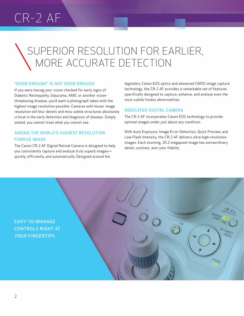
EASY-TO-MANAGE
CONTROLS RIGHT AT
YOUR FINGERTIPS
CR-2 AF
SUPERIOR RESOLUTION FOR EARLIER, MORE ACCURATE DETECTION
3
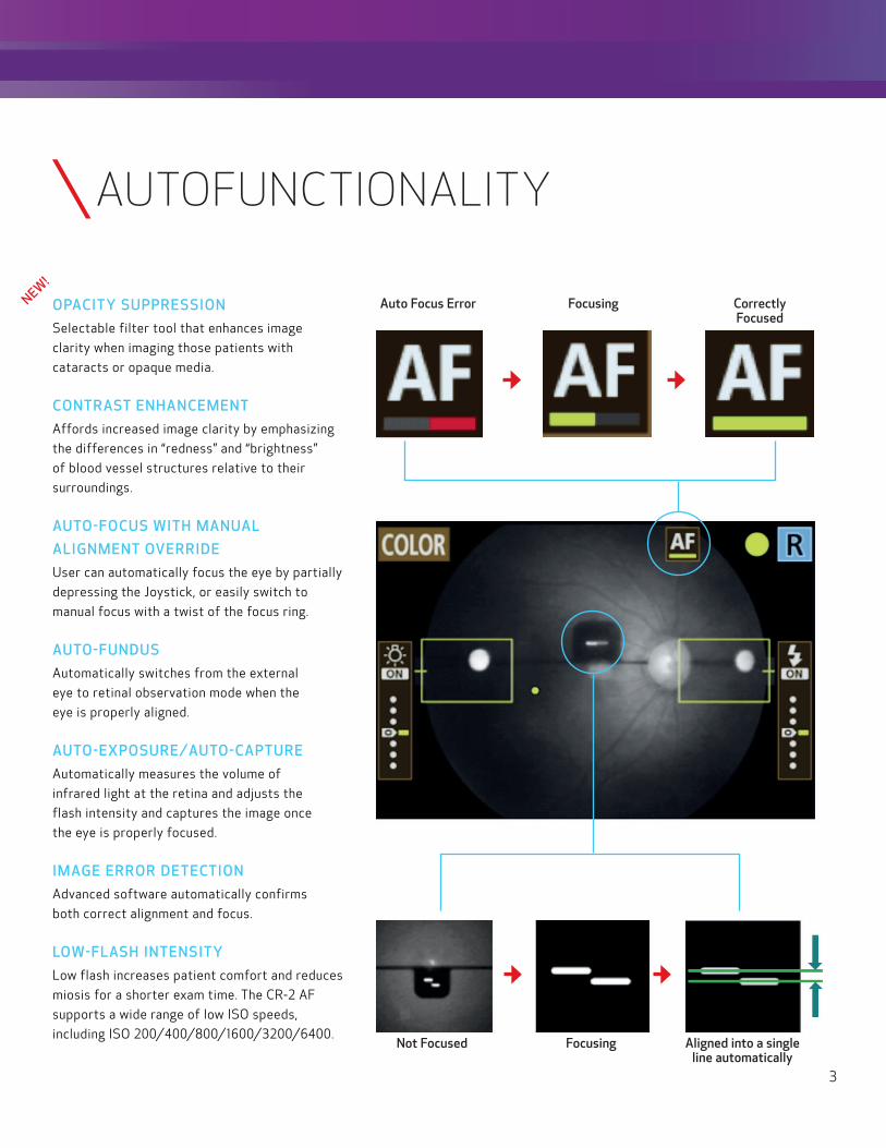
OPACITY SUPPRESSION
Selectable filter tool that enhances image
clarity when imaging those patients with
cataracts or opaque media.
CONTRAST ENHANCEMENT
Affords increased image clarity by emphasizing
the differences in “redness” and “brightness”
of blood vessel structures relative to their
surroundings.
AUTO-FOCUS WITH MANUAL
ALIGNMENT OVERRIDE
User can automatically focus the eye by partially
depressing the Joystick, or easily switch to
manual focus with a twist of the focus ring.
AUTO-FUNDUS
Automatically switches from the external
eye to retinal observation mode when the
eye is properly aligned.
AUTO-EXPOSURE/AUTO-CAPTURE
Automatically measures the volume of
infrared light at the retina and adjusts the
flash intensity and captures the image once
the eye is properly focused.
IMAGE ERROR DETECTION
Advanced software automatically confirms
both correct alignment and focus.
LOW-FLASH INTENSITY
Low flash increases patient comfort and reduces
miosis for a shorter exam time. The CR-2 AF
supports a wide range of low ISO speeds,
including ISO 200/400/800/1600/3200/6400.Not Focused Focusing Aligned into a single
line automatically
Auto Focus Error Focusing Correctly Focused
NEW!
AUTOFUNCTIONALITY

ADVANCED DIGITAL IMAGE FILTER PROCESSING
4
COLOR
CR-2 AF
CHANNEL MODES
BLUE
The color image provides brilliant,
full-spectrum images with superior
detail and color accuracy.
The blue channel mode provides a critical view
of the retinal nerve fiber layer, the internal
limiting membrane, retina folds, cysts, and
epiretinal membranes.
The CR-2 AF Digital Retinal Camera produces ultra-high-
resolution, 20.2-megapixel, wide-angle views with excellent
color, detail, and contrast. To further enhance your retinal
exam capabilities, the CR-2 AF has a full set of blue, green,
red, red-free, and cobalt digital processing modes to extract
more in-depth information from each image.

5
ADVANCED DIGITAL IMAGE FILTER PROCESSING
5
* Available when purchased with imageSPECTRUM Software.
RED
GREEN
EMBOSS*
CHANNEL MODES
The red channel mode provides specific
information deep into the choroidal areas
and is useful in identifying pigmentary
disturbances, choroidal melanomas,
ruptures, and nevi.
The Emboss tool enhances depth
perception with a 3D-like representation
of elevations and depressions. The entire
retina can be embossed, as well as the
optic disc or macula area. The Emboss tool
is also especially valuable in assessing
subtle areas not easily visualized with
color alone. The tool also assists in the
evaluation of macular degeneration,
glaucoma, and diabetic retinopathy.
The green channel mode provides excellent
overall contrast and enhances the retinal
vasculature. It’s also useful when highlighting
hemorrhages, drusen, and exudates.
6
GOING BEYOND THE CAPTURED IMAGE
Until recently, capturing, storing, and interpreting diagnostic
images were about all that most clinicians asked of their
diagnostic instruments. However, with today’s increased
workflow and practice efficiency expectations, rapidly
escalating PHI data security concerns, and data sharing
/access needs at an all-time high, things have changed.
High-resolution, diagnostic-quality images—not reduced
resolution thumbnails—now must be instantly accessible,
whether in your exam lane or half-way across the country.
That same image data must also be presented in a
coordinated fashion—along with the patient’s entire test
and image history—to meet your workflow and efficiency
requirements. Moreover, it must be done with security and
compliance with CMS and HIPAA regulations in mind.
ADVANCED IMAGE MANAGEMENT
imageSPECTRUM Image Management Software* is a
dedicated system designed to provide immediate access
to images from different eye care modalities. The system’s
core is built around today’s security and compliance
requirements and is based on the universal DICOM open
systems standard. It’s crisp and very adept at handling
literally hundreds of thousands of images from multiple
instruments and modalities, whether from Canon or
other manufacturers.** Furthermore, imageSPECTRUM
Image Management Software is highly scalable and can
help practices of any size and complexity to cleanly and
efficiently manage all their diagnostic information. It’s
also the ideal complement to your new Canon CR-2 AF
Digital Non-Mydriatic Retinal Camera.
CR-2 AF
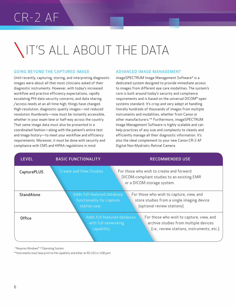
LEVEL BASIC FUNCTIONALITY RECOMMENDED USE
* Requires Windows® 7 Operating System.
** Instruments must have print-to-file capability and either an RS-232 or USB port.
IT’S ALL ABOUT THE DATA
Office
StandAlone
CapturePLUS Create and View Studies.
For those who wish to create and forward
DICOM-compliant studies to an existing EMR
or a DICOM storage system.
Adds full-featured database
functionality for capture
station user.
For those who wish to capture, view, and
store studies from a single imaging device
(optional review stations).
Adds full-featured database
with full networking
capability.
For those who wish to capture, view, and
archive studies from multiple devices
(i.e., review stations, instruments, etc.).
7
NEEDS HEADING
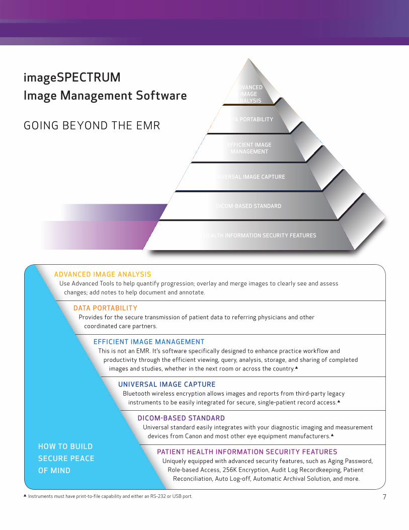
ADVANCED IMAGE ANALYSISUse Advanced Tools to help quantify progression; overlay and merge images to clearly see and assess
changes; add notes to help document and annotate.
DATA PORTABILITYProvides for the secure transmission of patient data to referring physicians and other
coordinated care partners.
EFFICIENT IMAGE MANAGEMENTThis is not an EMR. It’s software specifically designed to enhance practice workflow and
productivity through the efficient viewing, query, analysis, storage, and sharing of completed images and studies, whether in the next room or across the country.p
UNIVERSAL IMAGE CAPTUREBluetooth wireless encryption allows images and reports from third-party legacy
instruments to be easily integrated for secure, single-patient record access.p
DICOM-BASED STANDARDUniversal standard easily integrates with your diagnostic imaging and measurement
devices from Canon and most other eye equipment manufacturers.p
PATIENT HEALTH INFORMATION SECURITY FEATURESUniquely equipped with advanced security features, such as Aging Password,
Role-based Access, 256K Encryption, Audit Log Recordkeeping, Patient Reconciliation, Auto Log-off, Automatic Archival Solution, and more.
imageSPECTRUM Image Management Software
ADVANCED IMAGE
ANALYSIS
DATA PORTABILITY
EFFICIENT IMAGE MANAGEMENT
UNIVERSAL IMAGE CAPTURE
DICOM-BASED STANDARD
PATIENT HEALTH INFORMATION SECURITY FEATURES
HOW TO BUILD
SECURE PEACE
OF MIND
GOING BEYOND THE EMR
p Instruments must have print-to-file capability and either an RS-232 or USB port.

USA.CANON.COM/EYE-CARE
POST SALE SERVICE AND SUPPORT
MAINTAINING YOUR INVESTMENT IN EXCELLENCE.
The CR-2 AF Digital Non-Mydriatic Retinal Camera is backed by Canon, a global microprocessor-based company with 75 years of optical experience. Its superb customer
service and support organization is ready to answer your needs 24/7/365.
This common sense approach to service allows you to purchase a service plan that suits your specific needs—and your budget. The Canon service program may help you avoid costly instrument downtime while also helping you with the accessibility of your vital patient images and information.
To schedule a demo or for additional information, call 1-800-970-7227 or visit our Web site.
GeneralType: Digital Retinal Camera, Non-Mydriatic
Type of PhotographyAnterior, Stereo, Color, Digital Red-free, Digital Cobalt
Angle of View: 45º (35º SP Mode)
Magnification: 2x Digital
Minimal Pupil Size: 4.0 mm (3.3 mm SP mode)
Focus Adjustment Type: Split-Line Adjustment
Patient Diopter Compensation RangeWithout Compensation Lens: -10 D to +15 D
With “-“ Compensation Lens: -31 D to -7 D
With “+” Compensation Lens: +11 D to +33 D
Light SourceObservation: LED
Photography: White LED
Canthus Mark: 420 mm From Base
Internal Eye Fixation: LED Dot Matrix; four programmable patterns
External Eye Fixation: White LED (Sold Separately)
Working Distance: 35 mm
Working Distance AdjustmentAnterior Observation: Double Image Match Method
Fundus Observation: Working Distance Dots
Sensor Resolution: 20.2 megapixels
Camera Dedicated EOS Camera for CR-2 AF (Bundled)
Monitor 3.0-inch Color LCD Monitor
HDMI port for External Monitor (Optional)
Auto Function: Automatic Exposure
Mount MovementFront and Back: 70 mm
Side to Side: 100 mm
Up and Down: 30 mm
Chin Rest Movement: 60 mm
Electrical and Environmental PC Interface: USB 1.1, USB 2.0
Power Supply: AC 100-240 V, 50/60 Hz
Operating EnvironmentTemperature: 50º to 86º Fahrenheit (10º to 35º Celsius)
Humidity: 30% to 90% RH (No Condensation)
Atmospheric Pressure: 700 to 1060 hPa
Physical Characteristics Dimensions (H x W x D) 18.6” x 12” x 19.7” (473 mm x 305 mm x 500 mm)
Weight: 33 lb. (15 kg)
Optional AccessoriesAutoMosaic Function
External Fixation Target
Motorized Table
Auxiliary Monitor
Canon and EOS are registered trademarks of Canon Inc. in the United States and may also be registered trademarks or trademarks in other countries. DICOM is a registered trademark of the National Electrical Manufacturers Association for its standards publications relating to digital communications of medical information. All other referenced product names and marks are trademarks of their respective owners. Specifications and availability subject to change without notice. Not responsible for typographical errors.©2016 Canon U.S.A., Inc. All rights reserved.
EB-035, Rev. C0416R-CR-2AF-PDF-IH
CR-2 AFDIGITAL NON-MYDRIATIC RETINAL CAMERA