Cannabinoid Action Induces Autophagy-Mediated Cell Death Through Stimulation of ER Stress in Human...
14
Research article TheJournalofClinicalInvestigation http://www.jci.org Volume 119 Number 5 May 2009 1359 Cannabinoid action induces autophagy- mediated cell death through stimulation of ER stress in human glioma cells María S alazar , 1,2 Arkaitz Carracedo, 1 Íñigo J. Salanueva, 1 Sonia Hernández-Tiedra, 1 Mar Lorente, 1,2 Ainara Egia, 1 Patricia Vázquez, 3 Cristina Blázquez, 1,2 Sofía Torres, 1 Stephane García, 4 Jonathan Nowak, 4 Gian María Fimia, 5 Mauro Piacentini, 5 Francesco Cecconi, 6 Pier Paolo Pandolfi, 7 Luis González-Feria, 8 Juan L. Iovanna, 4 Manuel Guzmán, 1,2 Patricia Boya, 3 and Guillermo Velasco 1,2 1 Department of Biochemistry and Molecular Biology I, School of Biology, Complutense University, Madrid, Spain. 2 Centro de Investigación Biomédica en Red sobre Enfermedades Neurodegenerativas (CIBERNED), Madrid, Spain. 3 3D Lab (Development, Differentiation, and Degeneration), Department of Cellular and Molecular Physiopathology, Centro de Investigaciones Biológicas, Consejo Superior de Investigaciones Científicas (CSIC), Madrid, Spain. 4 INSERM U624, Campus de Luminy, Marseille, France. 5 National Institute for Infectious Diseases, IRCCS “L. Spallanzani,” Rome, Italy. 6 Laboratory of Molecular Neuroembryology , IRCCS Fondazione Santa Lucia and Department of B iology, University of Rome “Tor Vergata,” Rome, Italy. 7 Cancer Genetics Program, Beth Israel Deaconess Cancer Center and Department of Medicine, Beth Israel Deaconess Medical Center , Harvard Medical School, Boston, Massachusetts, USA. 8 Department of Neurosurgery, University Hospital, Tenerife, Spain. Autophagyca npromotece llsurvivalor celldeath, butthemole cularbasis underlyingi tsdualrole incance r remainsobscure.Herewedemonstratethat Δ 9 -tetrahydrocann abinol(THC),the mainactivecompon entof marijuana,induceshumangliomacelldeaththroughstimulationofautophagy.OurdataindicatethatTHC induced ceramideaccumulation andeukaryotictransl ationiniti ationfacto r2 α(eIF2α)phosphorylation andthereby activatedanERstressres ponsethatpromotedautopha gyviatribblesho molog3–depe ndent (TRB3-dep endent)inh ibition oftheAkt/mammal iantargetofrapamyc incomple x1(mTORC1)axis.We alsoshowedthatautophagyisupstreamofapoptosisincannabinoid-inducedhumanandmousecancercell deathandthatactivationofthispathwaywasnecessaryfortheantitumoractionofcannabinoidsinvivo. ThesefindingsdescribeamechanismbywhichTHCcanpromotetheautophagicdeathofhumanandmouse cancercellsandprovideevidencethatcannabinoidadministrationmaybeaneffectivetherapeuticstrategy fortargetinghumancancers. Introduction Macro-autophagy, hereafter referred to as “autophagy,” is a highly conserved cellular process in which cytoplasmic materials — including organelles — are sequestered into double-membrane vesicles called autophagosomes and delivered to lysosomes for degradation or recycling (1). In many cellular settings, triggering of autophagy relies on the inhibition of mammalian target of rapa- mycin complex 1 (mTORC1), an event that promotes the activa- tion (de-inhibition) of several autophagy proteins (Atgs) involved in the initial phase of membrane isolation (1). Enlargement of this complex to form the autophagosome requires the participation of 2 ubiquitin-like conjugation systems. One involves the conjuga- tion of ATG12 to ATG5 and the other of phosphatidylethanol- amine to LC3/ATG8 (1). The final outcome of the activation of the autophagy program is highly dependent on the cellular context and the strength and duration of the stress-inducing signals (2–5). Thus, besides its role in cellular homeostasis, autophagy can be a form of programmed cell death, designated “type II programmed cell death,” or play a cytoprotective role, for example in situations of nutrient starvation (6). Accordingly, autophagy has been pro- posed to play an important role in both tumor progression and promotion of cancer cell death (2–4), although the molecular mechanisms responsible for this dual action of autophagy in can- cer have not been elucidated. Δ 9 -T etrahydrocannabinol (THC), the main active component of marijuana (7), exerts a wide variety of biological effects by mim- icking endogenous substances — the endocannabinoids — that bind to and activate specific cannabinoid receptors (8). One of the most exciting areas of research in the cannabinoid field is the study of the potential application of cannabinoids as antitumoral agents (9). Cannabinoid administration has been found to curb the growth of several types of tumor xenografts in rats and mice (9, 10). Based on this preclinical evidence, a pilot clinical trial has been recently run to investigate the antitumoral action of THC on recurrent gliomas (11). Recent findings have also shown that the pro-apoptotic and tumor growth–inhibiting activity of cannabi- noids relies on the upregulation of the transcriptional co-activa- tor p8 (12) and its target the pseudo-kinase tribbles homolog 3 (TRB3) (13). However, the mechanisms that promote the activa- tion of this signaling route as well as the targets downstream of TRB3 that mediate its tumor cell–killing action remain elusive. In this study we found that ER stress–evoked upregulation of the p8/TRB3 pathway induced autophagy via inhibition of the Akt/ mTORC1 axis and that activation of autophagy promoted the apoptotic death of tumor cells. The uncovering of this pathway, Conflictofinterest: The authors have declared that no conflict of interest exists. Nonstandardabbreviationsused: Atg, autophagy protein; eIF2 α, eukaryotic translation initiation factor 2 α; MEF, mouse embryonic fibroblast; THC, Δ 9 -tetrahy- drocannabinol; mTORC1, mammalian target of rapamycin complex 1; PDI, protein disulphide isomerase; TRB3, tribbles homolog 3. Citationforthisarticle: J. Clin. Invest. 119:1359–1372 (2009). doi:10.1172/JCI37948.
-
Upload
leslie-bishop-paul -
Category
Documents
-
view
220 -
download
0
Transcript of Cannabinoid Action Induces Autophagy-Mediated Cell Death Through Stimulation of ER Stress in Human...

8/2/2019 Cannabinoid Action Induces Autophagy-Mediated Cell Death Through Stimulation of ER Stress in Human Glioma Cells.
http://slidepdf.com/reader/full/cannabinoid-action-induces-autophagy-mediated-cell-death-through-stimulation 1/14
Research article
TheJournalofClinicalInvestigation http://www.jci.org Volume 119 Number 5 May 2009 1359
Cannabinoid action induces autophagy-mediated cell death through stimulation
of ER stress in human glioma cellsMaría Salazar,1,2 Arkaitz Carracedo,1 Íñigo J. Salanueva,1 Sonia Hernández-Tiedra,1 Mar Lorente,1,2
Ainara Egia,1 Patricia Vázquez,3 Cristina Blázquez,1,2 Sofía Torres,1 Stephane García,4 Jonathan Nowak,4 Gian María Fimia,5 Mauro Piacentini,5 Francesco Cecconi,6 Pier Paolo Pandolfi,7 Luis González-Feria,8 Juan L. Iovanna,4 Manuel Guzmán,1,2 Patricia Boya,3 and Guillermo Velasco1,2
1Department of Biochemistry and Molecular Biology I, School of Biology, Complutense University, Madrid, Spain.2Centro de Investigación Biomédica en Red sobre Enfermedades Neurodegenerativas (CIBERNED), Madrid, Spain.
33D Lab (Development, Differentiation, and Degeneration), Department of Cellular and Molecular Physiopathology, Centro de Investigaciones Biológicas,Consejo Superior de Investigaciones Científicas (CSIC), Madrid, Spain. 4INSERM U624, Campus de Luminy, Marseille, France.
5National Institute for Infectious Diseases, IRCCS “L. Spallanzani,” Rome, Italy. 6Laboratory of Molecular Neuroembryology,IRCCS Fondazione Santa Lucia and Department of Biology, University of Rome “Tor Vergata,” Rome, Italy. 7Cancer Genetics Program,
Beth Israel Deaconess Cancer Center and Department of Medicine, Beth Israel Deaconess Medical Center, Harvard Medical School,
Boston, Massachusetts, USA.8
Department of Neurosurgery, University Hospital, Tenerife, Spain.
Autophagycanpromotecellsurvivalorcelldeath,butthemolecularbasisunderlyingitsdualroleincancerremainsobscure.HerewedemonstratethatΔ9-tetrahydrocannabinol(THC),themainactivecomponentofmarijuana,induceshumangliomacelldeaththroughstimulationofautophagy.OurdataindicatethatTHCinducedceramideaccumulationandeukaryotictranslationinitiationfactor2α(eIF2α)phosphorylationandtherebyactivatedanERstressresponsethatpromotedautophagyviatribbleshomolog3–dependent(TRB3-dependent)inhibitionoftheAkt/mammaliantargetofrapamycincomplex1(mTORC1)axis.Wealsoshowedthatautophagyisupstreamofapoptosisincannabinoid-inducedhumanandmousecancercelldeathandthatactivationofthispathwaywasnecessaryfortheantitumoractionofcannabinoidsinvivo.ThesefindingsdescribeamechanismbywhichTHCcanpromotetheautophagicdeathofhumanandmousecancercellsandprovideevidencethatcannabinoidadministrationmaybeaneffectivetherapeuticstrategy
fortargetinghumancancers.
Introduction
Macro-autophagy, hereafter referred to as “autophagy,” is a highly conserved cellular process in which cytoplasmic materials— including organelles — are sequestered into double-membrane vesicles called autophagosomes and delivered to lysosomes fordegradation or recycling (1). In many cellular settings, triggeringof autophagy relies on the inhibition of mammalian target of rapa-mycin complex 1 (mTORC1), an event that promotes the activa-tion (de-inhibition) of several autophagy proteins (Atgs) involvedin the initial phase of membrane isolation (1). Enlargement of thiscomplex to form the autophagosome requires the participation of
2 ubiquitin-like conjugation systems. One involves the conjuga-tion of ATG12 to ATG5 and the other of phosphatidylethanol-amine to LC3/ATG8 (1). The final outcome of the activation of theautophagy program is highly dependent on the cellular contextand the strength and duration of the stress-inducing signals (2–5).Thus, besides its role in cellular homeostasis, autophagy can be a form of programmed cell death, designated “type II programmedcell death,” or play a cytoprotective role, for example in situations
of nutrient starvation (6). Accordingly, autophagy has been pro-posed to play an important role in both tumor progression andpromotion of cancer cell death (2–4), although the molecularmechanisms responsible for this dual action of autophagy in can-cer have not been elucidated.Δ9-Tetrahydrocannabinol (THC), the main active component of
marijuana (7), exerts a wide variety of biological effects by mim-icking endogenous substances — the endocannabinoids — thatbind to and activate specific cannabinoid receptors (8). One of the most exciting areas of research in the cannabinoid field is thestudy of the potential application of cannabinoids as antitumoral
agents (9). Cannabinoid administration has been found to curbthe growth of several types of tumor xenografts in rats and mice(9, 10). Based on this preclinical evidence, a pilot clinical trial hasbeen recently run to investigate the antitumoral action of THC onrecurrent gliomas (11). Recent findings have also shown that thepro-apoptotic and tumor growth–inhibiting activity of cannabi-noids relies on the upregulation of the transcriptional co-activa-tor p8 (12) and its target the pseudo-kinase tribbles homolog 3(TRB3) (13). However, the mechanisms that promote the activa-tion of this signaling route as well as the targets downstream of TRB3 that mediate its tumor cell–killing action remain elusive.In this study we found that ER stress–evoked upregulation of thep8/TRB3 pathway induced autophagy via inhibition of the Akt/
mTORC1 axis and that activation of autophagy promoted theapoptotic death of tumor cells. The uncovering of this pathway,
Conflictofinterest:The authors have declared that no conflict of interest exists.
Nonstandardabbreviationsused: Atg, autophagy protein; eIF2α, eukaryotictranslation initiation factor 2α; MEF, mouse embryonic fibroblast; THC, Δ9-tetrahy-drocannabinol; mTORC1, mammalian target of rapamycin complex 1; PDI, protein
disulphide isomerase; TRB3, tribbles homolog 3.Citationforthisarticle: J. Clin. Invest. 119:1359–1372 (2009). doi:10.1172/JCI37948.

8/2/2019 Cannabinoid Action Induces Autophagy-Mediated Cell Death Through Stimulation of ER Stress in Human Glioma Cells.
http://slidepdf.com/reader/full/cannabinoid-action-induces-autophagy-mediated-cell-death-through-stimulation 2/14
research article
1360 TheJournalofClinicalInvestigation http://www.jci.org Volume 119 Number 5 May 2009
8/2/2019 Cannabinoid Action Induces Autophagy-Mediated Cell Death Through Stimulation of ER Stress in Human Glioma Cells.
http://slidepdf.com/reader/full/cannabinoid-action-induces-autophagy-mediated-cell-death-through-stimulation 3/14
research article
TheJournalofClinicalInvestigation http://www.jci.org Volume 119 Number 5 May 2009 1361
which we believe is novel, for promoting tumor cell death may havetherapeutic implications in the treatment of cancer.
Results
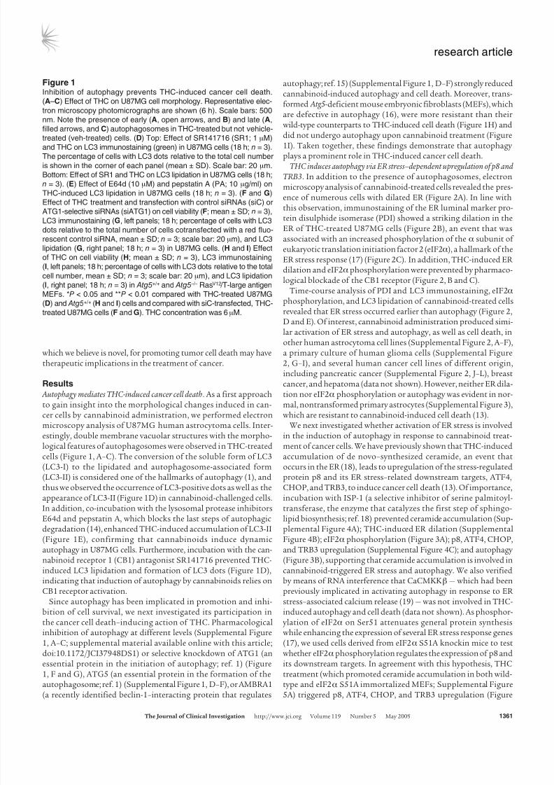
Autophagy mediates THC-induced cancer cell death. As a first approachto gain insight into the morphological changes induced in can-
cer cells by cannabinoid administration, we performed electronmicroscopy analysis of U87MG human astrocytoma cells. Inter-estingly, double membrane vacuolar structures with the morpho-logical features of autophagosomes were observed in THC-treatedcells (Figure 1, A–C). The conversion of the soluble form of LC3(LC3-I) to the lipidated and autophagosome-associated form(LC3-II) is considered one of the hallmarks of autophagy (1), andthus we observed the occurrence of LC3-positive dots as well as theappearance of LC3-II (Figure 1D) in cannabinoid-challenged cells.In addition, co-incubation with the lysosomal protease inhibitorsE64d and pepstatin A, which blocks the last steps of autophagicdegradation (14), enhanced THC-induced accumulation of LC3-II(Figure 1E), confirming that cannabinoids induce dynamic
autophagy in U87MG cells. Furthermore, incubation with the can-nabinoid receptor 1 (CB1) antagonist SR141716 prevented THC-induced LC3 lipidation and formation of LC3 dots (Figure 1D),indicating that induction of autophagy by cannabinoids relies onCB1 receptor activation.
Since autophagy has been implicated in promotion and inhi-bition of cell survival, we next investigated its participation inthe cancer cell death–inducing action of THC. Pharmacologicalinhibition of autophagy at different levels (Supplemental Figure1, A–C; supplemental material available online with this article;doi:10.1172/JCI37948DS1) or selective knockdown of ATG1 (anessential protein in the initiation of autophagy; ref. 1) (Figure1, F and G), ATG5 (an essential protein in the formation of the
autophagosome; ref. 1) (Supplemental Figure 1, D–F), or AMBRA1(a recently identified beclin-1–interacting protein that regulates
autophagy; ref. 15) (Supplemental Figure 1, D–F) strongly reducedcannabinoid-induced autophagy and cell death. Moreover, trans-formed Atg5-deficient mouse embryonic fibroblasts (MEFs), whichare defective in autophagy (16), were more resistant than theirwild-type counterparts to THC-induced cell death (Figure 1H) and
did not undergo autophagy upon cannabinoid treatment (Figure1I). Taken together, these findings demonstrate that autophagy plays a prominent role in THC-induced cancer cell death.
THC induces autophagy via ER stress–dependent upregulation of p8 andTRB3. In addition to the presence of autophagosomes, electronmicroscopy analysis of cannabinoid-treated cells revealed the pres-ence of numerous cells with dilated ER (Figure 2A). In line withthis observation, immunostaining of the ER luminal marker pro-tein disulphide isomerase (PDI) showed a striking dilation in theER of THC-treated U87MG cells (Figure 2B), an event that wasassociated with an increased phosphorylation of the α subunit of eukaryotic translation initiation factor 2 (eIF2α), a hallmark of theER stress response (17) (Figure 2C). In addition, THC-induced ER dilation and eIF2αphosphorylation were prevented by pharmaco-logical blockade of the CB1 receptor (Figure 2, B and C).
Time-course analysis of PDI and LC3 immunostaining, eIF2α phosphorylation, and LC3 lipidation of cannabinoid-treated cellsrevealed that ER stress occurred earlier than autophagy (Figure 2,D and E). Of interest, cannabinoid administration produced simi-lar activation of ER stress and autophagy, as well as cell death, inother human astrocytoma cell lines (Supplemental Figure 2, A–F),a primary culture of human glioma cells (Supplemental Figure2, G–I), and several human cancer cell lines of different origin,including pancreatic cancer (Supplemental Figure 2, J–L), breastcancer, and hepatoma (data not shown). However, neither ER dila -tion nor eIF2α phosphorylation or autophagy was evident in nor-mal, nontransformed primary astrocytes (Supplemental Figure 3),
which are resistant to cannabinoid-induced cell death (13).We next investigated whether activation of ER stress is involved
in the induction of autophagy in response to cannabinoid treat-ment of cancer cells. We have previously shown that THC-inducedaccumulation of de novo–synthesized ceramide, an event thatoccurs in the ER (18), leads to upregulation of the stress-regulatedprotein p8 and its ER stress–related downstream targets, ATF4,CHOP, and TRB3, to induce cancer cell death (13). Of importance,incubation with ISP-1 (a selective inhibitor of serine palmitoyl-transferase, the enzyme that catalyzes the first step of sphingo-lipid biosynthesis; ref. 18) prevented ceramide accumulation (Sup-plemental Figure 4A); THC-induced ER dilation (SupplementalFigure 4B); eIF2α phosphorylation (Figure 3A); p8, ATF4, CHOP,
and TRB3 upregulation (Supplemental Figure 4C); and autophagy (Figure 3B), supporting that ceramide accumulation is involved incannabinoid-triggered ER stress and autophagy. We also verifiedby means of RNA interference that CaCMKKβ — which had beenpreviously implicated in activating autophagy in response to ER stress–associated calcium release (19) — was not involved in THC-induced autophagy and cell death (data not shown). As phosphor-ylation of eIF2α on Ser51 attenuates general protein synthesiswhile enhancing the expression of several ER stress response genes(17), we used cells derived from eIF2α S51A knockin mice to testwhether eIF2αphosphorylation regulates the expression of p8 andits downstream targets. In agreement with this hypothesis, THCtreatment (which promoted ceramide accumulation in both wild-
type and eIF2α S51A immortalized MEFs; Supplemental Figure5A) triggered p8, ATF4, CHOP, and TRB3 upregulation (Figure
Figure 1Inhibition of autophagy prevents THC-induced cancer cell death.
(A–C) Effect of THC on U87MG cell morphology. Representative elec-
tron microscopy photomicrographs are shown (6 h). Scale bars: 500
nm. Note the presence of early (A, open arrows, and B) and late (A,
filled arrows, and C) autophagosomes in THC-treated but not vehicle-
treated (veh-treated) cells. (D) Top: Effect of SR141716 (SR1; 1 μM)and THC on LC3 immunostaining (green) in U87MG cells (18 h; n = 3).
The percentage of cells with LC3 dots relative to the total cell number
is shown in the corner of each panel (mean ± SD). Scale bar: 20 μm.
Bottom: Effect of SR1 and THC on LC3 lipidation in U87MG cells (18 h;
n = 3). (E) Effect of E64d (10 μM) and pepstatin A (PA; 10 μg/ml) on
THC-induced LC3 lipidation in U87MG cells (18 h; n = 3). (F and G)
Effect of THC treatment and transfection with control siRNAs (siC) or
ATG1-selective siRNAs (siATG1) on cell viability (F; mean ± SD; n = 3),
LC3 immunostaining (G, left panels; 18 h; percentage of cells with LC3
dots relative to the total number of cells cotransfected with a red fluo-
rescent control siRNA, mean ± SD; n = 3; scale bar: 20 μm), and LC3
lipidation (G, right panel; 18 h; n = 3) in U87MG cells. (H and I) Effect
of THC on cell viability (H; mean ± SD; n = 3), LC3 immunostaining
(I, left panels; 18 h; percentage of cells with LC3 dots relative to the total
cell number, mean ± SD; n = 3; scale bar: 20 μm), and LC3 lipidation(I, right panel; 18 h; n = 3) in Atg5+/+ and Atg5 –/– RasV12 /T-large antigen
MEFs. *P < 0.05 and **P < 0.01 compared with THC-treated U87MG
(D) and Atg5+/+ (H and I) cells and compared with siC-transfected, THC-
treated U87MG cells (F and G). THC concentration was 6 μM.

8/2/2019 Cannabinoid Action Induces Autophagy-Mediated Cell Death Through Stimulation of ER Stress in Human Glioma Cells.
http://slidepdf.com/reader/full/cannabinoid-action-induces-autophagy-mediated-cell-death-through-stimulation 4/14
research article
1362 TheJournalofClinicalInvestigation http://www.jci.org Volume 119 Number 5 May 2009
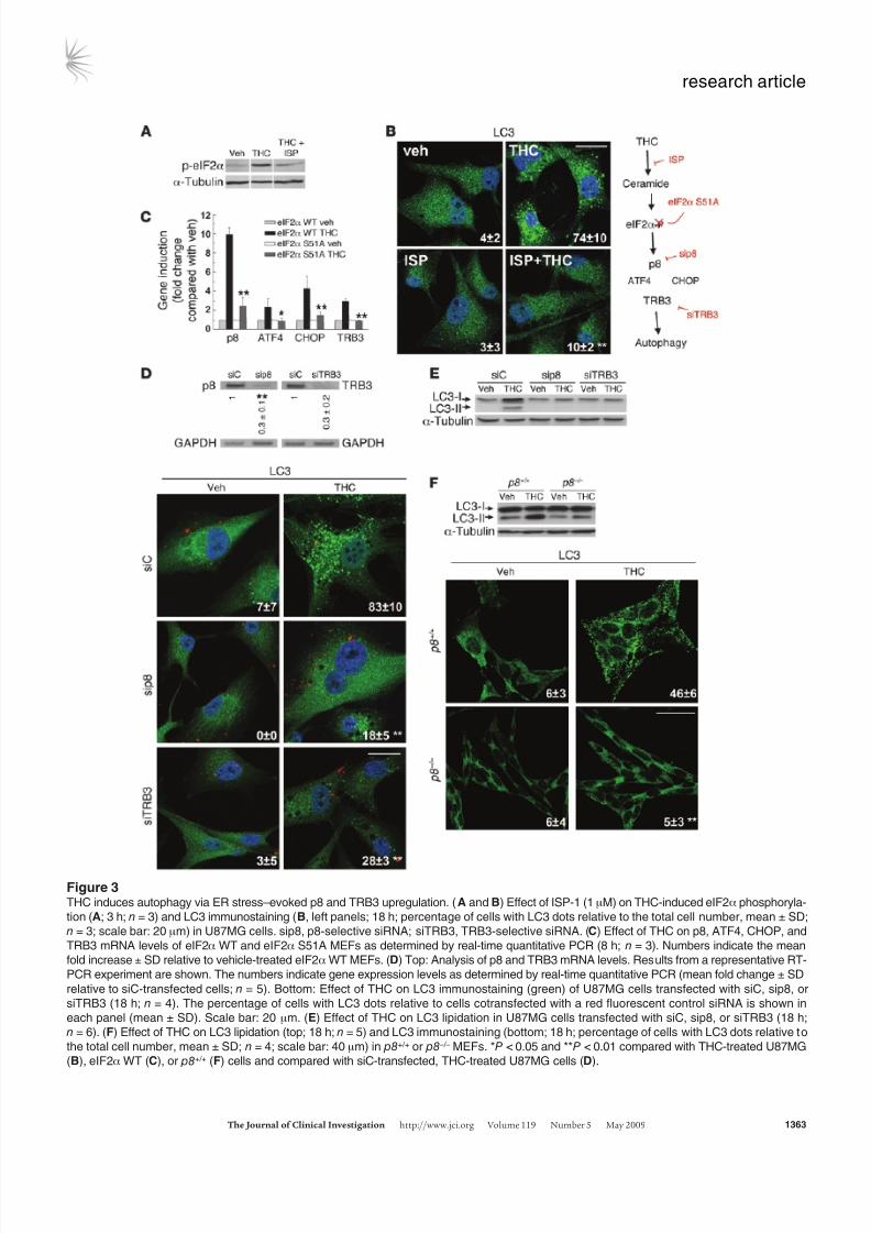
3C) as well as autophagy (Supplemental Figure 5B) in wild-typecells but not in their eIF2α S51A counterparts.
We subsequently asked whether p8 and its downstream tar-gets regulate autophagy. Knockdown of p8 or TRB3 preventedTHC-induced autophagy (Figure 3, D and E) but not ER dilation(Supplemental Figure 4D) in U87MG cells. Furthermore, THCinduced autophagy in p8+/+ but not p8-deficient transformed
MEFs (Figure 3F and Supplemental Figure 5C). Altogether, thesefindings reveal that THC induces autophagy of cancer cells via
activation of an ER stress–triggered signaling route that involvesstimulation of ceramide synthesis de novo, eIF2α phosphoryla-tion, and p8 and TRB3 upregulation.
THC inhibits Akt and mTORC1 via TRB3. Inhibition of mTORC1is considered a key step in the early triggering of autophagy (6).We therefore tested whether cannabinoid-induced upregulationof the p8 pathway leads to autophagy via inhibition of this com-
plex. THC treatment of U87MG cells reduced the phosphorylationof p70S6 kinase (a well-established mTORC1 substrate) and the
Figure 2ER stress precedes autophagy in cannabinoid action. (A) Effect of THC on U87MG cell morphology. Note the presence of the dilated ER in
THC- but not vehicle-treated cells (6 h). Arrows point to the ER. Scale bars: 500 nm. (B) Effect of SR1 (1 μM) and THC on PDI immunostaining
(red) in U87MG cells (8 h; n = 3). The percentage of cells with PDI dots relative to the total cell number is shown in the corner of each panel
(mean ± SD). Scale bar: 20 μm. (C) Effect of SR1 (1 μM) on THC-induced eIF2α phosphorylation of U87MG cells (3 h; OD relative to vehicle-treated cells, mean ± SD; n = 3). (D) Effect of THC on PDI (red) and LC3 (green) immunostaining in U87MG cells (n = 3). The percentage of
cells with PDI or LC3 dots relative to total cell number at each time point (mean ± SD) is shown. Scale bar: 20 μm. (E) Effect of THC on eIF2α
phosphorylation and LC3 lipidation in U87MG cells (n = 3). **P < 0.01 compared with THC-treated (B) or vehicle-treated (C and D) cells.
8/2/2019 Cannabinoid Action Induces Autophagy-Mediated Cell Death Through Stimulation of ER Stress in Human Glioma Cells.
http://slidepdf.com/reader/full/cannabinoid-action-induces-autophagy-mediated-cell-death-through-stimulation 5/14
research article
TheJournalofClinicalInvestigation http://www.jci.org Volume 119 Number 5 May 2009 1363
Figure 3THC induces autophagy via ER stress–evoked p8 and TRB3 upregulation. ( A and B) Effect of ISP-1 (1 μM) on THC-induced eIF2α phosphoryla-
tion (A; 3 h; n = 3) and LC3 immunostaining (B, left panels; 18 h; percentage of cells with LC3 dots relative to the total cell number, mean ± SD;
n = 3; scale bar: 20 μm) in U87MG cells. sip8, p8-selective siRNA; siTRB3, TRB3-selective siRNA. (C) Effect of THC on p8, ATF4, CHOP, and
TRB3 mRNA levels of eIF2α WT and eIF2α S51A MEFs as determined by real-time quantitative PCR (8 h; n = 3). Numbers indicate the mean
fold increase ± SD relative to vehicle-treated eIF2α WT MEFs. (D) Top: Analysis of p8 and TRB3 mRNA levels. Results from a representative RT-
PCR experiment are shown. The numbers indicate gene expression levels as determined by real-time quantitative PCR (mean fold change ± SD
relative to siC-transfected cells; n = 5). Bottom: Effect of THC on LC3 immunostaining (green) of U87MG cells transfected with siC, sip8, or
siTRB3 (18 h; n = 4). The percentage of cells with LC3 dots relative to cells cotransfected with a red fluorescent control siRNA is shown in
each panel (mean ± SD). Scale bar: 20 μm. (E) Effect of THC on LC3 lipidation in U87MG cells transfected with siC, sip8, or siTRB3 (18 h;
n = 6). (F) Effect of THC on LC3 lipidation (top; 18 h; n = 5) and LC3 immunostaining (bottom; 18 h; percentage of cells with LC3 dots relative to
the total cell number, mean ± SD; n = 4; scale bar: 40 μm) in p8+/+ or p8 –/– MEFs. *P < 0.05 and **P < 0.01 compared with THC-treated U87MG
(B), eIF2α WT (C), or p8+/+ (F) cells and compared with siC-transfected, THC-treated U87MG cells (D).

8/2/2019 Cannabinoid Action Induces Autophagy-Mediated Cell Death Through Stimulation of ER Stress in Human Glioma Cells.
http://slidepdf.com/reader/full/cannabinoid-action-induces-autophagy-mediated-cell-death-through-stimulation 6/14
research article
1364 TheJournalofClinicalInvestigation http://www.jci.org Volume 119 Number 5 May 2009
Figure 4THC inhibits the Akt/mTORC1 pathway via TRB3. (A) Effect of THC on p70S6K and S6 phosphorylation of U87MG cells (n = 6). (B) Effect of THC
on cell viability (left panel; 24 h; mean ± SD; n = 6) and LC3 lipidation (right panel; 18 h; n = 4) in Tsc2+/+ and Tsc2 –/– MEFs. (C) Effect of THC on
Akt, TSC2, PRAS40, p70S6K, and S6 phosphorylation of U87MG cells (18 h; OD relative to vehicle-treated cells, mean ± SD; n = 7). (D) Effect
of THC on cell viability (left panel; 24 h; mean ± SD; n = 4) and LC3 lipidation (right panel; 18 h; n = 4) of pBABE and myristoylated Akt (myr-Akt)
MEFs. (E) Effect of THC on Akt co-immunoprecipitation with TRB3 in U87MG cell extracts (8 h; OD relative to vehicle-treated cells, mean ± SD;
n = 9; input: TRB3). (F and G) Effect of THC on Akt, TSC2, PRAS40, p70S6K, and S6 phosphorylation and LC3 lipidation ( G only) of siC- and
siTRB3-transfected (F; 18 h; OD relative to vehicle-treated siC-transfected U87MG cells, mean ± SD; n = 7; upper panel shows an analysis of TRB3
mRNA levels) and EGFP (Ad-EGFP) or rat TRB3 (Ad-TRB3) adenoviral vector–infected (G; 18 h; OD relative to vehicle-treated Ad-EGFP–infected
U87MG cells, mean ± SD; n = 4; upper panel shows an analysis of rTRB3 mRNA levels) U87MG cells. ( H) Effect of THC on Akt, p70S6K, and
S6 phosphorylation of p8+/+ and p8 –/– MEFs (n = 7). *P < 0.05 and **P < 0.01 compared with THC-treated Tsc2+/+ (B) and pBABE (D) MEFs and
compared with vehicle-treated (C and E), vehicle-treated siC-transfected (F), or Ad-EGFP–infected (G) U87MG cells.

8/2/2019 Cannabinoid Action Induces Autophagy-Mediated Cell Death Through Stimulation of ER Stress in Human Glioma Cells.
http://slidepdf.com/reader/full/cannabinoid-action-induces-autophagy-mediated-cell-death-through-stimulation 7/14
research article
TheJournalofClinicalInvestigation http://www.jci.org Volume 119 Number 5 May 2009 1365
ribosomal protein S6 (a well-established p70S6 kinase substrate)(Figure 4, A and C), indicating that mTORC1 is inhibited in can-nabinoid-challenged cells. In addition, the cannabinoid-induceddecrease in p70S6 kinase and S6 phosphorylation, autophagy, andcell death were not evident in Tsc2–/– cells, in which mTORC1 is
constitutively active (20) (Figure 4B and Supplemental Figure 6, Aand B), further supporting a major role for mTORC1 inhibition inthe induction of autophagic cell death by cannabinoids.
The protein kinase Akt positively regulates the activity of themTORC1 complex by phosphorylating and inhibiting TSC2 andPRAS40 (a well-established Akt substrate within the mTORC1complex). Thus, Akt inhibition decreases mTORC1 activity andpromotes autophagy (20). In line with this idea, THC decreasedthe phosphorylation of Akt, TSC2, and PRAS40 as well as p70S6kinase and S6 (Figure 4C). This inhibition of the Akt/mTORC1pathway was abrogated by incubation with a CB1 receptor antago-nist (Supplemental Figure 6C) or a ceramide synthesis inhibitor(Supplemental Figure 6D). Likewise, cells overexpressing a myris-toylated (constitutively active) form of Akt were resistant to THC-induced mTORC1 inhibition, autophagy, and cell death (Figure4D and Supplemental Figure 6, E and F), further supporting thatTHC induces autophagy via Akt inhibition.
Since TRB3 has been shown to directly interact with and inhibit Akt (21, 22), we investigated whether upregulation of TRB3 wasresponsible for THC-induced Akt/mTORC1 inhibition. Sev-eral observations support that this is indeed the case: (a) THCincreased the amount of Akt coimmunoprecipitated with TRB3from U87MG extracts (Figure 4E), (b) knockdown of TRB3 pre-
vented the effect of THC on Akt, TSC2, PRAS-40, p70S6 kinase,and S6 phosphorylation (Figure 4F), and (c) TRB3 overexpressiondecreased Akt, TSC2, PRAS40, p70S6 kinase, and S6 phosphoryla-tion, enhanced the inhibitory effect of THC on the phosphoryla-
tion of these proteins, and promoted autophagy (Figure 4G). Inline with these observations, THC failed to inhibit Akt, p70S6kinase, and S6 phosphorylation of eIF2α S51A knockin or p8-deficient MEFs, in which TRB3 did not become upregulated uponcannabinoid treatment (Figure 4H and Supplemental Figure 6, Gand H). Altogether, these data demonstrate that upregulation of p8 and TRB3 induce autophagy of tumor cells via inhibition of the
Akt/mTORC1 pathway.THC-induced autophagy promotes the apoptotic death of cancer cells.
While analyzing the mechanism of cannabinoid cell-killingaction, we observed that incubation with the pan-caspase inhibi-tor ZVAD-fmk prevented cell death to the same extent as genetic(Figure 5A) or pharmacological (Supplemental Figure 7) inhibi-
tion of autophagy. Furthermore, Bax/Bak double knockout (DKO)immortalized MEFs, which are protected against mitochondrialapoptosis (23), were resistant to THC-induced cell death andapoptosis (Figure 5B) but underwent eIF2α phosphorylationand autophagy (Figure 5C) upon THC treatment. We thereforeinvestigated whether cannabinoid-induced autophagy promotedthe apoptotic death of cancer cells. Time-course analysis of LC3and active caspase-3 immunostaining in U87MG cells revealedthat autophagy preceded the appearance of apoptotic features inTHC-treated cells (Figure 5D). In addition, selective knockdownof ATG1 (Figure 5D) as well as of AMBRA1 or ATG5 (Supplemen-tal Figure 8) prevented THC-induced caspase-3 activation. More-over, unlike their wild-type counterparts, Atg5-deficient immor-
talized MEFs did not undergo phosphatidylserine translocationto the outer leaflet of the plasma membrane (Figure 5E), loss
of mitochondrial membrane potential (Figure 5F), or increasedproduction of reactive oxygen species (Supplemental Figure 9) inresponse to cannabinoid treatment. These findings indicate thatactivation of the autophagy-mediated cell death pathway occursupstream of apoptosis in cannabinoid antitumoral action.
Activation of autophagy is necessary for cannabinoid antitumoral actionin vivo. To determine the in vivo relevance of our findings, we firstinvestigated whether THC promotes the activation of the above-described autophagy-mediated cell death pathway in U87MG cell–derived tumor xenografts, in which we have recently shown thatcannabinoid treatment reduces tumor growth (specifically, THCadministration for 14 days decreased tumor growth by 50%; ref.13). Analysis of these tumors revealed that cannabinoid adminis-tration increases TRB3 expression and decreases S6 phosphoryla-tion (Figure 6A). Likewise, formation of LC3 dots as well as increasein LC3-II and active caspase-3 immunostaining were observed inTHC-treated, but not vehicle-treated, tumors (Figure 6B).
To further investigate whether activation of the p8 pathway mediates cannabinoid antitumoral action, we also analyzed tumorsderived from p8+/+ and p8–/– Ras V12 /E1A-transformed MEFs (in thiscase, THC administration for 8 days decreased by 45% the growthof p8+/+ tumors but had no significant effect on p8–/– tumors; ref.13). THC treatment increased TRB3 expression, decreased S6 phos-phorylation, and increased autophagy as well as TUNEL and activecaspase-3 immunostaining in p8+/+ but not p8–/– tumors (Figure 6Cand Supplemental Figure 10). Moreover, THC treatment enhancedthe number of cells with LC3 dots and TUNEL-positive nuclei inp8+/+ but not in p8–/– tumors (Figure 6C).
In order to verify the importance of autophagy for cannabinoidantitumoral action, we next generated tumors with Atg5+/+ and
Atg5–/– Ras V12 /T-large antigen transformed MEFs. THC adminis-tration reduced by more than 80% the growth of tumors derived
from wild-type cells but had no significant effect on those tumorsgenerated by autophagy-deficient cells (Figure 7A). Furthermore,cannabinoid administration increased autophagy, TUNEL (Fig-ure 7B), and active caspase-3 immunostaining (Supplemental Fig-ure 11) in Atg5+/+ but not Atg5–/– tumors. Likewise, cannabinoidadministration increased the number of cells with LC3 dots andTUNEL-positive nuclei in Atg5+/+ but not Atg5–/– tumors (Figure7B). Taken together, these findings demonstrate that activation of the autophagy-mediated cell death pathway is indispensable forcannabinoid antitumoral action.
Finally, we analyzed the tumors of 2 patients enrolled in a clinicaltrial aimed at investigating the effect of THC on recurrent glioblas-toma multiforme. The patients were subjected to intracranial THC
administration, and biopsies were taken before and after the treat-ment (11). In the 2 patients, cannabinoid inoculation increasedTRB3 immunostaining and decreased S6 phosphorylation (Figure8A). Interestingly, the number of cells with autophagic phenotype(Figure 8B) as well as with active caspase-3 immunostaining (Fig-ure 8C) was increased in the tumor samples obtained after THCtreatment. Although these studies were only conducted in speci-mens from 2 patients, they are in line with the preclinical evidenceshown above and suggest that cannabinoid administration mightalso trigger autophagy-mediated cell death in human tumors.
Discussion
In this study we show that cannabinoids, a new family of potential
antitumoral agents, induce autophagy of cancer cells and that thisprocess mediates the cell death–promoting activity of these com-

8/2/2019 Cannabinoid Action Induces Autophagy-Mediated Cell Death Through Stimulation of ER Stress in Human Glioma Cells.
http://slidepdf.com/reader/full/cannabinoid-action-induces-autophagy-mediated-cell-death-through-stimulation 8/14
research article
1366 TheJournalofClinicalInvestigation http://www.jci.org Volume 119 Number 5 May 2009
Figure 5Autophagy is upstream of apoptosis in cannabinoid-induced cancer cell death. (A) Effect of THC and the pan-caspase inhibitor ZVAD (10 μM) on
the viability of Atg5+/+ and Atg5 –/– MEFs (36 h; percentage of viable cells relative to the corresponding Atg5+/+ vehicle-treated cells, mean ± SD;
n = 3). (B) Effect of THC on the apoptosis of Bax/Bak WT and Bax/Bak DKO MEFs as determined by cytofluorometric analysis of Annexin V/
propidium iodide (PI) (24 h; mean ± SD; n = 3). The mean ± SD percentage of Annexin V–positive/PI-positive and Annexin V–positive, PI-nega-
tive cells is shown in the upper and lower corners, respectively. (C) Effect of THC on eIF2α phosphorylation (3 h; n = 3) and LC3 lipidation (18 h;
n = 4) of Bax/Bak WT and DKO MEFs. (D) Left: Effect of THC on autophagy and apoptosis of U87MG cells transfected with siC or siATG1.
Green bars, cells with LC3 dots; red bars, active caspase-3–positive cells; white bars, cells with both LC3 dots and active caspase-3 staining.
Data correspond to the percentage of cells with LC3 dots (green bars), active caspase-3–positive cells (red bars), and cells with LC3 dots and
active caspse-3 staining (white bars) relative to the total number of transfected cells at each time point (mean ± SD; n = 3). Right: Representative
photomicrographs (36 h; scale bar: 20 μm). (E and F) Effect of THC on apoptosis (E; 24 h; n = 3) and loss of mitochondrial membrane potential as
determined by DiOC6(3) staining (F; 24 h; n = 4) of Atg5+/+ and Atg5 –/– MEFs. In E, the mean ± SD percentage of Annexin V–positive/PI-positive
and Annexin V–positive, PI-negative cells is shown in the upper and lower corners, respectively. **P < 0.01 compared with THC-treated Atg5+/+
(A, E, and F) and Bax/Bak WT (B) MEFs and from THC-treated, siC-transfected cells (D).

8/2/2019 Cannabinoid Action Induces Autophagy-Mediated Cell Death Through Stimulation of ER Stress in Human Glioma Cells.
http://slidepdf.com/reader/full/cannabinoid-action-induces-autophagy-mediated-cell-death-through-stimulation 9/14
research article
TheJournalofClinicalInvestigation http://www.jci.org Volume 119 Number 5 May 2009 1367
Figure 6THC activates the autophagic cell death pathway in vivo. (A)
Effect of peritumoral THC administration on TRB3 and p-S6
immunostaining in U87MG tumors. TRB3- or p-S6–stained
area normalized to the total number of nuclei in each section;
numbers indicate the mean fold change ± SD; 18 sections
were counted for each of 3 dissected tumors for each condi-
tion. Scale bar: 50 μm. (B) Left: Effect of peritumoral THC
administration on LC3 and active caspase-3 immunostaining
in U87MG tumors. Arrows point to cells with LC3 dots. The
numbers indicate the percentage of active caspase-3–posi-
tive cells relative to the total number of nuclei in each sec-
tion ± SD. Ten sections were counted for each of 3 dissectedtumors for each condition. Scale bars: 20 μm. Right: Effect of
peritumoral THC administration on LC3 lipidation in U87MG
tumors. Representative samples from 1 vehicle-treated and
1 THC-treated tumor are shown. Numbers indicate the LC3-I
and LC3-II OD values relative to vehicle-treated tumors
(mean ± SD). n = 3. (C) Left: Effect of THC administration on
LC3 immunostaining (green) and TUNEL (red) in RasV12 /E1A
p8+/+ and p8 –/– tumor xenografts. Arrows point to cells with
LC3 dots and TUNEL-positive nuclei. Right: Bar graph shows
the percentage of TUNEL-positive nuclei or cells with TUNEL-
positive nuclei and LC3 dots relative to the total number of
nuclei in each section (mean ± SD). Eighteen sections were
counted from 3 dissected tumors for each condition. Scale
bars: 50 μm. Inset shows the magnification of 1 selected cell
(arrows point to LC3 dots; scale bar: 10 μm). *P < 0.05 and**P < 0.01 compared with vehicle-treated tumors.

8/2/2019 Cannabinoid Action Induces Autophagy-Mediated Cell Death Through Stimulation of ER Stress in Human Glioma Cells.
http://slidepdf.com/reader/full/cannabinoid-action-induces-autophagy-mediated-cell-death-through-stimulation 10/14
research article
1368 TheJournalofClinicalInvestigation http://www.jci.org Volume 119 Number 5 May 2009
pounds. Several observations strongly support this idea: (a) THCinduced autophagy and cell death in different types of cancer cellsbut not in nontransformed astrocytes, which are resistant to can-nabinoid killing action, (b) pharmacological or genetic inhibitionof autophagy prevented THC-induced cell death, (c) autophagy-deficient tumors were resistant to THC growth-inhibiting action,and (d) THC administration activated the autophagic cell deathpathway in 3 different models of tumor xenografts as well as in 2human tumor samples.
Depending on the cellular context and the strength and durationof the triggering stimulus, autophagy is involved in the promotion
or inhibition of cancer cell survival (4, 5, 24, 25). However, the molec-ular bases of this dual role of autophagy in cancer remain unknown.
Data presented here demonstrate that induction of autophagy by cannabinoids leads to cancer cell death and identify the signalingroute responsible for the activation of this cellular process. Thus,our findings suggest that THC — via activation of the CB1 recep-tor and stimulation of ceramide synthesis de novo — activates anearly ER stress response that leads to increased phosphorylation of eIF2α on Ser51. Experiments performed with eIF2α S51A mutantcells have shown that phosphorylation of this residue, which isknown to attenuate general protein translation while enhancing theexpression of several genes related with the ER stress response (17),is required for the upregulation of the stress protein p8 and its ER
stress–related downstream targets ATF4, CHOP, and TRB3 as wellas for the induction of autophagy by cannabinoids. Furthermore,
Figure 7Autophagy is essential for cannabinoid antitumoral action. (A) Effect of peritumoral THC administration on the growth of Atg5+/+ (upper panel)
and Atg5 –/– (lower panel) RasV12 /T-large antigen MEF tumor xenografts generated in nude mice (mean ± SD; n = 7 for each condition). Photo-
graphs show representative images of vehicle- and THC-treated tumors. (B) Left: Effect of THC administration on LC3 immunostaining (green)
and apoptosis as determined by TUNEL (red) in Atg5+/+ and Atg5 –/– MEF tumor xenografts. Representative images from 1 vehicle-treated and
1 THC-treated Atg5+/+ and Atg5 –/– tumors are shown. Right: Bar graphs show the percentage of TUNEL-positive nuclei and cells with TUNEL-
positive nuclei and LC3 dots relative to the total number of nuclei in each section (mean ± SD). Eighteen sections were counted from 3 dissected
tumors for each condition (vehicle-treated and THC-treated). Scale bar: 50 μm. (C) Schematic of the proposed mechanism of THC-induced cell
death (see text for details). **P < 0.01 compared with vehicle-treated tumors.

8/2/2019 Cannabinoid Action Induces Autophagy-Mediated Cell Death Through Stimulation of ER Stress in Human Glioma Cells.
http://slidepdf.com/reader/full/cannabinoid-action-induces-autophagy-mediated-cell-death-through-stimulation 11/14
research article
TheJournalofClinicalInvestigation http://www.jci.org Volume 119 Number 5 May 2009 1369
we demonstrate that the upregulation of p8 and TRB3, which hasbeen previously implicated in cannabinoid-evoked cell death (13), is
a crucial event in the triggering of autophagy. Ceramide accumula-tion has been proposed to induce ER stress (26, 27) and autoph-
agy (28), and eIF2α phosphorylation has been implicated in theinduction of autophagy in response to different situations (29–31).
However, the molecular mechanisms responsible for these actionshave not been clarified. Findings presented here now suggest that
Figure 8
THC administration promotes autophagy in glioblastomas of 2 patients. Analysis of different parameters in 2 patients with glioblastoma mul-tiforme before and after intracranial THC treatment (it was estimated that doses of 6–10 μM were reached at the site of administration). (A)
TRB3 and p-S6 immunostaining. Representative photomicrographs are shown. Numbers indicate the TRB3- or p-S6–stained area normalized
to the total number of nuclei in each section (mean fold change ± SD) relative to the corresponding pre-treatment sample. Fifteen sections were
counted for each tumor and each condition (before and after treatment). Scale bar: 50 μm. (B) Representative photomicrographs of LC3 diamino-
benzidine immunostaining. The mean percentage of cells with LC3 dots ± SD relative to the total number of nuclei in each section is noted in the
corner of each panel. Ten sections were counted from each biopsy for each condition. Arrows point to cells with LC3 dots. Scale bar: 20 μm. (C)
Representative photomicrographs of active caspase-3 diaminobenzidine immunostaining. Numbers indicate the percentage of cells with active
caspase-3 staining ± SD relative to the total number of nuclei in each section. Ten sections were counted from each biopsy for each condition.
Arrows point to cells with active caspase-3 staining. Scale bar: 20 μm. *P < 0.05 and **P < 0.01 compared with before treatment.
8/2/2019 Cannabinoid Action Induces Autophagy-Mediated Cell Death Through Stimulation of ER Stress in Human Glioma Cells.
http://slidepdf.com/reader/full/cannabinoid-action-induces-autophagy-mediated-cell-death-through-stimulation 12/14
research article
1370 TheJournalofClinicalInvestigation http://www.jci.org Volume 119 Number 5 May 2009
upregulation of the p8-TRB3 pathway constitutes a mechanism by which de novo–synthesized ceramide and eIF2α phosphorylationpromote autophagy, thus identifying what we believe is a novel con-nection between ER stress and autophagy.
Our data also demonstrate that the autophagy-promoting activ-
ity of the p8-regulated pathway is based on its ability to inhibit the Akt/mTORC1 axis. Regulation of mTORC1 largely relies on theactivity of the prosurvival kinase Akt, whose inhibition leads tomTORC1 inactivation and, in turn, to autophagy (20). Our find-ings reveal that THC upregulates TRB3, promoting its interactionwith Akt and leading to decreased phosphorylation of this kinaseas well as of its direct substrates TSC2 and PRAS40, which trig-gers mTORC1 inhibition and induction of autophagy. TRB3 hasbeen previously shown to inhibit Akt (21, 22), although the precisecontribution of this pseudo-kinase to the regulation of Akt activ-ity in different cellular contexts is unclear (32). Here we demon-strate that TRB3 inhibition of the Akt/mTORC1 axis is essentialfor cannabinoid-induced autophagy of cancer cells. Moreover, weshow that this pathway is essential for cannabinoid antitumoralaction. Thus, THC administration leads to TRB3 upregulation,mTORC1 inhibition, induction of autophagy, and reduction of tumor growth in different models of tumor xenografts, but not inp8-deficient tumors that are defective in the upregulation of thep8/TRB3 pathway. Furthermore, activation of this pathway wasalso evident in 2 glioma patients that had been treated with THC.These results thus uncover a role for TRB3 that may be of greatimportance in the regulation of cancer cell death.
Autophagy has been proposed to protect from apoptosis, actas an apoptosis-alternative pathway to induce cell death, or acttogether with apoptosis as a combined mechanism for cell death(6, 33). However, very little is known about the role of the interplay between these 2 cellular processes in the control of tumor growth
in response to anticancer agents. Our results now clearly demon-strate that induction of autophagy is involved in the mechanismby which cannabinoids promote the activation of the mitochon-drial pro-apoptotic pathway. Thus, neither tumors in which thep8-regulated pathway has been ablated (and in which, therefore,THC treatment does not induce autophagy) nor tumors intrin-sically deficient in autophagy undergo apoptosis in response toTHC, and so they are resistant to THC antitumoral action. Thesefindings reveal that autophagy is required for the activation of apoptosis in response to cannabinoid treatment in vivo.
It is worth noting that the concentrations of THC used in thisstudy are in the same range as those administered intracranially to the patients in which we observed activation of the autophagy-
mediated cell death pathway (11) and could be thus consideredclinically relevant. Of interest, intraperitoneal administration of THC to U87MG tumor xenografts produces a similar decrease intumor growth (that occurs in concert with increased autophagy and apoptosis) to that observed when the cannabinoid is adminis-tered peritumorally (our unpublished observations). Consideringthat no signs of toxicity were observed in the clinical trial patients(11) or in tumor-bearing animals treated intracranially, peritumor-ally, or intraperitoneally with THC (refs. 34 and 35 and data notshown), and that no overt toxic effects have been reported in otherclinical trials of cannabinoid use in cancer patients for variousapplications (e.g., inhibition of nausea, vomiting, and pain) andusing different routes of administration (e.g., oral, oro-mucosal)
(9, 36), our findings support that safe, therapeutically efficaciousdoses of THC may be reached in cancer patients.
In summary, in this study we identify what we believe is a new route that links the ER stress response to the activation of autoph-agy and promotes the apoptotic death of tumor cells (Figure 7C).The identification of this pathway will help to understand themolecular events that lead to activation of autophagy-mediated
cell death by anticancer drugs and may contribute to the design of new therapeutic strategies for inhibiting tumor growth.
Methods
Cell culture and viability. Cortical astrocytes were prepared from 24-hour-old
mice as previously described (13). Primary cultures of brain tumor cells
were prepared and cultured as described in the Supplemental Methods.
U87MG, T98G, U373MG, and MiaPaCa2 cells, p8+/+ and p8–/– Ras V12 /E1A
MEFs, Atg5+/+ and Atg5–/– T-large antigen MEFs (provided by Noboru
Mizushima, Tokyo Medical and Dental University, Tokyo, Japan), Bax/
Bak wild-type and Bax/Bak DKO T-large antigen MEFs (provided by Luca
Scorrano, Dulbecco Telethon Institute, Milan, Italy, and Patrizia Agosti-
nis, Catholic University of Leuven, Leuven, Belgium), eIF2α S51S WT and
eIF2α S51A T-large antigen MEFs (provided by Richard Kaufman, Uni-
versity of Michigan, Ann Arbor, Michigan, USA, and Cesar de Haro and
Juan J. Berlanga, Centro de Biología Molecular Severo Ochoa, Autonoma
University, Madrid, Spain), Tsc2+/+ and Tsc2–/– p53–/– MEFs, empty vector
(pBABE) and pBABE-myr-Akt MEFs, and Atg5+/+ and Atg5–/– Ras V12 /T-large
antigen MEFs were cultured in DMEM containing 10% FBS and trans-
ferred to medium containing 0.5% FBS (except Ras V12 /E1A-transformed
MEFs, which were transferred to medium containing 2% FBS) 18 h before
performing the different treatments. p8+/+ and p8–/– Ras V12 /E1A MEFs as
well as Atg5+/+ and Atg5–/– Ras V12 /T-large antigen MEFs correspond to a
polyclonal mix of at least 20 different selected clones. Unless otherwise
indicated, THC was used at a final concentration of 5 μM. Cell viability was
determined by the MTT [3-(4,5-dimethylthiazol-2-yl)-2,5-diphenyl tetrazo-
lium bromide] test (Sigma-Aldrich).
Flow cytometry. Briefly, cells (approximately 5 × 10⁵ cells per assay) were
trypsinized, divided in 2 tubes, washed, and collected by centrifugation
at 1,500 g for 5 min. One aliquot was incubated for 10 min at 37°C with
Annexin V–FITC (BD Biosciences). Propidium iodide (1 μg/ml) was added
just before cytofluorometric analysis. The other aliquot was simultane-
ously labeled with 3,3ʹ-dihexyloxacarbocyanine iodide (DiOC6[3], 40 nM;
Invitrogen) and hydroethidium (5 μM; Invitrogen) for 10 minutes at
37°C, followed by cytofluorometric analysis. Cells (10,000) were recorded
in each analysis. Fluorescence intensity was analyzed in an EPICS XL flow
cytometer (Beckman Coulter).
Western blot . Western blot analysis was performed following standard
procedures. A list of the antibodies used can be found in Supplemen-
tal Methods. Densitometric analysis was performed with Quantity One
software (Bio-Rad).Transfections. U87MG cells (75% confluent) were transfected with
siRNA duplexes using the DharmaFECT 1 Transfection reagent (Dhar-
macon). Cells were trypsinized and seeded 24 h after transfection, at a
density of 5,000 cells/cm2. Transfection efficiency was greater than 70%
as monitored with a control fluorescent (red) siRNA (siGLO RISC-Free
siRNA; Dharmacon). In immunofluorescence experiments, control and
selective siRNAs were used in a 1:5 ratio, and cells with red spots were
scored as transfected.
Infections with adenoviral vectors. U87MG cells (75% confluent) were trans-
duced for 1 h with supernatants obtained from HEK293 cells infected with
adenoviral vectors carrying EGFP (provided by Javier G. Castro, Hospital
Infantil Universitario Niño Jesús, Madrid, Spain), rat HA-tagged TRB3
(donated by Patrick Iynedjian, University of Geneva, Geneva, Switzerland)(32), or human EGFP-LC3 (provided by Aviva Tolkovsky and Christoph
8/2/2019 Cannabinoid Action Induces Autophagy-Mediated Cell Death Through Stimulation of ER Stress in Human Glioma Cells.
http://slidepdf.com/reader/full/cannabinoid-action-induces-autophagy-mediated-cell-death-through-stimulation 13/14
research article
TheJournalofClinicalInvestigation http://www.jci.org Volume 119 Number 5 May 2009 1371
Goemans, University of Cambridge, Cambridge, United Kingdom). Infec-
tion efficiency was greater than 80% as determined by EGFP fluorescence.
RNA interference. Double-stranded RNA duplexes were purchased from
Dharmacon. A list of sequences can be found in the Supplemental Methods.
RT-PCR analysis. RNA was isolated using Trizol Reagent (Invitrogen).
cDNA was obtained with Transcriptor Reverse transcriptase (Roche Applied Science). Primers and amplification conditions can be found in
the Supplemental Methods.
Real-time quantitative PCR . cDNA was obtained using Transcriptor (Roche
Applied Science). Real-time quantitative PCR assays were performed using
the FastStart Universal Probe Master mix with Rox (Roche Applied Sci-
ence), and probes were obtained from the Universal ProbeLibrary Set
(Roche Applied Science). Primer sequences can be found in the Supple-
mental Methods. Amplifications were run in a 7900 HT-Fast Real-Time
PCR System (Applied Biosystems). Each value was adjusted by using 18S
RNA levels as a reference.
Immunoprecipitation. U87MG cells were lysed in HEPES lysis buffer (see
Supplemental Methods for buffer composition). Lysate (1–4 mg) was pre-
cleared by incubating with 5–20 μl of protein G–Sepharose conjugated
to pre-immune IgG. The lysate extracts were then incubated with 5–20
μl of protein G–Sepharose conjugated to 5–20 μg of the anti-TRB3 anti-
body or pre-immune IgG. TRB3 antibody (aminoterminal end, ab50516;
Abcam) was covalently conjugated to protein G–Sepharose using dimethyl
pimelimidate. Immunoprecipitations were carried out for 1 h at 4°C
on a rotatory wheel. The immunoprecipitates were washed 4 times with
HEPES lysis buffer, followed by 2 washes with HEPES kinase buffer. The
immunoprecipitates were resuspended in 30 μl of sample buffer (not con-
taining 2-mercaptoethanol) and filtered through a 0.22-μm Spin-X filter,
and 2-mercaptoethanol was added to a concentration of 1% (vol/vol). Sam-
ples were subjected to electrophoresis and immunoblot analysis.
Ceramide levels. Ceramide levels were determined as previously described (37).
Confocal laser scanning microscopy. Standard protocols for immuno-
fluorescence microscopy were used (see Supplemental Methods for the
antibodies used). To quantify the percentage of cells with LC3 or PDI dots,
at least 200 cells per condition were counted in randomly selected fields.
In all cases, only those cells with 4 or more prominent dots of either LC3
or PDI were scored positively.
In vivo treatments. Tumors derived from U87MG cells and p8+/+ and p8–/–
MEFs were induced and treated as previously described (13). Tumors
derived from Atg5+/+ or Atg5–/– Ras V12 /T-large antigen MEFs (see Supplemen-
tal Methods for the procedure used to generate these cells) were induced
in nude mice by subcutaneous injection of 10 7 cells in PBS supplemented
with 0.1% glucose. Tumors were allowed to grow until an average volume of
200–250 mm3, and animals were assigned randomly to the different groups.
At this point, vehicle or THC (15 mg/kg/d) in 100 μl of PBS supplemented
with 5 mg/ml BSA was administered daily in a single peritumoral injection.Tumors were measured with an external caliper, and volume was calculated
as (4π /3) × (width/2)2 × (length/2). All procedures involving animals were
performed with the approval of the Complutense University Animal Experi-
mentation Committee according to Spanish official regulations.
Human tumor samples. Tumor biopsies were obtained from 2 recurrent
glioblastoma multiforme patients who had been treated with THC. The
characteristics of the patients and the clinical study have been described
in detail elsewhere (11). Briefly, THC dissolved in 30 ml of physiological
saline solution plus 0.5% (wt/vol) human serum albumin was administered
intratumorally to the patients. Patient 1 received a total of 1.46 mg of THC
for 30 days, while patient 2 received a total of 1.29 mg of THC for 26 days
(it was estimated that doses of 6–10 μM THC were reached at the site of
administration; ref. 11). Samples were fixed in formalin, embedded in par-affin, and used for immunomicroscopy.
Immunomicroscopy of tumor samples. Samples from tumor xenografts
were dissected, Tissue-Tek (Sakura) embedded, frozen, and, before the
staining procedures were performed, fixed in acetone for 10 min at room
temperature. Samples from human tumors were subjected to deparaf-
finization, rehydration, and antigen retrieval before the staining proce-
dures were performed. Standard protocols for immunofluorescence orimmunohistochemistry microscopy were used (see Supplemental Meth-
ods). Nuclei were counterstained with TOTO-3 iodide (U87MG and human
tumor samples; Invitrogen) or Hoechst 33342 (MEF tumors; Invitrogen).
Fluorescence images were acquired using Metamorph-Offline 6.2 software
(Universal Imaging) and Zeiss Axioplan 2 Microscope.
TUNEL. Tumor samples were fixed, blocked, and permeabilized, and
TUNEL was performed as previously described (13).
Electron microscopy. Ultrastructural analysis of vehicle- and THC-treated
cells was assessed by conventional embedding in the epoxy-resin EML-812
(Taab Laboratories). Ultrathin (20- to 30-nm-thick) sections of the sam-
ples were obtained using a Leica-Reichert-Jung ultramicrotome and then
stained with saturated uranyl acetate–lead citrate by standard procedures.
Ultrathin sections were analyzed in a JEOL 1200-EX II transmission elec-
tron microscope operating at 100 kV.
Statistics. Statistical analysis was performed by ANOVA with a post-hoc
analysis using the Student-Neuman-Keuls test. Differences were consid-
ered significant when the P value was less than 0.05.
Acknowledgments
This work was supported by grants from the Spanish Ministry of Education and Science (MEC) (HF2005/0021, to G. Velasco;SAF2006/00918, to M. Guzmán; and BFU2006-00508, to P.Boya), Santander-Complutense PR34/07-15856, to G. Velasco),Comunidad de Madrid (S-SAL/0261/2006, to M. Guzmán),and La Ligue contre le Cancer and Canceropole PACA (to J.L.Iovanna). M. Salazar was the recipient of a fellowship from
the MEC. A. Carracedo was the recipient of fellowships fromGobierno Vasco, the Federation of European Biochemical Soci-eties, and the European Molecular Biology Organization. M.Lorente and P. Boya have a Juan de la Cierva and a Ramón y Cajal contract from the MEC, respectively. S. Hernández-Tiedra has a technician contract from the Spanish Ministry of Educa-tion and the Fondo Social Europeo. The authors thank Dario
Alessi (University of Dundee, Dundee, United Kingdom) fordonating anti-PRAS40 antibodies and for technical support forimmunoprecipitation experiments; Gemma Fabriàs, Josefina Casas, and Eva Dalmau (Instituto de Investigaciones Químicasy Ambientales, Barcelona, Spain) for analyzing ceramide sam-ples; José Lizcano, José Bayascas, María M. Caffarel, and Patrizia
Agostinis for their experimental suggestions; and other mem-bers of our laboratory for their continual support.
Received for publication November 3, 2008, and accepted inrevised form February 11, 2009.
Address correspondence to: Guillermo Velasco, Department of Bio-chemistry and Molecular Biology I, School of Biology, ComplutenseUniversity, c/ José Antonio Novais s/n, 28040 Madrid, Spain. Phone:34-913944668; Fax: 34-913944672; E-mail: [email protected].
Arkaitz Carracedo and Ainara Egia’s present address is: Can-cer Genetics Program, Beth Israel Deaconess Cancer Center and
Department of Medicine, Beth Israel Deaconess Medical Center,Harvard Medical School, Boston, Massachusetts, USA.

8/2/2019 Cannabinoid Action Induces Autophagy-Mediated Cell Death Through Stimulation of ER Stress in Human Glioma Cells.
http://slidepdf.com/reader/full/cannabinoid-action-induces-autophagy-mediated-cell-death-through-stimulation 14/14
research article
1. Rubinsztein, D.C., Gestwicki, J.E., Murphy, L.O.,and Klionsky, D.J. 2007. Potential therapeuticapplications of autophagy. Nat. Rev. Drug Discov. 6:304–312.
2. Kondo, Y., Kanzawa, T., Sawaya, R., and Kondo,S. 2005. The role of autophagy in cancer develop-ment and response to therapy. Nat. Rev. Cancer.
5:726–734.3. Marx, J. 2006. Autophagy: is it cancer’s friend or
foe? Science. 312:1160–1161.4. Mathew, R., Karantza-Wadsworth, V., and White, E.
2007. Role of autophagy in cancer. Nat. Rev. Cancer. 7:961–967.
5. Levine, B., and Kroemer, G. 2008. Autophagy in thepathogenesis of disease. Cell. 132:27–42.
6. Maiuri, M.C., Zalckvar, E., Kimchi, A., and Kro-emer, G. 2007. Self-eating and self-killing: crosstalkbetween autophagy and apoptosis. Nat. Rev. Mol.Cell Biol. 8:741–752.
7. Gaoni, Y., and Mechoulam, R. 1964. Isolation,structure and partial synthesis of an active constit-uent of hashish. J. Am. Chem. Soc. 86:1646–1647.
8. Howlett, A.C., et al. 2002. International Union of Pharmacology. XXVII. Classification of cannabi-
noid receptors. Pharmacol. Rev. 54:161–202.9. Guzman, M. 2003. Cannabinoids: potential anti-cancer agents. Nat. Rev. Cancer. 3:745–755.
10. Velasco, G., et al. 2007. Cannabinoids and gliomas. Mol. Neurobiol. 36:60–67.
11. Guzman, M., et al. 2006. A pilot clinical study of Delta9-tetrahydrocannabinol in patients withrecurrent glioblastoma multiforme. Br. J. Cancer. 95:197–203.
12. Encinar, J.A., et al. 2001. Human p8 is a HMG-I/Y-like protein with DNA binding activity enhancedby phosphorylation. J. Biol. Chem. 276:2742–2751.
13. Carracedo, A., et al. 2006. The stress-regulated pro-tein p8 mediates cannabinoid-induced apoptosisof tumor cells. Cancer Cell. 9:301–312.
14. Klionsky, D.J., et al. 2008. Guidelines for the use and
interpretation of assays for monitoring autophagy in higher eukaryotes. Autophagy. 4:151–175.
15. Fimia, G.M., et al. 2007. Ambra1 regulates autopha-gy and development of the nervous system. Nature. 447:1121–1125.
16. Kuma, A., et al. 2004. The role of autophagy dur-ing the early neonatal starvation period. Nature.
432:1032–1036.17. Schroder, M., and Kaufman, R.J. 2005. The mam-
malian unfolded protein response. Annu. Rev. Bio-chem. 74:739–789.
18. Ogretmen, B., and Hannun, Y.A. 2004. Biologically active sphingolipids in cancer pathogenesis andtreatment. Nat. Rev. Cancer. 4:604–616.
19. Hoyer-Hansen, M., et al. 2007. Control of macroau-tophagy by calcium, calmodulin-dependent kinasekinase-beta, and Bcl-2. Mol. Cell. 25:193–205.
20. Guertin, D.A., and Sabatini, D.M. 2007. Definingthe role of mTOR in cancer. Cancer Cell. 12:9–22.
21. Du, K., Herzig, S., Kulkarni, R.N., and Montminy,M. 2003. TRB3: a tribbles homolog that inhib-its Akt/PKB activation by insulin in liver. Science. 300:1574–1577.
22. Matsushima, R., Harada, N., Webster, N.J., Tsut-
sumi, Y.M., and Nakaya, Y. 2006. Effect of TRB3on insulin and nutrient-stimulated hepatic p70 S6kinase activity. J. Biol. Chem. 281:29719–29729.
23. Scorrano, L., et al. 2003. BAX and BAK regulationof endoplasmic reticulum Ca2+: a control point forapoptosis. Science. 300:135–139.
24. Amaravadi, R.K., and Thompson, C.B. 2007. Theroles of therapy-induced autophagy and necrosis incancer treatment. Clin. Cancer Res. 13:7271–7279.
25. Lefranc, F., Facchini, V., and Kiss, R. 2007. Proau-tophagic drugs: a novel means to combat apopto-sis-resistant cancers, with a special emphasis onglioblastomas. Oncologist. 12:1395–1403.
26. Swanton, C., et al. 2007. Regulators of mitoticarrest and ceramide metabolism are determinantsof sensitivity to paclitaxel and other chemothera-
peutic drugs. Cancer Cell. 11:498–512.27. Kolesnick, R., Altieri, D., and Fuks, Z. 2007. A CER-
Tain role for ceramide in taxane-induced cell death.Cancer Cell. 11:473–475.
28. Lavieu, G., et al. 2007. Is autophagy the key mecha-nism by which the sphingolipid rheostat controlsthe cell fate decision? Autophagy. 3:45–47.
29. Talloczy, Z., et al. 2002. Regulation of starvation-and virus-induced autophagy by the eIF2alpha kinase signaling pathway. Proc. Natl. Acad. Sci. U. S. A. 99:190–195.
30. Kouroku, Y., et al. 2007. ER stress (PERK/eIF2alpha phosphorylation) mediates the polyglutamine-induced LC3 conversion, an essential step forautophagy formation. Cell Death Differ. 14:230–239.
31. Hoyer-Hansen, M., and Jaattela, M. 2007. Connect-ing endoplasmic reticulum stress to autophagy by unfolded protein response and calcium. Cell Death Differ. 14:1576–1582.
32. Iynedjian, P.B. 2005. Lack of evidence for a role of TRB3/NIPK as an inhibitor of PKB-mediated insu-lin signalling in primary hepatocytes. Biochem. J. 386:113–118.
33. Yousefi, S., et al. 2006. Calpain-mediated cleavage
of Atg5 switches autophagy to apoptosis. Nat. Cell Biol. 8:1124–1132.34. Galve-Roperh, I., et al. 2000. Anti-tumoral action of
cannabinoids: involvement of sustained ceramideaccumulation and extracellular signal-regulatedkinase activation. Nat. Med. 6:313–319.
35. Carracedo, A., et al. 2006. Cannabinoids induce apop-tosis of pancreatic tumor cells via endoplasmic reticu-lum stress-related genes. Cancer Res. 66:6748–6755.
36. Hall, W., Christie, M., and Currow, D. 2005. Can-nabinoids and cancer: causation, remediation, andpalliation. Lancet Oncol. 6:35–42.
37. Gomez del Pulgar, T., Velasco, G., Sanchez, C.,Haro, A., and Guzman, M. 2002. De novo-synthe-sized ceramide is involved in cannabinoid-inducedapoptosis. Biochem. J. 363:183–188.