Cannabidiol inhibits pathogenic T cells, decreases spinal ... · 1-mediated, psychotropic effects...
13
Themed Issue: Cannabinoids in Biology and Medicine, Part I RESEARCH PAPERCannabidiol inhibits pathogenic T cells, decreases spinal microglial activation and ameliorates multiple sclerosis-like disease in C57BL/6 mice Ewa Kozela 1 *, Nirit Lev 2 *, Nathali Kaushansky 3 , Raya Eilam 4 , Neta Rimmerman 5 , Rivka Levy 5 , Avraham Ben-Nun 3 , Ana Juknat 1 * and Zvi Vogel 1,5 * 1 The Dr. Miriam and Sheldon G. Adelson Center for the Biology of Addictive Diseases, Physiology and Pharmacology Department, Sackler School of Medicine, Tel Aviv University, Tel Aviv, Israel, 2 Neurology Department, Rabin Medical Center, Sackler School of Medicine, Tel Aviv University, Tel Aviv, Israel, 3 Immunology Department, 4 Histology Department, 5 Neurobiology Department, Weizmann Institute of Science, Rehovot, Israel Correspondence Zvi Vogel, Neurobiology Department, Weizmann Institute of Science, Rehovot 76100, Israel and Physiology and Pharmacology Department, Sackler School of Medicine, Tel Aviv University, Tel Aviv 69978, Israel. E-mail: [email protected] ---------------------------------------------------------------- *Authors contributed equally to this work. ---------------------------------------------------------------- Keywords cannabinoids; cannabidiol; microglia; T cells; EAE; MOG ---------------------------------------------------------------- Received 5 December 2010 Revised 10 March 2011 Accepted 10 March 2011 BACKGROUND AND PURPOSE Cannabis extracts and several cannabinoids have been shown to exert broad anti-inflammatory activities in experimental models of inflammatory CNS degenerative diseases. Clinical use of many cannabinoids is limited by their psychotropic effects. However, phytocannabinoids like cannabidiol (CBD), devoid of psychoactive activity, are, potentially, safe and effective alternatives for alleviating neuroinflammation and neurodegeneration. EXPERIMENTAL APPROACH We used experimental autoimmune encephalomyelitis (EAE) induced by myelin oligodendrocyte glycoprotein (MOG) in C57BL/6 mice, as a model of multiple sclerosis. Using immunocytochemistry and cell proliferation assays we evaluated the effects of CBD on microglial activation in MOG-immunized animals and on MOG-specific T-cell proliferation. KEY RESULTS Treatment with CBD during disease onset ameliorated the severity of the clinical signs of EAE. This effect of CBD was accompanied by diminished axonal damage and inflammation as well as microglial activation and T-cell recruitment in the spinal cord of MOG-injected mice. Moreover, CBD inhibited MOG-induced T-cell proliferation in vitro at both low and high concentrations of the myelin antigen. This effect was not mediated via the known cannabinoid CB1 and CB2 receptors. CONCLUSIONS AND IMPLICATIONS CBD, a non-psychoactive cannabinoid, ameliorates clinical signs of EAE in mice, immunized against MOG. Suppression of microglial activity and T-cell proliferation by CBD appeared to contribute to these beneficial effects. LINKED ARTICLES This article is part of a themed issue on Cannabinoids in Biology and Medicine. To view the other articles in this issue visit http://dx.doi.org/10.1111/bph.2011.163.issue-7 Abbreviations CBD, cannabidiol; EAE, experimental autoimmune encephalomyelitis; MOG, myelin oligodendrocyte glycoprotein BJP British Journal of Pharmacology DOI:10.1111/j.1476-5381.2011.01379.x www.brjpharmacol.org British Journal of Pharmacology (2011) 163 1507–1519 1507 © 2011 The Authors British Journal of Pharmacology © 2011 The British Pharmacological Society
Transcript of Cannabidiol inhibits pathogenic T cells, decreases spinal ... · 1-mediated, psychotropic effects...

Themed Issue: Cannabinoids in Biology and Medicine, Part I
RESEARCH PAPERbph_1379 1507..1519
Cannabidiol inhibitspathogenic T cells,decreases spinal microglialactivation and amelioratesmultiple sclerosis-likedisease in C57BL/6 miceEwa Kozela1*, Nirit Lev2*, Nathali Kaushansky3, Raya Eilam4,Neta Rimmerman5, Rivka Levy5, Avraham Ben-Nun3, Ana Juknat1* andZvi Vogel1,5*
1The Dr. Miriam and Sheldon G. Adelson Center for the Biology of Addictive Diseases,
Physiology and Pharmacology Department, Sackler School of Medicine, Tel Aviv University, Tel
Aviv, Israel, 2Neurology Department, Rabin Medical Center, Sackler School of Medicine, Tel Aviv
University, Tel Aviv, Israel, 3Immunology Department, 4Histology Department, 5Neurobiology
Department, Weizmann Institute of Science, Rehovot, Israel
CorrespondenceZvi Vogel, NeurobiologyDepartment, Weizmann Instituteof Science, Rehovot 76100, Israeland Physiology andPharmacology Department,Sackler School of Medicine, TelAviv University, Tel Aviv 69978,Israel. E-mail:zvi.vogel@weizmann.ac.il----------------------------------------------------------------
*Authors contributed equally tothis work.----------------------------------------------------------------
Keywordscannabinoids; cannabidiol;microglia; T cells; EAE; MOG----------------------------------------------------------------
Received5 December 2010Revised10 March 2011Accepted10 March 2011
BACKGROUND AND PURPOSECannabis extracts and several cannabinoids have been shown to exert broad anti-inflammatory activities in experimentalmodels of inflammatory CNS degenerative diseases. Clinical use of many cannabinoids is limited by their psychotropic effects.However, phytocannabinoids like cannabidiol (CBD), devoid of psychoactive activity, are, potentially, safe and effectivealternatives for alleviating neuroinflammation and neurodegeneration.
EXPERIMENTAL APPROACHWe used experimental autoimmune encephalomyelitis (EAE) induced by myelin oligodendrocyte glycoprotein (MOG) inC57BL/6 mice, as a model of multiple sclerosis. Using immunocytochemistry and cell proliferation assays we evaluated theeffects of CBD on microglial activation in MOG-immunized animals and on MOG-specific T-cell proliferation.
KEY RESULTSTreatment with CBD during disease onset ameliorated the severity of the clinical signs of EAE. This effect of CBD wasaccompanied by diminished axonal damage and inflammation as well as microglial activation and T-cell recruitment in thespinal cord of MOG-injected mice. Moreover, CBD inhibited MOG-induced T-cell proliferation in vitro at both low and highconcentrations of the myelin antigen. This effect was not mediated via the known cannabinoid CB1 and CB2 receptors.
CONCLUSIONS AND IMPLICATIONSCBD, a non-psychoactive cannabinoid, ameliorates clinical signs of EAE in mice, immunized against MOG. Suppression ofmicroglial activity and T-cell proliferation by CBD appeared to contribute to these beneficial effects.
LINKED ARTICLESThis article is part of a themed issue on Cannabinoids in Biology and Medicine. To view the other articles in this issue visithttp://dx.doi.org/10.1111/bph.2011.163.issue-7
AbbreviationsCBD, cannabidiol; EAE, experimental autoimmune encephalomyelitis; MOG, myelin oligodendrocyte glycoprotein
BJP British Journal ofPharmacology
DOI:10.1111/j.1476-5381.2011.01379.xwww.brjpharmacol.org
British Journal of Pharmacology (2011) 163 1507–1519 1507© 2011 The AuthorsBritish Journal of Pharmacology © 2011 The British Pharmacological Society

IntroductionThe anti-inflammatory properties of cannabinoids, constitu-ents of the Cannabis sativa plant, have been appreciated sinceancient times and have been supported experimentally, inmodels of inflammation in vitro and in vivo (Tanasescu andConstantinescu, 2010). Indeed, immune cells express the ele-ments of the cannabinoid system including endocannab-inoid ligands, endocannabinoid enzymatic machinery andcannabinoid CB receptors, mostly of the CB2 type and manyfewer of the CB1 type (Mackie, 2005; nomenclature followsAlexander et al. (2009). Moreover, expression of CB1 and CB2
receptors and the activity of the endocannabinoid system areregulated in response to inflammation, a finding that furtherconfirms the involvement of the cannabinoid system inimmune processes (Croxford and Yamamura, 2005; Mareszet al., 2005; Pietr et al., 2009).
The clear and promising therapeutic potential of cannab-inoids (Klein and Newton, 2007) is limited due to central,CB1-mediated, psychotropic effects of many of these materi-als. A good example is D9-tetrahydrocannabinol (THC) as themost studied psychoactive cannabinoid, activating both CB1
and CB2 receptors (Bayewitch et al., 1995). The preferentialCB2 receptor expression on immune cells offers an attractiveopportunity to regulate the function of the immune systemwith CB2 receptor ligands. Studies on CB1/CB2 receptor knock-down mice revealed the existence of other possible, receptorand non-receptor, cannabinoid targets (Járai et al., 1999),confirmed to be immunomodulatory (Kaplan et al., 2003;2008). Thus, cannabinoid compounds not acting on CB1 orCB2 receptors seem to offer new tools to manipulate inflam-mation. Cannabidiol (CBD) is one of these compounds and isthe major Cannabis-derived non-CB1/CB2 receptor ligand(Showalter et al., 1996). This compound is not only devoid ofpsychotropic effects but also is able to inhibit many centraleffects of CB1 receptor ligands, for example, the anxiogenicand psychotogenic activities of THC (Zuardi, 2008). Interest-ingly, despite different pharmacological and behaviouraleffects, CBD shares with ‘classic’ psychocannabinoidsmany beneficial effects, including its capacity to act as animmunomodulator.
Indeed, CBD exerts a wide range of anti-inflammatoryproperties and regulates cell cycle and function of variousimmune cells. These effects include suppression of humoralresponses, such as release of cytokines, chemokines, growthfactors, as well as suppression of immune cell proliferation,activation, maturation, migration and antigen presentation(Mechoulam et al., 2007). In an earlier publication, weshowed that CBD inhibited production of the cytokine IL-6and the chemokine CCL-2 by activated microglial cells and inparallel activated intracellular anti-inflammatory pathways(Kozela et al., 2010).
Among the many types of neurodegenerative diseases inwhich inflammation is involved, multiple sclerosis (MS) isone of those clearly induced and driven by dysfunctionalimmune system activity. In MS, myelin autoreactive periph-eral T cells migrate into the CNS and initiate cytotoxic,degenerative processes that include demyelination, oligoden-drocyte cell death and axonal degeneration. These effects leadto neurological deficits such as visual and sensory distur-bances, motor weakness, tremor, ataxia and progressive dis-
ability as the main clinical symptoms (Compston and Coles,2008). Infiltrating T cells are constantly reactivated withinthe CNS parenchyma by microglia. Microglial cells viachemokine and cytokine release and constant antigen pre-sentation potentiate T-cell recruitment to the CNS and facili-tate their polarization into cytotoxic phenotypes (Th1 orTh17). Moreover, via released chemokines such as CCL-2,microglial cells recruit other immune cells of myeloid origin,specialized in epitope spreading and phagocytosis of myelinincluding monocytes, macrophages, B cells and dendriticcells (Jack et al., 2005; Koning et al., 2009). Depletion ofmicroglia or impairment of their function can attenuatedisease progression in experimental animal models of MSsupporting their role in initiation and development of thisdisease (Huitinga et al., 1990; Heppner et al., 2005). Thus,suppression of microglia will potentially reduce inflamma-tory lesions and limit demyelination within the CNS.
One of the best described and commonly used animalmodels of MS is experimental autoimmune encephalomyeli-tis (EAE) which is induced when the animals are immunizedwith myelin components, for example, myelin basic protein,proteolipid protein or myelin oligodendrocyte glycoprotein(MOG) or by passive transfer of autoreactive myelin specific Tcells to produce demyelination and MS-like neurological andclinical signs (Shevach, 1999). Using the MOG-induced EAEmouse model, we investigated if systemically given CBD atthe time of symptomatic disease onset could affect the pro-gression of the disease. We observed that CBD amelioratedthe severity of the EAE in MOG-injected mice. Moreover,CBD attenuated microglia activation in MOG-immunizedanimals and inhibited MOG-induced proliferation ofencephalitogenic T cells.
Methods
Animals and experimental designAll animal care and experimental procedures complied withand were approved under the guidance and regulations of theWeizmann Institute of Science and Tel Aviv University. EAEwas induced in 30 8-week-old female C57BL/6 mice (HarlanLaboratories, Rehovot, Israel) by two subcutaneous injectionsof MOG35-55 fragment (encompassing amino acids 35–55 ofMOG) on days 1 and 8, injected in the left and right flanksrespectively. Each injection contained 300 mg MOG in 200 mLof complete Freund’s adjuvant (CFA) containing Mycobacte-rium tuberculosis at 200 mg (Sigma, Israel). Control micereceived only CFA without MOG (Ctrl, n = 10).
CBD (5 mg·kg-1; CBD + EAE group) or its vehicle (cremo-phor, ethanol and PBS, v : v : v 1:1:18; EAE group) wasinjected (i.p.) into 15 MOG-immunized mice immediately atthe onset of disease signs for 3 consecutive days (on days 19,20 and 21). Clinical disease scores were recorded daily until30 days after first immunization. At this time point, spinalcords were collected for further pathological and immuno-logical studies. The signs of EAE were scored as follows: 0, noclinical signs; 1, loss of tail tonicity; 2, partial hind limbparalysis; 3, complete hind limb paralysis; 4, partial frontallimb paralysis; 5, total paralysis; 6, death (Lev et al., 2004). Inparallel, we used control mice which received only CFA (Ctrl)
BJP E Kozela et al.
1508 British Journal of Pharmacology (2011) 163 1507–1519

and were also injected with CBD on days 19, 20 and 21 (Ctrl+ CBD group, each group n = 10). The days of EAE onset andrespective CBD injections were chosen based on our previousexperience with this model (Lev et al., 2004).
CBD (National Institute on Drug Abuse, Rockville, MD,USA) solution was prepared freshly before each of the treat-ments. The dose of CBD was chosen based on previousstudies with systemic administration of the drug in whichCBD at 5 mg·kg-1 exhibited maximal efficiency at relievingrheumatoid signs in collagen-induced arthritis (Malfait et al.,2000). MOG synthesis was carried out by the WeizmannInstitute Synthesis Unit, using a solid-phase technique on apeptide synthesizer (Applied Biosystems Inc., Foster City, CA,USA).
Histology and immunocytochemistryHistological analysis of spinal cord sections was used todefine severity of inflammation and demyelination and theeffect of CBD treatment on these parameters. Spinal cordswere dissected 30 days after first immunization of mice, fixedin 10% buffered formalin and embedded in paraffin. Thepresence of axonal pathology was supported by immunohis-tochemistry with anti-non-phosphorylated neurofilament H(SMI-32, Sternberg Antibodies, Emeryville, CA, USA) on 8 mmthick paraffin sections. Subsequently, 4 mm thick sectionswere stained with haematoxylin and eosin (H&E) andassessed for inflamed areas. The assessment of immune cellinfiltration was performed using immunocytochemistry on4 mm spinal cord sections and included staining for T cells(CD3+, Serotec, Kiddlington, Oxford, UK), microglia/macrophage number and their activation state (Iba-1, Wako,Richmond, VA, USA; and Mac-2/Galectin 3, Cedarlane, Burl-ington, Ontario, Canada).
It should be noted that the proteins, ionizing calcium-binding adaptor molecule 1 (Iba-1) and Mac-2/Galectin-3,do not discriminate between microglia, resident macroph-ages within the CNS, and invasive macrophage-like cellsfrom peripheral sites that crossed the blood-brain barrier.Thus, in further description of the stainings we refer tomicroglia/macrophage populations. The Iba-1 protein is spe-cifically expressed in microglia/macrophages and becomesup-regulated during the activation of these cells. Mac-2/Galectin-3 is another microglial/macrophage marker whoseexpression reflects specifically the activation state of thesecells. For example, Mac-2/Galectin-3 expression is known tobe increased in these cells following phagocytosis ofdamaged myelin and cell particles (Reichert and Rotshenker,1999).
The immunostainings were visualized with respective sec-ondary antibodies conjugated to Cy2 or Cy3 fluorochromes.Spinal cords from control, healthy mice that received 3 dailyinjections of either CBD or its vehicle were dissected andstained in parallel. Assessments of intensity and differences inimmunofluorescence staining for Iba-1 and Mac-2/Galectin-3were performed using the Image Pro Plus analysis software(Media cybernetics, Bethesda, MD, USA) and collected as arbi-trary units representing optical density per area. The statisticswas carried out on per cent values calculated from opticaldensity values and is given in the appropriate figure legend.In the case of T cells, CD3+ cells were counted (three areas per
mouse, three to four mice per treatment) and the cellnumbers were subjected to statistical analysis.
Encephalitogenic T-cell lineThe MOG35-55-specific T-cell line was established fromlymph node cells of C57BL/6 female mice that had beenprimed 10 days earlier with MOG35-55 emulsified in CFA aspreviously described (Kaushansky et al., 2006). The T-cell linewas maintained in vitro in RPMI-1640 (Biological Industries,Kibbutz Beit HaEmek, Israel) containing 5% fetal calf serumand supplemented with 10 U·mL-1 IL-2 (Peprotech Inc, RockyHill, NJ, USA), L-glutamine, 100 mg·mL-1 of streptomycin,100 U·mL-1 of penicillin, 50 mM b-mercaptoethanol, 1 mMnon-essential amino acids and 1 mM sodium pyruvate withalternate stimulation with MOG every 14 days as previouslydescribed (Ben-Nun and Lando, 1983; Kaushansky et al.,2006).
Encephalitogenic T-cell proliferationThe MOG35-55-reactive line of T cells raised from primedmice was cultured in 96-well plates (1.5 ¥ 104 cells·per well)together with irradiated (25Gy) splenic antigen presentingcells (APCs; 5 ¥ 105 cells per well). APCs were isolated fromspleens of naïve C57BL/6 mice. Lysis of erythrocytes wasperformed using ACK solution (150 mM NH4Cl, 10 mMKHCO3 and 0.1 mM EDTA). The cells were cultured in 0.2 mLRPMI-1640 containing 2.5% fetal calf serum and supple-mented with L-glutamine, 50 mM b-mercaptoethanol, and inthe presence of MOG35-55 peptide (1 or 2.5 mg·mL-1). CBD atfinal concentrations of 1, 5 or 10 mM or its vehicle (0.1%ethanol in RPMI-1640) was added to the cells together withthe MOG. The cells were then incubated for 72 h at 37°C in5% CO2 humidified air (Ben-Nun and Lando, 1983; Kaushan-sky et al., 2006). Cell proliferation was measured by pulsingthe cells with [3H]Thymidine (0.5 mCi·well-1) for the last 16 hof the incubation period and the cells were then harvestedand counted using a Matrix 96 Direct b counter (PackardInstr., Meriden, CT, USA). To evaluate if CB1 or CB2 receptorswere involved in the effects of CBD on T-cell proliferation,SR141716 (CB1 receptor antagonist; RTI International, NC,USA) and SR144528 (CB2 receptor antagonist; RTI Interna-tional, NC, USA) at 1 mM concentration were applied 30 minbefore the application of CBD (5 or 10 mM) and MOG at1 mg·mL-1. The proliferative responses of the cell microcul-tures were converted to per cent values with MOG effectexpressed as 100%. Per cent values were calculated fromStimulation Index (SI) values which are the fold changes inmean counts per minute (cpm) of MOG cultures over meancpm of cultures without MOG (spontaneous proliferation).Statistical analysis was performed on per cent values. Eachexperiment was carried out in triplicates and repeated threeor four times.
Statistical analysisEAE clinical scores were reported as average � SD. Repeatedmeasure analysis of variance (ANOVA) was used for statisticalanalysis of these data. The fluorescence intensity and T-cellproliferation data were expressed as the mean � SEM andanalysed for statistical significance using one-way ANOVA, fol-lowed by Bonferroni post hoc tests. P < 0.05 was considered
BJPCBD ameliorates MOG-induced EAE
British Journal of Pharmacology (2011) 163 1507–1519 1509

significant. Graph Pad Prism program (La Jolla, CA, USA) wasused for statistical analysis of the data.
Results
CBD ameliorates clinical signs ofMOG-induced EAE and disease progressionC57BL/6 mice immunized with MOG35-55 developed severeEAE disease with complete hind limb paralysis [EAE mice,average clinical score 2.52 � 0.54; Figure 1; with 13 out of 15mice exhibiting clinical signs of EAE (EAE ratio 13/15)]. Asshown in Figure 1, three injections (i.p.) of CBD (5 mg·kg-1,one a day) during the onset of clinical disease resulted inamelioration of the disease signs during the days of injectionsas well as markedly delaying disease progression. On day 21,MOG-induced EAE mice exhibited limp tail and partialparesis of the hind limbs (clinical score 1.03 � 0.29; EAE ratio9/15). However, MOG-induced EAE mice that received CBDduring days 19–21 exhibited minimal clinical signs of EAE(average clinical score 0.13 � 0.09, P < 0.05; Figure 1; EAEratio 1/15). Interestingly, 2 days after the CBD injections (day23), the clinical score of CBD-treated mice increased slowly,reaching mild disease severity at the end of the study (averageclinical score 1 and EAE ratio 6/15 vs. average clinical score of2.54 � 0.54 in untreated EAE mice, P < 0.05; EAE ratio 13/15).
CBD effect on spinal cord of healthy,non-EAE miceNeither mice that received CFA alone nor those that receivedCFA with 5 mg·kg-1 of CBD exhibited any clinical signs of EAE
(data not shown). Next, we evaluated if CBD injections inthese healthy mice affected the spinal cords. As shown onFigure 2A, CBD injections did not affect the pattern of H&Ehistological staining, compared with the group of healthymice, treated only with CFA. Moreover, Iba-1 microglialexpression in Ctrl + CBD group treated with CBD and CFA didnot differ from that in the mice given only CFA (Figure 2B).CD3+ T cells were absent in spinal cords of mice given CFA,with or without CBD (Figure 2C). The amount of staining ispresented in Figures 5C and 6C.
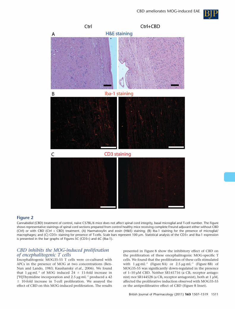
CBD slows down MOG-induced axonaldamage and inflammation in spinalcord of EAE miceMice were killed 30 days after EAE induction and spinal cordsections were analysed for the degree of damage to the whitematter tissue, using the SMI-32 antibody which stainsdamaged axons. SMI-32 staining revealed extensive axonaldegeneration in the MOG-immunized EAE mice. In contrast,CBD-treated EAE mice showed much lesser axonal damage(Figure 3). Next, we performed H&E histological staining tovisualize the inflamed areas in the spinal cord. Results pre-sented in Figure 4 show markedly weaker inflammation insections of spinal cord derived from CBD-treated EAE mice,compared with untreated EAE mice. These results werefurther confirmed by immunocytochemical staining forCD3+ T lymphocytes and markers of macrophages/microglialcells. Spinal cords derived from the EAE mice were rich inCD3+ T-cell infiltrates (Figure 5A,C), while treatment withCBD profoundly and significantly decreased CD3+ infiltratesinto the white matter (Figure 5B,C).
CBD decreases MOG-inducedmicroglial/macrophage activationAs shown above, immunohistochemical staining for the pres-ence of Iba-1 microglial marker showed that Iba-1 isexpressed in the spinal cord of control mice at a very low leveland that CBD injections to control naïve mice did not affectthis basal expression (Figure 2B). Iba-1 expression was,however, dramatically increased on day 30 post MOG immu-nization (Figure 6A,C). In contrast, a profoundly reducednumber of Iba-1 stained cells was present after CBD treatmentof these mice (Figure 6B,C). The Iba-1 expression in CBD-treated MOG-induced EAE group remained slightly higherthan that observed in naïve control animals showing thatCBD treatment did not completely eliminate microglial acti-vation. This result is in agreement with the mild but stillpresent clinical signs as shown in Figure 1.
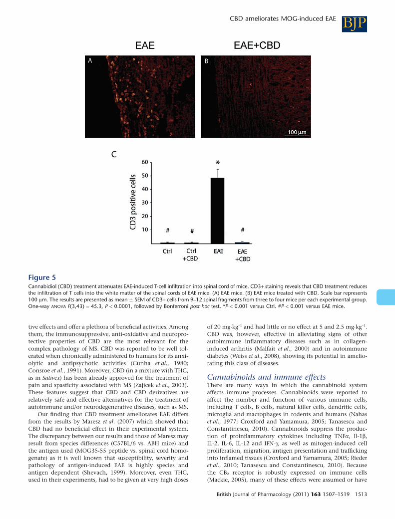
Mac-2/Galectin-3 is a microglial/macrophage markerwhose expression reflects the activation state of these cells.Staining of spinal cord of healthy, naïve animals for Mac-2/Galectin-3 shows absence (or presence of very low number) ofactivated microglial cells (Figure 7C). CBD injections did notchange Mac-2/Galectin-3 expression in these naïve controlanimals. However, staining of spinal cords derived from EAEmice resulted in dramatically increased number of Mac-2/Galectin-3 positive cells in the spinal cord (Figure 7A,C). Thisincrease was significantly reduced by CBD injections duringthe onset of clinical EAE (Figure 7B,C).
Figure 1Cannabidiol (CBD) ameliorates the clinical signs and disease progres-sion of EAE induced by myelin oligodendrocyte glycoprotein (MOG).EAE was induced in C57BL/6 mice by flank s.c. immunization withMOG35-55. Clinical disease scores were recorded daily until day 30after induction. CBD (CBD + EAE) or its vehicle (EAE group), wasinjected i.p. during the onset of the disease for 3 consecutive days(days 19–21; indicated by arrows). The mean clinical scores � SD areshown. Each group consisted of 15 mice. Repeated measure ANOVA
showed significant differences between the EAE and CBD + EAEgroups (P < 0.001).
BJP E Kozela et al.
1510 British Journal of Pharmacology (2011) 163 1507–1519
CBD inhibits the MOG-induced proliferationof encephalitogenic T cellsEncephalitogenic MOG35-55 T cells were co-cultured withAPCs in the presence of MOG at two concentrations (Ben-Nun and Lando, 1983; Kaushansky et al., 2006). We foundthat 1 mg·mL-1 of MOG induced 24 � 11-fold increase in[3H]Thymidine incorporation and 2.5 mg·mL-1 produced a 42� 10-fold increase in T-cell proliferation. We assayed theeffect of CBD on this MOG-induced proliferation. The results
presented in Figure 8 show the inhibitory effect of CBD onthe proliferation of these encephalitogenic MOG-specific Tcells. We found that the proliferation of these cells stimulatedwith 1 mg·mL-1 (Figure 8A) or 2.5 mg·mL-1 (Figure 8B) ofMOG35-55 was significantly down-regulated in the presenceof 1–10 mM CBD. Neither SR141716 (a CB1 receptor antago-nist) nor SR144528 (a CB2 receptor antagonist), both at 1 mM,affected the proliferative induction observed with MOG35-55or the antiproliferative effect of CBD (Figure 8 Inset).
Figure 2Cannabidiol (CBD) treatment of control, naïve C57BL/6 mice does not affect spinal cord integrity, basal microglial and T-cell number. The Figureshows representative stainings of spinal cord sections prepared from control healthy mice receiving complete Freund adjuvant either without CBD(Ctrl) or with CBD (Ctrl + CBD) treatment. (A) Haematoxylin and eosin (H&E) staining; (B) Iba-1 staining for the presence of microglial/macrophages; and (C) CD3+ staining for presence of T-cells. Scale bars represent 100 mm. Statistical analysis of the CD3+ and Iba-1 expressionis presented in the bar graphs of Figures 5C (CD3+) and 6C (Iba-1).
BJPCBD ameliorates MOG-induced EAE
British Journal of Pharmacology (2011) 163 1507–1519 1511

In parallel, we evaluated the effect of CBD on the spon-taneous proliferation of T cells (without MOG activation). Asdescribed above, this spontaneous proliferation was very lowcompared with the MOG-induced proliferation. We foundthat neither 0.15 mM (SI = 1.1 � 0.0), 5 mM (SI = 1.1 � 0.0) nor10 mM (SI = 0.9 � 0.0) of CBD affected the basal level ofproliferation of the T cells in the absence of MOG. Altogether,these results show that CBD selectively inhibited antigen-induced cell proliferation but did not affect basal (non-antigen-induced) proliferation.
Discussion
Our data showed that CBD, when administered systemicallyfor 3 days during clinical EAE disease onset, ameliorated the
EAE clinical signs and slowed its progression. The attenuationof EAE progression was accompanied by diminished axonaldamage and immune cell infiltration (including T cells andmicroglial cells) and activation. Using a highly specificautoreactive T-cell line, we observed that CBD decreased theMOG-induced proliferation of these cells.
The effectiveness of cannabinoids in ameliorating theclinical signs of EAE in rodents had been previously shownusing mostly mixed affinity CB1/CB2 receptor ligands (THC,WIN552122) in either myelin peptide or virus-inducedmodels (Lyman et al., 1989; Arévalo-Martín et al., 2003; Crox-ford and Miller, 2003). It should be noted that the immuno-suppressive effects of CB1 or mixed CB1/CB2 receptor ligandsare mostly evident at doses inducing central effects like seda-tion and hypothermia (Lyman et al., 1989; Croxford et al.,2008). CBD and its derivatives are devoid of such psychoac-
Figure 3Cannabidiol (CBD) reduces the MOG-induced axonal damage in spinal cords of EAE mice. Mice were killed 30 days after EAE disease inductionand sections of spinal cord were analysed for damaged axons using SMI-32 antibody. (A) MOG-induced EAE mice; (B) MOG-induced EAE micetreated with CBD (as described in Figure 1). Scale bar represents 200 mm.
Figure 4Cannabidiol (CBD) treatment attenuates EAE-induced immune cell infiltration into spinal cord of mice. Haematoxylin and eosin staining revealedthat CBD treatment reduces immune cell infiltration into the white matter of the spinal cords of EAE mice. (A) EAE mice and (B) EAE mice treatedwith CBD. Scale bar represents 100 mm.
BJP E Kozela et al.
1512 British Journal of Pharmacology (2011) 163 1507–1519
tive effects and offer a plethora of beneficial activities. Amongthem, the immunosuppressive, anti-oxidative and neuropro-tective properties of CBD are the most relevant for thecomplex pathology of MS. CBD was reported to be well tol-erated when chronically administered to humans for its anxi-olytic and antipsychotic activities (Cunha et al., 1980;Consroe et al., 1991). Moreover, CBD (in a mixture with THC,as in Sativex) has been already approved for the treatment ofpain and spasticity associated with MS (Zajicek et al., 2003).These features suggest that CBD and CBD derivatives arerelatively safe and effective alternatives for the treatment ofautoimmune and/or neurodegenerative diseases, such as MS.
Our finding that CBD treatment ameliorates EAE differsfrom the results by Maresz et al. (2007) which showed thatCBD had no beneficial effect in their experimental system.The discrepancy between our results and those of Maresz mayresult from species differences (C57BL/6 vs. ABH mice) andthe antigen used (MOG35-55 peptide vs. spinal cord homo-genate) as it is well known that susceptibility, severity andpathology of antigen-induced EAE is highly species andantigen dependent (Shevach, 1999). Moreover, even THC,used in their experiments, had to be given at very high doses
of 20 mg·kg-1 and had little or no effect at 5 and 2.5 mg·kg-1.CBD was, however, effective in alleviating signs of otherautoimmune inflammatory diseases such as in collagen-induced arthritis (Malfait et al., 2000) and in autoimmunediabetes (Weiss et al., 2008), showing its potential in amelio-rating this class of diseases.
Cannabinoids and immune effectsThere are many ways in which the cannabinoid systemaffects immune processes. Cannabinoids were reported toaffect the number and function of various immune cells,including T cells, B cells, natural killer cells, dendritic cells,microglia and macrophages in rodents and humans (Nahaset al., 1977; Croxford and Yamamura, 2005; Tanasescu andConstantinescu, 2010). Cannabinoids suppress the produc-tion of proinflammatory cytokines including TNFa, Il-1b,IL-2, IL-6, IL-12 and IFN-g, as well as mitogen-induced cellproliferation, migration, antigen presentation and traffickinginto inflamed tissues (Croxford and Yamamura, 2005; Riederet al., 2010; Tanasescu and Constantinescu, 2010). Becausethe CB2 receptor is robustly expressed on immune cells(Mackie, 2005), many of these effects were assumed or have
Figure 5Cannabidiol (CBD) treatment attenuates EAE-induced T-cell infiltration into spinal cord of mice. CD3+ staining reveals that CBD treatment reducesthe infiltration of T cells into the white matter of the spinal cords of EAE mice. (A) EAE mice. (B) EAE mice treated with CBD. Scale bar represents100 mm. The results are presented as mean � SEM of CD3+ cells from 9–12 spinal fragments from three to four mice per each experimental group.One-way ANOVA F(3,43) = 45.3, P < 0.0001, followed by Bonferroni post hoc test. *P < 0.001 versus Ctrl. #P < 0.001 versus EAE mice.
BJPCBD ameliorates MOG-induced EAE
British Journal of Pharmacology (2011) 163 1507–1519 1513

been shown to be mediated via the CB2 receptor (Buckleyet al., 2000). CB1 receptor-dependent immunosuppressiveeffects were also observed, despite their moderate expressionin immune cells (Cabral et al., 2001).
However, studies with CB1 and CB2 receptor knockoutmice as well as the use of specific CB1 or CB2 receptor antago-nists and of non-CB1/CB2 receptor ligands, revealed thatmany anti-inflammatory effects of cannabinoids do notinvolve the classic CB1/CB2 receptor pathways (Puffenbargeret al., 2000; Kaplan et al., 2003; 2008; Walter et al., 2003;Kozela et al., 2010). Indeed, it is known that non-CB1/CB2
receptor immunomodulatory effects exist and includecontrol of cytokine release (Kaplan et al., 2003; Kozela et al.,2010) and migratory activity of microglial cells (Walter et al.,2003; McHugh et al., 2010). Interestingly, some of the effectsof known CB1/CB2 receptor agonists also seem to involvenon-CB1/CB2 targets and the effects of non-CB1/CB2 receptorligands were slightly diminished in cells from CB1/CB2 recep-tor knockout mice (Kaplan et al., 2003; 2008) suggesting thatseveral types of cannabinoid targets may complement orinteract with each other.
Consequently, CBD, a compound active on non-CB1 orCB2 receptors (Showalter et al., 1996), has been shown to
possess a wide anti-inflammatory profile. CBD was shown todecrease TNFa, IL-2 and IFNg release from activated spleno-cytes and macrophages (Malfait et al., 2000; Jan et al., 2007;Kaplan et al., 2008). It also suppresses concavalin A and col-lagen induced T-cell proliferation (Malfait et al., 2000; Janet al., 2007), microglial migration (Walter et al., 2003) andcytokine release (Kozela et al., 2010). It suppresses antigen-specific antibody production in splenocytes (Jan et al., 2007),as well as attenuates endothelial inflammation and barrierdisruption (Rajesh et al., 2007). These activities of CBD maycontribute to its beneficial effects in EAE, as observed in ourhands, because many of these immune processes werereported to be involved in EAE pathology at different stages ofthe disease model.
Role of microglial cells in EAEIt is well established that microglia become rapidly activatedwhen the integrity of the CNS is disturbed as a consequenceof lesions, neurotoxicity, infections and autoimmune diseases(Hanisch and Kettenmann, 2007). This activation includesthe release of a variety of cytotoxic agents, for example,cytokines and chemokines, and of trophic factors that recruitother specialized cells from the periphery into the brain
Figure 6Cannabidiol (CBD) reduces the MOG-up-regulated Iba-1 expression in spinal cords of mice. Representative pictures showing that (A) MOGinjections increase Iba-1 staining in spinal cord, and (B) that CBD injections significantly decrease the level of Iba-1 staining. (C) Quantificationof Iba-1 fluorescence in sections of control (Ctrl) and of EAE mice with and without CBD treatment. The results are presented as mean � SEM oftotal fluorescence from 8–11 spinal fragments from three to four mice per each experimental group. One-way ANOVA F(3,37) = 30.23, P < 0.0001,followed by Bonferroni post hoc test. *P < 0.001 versus Ctrl. #P < 0.001 versus EAE mice. Scale bar represents 100 mm.
BJP E Kozela et al.
1514 British Journal of Pharmacology (2011) 163 1507–1519
parenchyma. Microglial activation is accompanied by mor-phological changes and surface expression of various mol-ecules that serve as adhesion molecules, enzymes,complement and immunoglobulin receptors and moleculesrelated to T-cell activation (Hanisch and Kettenmann, 2007).For example, the Iba-1 protein, located within a segment ofthe major histocompatibility complex class III region, is spe-cifically expressed in macrophages/microglia and its expres-sion is up-regulated during the activation of these cells.Mac-2 is a member of the Galectin-3 family of galactosebinding lectins, is another microglia differentiation and acti-vation marker. In this regard, microglial cells expressingMac-2 are capable of phagocytosis of myelin, while restingmicroglia are not (Reichert and Rotshenker, 1999).
Microglial cells are considered as key players in MS atvarious stages of the disease (Ponomarev et al., 2005; Koninget al., 2009). Depletion or blockade of microglia reducesinflammatory lesions, demyelination and EAE disease pro-gression (Huitinga et al., 1990; Heppner et al., 2005). Thus,arrest of microglial function is postulated to be beneficial inEAE/MS treatment. Indeed, we showed here that CBD injec-tions diminished the activation of microglia as measured by
Iba-1 and Mac-2 expression in the spinal cord of MOG-injected mice. As mentioned above, although Mac-2 and par-ticularly Iba-1 are generally accepted expression markers formicroglia (Reichert and Rotshenker, 1999; Heppner et al.,2005), these proteins are also expressed on some perivascularmacrophages and macrophages infiltrating the CNS duringpathological conditions. Thus, the inhibitory activity of CBDmay apply to microglia as well as to several othermacrophage-like cells. Previously, we and others have shownthat CBD in vitro decreases the production of TNFa, IL-1b,IL-6 and ROS species in LPS-activated microglia (El-Remessyet al., 2006; Liou et al., 2008; Kozela et al., 2010). Moreover,the microglial production of one of the most potent T-cellattracting chemokines, CCL-2, was also reduced followingCBD treatment (Kozela et al., 2010; Juknat et al., 2011). CBDalso reduces the migration of microglial cells (Walter et al.,2003). These data suggest that CBD is a potent inhibitor ofvarious functions of microglial cells. Thus, by silencingmicroglial cells, CBD may be able to prevent further inflam-matory processes within the CNS parenchyma, that is,myelin antigen exposure, T-cell infiltration, cytokine/chemokine release and recruitment of other immune cells.
Figure 7Cannabidiol (CBD) reduces the increased Mac-2/Galectin-3 expression in the spinal cords of EAE mice. Representative pictures showing that (A)MOG injections increase Mac-2 expression and (B) that CBD injections during the onset of EAE significantly decrease the level of Mac-2 staining.(C) Quantification of Mac-2 fluorescence in spinal cord sections of control and EAE mice with and without CBD treatment. The results arepresented as mean � SEM of total fluorescence from 8–11 spinal fragments from three to four mice per each experimental group. One-way ANOVA
F(3,34) = 63.25, P < 0.0001, followed by Bonferroni post hoc test. *P < 0.001 versus Ctrl. #P < 0.001 versus EAE mice. Scale bar represents 100 mm.
BJPCBD ameliorates MOG-induced EAE
British Journal of Pharmacology (2011) 163 1507–1519 1515

Antiproliferative effects of CBDWe showed that CBD decreased MOG-induced encephalito-genic T-cell proliferation. This antiproliferative effect of CBDwas not mediated via CB1 or CB2 receptors. High proliferationrate is a core response of immune cells (including T cells)following various stimuli, such as cytokines, mitogens andantigens presented by APCs. With regard to EAE or MS, it wasshown that myelin-specific T-cell clones penetrate the blood-brain barrier and the CNS parenchyma initiating demyelina-tion. We observed that CBD inhibited the MOG-inducedproliferation of encephalitogenic T cells isolated from mice,previously immunized against MOG. This potent antiprolif-erative activity of CBD probably contributes significantly tothe EAE-ameliorating effects of CBD and is in agreement withthe lower number of T cells present in the spinal cords ofCBD-treated EAE mice. Indeed, CBD has been reported toinduce apoptosis in transformed T-cell lines (McKallip et al.,2006; Lee et al., 2008) as well as in primary splenic lympho-
cytes including CD4+, CD8+ and B220+ subsets (Lee et al.,2008; Wu et al., 2008). It had a similar apoptotic activitytowards lymphocytes in peripheral blood (Ignatowska-Jankowska et al., 2009). Moreover, concavalin A-induced (Janet al., 2007) and collagen-induced stimulation of T cells(Malfait et al., 2000) was inhibited by CBD. On the otherhand, primary monocytes were observed to be resistant to theapoptotic effects of CBD (Gallily et al., 2003). This differencein apoptotic susceptibility of various immune cell types toCBD is of considerable significance but its mechanism is stillunknown.
Anti-oxidant and neuroprotective propertiesof CBDSeveral cannabinoids including THC and CBD exert anti-oxidative and neuroprotective properties (Mechoulam et al.,2007). Most of the current MS therapies are directed againstvarious immune cells to achieve immunosuppressive effects.
Figure 8Cannabidiol (CBD) inhibits MOG-induced T-cell proliferation. MOG-induced T-cell proliferation was determined by [3H]thymidine incorporation.Encephalitogenic T cells were co-cultured with antigen presenting cells and exposed to MOG35-55 at 1 (A) and 2.5 mg·mL-1 (B). CBD was added5 min before MOG. Assays were carried out each time in triplicate and mean per cent values � SEM are shown based on four independentexperiments. (A) One-way ANOVA F(4,19) = 124.7, P < 0.0001; (B) One-way ANOVA F(4,19) = 31.1, P < 0.0001, followed by Bonferroni post hoctest. *P < 0.05, ***P < 0.001 versus MOG alone at the respective concentration. Inset: neither the CB1 receptor antagonist (1 mM of SR141716;SR1) nor the CB2 receptor antagonist (1 mM of SR144528; SR2) applied 30 min before CBD and MOG affected MOG35-55 (1 mg·mL-1) – inducedT-cell proliferation either in the absence or presence of CBD; one-way ANOVA F(8,26) = 26.8, P < 0.05; ns, non-significant.
BJP E Kozela et al.
1516 British Journal of Pharmacology (2011) 163 1507–1519

However, increasing evidence shows that immunosuppres-sion alone was not sufficient for therapeutic effect especiallyin late, secondary progressive MS (Bennett and Stüve, 2009;Jones and Coles, 2010). In these cases, the neurodegenerativeprocesses become resistant to immunomodulation. Indeed,neurodegeneration that consists of neuronal and axonal losscan result from oxidative stress and excitotoxicity and isdriven by activated microglial cells and macrophagic/monocytic infiltrates (Hanisch and Kettenmann, 2007).It appears that the cannabinoid system could provide arescue mechanism in such conditions. Accordingly, the ame-liorating activity of THC-like cannabinoids combines CB2
receptor-mediated inhibition of autoreactive T cells and CB1
receptor-mediated neuroprotective activity on neurons(Maresz et al., 2007). Similarly, Croxford et al. (2008) pointedout that cannabinoid-mediated neuroprotection rather thanimmunosuppression, was relevant for the recovery process atthe later, remissive stages of MS.
The main source of reactive oxygen and nitrogen specieswithin the CNS under neurodegenerative conditions arereactive microglia and astrocytes (Hanisch and Kettenmann,2007). Oxidative signalling negatively affects neuronal andaxonal survival, especially of axons lacking a myelin sheath,and results in irreversible damage. Accordingly, deletion ofnuclear factor-erythroid 2-related 2 (Nrf2), a redox-sensitivetranscription factor that regulates expression of many pro-tective antioxidant and detoxication enzymes and transport-ers (Kobayashi and Yamamoto, 2005), resulted inexacerbation of EAE (Johnson et al., 2010). CBD has beenfrequently described as a potent neuroprotective andanti-oxidant agent. CBD reduced glutamate excitotoxicityand hydroperoxide-induced oxidative neuronal damage(Hampson et al., 1998) and provided neuroprotectionagainst 6-hydroxy-dopamine (Lastres-Becker et al., 2005),b-amyloid (Iuvone et al., 2004) and prion toxicities toneurons (Dirikoc et al., 2007) and against neuroinflamma-tion (Esposito et al., 2007; Zuardi, 2008). In this regard, wehave shown that exposure of microglial cells in culture toCBD up-regulates a number of anti-oxidative genes includ-ing genes that are involved in glutathione synthesis. Manyof these genes are under the control of the Nrf2 transcrip-tion factor (Juknat et al., 2011). Thus, the anti-oxidativeproperties of CBD may significantly contribute to thealleviation of EAE pathology and accompany its anti-inflammatory beneficial properties.
In summary, we have shown that CBD administered toMOG-immunized C57BL/6 mice, at the onset of EAE disease,reduced the severity of the clinical signs of EAE. CBD treat-ment was accompanied by diminished axonal loss andinflammation (infiltration of T cells and microglial activa-tion). Moreover, CBD prevented proliferation of myelin-specific T cells in vitro. These observations suggest that CBDmay have potential for alleviating MS-like pathology.
Acknowledgements
This work was supported by the Dr Miriam and Sheldon G.Adelson Center for the Biology of Addictive Diseases and bythe Dr Miriam and Sheldon G. Adelson Medical Research
Foundation. A. J. and N. R. were supported by the Center forAbsorption in Science in Israel.
Conflict of interest
The authors state no conflict of interest.
ReferencesAlexander SPH, Mathie A, Peters JA (2009). Guide to Receptors andChannels (GRAC), 4th edn. Br J Pharmacol 158: S1–S254.
Arévalo-Martín A, Vela JM, Molina-Holgado E, Borrell J, Guaza C(2003). Therapeutic action of cannabinoids in a murine model ofmultiple sclerosis. J Neurosci 23: 2511–2516.
Bayewitch M, Avidor-Reiss T, Levy R, Barg J, Mechoulam R, Vogel Z(1995). The peripheral cannabinoid receptor: adenylate cyclaseinhibition and G protein coupling. FEBS Lett 375: 143–147.
Bennett JL, Stüve O (2009). Update on inflammation,neurodegeneration, and immunoregulation in multiple sclerosis:therapeutic implications. Clin Neuropharmacol 32: 121–132.
Ben-Nun A, Lando Z (1983). Detection of autoimmune cellsproliferating to myelin basic protein and selection of T cell linesthat mediate experimental autoimmune encephalomyelitis (EAE) inmice. J Immunol 130: 1205–1209.
Buckley NE, McCoy KL, Mezey E, Bonner T, Zimmer A, Felder CCet al. (2000). Immunomodulation by cannabinoids is absent in micedeficient for the cannabinoid CB(2) receptor. Eur J Pharmacol 396:141–149.
Cabral GA, Harmon KN, Carlisle SJ (2001). Cannabinoid-mediatedinhibition of inducible nitric oxide production by rat microglialcells: evidence for CB1 receptor participation. Adv Exp Med Biol493: 207–214.
Compston A, Coles A (2008). Multiple sclerosis. Lancet 372:1502–1517.
Consroe P, Laguna J, Allender J, Snider S, Stern L, Sandyk R et al.(1991). Controlled clinical trial of cannabidiol in Huntington’sdisease. Pharmacol Biochem Behav 40: 701–708.
Croxford JL, Miller SD (2003). Immunoregulation of a viral modelof multiple sclerosis using the synthetic cannabinoid R+WIN55,212.J Clin Invest 111: 1231–1240.
Croxford JL, Yamamura T (2005). Cannabinoids and the immunesystem: potential for the treatment of inflammatory diseases? JNeuroimmunol 166: 3–18.
Croxford JL, Pryce G, Jackson SJ, Ledent C, Giovannoni G,Pertwee RG et al. (2008). Cannabinoid-mediated neuroprotection,not immunosuppression, may be more relevant to multiplesclerosis. J Neuroimmunol 193: 120–129.
Cunha JM, Carlini EA, Pereira AE, Ramos OL, Pimentel C,Gagliardi R et al. (1980). Chronic administration of cannabidiol tohealthy volunteers and epileptic patients. Pharmacology 21:175–185.
Dirikoc S, Priola SA, Marella M, Zsürger N, Chabry J (2007).Nonpsychoactive cannabidiol prevents prion accumulation andprotects neurons against prion toxicity. J Neurosci 27: 9537–9544.
BJPCBD ameliorates MOG-induced EAE
British Journal of Pharmacology (2011) 163 1507–1519 1517

El-Remessy AB, Al-Shabrawey M, Khalifa Y, Tsai NT, Caldwell RB,Liou GI (2006). Neuroprotective and blood-retinal barrier-preservingeffects of cannabidiol in experimental diabetes. Am J Pathol 168:235–244.
Esposito G, Scuderi C, Savani C, Steardo L Jr, De Filippis D,Cottone P et al. (2007). Cannabidiol in vivo blunts b-amyloidinduced neuroinflammation by suppressing IL-1 b and iNOSexpression. Br J Pharmacol 151: 1272–1279.
Gallily R, Even-Chena T, Katzavian G, Lehmann D, Dagan A,Mechoulam R (2003). g-irradiation enhances apoptosis induced bycannabidiol, a non-psychotropic cannabinoid, in cultured HL-60myeloblastic leukemia cells. Leuk Lymphoma 44: 1767–1773.
Hampson AJ, Grimaldi M, Axelrod J, Wink D (1998). Cannabidioland (-)Delta9-tetrahydrocannabinol are neuroprotectiveantioxidants. Proc Natl Acad Sci U S A 95: 8268–8273.
Hanisch UK, Kettenmann H (2007). Microglia: active sensor andversatile effector cells in the normal and pathologic brain. NatNeurosci 10: 1387–1394.
Heppner FL, Greter M, Marino D, Falsig J, Raivich G, Hovelmeyer Net al. (2005). Experimental autoimmune encephalomyelitisrepressed by microglial paralysis. Nat Med 11: 146–152.
Huitinga I, Van Rooijen N, De Groot CJ, Uitdehaag BM,Dijkstra CD (1990). Suppression of experimental allergicencephalomyelitis in Lewis rats after elimination of macrophages. JExp Med 172: 1025–1033.
Ignatowska-Jankowska B, Jankowski M, Glac W, Swiergel AH (2009).Cannabidiol-induced lymphopenia does not involve NKT and NKcells. J Physiol Pharmacol 3 (Suppl.): 99–103.
Iuvone T, Esposito G, Esposito R, Santamaria R, Di Rosa M, Izzo AA(2004). Neuroprotective effect of cannabidiol, a non-psychoactivecomponent from Cannabis sativa, on b-amyloid-induced toxicity inPC12 cells. J Neurochem 89: 134–141.
Jack C, Ruffini F, Bar-Or A, Antel JP (2005). Microglia and multiplesclerosis. J Neurosci Res 81: 363–373.
Jan TR, Su ST, Wu HY, Liao MH (2007). Suppressive effects ofcannabidiol on antigen-specific antibody production and functionalactivity of splenocytes in ovalbumin-sensitized BALB/c mice. IntImmunopharmacol 7: 773–780.
Járai Z, Wagner JA, Varga K, Lake KD, Compton DR, Martin BR et al.(1999). Cannabinoid-induced mesenteric vasodilation through anendothelial site distinct from CB1 or CB2 receptors. Proc Natl AcadSci U S A 96: 14136–14141.
Johnson DA, Amirahmadi S, Ward C, Fabry Z, Johnson JA (2010).The absence of the pro-antioxidant transcription factor Nrf2exacerbates experimental autoimmune encephalomyelitis. ToxicolSci 114: 237–246.
Jones JL, Coles AJ (2010). New treatment strategies in multiplesclerosis. Exp Neurol 225: 34–39.
Juknat A, Pietr M, Kozela E, Rimmerman N, Levy R, Coppola Get al. (2011). Differential transcriptional profiles mediatedby exposure to the cannabinoids cannabidiol andD9-tetrahydrocannabinol in BV-2 microglial cells. Br J Pharm DOI:10.1111/j.1476-5381.2011.01461.x.
Kaplan BL, Rockwell CE, Kaminski NE (2003). Evidence forcannabinoid receptor-dependent and -independent mechanisms ofaction in leukocytes. J Pharmacol Exp Ther 306: 1077–1085.
Kaplan BL, Springs AE, Kaminski NE (2008). The profile of immunemodulation by cannabidiol (CBD) involves deregulation of nuclearfactor of activated T cells (NFAT). Biochem Pharmacol 76: 726–737.
Kaushansky N, Zhong MC, Kerlero de Rosbo N, Hoeftberger R,Lassmann H, Ben-Nun A (2006). Epitope specificity of autoreactiveT and B cells associated with experimental autoimmuneencephalomyelitis and optic neuritis induced by oligodendrocyte-specific protein in SJL/J mice. J Immunol 177: 7364–7376.
Klein TW, Newton CA (2007). Therapeutic potential ofcannabinoid-based drugs. Adv Exp Med Biol 601: 395–413.
Kobayashi M, Yamamoto M (2005). Molecular mechanismsactivating the Nrf2-Keap1 pathway of antioxidant gene regulation.Antioxid Redox Signal 7: 385–394.
Koning N, Uitdehaag BM, Huitinga I, Hoek RM (2009). Restoringimmune suppression in the multiple sclerosis brain. Prog Neurobiol89: 359–368.
Kozela E, Pietr M, Juknat A, Rimmerman N, Levy R, Vogel Z (2010).Cannabinoids Delta(9)-tetrahydrocannabinol and cannabidioldifferentially inhibit the lipopolysaccharide-activated NF-kappaBand interferon- b/STAT proinflammatory pathways in BV-2microglial cells. J Biol Chem 285: 1616–1626.
Lastres-Becker I, Molina-Holgado F, Ramos JA, Mechoulam R,Fernández-Ruiz J (2005). Cannabinoids provide neuroprotectionagainst 6-hydroxydopamine toxicity in vivo and in vitro: relevanceto Parkinson’s disease. Neurobiol Dis 19: 96–107.
Lee CY, Wey SP, Liao MH, Hsu WL, Wu HY, Jan TR (2008). Acomparative study on cannabidiol-induced apoptosis in murinethymocytes and EL-4 thymoma cells. Int Immunopharmacol 8:732–740.
Lev N, Barhum Y, Melamed E, Offen D (2004). Bax-ablationattenuates experimental autoimmune encephalomyelitis in mice.Neurosci Lett 359: 139–142.
Liou GI, Auchampach JA, Hillard CJ, Zhu G, Yousufzai B, Mian Set al. (2008). Mediation of cannabidiol anti-inflammation in theretina by equilibrative nucleoside transporter and A2A adenosinereceptor. Invest Ophthalmol Vis Sci 49: 5526–5531.
Lyman WD, Sonett JR, Brosnan CF, Elkin R, Bornstein MB (1989).Delta 9-tetrahydrocannabinol: a novel treatment for experimentalautoimmune encephalomyelitis. J Neuroimmunol 23: 73–81.
McHugh D, Hu SS, Rimmerman N, Juknat A, Vogel Z, Walker JMet al. (2010). N-arachidonoyl glycine, an abundant endogenouslipid, potently drives directed cellular migration through GPR18,the putative abnormal cannabidiol receptor. BMC Neurosci 11: 44.
McKallip RJ, Jia W, Schlomer J, Warren JW, Nagarkatti PS,Nagarkatti M (2006). Cannabidiol-induced apoptosis in humanleukemia cells: a novel role of cannabidiol in the regulation ofp22phox and nox4 expression. Mol Pharmacol 70: 897–908.
Mackie K (2005). Distribution of cannabinoid receptors in thecentral and peripheral nervous system. Handb Exp Pharmacol 168:299–325.
Malfait AM, Gallily R, Sumariwalla PF, Malik AS, Andreakos E,Mechoulam R et al. (2000). The nonpsychoactive cannabisconstituent cannabidiol is an oral anti-arthritic therapeutic inmurine collagen-induced arthritis. Proc Natl Acad Sci U S A 97:9561–9566.
Maresz K, Carrier EJ, Ponomarev ED, Hillard CJ, Dittel BN (2005).Modulation of the cannabinoid CB2 receptor in microglial cells inresponse to inflammatory stimuli. J Neurochem 95: 437–445.
Maresz K, Pryce G, Ponomarev ED, Marsicano G, Croxford JL,Shriver LP et al. (2007). Direct suppression of CNS autoimmuneinflammation via the cannabinoid receptor CB1 on neurons andCB2 on autoreactive T cells. Nat Med 13: 492–497.
BJP E Kozela et al.
1518 British Journal of Pharmacology (2011) 163 1507–1519

Mechoulam R, Peters M, Murillo-Rodriguez E, Hanus LO (2007).Cannabidiol–recent advances. Chem Biodivers 4: 1678–1692.
Nahas GG, Morishima A, Desoize B (1977). Effects of cannabinoidson macromolecular synthesis and replication of culturedlymphocytes. Fed Proc 36: 1748–1752.
Pietr M, Kozela E, Levy R, Rimmerman N, Lin YH, Stella N et al.(2009). Differential changes in GPR55 during microglial cellactivation. FEBS Lett 583: 2071–2076.
Ponomarev ED, Shriver LP, Maresz K, Dittel BN (2005). Microglialcell activation and proliferation precedes the onset of CNSautoimmunity. J Neurosci Res 81: 374–389.
Puffenbarger RA, Boothe AC, Cabral GA (2000). Cannabinoidsinhibit LPS-inducible cytokine mRNA expression in rat microglialcells. Glia 29: 58–69.
Rajesh M, Pan H, Mukhopadhyay P, Ba’ tkai S, Osei-Hyiaman D,Hasko G et al. (2007). Cannabidiol attenuates high glucose-inducedendothelial cell inflammatory response and barrier disruption. JLeukoc Biol 82: 1382–1389.
Reichert F, Rotshenker S (1999). Galectin-3/MAC-2 in experimentalallergic encephalomyelitis. Exp Neurol 160: 508–514.
Rieder SA, Chauhan A, Singh U, Nagarkatti M, Nagarkatti P (2010).Cannabinoid-induced apoptosis in immune cells as a pathway toimmunosuppression. Immunobiology 215: 598–560.
Shevach EM (1999). Animal models for autoimmune andinflammatory disease. In: Coligan JE, Kruisbeek AM, Margulies DH,
Shevach EM, Strober W (eds). Current Protocols in Immunology.John Wiley & Sons, Inc: New York, pp. 15.0.1–15.0.5.
Showalter VM, Compton DR, Martin BR, Abood ME (1996).Evaluation of binding in a transfected cell line expressing aperipheral cannabinoid receptor (CB2): identification ofcannabinoid receptor subtype selective ligands. J Pharmacol ExpTher 278: 989–999.
Tanasescu R, Constantinescu CS (2010). Cannabinoids and theimmune system: an overview. Immunobiology 215: 588–597.
Walter L, Franklin A, Witting A, Wade C, Xie Y, Kunos G et al.(2003). Nonpsychotropic cannabinoid receptors regulate microglialcell migration. J Neurosci 23: 1398–1405.
Weiss L, Zeira M, Reich S, Slavin S, Raz I, Mechoulam R et al.(2008). Cannabidiol arrests onset of autoimmune diabetes in NODmice. Neuropharmacology 54: 244–249.
Wu HY, Chu RM, Wang CC, Lee CY, Lin SH, Jan TR (2008).Cannabidiol-induced apoptosis in primary lymphocytes isassociated with oxidative stress-dependent activation of caspase-8.Toxicol Appl Pharmacol 226: 260–270.
Zajicek J, Fox P, Sanders H, Wright D, Vickery J, Nunn A et al.(2003). Cannabinoids for treatment of spasticity and otherssymptoms related to multiple sclerosis (CAMS study): multicenterrandomized placebo-controlled trial. Lancet 362: 1517–1526.
Zuardi AW (2008). Cannabidiol: from an inactive cannabinoid to adrug with wide spectrum of action. Rev Bras Psiquiatr 30: 271–280.
BJPCBD ameliorates MOG-induced EAE
British Journal of Pharmacology (2011) 163 1507–1519 1519


![Web view23 March 2017 [08–17] Supporting document 1 . Updated estimates of . dietary . exposure to 9. tetrahydrocannabinol (THC) and cannabidiol (CBD) from](https://static.fdocuments.in/doc/165x107/5ab2020e7f8b9aea528d163f/web-view23-march-2017-0817-supporting-document-1-updated-estimates-of-.jpg)