Cannabidiol and Other Cannabinoids Reduce Microglial ... · Cannabidiol and Other Cannabinoids...
10
Cannabidiol and Other Cannabinoids Reduce Microglial Activation In Vitro and In Vivo: Relevance to Alzheimer’s Disease Ana María Martín-Moreno, David Reigada, 1 Bele ´ n G. Ramírez, 2 R. Mechoulam, Nadia Innamorato, Antonio Cuadrado, and María L. de Ceballos Neurodegeneration Group, Department of Cellular, Molecular and Developmental Neurobiology, Instituto Cajal, Consejo Superior de Investigaciones Cientificas, Madrid, Spain (A.M.M.-M., D.R., B.G.R., M.L.C.); Centro de Investigacio ´n en Red en Enfermedades Neurodegenerativas (A.M.M.-M., D.R., B.G.R., M.L.C., N.I., A.C.) and Institute for Drug Research, Medical Faculty, Hebrew University (R.M.), Jerusalem, Israel; and Departamento de Bioquímica, Instituto de Investigaciones Biome ´ dicas “Alberto Sols” UAM-Consejo Superior de Investigaciones Cientificas, Facultad de Medicina, Universidad Auto ´ noma de Madrid, Madrid, Spain (N.I., A.C.) Received January 18, 2011; accepted February 11, 2011 ABSTRACT Microglial activation is an invariant feature of Alzheimer’s disease (AD). It is noteworthy that cannabinoids are neuroprotective by pre- venting -amyloid (A)-induced microglial activation both in vitro and in vivo. On the other hand, the phytocannabinoid cannabidiol (CBD) has shown anti-inflammatory properties in different paradigms. In the present study, we compared the effects of CBD with those of other cannabinoids on microglial cell functions in vitro and on learning behavior and cytokine expression after A intraventricular adminis- tration to mice. CBD, ( R)-( +)-[2,3-dihydro-5-methyl-3-(4-morpholinyl- methyl) pyrrolo-[1,2,3-d,e]-1,4-benzoxazin-6-yl]-1-naphthalenyl- methanone [WIN 55,212-2 (WIN)], a mixed CB 1 /CB 2 agonist, and 1,1-dimethylbutyl-1-deoxy- 9 -tetrahydrocannabinol [JWH-133 (JWH)], a CB 2 -selective agonist, concentration-dependently de- creased ATP-induced (400 M) increase in intracellular calcium ([Ca 2+ ] i ) in cultured N13 microglial cells and in rat primary microglia. In contrast, 4-[4-(1,1-dimethylheptyl)-2,6-dimethoxyphenyl]-6,6- dimethyl-bicyclo[3.1.1]hept-2-ene-2-methanol [HU-308 (HU)], an- other CB 2 agonist, was without effect. Cannabinoid and aden- osine A 2A receptors may be involved in the CBD action. CBD- and WIN-promoted primary microglia migration was blocked by CB 1 and/or CB 2 antagonists. JWH and HU- induced migration was blocked by a CB 2 antagonist only. All of the cannabinoids decreased lipopolysaccharide-induced nitrite generation, which was insensitive to cannabinoid an- tagonism. Finally, both CBD and WIN, after subchronic ad- ministration for 3 weeks, were able to prevent learning of a spatial navigation task and cytokine gene expression in -amyloid-injected mice. In summary, CBD is able to mod- ulate microglial cell function in vitro and induce beneficial effects in an in vivo model of AD. Given that CBD lacks psychoactivity, it may represent a novel therapeutic ap- proach for this neurological disease. Introduction Alzheimer’s disease (AD) is characterized by -amyloid (A) deposition in senile plaques, neurofibrillary tangles, selective neuronal loss, and progressive cognitive deficits. Another invariant feature of the neurological disease is glial activation and is considered to be responsible of the ongoing inflammatory condition occurring in AD brain (Akiyama et al., 2000). Microglial activation is also present in AD exper- imental models in vivo, such as rats injected with A either focally or intraventricularly, and in transgenic models of the This work was supported by the Spanish Ministry of Science and Technol- ogy [Grant SAF 2005-02845]; the National Institutes of Health National In- stitute on Drug Abuse [Grant DA9789]; a fellowship from the Community of Madrid (to B.G.R.); and a fellowship from the Ministry of Science and Inno- vation (to A.M.M.-M.). 1 Current affiliation: Molecular Neuroprotection Group, Experimental Neu- rology Unit, Paraplejic National Hospital, Toledo, Spain. 2 Current affiliation: Unidad de Apoyo a la Investigacio ´n, Fundacio ´n MD Anderson Internacional Espan ˜ a, Madrid, Spain. A.M.M.-M. and D.R. contributed equally to this work. Article, publication date, and citation information can be found at http://molpharm.aspetjournals.org. doi:10.1124/mol.111.071290. ABBREVIATIONS: AD, Alzheimer’s disease; A, -amyloid peptide; IL-6, interleukin 6; TNF-, tumor necrosis factor-; WIN, WIN 55,212-2, (R)-(+)- [2,3-dihydro-5-methyl-3-(4-morpholinylmethyl) pyrrolo-[1,2,3-d,e]-1,4-benzoxazin-6-yl]-1-naphthalenyl-methanone; JWH, JWH-133; JWH-133, 1,1- dimethylbutyl-1-deoxy- 9 -tetrahydrocannabinol; HU, HU-308, 4-[4-(1,1-dimethylheptyl)-2,6-dimethoxyphenyl]-6,6-dimethyl-bicyclo[3.1.1]hept-2-ene- 2-methanol; LPS, lipopolysaccharide; iNOS, inducible nitric-oxide synthase; SCR, scrambled peptide; FCS, fetal calf serum; PCR, polymerase chain reaction; ANOVA, analysis of variance; SR1, SR141716, N-piperidino-5-(4-chlorophenyl)-l-(2, 4-dichlorophenyl)-4-methylpyrazole-3-carboxamide; SR2, SR144528, N-[(1S)-endo-1,3,3-trimethyl bicyclo [2.2.1] heptan-2-yl]-5-(4-chloro-3-methylphenyl)-1-(4-methylbenzyl)-pyrazole-3-carboxamide; CGS, CGS21680, 2-[p-(2-carboxyethyl)phenethylamino]-5'-N-ethylcarboxamidoadenosine; ZM, ZM241385, 4-{2-[7-amino-2-(2-furyl)[1,2,4]triazolo-[2,3- a][1,3,5]triazin-5-ylamino]ethyl}phenol; VDM11, N-(4-hydroxy-2-methylphenyl)-5Z,8Z,11Z,14Z-eicosatetraenamide. 0026-895X/11/7906-964–973$25.00 MOLECULAR PHARMACOLOGY Vol. 79, No. 6 Copyright © 2011 The American Society for Pharmacology and Experimental Therapeutics 71290/3686012 Mol Pharmacol 79:964–973, 2011 Printed in U.S.A. 964
-
Upload
nguyennhan -
Category
Documents
-
view
228 -
download
0
Transcript of Cannabidiol and Other Cannabinoids Reduce Microglial ... · Cannabidiol and Other Cannabinoids...

Cannabidiol and Other Cannabinoids Reduce Microglial ActivationIn Vitro and In Vivo: Relevance to Alzheimer’s Disease
Ana María Martín-Moreno, David Reigada,1 Belen G. Ramírez,2 R. Mechoulam,Nadia Innamorato, Antonio Cuadrado, and María L. de Ceballos
Neurodegeneration Group, Department of Cellular, Molecular and Developmental Neurobiology, Instituto Cajal, ConsejoSuperior de Investigaciones Cientificas, Madrid, Spain (A.M.M.-M., D.R., B.G.R., M.L.C.); Centro de Investigacion en Red enEnfermedades Neurodegenerativas (A.M.M.-M., D.R., B.G.R., M.L.C., N.I., A.C.) and Institute for Drug Research, MedicalFaculty, Hebrew University (R.M.), Jerusalem, Israel; and Departamento de Bioquímica, Instituto de Investigaciones Biomedicas“Alberto Sols” UAM-Consejo Superior de Investigaciones Cientificas, Facultad de Medicina, Universidad Autonoma de Madrid,Madrid, Spain (N.I., A.C.)
Received January 18, 2011; accepted February 11, 2011
ABSTRACT
Microglial activation is an invariant feature of Alzheimer’s disease(AD). It is noteworthy that cannabinoids are neuroprotective by pre-venting �-amyloid (A�)-induced microglial activation both in vitro andin vivo. On the other hand, the phytocannabinoid cannabidiol (CBD)has shown anti-inflammatory properties in different paradigms. In thepresent study, we compared the effects of CBD with those of othercannabinoids on microglial cell functions in vitro and on learningbehavior and cytokine expression after A� intraventricular adminis-tration to mice. CBD, (R)-(�)-[2,3-dihydro-5-methyl-3-(4-morpholinyl-methyl) pyrrolo-[1,2,3-d,e]-1,4-benzoxazin-6-yl]-1-naphthalenyl-methanone [WIN 55,212-2 (WIN)], a mixed CB1/CB2 agonist, and1,1-dimethylbutyl-1-deoxy-�9-tetrahydrocannabinol [JWH-133(JWH)], a CB2-selective agonist, concentration-dependently de-creased ATP-induced (400 �M) increase in intracellular calcium([Ca2�]i) in cultured N13 microglial cells and in rat primary microglia.In contrast, 4-[4-(1,1-dimethylheptyl)-2,6-dimethoxyphenyl]-6,6-
dimethyl-bicyclo[3.1.1]hept-2-ene-2-methanol [HU-308 (HU)], an-other CB2 agonist, was without effect. Cannabinoid and aden-osine A2A receptors may be involved in the CBD action.CBD- and WIN-promoted primary microglia migration wasblocked by CB1 and/or CB2 antagonists. JWH and HU-induced migration was blocked by a CB2 antagonist only. Allof the cannabinoids decreased lipopolysaccharide-inducednitrite generation, which was insensitive to cannabinoid an-tagonism. Finally, both CBD and WIN, after subchronic ad-ministration for 3 weeks, were able to prevent learning of aspatial navigation task and cytokine gene expression in�-amyloid-injected mice. In summary, CBD is able to mod-ulate microglial cell function in vitro and induce beneficialeffects in an in vivo model of AD. Given that CBD lackspsychoactivity, it may represent a novel therapeutic ap-proach for this neurological disease.
Introduction
Alzheimer’s disease (AD) is characterized by �-amyloid(A�) deposition in senile plaques, neurofibrillary tangles,selective neuronal loss, and progressive cognitive deficits.Another invariant feature of the neurological disease is glialactivation and is considered to be responsible of the ongoinginflammatory condition occurring in AD brain (Akiyama etal., 2000). Microglial activation is also present in AD exper-imental models in vivo, such as rats injected with A� eitherfocally or intraventricularly, and in transgenic models of the
This work was supported by the Spanish Ministry of Science and Technol-ogy [Grant SAF 2005-02845]; the National Institutes of Health National In-stitute on Drug Abuse [Grant DA9789]; a fellowship from the Community ofMadrid (to B.G.R.); and a fellowship from the Ministry of Science and Inno-vation (to A.M.M.-M.).
1 Current affiliation: Molecular Neuroprotection Group, Experimental Neu-rology Unit, Paraplejic National Hospital, Toledo, Spain.
2 Current affiliation: Unidad de Apoyo a la Investigacion, Fundacion MDAnderson Internacional Espana, Madrid, Spain.
A.M.M.-M. and D.R. contributed equally to this work.Article, publication date, and citation information can be found at
http://molpharm.aspetjournals.org.doi:10.1124/mol.111.071290.
ABBREVIATIONS: AD, Alzheimer’s disease; A�, �-amyloid peptide; IL-6, interleukin 6; TNF-�, tumor necrosis factor-�; WIN, WIN 55,212-2, (R)-(�)-
[2,3-dihydro-5-methyl-3-(4-morpholinylmethyl) pyrrolo-[1,2,3-d,e]-1,4-benzoxazin-6-yl]-1-naphthalenyl-methanone; JWH, JWH-133; JWH-133, 1,1-
dimethylbutyl-1-deoxy-�9-tetrahydrocannabinol; HU, HU-308, 4-[4-(1,1-dimethylheptyl)-2,6-dimethoxyphenyl]-6,6-dimethyl-bicyclo[3.1.1]hept-2-ene-
2-methanol; LPS, lipopolysaccharide; iNOS, inducible nitric-oxide synthase; SCR, scrambled peptide; FCS, fetal calf serum; PCR, polymerase chain
reaction; ANOVA, analysis of variance; SR1, SR141716, N-piperidino-5-(4-chlorophenyl)-l-(2, 4-dichlorophenyl)-4-methylpyrazole-3-carboxamide; SR2,
SR144528, N-[(1S)-endo-1,3,3-trimethyl bicyclo [2.2.1] heptan-2-yl]-5-(4-chloro-3-methylphenyl)-1-(4-methylbenzyl)-pyrazole-3-carboxamide; CGS,
CGS21680, 2-[p-(2-carboxyethyl)phenethylamino]-5�-N-ethylcarboxamidoadenosine; ZM, ZM241385, 4-{2-[7-amino-2-(2-furyl)[1,2,4]triazolo-[2,3-
a][1,3,5]triazin-5-ylamino]ethyl}phenol; VDM11, N-(4-hydroxy-2-methylphenyl)-5Z,8Z,11Z,14Z-eicosatetraenamide.
0026-895X/11/7906-964–973$25.00MOLECULAR PHARMACOLOGY Vol. 79, No. 6Copyright © 2011 The American Society for Pharmacology and Experimental Therapeutics 71290/3686012Mol Pharmacol 79:964–973, 2011 Printed in U.S.A.
964

disease (Masliah et al., 1996; Jantzen et al., 2002; Ramírez etal., 2005). Furthermore, A� addition to cultures induces mi-croglial activation, reflected in increased secretion of toxicspecies such as NO and cytokines (Combs et al., 2001;Ramírez et al., 2005). Given that microglial activation mayresult in neurodegeneration, currently, it is considered thatpharmacological targeting of microglial activity may be afeasible therapeutic strategy for neurodegenerative diseasesin general and for AD in particular.
Cannabinoids, whether plant-derived, synthetic, or endo-cannabinoids, exert their functions through activation of can-nabinoid receptors, two of which have been well character-ized to date: CB1 and CB2 (Howlett et al., 2002; Piomelli,2003). Cannabinoids are neuroprotective against excitotoxic-ity and acute brain damage, both in vitro and in vivo (van derStelt et al., 2002; Mechoulam and Shohami, 2007). Severalmechanisms account for the neuroprotection afforded by thistype of drug such as blockade of excitotoxicity, reduction ofcalcium influx, antioxidant properties of the compounds, orenhanced trophic factor support. A decrease in proinflamma-tory mediators brought about by cannabinoids (Walter andStella, 2004) may be also involved in their neuroprotection.Indeed, several reports have shown that cannabinoids reduceNO and cytokine generation and/or their mRNA expressionin microglia cultures (Waksman et al., 1999; Puffenbarger etal., 2000; Facchinetti et al., 2003). Cannabidiol (CBD), themajor plant-derived nonpsychotropic constituent of mari-juana, is of potential therapeutic interest in different diseaseconditions (e.g., inflammation) (Mechoulam et al., 2007).Oral treatment with CBD decreases edema and hyperalgesiain a rat paw model of carrageenan-induced inflammation(Costa et al., 2004). A single dose of the phytocannabinoidreduces tumor necrosis factor-� (TNF-�) levels in lipopoly-saccharide (LPS)-injected mice and improves collagen-in-duced arthritis (Malfait et al., 2000). CBD binds to CB recep-tors with low affinity (Showalter et al., 1996) and may exertcannabinoid receptor-independent effects as well. For in-stance, CBD inhibition of the equilibrative nucleoside trans-porter, which results in enhancement of adenosine signalingthrough A2A receptors, is involved in its immunosuppressiveeffects (Carrier et al., 2006).
In the context of AD, CBD has shown to be neuroprotectiveagainst the A� addition to cultured cells. Several mecha-nisms seem to be involved, including CBD reduction of oxi-dative stress and blockade of apoptosis (Iuvone et al., 2004),�-phosphorylation inhibition through the Wnt/�-cateninpathway (Esposito et al., 2006a), and the decrease in iNOSexpression and nitrite generation (Esposito et al., 2006b). Wehave shown that cannabinoids prevent A�-induced neurode-generation by reducing microglial activation (Ramírez et al.,2005), and both CB1 and CB2 receptors in microglia partici-pate in such an action. More importantly, cannabinoids pre-vented microglial activation, loss of neuronal markers, andcognitive deficits in A�-treated rats (Ramírez et al., 2005). Invivo, CBD also suppressed neuroinflammation in mice in-jected with A� into the hippocampus by inhibiting the in-creased glial fibrillary acidic protein and iNOS expression,along with nitrite and interleukin-1� generation (Esposito etal., 2007). However, previous studies have not investigatedthe effects of CBD on microglial cell function.
Taken together, these results prompted us to study the effectsCBD in comparison with other cannabinoids on functions in-
volved in microglial activation, namely intracellular calciumlevels, migration, and NO generation in cultured microglialcells. To that end, we have used [(R)-(�)-[2,3-dihydro-5-methyl-3-(4-morpholinylmethyl) pyrrolo-[1,2,3-d,e]-1,4-benzoxazin-6-yl]-1-naphthalenyl-methanone [WIN 55,212-2 (WIN)], a mixedCB1/CB2 agonist (Howlett et al., 2002) and 1,1-dimethylbutyl-1-deoxy-�9-tetrahydrocannabinol [JWH-133 (JWH)] and 44-[4-(1,1-dimethylheptyl)-2,6-dimethoxyphenyl]-6,6-dimethyl-bicyclo[3.1.1]hept-2-ene-2-methanol [HU-308 (HU)]s asselective CB2 agonists (Hanus et al., 1999; Huffman et al.,1999). Furthermore, we assessed whether these cannabinoidsadministered to A�-injected mice were able to counteract in-flammation and the cognitive deficits.
Materials and Methods
Materials
A�1–40 (NeoMPS, Strasbourg, France) was dissolved in phosphatebuffer (1.72 mg/ml) and aged at 37°C for 24 h (“fibrillar” peptide), beingvortexed several times during that period, and aliquots were stored at�80°C until use. The control peptide was not subjected to ageing (“sol-uble” peptide). Aggregation of all peptides was confirmed by electronmicroscopy after staining with 2% uranyl acetate or 1% tungstic acid. Apeptide containing the same 11 amino acids of A�25–35 fragment butwith a scrambled sequence (SCR; Neosystem France, Strasbourg,France) was used as an additional control. The scrambled peptide wasdissolved in oxygen-free distilled water at a concentration of 2.5 mg/mland stored at �80°C until used. WIN and JWH were from TocrisBioscience (Bristol, UK); CBD and HU were provided by one of us (R.Mechoulam); and N-piperidino-5-(4-chlorophenyl)-l-(2, 4-dichlorophe-nyl)-4-methylpyrazole-3-carboxamide [SR141716 (SR1)] (Rinaldi-Carmona et al., 1994) and N-[(1S)-endo-1,3,3-trimethyl bicyclo [2.2.1]heptan-2-yl]-5-(4-chloro-3-methylphenyl)-1-(4-methylbenzyl)-pyrazole-3-carboxamide [SR144528 (SR2)] (Rinaldi-Carmona et al., 1998) werekindly donated by Sanofi-Synthelabo (Montpellier, France). Each of thesecompounds was dissolved in dimethyl sulfoxide at 10 mM concentra-tion, and aliquots were stored at �80°C. Before their use, drugs werediluted in appropriate solvent (e.g., phosphate-buffered saline or cellculture medium) and dimethyl sulfoxide never exceeded 0.1% in cellculture experiments. Cell culture reagents were from Sigma-Aldrich(St. Louis, MO) unless otherwise stated. Salts and other reagents wereof analytical grade from Merck (Darmstadt, Germany).
Cell Cultures and Treatments
Primary Rat Microglial Cultures. Primary mixed glial cultureswere prepared from neonatal rat cortex as described previously(Ramírez et al., 2005). In brief, mechanically dissociated corticeswere seeded onto 75-cm2 flasks in Dulbecco’s modified Eagle’s me-dium (Lonza France Sarl, Paris, France), supplemented with 10%fetal calf serum (FCS; Invitrogen, Carlsbad, CA) and 40 �g/ml gen-tamicin. Cells were cultured in a humidified atmosphere of 5% CO2/95% air at 37°C, and the medium was changed the day after seedingand once every week afterward. When confluence was reached, afterbeing cultured for 2 to 3 weeks, flasks were shaken for 2 to 3 h at 230rpm at 37°C, and floating cells were pelleted and seeded onto poly-(lysine)-coated 96-well plates in medium with 0.1% FCS. The cul-tures were at least 99% pure, as judged by immunocytochemicalcriteria. Drugs were added in one tenth of the final volume to main-tain the aggregation of peptides.
Because of the poor yield of the microglial cultures, we took ad-vantage of microglial cell lines N13 and BV-2 to construct concen-tration-response curves with the cannabinoids under study. There-after, concentrations that approximated the EC50 were assayed inprimary microglial cultures. Furthermore, the involvement of eitherCB1 or CB2 receptors in the microglial cell functions, difficult tostudy in vivo, was investigated.
Effects of Cannabinoids on Microglial Cell Function 965
BV-2 Microglial Cells. The transformed microglial cell line (v-raf/v-mic) was obtained from the Interlab Cell Line Collection (Na-tional Institute for Cancer Research and Advanced BiotechnologyCenter, Geneva, Italy).The cells were grown in RPMI 1640 (Sigma-Aldrich) supplemented with 10% fetal calf serum and 2 mM glu-tamine, detached from the flasks by manual shaking, and seededonto poly(L-lysine) (10 �g/ml)-coated plates.
N13 Microglial Cells. In other experiments, the microglial cellline N13 was used (Righi et al., 1989). Cells were cultured in RPMI1640 containing 10% FCS in F75 flasks, and the medium waschanged every 2 or 3 days. At confluence, the cells were trypsinized(trypsin-EDTA; Sigma-Aldrich) and seeded onto poly(L-lysine)-coated plates in medium containing 0.1% FCS.
Measurement of [Ca2�]i. Microglial cells, either N13 cells orprimary rat microglia, were plated in 96-well plates (90.000 cells/well in 100 �l of medium) in RPMI culture medium with 0.1% FCSfor 1 day. After washing twice with Krebs’ solution (105 mM NaCl; 5mM KCl; 10 mM HEPES-sodium; 5 mM NaHCO3; 60 mM mannitol;5 mM sucrose; 0.5 mM MgCl2; and 1.3 mM CaCl2, pH 7.4), the cellswere loaded with 50 �l/well of 10 �M Fura-2 containing 0.2% Plu-ronic acid in Krebs’ solution for 30 min and 37°C. Antagonists wereadded to the dye solution at final concentration in the correspondingwells. Then, the wells were washed twice with Krebs’ solution, and40 �l were added to each well, adding the antagonist to final con-centration where appropriate. The intracellular calcium concentra-tion was estimated by alternatively exciting with 340 and 380 nmand measuring the fluorescence emission at 510 nm in a fluorescenceplate reader (Fluostar Optima; BMG Labtech GmbH, Offenburg,Germany) at 37°C. After 15-s reading, 10 �l of control (Krebs’ buffer)or a 5-fold concentrated agonist solution was added to each well bymeans of the injector system, and the fluorescence was measured foran additional 1 to 2 min. The ratio of emission levels at 340 and 380nm was calculated at each time point. The conversion to intracellularcalcium concentration was performed using calibrating solutionsconsisting in 5 �M ionomycin in Krebs’ buffer (maximal response) or5 �M ionomycin plus 20 mM EGTA in Krebs’ buffer (minimal re-sponse). In addition, any background signal was subtracted as mea-sured in nonloaded wells. The increase in [Ca2�]i was expressed as apercentage of the peak level in comparison with the preinjectionbaseline according to the following formula: �[Ca2�]i � [(peak �
baseline)/baseline] � 100. In other experiments, data were normal-ized versus the ATP response. In preliminary experiments, it wasshown that the reduction of extracellular calcium, without addedcalcium in the buffer and addition of EGTA 10 mM, decreased theintracellular calcium levels by 50%.
Migration Studies in Chemotaxis Chambers. Chemotaxisthrough porous membranes was assessed according to Boyden withsome modifications. N13 cells or primary microglial cells (90,000cells) were seeded onto the upper compartment of inserts (6.5 mmdiameter) with 8-�m porous polycarbonate membranes in 24-wellplates (Transwell Costar 3422; Corning Life Sciences, Lowell, MA).
In preliminary experiments, it was confirmed that to obtain amigratory response, it was necessary to activate the cells by exposureto LPS for 24 h, in agreement with previous studies (Cui et al., 2002).Under these conditions, LPS dose-dependently (1500–6000 ng/ml) orthe chemotactic peptide fMLP (25–200 nM) induced migration of N13cells after 3 h of its addition to the cultures (data not shown). Cellswere treated by the addition of LPS (3 �g/ml final concentration;from Escherichia coli 0127:B8, Difco Laboratories, Detroit, MI) inculture medium to both compartments. After 24 h, the medium waschanged, and the treatments were added to the lower compartment.Three hours later, cells were fixed with 4% paraformaldehyde for 30min and stained with Coomassie Brilliant Blue (0.2% in 10% aceticacid/40% methanol). The insert membrane was cut and mountedonto a microscope slide, and cells on the lower face of the filter werecounted by phase-contrast microscopy by an observer unaware of thetreatments (four fields per condition in triplicate) in an AxiovertZeiss microscope at 400� magnification.
Nitrite Assay. These experiments were performed with BV-2cells. Cells were plated (50,000 cells/well in 100 �l of medium) onto96-well precoated plates in RPMI culture medium containing 0.1%FCS. After 24 h, the culture cells were treated with LPS (300 ng/ml)alone or with the cannabinoids, and they were cultured for an addi-tional 24 h. In preliminary studies, we determined the appropriateLPS concentration and incubation time. Nitrite oxide production wasassessed by the colorimetric Griess reaction (Sigma-Aldrich), whichdetects nitrite (NO2
�), a stable reaction product of NO and molecularoxygen, in cell cultures supernatants. Eighty microliters of eachsample was incubated with 80 �l of Griess reagent for 15 min, andabsorbance was measured at 540 nm in a microplate reader. Thenitrite concentration was determined from a sodium nitrite standardcurve.
Immunocytochemistry
Immunostaining of microglial cell cultures was performed afterfixation with paraformaldehyde (4% paraformaldehyde in 0.1 Mphosphate buffer) for 30 min, followed by rinses with phosphate-buffered saline as described previously (Ramírez et al., 2005). Thecells were incubated with the different antibodies overnight at 4°C.Dilutions of antibodies were as follows: polyclonal anti-CB1 (Dr. K.Mackie, University of Washington, Seattle, WA), 1:900; polyclonalanti-CB2 (Thermo Fisher Scientific, Waltham, MA), 1:900, and bio-tinylated tomato lectin (Sigma-Aldrich), 1:150. Development wasconducted by the ABC method (Thermo Fisher Scientific), and im-munoreactivity was visualized by 3,3�-diaminobenzidine oxidation aschromogen, with nickel enhancement. Omission of primary or sec-ondary antibodies resulted in no immunostaining. Specificity of anti-CB1 and anti-CB2 staining was assessed by preabsorption of theantibodies with the antigenic peptides (kindly given by Dr. K.Mackie), which completely abolished labeling.
A�-Injected Mice. All experiments were performed according toethical regulations on the use and welfare of experimental animals ofthe European Union and the Spanish Ministry of Agriculture, andthe procedures were approved by the bioethical committee of theConsejo Superior de Investigaciones Cientificas.
These animals were used as a partial AD model, which developsglial activation and cognitive deficit in learning a spatial task(Ramírez et al., 2005). C57/Bl6 mice of 3 months of age were intra-ventricularly injected with 2.5 �g of fibrillar A� or saline (5 �l). TheHamilton syringe used for intraventricular injections was repeatedlywashed with distilled water followed by flushing with 1 mg/ml bovineserum albumin solution, which reduces drastically binding to glass.This procedure was performed before every injection. The next day,the intraperitoneal treatment with the cannabinoids (20 mg/kg CBD;0.5 mg/kg HU-308, JWH, and WIN) was initiated. During the firstweek, the mice were treated daily, then for 2 weeks, they weretreated 3 days/week. Performance in the Morris water maze wasconducted at the same time of the day (9:00 AM to 2:00 PM). Todetermine spatial learning, rats were trained to find a hidden plat-form in a water tank 100 cm in diameter. Four trials per day withdifferent start positions, each 30 min apart, were conducted for 5days (Ramírez et al., 2005), and latency to reach the platform wasrecorded. Cutoff time to find the platform was 60 s, and mice failingto find the platform were placed on it and left there for 15 s. Dataacquisition was performed with a video camera (Noldus InformationTechnology, Wageningen, the Netherlands). The animals were sac-rificed 18 days after the A� injection, and their brains were dis-sected, frozen, and stored at �80°C until assayed.
Analysis of mRNA Levels by Quantitative Real-Time PCR
Total RNA from cortex was extracted using TRIzol reagent accordingto the manufacturer’s instructions (Invitrogen, Carlsbad, CA). To avoidinterference with potential genomic DNA amplification, we treated 1 �gof total RNA with 1 �l of DNase I (Invitrogen) plus 1 �l of 10� buffer(Invitrogen). The samples were incubated at 37°C for 15 min. EDTA (25
966 Moreno et al.

mM) was added to the mixture and the samples were incubated at 65°Cfor 15 min to heat-inactivate the DNase I. Then, the samples wereincubated at 40°C for 1 min. The reaction was collected after centrifu-gation at 10,000 rpm (pulse), and 1 �g of DNA-free RNA was used forreverse transcription. For cDNA synthesis, a total of 1 �g of RNA fromthe different samples was reverse-transcribed for 75 min at 42°C using5 U of avian myeloblastosis virus reverse transcriptase (Promega, Mad-ison, WI) in the presence of 20 U of RNasin (Promega). The real-timePCR was performed in 25 �l using the fluorescent dye SYBR GreenMaster mix (Applied Biosystems, Foster City, CA) and a mixture of 5pmol of reverse and forward primers. The primers used were for TNF-�,forward primer 5� CATCTTCTCAAAATTCGAGTGACAA 3�, and re-verse primer 5� TGGGAGTAGACAAGGTACAACCC 3� (fragment size175, 30 cycles for linear range), and for IL-6, forward primer 5� GAG-GATACCACTCCCAACAGACC 3�, and reverse primer 5� AAGTGCAT-CATCGTTGTTCATACA 3� (fragment size 141, 30 cycles for linearrange). Quantification was performed on an ABI PRISM 7900 sequencedetection system (Applied Biosystems). PCR cycles proceeded as fol-lows: initial denaturation for 10 min at 95°C, then 40 cycles of dena-turation (15 s, 95°C), annealing (30 s, 60°C), and extension (30 s, 60°C).The melting-curve analysis showed the specificity of the amplifications.Threshold cycle, which inversely correlates with the target mRNA level,was measured as the cycle number at which the reporter fluorescentemission appears above the background threshold (data not shown).Data analysis is based on the �CT method with normalization of rawdata to a housekeeping gene (�-actin). All of the PCRs were performedin triplicate.
Statistical Analysis
Statistical significance analysis was assessed by using one-way ortwo-way analysis of variance (ANOVA) followed by unpaired Stu-dent’s t test (Prism software version 5.0; GraphPad Software Inc.,San Diego, CA). A value of p � 0.05 was considered significant.
Results
Expression of CB1 and CB2 in Microglial Cell Line
N13. N13 cells have been immortalized from primary micemicroglial cell cultures and shown to express microglialmarkers and release several cytokines upon LPS stimulationwhile being capable of FcR-mediated phagocytosis (Righi etal., 1989). Previous evidence has shown that primary micro-glia and BV-2 microglial cells express both CB1 and CB2
(Walter et al., 2003; Ramírez et al., 2005). Accordingly, theexpression of cannabinoid receptor subtypes by N13 micro-glial cells was assessed by immunocytochemistry and com-pared with that in cultured primary microglia. As expected,the N13 cells expressed both receptors (Fig. 1, top). Immu-
noreaction was present in the cell membrane and excludedthe nuclei. Similar results were obtained in rat microglialcells (Fig. 1, bottom). Therefore, microglial cells should besusceptible of activation by the different cannabinoid ago-nists selected for the present study.
Cannabinoid Agonists Inhibit ATP-Induced Intra-
cellular Calcium Increase in Cultured Microglia. Vari-ations in intracellular calcium concentration ([Ca2�]i) under-lie several important cell signaling functions, and they areinvolved in microglia activation. Indeed, an increase in ex-tracellular ATP, released by dying neurons and glia, caninteract with purinergic receptors, increase [Ca2�]i, and ac-tivate microglia (Farber and Kettenmann, 2006). On theother hand, cannabinoid agonists have been shown to inhibitcalcium responses in a variety of cells, including glial cells(Mato et al., 2009).
Indeed, we found that ATP increased [Ca2�]i in a concen-tration-dependent manner (10–400 �M; data not shown) inN13 cells, reaching concentrations as high as 700 nM (nearlya 3-fold increase over basal levels) with the highest concen-tration tested. Intracellular calcium increased almost imme-diately after adding ATP to the N13 cells (see Fig. 2 C), andafter reaching its maximal, even in the presence of ATP, itreturned to baseline calcium levels.
For the subsequent experiments, we selected the ATP con-centration of 400 �M that may mimic the high concentrationsreleased by dying neurons and glia (e.g., pathological concen-trations). Cannabinoids per se did not affect basal [Ca2�]ilevels when added to the cultures in a wide concentrationrange (10–1000 nM). However, CBD did reduce ATP-induced[Ca2�]i in a concentration-dependent manner (Fig. 2A), andthe maximal effect attained was a 25% reduction. The effectof CBD in N13 microglia was not changed in the presence ofeither the CB1- or the CB2-selective antagonists (100 nM;Fig. 2E), which per se showed no effect (data not shown). The[Ca2�]i response of primary microglia to ATP was differentfrom that observed in N13 cells, because after reaching itspeak effect, approximately 20 s after its addition to the cul-ture, it was maintained at least for 50 s (Fig. 2, B and C). Itis noteworthy that the CB2 antagonist fully reversed theCBD effect (Fig. 2, B and F). WIN reduced ATP-inducedintracellular calcium (Fig. 2), and although both antagonistsblocked its effect in N13 cells (Fig. 2E), the CB2-selectiveagonist fully reversed its effect in microglia (Fig. 2F). Finally,JWH also decreased the calcium intracellular levels afterATP addition. This effect was counteracted by the CB2-selec-tive antagonist in primary microglia (Fig. 2F), although itwas not the case in N13 cells (Fig. 2E). HU-308 did notchange ATP-induced increase in [Ca2�]i at any concentrationtested (10–300 nM; data not shown).
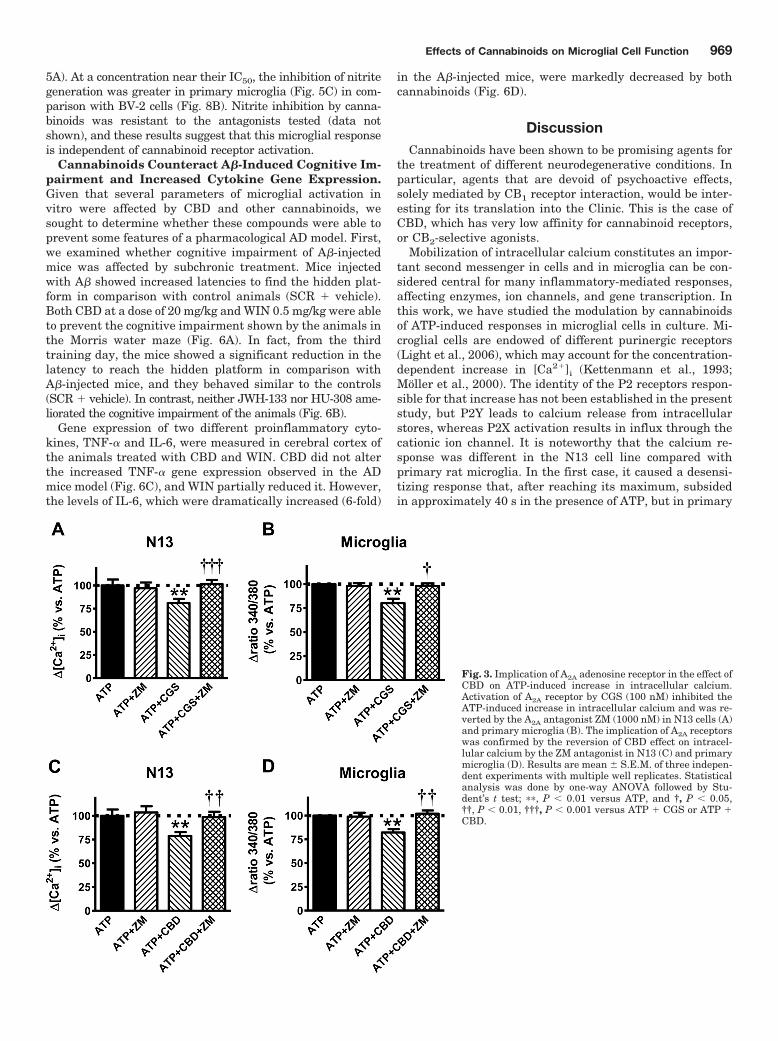
Previous work has shown that the immunosuppressive effects ofCBD involved the activation of adenosine receptors, given itsblockade of the equilibrative nucleoside transporter (Carrier et al.,2006). To ascertain the possible involvement of a similar mecha-nism responsible for the CBD reduction of ATP-induced calciumresponses in microglial cells, we examined whether an A2A agonistcould mimic the CBD action (Fig. 3). In fact, 2-[p-(2-carboxyethyl)phenethylamino]-5�-N-ethylcarboxamidoadenosine [CGS21680(CGS)], an A2A agonist (Klotz et al., 1998), decreased at thesame extent the ATP-induce increase in [Ca2�]i in N13 andprimary microglial cells (Fig. 3, A and B), which wasblocked by 4-{2-[7-amino-2-(2-furyl)[1,2,4]triazolo-[2,3-
Fig. 1. N13 microglial cells and rat primary microglia express CB1 andCB2 receptors. Cells were immunostained with anti-CB1 (1:900 dilution)and anti-CB2 antibodies (1:900 dilution). Top, N13 cells; bottom, primarymicroglia. Initial magnification, 200�.
Effects of Cannabinoids on Microglial Cell Function 967

a][1,3,5]triazin-5-ylamino]ethyl}phenol [ZM241385 (ZM)], anA2A antagonist (Palmer et al., 1995), that per se had no effect. Ofnote is that ZM was able to inhibit the CBD effect both in N13 andprimary microglial cells (Fig. 3, C and D). Therefore, we haveshown that CBD and other cannabinoids counteract the effects ofATP on microglial cells by a mechanism involving both cannabi-noid receptor-dependent and independent mechanisms.
CBD and Other Cannabinoids Promote Microglial
Cell Migration. The A� peptide (Tiffany et al., 2002) anddifferent cannabinoids trigger migration (Walter et al., 2003)that may subserve a beneficial function as a requisite forphagocytosing aggregated A�. Therefore, migration of micro-glial cells through porous membranes toward the lowerchamber containing the compounds was investigated.
The mixed CB1/CB2 agonist WIN and the CB2-selectiveagonist JWH promoted N13 cell migration (Fig. 4, A and B).Note that in these experiments, the cells changed their ovalmorphology (Fig. 1) to a fully round morphology with nolamellipodia (Fig. 4 B). Migration was similarly increased(approximately 20% compared with controls) by the two ago-nists (200 nM). The CB1-selective agonist SR1 did not affectmigration, whereas the CB2-selective antagonist SR2 in-duced a partial although statistically significant effect onmigration (approximately 10% increase; Fig. 2A). The effectof WIN on N13 cells was not altered by either of the canna-binoid antagonists, but full prevention of its migratory effectwas obtained when both antagonists were combined. In con-
trast, the CB2 antagonist completely blocked the responseinduced by JWH, whereas the CB1 antagonist had no effect(Fig. 4A). Fibrillar A� (2 �M) induced microglial migration(20%) in comparison with vehicle-treated control cultures(Fig. 4C) and to those treated with a scrambled peptide,which had no effect. A� combined with the cannabinoidsexerted a similar effect to that obtained with the agonistsalone (data not shown). Primary microglial cells showed amore robust response (2.5-fold compared with controls) uponthe addition of A�, LPS, or the cannabinoid agonists (Fig.4C). The migratory effect induced by CBD and WIN was fullyreversed by either of the selective antagonists (Fig. 4C). Incontrast, the migration promoted by JWH and HU were onlyinhibited by the CB2 antagonist (Fig. 4C). Taken together,these results indicate that CBD and the other cannabinoidspromote microglial cell migration in a cannabinoid receptor-dependent manner.
NO Generation Is Inhibited by Cannabinoids. Canna-binoids inhibit LPS NO synthase stimulation and NO gener-ation as reflected in nitrate accumulation in the culturemedia (Waksman et al., 1999). Given that LPS challenge didnot generate nitrites in N13 cells, BV-2 microglial cells wereused in these experiments.
Nitrite generation, measured in the culture media after LPSstimulation, was concentration-dependently reduced by all thecannabinoids tested (Fig. 5). According to their IC50, their rel-ative potencies were CBD � HU-308�JWH-133��WIN (Fig.
Fig. 2. CBD, WIN, and JWH inhibited the ATP-induced increase in intracellular calcium in cultured N13 and primary microglial cells.A, concentration-dependent inhibition of ATP-induced increase in [Ca2�]i by cannabinoids in N13 cells. The effect of CBD (B; 100 nM), of WIN (C; 400nM), and of JWH (D; 100 nM) was reverted by SR2 antagonist (100 nM). Effect of cannabinoids on ATP-induced increase in [Ca2�]i in N13 cells (E)and an primary microglial cells (F). The antagonist SR2 blocked the effect of WIN in N13 cells and the effect of all cannabinoids in primary microglia.Results are mean S.E.M. of three independent experiments with multiple well replicates. Statistical analysis was done by one-way ANOVA followedby Student’s t test; �, P � 0.05 and ��, P � 0.01 versus ATP, and †, P � 0.05 and †††, P � 0.001 versus ATP � cannabinoid.
968 Moreno et al.
5A). At a concentration near their IC50, the inhibition of nitritegeneration was greater in primary microglia (Fig. 5C) in com-parison with BV-2 cells (Fig. 8B). Nitrite inhibition by canna-binoids was resistant to the antagonists tested (data notshown), and these results suggest that this microglial responseis independent of cannabinoid receptor activation.
Cannabinoids Counteract A�-Induced Cognitive Im-
pairment and Increased Cytokine Gene Expression.
Given that several parameters of microglial activation invitro were affected by CBD and other cannabinoids, wesought to determine whether these compounds were able toprevent some features of a pharmacological AD model. First,we examined whether cognitive impairment of A�-injectedmice was affected by subchronic treatment. Mice injectedwith A� showed increased latencies to find the hidden plat-form in comparison with control animals (SCR � vehicle).Both CBD at a dose of 20 mg/kg and WIN 0.5 mg/kg were ableto prevent the cognitive impairment shown by the animals inthe Morris water maze (Fig. 6A). In fact, from the thirdtraining day, the mice showed a significant reduction in thelatency to reach the hidden platform in comparison withA�-injected mice, and they behaved similar to the controls(SCR � vehicle). In contrast, neither JWH-133 nor HU-308 ame-liorated the cognitive impairment of the animals (Fig. 6B).
Gene expression of two different proinflammatory cyto-kines, TNF-� and IL-6, were measured in cerebral cortex ofthe animals treated with CBD and WIN. CBD did not alterthe increased TNF-� gene expression observed in the ADmice model (Fig. 6C), and WIN partially reduced it. However,the levels of IL-6, which were dramatically increased (6-fold)
in the A�-injected mice, were markedly decreased by bothcannabinoids (Fig. 6D).
Discussion
Cannabinoids have been shown to be promising agents forthe treatment of different neurodegenerative conditions. Inparticular, agents that are devoid of psychoactive effects,solely mediated by CB1 receptor interaction, would be inter-esting for its translation into the Clinic. This is the case ofCBD, which has very low affinity for cannabinoid receptors,or CB2-selective agonists.
Mobilization of intracellular calcium constitutes an impor-tant second messenger in cells and in microglia can be con-sidered central for many inflammatory-mediated responses,affecting enzymes, ion channels, and gene transcription. Inthis work, we have studied the modulation by cannabinoidsof ATP-induced responses in microglial cells in culture. Mi-croglial cells are endowed of different purinergic receptors(Light et al., 2006), which may account for the concentration-dependent increase in [Ca2�]i (Kettenmann et al., 1993;Moller et al., 2000). The identity of the P2 receptors respon-sible for that increase has not been established in the presentstudy, but P2Y leads to calcium release from intracellularstores, whereas P2X activation results in influx through thecationic ion channel. It is noteworthy that the calcium re-sponse was different in the N13 cell line compared withprimary rat microglia. In the first case, it caused a desensi-tizing response that, after reaching its maximum, subsidedin approximately 40 s in the presence of ATP, but in primary
Fig. 3. Implication of A2A adenosine receptor in the effect ofCBD on ATP-induced increase in intracellular calcium.Activation of A2A receptor by CGS (100 nM) inhibited theATP-induced increase in intracellular calcium and was re-verted by the A2A antagonist ZM (1000 nM) in N13 cells (A)and primary microglia (B). The implication of A2A receptorswas confirmed by the reversion of CBD effect on intracel-lular calcium by the ZM antagonist in N13 (C) and primarymicroglia (D). Results are mean S.E.M. of three indepen-dent experiments with multiple well replicates. Statisticalanalysis was done by one-way ANOVA followed by Stu-dent’s t test; ��, P � 0.01 versus ATP, and †, P � 0.05,††, P � 0.01, †††, P � 0.001 versus ATP � CGS or ATP �
CBD.
Effects of Cannabinoids on Microglial Cell Function 969

microglial cells, the response was sustained. Dying neuronsrelease high amounts of ATP; therefore, we decided to usethose pathological concentrations for subsequent experi-ments. CBD and the other cannabinoid agonists, with theexception of HU, decreased ATP-induced [Ca2�]i, both in N13microglial cells and in primary rat microglia in culture. Theeffect of the compounds was either independent of cannabi-noid receptor activation (e.g., CBD and JWH in N13 cells),
given that it was resistant to cannabinoid receptor antago-nism, or CB2 receptor-mediated (WIN in N13 cells and allcompounds in primary microglia). The cannabinoid receptorindependence of the effect of CBD in N13 cells prompted us toinvestigate a possible A2A receptor mediation. We found thatthe A2A-selective agonist CGS (Klotz et al., 1998) also re-duced the ATP-induced [Ca2�]i to a similar extent and theinhibition elicited by CBD and CGS was blocked by the
Fig. 4. Cannabinoid agonists promote microglial cell migra-tion. WIN and JWH (200 nM) promoted N13 cell migration (Aand B), which was inhibited by the combination of CB1 andCB2 antagonist (100 nM each), or the CB2 antagonist (SR2),respectively. Cannabinoid agonists promoted primary micro-glia migration, which was blocked by CB1 and/or CB2 antag-onism (C). Fib, fibrillar A�1–40 (2 �M). Results are themean S.E.M. of four independent experiments in duplicate.Statistical analysis was done by one-way ANOVA followed byStudent’s t test; ��, P � 0.01, ���, P � 0.001 versus control (notreatment); and †, P � 0.05 versus the cannabinoid agonistalone.
Fig. 5. Cannabinoid agonists decreasedLPS-induced nitrite generation in micro-glial cells. A, cannabinoid agonists concen-tration-dependently inhibited LPS-inducednitrite generation in BV-2 microglial cells.Cannabinoid agonist inhibition of LPS-in-duced nitrite generation in BV-2 (B) andprimary microglial cells (C). Agent concen-trations are depicted. In B and C, resultsare expressed as a percentage of control.Nitrite levels were the following: BV-2cells: control, 2.47 0.21, and LPS-in-duced, 12.54 0.98 pg/ml; primary micro-glia: control, 3.65 0.40, and LPS-induced,18.5 1.2. Results are mean S.E.M. offour independent experiments in duplicate.Statistical analysis was done by one-wayANOVA followed by Student’s t test; ���,P � 0.001 versus control (vehicle); and †,P � 0.05 versus LPS.
970 Moreno et al.

A2A-selective antagonist ZM (Palmer et al., 1995). Thoseresults indicate that cannabinoid inhibition of the intracel-lular calcium increase brought about by high concentrationsof ATP in microglial cells may be mediated by cannabinoid orA2A receptors.
In some works in which micromolar concentrations of can-nabinoids enhance [Ca2�]i, ryanodine receptors seem to beinvolved. Indeed, the increase in [Ca2�]i induced by ACEAand JWH-133 was partially blocked by a ryanodine antago-nist in RIN insulinoma cells (De Petrocellis et al., 2007). Theinvolvement of intracellular calcium stores in cannabidiolelevation of [Ca2�]i has also been described in hippocampalcells (neurons and glia) in culture (Drysdale et al., 2006).Given that we found a decrease by cannabinoids of ATP-induced increase in [Ca2�]i and that in microglial cells, CB1
(e.g., in the case of WIN) and/or CB2 antagonists were effec-tive at blocking their effect, we have not addressed the in-volvement of ryanodine receptors.
Activation of microglia by A� is associated with chemotac-tic responses toward it, consistent with the extensive clus-tering of activated microglia at sites of A� deposition in ADbrain. Furthermore, A� induces migration across porousmembranes through the interaction with chemotactic recep-tors such as the formyl peptide receptors FPR2 and FPR-like1 receptor, its human counterpart (Cui et al., 2002). Indeed,we observed that the A� peptide induced chemotactic re-sponses in cultured microglia cells. Cannabinoids, whetherplant-derived or endocannabinoids, have been shown to in-duce migration of BV-2 microglial cells (Walter et al., 2003).In the present work, the synthetic cannabinoids WIN andJWH promoted migration at similar concentrations in theN13 cell line, and their effect was greater in primary micro-glia. According to the work of Walter et al. (2003), the can-nabinoid effects were mediated by CB2 and abnormal canna-bidiol receptors. The results of the present work also showthe involvement of CB2 receptors in migration of N13 andprimary microglial cells. Indeed, the two CB2-selective ago-nists JWH and HU promoted it, and their effect was com-pletely blocked by the CB2-selective antagonist but un-changed by the CB1-selective antagonist, as expected.
However, in our hands, CB1 receptor activation was effectiveas well. In fact, the migration induced by WIN, a mixedCB1/CB2 agonist not tested previously, and by CBD wasprevented by both selective antagonists.
In agreement with previous work that has shown de-creased NO generation by microglia in the presence of can-nabinoids (Waksman et al., 1999), we observed a concentra-tion-dependent decrease in nitrites in the culture media ofLPS-stimulated microglia. Although in BV-2 microglial cellsCBD and HU seemed to be more potent at decreasing ni-trites, in primary microglia, all of the cannabinoid agonistswere equipotent. The effect of cannabinoids were indepen-dent of cannabinoid receptor activation, given that it wasunaltered by the selective antagonists used in the presentstudy.
We and other authors have shown that treatment with can-nabinoid agonists and agents that increase endocannabinoidavailability, such as the inhibitor of endocannabinoid reuptakeN-(4-hydroxy-2-methylphenyl)-5Z,8Z,11Z,14Z-eicosatetraen-amide (VDM11), are able to prevent A�-induced cognitive def-icits (Ramírez et al., 2005; van der Stelt et al., 2006). Further-more, in the work by van der Stelt et al. (2006), it was foundthat VDM11 treatment inhibited different glial parameters (cy-clooxygenase-2, iNOS, S100�) in hippocampus, which were in-creased by A� intracortical injection. In the present work, A�-injected mice subjected to subchronic systemic administrationof WIN or CBD showed better performance in the Morris watermaze compared with vehicle-treated animals. In this paradigm,the two selective CB2 agonists were ineffective in that respect,although JWH-prolonged oral administration showed beneficialeffects in the transgenic model of AD Tg APP (A. M. Martín-Moreno, B. Brera, E. Carro, M. Delgado, M. A. Pozo, N. Innam-orato, A. Cuadrado, and M. L. de Ceballos, in preparation). Thepossible involvement of glial activation modulation was as-sessed by measuring cytokine gene expression. In mice injectedwith A�, both TNF-� and IL-6 expression was markedly in-creased. WIN and CBD treatment abolished IL-6 expres-sion increase, and WIN partially reduced that of TNF-�.Therefore, in an in vivo short-term pharmacological modelof AD, cannabinoids showed beneficial behavioral effects
Fig. 6. CBD and WIN 55,212-2 prevented A�-inducedlearning deficit and cytokine expression. Mice received asingle A� intraventricular injection (2.5 �g/5 �l) andwere treated with the cannabinoid agonists daily (seeMaterials and Methods for details). Training in the wa-ter maze was conducted during the second week of treat-ment. CBD and WIN prevented A�-induced cognitiveimpairment (A), whereas HU and JWH were withouteffect (B). WIN and CBD prevented IL-6 increased geneexpression induced by A� (D), but did not affect TNF�
increase (C). Results are mean S.E.M. of eight mice pergroup. In the water maze, results S.E.M., which wereless than 15%, were omitted for the sake of clarity. SCR,scrambled peptide; Fib, fibrillar A�1–40. Statistical anal-ysis of water maze performance was done by two-wayANOVA followed by Student’s t test: �, P � 0.05 versusSCR-treated mice; †, P � 0.05 versus A�-injected miceand of cytokine expression by one-way ANOVA: �, P �
0.05 and ���, P � 0.001 versus SCR, and †††, P � 0.001versus Fib.
Effects of Cannabinoids on Microglial Cell Function 971

that seem to be mediated through glial activation modula-tion.
The pharmacology of endocannabinoids and phytocannabi-noids seems to be increasingly complicated. According tobinding studies performed in cells transfected with the CB1
and CB2 human receptors (Showalter et al., 1996), CBD hasshown very low affinity (approximately 2–4 �M). However,one interesting finding of this work is that CBD exerts sev-eral effects on microglial function in the high nanomolarrange and at similar concentrations of other cannabinoidstested. Moreover, many of CBD effects seem to be CB2 recep-tor-mediated. Expression of CB2 receptors in normal brain isnegligible and only measurable by quantitative PCR. Thisfact can explain that the interaction of CBD with CB2 recep-tors has been unnoticed and unraveled in studies involvingmicroglial cells.
There is no doubt that in AD, a pronounced inflammationoccurs in which astrocytes and microglial cells are involvedand A�, which is central to AD pathology and is at least inpart responsible of it. However, the inflammatory responseaccounts for both detrimental and beneficial effects in thepathology. Indeed, activated microglia release toxic mole-cules, such as NO and proinflammatory cytokines, as initialplayers that may induce neurodegeneration. At the sametime, these cells release trophic factors and thereafter mi-grate to affected brain areas and phagocytose dead neuronsand A� deposits, therefore contributing to neuroprotection.The resulting outcome of the inflammatory process would bethe combination of both effects. It is noteworthy that canna-binoids seem to differentially regulate those separate cellularevents of activated microglial in a positive direction. On theone hand, these compounds effectively counteract A�-medi-ated increase in the proinflammatory cytokine TNF-� andthe ensuing neurodegeneration after its administration invitro and in vivo (Ramírez et al., 2005). In contrast, as shownhere, cannabinoids promote migration, a cellular mechanismthat ultimately will allow the removal of the deposited A�
peptide. Therefore, this kind of drug with neuroprotectiveand anti-inflammatory effects (Walter and Stella, 2004) maybe of interest in the prevention of AD inflammation, in par-ticular CB2-selective agonists, which are devoid of psychoac-tive effects (Hanus et al., 1999).
Acknowledgments
We thank Dr. K. Mackie for anti-CB1 antibody and CB1 and CB2
antigenic peptides, Sanofi-Synthelabo for SR141716 and SR144528,and M. E. Fernandez de Molina and S. Plazuelo for excellent tech-nical assistance.
Authorship Contributions
Participated in research design: Mechoulam, Cuadrado, and deCeballos.
Conducted experiments: Martín-Moreno, Reigada, Ramírez, andInnamorato.
Contributed new reagents or analytic tools: Mechoulam.Performed data analysis: Martín-Moreno, Reigada, Ramírez, In-
namorato, and de Ceballos.Wrote or contributed to the writing of the manuscript: Mechoulam,
Cuadrado, and de Ceballos.
References
Akiyama H, Barger S, Barnum S, Bradt B, Bauer J, Cole GM, Cooper NR, Eikelen-boom P, Emmerling M, Fiebich BL, et al. (2000) Inflammation and Alzheimer’sdisease. Neurobiol Aging 21:383–421.
Carrier EJ, Auchampach JA, and Hillard CJ (2006) Inhibition of an equilibrativenucleoside transporter by cannabidiol: a mechanism of cannabinoid immunosup-pression. Proc Natl Acad Sci USA 103:7895–7900.
Combs CK, Karlo JC, Kao SC, and Landreth GE (2001) beta-Amyloid stimulation ofmicroglia and monocytes results in TNFalpha-dependent expression of induciblenitric oxide synthase and neuronal apoptosis. J Neurosci 21:1179–1188.
Costa B, Colleoni M, Conti S, Parolaro D, Franke C, Trovato AE, and Giagnoni G(2004) Oral anti-inflammatory activity of cannabidiol, a non-psychoactive constit-uent of cannabis, in acute carrageenan-induced inflammation in the rat paw.Naunyn Schmiedebergs Arch Pharmacol 369:294–299.
Cui YH, Le Y, Gong W, Proost P, Van Damme J, Murphy WJ, and Wang JM (2002)Bacterial lipopolysaccharide selectively up-regulates the function of the chemot-actic peptide receptor formyl peptide receptor 2 in murine microglial cells. J Im-munol 168:434–442.
De Petrocellis L, Marini P, Matias I, Moriello AS, Starowicz K, Cristino L, Nigam S,and Di Marzo V (2007) Mechanisms for the coupling of cannabinoid receptors tointracellular calcium mobilization in rat insulinoma beta-cells. Exp Cell Res 313:2993–3004.
Drysdale AJ, Ryan D, Pertwee RG, and Platt B. (2006) Cannabidiol-induced intra-cellular Ca2� elevations in hippocampal cells. Neuropharmacology 50: 621–631.
Esposito G, De Filippis D, Carnuccio R, Izzo AA, and Iuvone T (2006a) The mari-juana component cannabidiol inhibits beta-amyloid-induced tau protein hyper-phosphorylation through Wnt/beta-catenin pathway rescue in PC12 cells. J MolMed 84:253–258.
Esposito G, De Filippis D, Maiuri MC, De Stefano D, Carnuccio R, and Iuvone T(2006b) Cannabidiol inhibits inducible nitric oxide synthase protein expressionand nitric oxide production in beta-amyloid stimulated PC12 neurons through p38MAP kinase and NF-kappaB involvement. Neurosci Lett 399:91–95.
Esposito G, Scuderi C, Savani C, Steardo L Jr, De Filippis D, Cottone P, Iuvone T,Cuomo V, and Steardo L (2007) Cannabidiol in vivo blunts beta-amyloid inducedneuroinflammation by suppressing IL-1beta and iNOS expression. Br J Pharma-col 151:1272–1279.
Facchinetti F, Del Giudice E, Furegato S, Passarotto M, and Leon A (2003) Canna-binoids ablate release of TNFalpha in rat microglial cells stimulated with lipo-polysaccharide. Glia 41:161–168.
Farber K and Kettenmann H (2006) Functional role of calcium signals for microglialfunction. Glia 54:656–665.
Hanus L, Breuer A, Tchilibon S, Shiloah S, Goldenberg D, Horowitz M, Pertwee RG,Ross RA, Mechoulam R, and Fride E (1999) HU-308: a specific agonist for CB(2),a peripheral cannabinoid receptor. Proc Natl Acad Sci USA 96:14228–14233.
Howlett AC, Barth F, Bonner TI, Cabral G, Casellas P, Devane WA, Felder CC,Herkenham M, Mackie K, Martin BR, Mechoulam R, and Pertwee RG., Herkenha,M, Mackie K, Martin BR, Mechoulan R, Pertwee RG (2002) International Union ofPharmacology. XXVII. Classification of cannabinoid receptors. Pharmacol Rev54:161–202.
Huffman JW, Liddle J, Yu S, Aung MM, Abood ME, Wiley JL, and Martin BR (1999)3-(1�,1�-Dimethylbutyl)-1-deoxy-delta-8-THC and related compounds: synthesisand selective ligands for the CB2 receptor. Bioorg Med Chem 7:2905–2914.
Iuvone T, Esposito G, Esposito R, Santamaria R, Di Rosa M, and Izzo AA (2004)Neuroprotective effect of cannabidiol, a non-psychoactive component from Canna-bis sativa, on beta-amyloid-induced toxicity in PC12 cells. J Neurochem 89:134–141.
Jantzen PT, Connor KE, DiCarlo G, Wenk GL, Wallace JL, Rojiani AM, Coppola D,Morgan D, and Gordon MN (2002) Microglial activation and �-amyloid depositreduction caused by nitric oxide-releasing nonsteroidal anti-inflammatory drug inamyloid precursor protein plus presenilin-1 transgenic mice. J Neurosci 22:2246–2254.
Kettenmann H, Banati R, and Walz W (1993) Electrophysiological behavior ofmicroglia. Glia 7:93–101.
Klotz KN, Hessling J, Hegler J, Owman C, Kull B, Fredholm BB, and Lohse MJ(1998) Comparative pharmacology of human adenosine receptor subtypes - char-acterization of stably transfected receptors in CHO cells. Naunyn SchmiedebergsArch Pharmacol 357:1–9.
Light AR, Wu Y, Hughen RW, and Guthrie PB (2006) Purinergic receptors activatingrapid intracellular Ca increases in microglia. Neuron Glia Biol 2:125–138.
Malfait AM, Gallily R, Sumariwalla PF, Malik AS, Andreakos E, Mechoulam R, andFeldmann M (2000) The nonpsychoactive cannabis constituent cannabidiol is anoral anti-arthritic therapeutic in murine collagen-induced arthritis. Proc NatlAcad Sci USA 97:9561–9566.
Masliah E, Sisk A, Mallory M, Mucke L, Schenk D, and Games D (1996) Comparisonof neurodegenerative pathology in transgenic mice overexpressing V717F beta-amyloid precursor protein and Alzheimer’s disease. J Neurosci 16:5795–5811.
Mato S, Alberdi E, Ledent C, Watanabe M, and Matute C (2009) CB1 cannabinoidreceptor-dependent and -independent inhibition of depolarization-induced calciuminflux in oligodendrocytes. Glia 57:295–306.
Mechoulam R, Peters M, Murillo-Rodriguez E, and Hanus LO (2007) Cannabidiol–recent advances. Chem Biodivers 4:1678–1692.
Mechoulam R and Shohami E (2007) Endocannabinoids and traumatic brain injury.Mol Neurobiol 36:68–74.
Moller T, Kann O, Verkhratsky A, and Kettenmann H (2000) Activation of mousemicroglial cells affects P2 receptor signaling. Brain Res 853:49–59.
Palmer TM, Poucher SM, Jacobson KA, and Stiles GL (1995) 125I-4-(2-[7-amino-2-[2-furyl][1,2,4]triazolo[2,3-a][1,3,5] triazin-5-yl-amino]ethyl)phenol, a high affinityantagonist radioligand selective for the A2a adenosine receptor. Mol Pharmacol48:970–974.
Piomelli D (2003) The molecular logic of endocannabinoid signalling. Nat Rev Neu-rosci 4:873–884.
Puffenbarger RA, Boothe AC, and Cabral GA (2000) Cannabinoids inhibit LPS-inducible cytokine mRNA expression in rat microglial cells. Glia 29:58–69.
Ramírez BG, Blazquez C, Gomez del Pulgar T, Guzman M, and de Ceballos ML
972 Moreno et al.

(2005) Prevention of Alzheimer’s disease pathology by cannabinoids: neuroprotec-tion mediated by blockade of microglial activation. J Neurosci 25:1904–1913.
Righi M, Mori L, De Libero G, Sironi M, Biondi A, Mantovani A, Donini SD, andRicciardi-Castagnoli P (1989) Monokine production by microglial cell clones. EurJ Immunol 19:1443–1448.
Rinaldi-Carmona M, Barth F, Heaulme M, Shire D, Calandra B, Congy C, MartinezS, Maruani J, Neliat G, and Caput D (1994) SR141716A, a potent and selectiveantagonist of the brain cannabinoid receptor. FEBS Lett 350:240–244.
Rinaldi-Carmona M, Barth F, Millan J, Derocq JM, Casellas P, Congy C, Oustric D,Sarran M, Bouaboula M, Calandra B, et al. (1998) SR 144528, the first potent andselective antagonist of the CB2 cannabinoid receptor. J Pharmacol Exp Ther284:644–650.
Showalter VM, Compton DR, Martin BR, and Abood ME (1996) Evaluation ofbinding in a transfected cell line expressing a peripheral cannabinoid receptor(CB2): identification of cannabinoid receptor subtype selective ligands. J Pharma-col Exp Ther 278:989–999.
Tiffany HL, Lavigne MC, Cui YH, Wang JM, Leto TL, Gao JL, and Murphy PM(2002) Amyloid-beta induces chemotaxis and oxidant stress by acting at formyl-peptide receptor 2, a G protein-coupled receptor expressed in phagocytes andbrain. J Biol Chem 276:23645–23652.
van der Stelt M, Mazzola C, Esposito G, Matias I, Petrosino S, De Filippis D, Micale
V, Steardo L, Drago F, Iuvone T, et al. (2006) Endocannabinoids and beta-amyloid-induced neurotoxicity in vivo: effect of pharmacological elevation of endocannabi-noid levels. Cell Mol Life Sci 63:1410–1424.
van der Stelt M, Veldhuis WB, Maccarrone M, Bar PR, Nicolay K, Veldink GA, DiMarzo V, and Vliegenthart JF (2002) Acute neuronal injury, excitotoxicity, and theendocannabinoid system. Mol Neurobiol 26:317–346.
Waksman Y, Olson JM, Carlisle SJ, and Cabral GA (1999) The central cannabinoidreceptor (CB1) mediates inhibition of nitric oxide production by rat microglial cells.J Pharmacol Exp Ther 288: 1357–1366.
Walter L, Franklin A, Witting A, Wade C, Xie Y, Kunos G, Mackie K, and Stella N(2003) Nonpsychotropic cannabinoid receptors regulate microglial cell migration.J Neurosci 23:1398–1405.
Walter L and Stella N (2004) Cannabinoids and neuroinflammation. Br J Pharmacol
141:775–785.
Address correspondence to: Dr. M. L. de Ceballos, NeurodegenerationGroup, Department of Cellular, Molecular and Developmental Neurobiology,Instituto Cajal, CSIC, Doctor Arce, 37, 28002 Madrid, Spain. E-mail:[email protected]
Effects of Cannabinoids on Microglial Cell Function 973