Canine Atopic Dermatitis...
160
Canine Atopic Dermatitis Immunotherapeutic* First to Know Slides September 2015 *This product license is conditional. Safety and efficacy studies are in progress .
-
Upload
duongduong -
Category
Documents
-
view
226 -
download
2
Transcript of Canine Atopic Dermatitis...
Canine Atopic
Dermatitis
Immunotherapeutic*
First to Know Slides
September 2015
*This product license is conditional. Safety and efficacy studies are in progress.
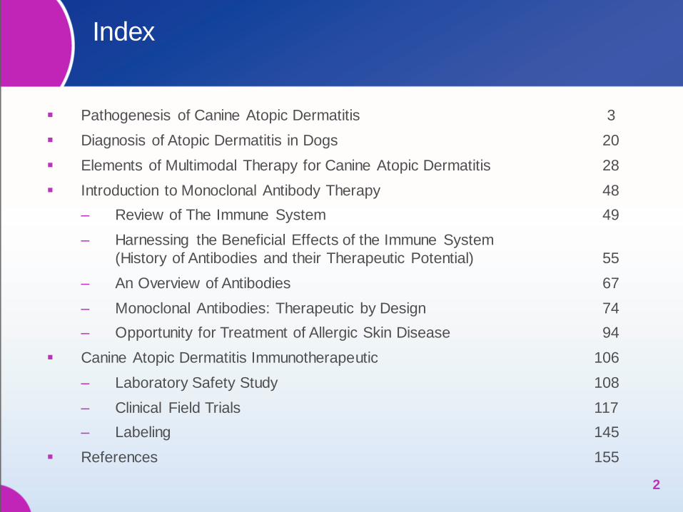
Index
Pathogenesis of Canine Atopic Dermatitis 3
Diagnosis of Atopic Dermatitis in Dogs 20
Elements of Multimodal Therapy for Canine Atopic Dermatitis 28
Introduction to Monoclonal Antibody Therapy 48
‒ Review of The Immune System 49
‒ Harnessing the Beneficial Effects of the Immune System
(History of Antibodies and their Therapeutic Potential) 55
‒ An Overview of Antibodies 67
‒ Monoclonal Antibodies: Therapeutic by Design 74
‒ Opportunity for Treatment of Allergic Skin Disease 94
Canine Atopic Dermatitis Immunotherapeutic 106
‒ Laboratory Safety Study 108
‒ Clinical Field Trials 117
‒ Labeling 145
References 155
2
PATHOGENESIS OF
CANINE ATOPIC DERMATITIS
3 3
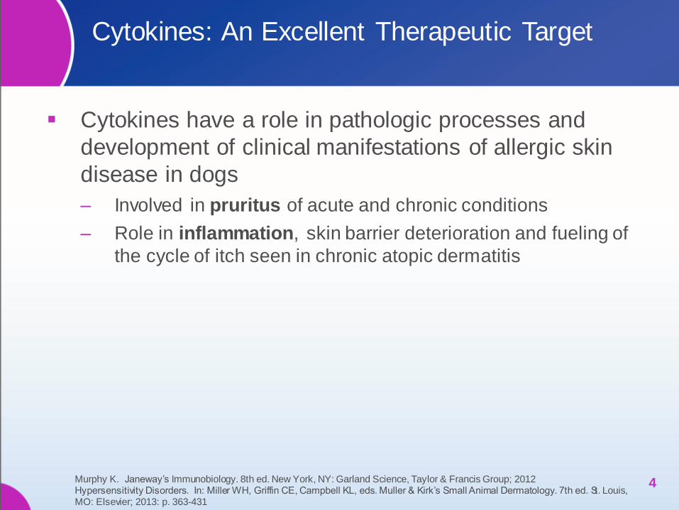
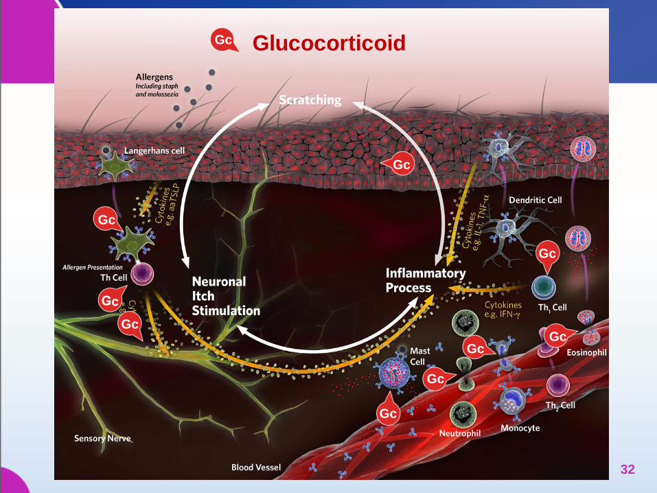
Cytokines: An Excellent Therapeutic Target
Cytokines have a role in pathologic processes and
development of clinical manifestations of allergic skin
disease in dogs
‒ Involved in pruritus of acute and chronic conditions
‒ Role in inflammation, skin barrier deterioration and fueling of
the cycle of itch seen in chronic atopic dermatitis
4 Murphy K. Janeway’s Immunobiology. 8th ed. New York, NY: Garland Science, Taylor & Francis Group; 2012 Hypersensitivity Disorders. In: Miller WH, Griffin CE, Campbell KL, eds. Muller & Kirk’s Small Animal Dermatology. 7th ed. St. Louis, MO: Elsevier; 2013: p. 363-431
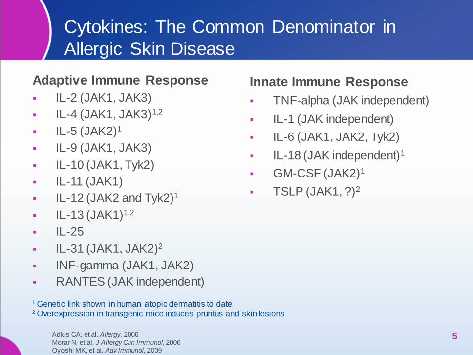
Cytokines: The Common Denominator in
Allergic Skin Disease
Adaptive Immune Response
IL-2 (JAK1, JAK3)
IL-4 (JAK1, JAK3)1,2
IL-5 (JAK2)1
IL-9 (JAK1, JAK3)
IL-10 (JAK1, Tyk2)
IL-11 (JAK1)
IL-12 (JAK2 and Tyk2)1
IL-13 (JAK1)1,2
IL-25
IL-31 (JAK1, JAK2)2
INF-gamma (JAK1, JAK2)
RANTES (JAK independent)
Innate Immune Response
TNF-alpha (JAK independent)
IL-1 (JAK independent)
IL-6 (JAK1, JAK2, Tyk2)
IL-18 (JAK independent)1
GM-CSF (JAK2)1
TSLP (JAK1, ?)2
5
1 Genetic link shown in human atopic dermatitis to date 2 Overexpression in transgenic mice induces pruritus and skin lesions
Adkis CA, et al. Allergy, 2006 Morar N, et al. J Allergy Clin Immunol, 2006 Oyoshi MK, et al. Adv Immunol, 2009
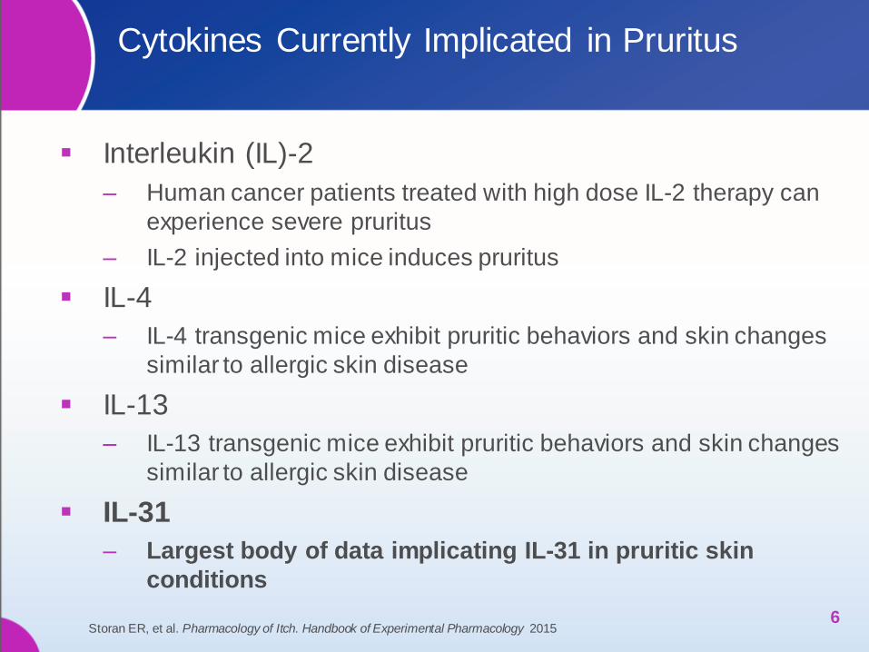
Cytokines Currently Implicated in Pruritus
Interleukin (IL)-2
‒ Human cancer patients treated with high dose IL-2 therapy can
experience severe pruritus
‒ IL-2 injected into mice induces pruritus
IL-4
‒ IL-4 transgenic mice exhibit pruritic behaviors and skin changes
similar to allergic skin disease
IL-13
‒ IL-13 transgenic mice exhibit pruritic behaviors and skin changes
similar to allergic skin disease
IL-31
‒ Largest body of data implicating IL-31 in pruritic skin
conditions
6 Storan ER, et al. Pharmacology of Itch. Handbook of Experimental Pharmacology 2015
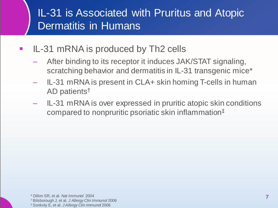
IL-31 is Associated with Pruritus and Atopic
Dermatitis in Humans
IL-31 mRNA is produced by Th2 cells
‒ After binding to its receptor it induces JAK/STAT signaling,
scratching behavior and dermatitis in IL-31 transgenic mice*
‒ IL-31 mRNA is present in CLA+ skin homing T-cells in human
AD patients†
‒ IL-31 mRNA is over expressed in pruritic atopic skin conditions
compared to nonpruritic psoriatic skin inflammation‡
7 * Dillon SR, et al. Nat Immunol 2004 † Bilsborough J, et al. J Allergy Clin Immunol 2006 ‡ Sonkoly E, et al. J Allergy Clin Immunol 2006
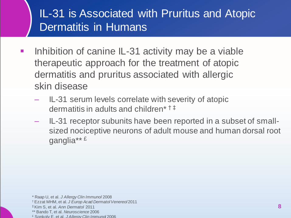
IL-31 is Associated with Pruritus and Atopic
Dermatitis in Humans
Inhibition of canine IL-31 activity may be a viable
therapeutic approach for the treatment of atopic
dermatitis and pruritus associated with allergic
skin disease
‒ IL-31 serum levels correlate with severity of atopic
dermatitis in adults and children* † ‡
‒ IL-31 receptor subunits have been reported in a subset of small-
sized nociceptive neurons of adult mouse and human dorsal root
ganglia** £
8
* Raap U, et al. J Allergy Clin Immunol 2008 † Ezzat MHM, et al. J Europ Acad Dermatol Venereol 2011 ‡ Kim S, et al. Ann Dermatol 2011 ** Bando T, et al. Neuroscience 2006 £ Sonkoly E, et al. J Allergy Clin Immunol 2006
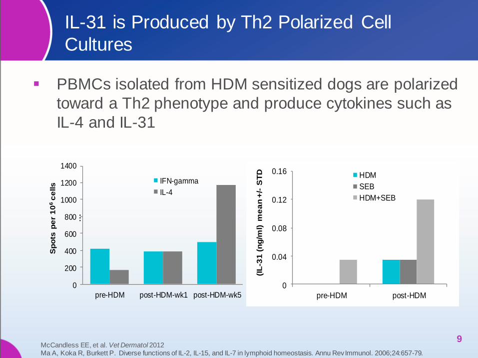
IL-31 is Produced by Th2 Polarized Cell
Cultures
PBMCs isolated from HDM sensitized dogs are polarized
toward a Th2 phenotype and produce cytokines such as
IL-4 and IL-31
9
0
200
400
600
800
1000
1200
1400
pre-HDM post-HDM-wk1 post-HDM-wk5
Sp
ots
pe
r 1
06
ce
lls
…
IFN-gamma
IL-4
0
0.04
0.08
0.12
0.16
pre-HDM post-HDM
(IL
-31
(n
g/m
l) m
ea
n +
/-S
TD HDM
SEB
HDM+SEB
McCandless EE, et al. Vet Dermatol 2012 Ma A, Koka R, Burkett P. Diverse functions of IL-2, IL-15, and IL-7 in lymphoid homeostasis. Annu Rev Immunol. 2006;24:657-79.
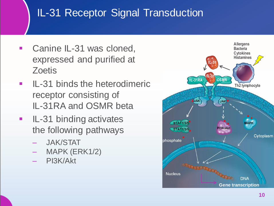
IL-31 Receptor Signal Transduction
Canine IL-31 was cloned,
expressed and purified at
Zoetis
IL-31 binds the heterodimeric
receptor consisting of
IL-31RA and OSMR beta
IL-31 binding activates
the following pathways
‒ JAK/STAT
‒ MAPK (ERK1/2)
‒ PI3K/Akt
10
Allergens Bacteria Cytokines Histamines
IL-31
Gene transcription
IL-31
OSMR
Th2 lymphocyte
IL-31RA
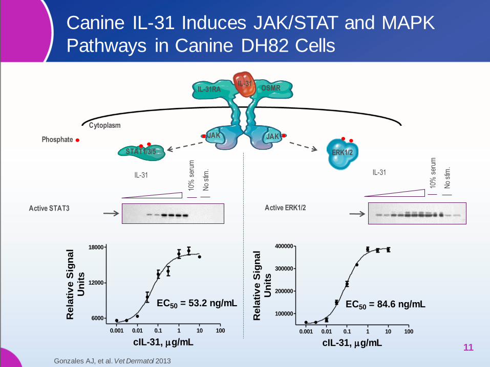
Canine IL-31 Induces JAK/STAT and MAPK
Pathways in Canine DH82 Cells
11
0.001 0.01 0.1 1 10 100
6000
12000
18000
EC50 = 53.2 ng/mL
cIL-31, g/mL
Re
lati
ve
Sig
na
lU
nit
s
0.001 0.01 0.1 1 10 100
100000
200000
300000
400000
EC50 = 84.6 ng/mL
cIL-31, g/mL
Rela
tiv
e S
ign
al
Un
its
10%
ser
um
No
stim
.
Active ERK1/2 Active STAT3
IL-31 IL-31
10%
ser
um
No
stim
.
Cytoplasm
ERK1/2 STAT1/3/5
IL-31RA OSMR IL-31
JAK JAK Phosphate
Gonzales AJ, et al. Vet Dermatol 2013

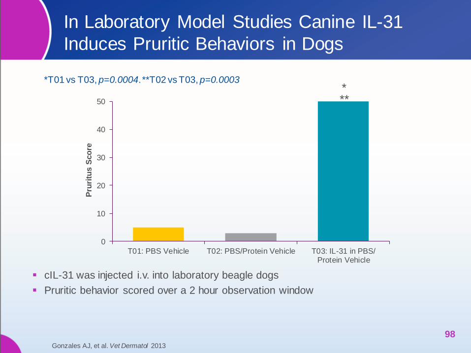
In Laboratory Model Studies Canine IL-31
Induces Pruritic Behaviors in Dogs
cIL-31 was injected i.v. into laboratory beagle dogs
Pruritic behavior scored over a 2 hour observation window
12 Gonzales AJ, et al. Vet Dermatol 2013
0
10
20
30
40
50
T01: PBS Vehicle T02: PBS/Protein Vehicle T03: IL-31 in PBS/Protein Vehicle
Pru
ritu
s S
co
re
* **
*T01 vs T03, p=0.0004. **T02 vs T03, p=0.0003
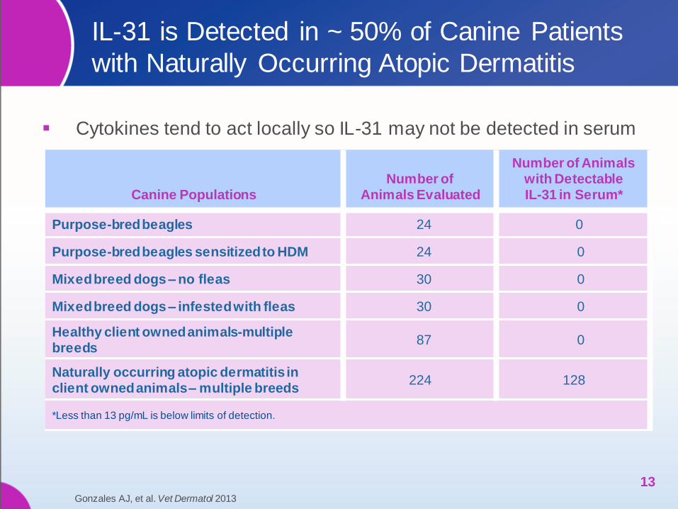
IL-31 is Detected in ~ 50% of Canine Patients
with Naturally Occurring Atopic Dermatitis
Cytokines tend to act locally so IL-31 may not be detected in serum
13
Canine Populations Number of
Animals Evaluated
Number of Animals with Detectable IL-31 in Serum*
Purpose-bred beagles 24 0
Purpose-bred beagles sensitized to HDM 24 0
Mixed breed dogs – no fleas 30 0
Mixed breed dogs – infested with fleas 30 0
Healthy client owned animals-multiple breeds
87 0
Naturally occurring atopic dermatitis in client owned animals – multiple breeds
224 128
*Less than 13 pg/mL is below limits of detection.
Gonzales AJ, et al. Vet Dermatol 2013

Caninized (c)IL-31 Induces Pruritus in Dogs
Regardless of the Route of Injection
Animal behavior recorded by video monitors
Pruritic behavior displayed during a 4 hr window post-cIL-31 treatment-measured in actual time (seconds)
14
0
200
400
600
800
1000
1200
761 448 538 488 044
Pru
riti
c B
eh
avio
r (s
ec)
Animal Number
Baseline
Post ID injection (10 ug IL-31)
0
200
400
600
800
1000
1200
1400
761 488 138 044
Pru
riti
c B
eh
avio
r (s
ec)
Animal Number
Baseline
Post IV infusion (10 ug IL-31)
Mock protein
Mock protein
cIL-31 Treated
Intradermal (ID) Injection Intravenous (IV) Injection
cIL-31 Treated Mock proteinTreated
Gonzales AJ, et al. Vet Dermatol 2013
Mock proteinTreated

cIL-31-induced Pruritus Model Validated by
Response to Prednisolone
59
85
0
20
40
60
80
100
Day 0 Day 6
Pru
riti
c s
co
re
Placebo
Prednisolone @ 0.50 mg/kg BID
*p=0.0656
**p= 0.0003
* **
15
Reference: Study Report No. 7D61R-60-11-B68, Zoetis Inc.
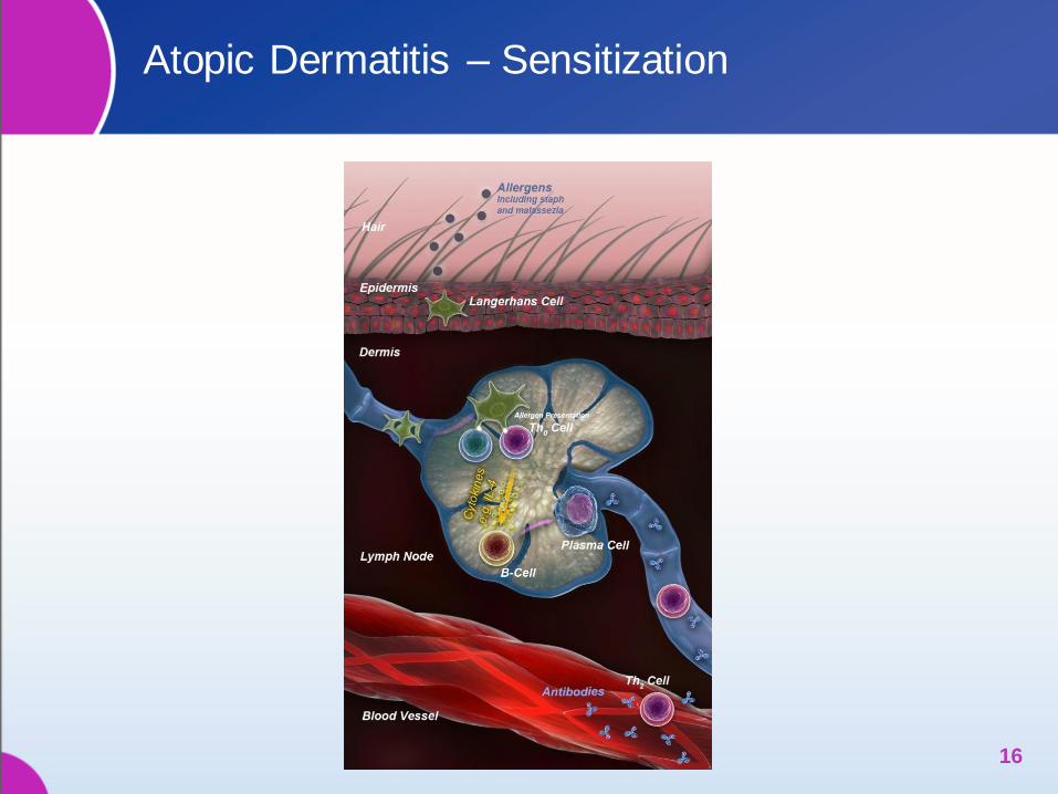
Atopic Dermatitis – Sensitization
16

Atopic Dermatitis – Progression
17
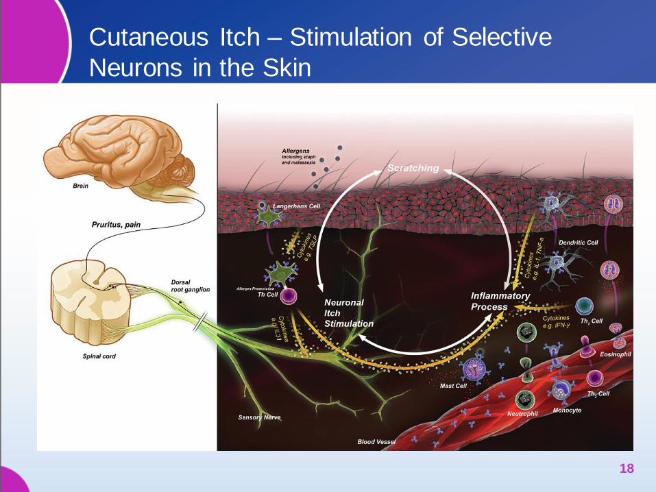
Cutaneous Itch – Stimulation of Selective
Neurons in the Skin
18

IL-31 Receptors Located on Many Different
Cell Types
19
DIAGNOSIS OF ATOPIC
DERMATITIS IN DOGS
20
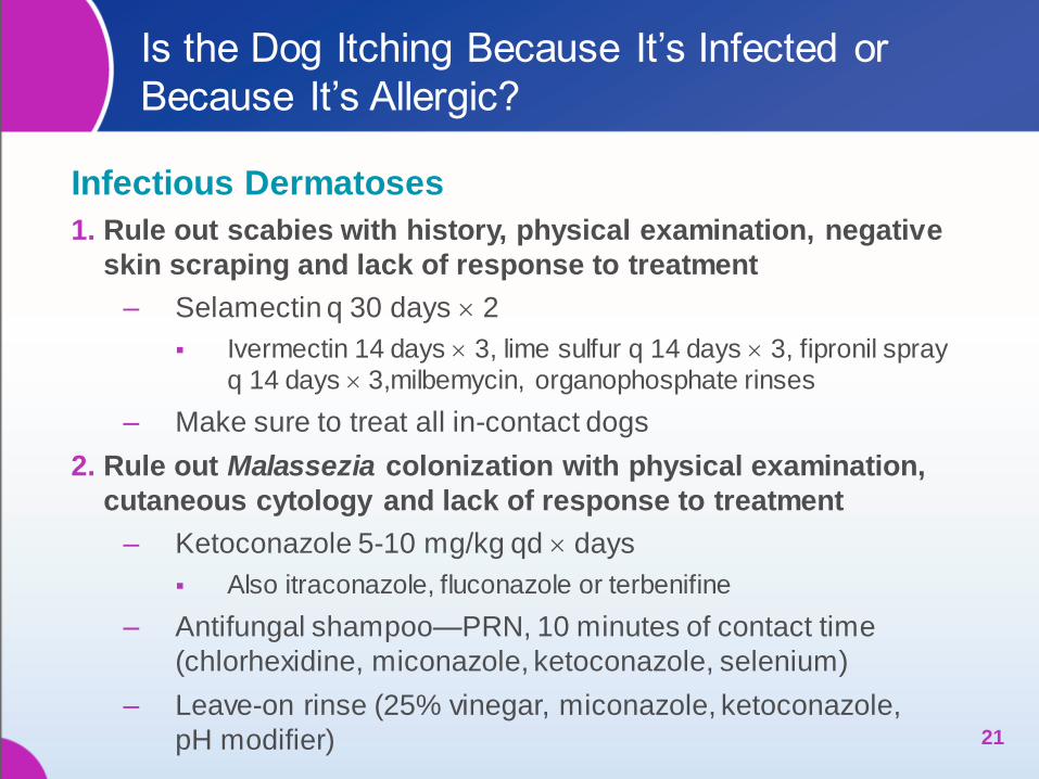
Is the Dog Itching Because It’s Infected or
Because It’s Allergic?
Infectious Dermatoses
1. Rule out scabies with history, physical examination, negative
skin scraping and lack of response to treatment
‒ Selamectin q 30 days 2
Ivermectin 14 days 3, lime sulfur q 14 days 3, fipronil spray
q 14 days 3,milbemycin, organophosphate rinses
‒ Make sure to treat all in-contact dogs
2. Rule out Malassezia colonization with physical examination,
cutaneous cytology and lack of response to treatment
‒ Ketoconazole 5-10 mg/kg qd days
Also itraconazole, fluconazole or terbenifine
‒ Antifungal shampoo—PRN, 10 minutes of contact time
(chlorhexidine, miconazole, ketoconazole, selenium)
‒ Leave-on rinse (25% vinegar, miconazole, ketoconazole,
pH modifier) 21
Is the Dog Itching Because It’s Infected or
Because It’s Allergic? (cont’d)
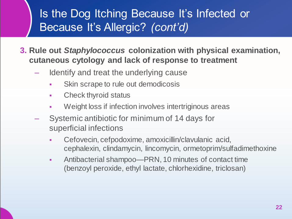
3. Rule out Staphylococcus colonization with physical examination,
cutaneous cytology and lack of response to treatment
‒ Identify and treat the underlying cause
Skin scrape to rule out demodicosis
Check thyroid status
Weight loss if infection involves intertriginous areas
‒ Systemic antibiotic for minimum of 14 days for
superficial infections
Cefovecin, cefpodoxime, amoxicillin/clavulanic acid,
cephalexin, clindamycin, lincomycin, ormetoprim/sulfadimethoxine
Antibacterial shampoo—PRN, 10 minutes of contact time
(benzoyl peroxide, ethyl lactate, chlorhexidine, triclosan)
22
Is the Dog Itching Because It’s Infected or
Because It’s Allergic? (cont’d)
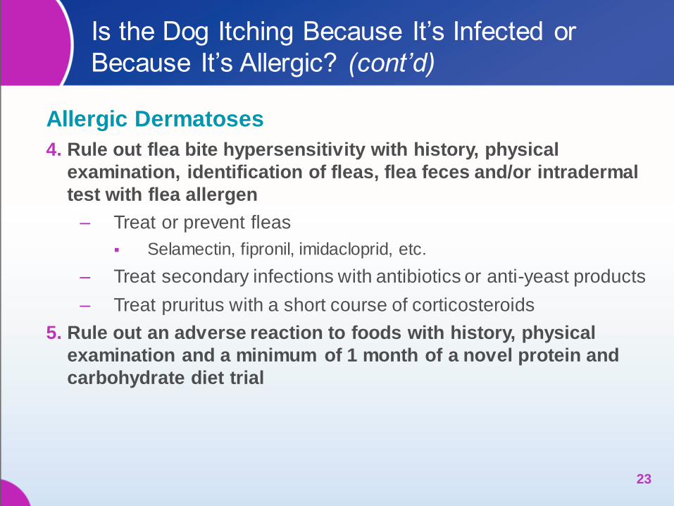
Allergic Dermatoses
4. Rule out flea bite hypersensitivity with history, physical
examination, identification of fleas, flea feces and/or intradermal
test with flea allergen
‒ Treat or prevent fleas
Selamectin, fipronil, imidacloprid, etc.
‒ Treat secondary infections with antibiotics or anti-yeast products
‒ Treat pruritus with a short course of corticosteroids
5. Rule out an adverse reaction to foods with history, physical
examination and a minimum of 1 month of a novel protein and
carbohydrate diet trial
23
Is the Dog Itching Because It’s Infected or
Because It’s Allergic? (cont’d)
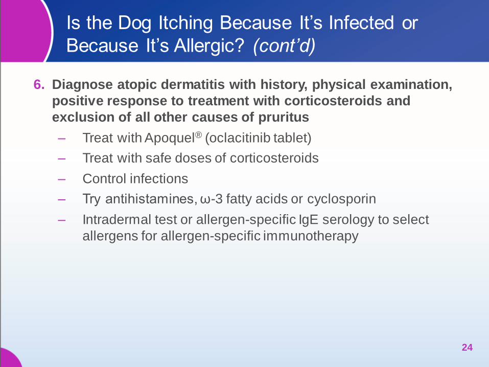
6. Diagnose atopic dermatitis with history, physical examination,
positive response to treatment with corticosteroids and
exclusion of all other causes of pruritus
‒ Treat with Apoquel® (oclacitinib tablet)
‒ Treat with safe doses of corticosteroids
‒ Control infections
‒ Try antihistamines, ω-3 fatty acids or cyclosporin
‒ Intradermal test or allergen-specific IgE serology to select
allergens for allergen-specific immunotherapy
24
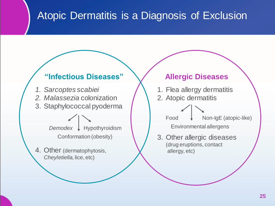
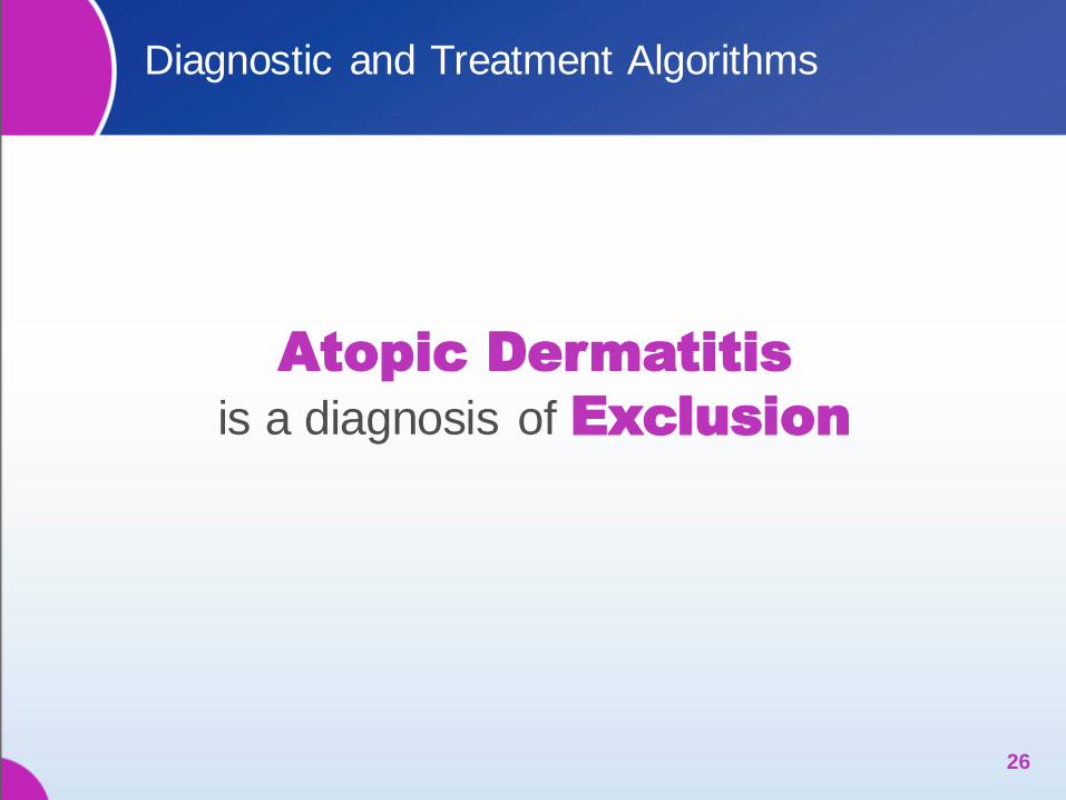
Atopic Dermatitis is a Diagnosis of Exclusion
25
“Infectious Diseases” Allergic Diseases
1. Flea allergy dermatitis
2. Atopic dermatitis
3. Other allergic diseases (drug eruptions, contact
allergy, etc)
Food
Environmental allergens
Non-IgE (atopic-like)
1. Sarcoptes scabiei
2. Malassezia colonization
3. Staphylococcal pyoderma
Demodex Hypothyroidism
Conformation (obesity)
4. Other (dermatophytosis,
Cheyletiella, lice, etc)
Diagnostic and Treatment Algorithms
26
Atopic Dermatitis
is a diagnosis of Exclusion
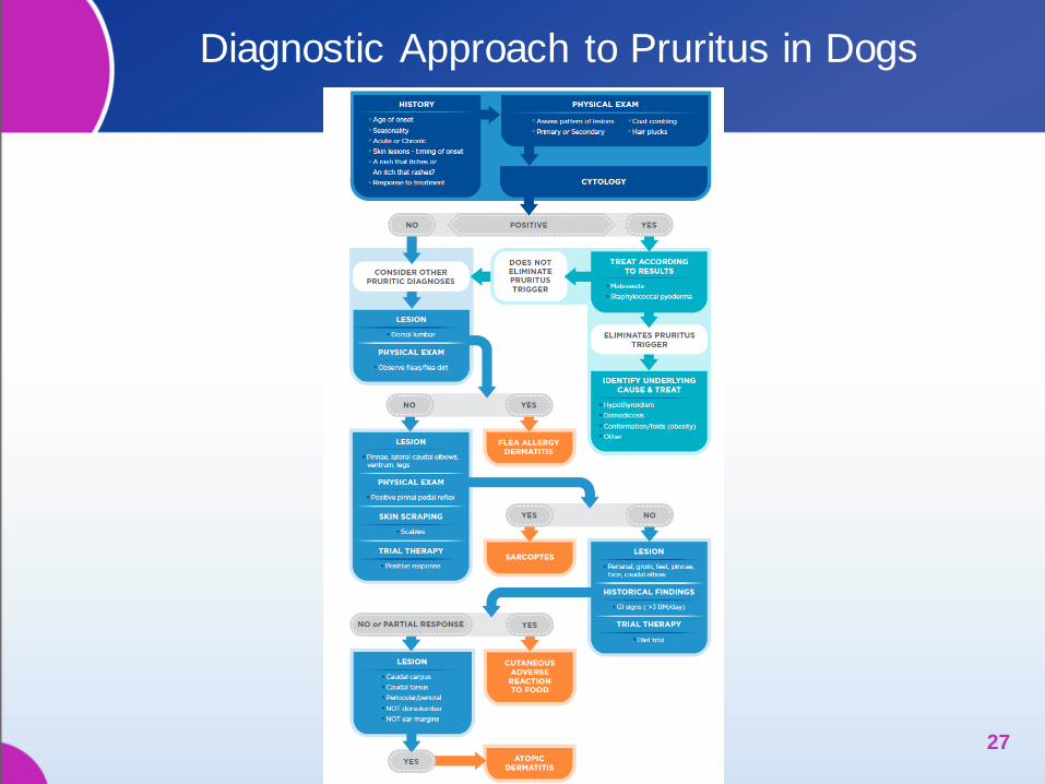
Diagnostic Approach to Pruritus in Dogs
27
ELEMENTS OF
MULTIMODAL THERAPY FOR
CANINE ATOPIC DERMATITIS
28

Treatment for Canine AD is Often Multimodal
29
Options Efficacy Advantages Disadvantages
Essential Fatty
Acids (ω3 and ω6) * ~ 25%
Improvement in
haircoat and skin
Hi dose needed
No evidence of efficacy
Antihistamines * 25 – 30% Inexpensive
Perceived safety
No evidence of efficacy
No FDA-approved
product for dogs
Glucocorticoids * Very effective in treating
itching & inflammation Rapid onset
Immediate annoying
side effects
Serious over time
Cyclosporin * Shown to work in
74% of case of AD FDA-approved
GI upset
Delayed efficacy
ASIT *
Varying (~ 2/3 dogs
show some improvement
after 1 year)
May target the root
of the condition
May take up to a
year to work
Topicals
(shampoos, rinses,
sprays, ointments)
Varies Removes allergen
from the skin
Inconvenient
for owners
Oclacitinib +
Up to 67% reduction in
pruritus
~ 48% reduction in skin
lesions
Rapid onset
Improvement in pruritus and
skin condition
No PU/PD
Daily oral administration
* Olivry T, et al. Vet Dermatol. 2010 + Cosgrove SB, et al. Vet Dermatol. 2013

Fa Fatty Acids
Fa
Fa
Fa
Fa
30

Ah Antihistamines
Ah
31
32
Gc
Gc
Gc
Gc
Gc Gc
Gc
Gc
Gc Glucocorticoid
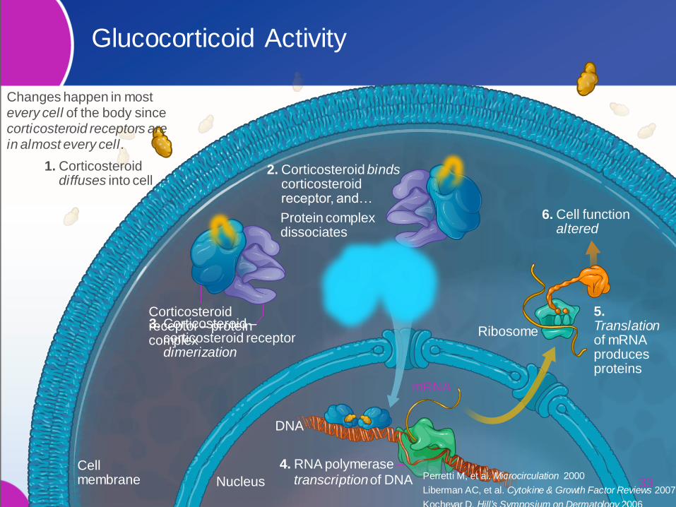
Glucocorticoid Activity
DNA
5. Translation of mRNA produces proteins
Ribosome
mRNA
Cell membrane Nucleus
Corticosteroid receptor – protein complex
1. Corticosteroid diffuses into cell
2. Corticosteroid binds corticosteroid receptor, and…
Protein complex dissociates
3. Corticosteroid – corticosteroid receptor dimerization
4. RNA polymerase transcription of DNA
6. Cell function altered
33
Changes happen in most every cell of the body since corticosteroid receptors are in almost every cell.
Perretti M, et al. Microcirculation 2000
Liberman AC, et al. Cytokine & Growth Factor Reviews 2007
Kochevar D. Hill’s Symposium on Dermatology 2006
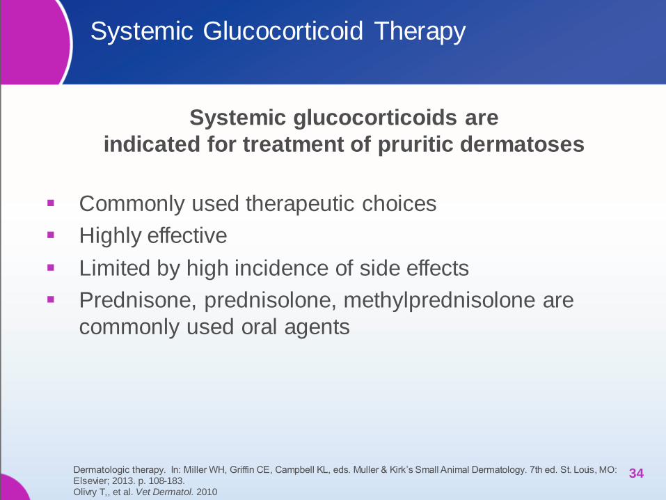
Systemic Glucocorticoid Therapy
Systemic glucocorticoids are
indicated for treatment of pruritic dermatoses
Commonly used therapeutic choices
Highly effective
Limited by high incidence of side effects
Prednisone, prednisolone, methylprednisolone are
commonly used oral agents
34 Dermatologic therapy. In: Miller WH, Griffin CE, Campbell KL, eds. Muller & Kirk’s Small Animal Dermatology. 7th ed. St. Louis, MO: Elsevier; 2013. p. 108-183. Olivry T,, et al. Vet Dermatol. 2010
Side Effects of Glucocorticoid Therapy can be
Bothersome and Medically Important
Common side effects in dogs
Polyuria, Polydipsia, Polyphagia
Weight gain
GI disturbance
Diarrhea, Melena
Vomiting
Possible behavioral changes (depression, hyperactivity, aggression)
Panting
Hyperlipidemia, Elevated liver enzymes
Diabetes mellitus
GI ulceration, Pancreatitis (high doses)
Muscle wasting (high doses)
Poor hair coat (long-term treatment)
35 Dermatologic therapy. In: Miller WH, Griffin CE, Campbell KL, eds. Muller & Kirk’s Small Animal Dermatology. 7th ed. St. Louis, MO: Elsevier; 2013. p. 108-183. Olivry T,, et al. Vet Dermatol. 2010
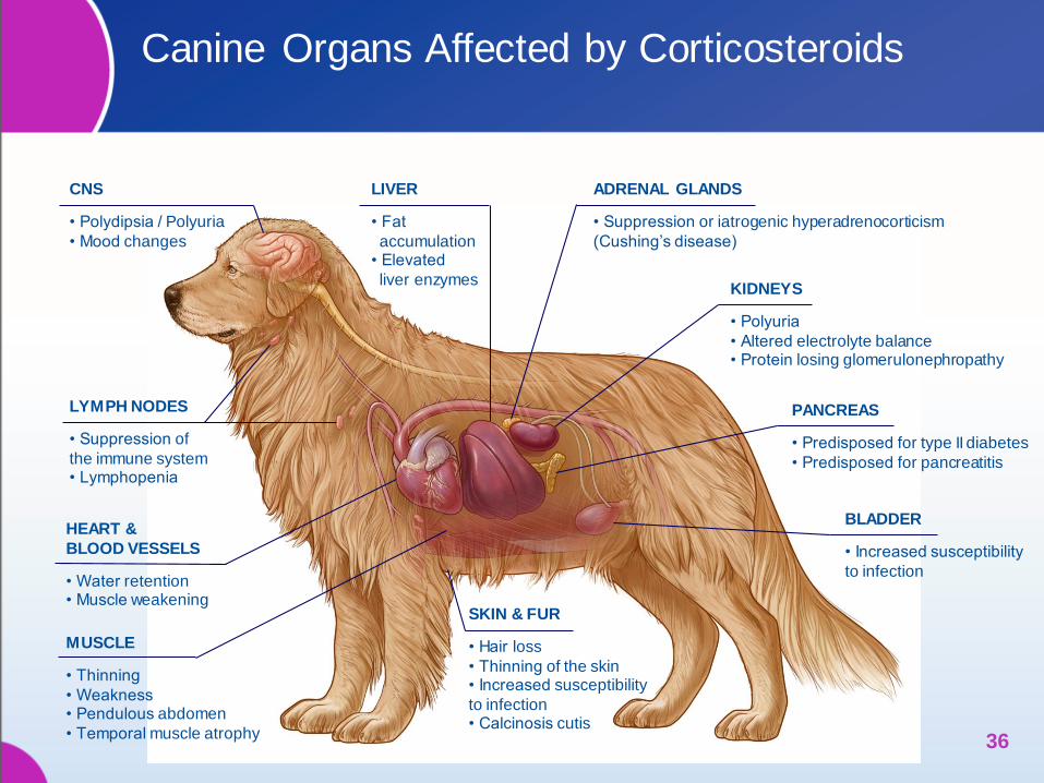
Canine Organs Affected by Corticosteroids
36
CNS • Polydipsia / Polyuria
• Mood changes
LYMPH NODES • Suppression of
the immune system • Lymphopenia
HEART &
BLOOD VESSELS • Water retention • Muscle weakening
MUSCLE • Thinning
• Weakness • Pendulous abdomen
• Temporal muscle atrophy
LIVER • Fat
accumulation • Elevated
liver enzymes
ADRENAL GLANDS • Suppression or iatrogenic hyperadrenocorticism
(Cushing’s disease)
KIDNEYS • Polyuria
• Altered electrolyte balance • Protein losing glomerulonephropathy
PANCREAS • Predisposed for type II diabetes
• Predisposed for pancreatitis
BLADDER • Increased susceptibility
to infection
SKIN & FUR • Hair loss
• Thinning of the skin • Increased susceptibility
to infection • Calcinosis cutis
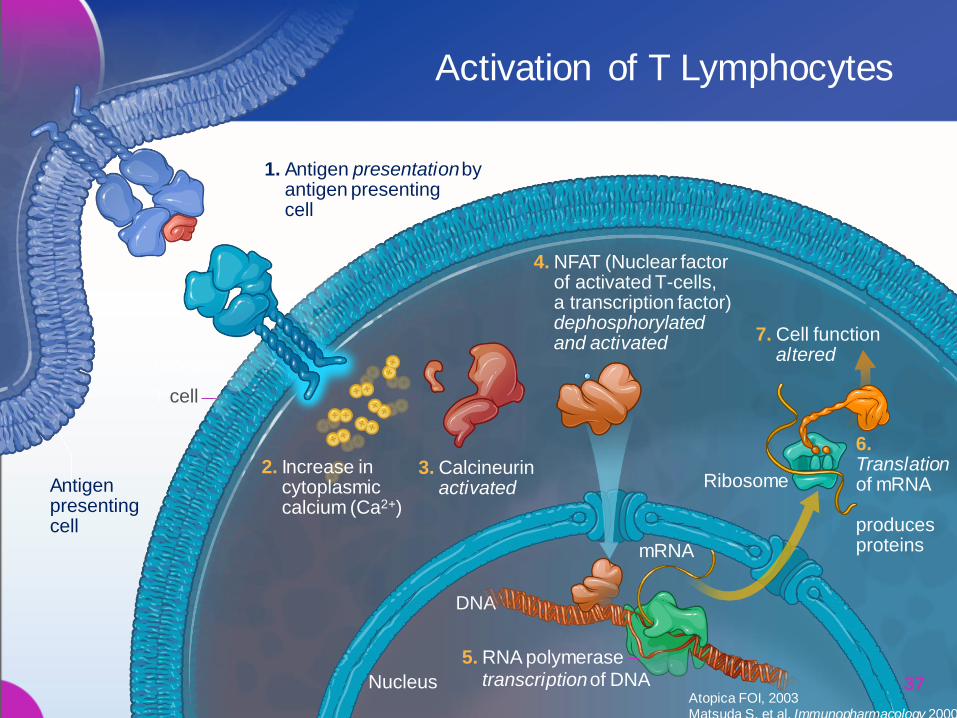
1. Antigen presentation by antigen presenting cell
T cell
T cell receptor
Antigen presenting cell
2. Increase in cytoplasmic calcium (Ca2+)
3. Calcineurin activated
4. NFAT (Nuclear factor of activated T-cells, a transcription factor) dephosphorylated and activated
DNA
Ribosome
mRNA
Nucleus
6. Translation of mRNA produces proteins
7. Cell function altered
5. RNA polymerase transcription of DNA
Activation of T Lymphocytes
37 Atopica FOI, 2003 Matsuda S, et al. Immunopharmacology 2000
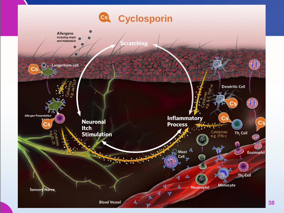
Cs
Cs
Cs Cs
Cs
Cs Cyclosporin
38
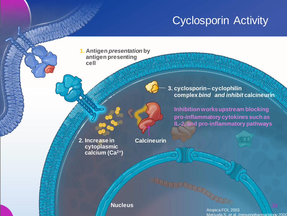
1. Antigen presentation by antigen presenting cell
2. Increase in cytoplasmic calcium (Ca2+)
Calcineurin
Nucleus
3. cyclosporin – cyclophilin complex bind and inhibit calcineurin
Inhibition works upstream blocking
pro-inflammatory cytokines such as IL-2, and pro-inflammatory pathways
Cyclosporin Activity
39 Atopica FOI, 2003 Matsuda S, et al. Immunopharmacology 2000
Cyclosporin Therapy
Atopica® (cyclosporin capsules) is indicated
for control of atopic dermatitis in dogs
Oral immunosuppressive agent
Delayed action of efficacy
Efficacy comparable to glucocorticoids after 3 weeks
of therapy
Evaluated for control of canine atopic dermatitis
Capsules can’t be broken or split
40
Atopica [package insert]. Greensboro, NC: Novartis Animal Health; 2008. Dermatologic therapy. In: Miller WH, Griffin CE, Campbell KL, eds. Muller & Kirk’s Small Animal Dermatology. 7th ed. St. Louis, MO: Elsevier; 2013. p. 108-183 Olivry T, et al. Vet Dermatol 2010
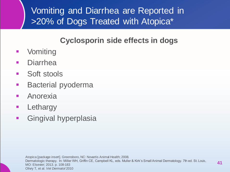
Vomiting and Diarrhea are Reported in
>20% of Dogs Treated with Atopica*
Cyclosporin side effects in dogs
Vomiting
Diarrhea
Soft stools
Bacterial pyoderma
Anorexia
Lethargy
Gingival hyperplasia
41
Atopica [package insert]. Greensboro, NC: Novartis Animal Health; 2008. Dermatologic therapy. In: Miller WH, Griffin CE, Campbell KL, eds. Muller & Kirk’s Small Animal Dermatology. 7th ed. St. Louis, MO: Elsevier; 2013. p. 108-183 Olivry T, et al. Vet Dermatol 2010

Successful Therapy for Canine Atopic Dermatitis:
A Historical Perspective from the Literature
Published reports of masked controlled clinical trials treating atopic
dermatitis in dogs show that glucocorticoids decreased owner VAS
measurement of pruritus 33%-81%
42
Diagnosis Design Efficacy Safety
Glucocorticoid Therapy
Atopic dermatitis*
Prednisolone 0.5 mg/kg SID for 6 weeks (double-masked; n=14)
• 69% (30-84%) mean reduction from baseline in CADESI-02 lesion scores
• 81% (45-86%) mean reduction from baseline in owner VAS scores
• 43% (6/14) of dogs experienced an adverse event
Atopic dermatitis†
Methylprednisolone 0.5-1 mg/kg SID for 1 week, then EOD for 3 weeks; dose tapered at the end of the 4 week period (single-masked; n=59)
• 45% (35-56%) mean reduction from baseline in CADESI-02 lesion scores
• 33% (23-43%) mean reduction from baseline in the owner VAS scores
• 15% (9/59) of dogs dropped out for inefficacy
• 19% (11/59) did not complete study due to worsening clinical signs
• Polyuria/polydipsia occurred in 25% (15/59)
* Olivry T, et al. Vet Dermatol 2002 † Steffan J, et al. Vet Dermatol 2003

Successful Therapy for Canine Atopic Dermatitis: A
Historical Perspective from the Literature
Published reports of masked controlled clinical trials treating atopic dermatitis in
dogs show that cyclosporin decreased owner VAS measurement of pruritus 36%-78%
43
Diagnosis Design Efficacy Safety
Cyclosporin
Atopic dermatitis* cyclosporin 5 mg/kg SID for 6 weeks (double-blinded; n=13)
• 58% (43-74%) mean reduction from baseline in CADESI-02 lesion scores
• 78% (52-87%) mean reduction from baseline in owner VAS scores
• 31% (4/13) of dogs experienced adverse events
• 20% of dogs on cyclosporin occasionally developed diarrhea or soft stools
Atopic dermatitis† cyclosporin 5 mg/kg SID for 4 months; dose tapered at the end of 4 weeks of treatment according to clinical response (single-blinded; n=117)
• 52% (44-59%) mean reduction from baseline in CADESI-02 lesion scores
• 36% (27-43%) mean reduction from baseline in owner VAS scores
• 9% (10/117) of dogs dropped out for inefficacy (increase CADESI-02 scores from baseline
• Vomiting 37% (43/117)
• Diarrhea 18% (21/117)
Atopic dermatitis ǂ cyclosporin 5 mg/kg SID for 30 days followed by up to 16 weeks at SID, EOD, or twice per week (Phase 2; n=192)
• 45% reduction in CADESI-02 lesion scores versus 9% in placebo-treated dogs
• 74% of dogs showed an improvement in pruritus versus 24% in placebo-treated dogs
Of the 265 total number of dogs treated:
• Vomiting 30.9%
• Diarrhea 20.0%
• Persistent otitis externa 6.8%
• UTI 3.8%
• Anorexia 3.0%
• Lethargy 2.3%
• Gingival hyperplasia 2.3%
• Lymphadenopathy 2.3%
* Olivry T, et al. Vet Dermatol 2002 † Steffan J, et al. Vet Dermatol 2003 ǂ Atopica [package insert]. Greensboro, NC: Novartis Animal Health; 2008

Ai
Ai
Ai Allergen-specific Immunotherapy
44
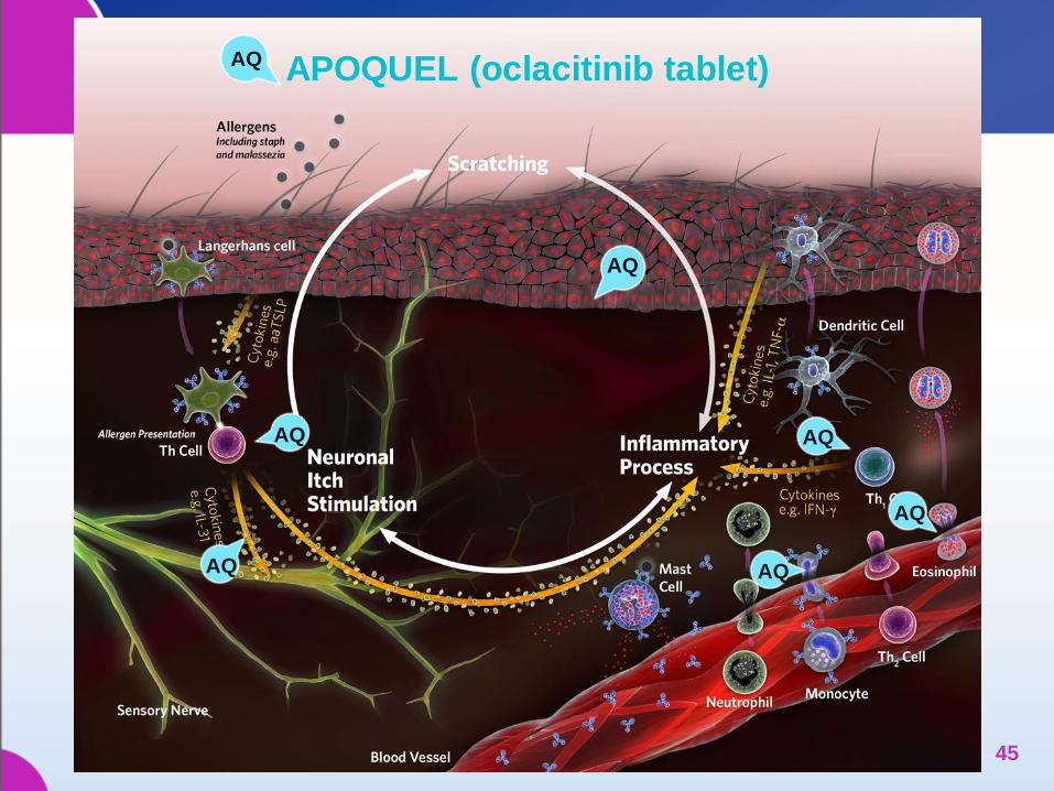
AQ
AQ APOQUEL (oclacitinib tablet)
AQ
AQ
AQ
AQ
AQ
45
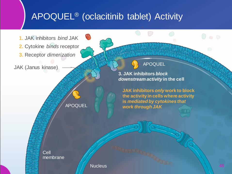
JAK (Janus kinase)
1. JAK inhibitors bind JAK
2. Cytokine binds receptor
3. Receptor dimerization
APOQUEL
APOQUEL
3. JAK inhibitors block downstream activity in the cell
JAK inhibitors only work to block the activity in cells where activity is mediated by cytokines that work through JAK
Nucleus
APOQUEL® (oclacitinib tablet) Activity
46
Cell membrane
APOQUEL Important Safety Information
APOQUEL® should not be used in dogs less than 12 months of age
or in dogs with serious infections. APOQUEL may increase the
susceptibility to infection and demodicosis and may exacerbate
neoplastic conditions. APOQUEL has not been evaluated in
combination with systemic immunosuppressive agents such as
glucocorticoids or cyclosporine. APOQUEL should not be used in
breeding dogs, or pregnant or lactating dogs. The most common
side effects seen in dogs administered APOQUEL were vomiting and
diarrhea. APOQUEL has been safely used in conjunction with other
common medications including antibiotics and parasiticides and with
vaccinations.
See full prescribing information at:
www.APOQUEL.com/APOQUEL_PI
INTRODUCTION TO
MONOCLONAL
ANTIBODY THERAPY
48 48
REVIEW OF
THE IMMUNE SYSTEM
49
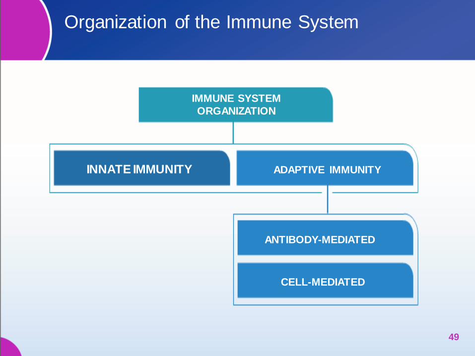
Organization of the Immune System
49
IMMUNE SYSTEM
ORGANIZATION
ADAPTIVE IMMUNITY INNATE IMMUNITY
ANTIBODY-MEDIATED
CELL-MEDIATED

Cells of the Innate Immune System
Myeloid cells
‒ Rapid Response (hours)
‒ Orchestrates adaptive immune responses
51
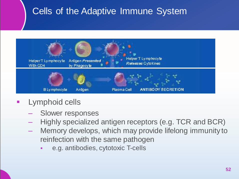
Cells of the Adaptive Immune System
Lymphoid cells
‒ Slower responses
‒ Highly specialized antigen receptors (e.g. TCR and BCR)
‒ Memory develops, which may provide lifelong immunity to
reinfection with the same pathogen e.g. antibodies, cytotoxic T-cells
52
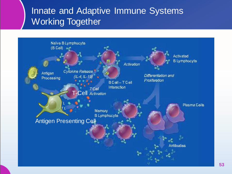
Innate and Adaptive Immune Systems
Working Together
53
Antigen Presenting Cell
T-Cell
HARNESSING THE
BENEFICIAL EFFECTS
OF THE IMMUNE SYSTEM
History of Antibodies
and their Therapeutic Potential
54
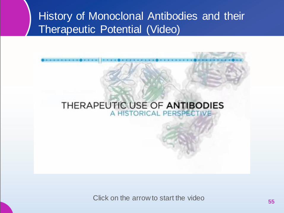
History of Monoclonal Antibodies and their
Therapeutic Potential (Video)
55 Click on the arrow to start the video

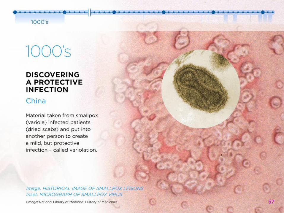
1000’s DISCOVERING
A PROTECTIVE INFECTION
China
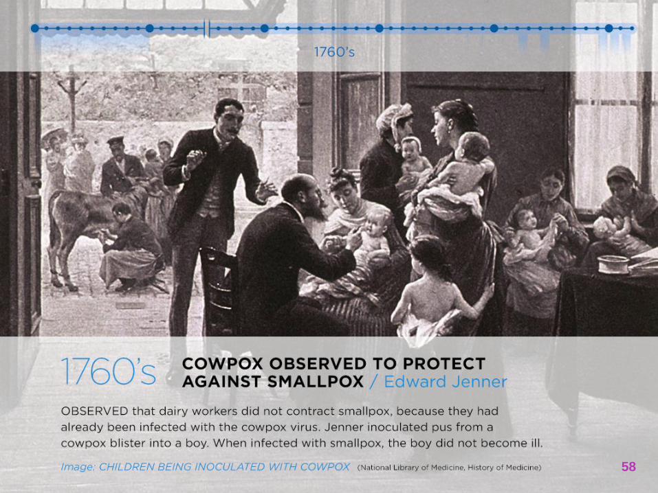
1760’s COWPOX OBSERVED
TO PROTECT AGAINST SMALLPOX
Edward Jenner
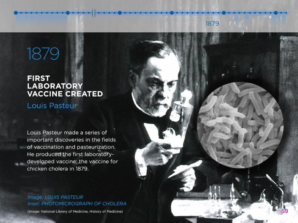
1879 FIRST LABORATORY
VACCINE CREATED
Louis Pasteur
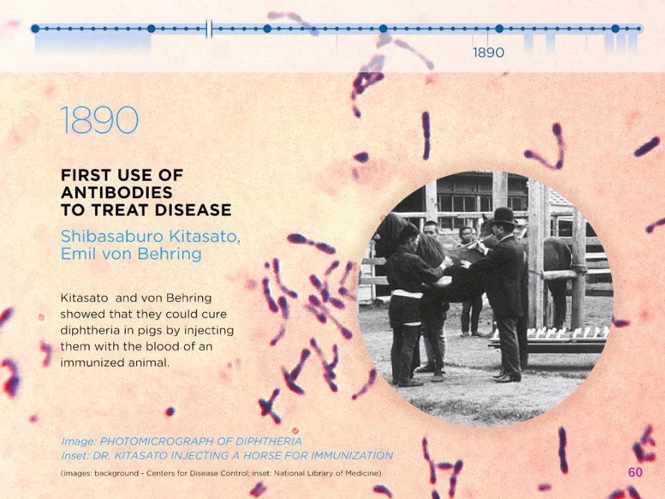
1890 FIRST USE
OF ANTIBODIES TO TREAT DISEASE
Shibasaburo Kitasato,
Emil von Behring
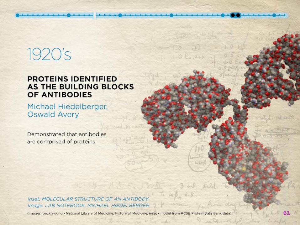
1920’s PROTEINS IDENTIFIED
AS THE BUILDING BLOCKS OF ANTIBODIES
Michael Hiedelberger,
Oswald Avery
1940’s ANTIBODY EXQUISTE SPECIFICITY
DEMONSTRATED, LOCK-AND-KEY MECHANISM CONFIRMED
Linus Pauling
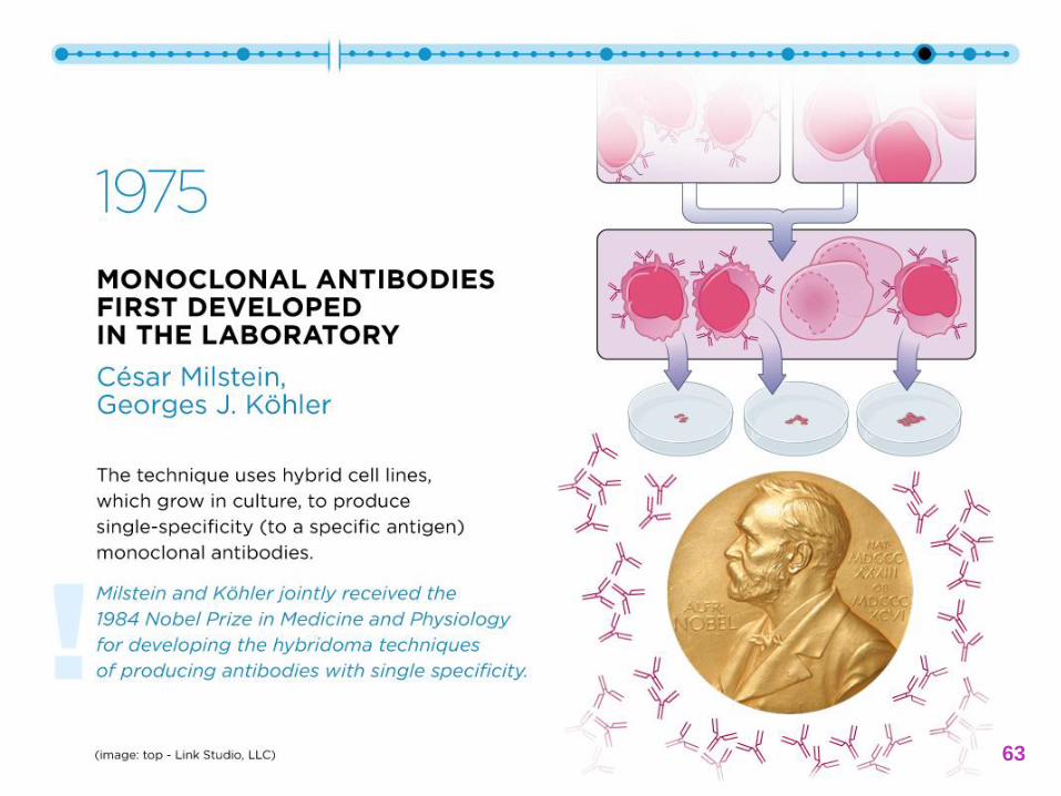
1975 MONOCLONAL ANTIBODIES
FIRST DEVELOPED IN THE LABORATORY
César Milstein,
Georges J. Köhler
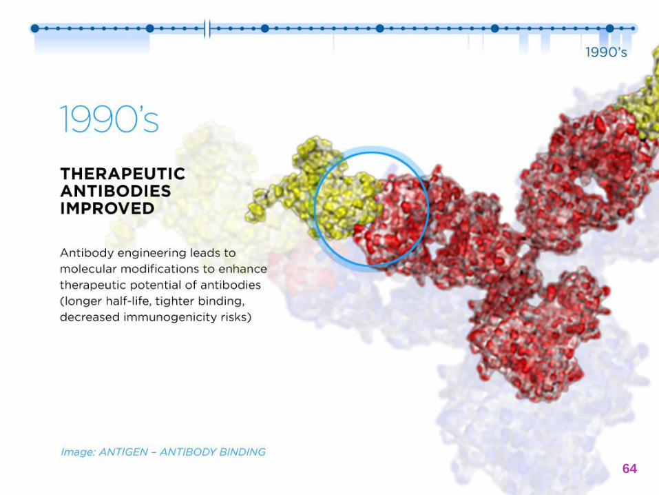
1990’s THERAPEUTIC ANTIBODIES
IMPROVED
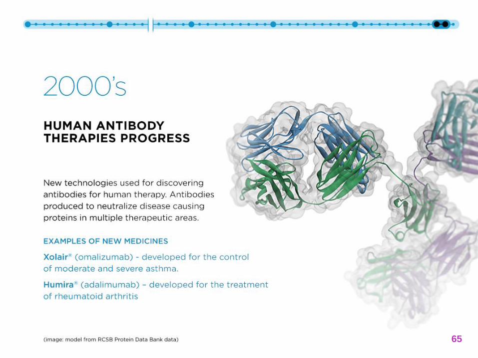
2000’s HUMAN ANTIBODY
THERAPIES PROGRESS
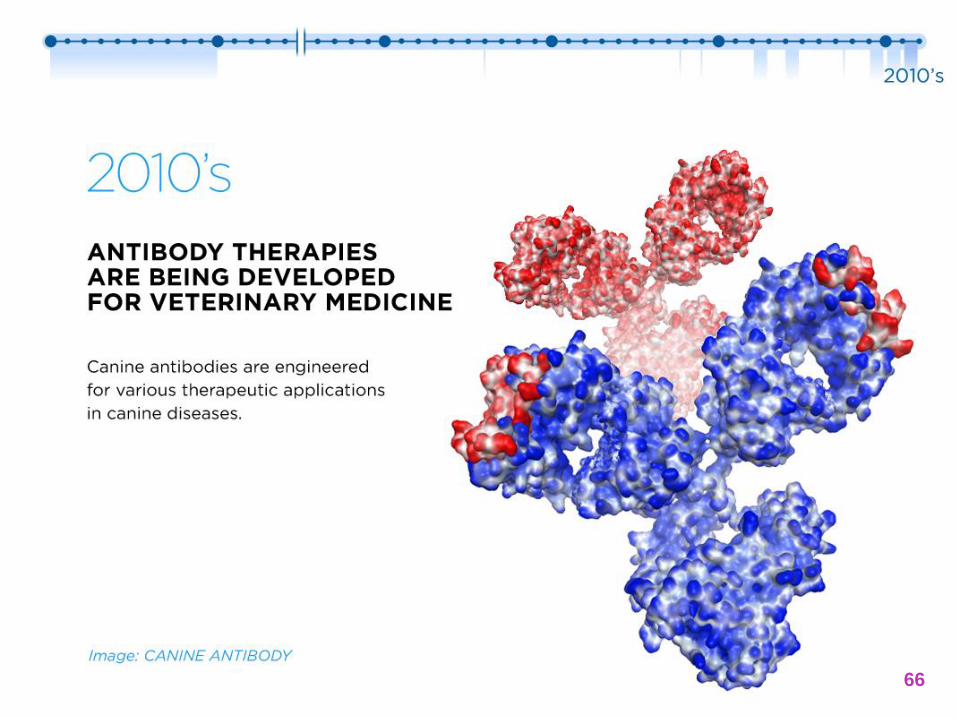
2010’s ANTIBODY THERAPIES
ARE BEING DEVELOPED FOR VETERINARY MEDICINE
IMAGE CREDITS: Historical images: National Library of Medicine, History of Medicine; Antibody models: (top) Data from RCSB Protein Data Bank, (bottom) Zoetis, Inc.
56
57
58
59
60
61

62
63
64
65
66
AN OVERVIEW
OF ANTIBODIES
67

Antibodies
Y-shaped proteins
Produced by mature B-cells (plasma cells)
Found in the blood or other tissue fluids
Used by the immune system to identify and neutralize foreign substances
Isotypes in mammals
‒ IgG, IgM, IgA, IgE, IgD
68
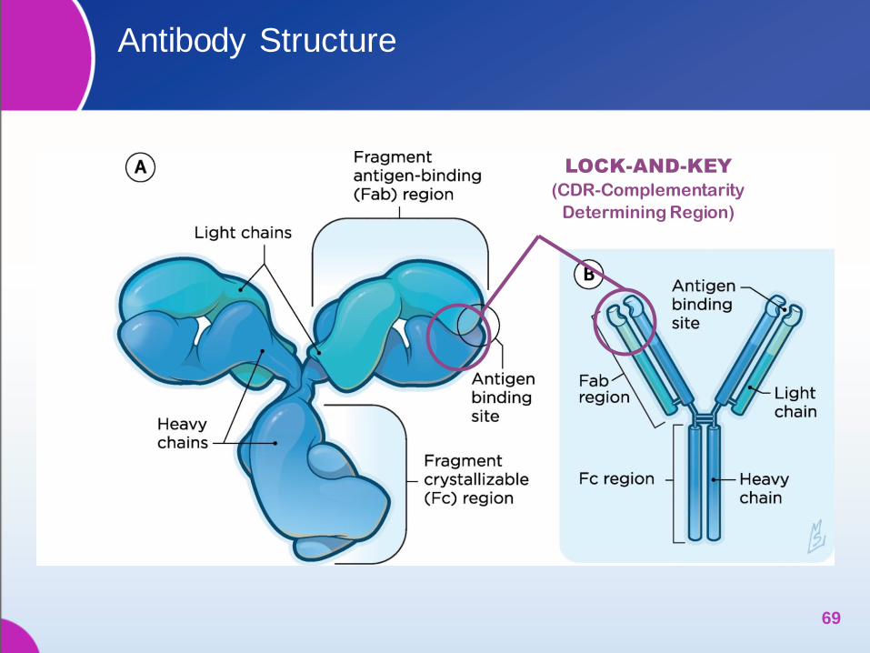
Antibody Structure
69
LOCK-AND-KEY
(CDR-Complementarity
Determining Region)
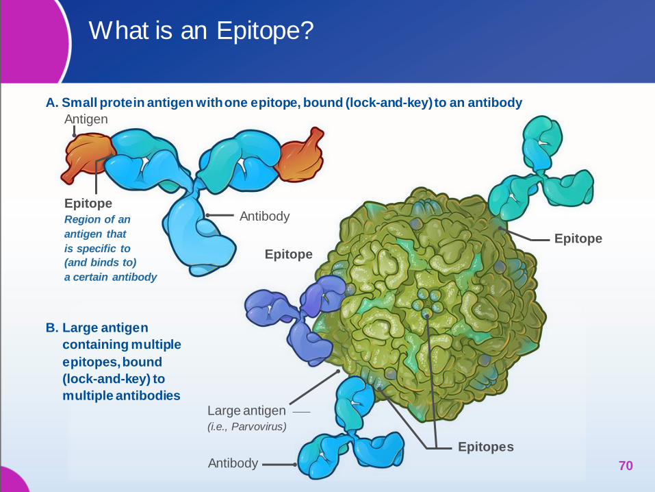
What is an Epitope?
70
A. Small protein antigen with one epitope, bound (lock-and-key) to an antibody
Antigen
Epitope Region of an
antigen that
is specific to
(and binds to)
a certain antibody
Antibody
Large antigen (i.e., Parvovirus)
Antibody
Epitope
Epitope
Epitopes
B. Large antigen
containing multiple
epitopes, bound
(lock-and-key) to
multiple antibodies
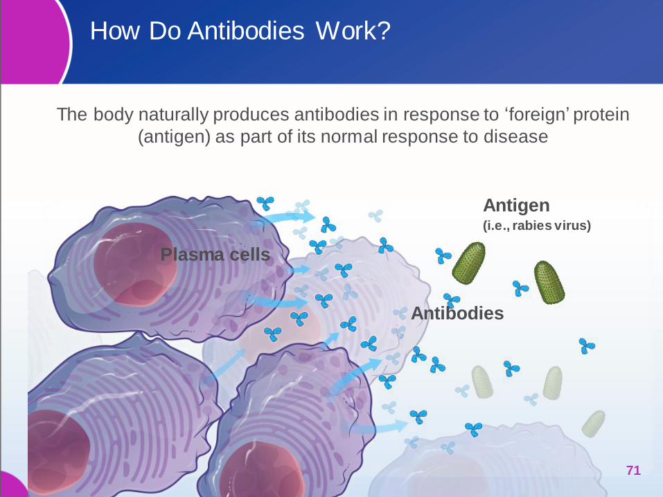
How Do Antibodies Work?
The body naturally produces antibodies in response to ‘foreign’ protein
(antigen) as part of its normal response to disease
71
Plasma cells
Antibodies
Antigen (i.e., rabies virus)

Therapeutic
antibody
How Do Therapeutic Antibodies Work?
Using the same principles, antibodies can now be administered by
injection and used therapeutically
72
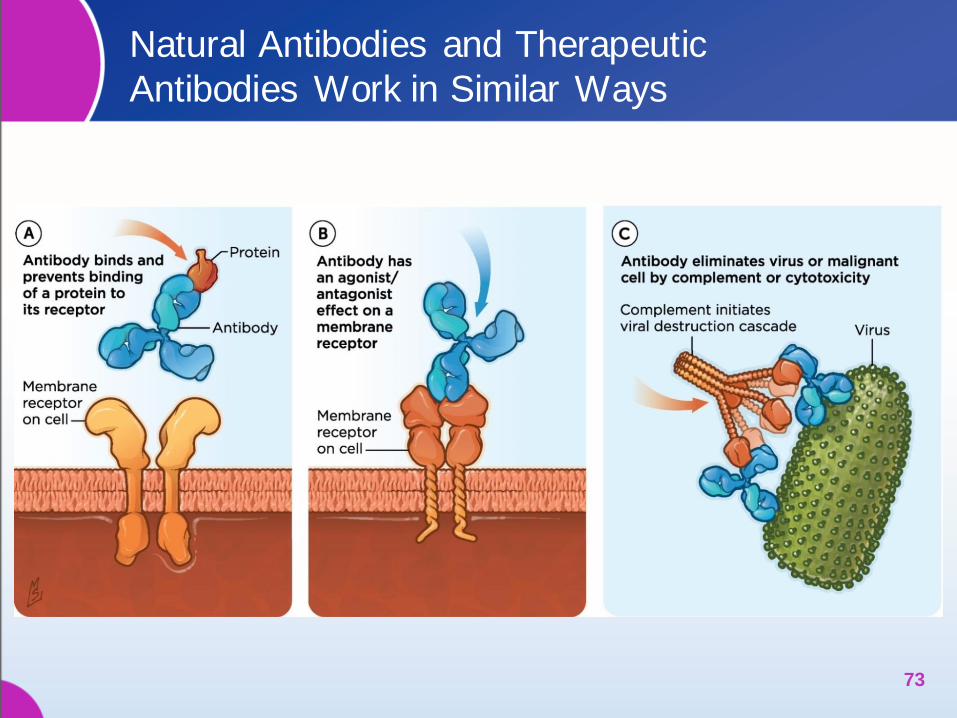
Natural Antibodies and Therapeutic
Antibodies Work in Similar Ways
73
MONOCLONAL ANTIBODIES:
THERAPEUTIC BY DESIGN
74
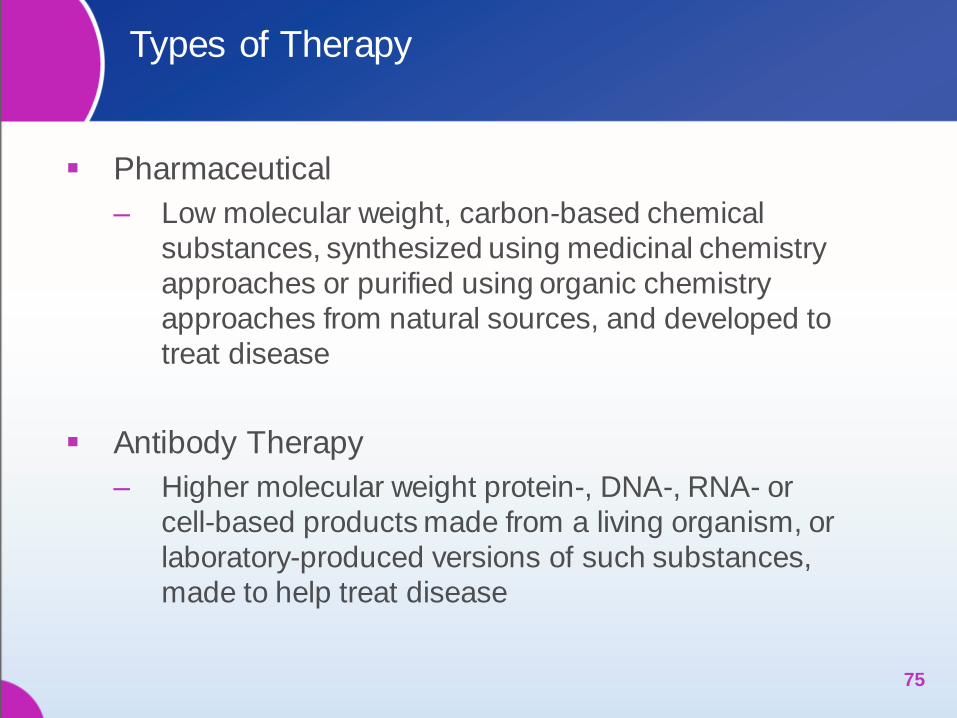
Types of Therapy
Pharmaceutical
‒ Low molecular weight, carbon-based chemical
substances, synthesized using medicinal chemistry
approaches or purified using organic chemistry
approaches from natural sources, and developed to
treat disease
Antibody Therapy
‒ Higher molecular weight protein-, DNA-, RNA- or
cell-based products made from a living organism, or
laboratory-produced versions of such substances,
made to help treat disease
75
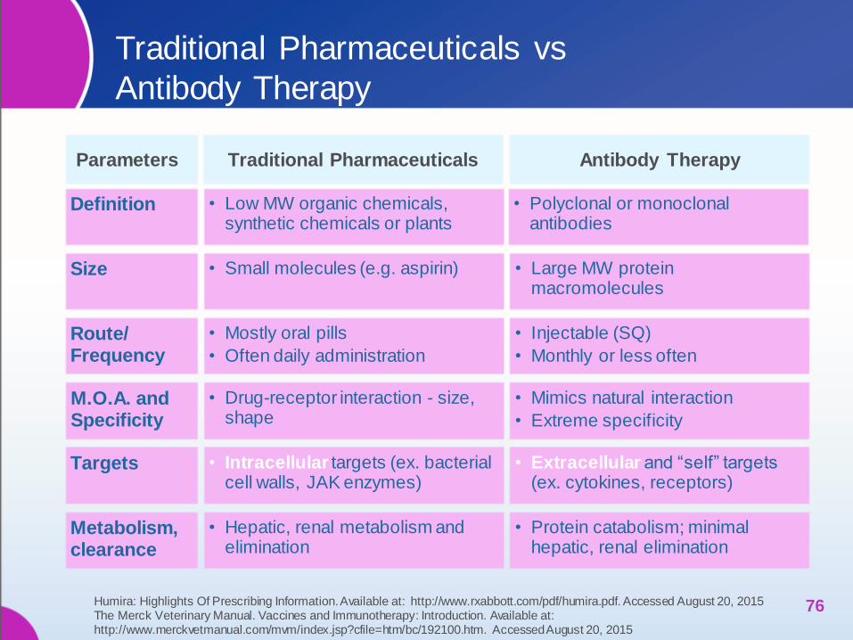
Antibody Therapy Traditional Pharmaceuticals Parameters
Definition
Size
• Low MW organic chemicals, synthetic chemicals or plants
• Polyclonal or monoclonal antibodies
• Small molecules (e.g. aspirin) • Large MW protein macromolecules
• Injectable (SQ)
• Monthly or less often
Route/ Frequency
• Mostly oral pills
• Often daily administration
M.O.A. and Specificity
• Mimics natural interaction
• Extreme specificity
Targets • Intracellular targets (ex. bacterial cell walls, JAK enzymes)
• Extracellular and “self” targets (ex. cytokines, receptors)
Metabolism, clearance
• Protein catabolism; minimal hepatic, renal elimination
• Drug-receptor interaction - size, shape
• Hepatic, renal metabolism and elimination
Traditional Pharmaceuticals vs
Antibody Therapy
76 Humira: Highlights Of Prescribing Information. Available at: http://www.rxabbott.com/pdf/humira.pdf. Accessed August 20, 2015 The Merck Veterinary Manual. Vaccines and Immunotherapy: Introduction. Available at: http://www.merckvetmanual.com/mvm/index.jsp?cfile=htm/bc/192100.htm. Accessed August 20, 2015

Why Develop Monoclonal Biotherapeutics
Using Monoclonal Antibodies?
Expand drug targets small
molecules can’t reach
More target-selective, less
side-effects
Mimic cellular interactions in
the body
Less frequent administration
Large amounts produced in
lab, can be frozen for future
use
77
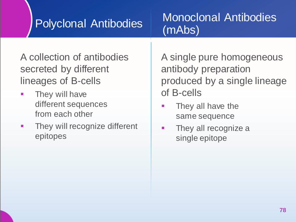
Polyclonal Antibodies
A collection of antibodies
secreted by different
lineages of B-cells
They will have
different sequences
from each other
They will recognize different
epitopes
A single pure homogeneous
antibody preparation
produced by a single lineage
of B-cells
They all have the
same sequence
They all recognize a
single epitope
Monoclonal Antibodies (mAbs)
78
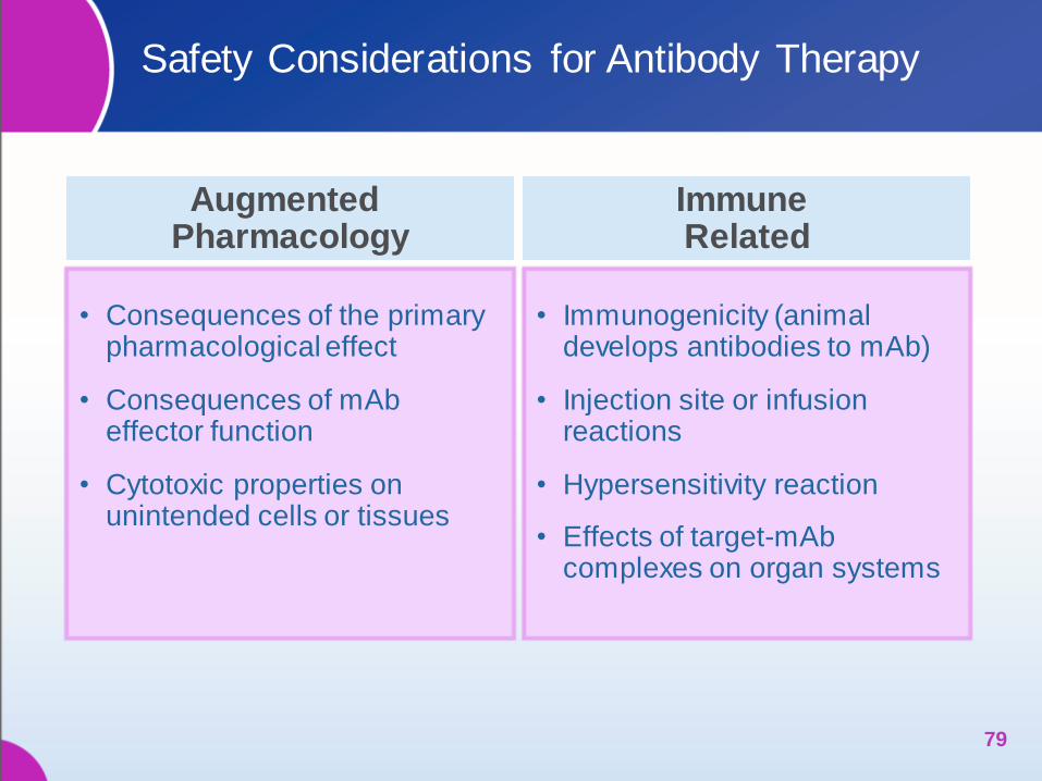
• Consequences of the primary pharmacological effect
• Consequences of mAb effector function
• Cytotoxic properties on unintended cells or tissues
• Immunogenicity (animal develops antibodies to mAb)
• Injection site or infusion reactions
• Hypersensitivity reaction
• Effects of target-mAb complexes on organ systems
Safety Considerations for Antibody Therapy
Augmented Pharmacology
Immune Related
79
Safety Evaluation 101: Where to Start?
80
* Guengerich, FP. Drug Metab Pharmacokinet. 2011
Contexts of Toxic
Responses*
Illustrative Safety studies and
tools…
Illustrative
Examples
Receptor-driven / dose-titratable responses
Target-related: interaction with
the targeted receptor (incl.
excess effect)
Drug:receptor binding
Dose/Response, Overdose, Duration
PK, ADME
Rimadyl®
(carprofen) (COX
inhibition)
“Off-target”-unintended &
undesired receptor binding
Above plus in vitro screening studies of
arrays of possible target receptors
Benadryl®
(anticholinergic)
Metabolic Activation:
biotransformation to new active
compound
Above plus special tests when
indicated
Enalapril,
Nitrofurazone
Responses much less closely related to dose
Immunogenicity—
adaptive/acquired immunity
PV, follow-up studies; ADA & clinical
consequences
Beta-lactams;
Vaccines
Idiosyncratic—low frequency,
difficult to predict or study
Large field studies; Pharmacovigilance [Pharma’s]
Vaccines
[Route of administration] Clinical & pathology evaluations [Injectables]

Pre-Approval Evaluation for Adverse Effects:
Classical Products
Contexts of Toxic
Responses1
Classical Pharmaceutical “Small Molecule”
Classical Vaccine
Receptor-driven dose-titratable responses
Target related Directly Evaluated n/a 2
Off-Target Directly Evaluated n/a
Metabolic Activation Dir. Eval. or Monitored 3 n/a
Responses not closely related to dose
Immunogenicity Monitored Desired 4
Idiosyncratic Monitored Directly Evaluated 4
81
1 Guengerich, FP. Drug Metab Pharmacokinet 2011 2 n/a: potential for this context of toxic response is ~nil. 3 Monitored: response is low-frequency & unpredictable, difficult to methodically evaluate. 4 Cytokine-related or immune-related clinical signs can be more frequent for vaccines than other classes, yet still be considered acceptable.
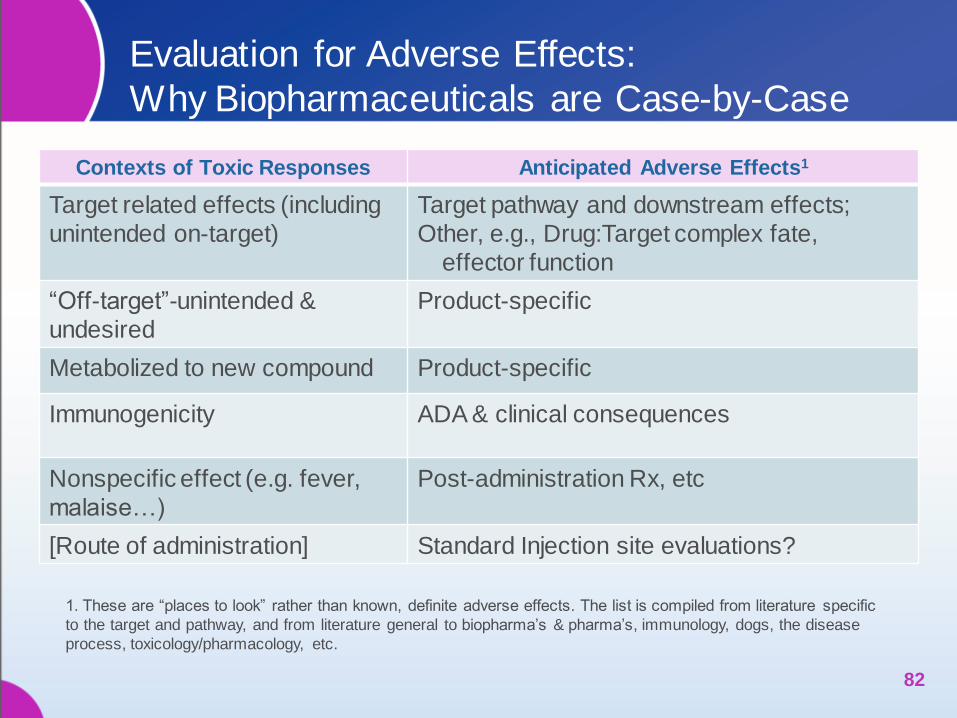
Evaluation for Adverse Effects:
Why Biopharmaceuticals are Case-by-Case
82
1. These are “places to look” rather than known, definite adverse effects. The list is compiled from literature specific
to the target and pathway, and from literature general to biopharma’s & pharma’s, immunology, dogs, the disease
process, toxicology/pharmacology, etc.
Contexts of Toxic Responses Anticipated Adverse Effects1
Target related effects (including
unintended on-target)
Target pathway and downstream effects;
Other, e.g., Drug:Target complex fate,
effector function
“Off-target”-unintended &
undesired
Product-specific
Metabolized to new compound Product-specific
Immunogenicity ADA & clinical consequences
Nonspecific effect (e.g. fever,
malaise…)
Post-administration Rx, etc
[Route of administration] Standard Injection site evaluations?
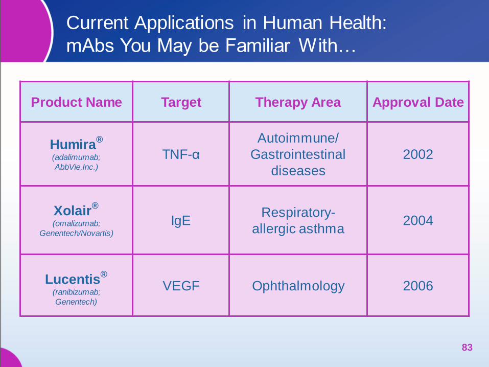
Current Applications in Human Health:
mAbs You May be Familiar With…
Product Name Target Therapy Area Approval Date
Humira®
(adalimumab;
AbbVie,Inc.)
TNF-α
Autoimmune/
Gastrointestinal
diseases
2002
Xolair®
(omalizumab;
Genentech/Novartis)
IgE Respiratory-
allergic asthma 2004
Lucentis®
(ranibizumab;
Genentech)
VEGF Ophthalmology 2006
83
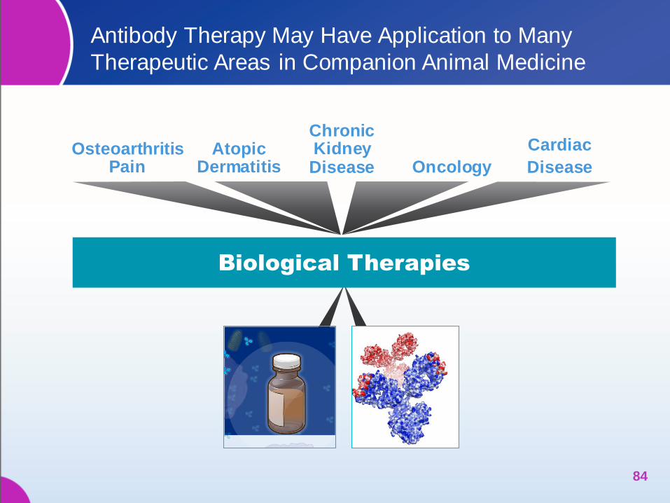
Antibody Therapy May Have Application to Many
Therapeutic Areas in Companion Animal Medicine
84
Osteoarthritis Pain Oncology
Atopic Dermatitis
Cardiac
Disease
Chronic Kidney Disease
Biological Therapies
0 .1 1 1 0 1 0 0
-2 0
0
2 0
4 0
6 0
8 0
1 0 0
1 2 0
IC 5 0 = 1 5 .3 n M
[m A b ], g /m L
% I
nh
ibit
ion
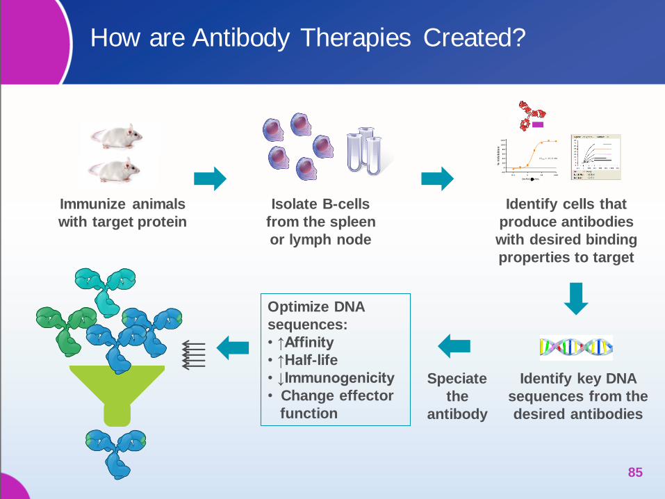
Isolate B-cells
from the spleen
or lymph node
Identify key DNA
sequences from the
desired antibodies
Optimize DNA
sequences:
• ↑Affinity
• ↑Half-life
• ↓Immunogenicity
• Change effector
function
Speciate
the
antibody
Immunize animals
with target protein
Identify cells that
produce antibodies
with desired binding
properties to target
How are Antibody Therapies Created?
85

Caninized Antibodies: Designed to be a Less
Immunogenic Therapeutic Antibody for Dogs
86
Less Immunogenic
Speciation is a key step to decreasing the
potential immunogenicity of a therapeutic antibody
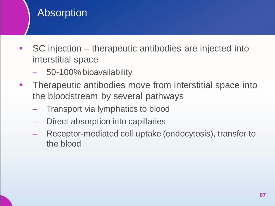
Absorption
SC injection – therapeutic antibodies are injected into
interstitial space
‒ 50-100% bioavailability
Therapeutic antibodies move from interstitial space into
the bloodstream by several pathways
‒ Transport via lymphatics to blood
‒ Direct absorption into capillaries
‒ Receptor-mediated cell uptake (endocytosis), transfer to
the blood
87
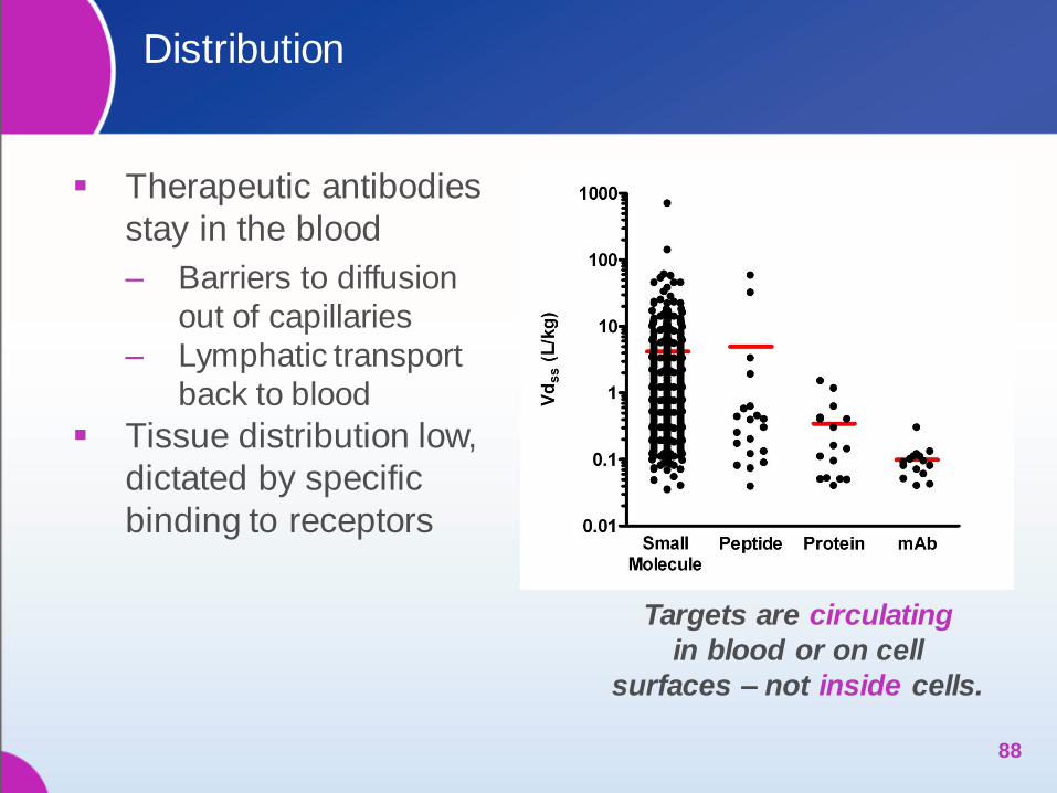
Distribution
Therapeutic antibodies
stay in the blood
‒ Barriers to diffusion
out of capillaries
‒ Lymphatic transport
back to blood
Tissue distribution low,
dictated by specific
binding to receptors
88
Targets are circulating
in blood or on cell
surfaces – not inside cells.
Metabolism and Clearance
Clearance mechanisms include:
‒ Binding to target
‒ Anti-mAb antibodies in circulation
‒ Flow out of capillaries into interstitial space, taken up by
cells and catabolized
Therapeutic antibodies that attach to the FcRn within
the endosome are protected from catabolization and
are recycled back into the blood or lymph which
extends half-life
89
Metabolism and Clearance (cont’d.)
Antibodies are catabolized to peptides and amino
acids within cells
Antibodies are NOT metabolized by traditional
metabolizing enzymes in the kidney or liver
‒ Drug-drug interactions rare
‒ Are not converted into reactive or toxic metabolites
Antibodies are NOT excreted in urine by the kidneys
90
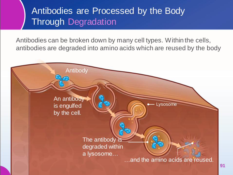
Antibodies are Processed by the Body
Through Degradation
Antibodies can be broken down by many cell types. Within the cells,
antibodies are degraded into amino acids which are reused by the body
91
Antibody
An antibody
is engulfed
by the cell.
The antibody is
degraded within
a lysosome… …and the amino acids are reused.
Lysosome
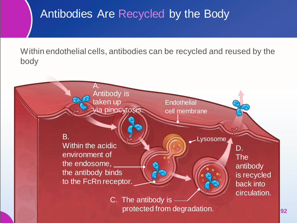
Antibodies Are Recycled by the Body
Within endothelial cells, antibodies can be recycled and reused by the
body
92
A. Antibody is taken up via pinocytosis.
B.
Within the acidic
environment of
the endosome,
the antibody binds
to the FcRn receptor.
C. The antibody is
protected from degradation.
Lysosome
D.
The
antibody
is recycled
back into
circulation.
Endothelial
cell membrane

Unlike Pharmaceuticals, Hepatic and Renal
Elimination is Minimal with Therapeutic Antibodies
Therapeutic antibodies are degraded by normal pathways for protein
catabolism, thus highly unlikely to induce liver or kidney toxicity
Is the liver important for metabolism?
‒ Radiolabeled mAb given to mice, at 24 h
3.6% in liver
Is the kidney important for metabolism?
‒ Radiolabeled mAb given to mice, at 24 h
2-3% in kidney
‒ Renal filtration has a MW cutoff
of approximately 30-50 kDa,
mAbs (150 KD) not filtered
93 Hnatowich DJ, et al. Science 1983
Liver Kidneys
OPPORTUNITY FOR
TREATMENT OF
CANINE ATOPIC DERMATITIS
94 94
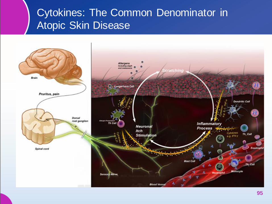
Cytokines: The Common Denominator in
Atopic Skin Disease
95
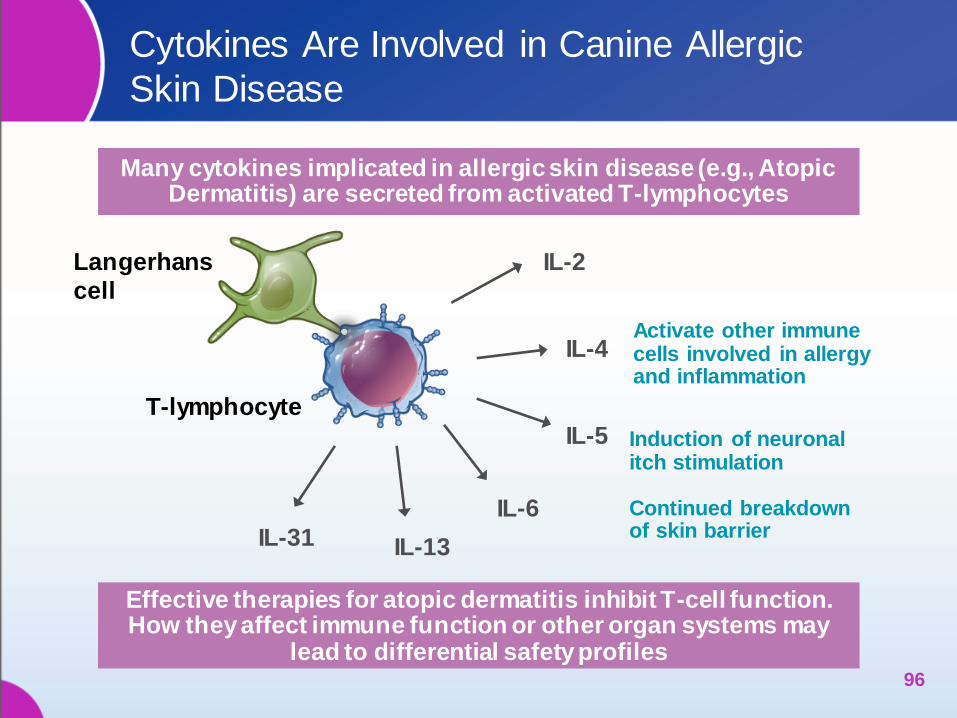
Cytokines Are Involved in Canine Allergic
Skin Disease
96
Langerhans
cell
T-lymphocyte
IL-2
IL-4
IL-6
IL-13 IL-31
IL-5
Activate other immune cells involved in allergy and inflammation
Many cytokines implicated in allergic skin disease (e.g., Atopic Dermatitis) are secreted from activated T-lymphocytes
Effective therapies for atopic dermatitis inhibit T-cell function. How they affect immune function or other organ systems may
lead to differential safety profiles
Induction of neuronal itch stimulation Continued breakdown of skin barrier

~ 50% of Dogs with Naturally Occurring Atopic
Dermatitis Have Measurable Serum Levels of IL-31
97
Canine Populations Number of
Animals Evaluated
Number of Animals with Detectable IL-31 in Serum*
Purpose-bred beagles 24 0
Purpose-bred beagles sensitized to HDM 24 0
Mixed breed dogs – no fleas 30 0
Mixed breed dogs – infested with fleas 30 0
Healthy client owned animals-multiple breeds
87 0
Naturally occurring atopic dermatitis in client owned animals – multiple breeds
224 128
*Less than 13 pg/mL is below limits of detection.
Gonzales AJ, et al. Vet Dermatol 2013
In Laboratory Model Studies Canine IL-31
Induces Pruritic Behaviors in Dogs
cIL-31 was injected i.v. into laboratory beagle dogs
Pruritic behavior scored over a 2 hour observation window
98
0
10
20
30
40
50
T01: PBS Vehicle T02: PBS/Protein Vehicle T03: IL-31 in PBS/Protein Vehicle
Pru
ritu
s S
co
re
* **
*T01 vs T03, p=0.0004. **T02 vs T03, p=0.0003
Gonzales AJ, et al. Vet Dermatol 2013
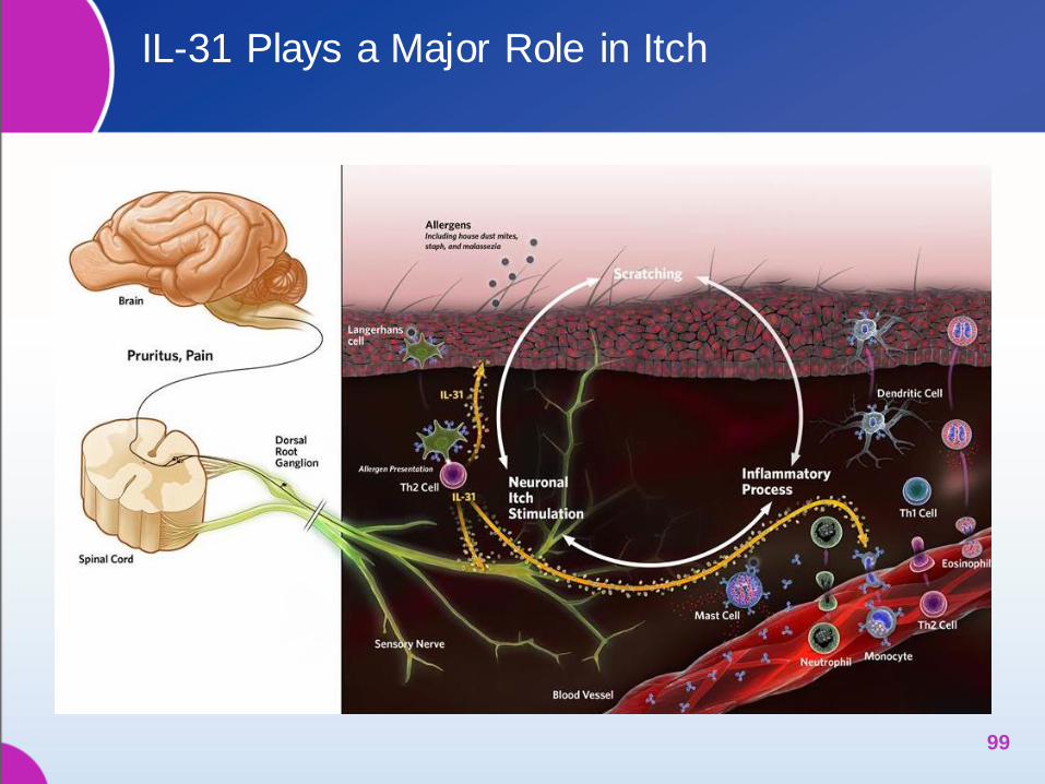
IL-31 Plays a Major Role in Itch
99
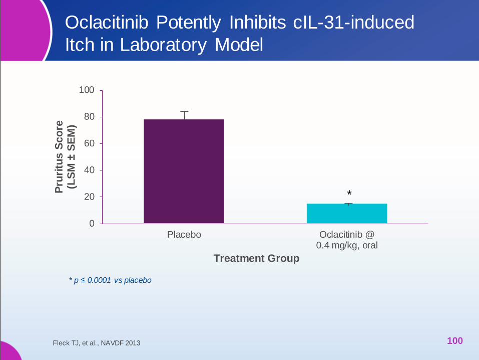
Oclacitinib Potently Inhibits cIL-31-induced
Itch in Laboratory Model
100
* p ≤ 0.0001 vs placebo
Fleck TJ, et al., NAVDF 2013
0
20
40
60
80
100
Placebo Oclacitinib @0.4 mg/kg, oral
Pru
ritu
s S
co
re
(LS
M ±
SE
M)
Treatment Group
*

APOQUEL (oclacitinib tablet) Inhibits the
Activity of Many Cytokines – Including IL-31
101
Langerhans
cell
T-lymphocyte
IL-2
IL-4
IL-6
IL-13 IL-31
IL-5
X
X
X X X
APOQUEL Blocks the Activity of Pruritogenic and
Pro-inflammatory Cytokines That Utilize JAK 1/JAK3
X X
Many Cytokines Implicated in Allergic Skin Disease (e.g., Atopic Dermatitis) Are Secreted from Activated T-lymphocytes
What Is the Role of IL-31 in the Animal?
Regulates aspects of innate as well as adaptive immunity
in tissues that are exposed to the environment
To date, IL-31 has only been identified to play a role in
inflammatory diseases
‒ Skin disease – atopic dermatitis
‒ Lung disease – allergic asthma
‒ Bowel diseases – IBD
Preliminary evidence that IL-31 controls the proliferation
and differentiation of cells of non-hematopoietic origin,
lending support to the concept that this cytokine plays an
integrative role in the proper formation of epithelia
102 Cornelissen C, et al. Europ J Cell Bio 2012
Inhibiting IL-31 Using a Monoclonal Antibody Approach
Holds Potential for Specificity and Low Toxicity
103
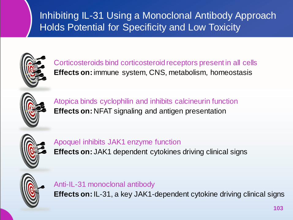
Anti-IL-31 monoclonal antibody
Effects on: IL-31, a key JAK1-dependent cytokine driving clinical signs
Corticosteroids bind corticosteroid receptors present in all cells
Effects on: immune system, CNS, metabolism, homeostasis
Atopica binds cyclophilin and inhibits calcineurin function
Effects on: NFAT signaling and antigen presentation
Apoquel inhibits JAK1 enzyme function
Effects on: JAK1 dependent cytokines driving clinical signs

Zoetis’ Target Product Profile for the Caninized
Anti-cIL-31 Monoclonal Antibody Therapy
A novel therapy for atopic dogs
‒ Not a pharmaceutical therapy
‒ Not a corticosteroid
‒ Targets a single cytokine (IL-31)
Injectable
Duration of effect of one month
Rapid onset of efficacy
‒ Similar to prednisolone and APOQUEL in ability to reduce pruritus
‒ Improvement in dermatitis/skin lesions within 7 days
Unique Safety Profile
‒ No immune suppression
‒ No production of anti-mAb antibodies
‒ No contraindications for other drugs or disease
‒ Use in dogs without any limitation to age
104 104

So Zoetis Scientists Asked…
Would inhibiting only IL-31
provide sufficient relief from
itch and inflammation in canine
patients with atopic dermatitis?
105
“
”

CANINE ATOPIC DERMATITIS
IMMUNOTHERAPEUTIC*
An Antibody Therapy Targeting cIL-31
from Zoetis
106 106 * Product license is conditional. Safety and efficacy studies are ongoing.
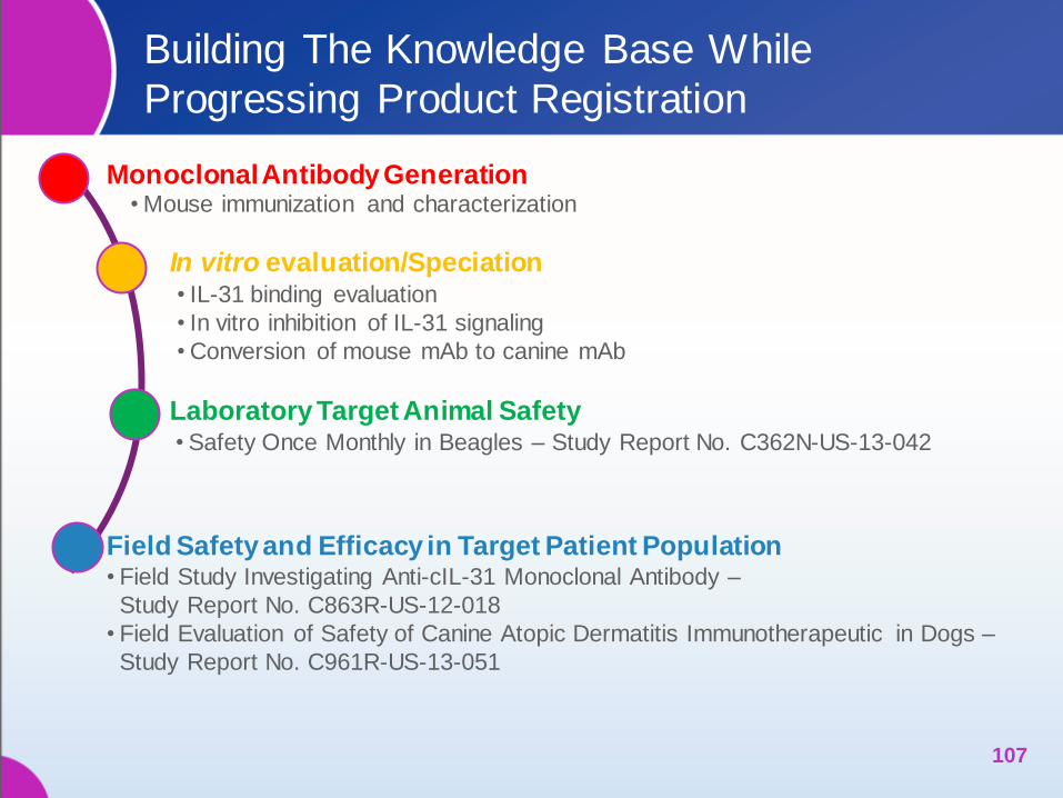
Building The Knowledge Base While
Progressing Product Registration
107
Monoclonal Antibody Generation • Mouse immunization and characterization
In vitro evaluation/Speciation • IL-31 binding evaluation
• In vitro inhibition of IL-31 signaling
• Conversion of mouse mAb to canine mAb
Laboratory Target Animal Safety • Safety Once Monthly in Beagles – Study Report No. C362N-US-13-042
Field Safety and Efficacy in Target Patient Population • Field Study Investigating Anti-cIL-31 Monoclonal Antibody –
Study Report No. C863R-US-12-018
• Field Evaluation of Safety of Canine Atopic Dermatitis Immunotherapeutic in Dogs –
Study Report No. C961R-US-13-051

LABORATORY SAFETY STUDY
108

Laboratory Target Animal Safety Study in Dogs
Demonstrates Margin of Safety Over 7 Month Study
Objective
‒ To demonstrate the margin of safety of Canine Atopic Dermatitis
Immunotherapeutic in normal laboratory Beagles when
administered SC at up to 10 mg/kg once monthly for 7
consecutive monthly doses
109
Treatment (mg/kg) * Administration Route
Animals
Enrolled
0
Days 0, 28, 56, 84,
112, 140, 168
SC
6/sex/group
(n=12) 3.3
10.0
Reference: Study Report No. C362N-US-13-042, Zoetis Inc.
* All dogs dosed according to the dosing chart
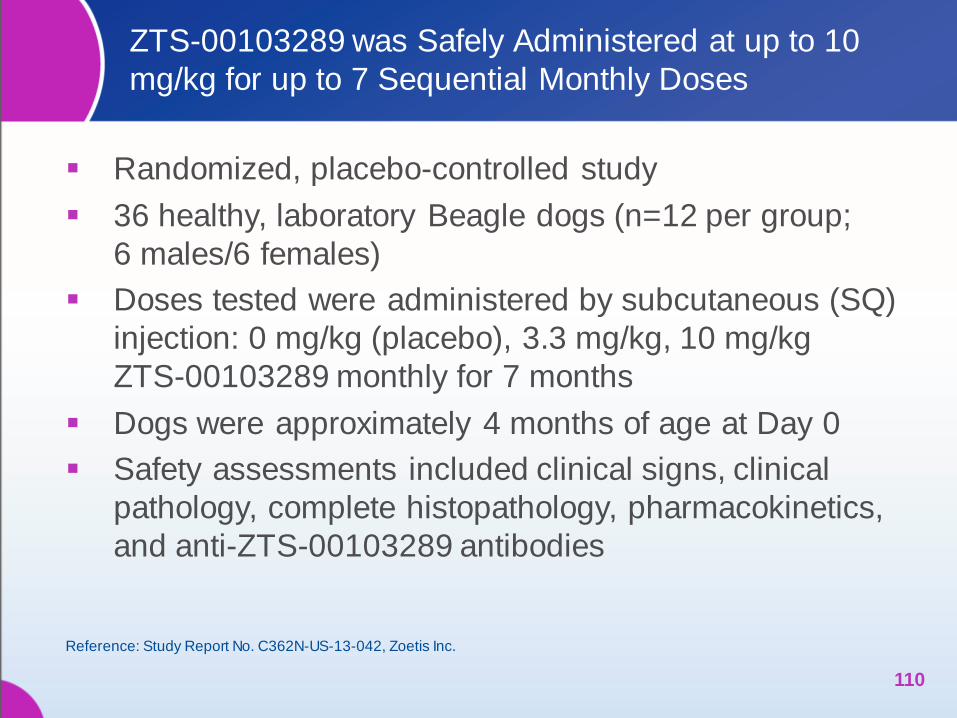
ZTS-00103289 was Safely Administered at up to 10
mg/kg for up to 7 Sequential Monthly Doses
Randomized, placebo-controlled study
36 healthy, laboratory Beagle dogs (n=12 per group;
6 males/6 females)
Doses tested were administered by subcutaneous (SQ)
injection: 0 mg/kg (placebo), 3.3 mg/kg, 10 mg/kg
ZTS-00103289 monthly for 7 months
Dogs were approximately 4 months of age at Day 0
Safety assessments included clinical signs, clinical
pathology, complete histopathology, pharmacokinetics,
and anti-ZTS-00103289 antibodies
110
Reference: Study Report No. C362N-US-13-042, Zoetis Inc.
111
Abnormal Clinical Observations (Data = Number of Dogs)
Clinical Sign Treatment
Period (days)
0-7 28-35 56-63 84-91 112-119 140-147 168-175
Lameness Placebo 0 0 0 0 0 0 1
Abdominal pain Placebo 0 0 0 0 0 0 2
Erythema ventral
abdominal area
ZTS-00103289
3.3 mg/kg 0 0 0 0 1 0 0
Loss of condition Placebo 5 4 2 1 0 0 0
ZTS-00103289
3.3 mg/kg 3 2 0 0 0 0 0
Swollen urogenital
area
ZTS-00103289
3.3 mg/kg 0 0 0 0 0 0 2
Thin Placebo 0 0 0 1 1 1 1
Dose site mild
localized erythema resembling a razor
burn
Placebo
1 0 0 0 0 0 0
Dose site mild
superficial scabbing
Placebo 1 0 0 0 0 0 0
ZTS-00103289
3.3 mg/kg 0 0 0 0 0 0 1
Reference: Study Report No. C362N-US-13-042, Zoetis Inc.
ZTS-00103289 was Safely Administered at up to 10
mg/kg for up to 7 Sequential Monthly (con’t)
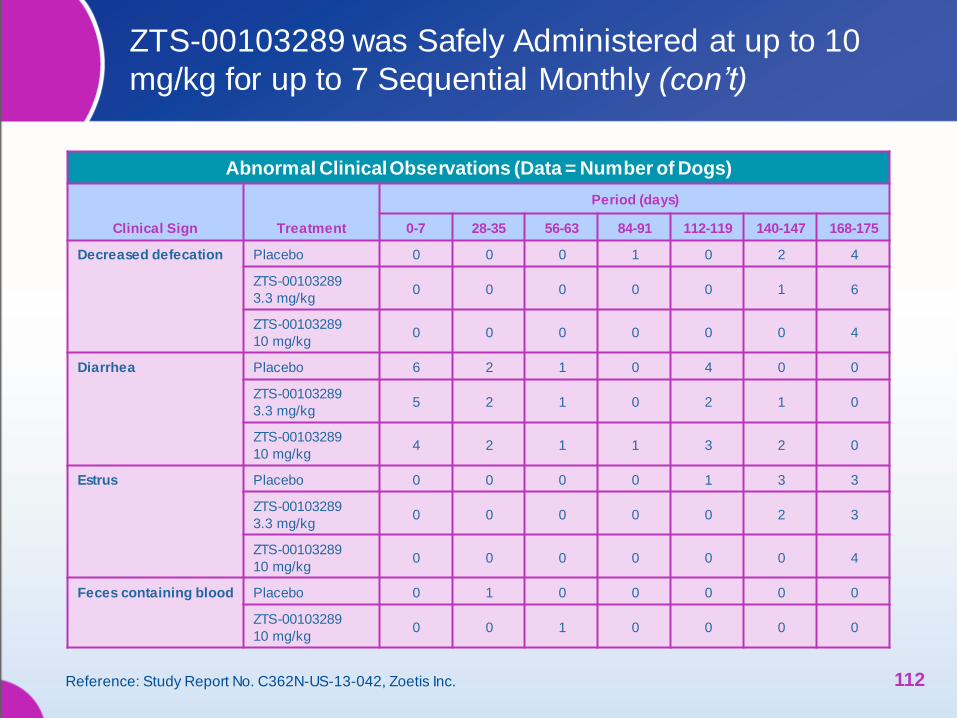
112
Abnormal Clinical Observations (Data = Number of Dogs)
Clinical Sign Treatment
Period (days)
0-7 28-35 56-63 84-91 112-119 140-147 168-175
Decreased defecation Placebo 0 0 0 1 0 2 4
ZTS-00103289
3.3 mg/kg 0 0 0 0 0 1 6
ZTS-00103289
10 mg/kg 0 0 0 0 0 0 4
Diarrhea Placebo 6 2 1 0 4 0 0
ZTS-00103289
3.3 mg/kg 5 2 1 0 2 1 0
ZTS-00103289
10 mg/kg 4 2 1 1 3 2 0
Estrus Placebo 0 0 0 0 1 3 3
ZTS-00103289
3.3 mg/kg 0 0 0 0 0 2 3
ZTS-00103289
10 mg/kg 0 0 0 0 0 0 4
Feces containing blood Placebo 0 1 0 0 0 0 0
ZTS-00103289
10 mg/kg 0 0 1 0 0 0 0
Reference: Study Report No. C362N-US-13-042, Zoetis Inc.
ZTS-00103289 was Safely Administered at up to 10
mg/kg for up to 7 Sequential Monthly (con’t)

ZTS-00103289 was Safely Administered at up to 10
mg/kg for up to 7 Sequential Monthly (con’t)
113
Abnormal Clinical Observations (Data = Number of Dogs)
Clinical Sign Treatment
Period (days)
0-7 28-35 56-63 84-91 112-119 140-147 168-175
Mucous diarrhea Placebo 0 0 1 0 0 0 0
ZTS-00103289
3.3 mg/kg 0 1 0 0 0 1 0
ZTS-00103289
10 mg/kg 0 0 1 0 0 1 1
Mucous stool Placebo 2 3 3 0 1 0 0
ZTS-00103289
3.3 mg/kg 2 6 2 0 0 0 1
ZTS-00103289
10 mg/kg 2 1 2 2 0 0 0
Mucous stool
containing blood
ZTS-00103289
10 mg/kg 0 0 0 1 0 0 1
No defecation Placebo 0 1 0 0 0 1 0
ZTS-00103289
3.3 mg/kg 0 2 0 3 0 0 0
ZTS-00103289
10 mg/kg 0 3 0 0 0 0 0
Reference: Study Report No. C362N-US-13-042, Zoetis Inc.

114
Abnormal Clinical Observations (Data = Number of Dogs)
Clinical Sign Treatment
Period (days)
0-7 28-35 56-63 84-91 112-119 140-147 168-175
Soft feces Placebo 11 5 7 5 6 2 4
ZTS-00103289
3.3 mg/kg 7 7 4 3 6 5 5
ZTS-00103289
10 mg/kg 11 7 10 8 8 8 7
Emesis Placebo 0 0 0 1 1 0 1
ZTS-00103289
3.3 mg/kg 0 0 0 0 1 0 0
ZTS-00103289
10 mg/kg 1 1 1 0 0 1 0
Emesis containing
food
ZTS-00103289
3.3 mg/kg 0 0 0 0 0 1 1
ZTS-00103289
10 mg/kg 0 0 0 0 0 1 2
Regurgitation Placebo 0 0 0 0 0 0 1
Reference: Study Report No. C362N-US-13-042, Zoetis Inc.
ZTS-00103289 was Safely Administered at up to 10
mg/kg for up to 7 Sequential Monthly (con’t)
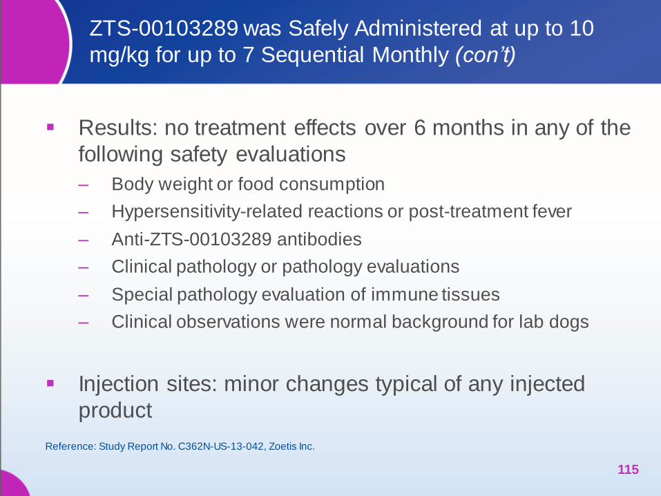
Results: no treatment effects over 6 months in any of the
following safety evaluations
‒ Body weight or food consumption
‒ Hypersensitivity-related reactions or post-treatment fever
‒ Anti-ZTS-00103289 antibodies
‒ Clinical pathology or pathology evaluations
‒ Special pathology evaluation of immune tissues
‒ Clinical observations were normal background for lab dogs
Injection sites: minor changes typical of any injected
product
115
Reference: Study Report No. C362N-US-13-042, Zoetis Inc.
ZTS-00103289 was Safely Administered at up to 10
mg/kg for up to 7 Sequential Monthly (con’t)
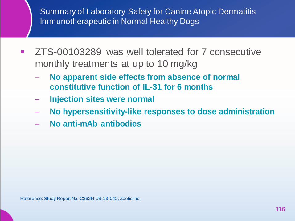
Summary of Laboratory Safety for Canine Atopic Dermatitis
Immunotherapeutic in Normal Healthy Dogs
ZTS-00103289 was well tolerated for 7 consecutive
monthly treatments at up to 10 mg/kg
‒ No apparent side effects from absence of normal
constitutive function of IL-31 for 6 months
‒ Injection sites were normal
‒ No hypersensitivity-like responses to dose administration
‒ No anti-mAb antibodies
116
Reference: Study Report No. C362N-US-13-042, Zoetis Inc.

CLINICAL FIELD TRIALS
Safety and Efficacy in Client-owned Dogs
with Atopic Dermatitis
117
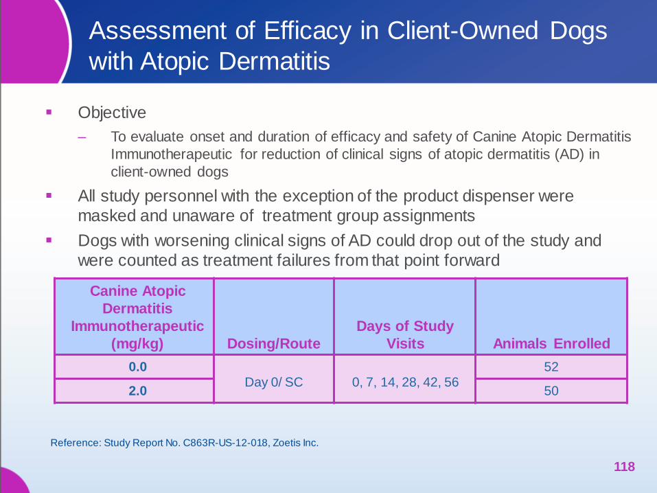
Assessment of Efficacy in Client-Owned Dogs
with Atopic Dermatitis
Objective
‒ To evaluate onset and duration of efficacy and safety of Canine Atopic Dermatitis
Immunotherapeutic for reduction of clinical signs of atopic dermatitis (AD) in
client-owned dogs
All study personnel with the exception of the product dispenser were
masked and unaware of treatment group assignments
Dogs with worsening clinical signs of AD could drop out of the study and
were counted as treatment failures from that point forward
118
Canine Atopic
Dermatitis
Immunotherapeutic
(mg/kg) Dosing/Route
Days of Study
Visits Animals Enrolled
0.0 Day 0/ SC
0, 7, 14, 28, 42, 56
52
2.0 50
Reference: Study Report No. C863R-US-12-018, Zoetis Inc.

Owner Assessment of Pruritus Assessed
Using Visual Analog Scale (VAS)
119
Owner Assessment of Pruritus VAS
10 cm lines with text descriptors at 2 cm intervals
Extremely severe itching. Dog is scratching, chewing, licking
almost continuously. Itching practically never stops regardless
of what else is happening around the dog.
Severe itching. Prolonged episodes of itching when the dog is
awake. Itching occurs at night and also when eating, playing,
exercising, or when otherwise distracted.
Moderate itching. Regular episodes of itching when the dog is
awake. Itching might occur at night and wake the dog. No itching
when eating, playing, exercising, or when being distracted.
Mild itching. More frequent episodes of itching. May notice
occasional episodes of itching at night. No itching when
sleeping, eating, playing, exercising or when being distracted.
Very mild itching. Occasional episodes of itching. The
dog is slightly more itchy than before the problem began.
Normal Dog. Itching is not a problem.
10 cm (100 mm) line
Owner places mark on
line that best represents
the dogs level of pruritus
(itch)
Measurement from the
bottom of the line (“normal
dog”) to the owner’s mark
on the line is recorded
and analyzed
Reference: Study Report No. C863R-US-12-018, Zoetis Inc.
Site E L X A SITE E L X A Site E L X A Site E L X A
Face
- Periauricular
Forelimb
- L Medial
Hind Limb
- L Medial
Sternum
- Periocular - L Lateral - L Lateral Axilla
- Left
- Perilabial - L Cubital
Flexor
- L Stifle
Flexor
- Right
- Muzzle L-Carpal
Flexor
- L Tarsal
Flexor
Thorax
- Dorsal
- Chin - R Medial - R Medial - L Lateral
Head-Dorsal - R Lateral - R Lateral - R Lateral
Pinna
- Left Concave
- R Cubital
Flexor
- R Stifle
Flexor
Inguinal
- Left
- Right
Concave
- R Carpal
Flexor
- R Tarsal
Flexor
- Right
- Left Convex Forefoot
- L Palmar
Metacarpal
Hind Foot
- L Plantar
Metatarsal
Abdomen
- Right Convex - L Dorsal
Metacarpal
- L Dorsal
Metatarsal
Lumbar
- Dorsal
Neck
- Dorsal
- L Palmar
Phalangeal
- L Plantar
Phalangeal
Flank
- Left
- Ventral - L Dorsal
Interdigital
- L Dorsal
Interdigital
- Right
- Left Lateral - R Palmar
Metacarpal
-R Plantar
Metatarsal
Tail
- Ventral
- Right Lateral - R Dorsal
Metacarpal
- R Dorsal
Metatarsal
- Dorsal
Perigenital - R Palmar
Phalangeal
- R Plantar
Phalangeal
Perianal - R Dorsal
Interdigital
- R Dorsal
Interdigital
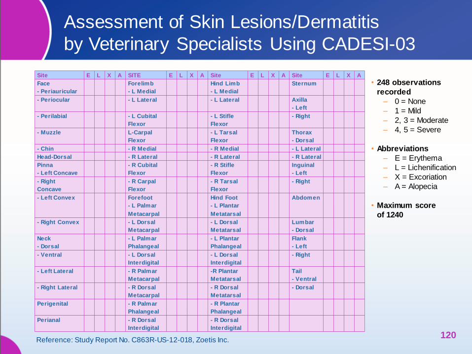
• 248 observations
recorded
– 0 = None
– 1 = Mild
– 2, 3 = Moderate
– 4, 5 = Severe
• Abbreviations
– E = Erythema
– L = Lichenification
– X = Excoriation
– A = Alopecia
• Maximum score
of 1240
Assessment of Skin Lesions/Dermatitis
by Veterinary Specialists Using CADESI-03
120 Reference: Study Report No. C863R-US-12-018, Zoetis Inc.
Dogs with Chronic Atopic Dermatitis Were
Included in Field Trial of Efficacy and Safety
Owner indicated a pruritus score of ≥30 (of 100) on a
Visual Analog Scale
Dermatologists assigned a CADESI-03 score of ≥30
Dogs were ≥1 year of age, weighed 2.0-80.0 kg and
were physically healthy other than their atopic disease
Dogs had a ≥1 year documented history of chronic
non-seasonal atopic dermatitis
‒ Based on Favrot (2010): requires presence of ≥5 of the following
1) Age at onset <3 years 5) Mostly indoor
2) Non-affected dorso-lumbar area 6) Affected ear pinnae
3) Corticosteroid-responsive pruritus 7) Non-affected ear margins
4) Chronic or recurrent yeast infections 8) Affected front feet
121
Reference: Study Report No. C863R-US-12-018, Zoetis Inc.
Dogs with Chronic Atopic Dermatitis Were
Included in Field Trial of Efficacy and Safety
Dogs were flea free and received appropriate
preventatives/treatments
Dogs had been appropriately withdrawn from protocol
specified medications and therapies that had the
potential to confound efficacy assessments
‒ Including but not limited to corticosteroids, cyclosporins,
antimicrobials and antihistamines
122
Reference: Study Report No. C863R-US-12-018, Zoetis Inc.
Dogs with Non-atopic Pruritus Were
Not Included
Dogs with malignant neoplasia
Dogs with evidence of immune suppression
Dogs with evidence of demodicosis within the past year
‒ If history of demodicosis, two or more negative skin
scrapings required
Lactating bitches or dogs intended for breeding
Dogs receiving systemic antimicrobial therapy for
treatment of bacterial or fungal skin infections
123
Reference: Study Report No. C863R-US-12-018, Zoetis Inc.

Response Based on Owner Assessed
Pruritus and CADESI-03 Score: Study Design
124
Visit 1 Visit 2 Visit 5 Visit 6 Visit 4
↑28 ↑0 ↑1 ↑2 ↑7 ↑56
Owner Pruritus Assessment (VAS)
Visit 1, Day 0, Randomization
Physical examination
Blood and Urine sample
Inclusion/Exclusion criteria
Ow ner pruritus assessment
Investigator CADESI-03 scoring Investigator visual analog assessment
Test article dispensing and administration
Concomitant Treatment record
Visit 2, Day 7±3
Physical examination
Blood sample
Investigator CADESI-03 scoring
Investigator visual analog assessment
Ow ner pruritus assessment
Visit 6, Day 56±7, Final Study Day
Physical examination
Blood and Urine sample
Ow ner pruritus assessment
Investigator CADESI-03 scoring
Investigator visual analog assessment Investigator response to treatment VAS
Concomitant Treatment record
Study Completion form
Ow ner response to treatment VAS
Visit 4, Day 28±3
Physical examination
Blood and Urine sample
Ow ner pruritus assessment
Investigator CADESI-03 scoring
Investigator visual analog assessment
Visit 5, Day 42±3
Physical examination
Blood sample
Investigator CADESI-03 scoring
Investigator visual analog assessment
Ow ner pruritus assessment
Visit 3, Day 14±3
Physical examination
Blood sample
Investigator CADESI-03 scoring
Investigator visual analog assessment
Ow ner pruritus assessment
↑14
Visit 3
↑42 ↑3 Day ↑21 ↑35 ↑49
Reference: Study Report No. C863R-US-12-018, Zoetis Inc.
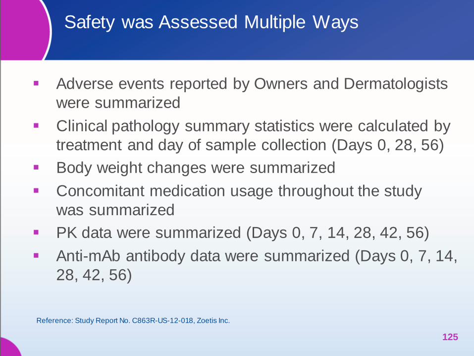
Safety was Assessed Multiple Ways
Adverse events reported by Owners and Dermatologists
were summarized
Clinical pathology summary statistics were calculated by
treatment and day of sample collection (Days 0, 28, 56)
Body weight changes were summarized
Concomitant medication usage throughout the study
was summarized
PK data were summarized (Days 0, 7, 14, 28, 42, 56)
Anti-mAb antibody data were summarized (Days 0, 7, 14,
28, 42, 56)
125
Reference: Study Report No. C863R-US-12-018, Zoetis Inc.
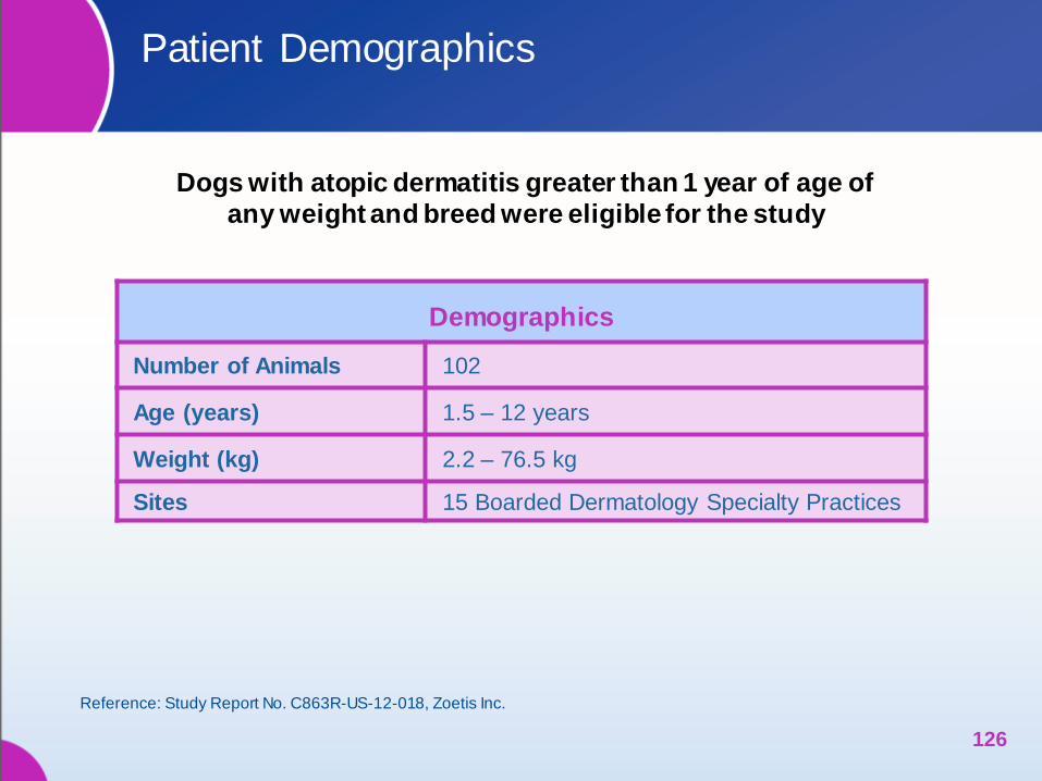
Patient Demographics
126
Dogs with atopic dermatitis greater than 1 year of age of
any weight and breed were eligible for the study
Reference: Study Report No. C863R-US-12-018, Zoetis Inc.
Demographics
Number of Animals 102
Age (years) 1.5 – 12 years
Weight (kg) 2.2 – 76.5 kg
Sites 15 Boarded Dermatology Specialty Practices
0
10
20
30
40
50
60
70
80
90
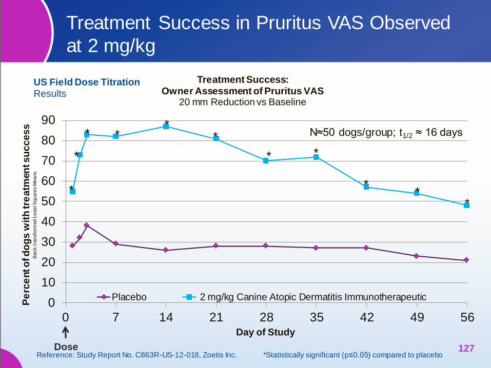
0 7 14 21 28 35 42 49 56Day of Study
Treatment Success: Owner Assessment of Pruritus VAS
20 mm Reduction vs Baseline
Placebo 2 mg/kg Canine Atopic Dermatitis Immunotherapeutic
N≈50 dogs/group; t1/2 ≈ 16 days
Pe
rce
nt o
f d
og
s w
ith
tre
atm
en
t s
uc
ce
ss
Back
-tra
nsf
orm
ed L
ea
st S
qu
are
s M
ean
s
*
*
* * *
*
*
*
* *
*
Treatment Success in Pruritus VAS Observed
at 2 mg/kg
127
US Field Dose Titration Results
Dose Reference: Study Report No. C863R-US-12-018, Zoetis Inc. *Statistically significant (p≤0.05) compared to placebo
0
10
20
30
40
50
60
70
80
0 7 14 21 28 35 42 49 56
Ow
ne
r P
ruri
tus
VA
S s
co
re (m
m)
Le
ast
Sq
ua
res
Me
an
s
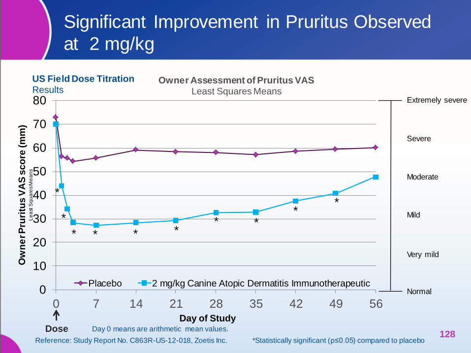
Owner Assessment of Pruritus VAS Least Squares Means
Placebo 2 mg/kg Canine Atopic Dermatitis Immunotherapeutic
Moderate
Extremely severe
Mild
Severe
Very mild
Normal
Significant Improvement in Pruritus Observed
at 2 mg/kg
128
Day of Study
*
*
*
* *
* * * * *
Reference: Study Report No. C863R-US-12-018, Zoetis Inc. *Statistically significant (p≤0.05) compared to placebo
Dose
US Field Dose Titration Results
Day 0 means are arithmetic mean values.
0
10
20
30
40
50
60
0 7 14 21 28 35 42 49 56
Pe
rce
nt o
f D
og
s w
ith
Tre
atm
en
t S
ucc
ess
Ba
ck-
tra
nsf
orm
ed
Le
ast
Sq
ua
res
Me
an
s
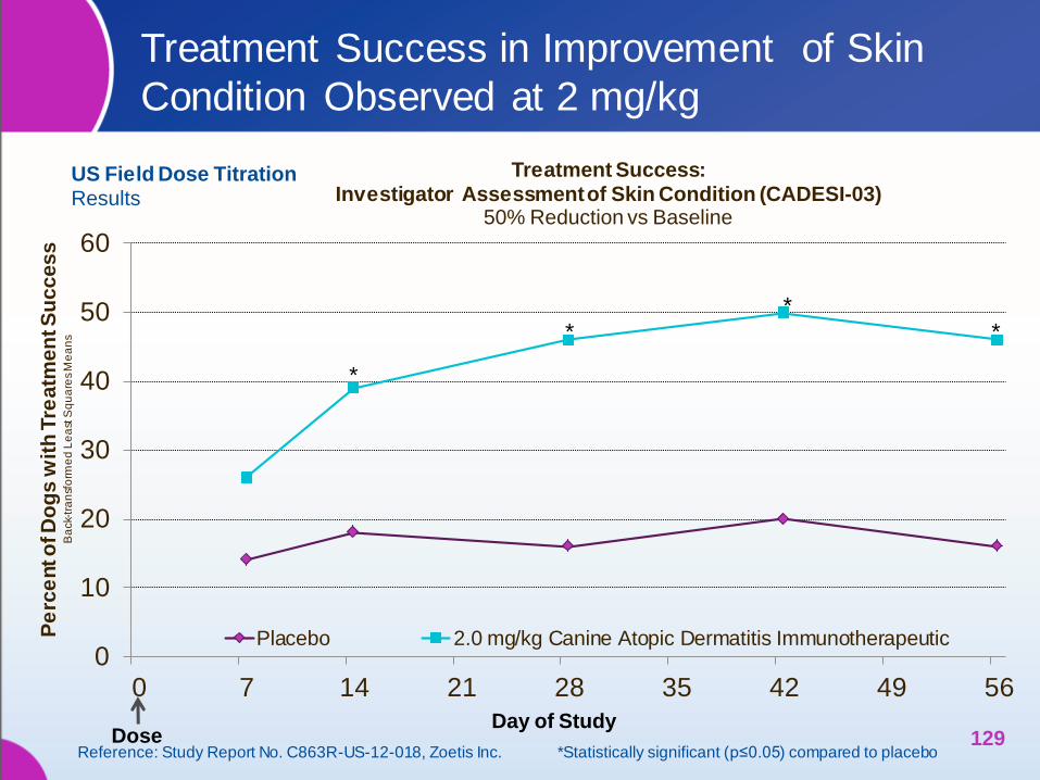
Treatment Success: Investigator Assessment of Skin Condition (CADESI-03)
50% Reduction vs Baseline
Placebo 2.0 mg/kg Canine Atopic Dermatitis Immunotherapeutic
* * *
*
Treatment Success in Improvement of Skin
Condition Observed at 2 mg/kg
129 Reference: Study Report No. C863R-US-12-018, Zoetis Inc. *Statistically significant (p≤0.05) compared to placebo
Day of Study
US Field Dose Titration Results
Dose

0
20
40
60
80
100
120
140
160
180
0 7 14 21 28 35 42 49 56
Investigator Assessment of Skin Condition (CADESI-03) Least Squares Means
Placebo 2 mg/kg Canine Atopic Dermatitis Immunotherapeutic
Improvement in Skin Condition Mirrored
Decrease in Owner-Assessed Itch at 2 mg/kg
130 Day 0 means are arithmetic mean values. Dose
In
ve
stig
ato
r C
AD
ES
I-0
3 S
co
res
Le
as
t S
qu
are
s M
ea
ns
* *
* * *
Day of Study
Reference: Study Report No. C863R-US-12-018, Zoetis Inc. *Statistically significant (p≤0.05) compared to placebo
US Field Dose Titration Results
Canine Atopic Dermatitis Immunotherapeutic was Safely
Administered with Commonly-Used Concurrent Medications
Concomitant Medications Administered During Study (%, (n))
Functional use term
Placebo
(n=52)
Canine Atopic Dermatitis
Immunotherapeutic
2 mg/kg
(n=50)
Diet formulations for maintenance 65.4 (34) 76.0 (38)
Endectocides 75.0 (39) 74.0 (37)
Medicated shampoos 48.1 (25) 36.0 (18)
Diet formulations for treatment of food allergies 34.6 (18) 26.0 (13)
Ectoparasiticides, insecticides and repellants 21.2 (11) 24.0 (12)
Emollients and protectants 21.2 (11) 24.0 (12)
Antigen Specific Immunotherapy 21.2 (11) 18.0 (9)
Omega 3 Fatty Acids 11.5 (6) 16.0 (8)
Antiseptics and disinfectants other than shampoos 23.1 (12) 14.0 (7)
Other anthelmintic agents, optional classification 17.3 (9) 12.0 (6)
Otic cleansers, non-medicated 11.5 (6) 8.0 (4)
Otic antifungals, topical 7.7 (4) 8.0 (4)
Non-medicated shampoos 5.8 (3) 6.0 (3)
Antibiotics for topical use 1.9 (1) 4.0 (2)
All other non-therapeutic products 0.0 (0) 4.0 (2)
Diet formulations – homemade 1.9 (1) 4.0 (2)
General nutrients 5.8 (3) 4.0 (2)
Antiemetics and antinauseants 0.0 (0) 4.0 (2)
Thyroid preparations 5.8 (3) 2.0 (1)
Reference: Study Report No. C863R-US-12-018, Zoetis Inc. 131
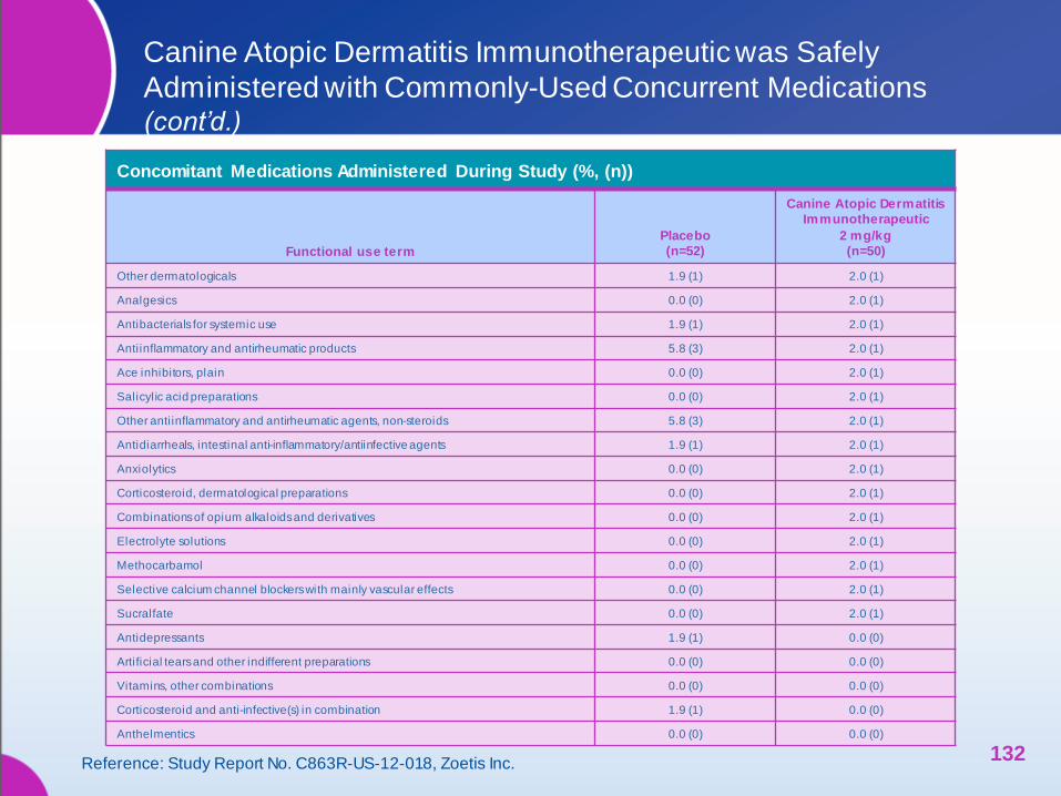
Canine Atopic Dermatitis Immunotherapeutic was Safely
Administered with Commonly-Used Concurrent Medications (cont’d.)
Concomitant Medications Administered During Study (%, (n))
Functional use term
Placebo
(n=52)
Canine Atopic Dermatitis
Immunotherapeutic
2 mg/kg
(n=50)
Other dermatologicals 1.9 (1) 2.0 (1)
Analgesics 0.0 (0) 2.0 (1)
Antibacterials for systemic use 1.9 (1) 2.0 (1)
Antiinflammatory and antirheumatic products 5.8 (3) 2.0 (1)
Ace inhibitors, plain 0.0 (0) 2.0 (1)
Salicylic acid preparations 0.0 (0) 2.0 (1)
Other anti inflammatory and antirheumatic agents, non-steroids 5.8 (3) 2.0 (1)
Antidiarrheals, intestinal anti-inflammatory/antiinfective agents 1.9 (1) 2.0 (1)
Anxiolytics 0.0 (0) 2.0 (1)
Corticosteroid, dermatological preparations 0.0 (0) 2.0 (1)
Combinations of opium alkaloids and derivatives 0.0 (0) 2.0 (1)
Electrolyte solutions 0.0 (0) 2.0 (1)
Methocarbamol 0.0 (0) 2.0 (1)
Selective calcium channel blockers with mainly vascular effects 0.0 (0) 2.0 (1)
Sucralfate 0.0 (0) 2.0 (1)
Antidepressants 1.9 (1) 0.0 (0)
Artificial tears and other indifferent preparations 0.0 (0) 0.0 (0)
Vitamins, other combinations 0.0 (0) 0.0 (0)
Corticosteroid and anti-infective(s) in combination 1.9 (1) 0.0 (0)
Anthelmentics 0.0 (0) 0.0 (0)
Reference: Study Report No. C863R-US-12-018, Zoetis Inc. 132
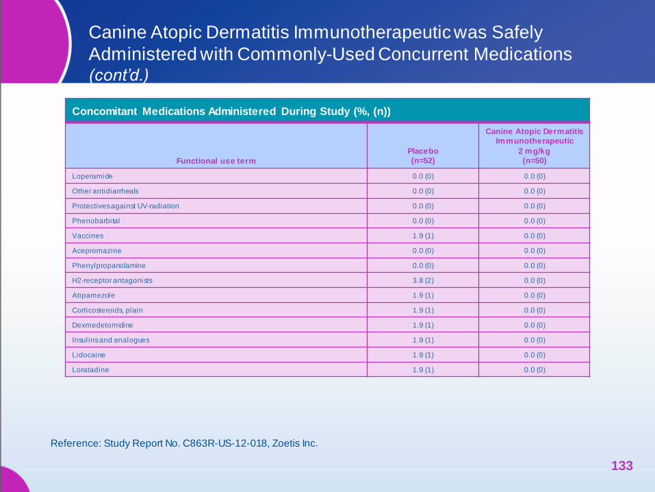
Canine Atopic Dermatitis Immunotherapeutic was Safely
Administered with Commonly-Used Concurrent Medications (cont’d.)
Concomitant Medications Administered During Study (%, (n))
Functional use term
Placebo
(n=52)
Canine Atopic Dermatitis
Immunotherapeutic
2 mg/kg
(n=50)
Loperamide 0.0 (0) 0.0 (0)
Other antidiarrheals 0.0 (0) 0.0 (0)
Protectives against UV-radiation 0.0 (0) 0.0 (0)
Phenobarbital 0.0 (0) 0.0 (0)
Vaccines 1.9 (1) 0.0 (0)
Acepromazine 0.0 (0) 0.0 (0)
Phenylpropanolamine 0.0 (0) 0.0 (0)
H2-receptor antagonists 3.8 (2) 0.0 (0)
Atipamezole 1.9 (1) 0.0 (0)
Corticosteroids, plain 1.9 (1) 0.0 (0)
Dexmedetomidine 1.9 (1) 0.0 (0)
Insulins and analogues 1.9 (1) 0.0 (0)
Lidocaine 1.9 (1) 0.0 (0)
Loratadine 1.9 (1) 0.0 (0)
Reference: Study Report No. C863R-US-12-018, Zoetis Inc.
133
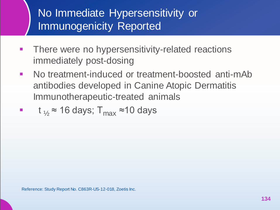
No Immediate Hypersensitivity or
Immunogenicity Reported
There were no hypersensitivity-related reactions
immediately post-dosing
No treatment-induced or treatment-boosted anti-mAb
antibodies developed in Canine Atopic Dermatitis
Immunotherapeutic-treated animals
t ½ ≈ 16 days; Tmax ≈10 days
134
Reference: Study Report No. C863R-US-12-018, Zoetis Inc.
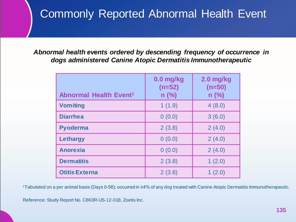
Abnormal Health Event1
0.0 mg/kg
(n=52)
n (%)
2.0 mg/kg
(n=50)
n (%)
Vomiting 1 (1.9) 4 (8.0)
Diarrhea 0 (0.0) 3 (6.0)
Pyoderma 2 (3.8) 2 (4.0)
Lethargy 0 (0.0) 2 (4.0)
Anorexia 0 (0.0) 2 (4.0)
Dermatitis 2 (3.8) 1 (2.0)
Otitis Externa 2 (3.8) 1 (2.0)
1Tabulated on a per animal basis (Days 0-56); occurred in ≥4% of any dog treated with Canine Atopic Dermatitis Immunotherapeutic.
Commonly Reported Abnormal Health Event
135
Reference: Study Report No. C863R-US-12-018, Zoetis Inc.
Abnormal health events ordered by descending frequency of occurrence in
dogs administered Canine Atopic Dermatitis Immunotherapeutic

Safety Assessment
One dog had a possible injection site reaction that
resolved by the end of the study without treatment
No dogs were withdrawn for abnormal clinical pathology
results or possible adverse health event attributed to
treatment
Overall means for RBC and WBC counts, clinical
chemistry variables and urine protein:creatinine
remained within reference range for each analyte;
urinalysis results were unremarkable
A wide variety of concomitant medications were
well tolerated
136
Reference: Study Report No. C863R-US-12-018, Zoetis Inc.
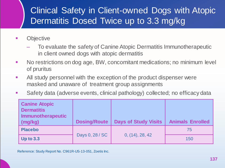
Clinical Safety in Client-owned Dogs with Atopic
Dermatitis Dosed Twice up to 3.3 mg/kg
Objective
‒ To evaluate the safety of Canine Atopic Dermatitis Immunotherapeutic
in client owned dogs with atopic dermatitis
No restrictions on dog age, BW, concomitant medications; no minimum level
of pruritus
All study personnel with the exception of the product dispenser were
masked and unaware of treatment group assignments
Safety data (adverse events, clinical pathology) collected; no efficacy data
137
Canine Atopic
Dermatitis
Immunotherapeutic
(mg/kg) Dosing/Route Days of Study Visits Animals Enrolled
Placebo
Days 0, 28 / SC 0, (14), 28, 42
75
Up to 3.3 150
Reference: Study Report No. C961R-US-13-051, Zoetis Inc.
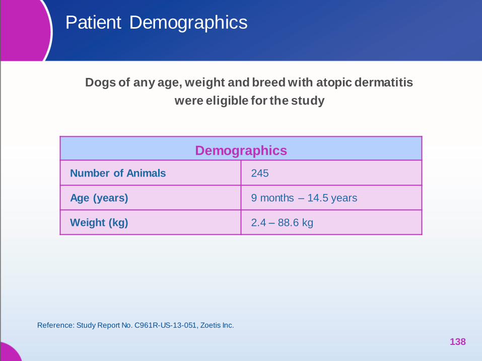
Patient Demographics
Dogs of any age, weight and breed with atopic dermatitis
were eligible for the study
138
Reference: Study Report No. C961R-US-13-051, Zoetis Inc.
Demographics
Number of Animals 245
Age (years) 9 months – 14.5 years
Weight (kg) 2.4 – 88.6 kg
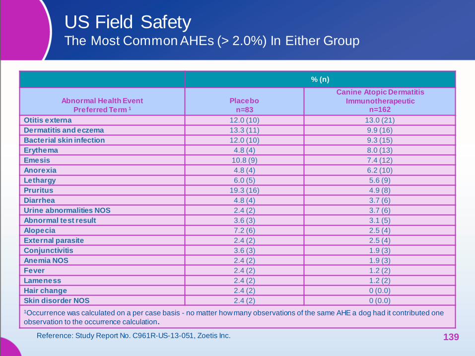
US Field Safety The Most Common AHEs (> 2.0%) In Either Group
% (n)
Abnormal Health Event
Preferred Term 1
Placebo
n=83
Canine Atopic Dermatitis
Immunotherapeutic n=162
Otitis externa 12.0 (10) 13.0 (21)
Dermatitis and eczema 13.3 (11) 9.9 (16)
Bacterial skin infection 12.0 (10) 9.3 (15)
Erythema 4.8 (4) 8.0 (13)
Emesis 10.8 (9) 7.4 (12)
Anorexia 4.8 (4) 6.2 (10)
Lethargy 6.0 (5) 5.6 (9)
Pruritus 19.3 (16) 4.9 (8)
Diarrhea 4.8 (4) 3.7 (6)
Urine abnormalities NOS 2.4 (2) 3.7 (6)
Abnormal test result 3.6 (3) 3.1 (5)
Alopecia 7.2 (6) 2.5 (4)
External parasite 2.4 (2) 2.5 (4)
Conjunctivitis 3.6 (3) 1.9 (3)
Anemia NOS 2.4 (2) 1.9 (3)
Fever 2.4 (2) 1.2 (2)
Lameness 2.4 (2) 1.2 (2)
Hair change 2.4 (2) 0 (0.0)
Skin disorder NOS 2.4 (2) 0 (0.0)
1Occurrence was calculated on a per case basis - no matter how many observations of the same AHE a dog had it contributed one
observation to the occurrence calculation.
139 Reference: Study Report No. C961R-US-13-051, Zoetis Inc.
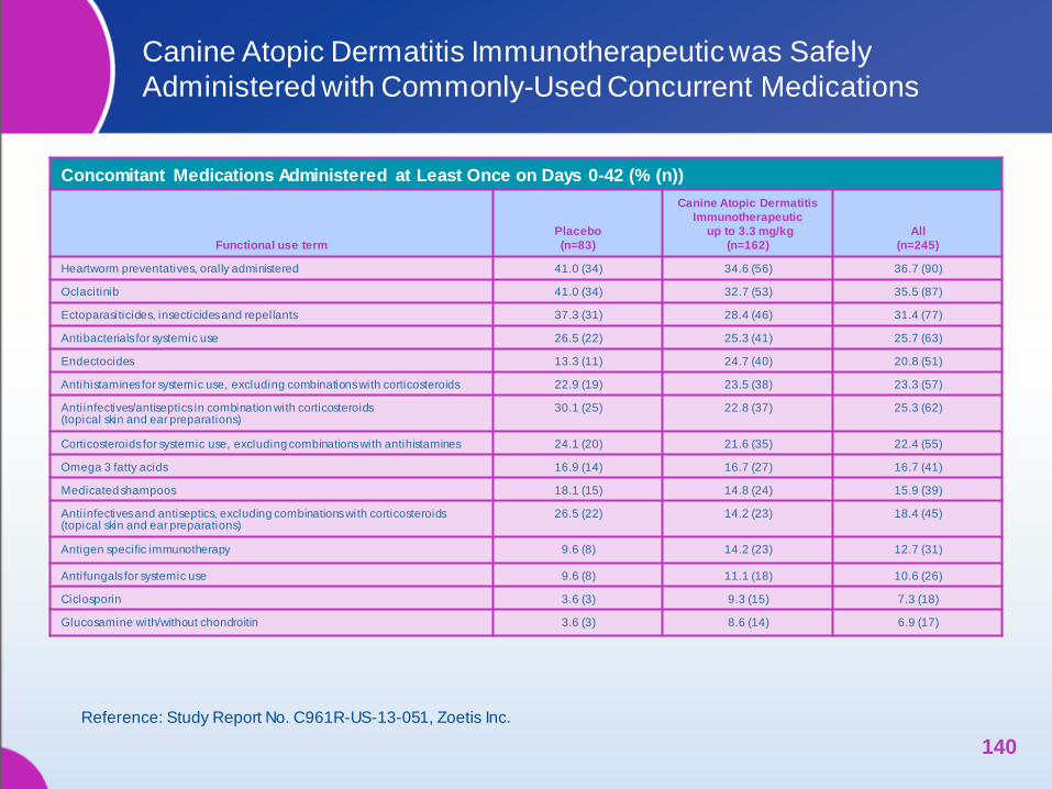
Canine Atopic Dermatitis Immunotherapeutic was Safely
Administered with Commonly-Used Concurrent Medications
•
Concomitant Medications Administered at Least Once on Days 0-42 (% (n))
Functional use term
Placebo
(n=83)
Canine Atopic Dermatitis
Immunotherapeutic
up to 3.3 mg/kg
(n=162)
All
(n=245)
Heartworm preventatives, orally administered 41.0 (34) 34.6 (56) 36.7 (90)
Oclacitinib 41.0 (34) 32.7 (53) 35.5 (87)
Ectoparasiticides, insecticides and repellants 37.3 (31) 28.4 (46) 31.4 (77)
Antibacterials for systemic use 26.5 (22) 25.3 (41) 25.7 (63)
Endectocides 13.3 (11) 24.7 (40) 20.8 (51)
Antihistamines for systemic use, excluding combinations with corticosteroids 22.9 (19) 23.5 (38) 23.3 (57)
Antiinfectives/antiseptics in combination with corticosteroids (topical skin and ear preparations)
30.1 (25) 22.8 (37) 25.3 (62)
Corticosteroids for systemic use, excluding combinations with antihistamines 24.1 (20) 21.6 (35) 22.4 (55)
Omega 3 fatty acids 16.9 (14) 16.7 (27) 16.7 (41)
Medicated shampoos 18.1 (15) 14.8 (24) 15.9 (39)
Antiinfectives and antiseptics, excluding combinations with corticosteroids (topical skin and ear preparations)
26.5 (22) 14.2 (23) 18.4 (45)
Antigen specific immunotherapy 9.6 (8) 14.2 (23) 12.7 (31)
Antifungals for systemic use 9.6 (8) 11.1 (18) 10.6 (26)
Ciclosporin 3.6 (3) 9.3 (15) 7.3 (18)
Glucosamine with/without chondroitin 3.6 (3) 8.6 (14) 6.9 (17)
Reference: Study Report No. C961R-US-13-051, Zoetis Inc.
140

Canine Atopic Dermatitis Immunotherapeutic was Safely
Administered with Commonly-Used Concurrent Medications (cont’d.)
141
Concomitant Medications Administered at Least Once on Days 0-42 (% (n))
Functional use term
Placebo
(n=83)
Canine Atopic Dermatitis
Immunotherapeutic
up to 3.3 mg/kg
(n=162)
All
(n=245)
Vaccines 7.2 (6) 7.4 (12) 7.3 (18)
Otic cleanser, non-medicated 8.4 (7) 6.8 (11) 7.3 (18)
Thyroid preparations 7.2 (6) 6.8 (11) 6.9 (17)
Trimeprazine with prednisolone 12.0 (10) 6.2 (10) 8.2 (20)
Antibiotics for topical use (topical skin)
2.4 (2) 5.6 (9) 4.5 (11)
Antiinflammatory agents and anti -infectives in combination (ophthalmologicals)
4.8 (4) 5.6 (9) 5.3 (13)
Carprofen 2.4 (2) 4.9 (8) 4.1 (10)
Drugs for peptic ulcer and gastro-esophageal reflux disease 2.4 (2) 4.9 (8) 4.1 (10)
Non-medicated shampoos 2.4 (2) 4.9 (8) 4.1 (10)
Vitamins, other combinations 2.4 (2) 4.9 (8) 4.1 (10)
Antiemetics and antinauseants 1.2 (1) 4.3 (7) 3.3 (8)
All other non-therapeutic products 2.4 (2) 3.7 (6) 3.3 (8)
Corticosteroids, dermatological preparations (shampoos, lotions, ear preparations such as “Synotic”)
1.2 (1) 3.7 (6) 2.9 (7)
Anthelmintics 2.4 (2) 3.1 (5) 2.9 (7)
Antidepressants 2.4 (2) 3.1 (5) 2.9 (7)
Reference: Study Report No. C961R-US-13-051, Zoetis Inc.
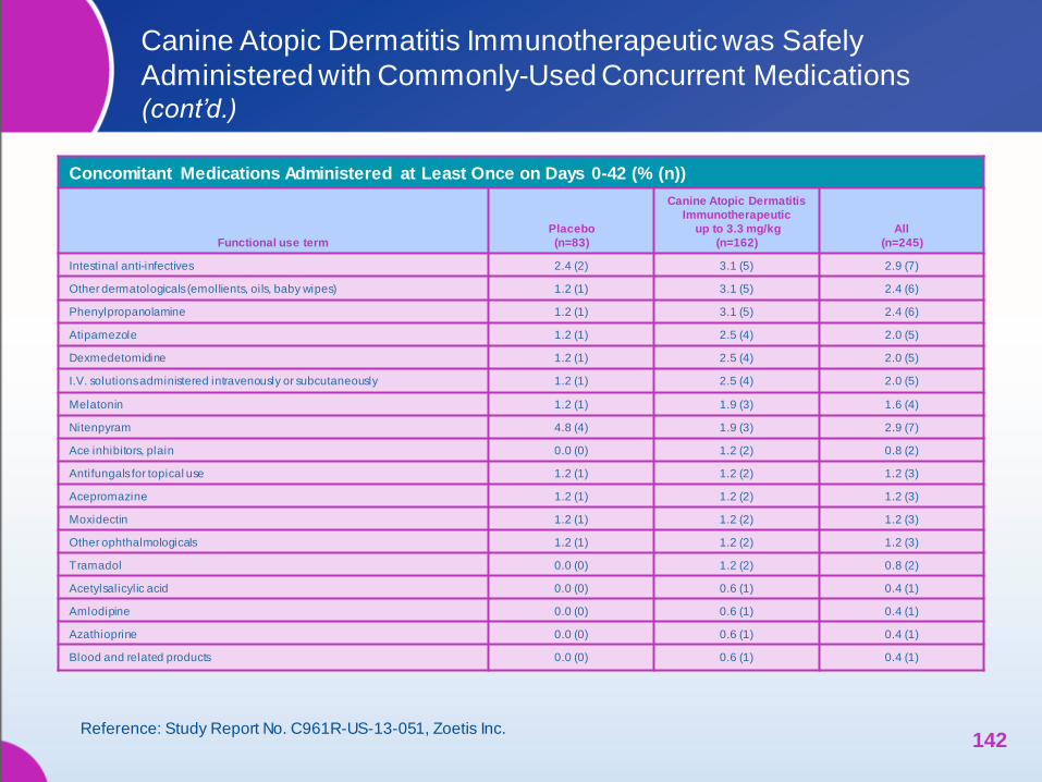
Canine Atopic Dermatitis Immunotherapeutic was Safely
Administered with Commonly-Used Concurrent Medications (cont’d.)
142
Concomitant Medications Administered at Least Once on Days 0-42 (% (n))
Functional use term
Placebo
(n=83)
Canine Atopic Dermatitis
Immunotherapeutic
up to 3.3 mg/kg
(n=162)
All
(n=245)
Intestinal anti-infectives 2.4 (2) 3.1 (5) 2.9 (7)
Other dermatologicals (emollients, oils, baby wipes) 1.2 (1) 3.1 (5) 2.4 (6)
Phenylpropanolamine 1.2 (1) 3.1 (5) 2.4 (6)
Atipamezole 1.2 (1) 2.5 (4) 2.0 (5)
Dexmedetomidine 1.2 (1) 2.5 (4) 2.0 (5)
I.V. solutions administered intravenously or subcutaneously 1.2 (1) 2.5 (4) 2.0 (5)
Melatonin 1.2 (1) 1.9 (3) 1.6 (4)
Nitenpyram 4.8 (4) 1.9 (3) 2.9 (7)
Ace inhibitors, plain 0.0 (0) 1.2 (2) 0.8 (2)
Antifungals for topical use 1.2 (1) 1.2 (2) 1.2 (3)
Acepromazine 1.2 (1) 1.2 (2) 1.2 (3)
Moxidectin 1.2 (1) 1.2 (2) 1.2 (3)
Other ophthalmologicals 1.2 (1) 1.2 (2) 1.2 (3)
Tramadol 0.0 (0) 1.2 (2) 0.8 (2)
Acetylsalicylic acid 0.0 (0) 0.6 (1) 0.4 (1)
Amlodipine 0.0 (0) 0.6 (1) 0.4 (1)
Azathioprine 0.0 (0) 0.6 (1) 0.4 (1)
Blood and related products 0.0 (0) 0.6 (1) 0.4 (1)
Reference: Study Report No. C961R-US-13-051, Zoetis Inc.
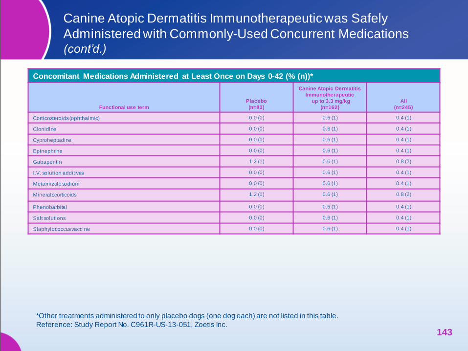
Canine Atopic Dermatitis Immunotherapeutic was Safely
Administered with Commonly-Used Concurrent Medications (cont’d.)
143
Concomitant Medications Administered at Least Once on Days 0-42 (% (n))*
Functional use term
Placebo
(n=83)
Canine Atopic Dermatitis
Immunotherapeutic
up to 3.3 mg/kg
(n=162)
All
(n=245)
Corticosteroids (ophthalmic) 0.0 (0) 0.6 (1) 0.4 (1)
Clonidine 0.0 (0) 0.6 (1) 0.4 (1)
Cyproheptadine 0.0 (0) 0.6 (1) 0.4 (1)
Epinephrine 0.0 (0) 0.6 (1) 0.4 (1)
Gabapentin 1.2 (1) 0.6 (1) 0.8 (2)
I.V. solution additives 0.0 (0) 0.6 (1) 0.4 (1)
Metamizole sodium 0.0 (0) 0.6 (1) 0.4 (1)
Mineralocorticoids 1.2 (1) 0.6 (1) 0.8 (2)
Phenobarbital 0.0 (0) 0.6 (1) 0.4 (1)
Salt solutions 0.0 (0) 0.6 (1) 0.4 (1)
Staphylococcus vaccine 0.0 (0) 0.6 (1) 0.4 (1)
*Other treatments administered to only placebo dogs (one dog each) are not listed in this table.
Reference: Study Report No. C961R-US-13-051, Zoetis Inc.
Canine Atopic Dermatitis Immunotherapeutic
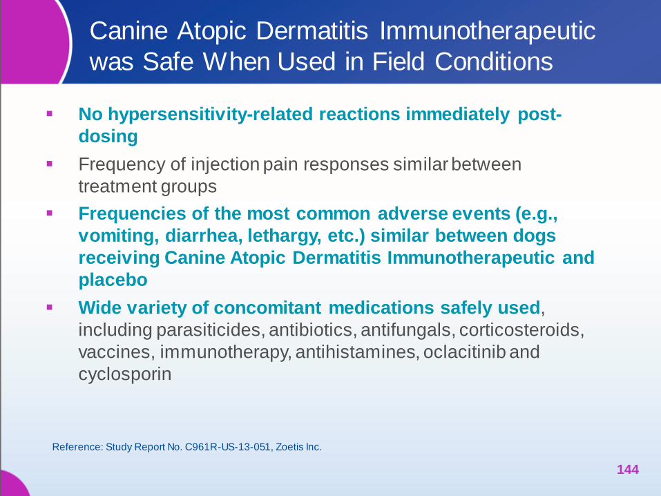
was Safe When Used in Field Conditions
No hypersensitivity-related reactions immediately post-
dosing
Frequency of injection pain responses similar between
treatment groups
Frequencies of the most common adverse events (e.g.,
vomiting, diarrhea, lethargy, etc.) similar between dogs
receiving Canine Atopic Dermatitis Immunotherapeutic and
placebo
Wide variety of concomitant medications safely used,
including parasiticides, antibiotics, antifungals, corticosteroids,
vaccines, immunotherapy, antihistamines, oclacitinib and
cyclosporin
144
Reference: Study Report No. C961R-US-13-051, Zoetis Inc.

LABELING
145
Canine Atopic Dermatitis Immunotherapeutic
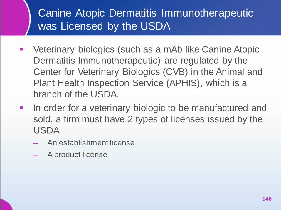
was Licensed by the USDA
Veterinary biologics (such as a mAb like Canine Atopic
Dermatitis Immunotherapeutic) are regulated by the
Center for Veterinary Biologics (CVB) in the Animal and
Plant Health Inspection Service (APHIS), which is a
branch of the USDA.
In order for a veterinary biologic to be manufactured and
sold, a firm must have 2 types of licenses issued by the
USDA
‒ An establishment license
‒ A product license
146
USDA Conditional License
The USDA will grant conditional licenses to meet an
emergency condition, limited market, local situation, or
other special circumstance.
The product must demonstrate the same safety and
purity requirements as fully licensed products but only
needs to have a “reasonable expectation” of efficacy.
147
USDA Conditional License
The firm must provide the following information in the
application for a conditional license
‒ evidence from scientific journals or experts that there is urgent
need in the field for the product
‒ evidence of a “reasonable expectation” of efficacy with data from
studies of the product
‒ all requirements for safety and purity (same as fully licensed
products)
‒ testing that shows consistency between batches of
manufactured product
148
USDA Conditional License
Once a conditional license is granted, the firm is
expected to make progress toward completion of the full
efficacy, potency and safety data in order to obtain a full
license.
While under a conditional license, products may need
additional authorization for distribution in each state and
in foreign countries. The product label must clearly state
that the product license is conditional and no trade
names may be used.
At this time, the USDA has communicated that all
products for canine atopic dermatitis will be considered
for conditional licensure only
149
Canine Atopic Dermatitis lmmunotherapeutic
Label
For Use in Dogs Only
This product license is conditional. Safety and efficacy studies
are in progress.
Canine Atopic Dermatitis lmmunotherapeutic aids in the reduction of
clinical signs associated with atopic dermatitis in dogs.
Canine Atopic Dermatitis lmmunotherapeutic is a ready-to-use,
sterile liquid containing a caninized monoclonal antibody (mAb)
against canine interleukin-31 (IL-31). IL-31 has been shown to
induce pruritus in dogs in laboratory studies.
Canine Atopic Dermatitis lmmunotherapeutic remains in circulation
for several weeks. It exerts a therapeutic effect by binding to and
neutralizing soluble cIL-31, thus inhibiting pruritus and reducing skin
lesions. Like other naturally-occurring antibodies and antibody-
antigen complexes, elimination is via normal protein degradation
pathways.
150
Directions
Canine Atopic Dermatitis lmmunotherapeutic is available
in 1-mL vials in four concentrations (10, 20, 30 or 40 mg).
Administer Canine Atopic Dermatitis lmmunotherapeutic
by subcutaneous injection at a minimum dose of 2 mg/kg
body weight according to the dosing table. Repeat
administration monthly, as needed.
The product does not contain a preservative. Each vial is
for single use only, and should be discarded after
puncture.
151
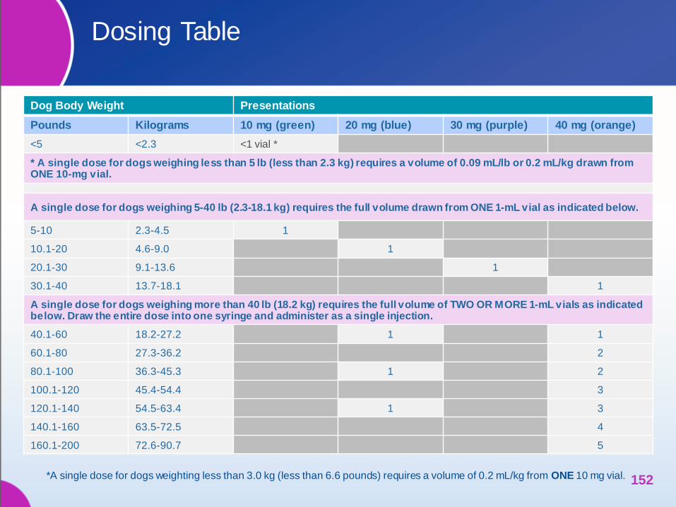
Dosing Table
Dog Body Weight Presentations
Pounds Kilograms 10 mg (green) 20 mg (blue) 30 mg (purple) 40 mg (orange)
<5 <2.3 <1 vial *
* A single dose for dogs weighing less than 5 lb (less than 2.3 kg) requires a volume of 0.09 mL/lb or 0.2 mL/kg drawn from ONE 10-mg vial.
A single dose for dogs weighing 5-40 lb (2.3-18.1 kg) requires the full volume drawn from ONE 1-mL vial as indicated below.
5-10 2.3-4.5 1
10.1-20 4.6-9.0 1
20.1-30 9.1-13.6 1
30.1-40 13.7-18.1 1
A single dose for dogs weighing more than 40 lb (18.2 kg) requires the full volume of TWO OR MORE 1-mL vials as indicated below. Draw the entire dose into one syringe and administer as a single injection.
40.1-60 18.2-27.2 1 1
60.1-80 27.3-36.2 2
80.1-100 36.3-45.3 1 2
100.1-120 45.4-54.4 3
120.1-140 54.5-63.4 1 3
140.1-160 63.5-72.5 4
160.1-200 72.6-90.7 5
152 *A single dose for dogs weighting less than 3.0 kg (less than 6.6 pounds) requires a volume of 0.2 mL/kg from ONE 10 mg vial.
Precautions
1. The product does not have preservative. Each vial is for single use
only, and should be discarded after puncture.
2. This product is intended for subcutaneous use only.
3. Store upright at 2°-8°C. Prolonged exposure to higher temperatures
and/or direct sunlight may adversely affect potency. Do not freeze.
4. Use entire contents when first opened.
5. Sterilized syringes and needles should be used to administer this
product. Do not sterilize with chemicals because traces of
disinfectant may inactivate the product.
6. Burn containers and all unused contents.
7. This product has not been tested in pregnant, lactating or breeding
animals.
153
Product Insert – Inquires
Technical inquiries should be directed to Zoetis, Inc.
Veterinary Services, (888) 963-8471 (USA),
(800) 461-0917 (Canada).
This product has been shown to be efficacious in treating
healthy dogs with canine atopic dermatitis. Canine Atopic
Dermatitis Immunotherapeutic should be used under the
supervision of a veterinarian.
References
Akdis CA, Akdis M, Bieber T, Bindslev-Jensen C, Boguniewicz M, Eigenmann P, et al. Diagnosis and treatment of atopic dermatitis in children and adults: European Academy of Allergology and Clinical Immunology/American Academy of Allergy, Asthma and Immunology/PRACTALL Consensus Report. Allergy. 2006 Aug; 61(8):969-87.
Aleo MM, McCall RB. Exploratory safety margin of PF-06443537, an anti-IL-31 monoclonal antibody in beagles. Zoetis Study Report No. 7462W-60-11-B43. 2 May 2012.
Atopica FOI, 2003.
Atopica [package insert]. Greensboro, NC: Novartis Animal Health; 2008.
Bando T, Morikawa Y, Komori T, Senba E. Complete overlap of interleukin-31 receptor A and oncostatin M receptor beta in the adult dorsal root ganglia with distinct developmental expression patterns. Neuroscience. 2006 Nov 3;142(4):1263–71.
Bilsborough J, Leung DY, Maurer M, Howell M, Boguniewicz M, Yao L, et al. IL-31 is associated with cutaneous lymphocyte antigen-positive skin homing T cells in patients with atopic dermatitis. J Allergy Clin Immunol. 2006 Feb;117(2):418–25.
Cornelissen C, Lüscher-Firzlaff J, Malte Baron J, Lüscher B. Signaling by IL-31 and functional consequences. Europ J Cell Bio. 2012 91:552– 66.
Cosgrove SB, Wren JA, Cleaver DM, Walsh KF, Follis SI, King VI, et al. A blinded, randomized, placebo-controlled trial of the efficacy and safety of the Janus kinase inhibitor oclacitinib (Apoquel®) in client-owned dogs with atopic dermatitis. Vet Dermatol. 2013 Dec;24(6): 587–97, e141-2.
Dermatologic therapy. In: Miller WH, Griffin CE, Campbell KL, eds. Muller & Kirk’s Small Animal Dermatology. 7th ed. St. Louis, MO: Elsevier; 2013. p. 108-83. 155
References (cont’d.)
Dillon SR, Sprecher C, Hammond A, Bilsborough J, Rosenfeld-Franklin M, Presnell SR, et al. Interleukin 31, a cytokine produced by activated T cells, induces dermatitis in mice. Nat Immunol. 2004 Jul;5(7):752–60.
Ezzat MH, Hasan ZE, Shaheen KY. Serum measurement of interleukin-31 (IL-31) in paediatric atopic dermatitis: elevated levels correlate with severity scoring. J Europ Acad Dermatol Venereol. 2011 Mar;25(3):334-9.
Favrot C, Steffan J, Seewald W, Picco F. A prospective study on the clinical features of chronic canine atopic dermatitis and its diagnosis. Vet Dermatol 2010 Feb;21(1):23–31.
Fleck TJ, Humphrey WH, Galvan BA, Aleo MM, Gonzales AJ, Shelly JA, et al. Comparison of the onset and anti-pruritic activity of the JAK inhibitor oclacitinib to prednisolone and dexamethasone in an IL-31 canine model of pruritus. [abstract] Presented at the 2013 North American Veterinary Dermatology Forum (NAVDF); April 17-20, 2013; Louisville, Kentucky.
Fleck T, Michels G, Martinon O, Mahabir S, Hoevers J. Laboratory effectiveness study of an anti-IL-31 monoclonal antibody in dogs compared to a negative control in an induced-pruritus model following a single subcutaneous dose. Zoetis Study C166R-US-13-037. 30 September 2013.
Fleck T, Michels G, Martinon O, Mahabir S, Hoevers J. Laboratory onset of effectiveness study of canine atopic dermatitis immunotherapeutic in dogs compared to placebo in an induced-pruritus model following a single subcutaneous dose of anti-IL-31 monoclonal antibody at 1.0 mg/kg body weight. Zoetis Study C860R-US-14-097. 12 January 2015.
Gonzales AJ, Humphrey WR, Messamore JE, Fleck TJ, Fici GJ, Shelly JA, et al. Interleukin-31: its role in canine pruritus and naturally occurring canine atopic dermatitis. Vet Dermatol. 2013 Feb;24(1):48–53.e11-2. 156
References (cont’d.)
Guengerich, FP. Mechanisms of drug toxicity and relevance to pharmaceutical development. Drug Metab Pharmacokinet. 2011 26(1): 3-14, 2011.
Hnatowich DJ, Layne WW, Childs RL, Lanteigne D, Davis MA, Griffin TW, et al. Radioactive labeling of antibody: a simple and efficient method. Science. 1983 May 6; 220(4597):613-5.
Humira: Highlights Of Prescribing Information. Available at: http://www.rxabbott.com/pdf/humira.pdf. Accessed August 20, 2015.
Hypersensitivity Disorders. In: Miller WH, Griffin CE, Campbell KL, eds. Muller & Kirk’s Small Animal Dermatology. 7th ed. St. Louis, MO: Elsevier; 2013: p. 363-431.
Kim S, Kim HJ, Yang HS, Kim E, Huh IS, Yang JM. IL-31 Serum protein and tissue mRNA levels in patients with atopic dermatitis. Ann Dermatol. 2011 Nov;23(4):468-73.
Kochevar D. Glucocorticoids: new mechanisms for old drugs [presentation]. Presented at the Hill’s Symposium on Dermatology. April 2-4, 2006; Palm Springs, CA.
Liberman AC, Druker J, Perone MJ, Arst E. Glucocorticoids in the regulation of transcription factors that control cytokine synthesis. Cytokine Growth Factor Rev. 2007 Feb-Apr;18(1-2):45–56.
Ma A, Koka R, Burkett P. Diverse functions of IL-2, IL-15, and IL-7 in lymphoid homeostasis. Ann Rev Immunol. 2006;24:657-79.
Marsella R, Sousa CA, Gonzales AJ, Fadok VA. Current understanding of the pathophysiologic mechanisms of canine atopic dermatitis. J Am Vet Med Assoc. 2012 Jul 15;241(2):194-207.
Matsuda S, Koyasu S. Mechanisms of action of cyclosporin. Immunopharmacology. 2000 May; 47(2-3):119–25.
157
References (cont’d.)
McCall RB, Fleck TJ. Evaluation of mock protein vs. Canine IL-31 as a pruritus-inducing agent in dogs when given via intravenous injection. Zoetis Study C761R-US-12-008. 9 October 2013.
McCall B, Fleck T. Evaluation of the anti-pruritic effect of PF-06443537, an anti-IL-31 monoclonal antibody in a canine model of IL-31-induced pruritus. Zoetis Study C863R-US-12-024. 12 March 2014.
McCall RB, Fleck TJ. Evaluation of the anti-pruritic effects of dexamethasone when administered as a single IM dose vs. oral Apoquel in a canine model of IL-31 induced pruritus. Zoetis Study A160R-US-12-057. 14 November 2012.
McCall RB, Fleck TJ. Evaluation of the anti-pruritic effects of prednisolone when administered for 1 week at 1 mg/kg/day in a canine model of IL-31 induced pruritus. Zoetis Study 7D61R-60-11-B68. 1 May 2012.
McCall RB, Humphrey WR. Pilot Study: Evaluation of the anti-pruritic effect of anti-IL-31 monoclonal antibody CAN 34D03-65 (PF-6443537) in a canine model of lL-31-induced pruritus. Zoetis Study 7D61W-60-11-B00. 16 October 2012.
McCandless EE, Messamore J, Rugg C, Fici GJ, Gonzales AJ. Production of IL-31 by canine Th2 cells and identification of inflammatory and neuronal target cells Vet Dermatol, 23, 2012 FC-64.
Michels GM, Martinon O, Mahabir S, Hoevers JD. A field study investigating three dose levels of an anti-IL-31 monoclonal antibody for treatment of atopic dermatitis in dogs. Zoetis Study C863R-US-12-018. 18 April 2014.
Michels GM, Martinon O, Mahabir S, Hoevers JD. Field evaluation of the safety of canine atopic dermatitis immunotherapeutic [Product Code 9580.02] administered to dogs by the subcutaneous route. Zoetis Study C961R-US-13-051. 10 November 2014. 158
References (cont’d.)
Miller WR. A safety study of PF-06443537 administered subcutaneously once monthly in beagle
dogs. Zoetis Study C362N-US-13-042. 12 December 2014.
Morar N, Willis-Owen, SA, Moffatt MF, Cookson WO. The genetics of atopic dermatitis. J Allergy Clin
Immunol. 2006 Jul;118(1):24-34.
Murphy K. Janeway’s Immunobiology. 8th ed. New York, NY: Garland Science, Taylor & Francis
Group; 2012.
Olivry T, DeBoer DJ, Favrot C, Jackson HA, Nuttall T, Prélaud P, et al. Treatment of canine atopic
dermatitis: 2010 clinical practice guidelines from the International Task Force on Canine Atopic
Dermatitis. Vet Dermatol. 2010 Jun;21(3):233-48.
Olivry T, Foster AP, Mueller RS, McEwan NA, Chesney C, Williams HC. Interventions for atopic
dermatitis in dogs: a systematic review of randomized controlled trials. Vet Dermatol. 2010
Feb;21(1):4-22.
Olivry T, Rivierre C, Jackson HA, Murphy KM, Davidson G, Sousa CA. Cyclosporine decreases skin
lesions and pruritus in dogs with atopic dermatitis: a blinded randomized prednisolone-controlled
trial. Vet Dermatol. 2002 Apr;13(2):77–87.
Oyoshi MK, He R, Kumar L, Yoon J, Geha RS. Cellular and molecular mechanisms in atopic
dermatitis. Adv Immunol. 2009; 102:135–226.
Perretti M, Ahluwalia A. The microcirculation and inflammation: site of action for glucocorticoids.
Microcirculation. 2000 Jun;7(3):147–61.
159
References (cont’d.)
Raap U, Wichmann K, Bruder M, Ständer S, Wedi B, Kapp A, et al. Correlation of IL-31 serum levels with severity of atopic dermatitis. J Allergy Clin Immunol. 2008 Aug;122(2):421-3.
Ramsey DS, Dunham SA, Martinon O, Hoevers JD, Mahabir S. Proof of efficacy and safety of an anti-IL-31 monoclonal antibody (IL-31 mAb) for the treatment of atopic dermatitis in client-owned dogs. Zoetis Study 4962R-60-11-277. 15 February 2013.
Sonkoly E, Muller A, Lauerma AI, Pivarcsi A, Soto H, Kemeny L, et al. IL-31: a new link between T cells and pruritus in atopic skin inflammation. J Allergy Clin Immunol. 2006 Feb;117(2):411–7.
Steffan J, Alexander D, Brovedani F, Fisch RD. Comparison of cyclosporin A with methylprednisolone for treatment of canine atopic dermatitis: a parallel, blinded, randomized controlled trial. Vet Dermatol. 2003 Feb;14(1):11–22.
Storan ER, O’Gorman SM, McDonald ID, Steinhoff M. Role of Cytokines and Chemokines in Itch. Pharmacology of Itch. Handbook of Experimental Pharmacology Volume 226, 2015;(226):163-76.
The Merck Veterinary Manual. Vaccines and Immunotherapy: Introduction. Available at: http://www.merckvetmanual.com/mvm/index.jsp?cfile=htm/bc/192100.htm. Accessed August 20, 2015.
Walters RR. Pharmacokinetic comparison of anti-IL-31 monoclonal antibody formulations in beagle dogs. Zoetis Study 7562W-60-11-B02. 10 April 2012.
All trademarks are the property of Zoetis, Inc., its affiliates and/or licensors.
All rights reserved. September, 2015. CYT-00028 160