Candida tropicalis - ARCA: Home Junior RA... · Candida tropicalis Rafael Araújo Gomes ... Oswaldo...
25
1 Antifungal Mechanism of [Ru III (NH 3 ) 4 catechol] + Complex on Fluconazole-Resistant Candida tropicalis Rafael Araújo Gomes-Junior a , Roberto Santana da Silva b , Renata Galvão de Lima c , Marcos A. Vannier-Santos a1 a Gonçalo Moniz Institute, Oswaldo Cruz Foundation (FIOCRUZ), Salvador, BA, Brazil; b Faculdade de Ciências Farmacêuticas de Ribeirão Preto, Universidade de São Paulo, Av. do Café s/n, 14040-903, Ribeirão Preto, SP, Brazil., c Faculdade de Ciências Integradas do Pontal, Universidade Federal de Uberlândia, Rua Vinte, 1600, Tupã, Ituiutaba, MG, Brazil. 1- Corresponding author: Dr. Marcos André Vannier-Santos Laboratório de Biologia Parasitária, Instituto Gonçalo Moniz; Fundação Oswaldo Cruz – FIOCRUZ; Rua Waldemar Falcão, 121 Brotas, Salvador, Bahia, Brazil CEP 40295-00, Phone: +55-071-31762208. Emails: [email protected] or [email protected]
Transcript of Candida tropicalis - ARCA: Home Junior RA... · Candida tropicalis Rafael Araújo Gomes ... Oswaldo...
1
Antifungal Mechanism of [RuIII
(NH3)4catechol]+ Complex on Fluconazole-Resistant
Candida tropicalis
Rafael Araújo Gomes-Juniora, Roberto Santana da Silva
b, Renata Galvão de Lima
c,
Marcos A. Vannier-Santosa1
aGonçalo Moniz Institute, Oswaldo Cruz Foundation (FIOCRUZ), Salvador, BA,
Brazil; bFaculdade de Ciências Farmacêuticas de Ribeirão Preto, Universidade de São
Paulo, Av. do Café s/n, 14040-903, Ribeirão Preto, SP, Brazil., cFaculdade de Ciências
Integradas do Pontal, Universidade Federal de Uberlândia, Rua Vinte, 1600, Tupã,
Ituiutaba, MG, Brazil.
1- Corresponding author: Dr. Marcos André Vannier-Santos
Laboratório de Biologia Parasitária, Instituto Gonçalo Moniz; Fundação
Oswaldo Cruz – FIOCRUZ; Rua Waldemar Falcão, 121 Brotas, Salvador,
Bahia, Brazil CEP 40295-00, Phone: +55-071-31762208. Emails:
2
Abstract
Candidiasis, a major opportunistic mycosis caused by Candida sp. may comprise life-
threatening systemic infections. The incidence of non-albicans species is rising,
particularly in South America and they are frequently drug resistant, causing
unresponsive cases. Thus, novel antimycotic agents are required. Here we tested the
antifungal activity of [RuIII
(NH3)4catechol]+ complex (RuCat), approaching possible
action mechanisms on fluconazole-resistant Candida tropicalis. RuCat significantly
(p<0.05) inhibited the growth and viability of C. tropicalis dose-dependently, (IC50
20.3µM). Cytotoxicity of RuCat upon murine splenocytes was lower (Selectivity Index
= 16). Scanning electron microscopy analysis showed pseudohyphae formation, yeast
aggregation and surface damage. RuCat-treated samples investigated by transmission
electron microscopy showed melanin granule trafficking to cell surfaces and
extracellular milieu. Surface adherent membrane fragments and extracellular debris
were also observed. RuCat treatment produced intense H2DCFDA labeling, indicating
reactive oxygen species (ROS) production which caused increased lipoperoxidation.
ROS are involved in the fungicidal effect as N-acetyl-L-cysteine completely restored
cell viability. Calcofluor White chitin staining suggest that 70 or 140 µM RuCat
treatment for 2h affected cell wall structure. PI labeling indicated necrotic cell death.
The present data indicate that RuCat triggers ROS production, lipoperoxidation and cell
surface damage, culminating in selective necrotic death of drug-resistant C. tropicalis.
Keywords: Candida tropicalis, fluconazole-resistant, ruthenium complex, catechol,
antifungal agents, candidiasis
3
Introduction
Candida genus includes about 350 species found in humans among other
mammals (Nunn et al. 2007). Candidiasis is an opportunistic infection increased over
the last decades (Ghannoum 2001; Melo et al. 2004). Despite its diversity, few species
(C. albicans, C. glabrata, C. tropicalis, C. krusei, C. parapsilosis, C. kefyr, C.
guilliermondii, C. pseudotropicalis, C. dubliniensis and C. lusitaniae) are implicated in
human disease.
C. albicans is a common pathogen found in the microbiome of the skin,
gastrointestinal mucosa and human cavities. The spectrum of diseases caused by
Candida ranges from oral candidiasis to life-threatening systemic infections.
Immunocompromising conditions significantly promote the fungal spread (Gudlaugsson
et al. 2003; Moran et al. 2012). Microbiome changes, gastrointestinal mucosa injury,
chemotherapy, intestinal obstruction, prolonged fasting, parenteral nutrition and
hypotension potentiate the mesenteric capillaries colonization by Candida sp., leading
to candidemia (Takahashi et al. 2003).
Increased invasive fungal infections are often caused by Candida sp. (Warnock
2007), are associated with 30% and 50% mortality rates among children and adults
respectively (Oliveira et al. 2014). Non-albicans species were reported, to cause 50% of
the invasive infections (Ruiz et al. 2005; García-Rodríguez et al. 2013). C. tropicalis is
the second/third non-albicans causative agent of bloodstream infections (Cantón et al.
2001; Colombo and Guimarães 2003). Moreover, these infections are more invasive and
often chemotherapy unresponsive (Cantón et al. 2001; Morace et al. 2011), mainly in
Latin America (Nucci et al. 2010).
4
Candidiasis treatment usually employs azole derivatives, as fluconazole,
miconazole, itraconazole, however azole-resistant isolates are occurring more frequently
(Canuto and Rodero 2002), so comprise a matter of concern. Thus, it is necessary to
develop new strategies to prevent the spread of these fungi, so new antifungal drugs are
required.
Catechols are phenolic compounds commonly found in nature and play a pivotal
role in neurotransmission, taking part in the catecholamine structure. Phenolic
allelochemicals as catechol and pyrogallol may be microbicidal upon both bacteria and
fungi (Kocac and Ismet 2006). Redox reactions involving catechols in the presence of
oxygen play important roles such as antimicrobial activity (Schweigert et al. 2001)
mediated by reactive oxygen species (ROS).
From this perspective, the synergism by combining a ruthenium (III) ion and
catechol ligand, as [RuIII
(NH3)4(catechol)]+ complex (RuCat), was shown to alter the
redox behavior as compared to free catechol (de Lima et al. 2003; 2004; Almeida et al.
2007). The coordination of the ruthenium (III) ion enhances oxygen consumption rates
indicating that the reaction of the complex with oxygen is more thermodynamically
favored facilitating the ROS production (Almeida et al. 2007).
Here we evaluated the RuCat antifungal activity against fluconazole-resistant
Candida tropicalis.
Materials and methods
5
Apparatus for Ruthenium Complex Characterization
The ultraviolet-visible (UV-vis.) spectra were recorded Varian Cary 50 Conc. Infrared
(IR) spectra were recorded on a Protegé 460 series FT-IR spectrometer, using solid
samples pressed in KBr pellets. Chromatography was carried out in a Shimadzu system
using a UV–vis. SPD- M20A detector, a LC-20AT pump with CBM-10A interface, a
DGU-20A3 degasser and Chemstation software. A CLC-ODS column (5 µm; 250 mm 9
4, 6 mm i.d.; Shimadzu) was used. Samples were dissolved in the appropriate mobile
phase, and 20 µL injection.
Chemicals and Reagents
RuCl3.3H2O, hydrazines and catechol were acquired as high purity reagents from
Aldrich Chemicals (Brazil) and were used as supplied. All preparations and
measurements were performed protected from light. Other chemicals were of reagent
grade, so used without further purification.
[RuIII
(NH3)4(catechol)](PF6) complex synthesis
[RuIII
(NH3)4(catechol)](PF6) complex was synthesized as previously described (da Silva
et al. 2000). The purity of the complexes was checked by HPLC analysis (Suppl. Figure
1), characterized by UV-vis. and IR spectroscopy and compared to published results
(Santana da Silva et al. 2000). UV-vis. spectra: λ (nm) [RuIII
(NH3)4(catechol)]+: 280
and 670 and FTIR ν(C–O): 1282 cm−1
.
Strain and growth conditions

6
Strain and growth conditions. A fresh C. tropicalis clinical isolate was isolated and
characterized by the Central Public Health Laboratory (LACEN-BA). The resultant
strain (Ct-LACEN 201103) was used here. Cultures were carried out in 4% potato
dextrose agar solid medium and in RPMI 1640 liquid medium buffered with MOPS (3
acid [N-morpholino] propane sulfonic) pH 7.0, at 35 °C. C. tropicalis proliferation was
determined daily under phase-microscopy on hemocytometric chambers and IC50
determined using GraphPad Prism 5.01. C. tropicalis viability was assessed by colony-
forming unit (CFU) counting.
Cytotoxicity Assays
Splenocytes from 8-10 weeks-old BALB/c mice were incubated in 24-well culture
plates at 37 °C, 5%CO2 in RPMI supplemented with 10% fetal bovine serum (Sigma-
Aldrich), with 125, 250, 500 and 1000 µM RuCat for 24h. After incubation, 5 mg mL-1
MTT in phosphate-buffered saline (PBS) were added for 2h, then DMSO was added to
dissolve the formazan crystals and absorbance values were determined at 540 nm. EC50
was determined using GraphPad Prism 5.01.
Propidium iodide staining
C. tropicalis treated with 40µM RuCat for 1 h at 35 °C in RPMI 1640 were incubated
with 10 μg/mL propidium iodide (PI - Sigma Chemical Co. St. Louis Mo USA) for 5
min. Acquisition was performed using a BD FACSCaliburTM
flow cytometer and data
were analyzed in BD CellQuestTM
software (San Jose, CA).
Fluorescence microscopy for detection of reactive species
7
For detection of ROS, yeasts were cultured in the presence and absence of 38 µM
RuCat for 60 min. Then the cells were washed in PBS, stained with 10 µM of the
fluorescent probe 2',7'-dichlorodihydrofluorescein diacetate (H2DCFDA) (Molecular
Probes, Eugene, OR) for 30 min and observed in a Olympus BX51 fluorescence
microscope at 460 to 495nm. 5 mM H2O2 were used for 1 hour as a positive control, and
5 mM ascorbic acid 2h pretreatments, prior to H2O2, as negative control. The
fluorescence was detected by using excitation and emission spectra of 495 nm and 529
nm.
Evaluation of Lipid Peroxidation
After treatment with 24, 70 and 140 µM RuCat for 1h, the samples were washed in
PBS, 200 µL of thiobarbituric acid (1%) were added and diluted with 50% acetic acid.
Then the samples were incubated at 100 °C for 2h and the reaction was stopped in ice
bath for 15 min. Thiobarbituric acid reactive substances (TBARS) were measured by
spectrophotometry at 532 nm.
Cell Wall Staining
Yeast cells treated with 70 and 140 µM RuCat for 2 h were washed in PBS, stained with
30 µg mL-1
Calcofluor White (CW - MP Biomedicals) and observed at 355 nm in a
Olympus BX51 fluorescence microscope.
Transmission Electron Microscopy (TEM).
Cells were fixed in 2.5% glutaraldehyde and 4% paraformaldehyde in 0.1 M sodium
cacodylate buffer, pH 7.4 and post-fixed in 1.5% of potassium permanganate for 1h,
8
protected from light (Vannier-Santos and Lins 2001). Samples were dehydrated in
acetone series and imbed in Spurr's resin (Ted Pella, Redding, CA). Ultrathin sections
were, collected on 300-mesh cooper grids, contrasted in 5% uranyl acetate and 3% lead
citrate. The ultrastructural analysis was performed on a Zeiss109 transmission electron
microscope at 80 kV.
Scanning Electron Microscopy (SEM). Yeast cells were fixed and dehydrated as
above, followed by critical point drying and gold metallization and observation in JEOL
JSM 6390 scanning electron microscope at 20kV (Vannier-Santos and De Castro 2009).
Statistical Analysis
Data represent the mean and standard deviation, of three independent experiments and
statistical analysis was performed employing ANOVA and Dunnett's post-test with p
<0.05, using GraphPad Prism 5.01.
Ethical Aspects
All the procedures performed were approved by the Fiocruz Ethics Committee (lic. no.
20/2015) and followed the ethics standards guidelines of International Council for
Laboratory Animal Science.
9
RESULTS
Effect of RuCat on C. tropicalis proliferation
RuCat incubation significantly (p <0.05) inhibited the growth of fluconazole-resistant
C. tropicalis, in RPMI 1640 for 24h at 35 °C, from 20 µM, dose-dependently. The
obtained RuCat IC50 was 20.3 µM (Figure 1A).
Cytotoxicity evaluation of RuCat in splenocytes cultures
In order to determine the RuCat effects on mammalian cells, MTT cytotoxicity assays
using Balb/c splenocytes were performed. Splenocyte cultures incubated with different
concentrations of the compound for 24 h showed significant toxicity only at
concentrations of 500 and 1000 µM (Figure 1B) and the obtained EC50 was 325 µM.
Cell Surface Analysis
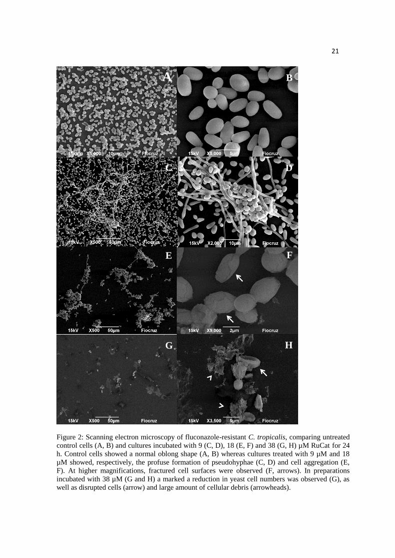
We used SEM to analyze the possible RuCat effects upon fluconazole-resistant C.
tropicalis incubated with 9, 18, 38 µM of the compound for 24 h (Figure 2). Untreated
control samples showed numerous ovoid yeast with smooth, continuous surfaces
(Figure 2 A, B); Cultures treated with 9 µM RuCat presented remarkable pseudohyphae
formation (Figure 2 C, D). Cultures incubated with 18 µM RuCat displayed intense
cellular aggregation (Figure 2 E) and higher magnifications revealed the presence of
10
surface cracks (Figure 2 F), whereas cultures incubated with 38 µM cells showed scarce
cells and large amount of cellular debris (Figure 2 G, H).
Necrosis Detection
In order to verify whether RuCat caused C. tropicalis necrosis, cultures were incubated
with PI to test membrane discontinuity assessed by flow cytometry. Contrary to
untreated controls, after 1 hour of treatment with 40 µM RuCat we found 30% of the
population was PI-labeled (Suppl. Fig. 2).
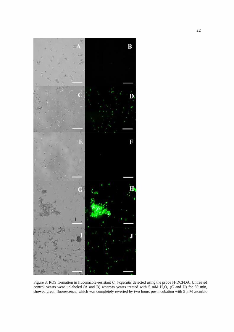
Evaluation of ROS formation
The probe 2',7'-dichlorofluorescein diacetate (H2DCFDA) was employed to detect the
ROS production caused by 38 µM RuCat for 1h (Figure 3). Untreated control cells were
unlabeled (Fig. 3A, B). The positive control incubated with 5 mM H2O2 for 1h showed
positive reaction (Figure 3C, D), which was completely reversed by 5 mM ascorbic acid
preincubation (Figure 3E, F). RuCat treatment resulted in intense H2DCFDA labeling
(G, H) which was partially reverted by ascorbic acid preincubation (I, J).
In order to evaluate the enhanced ROS production-induced oxidative stress we
measured lipoperoxidation in RuCat-treated yeasts.
Oxidative Stress Role in the RuCat Anti-Candida tropicalis Action
The measurement of TBARS was employed evaluate oxidative stress changes indicated
by lipid peroxidation. Increased ROS production led to enhanced lipoperoxidation and
presumably membrane damage. We measured TBARS formation after 1h exposure to
different RuCat concentrations (24, 70 and 140 µM). The concentrations tested
significantly (p <0.05) increased lipoperoxidation (Figure 4A).

11
In order to test the possible participation of the oxidative stress in the RuCat anti-
Candida activity, we preincubated cultures with 20 mM N-acetyl-L-cysteine (NAC) for
2 h before treatment with 150 µM RuCat. CFU counting was performed after 24h. We
notice that NAC completely restored the viability of these cells reverting the RuCat
activity (Figure 4B).
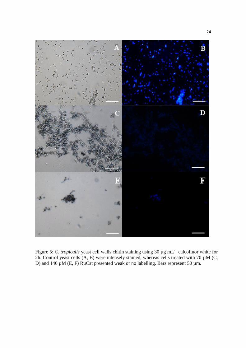
Candida tropicalis cell wall staining
The CW chitin-binding probe was used for labeling C. tropicalis cell walls to assess the
integrity of the cell surfaces, after treatment with RuCat for 2h. Untreated yeasts were
intensely labeled with CW (Figure 5 A, B), whereas fungal cells treated with 70 µM
(Figure 5C, D) or 140 µM RuCat (Figure 5E, F) were weakly labeled.
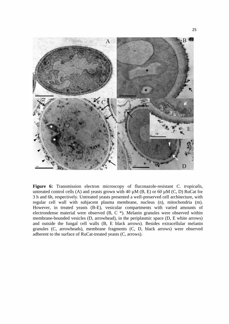
In order to approach the RuCat-induced mechanism of yeasts cell death we
evaluated the ultrastructural changes caused by 40 and 60 µM RuCat for 3h and 24 h.
Untreated control yeasts displayed intact and regular cell wall structure, polymorphic
nuclei and well-preserved mitochondria (Figure 6A), whereas RuCat-treated cells
showed vesicular compartments with different amounts of amorphous electrondense
material (Figure 6B, C), as well as melanin granules in membrane-bounded organelles
(Figure 6D), periplasmic region and covering the external cell surface (Figure 6B) and
free in the extracellular milieu (Figure 6 C-E). Surface adherent membrane fragments
(Figure 6 C, D) were also observed.
DISCUSSION
12
In plants catechol oxidation provides a natural protection against infections via ROS
production (Kumar and Pandey 2013). Its pro-oxidant effect may result from an auto-
oxidation in aqueous solution and physiological pH, leading to the formation of quinone
and superoxide radicals which are highly cytotoxic (O’Brien 1991). ROS production
may account at least in part for the SEM alterations and H2DCFDA staining. CW
labelling could also be precluded by melanin and or membrane fragments lining fungal
cell surfaces.
The high instability of catechol in aqueous solution, can be blocked by
ruthenium (III) ion binding via coordination linked in the oxygen atom producing
RuCat causing stability in aqueous solution for at least one week (de Lima et al. 2004).
It was previously reported that the combination of the ruthenium (III) and
catecholamine ligands led to fungicidal activity significantly higher than free ligands
due to metal ion playing an important role in the action of the complex on Candida sp.
(de Lima et al. 2003).
Determination of RuCat Selectivity Index comparing RuCat effects on C.
tropicalis and murine splenocytes in vitro [IS= CC50 (325 µM)/EC50 (20.3 µM)], may be
indicative of a promising agent as IS should be over 10-fold (Nwaka et al. 2009) and the
IS value obtained here was 16.
Ultrastructural analysis showed that treatment of C. tropicalis yeasts with RuCat
increased melanin granules, trafficking in vesicles to the fungal surface and release to
extracellular milieu. C. albicans was shown to produce and externalize melanin both in
vitro and in vivo (Morris-Jones et al. 2005). Melanin is a generic term for a
heterogeneous class of natural pigments widely present in mammals, protozoa, plants,
fungi and bacteria with a multitude of biological functions. The production of melanin

13
by fungi is of fundamental importance, comprising a relevant virulence factor for
several species. In fungi the final destination of melanin is the cell wall, which can be
found within the wall itself or as a layer in the periplasmic space (Langfelder et al.
2003), contributing to their protection against oxidative damage caused by phagocytes,
antifungal substances action among other stress conditions (van Duin et al. 2002;
Gómez and Nosanchuk, 2003; Nosanchuk and Casadevall 2006).
Previous studies have reported the presence of melanin granules located in the
cytoplasm similarly to mammalian melanosomes (e.g. Cladosporium carrionii and
Fonsecaea pedrosoi) (San-Blas et al. 1996; Franzen et al. 2008) and indicating a
possible association between melanin production and chitin synthesis (Walker et al.
2010). The membrane-bounded compartments detected by TEM may comprise fungal
melanosomes that are transported to the cell periphery, where melanin granules are
transferred to the cell wall (Nosanchuk et al. 2015). Although the mechanism of
melanin externalization has not yet been fully established the export process probably
involves the chitinase genes (CHT2 and CHT3) and the chitin regulatory pathway
suggesting association between cell surface disruption seen by SEM (Walker et al.
2010). Part of the granular material observed by SEM in 38 µM RuCat-treated cells may
be due to melanin, as these structures were previously described by electron microscopy
(Walker et al. 2010).
There is a wide variety of biological reactions which underlie the RuCat toxicity
and the ruthenium combination is possibly implicated in the ROS production and its
deleterious effect (Almeida et al. 2007). The oxidation of RuCat in the presence of
oxygen leads to the formation of superoxide radical (O2•-) and melanin may exert
antioxidant effect quenching free radicals (Fernandes et al. 2015). Interestingly

14
catecholamine precursors are involved in Cryptococcus neoformans melanization
(Chatterjee et al. 2012).
The RuCat treatment presumably triggered the antioxidant defense system for
detoxification of O2•-
into H2O2 by the cytoplasmic enzyme superoxide dismutase
(SOD). Then H2O2 is converted by catalase into water and oxygen, however, formation
of such ROS may exceed its protection capacity resulting in the oxidative stress
reported here. The pseudohyphal development and aggregation phenotypes observed
maybe related to oxidant stress adaptation (Boisnard et al. 2008).
Taken together the present data indicate that RuCat may comprise useful tools in
the development of fungicidal agents.
Acknowledgments
The authors acknowledge LACEN for providing the fungal isolate.
Funding
Supported by the Conselho Nacional de Desenvolvimento Científico e Tecnológico
(CNPq/PROEP); Coordenação de Aperfeiçoamento de Pessoal de Nível Superior
(Capes/PROCAD); Fundação de Amparo à Pesquisa do Estado da Bahia (FAPESB);
Programa Pesquisa para o Sistema Único de Saúde (PPSUS) and FIOCRUZ MAVS is a
CNPq research fellow.
Conflict of interest: None declared.
5. REFERENCES
Almeida WLC, Vítor DN, Pereira MRG et al. Redox properties of ruthenium complex with
catechol are involved in toxicity to glial cells. J Chil Chem Soc 2007; 52: 1240–3.
Boisnard S, Ruprich-Robert G, Florent M et al. Role of Sho1p adaptor in the pseudohyphal
development, drugs sensitivity, osmotolerance and oxidant stress adaptationin the
opportunistic yeast Candida lusitaniae. Yeast 2008; 25: 849–59.
15 Cantón E, Viudes A, Pemán J. Systemic nosocomial infection by yeasts. Rev Iberoam Micol
2001; 18: 51–5.
Canuto MM, Rodero FG. Antifungal drug resistance to azoles and polyenes. Lancet Infect Dis
2002; 2: 550–63.
Chatterjee S, Prados-Rosales R, Frases S et al. Using solid-state NMR to monitor the molecular
consequences of Cryptococcus neoformans melanization with different catecholamine
precursors. Biochemistry 2012; 51: 6080–8.
Colombo AL, Guimarães T. Epidemiologia das infecções hematogênicas por Candida spp. Rev
Soc Bras Med Trop 2003; 36: 599–607.
van Duin D, Casadevall A, Nosanchuk JD. Melanization of Cryptococcus neoformans and
Histoplasma capsulatum reduces their susceptibilities to amphotericin B and caspofungin.
Antimicrob Agents Chemother 2002; 46: 3394–400.
Fernandes C, Prados-Rosales R, Silva B et al. Activation of melanin synthesis in Alternaria
infectoria by antifungal drugs. Antimicrob Agents Chemother 2015; 60: 1646-55.
Franzen AJ, Cunha MML, Miranda K et al. Ultrastructural characterization of melanosomes of
the human pathogenic fungus Fonsecaea pedrosoi. J Struct Biol 2008; 162: 75–84.
Fuentes M, Hernández R, Gordillo D et al. Antifungal activity of melanin in clinical isolates of
Candida spp. 2014; 31: 28–33.
García-Rodríguez J, Cantón E, Pemán J et al. Age group, geographical incidence and patterns of
antifungal susceptibility of Candida species causing candidemia in the Spanish paediatric
population. Enferm Infecc Microbiol Clin 2013; 31: 363–8.
Ghannoum MA. Candida: a causative agent of an emerging infection. J Investig Dermatol Symp
Proc 2001; 6: 188–96.
Gómez BL, Nosanchuk JD. Melanin and fungi. Curr Opin Infect Dis 2003; 16: 91–6.
Gudlaugsson O, Gillespie S, Lee K et al. Attributable mortality of nosocomial candidemia,
revisited. Clin Infect Dis 2003; 37: 1172–7.
Kocac Ismail Alıskan, Ismet Talan IT. Antimicrobial Activity of Catechol and Pyrogallol as
16
Allelochemicals. Z Naturforsch 2006; 61c: 639–42.
Kumar S, Pandey AK. Chemistry and biological activities of flavonoids: an overview. Sci
World J 2013; 2013: 162750.
Langfelder K, Streibel M, Jahn B et al. Biosynthesis of fungal melanins and their importance
for human pathogenic fungi. Fungal Genet Biol 2003; 38: 143–58.
de Lima RG, Lever ABP, Ito IY et al. Antifungal activity of novel catecholamine ruthenium(III)
complexes. Transit Met Chem 2003; 28: 272–5.
de Lima RG, Marchesi MSP, de Godoy MAF et al. Structure-activity relationship of
coordinated catecholamine in the [Ru(III)(NH3)4(catecholamine)]+ complex. Int J Pharm
2004; 271: 21–30.
Melo NR, Taguchi H, Jorge J et al. Oral Candida flora from Brazilian human immunodeficiency
virus-infected patients in the highly active antiretroviral therapy era. Mem Inst Oswaldo Cruz
2004; 99: 425–31.
Morace G, Borghi E, Iatta R et al. Antifungal susceptibility of invasive yeast isolates in Italy:
the GISIA3 study in critically ill patients. BMC Infect Dis 2011; 11: 130.
Moran, G; Coleman, D.; Sullivan D. An Introduction to the Medically Important Candida
Species. In: Calderone R A and Clancy C J (Eds). Candida and Candidiasis, Second Edition.
American Society of Microbiology, 2012, 11–25.
Morris-Jones R, Gomez BL, Diez S et al. Synthesis of melanin pigment by Candida albicans in
vitro and during infection. Infect Immun 2005; 73: 6147–50.
Nosanchuk JD, Casadevall A. Impact of melanin on microbial virulence and clinical resistance
to antimicrobial compounds. Antimicrob Agents Chemother 2006; 50: 3519–28.
Nosanchuk JD, Stark RE, Casadevall A. Fungal Melanin: What do We Know About Structure?
Front Microbiol 2015; 6: 1463.
Nucci M, Queiroz-Telles F, Tobón AM et al. Epidemiology of opportunistic fungal infections in
Latin America. Clin Infect Dis 2010; 51: 561–70.

17 Nunn MA, Schäfer SM, Petrou MA et al. Environmental source of Candida dubliniensis. Emerg
Infect Dis 2007; 13: 747–50.
Nwaka S, Ramirez B, Brun R et al. Advancing drug innovation for neglected diseases-criteria
for lead progression. PLoS Negl Trop Dis 2009; 3: e440.
O’Brien PJ. Molecular mechanisms of quinone cytotoxicity. Chem Biol Interact 1991; 80: 1–41.
Oliveira VKP, Ruiz L da S, Oliveira NAJ et al. Fungemia caused by Candida species in a
children’s public hospital in the city of São Paulo, Brazil: study in the period 2007-2010. Rev
Inst Med Trop São Paulo 2014; 56: 301–5.
Ruiz LS, Sugizaki MF, Montelli AC et al. Fungemia by yeasts in Brazil: Occurrence and
phenotypic study of strains isolated at the Public Hospital, Botucatu, São Paulo. J Mycol
Med 2005; 15: 13–21.
San-Blas G, Guanipa O, Moreno B et al. Cladosporium carrionii and Hormoconis resinae (C.
resinae): Cell wall and melanin studies. Curr Microbiol 1996; 32: 11–6.
Santana da Silva R, Gorelsky SI, Dodsworth ES et al. Synthesis, spectral and redox properties
of tetraammine dioxolene ruthenium complexes. J Chem Soc Dalt Trans 2000; 4078–88.
Schweigert N, Zehnder AJB, Eggen RIL. Chemical properties of catechols and their molecular
modes of toxic action in cells, from microorganisms to mammals. Environ Microbiol 2001;
3: 81–91.
Takahashi K, Kita E, Konishi M et al. Translocation model of Candida albicans in DBA-2/J
mice with protein calorie malnutrition mimics hematogenous candidiasis in humans. Microb
Pathog 2003; 35: 179–87.
Tapia C V, Falconer M, Tempio F et al. Melanocytes and melanin represent a first line of innate
immunity against Candida albicans. Med Mycol 2014; 52: 445–54.
Vannier-Santos MA, De Castro SL. Electron microscopy in antiparasitic chemotherapy: a
(close) view to a kill. Curr Drug Targets 2009; 10: 246–60.
Vannier-Santos MA, Lins U. Cytochemical techniques and energy-filtering transmission
electron microscopy applied to the study of parasitic protozoa. Biol Proced Online 2001; 3:

18
8–18.
Walker CA, Gómez BL, Mora-Montes HM et al. Melanin externalization in Candida albicans
depends on cell wall chitin structures. Eukaryot Cell 2010; 9: 1329–42.
Warnock DW. Trends in the epidemiology of invasive fungal infections. Jpn J Med Mycol
2007; 48: 1-12.
Figure 1: Antifungal activity of different concentrations of RuCat on fluconazole-
resistant C. tropicalis strain, determined by cell counting in RPMI 1640 for 24h at 35 °C
(A); MTT activity of Balb/c mice splenocytes treated in vitro with different RuCat
concentrations in RPMI-1640 medium containing 10% fetal calf serum (B). Data are
representative of at least three independent experiments, expressed as means plus
standard deviations and analyzed using ANOVA and Dunnett's post-test. * p <0.05.
Figure 2: Scanning electron microscopy of fluconazole-resistant C. tropicalis,
comparing untreated control cells (A, B) and cultures incubated with 9 (C, D), 18 (E, F)
and 38 (G, H) µM RuCat for 24 h. Control cells showed a normal oblong shape (A, B)
whereas cultures treated with 9 µM and 18 µM showed, respectively, the profuse
formation of pseudohyphae (C, D) and cell aggregation (E, F). At higher
magnifications, fractured cell surfaces were observed (F, arrows). In preparations
incubated with 38 µM (G and H) a marked a reduction in yeast cell numbers was
observed (G), as well as disrupted cells (arrow) and large amount of cellular debris
(arrowheads).
Figure 3: ROS formation in fluconazole-resistant C. tropicalis detected using the probe
H2DCFDA. Untreated control yeasts were unlabeled (A and B) whereas yeasts treated
with 5 mM H2O2 (C and D) for 60 min, showed green fluorescence, which was
completely reverted by two hours pre-incubation with 5 mM ascorbic acid (E and F).

19
Fungal cells treated with 38 µM RuCat for 60 min displayed intense H2DCFDA labeling
(G and H), which was partially reverted by 5 mM ascorbic acid pretreatment (I, J). Bars
represent 50 µm.
Figure 4: RuCat for 1h significantly and dose-dependently enhanced lipid peroxidation
in fluconazole-resistant C. tropicalis accessed by thiobarbituric acid-reacting substances
(TBARS) measurements (A). RuCat-induced C. tropicalis impaired proliferation was
reverted by 20 mM N-acetyl-L-cysteine (NAC) preincubation. Cell densities (A) and
CFU counts (B) determined after 24 h are representative of at least three independent
replicates. Data expressed as means ± standard deviations were analyzed using ANOVA
and Dunnett's post-test, *p <0.05.
Figure 5: C. tropicalis yeast cell walls chitin staining using 30 µg mL-1
calcofluor white
for 2h. Control yeast cells (A, B) were intensely stained, whereas cells treated with 70
µM (C, D) and 140 µM (E, F) RuCat presented weak or no labelling. Bars represent 50
µm.
Figure 6: Transmission electron microscopy of fluconazole-resistant C. tropicalis,
untreated control cells (A) and yeasts grown with 40 µM (B, E) or 60 µM (C, D) RuCat
for 3 h and 6h, respectively. Untreated yeasts presented a well-preserved cell
architecture, with regular cell wall with subjacent plasma membrane, nucleus (n),
mitochondria (m). However, in treated yeasts (B-E), vesicular compartments with
varied amounts of electrondense material were observed (B, C *). Melanin granules
were observed within membrane-bounded vesicles (D, arrowhead), in the periplasmic
space (D, E white arrows) and outside the fungal cell walls (B, E black arrows). Besides
extracellular melanin granules (C, arrowheads), membrane fragments (C, D, black
arrows) were observed adherent to the surface of RuCat-treated yeasts (C, arrows).

20
Figure 1: Antifungal activity of different concentrations of RuCat on fluconazole-resistant
C. tropicalis strain, determined by cell counting in RPMI 1640 for 24h at 35 °C (A); MTT
activity of Balb/c mice splenocytes treated in vitro with different RuCat concentrations in
RPMI-1640 medium containing 10% fetal calf serum (B). Data are representative of at least
three independent experiments, expressed as means plus standard deviations and analyzed
using ANOVA and Dunnett's post-test. * p <0.05.
A
B
21
Figure 2: Scanning electron microscopy of fluconazole-resistant C. tropicalis, comparing untreated
control cells (A, B) and cultures incubated with 9 (C, D), 18 (E, F) and 38 (G, H) µM RuCat for 24
h. Control cells showed a normal oblong shape (A, B) whereas cultures treated with 9 µM and 18
µM showed, respectively, the profuse formation of pseudohyphae (C, D) and cell aggregation (E,
F). At higher magnifications, fractured cell surfaces were observed (F, arrows). In preparations
incubated with 38 µM (G and H) a marked a reduction in yeast cell numbers was observed (G), as
well as disrupted cells (arrow) and large amount of cellular debris (arrowheads).
A B
C
E
D
G
F
H
22
Figure 3: ROS formation in fluconazole-resistant C. tropicalis detected using the probe H2DCFDA. Untreated
control yeasts were unlabeled (A and B) whereas yeasts treated with 5 mM H2O2 (C and D) for 60 min,
showed green fluorescence, which was completely reverted by two hours pre-incubation with 5 mM ascorbic

23
Figure 4: RuCat for 1h significantly and dose-dependently enhanced lipid peroxidation in
fluconazole-resistant C. tropicalis accessed by thiobarbituric acid-reacting substances
(TBARS) measurements (A). RuCat-induced C. tropicalis impaired proliferation was
reverted by 20 mM N-acetyl-L-cysteine (NAC) preincubation. Cell densities (A) and CFU
counts (B) determined after 24 h are representative of at least three independent replicates.
Data expressed as means ± standard deviations were analyzed using ANOVA and Dunnett's
post-test, *p <0.05.
2.5105
5.0107
1.0108
1.5108
Control
150 M RuCat
RuCat(150 M)+NAC(20 mM)
*
CF
U/m
L
A B
24
Figure 5: C. tropicalis yeast cell walls chitin staining using 30 µg mL-1
calcofluor white for
2h. Control yeast cells (A, B) were intensely stained, whereas cells treated with 70 µM (C,
D) and 140 µM (E, F) RuCat presented weak or no labelling. Bars represent 50 µm.
25
Figure 6: Transmission electron microscopy of fluconazole-resistant C. tropicalis,
untreated control cells (A) and yeasts grown with 40 µM (B, E) or 60 µM (C, D) RuCat for
3 h and 6h, respectively. Untreated yeasts presented a well-preserved cell architecture, with
regular cell wall with subjacent plasma membrane, nucleus (n), mitochondria (m).
However, in treated yeasts (B-E), vesicular compartments with varied amounts of
electrondense material were observed (B, C *). Melanin granules were observed within
membrane-bounded vesicles (D, arrowhead), in the periplasmic space (D, E white arrows)
and outside the fungal cell walls (B, E black arrows). Besides extracellular melanin
granules (C, arrowheads), membrane fragments (C, D, black arrows) were observed
adherent to the surface of RuCat-treated yeasts (C, arrows).


![Joubert syndrome: brain and spinal cord malformations in … · 2012-03-05 · pneumoniae, Staphylococcus sp. and Candida tropicalis. Imaging studies Fetal subjects [1–3] were imaged](https://static.fdocuments.in/doc/165x107/5f4b097b817bbb35cd4c784e/joubert-syndrome-brain-and-spinal-cord-malformations-in-2012-03-05-pneumoniae.jpg)