Candida biofilms and oral candidosis: treatment and prevention
16
Candida biofilms and oral candidosis: treatment and prevention D AVID W. W ILLIAMS ,T OMOARI K URIYAMA ,S ONIA S ILVA ,S LADJANA M ALIC & M ICHAEL A. O. L EWIS In recent years, there has been a significant increase in the incidence of human fungal infections (60). A number of factors have been implicated with this increase, but it is generally accepted that the main influences relate to the more widespread provision of new medical practices, such as immunosuppressive therapy and use of broad spectrum antibiotics, and invasive surgical procedures such as solid organ or bone marrow transplantation. Infections may either be superficial, affecting the skin, hair, nails and mucosal membranes, or systemic, involving major body organs (95). The risk of systemic infection ap- pears to be enhanced in cases where the individual is already colonized by Candida (71). With regard to superficial mucosal infections, the continued spread of HIV infection (29) and the more extensive use of inhaled steroids (36) have also played significant roles. Of the fungi regarded as human pathogens, mem- bers of the genus Candida are amongst the most frequently recovered from disease. The Candida genus is a taxomomic grouping that was originally used to define Ôyeast-likeÕ organisms that were not considered to have a sexual reproductive life cycle. Candida contains over 350 heterogeneous species, but only a minority of these have been implicated in human disease (Table 1). Infections caused by Can- dida are collectively referred to in the plural as can- didoses (singular candidosis) or candidiases (singular candidiasis). Both terms are used in the literature although candidosis is preferred to candidiasis by many due to the Ô-osisÕ part of the word being con- sistent with the nomenclature used for other fungal infections. Of the Candida species isolated from humans, Candida albicans is the most prevalent in both health and disease. It is generally accepted that commensal carriage of this species occurs in approximately 50% of individuals (81, 107), although figures do vary depending on the population examined. Mycological studies have shown that C. albicans represents over 80% of isolates from all forms of human candidosis (85). However, the so-called non-Candida albicans Candida species are increasingly recognized as important agents of human infection (41, 46, 55, 72). The apparently increased involvement of non-Can- dida albicans Candida species in human candidoses may partly relate to improvements in diagnostic methods, such as the use of primary agars with the ability to differentiate species, and the introduction of molecular techniques in the routine diagnosis of fungaemia (64). However, the increased prevalence of non-Candida albicans Candida species in disease could also be a reflection of the inherently higher level of antifungal drug resistance in some non- Candida albicans Candida species (39) compared with C. albicans, as this would promote their persis- tence, possibly to the detriment of C. albicans, in mixed-species infections treated with traditional antifungal agents. Candidoses have been recognized throughout hu- man history and are often described as being Ôdis- eases of the diseasedÕ, reflecting the opportunistic pathogenic nature of Candida. Whilst Candida spe- cies are generally regarded as harmless members of the healthy commensal microflora of humans, infection can arise if a colonized individual becomes immunocompromised. Candida species have been encountered in a wide spectrum of diseases, and almost all human body organs can become infected (79). Systemic infections are rare, but are serious when they do occur, with 250 Periodontology 2000, Vol. 55, 2011, 250–265 Printed in Singapore. All rights reserved Ó 2011 John Wiley & Sons A/S PERIODONTOLOGY 2000
Transcript of Candida biofilms and oral candidosis: treatment and prevention

Candida biofilms and oralcandidosis: treatment andprevention
DA V I D W. WI L L I A M S, TO M O A R I KU R I Y A M A, SO N I A SI L V A, SL A D J A N A MA L I C &MI C H A E L A. O. LE W I S
In recent years, there has been a significant increase
in the incidence of human fungal infections (60). A
number of factors have been implicated with this
increase, but it is generally accepted that the main
influences relate to the more widespread provision of
new medical practices, such as immunosuppressive
therapy and use of broad spectrum antibiotics, and
invasive surgical procedures such as solid organ or
bone marrow transplantation. Infections may either
be superficial, affecting the skin, hair, nails and
mucosal membranes, or systemic, involving major
body organs (95). The risk of systemic infection ap-
pears to be enhanced in cases where the individual is
already colonized by Candida (71). With regard to
superficial mucosal infections, the continued spread
of HIV infection (29) and the more extensive use of
inhaled steroids (36) have also played significant
roles.
Of the fungi regarded as human pathogens, mem-
bers of the genus Candida are amongst the most
frequently recovered from disease. The Candida
genus is a taxomomic grouping that was originally
used to define �yeast-like� organisms that were not
considered to have a sexual reproductive life cycle.
Candida contains over 350 heterogeneous species,
but only a minority of these have been implicated in
human disease (Table 1). Infections caused by Can-
dida are collectively referred to in the plural as can-
didoses (singular candidosis) or candidiases (singular
candidiasis). Both terms are used in the literature
although candidosis is preferred to candidiasis by
many due to the �-osis� part of the word being con-
sistent with the nomenclature used for other fungal
infections.
Of the Candida species isolated from humans,
Candida albicans is the most prevalent in both health
and disease. It is generally accepted that commensal
carriage of this species occurs in approximately 50%
of individuals (81, 107), although figures do vary
depending on the population examined. Mycological
studies have shown that C. albicans represents over
80% of isolates from all forms of human candidosis
(85). However, the so-called non-Candida albicans
Candida species are increasingly recognized as
important agents of human infection (41, 46, 55, 72).
The apparently increased involvement of non-Can-
dida albicans Candida species in human candidoses
may partly relate to improvements in diagnostic
methods, such as the use of primary agars with the
ability to differentiate species, and the introduction
of molecular techniques in the routine diagnosis of
fungaemia (64). However, the increased prevalence of
non-Candida albicans Candida species in disease
could also be a reflection of the inherently higher
level of antifungal drug resistance in some non-
Candida albicans Candida species (39) compared
with C. albicans, as this would promote their persis-
tence, possibly to the detriment of C. albicans, in
mixed-species infections treated with traditional
antifungal agents.
Candidoses have been recognized throughout hu-
man history and are often described as being �dis-
eases of the diseased�, reflecting the opportunistic
pathogenic nature of Candida. Whilst Candida spe-
cies are generally regarded as harmless members of
the healthy commensal microflora of humans,
infection can arise if a colonized individual becomes
immunocompromised.
Candida species have been encountered in a wide
spectrum of diseases, and almost all human body
organs can become infected (79). Systemic infections
are rare, but are serious when they do occur, with
250
Periodontology 2000, Vol. 55, 2011, 250–265
Printed in Singapore. All rights reserved
� 2011 John Wiley & Sons A/S
PERIODONTOLOGY 2000
mortality rates of up to 60% (22, 62). The incidence of
systemic fungal infection has increased in recent
decades, although exact figures are difficult to
ascertain as most are only diagnosed following au-
topsy. However, in the past 10 years, a fivefold in-
crease in candidaemia has been reported (14), and
the current incidence of candidaemia per 1,000
admissions in Europe ranges from 0.17 to 20
depending on the country and patient group studied
(60). Particularly susceptible patients are those
suffering from leukaemia or recipients of haemato-
poietic stem cell transplants (i.e. bone marrow
transplants) (85). However, the vast majority of
infections remain superficial, affecting moist muco-
sal membranes, particularly of the vagina and oral
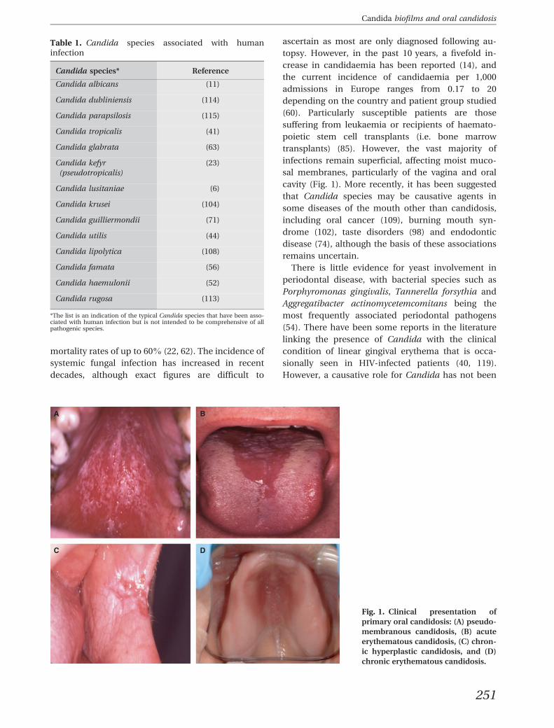
cavity (Fig. 1). More recently, it has been suggested
that Candida species may be causative agents in
some diseases of the mouth other than candidosis,
including oral cancer (109), burning mouth syn-
drome (102), taste disorders (98) and endodontic
disease (74), although the basis of these associations
remains uncertain.
There is little evidence for yeast involvement in
periodontal disease, with bacterial species such as
Porphyromonas gingivalis, Tannerella forsythia and
Aggregatibacter actinomycetemcomitans being the
most frequently associated periodontal pathogens
(54). There have been some reports in the literature
linking the presence of Candida with the clinical
condition of linear gingival erythema that is occa-
sionally seen in HIV-infected patients (40, 119).
However, a causative role for Candida has not been
Table 1. Candida species associated with humaninfection
Candida species* Reference
Candida albicans (11)
Candida dubliniensis (114)
Candida parapsilosis (115)
Candida tropicalis (41)
Candida glabrata (63)
Candida kefyr
(pseudotropicalis)
(23)
Candida lusitaniae (6)
Candida krusei (104)
Candida guilliermondii (71)
Candida utilis (44)
Candida lipolytica (108)
Candida famata (56)
Candida haemulonii (52)
Candida rugosa (113)
*The list is an indication of the typical Candida species that have been asso-ciated with human infection but is not intended to be comprehensive of allpathogenic species.
A
C
B
D
Fig. 1. Clinical presentation of
primary oral candidosis: (A) pseudo-
membranous candidosis, (B) acute
erythematous candidosis, (C) chron-
ic hyperplastic candidosis, and (D)
chronic erythematous candidosis.
251
Candida biofilms and oral candidosis

confirmed and the condition is rarely observed today,
possibly due to the frequent use of systemic anti-
fungal agents in these patients.
Interestingly, whilst the microflora of peri-
implantitis resembles that of chronic periodontitis,
Candida has been recovered from 55% of peri-im-
plant lesions and appears to be absent at healthy
implant sites (61). However, the aetiological
involvement of Candida in peri-implantitis requires
further longitudinal studies.
Virulence factors of Candida
Infection models of candidosis in animals suggest
that C. albicans is the most pathogenic species, and
in vitro investigations indicate that it also expresses
higher levels of putative virulence factors compared
with other Candida species. Several potential viru-
lence factors have been proposed in the pathoge-
nicity of Candida species (Table 2), with adhesion to
host surfaces, secretion of proteinases and hyphal
formation apparently the most significant.
Adherence of Candida to host surfaces
Adherence of Candida to host surfaces is required for
initial colonization and contributes to persistence of
the organism within the host. Candida cells adhere to
several host cell types, including epithelial, endothe-
lial and phagocytic cells. Among the many adhesins
expressed by C. albicans, agglutinin-like sequence
proteins have been implicated in pathogenesis and
biofilm formation (78). These cell wall-bound adhe-
sins bind to diverse mammalian peptide ligands,
causing cellular aggregation through homotypic
adhesion, and also co-aggregate with other microbial
pathogens to mediate polymicrobial infections.
Candida can adhere to the surface of medical devices,
in particular denture acrylic and silicone soft liners,
which often promotes biofilm formation.
Secretion of hydrolytic enzymes byCandida
Candida species secrete several hydrolytic enzymes
associated with pathogenicity, including secreted
aspartyl proteinases, phospholipases, lipases, phos-
phomonoesterase and hexosaminidase (80). Of these
enzymes, the secreted aspartyl proteinases have at-
tracted most interest and are widely considered to be
central to the development of Candida infection. In
contrast with other types of proteinases, secreted
aspartyl proteinases show proteolytic activity only
under acid conditions (pH < 4.0). Importantly for
oral infection, the environment under a removable
denture is acidic, which provides conditions suitable
for both production and activity of secreted aspartyl
proteinases. Secreted aspartyl proteinases are only
produced by certain Candida species, with C. albi-
cans secreting nine distinct aspartyl proteinases,
often at much higher levels compared with other
species. Interestingly, strains of C. albicans isolated
from clinically apparent oral candidosis have been
Table 2. Virulence factors associated with Candida and oral candidosis
Virulence factor Effect
Adherence to host surfaces Promotes retention in the mouth
• Relative cell-surface hydrophobicity • Non-specific adherence process
• Cell surface adhesin molecules • Specific adherence mechanisms
Evasion of host defence mechanisms Promotes retention in the mouth
• High-frequency phenotypic switching • Antigenic modification through frequent cell-surface changes
• Hyphal development • impairs phagocytosis
• Secreted aspartyl proteinase production • Secretory IgA destruction
• Binding of complement molecules • Antigenic masking
Invasion and destruction of host tissue Enhances pathogenicity
• Hyphal development • Promotes invasion of oral epithelium
• Secreted aspartyl proteinase production • Host cell and extracellular matrix damage
• Phospholipase production • Damage to host cells
252
Williams et al.

shown to produce higher levels of secreted aspartyl
proteinases compared with strains obtained from
carriers with no mucosal signs (58). These findings
suggest that strains of C. albicans that are actively
involved in candidosis could be inherently more vir-
ulent than commensal strains, possibly by being able
to upregulate secreted aspartyl proteinase gene
expression. In contrast, there is no conclusive evi-
dence that proteinase activity is always associated
with infection, and this probably reflects the multi-
factorial nature of Candida infections (80). Phos-
pholipases hydrolyse one or more ester linkages of
glycerophospholipids. Phospholipase activity has
been demonstrated for many fungal pathogens,
including Candida species. It has been reported that
the phospholipase activity is enhanced when hyphae
are in direct contact with host tissue.
Morphological transition of Candida
Candida species, in particular C. albicans, can exhibit
morphological alternation from yeast, pseudohyphal
and hyphal forms, depending on environmental
conditions. Hyphae are believed to play an important
role in tissue and biomaterial invasion, and in vitro
research has shown that C. albicans hyphal mutants
and non-C. albicans strains lacking hyphal formation
exhibit lower ability to invade tissue compared with
wild-type C. albicans strains (50). Candida hyphae
also demonstrate increased adherence properties (53,
80, 103) and greater resistance to phagocytosis com-
pared with yeast. Thus hyphal formation is consid-
ered to be significant to the pathogenicity of Candida.
Candida biofilms and infection
Biofilms can be defined as communities of microor-
ganisms, often attached to a surface and encased
within an extracellular polysaccharide matrix that is
produced by the microorganisms (25). The biofilm
state is the preferred mode of growth of microor-
ganisms in natural environments (24), and recent
reports have linked biofilms with over 65% of hos-
pital-acquired infections (28, 51). It has also been
suggested that Candida strains with a high ability to
form biofilms are generally more virulent than others
(79, 104). The explanation for this is likely to be
multifactorial and relate to the differences observed
between biofilm cells and their free-living or plank-
tonic counterparts. Indeed, it is now known that
significant phenotypic differences occur between
biofilm and planktonic lifestyles (8, 28, 73, 91, 92).
Perhaps the most important of these are those factors
that relate to the promotion, persistence and viru-
lence of the organisms within the host environment.
A recent investigation of candidaemia highlighted the
importance of biofilms in infection, with higher
mortality rates evident when a Candida biofilm was
present (116).
In the oral cavity, not only will adherent biofilm
cells be protected from the normal mechanical
flushing action of saliva and gingival crevicular fluid,
but the biofilm itself is a defensive barrier against
penetration of host immune factors and adminis-
tered antimicrobials (43, 121). Candida biofilms were
first shown to exhibit resistance to antifungals by
Hawser & Douglas (43), and this has been reported by
numerous other researchers since (19, 92). The exact
mechanism of biofilm resistance to antifungals
remains unclear, but it is probably multifactorial. The
extracellular polysaccharide of the biofilm could
serve as an inhibitor to diffusion of an antimicrobial
agent or ionically bind the drug as it diffuses through
the biofilm, thereby effectively reducing its �bio-
availability� (3, 8). A feature of multilayered biofilms is
the reduced activity and growth rates of cells that are
in areas of limited exposure to required gases and
nutrients, and it could readily be envisaged how these
cells would be less susceptible to an antimicrobial
that relies on inhibiting biochemical pathways asso-
ciated with actively growing cells. These cells could
represent the �persister cells� that have been sug-
gested to be the resistant phenotype within a biofilm
community (59). Conversely, other studies have
demonstrated that biofilm resistance mechanisms
are actually not completely dependent on changes in
growth rates (7), and may be the result of upregula-
tion of particular genes by biofilm cells. Indeed, the
genes encoding ATP-binding cassette (ABC) trans-
porter proteins that are particularly associated with
azole drug resistance by efflux pump mechanisms in
C. albicans have been shown to be upregulated in
biofilms (69, 73).
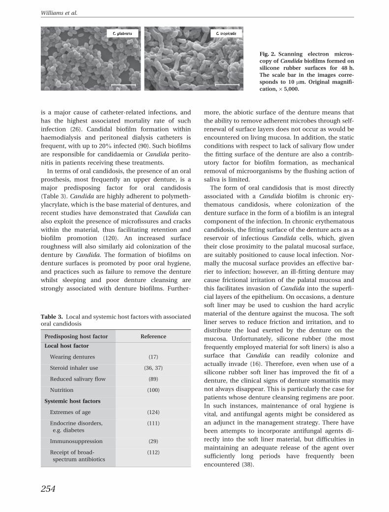
Involvement of Candida biofilms in human infec-
tion is well recognized, particularly when occurring
on biomaterials used for implantable medical devices
(Fig. 2). Away from the oral cavity, biofilms of Can-
dida on the silicone rubber of artificial voice box
prostheses in laryngectomized patients have been
identified as a major cause of their failure. Urinary
tract infection by Candida in catheterized patients is
associated with biofilm formation on the inner lumen
of the catheter, and other infections including those
of prosthetic heart valves and replacement joints
have also been linked with Candida biofilms (18, 49,
96). Candida colonization of intravascular catheters
253
Candida biofilms and oral candidosis
is a major cause of catheter-related infections, and
has the highest associated mortality rate of such
infection (26). Candidal biofilm formation within
haemodialysis and peritoneal dialysis catheters is
frequent, with up to 20% infected (90). Such biofilms
are responsible for candidaemia or Candida perito-
nitis in patients receiving these treatments.
In terms of oral candidosis, the presence of an oral
prosthesis, most frequently an upper denture, is a
major predisposing factor for oral candidosis
(Table 3). Candida are highly adherent to polymeth-
ylacrylate, which is the base material of dentures, and
recent studies have demonstrated that Candida can
also exploit the presence of microfissures and cracks
within the material, thus facilitating retention and
biofilm promotion (120). An increased surface
roughness will also similarly aid colonization of the
denture by Candida. The formation of biofilms on
denture surfaces is promoted by poor oral hygiene,
and practices such as failure to remove the denture
whilst sleeping and poor denture cleansing are
strongly associated with denture biofilms. Further-
more, the abiotic surface of the denture means that
the ability to remove adherent microbes through self-
renewal of surface layers does not occur as would be
encountered on living mucosa. In addition, the static
conditions with respect to lack of salivary flow under
the fitting surface of the denture are also a contrib-
utory factor for biofilm formation, as mechanical
removal of microorganisms by the flushing action of
saliva is limited.
The form of oral candidosis that is most directly
associated with a Candida biofilm is chronic ery-
thematous candidosis, where colonization of the
denture surface in the form of a biofilm is an integral
component of the infection. In chronic erythematous
candidosis, the fitting surface of the denture acts as a
reservoir of infectious Candida cells, which, given
their close proximity to the palatal mucosal surface,
are suitably positioned to cause local infection. Nor-
mally the mucosal surface provides an effective bar-
rier to infection; however, an ill-fitting denture may
cause frictional irritation of the palatal mucosa and
this facilitates invasion of Candida into the superfi-
cial layers of the epithelium. On occasions, a denture
soft liner may be used to cushion the hard acrylic
material of the denture against the mucosa. The soft
liner serves to reduce friction and irritation, and to
distribute the load exerted by the denture on the
mucosa. Unfortunately, silicone rubber (the most
frequently employed material for soft liners) is also a
surface that Candida can readily colonize and
actually invade (16). Therefore, even when use of a
silicone rubber soft liner has improved the fit of a
denture, the clinical signs of denture stomatitis may
not always disappear. This is particularly the case for
patients whose denture cleansing regimens are poor.
In such instances, maintenance of oral hygiene is
vital, and antifungal agents might be considered as
an adjunct in the management strategy. There have
been attempts to incorporate antifungal agents di-
rectly into the soft liner material, but difficulties in
maintaining an adequate release of the agent over
sufficiently long periods have frequently been
encountered (38).
Fig. 2. Scanning electron micros-
copy of Candida biofilms formed on
silicone rubber surfaces for 48 h.
The scale bar in the images corre-
sponds to 10 lm. Original magnifi-
cation, · 5,000.
Table 3. Local and systemic host factors with associatedoral candidosis
Predisposing host factor Reference
Local host factor
Wearing dentures (17)
Steroid inhaler use (36, 37)
Reduced salivary flow (89)
Nutrition (100)
Systemic host factors
Extremes of age (124)
Endocrine disorders,
e.g. diabetes
(111)
Immunosuppression (29)
Receipt of broad-
spectrum antibiotics
(112)
254
Williams et al.

The wearing of a denture is also a predisposing
factor to other forms of oral candidosis, such as
hyperplastic candidosis and angular cheilitis (2).
Whilst these types of oral candidosis occur at sites
away from the denture biofilm, it is probable that the
increase in Candida numbers resulting from the
presence of a denture is a major contributory factor
in these infections (1).
Most natural host surfaces such as the oral mucosa
have major advantages compared with implanted
biomaterials and other non-shedding surfaces in
preventing Candida colonization and biofilm forma-
tion. Clearly, the innate immune response of the host
will be effective within the oral mucosa, and, as
previously mentioned, the fact that the oral epithe-
lium is continually replenished means that, in order
to colonize the oral mucosa, Candida must be pres-
ent in the mouth in sufficient numbers and with a
high enough growth rate to allow their continued
persistence. In healthy individuals, candidal biofilms
are therefore not generally seen on the palatal or
buccal mucosa, although low-level colonization will
be detected in cases of commensal carriage. In the
event of host debilitation causing an ecological shift
in favour of Candida growth, candidal biofilms may
develop on the mucosa itself (Fig. 3) (27). Indeed, in
cases of pseudomembranous candidosis and hyper-
plastic candidosis, multilayered growth of Candida
adhering to the mucosal surface can be seen using
histological staining methods applied to either
mucosa smears or biopsy sections (Fig. 4).
Candida biofilm formation
As described above, the presence of Candida biofilms
can play a significant role in clinical infection be-
cause of their resilience and resistance to normal host
removal mechanisms and also antimicrobial therapy.
In order to adequately treat and prevent these
infections, it is therefore first necessary to understand
the processes involved in candidal biofilm formation
and growth, which could then lead to the identifica-
tion of suitable targets for therapy.
Candida biofilm formation and infection is a
staged process comprising (i) adherence to the sur-
face, (ii) colonization, proliferation and invasion, and
(iii) detachment of biofilm cells to promote coloni-
zation and infection of distal sites. Altered gene
expression accompanies development of the biofilm,
and such changes can be detected soon after initial
attachment to the surface (75). In the case of
C. albicans, biofilm formation has been reported to
be associated with the upregulation of genes involved
in adherence (such as agglutinin-like sequence genes
and hyphal wall protein 1; Hwp1) (78) and also those
genes involved in amino acid biosynthesis and
metabolism (126).
The initial stage of adhesion of Candida to host
surfaces occurs through the combined effects of
C. glabrata C. tropicalis
Fig. 3. Confocal laser scanning
microscopy images of (A) Candida
glabrata and (B) Candida tropicalis
colonizing and invading human oral
epithelium after 24 h incubation.
Candida yeast and filaments are
shown in red, and the nuclei of the
epithelial cells appear blue.
Fig. 4. Periodic acid Schiff-stained tissue section of
chronic hyperplastic candidosis showing tissue invading
Candida hyphae (purple-stained filaments).
255
Candida biofilms and oral candidosis
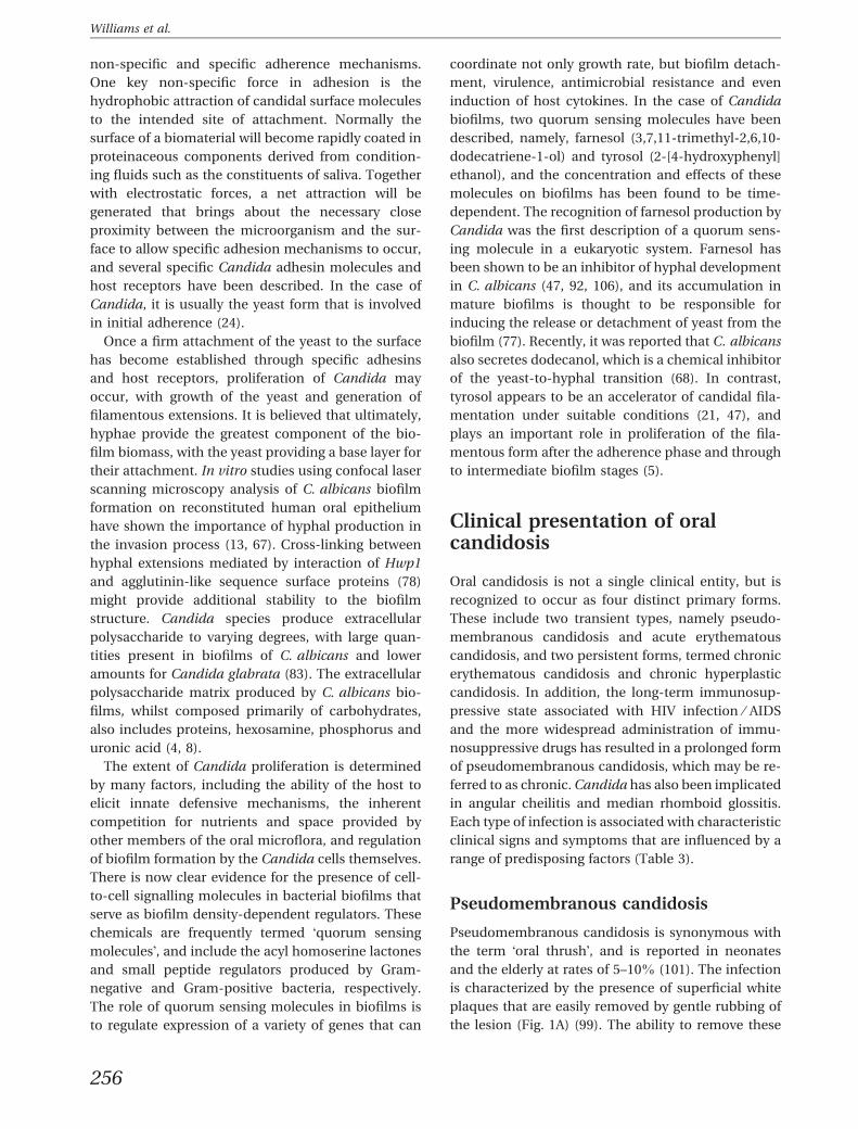
non-specific and specific adherence mechanisms.
One key non-specific force in adhesion is the
hydrophobic attraction of candidal surface molecules
to the intended site of attachment. Normally the
surface of a biomaterial will become rapidly coated in
proteinaceous components derived from condition-
ing fluids such as the constituents of saliva. Together
with electrostatic forces, a net attraction will be
generated that brings about the necessary close
proximity between the microorganism and the sur-
face to allow specific adhesion mechanisms to occur,
and several specific Candida adhesin molecules and
host receptors have been described. In the case of
Candida, it is usually the yeast form that is involved
in initial adherence (24).
Once a firm attachment of the yeast to the surface
has become established through specific adhesins
and host receptors, proliferation of Candida may
occur, with growth of the yeast and generation of
filamentous extensions. It is believed that ultimately,
hyphae provide the greatest component of the bio-
film biomass, with the yeast providing a base layer for
their attachment. In vitro studies using confocal laser
scanning microscopy analysis of C. albicans biofilm
formation on reconstituted human oral epithelium
have shown the importance of hyphal production in
the invasion process (13, 67). Cross-linking between
hyphal extensions mediated by interaction of Hwp1
and agglutinin-like sequence surface proteins (78)
might provide additional stability to the biofilm
structure. Candida species produce extracellular
polysaccharide to varying degrees, with large quan-
tities present in biofilms of C. albicans and lower
amounts for Candida glabrata (83). The extracellular
polysaccharide matrix produced by C. albicans bio-
films, whilst composed primarily of carbohydrates,
also includes proteins, hexosamine, phosphorus and
uronic acid (4, 8).
The extent of Candida proliferation is determined
by many factors, including the ability of the host to
elicit innate defensive mechanisms, the inherent
competition for nutrients and space provided by
other members of the oral microflora, and regulation
of biofilm formation by the Candida cells themselves.
There is now clear evidence for the presence of cell-
to-cell signalling molecules in bacterial biofilms that
serve as biofilm density-dependent regulators. These
chemicals are frequently termed �quorum sensing
molecules�, and include the acyl homoserine lactones
and small peptide regulators produced by Gram-
negative and Gram-positive bacteria, respectively.
The role of quorum sensing molecules in biofilms is
to regulate expression of a variety of genes that can
coordinate not only growth rate, but biofilm detach-
ment, virulence, antimicrobial resistance and even
induction of host cytokines. In the case of Candida
biofilms, two quorum sensing molecules have been
described, namely, farnesol (3,7,11-trimethyl-2,6,10-
dodecatriene-1-ol) and tyrosol (2-[4-hydroxyphenyl]
ethanol), and the concentration and effects of these
molecules on biofilms has been found to be time-
dependent. The recognition of farnesol production by
Candida was the first description of a quorum sens-
ing molecule in a eukaryotic system. Farnesol has
been shown to be an inhibitor of hyphal development
in C. albicans (47, 92, 106), and its accumulation in
mature biofilms is thought to be responsible for
inducing the release or detachment of yeast from the
biofilm (77). Recently, it was reported that C. albicans
also secretes dodecanol, which is a chemical inhibitor
of the yeast-to-hyphal transition (68). In contrast,
tyrosol appears to be an accelerator of candidal fila-
mentation under suitable conditions (21, 47), and
plays an important role in proliferation of the fila-
mentous form after the adherence phase and through
to intermediate biofilm stages (5).
Clinical presentation of oralcandidosis
Oral candidosis is not a single clinical entity, but is
recognized to occur as four distinct primary forms.
These include two transient types, namely pseudo-
membranous candidosis and acute erythematous
candidosis, and two persistent forms, termed chronic
erythematous candidosis and chronic hyperplastic
candidosis. In addition, the long-term immunosup-
pressive state associated with HIV infection ⁄ AIDS
and the more widespread administration of immu-
nosuppressive drugs has resulted in a prolonged form
of pseudomembranous candidosis, which may be re-
ferred to as chronic. Candida has also been implicated
in angular cheilitis and median rhomboid glossitis.
Each type of infection is associated with characteristic
clinical signs and symptoms that are influenced by a
range of predisposing factors (Table 3).
Pseudomembranous candidosis
Pseudomembranous candidosis is synonymous with
the term �oral thrush�, and is reported in neonates
and the elderly at rates of 5–10% (101). The infection
is characterized by the presence of superficial white
plaques that are easily removed by gentle rubbing of
the lesion (Fig. 1A) (99). The ability to remove these
256
Williams et al.

plaques is a diagnostic feature that differentiates
pseudomembranous candidosis from other forms of
white patch lesions of the oral mucosa. Histological
examination of the plaques reveals fungal elements
in the form of yeast and filamentous forms, together
with epithelial cells.
Acute erythematous candidosis
This condition is often referred to as �antibiotic sore
mouth�, as it tends to develop following a reduction
in the levels of the bacterial component of the oral
microflora after the receipt of broad-spectrum anti-
biotics. A decrease in bacterial numbers results in
reduced microbial competition for Candida in terms
of nutrition and adherence sites. The clinical signs
and symptoms of acute erythematous candidosis are
therefore a direct consequence of an ecological shift
in the normal homeostatic balance of the microbial
community. Acute erythematous candidosis presents
as a painful reddened lesion, and although the palate
or buccal mucosa may be involved, the most com-
mon site of infection is the dorsum of the tongue
(Fig. 1B). Cessation of antibiotic therapy results in a
return to normal levels of bacteria, which subse-
quently resolves the candidosis without intervention.
Concomitant use of steroid therapy, particularly in
inhaler form, is an additional contributing factor, as
this can create a localized area of immune suppres-
sion within the mucosa that permits overgrowth of
Candida.
Chronic hyperplastic candidosis
This form of candidosis characteristically presents as
a thickened white plaque, most frequently at the
commissure region of the mouth or on the dorsum
of the tongue (Fig. 1C). Of particular concern with
this form of infection is the potential for the devel-
opment of squamous cell carcinoma at lesional
sites, although the role of Candida in the process of
malignant change remains unclear (109, 122). Two
clinical types of hyperplastic candidosis have been
described based on the appearance of the lesion.
Homogeneous hyperplastic candidosis is described
as having smooth white lesions that are notably
distinct from those of heterogeneous hyperplastic
candidosis in which areas of erythema occur
resulting in a nodular, speckled appearance. It has
been suggested that heterogeneous lesions have a
greater likelihood of malignant transformation (12,
94). In contrast to pseudomembranous candidosis,
the white patch lesions of hyperplastic candidosis
do not rub off with gentle rubbing. Hyperplastic
candidosis can only be diagnosed by histopatho-
logical examination of lesional biopsy material,
which reveals candidal hyphae invading the epithe-
lium and an underlying chronic inflammatory cell
infiltrate (125). Uncertainty remains over whether
candidal invasion is the primarily ecological factor
of hyperplastic candidosis, or whether Candida
infection is secondary to the formation of an altered
epithelium. Almost all patients with hyperplastic
candidosis are smokers.
Chronic erythematous candidosis
Commonly known as Candida-associated denture
stomatitis, chronic erythematous candidosis presents
as a reddening of the mucosa beneath the fitting
surface of a denture (Fig. 1D). The infection may
develop under any acrylic denture or intra-oral
appliance, but is almost exclusively encountered on
palatal tissues. Principle host factors associated with
this condition are inadequate oral hygiene, failure to
remove dentures whilst sleeping, or poor denture fit
(32). Chronic erythematous candidosis is the most
prevalent form of oral candidosis, with up to 75% of
denture wearers having clinical signs of this condi-
tion, although the sufferer is often unaware of the
presence of infection (9).
Angular cheilitis
This condition presents as erythematous lesions at
one or more, or usually both of the angles of the
mouth. In addition to Candida, the spectrum of
microorganisms recovered from this condition
includes Staphylococcus aureus and streptococcal
species, either alone or in combination. Therefore,
the exact role that Candida itself plays in angular
cheilitis remains uncertain (110). Often, angular
cheilitis involving Candida occurs in patients with a
pre-existing primary form of oral candidosis, most
frequently chronic erythematous candidosis. The in-
creased level of Candida within the mouth of such
patients is most likely to be the major contributing
factor.
Other oral candidoses
Median rhomboid glossitis is a chronic mucosal
condition that, as its name implies, characteristically
presents as a symmetrically shaped lesion on the
midline of the dorsum of the tongue. Candida can
often be recovered from the surface of the lesion, has
257
Candida biofilms and oral candidosis

and is thus implicated in the aetiology. Furthermore,
resolution of the condition following provision of
systemic antifungal therapy supports the role of
Candida in the infection. As with other forms of oral
candidosis, tobacco smoking and steroid inhaler use
are recognized predisposing factors.
Chronic mucocutaneous candidosis is a rare
condition in which a range of chronic Candida
infections of mucous membranes, skin and nails are
encountered. The principle predisposing factor for
chronic mucocutaneous candidosis is congenital
impaired cellular immunity against Candida.
Management of oral candidosis
Principles of treatment
Successful management of patients with oral candi-
dosis requires identification, and where possible
correction, of the specific underlying predisposing
factors in an individual patient. Without this recog-
nition, subsequent treatment using antifungal
therapy may only result in the temporary relief of
infection, with relapses inevitably following. Instruc-
tions should be provided on appropriate oral hygiene
practices. Use of steroid inhalers should be coupled
with rinsing the mouth with water after administra-
tion. All patients should be advised on the importance
of reduction or cessation of any smoking habits. As
described below, oral hygiene practices are also
essential in removal of candidal biofilms on host
surfaces and oral prostheses.
Any identified nutritional deficiency should be
corrected, and advice provided on dietary habits such
as appropriate carbohydrate intake. However, despite
these interventions, situations arise in which the
underlying cause cannot be resolved, such as HIV
infection or immunosuppressive therapy following
organ or bone marrow transplant. In these circum-
stances, treatment of oral candidosis is based on the
use of antifungal therapy. Details on antifungal
agents are provided below, and Table 4 shows the
typical antifungal drugs and the treatment regimes
used specifically for oral candidosis.
Antifungal agents
Relatively few antifungal drugs are available when
compared to the range of antibiotics that have been
produced, which probably reflects both the relatively
recent recognition of the importance of fungal
infections in humans and the difficulty involved in
developing an agent with activity against a eukaryotic
cell type without problems of associated host cell
toxicity. Antifungal drugs are classified according to
their mode of action: (i) disruption of fungal cell
membranes, as seen with the polyene antifungals
(nystatin and amphotericin), (ii) inhibition of ergos-
terol synthesis, exemplified by the azole group of
antifungals (fluconazole and itraconazole), (iii) inhi-
bition of b-1,3-D-glucan synthesis (echinocandin
antifungals), and (iv) induction of incorrect RNA
synthesis and interference with DNA replication
(5-fluorocytosine).
Polyene antifungals exert their fungicidal activity
by inducing cell membrane porosity following
interaction with the ergosterol component of the
membrane, with the subsequent effect of loss of
cytoplasmic content. Polyenes have a broad spec-
trum of antifungal activity, but, due to their poor
absorption through the gut, their use in treatment of
oral candidosis is extremely limited. However, in view
of the fact that these agents are not absorbed, their
safety profile is good. A specific role for the topical
use of these agents is in cream format for the treat-
ment of chronic erythematous candidosis. Despite
wide use over several decades, the actual incidence of
resistance of Candida to polyenes is rare, but can
sometimes arise through a reduction in the ergosterol
content of cell membranes (105).
Azole antifungals are fungistatic through interfer-
ence with the fungal enzyme lanosterol demethylase,
which is a key enzyme in the biosynthesis of ergos-
terol. Both fluconazole and itraconazole are well ab-
sorbed through the gut, which means that oral
administration is an effective means of systemic
delivery. Furthermore, the effects of fluconazole in
the oral cavity are enhanced as it is secreted in saliva
at levels equivalent to those achieved in the blood.
Fluconazole is the agent of first choice for all forms of
oral candidosis apart from chronic erythematous
candidosis. The clinical effectiveness of agents that
can only be delivered topically, such as amphotericin
or nystatin, is limited due to problems in maintaining
sufficient levels of drug at the site of infection. The
taste of topical agents stimulates salivary secretion,
which rapidly dilutes and removes the antifungal
agent from the mouth. In view of this, their clinical
use is limited. Fluconazole has a good safety profile
when given systemically, with few contra-indications
or side effects. Important interactions occur with
coumarin anticoagulants and sulfonylurea antidia-
betic agents. Acquired resistance to azole antifungals
has emerged in recent years, and certain Candida
species are also inherently resistant to these agents.
258
Williams et al.

Several mechanisms of azole resistance have been
reported including (i) an alteration in the chemical
structure of the demethylase enzyme, (ii) removal of
the azole from the cell by multidrug transporter
pumps, and (iii) compensation by other sterol syn-
thesis enzymes in membrane biosynthesis. Even in
the absence of a defined resistance mechanism, the
in vitro susceptibility of a given Candida strain often
does not correlate with the subsequent clinical out-
come for patients with oral candidosis. One possible
explanation for this could relate to the phenotypic
differences described above for planktonic and bio-
film cultured cells, as it is the former that are most
frequently used for in vitro antifungal susceptibility
testing.
Management of candidal biofilms
As described previously, the presence of candidal
biofilms reduces the likelihood of removal of organ-
isms by host defence mechanisms and antifungal
agents. Thus appropriate management of biofilms is
essential. There is no single approach that can be
used to specifically counter candidal biofilms, and a
variety of mechanical and chemical methods to im-
prove oral hygiene are generally adopted. Ideally an
�anti-biofilm� approach will prevent development of
the biofilm in the first instance, as well as being
effective against established biofilms.
Standard oral hygiene practices including tooth-
brushing and the use of mouthwashes are important
Table 4. Recommended antifungal agents for treatment of oral candidosis
PMC AEC CEC CHC Typical adult dose
Topical
administration
Nystatin Yes one lozenge:
100,000 units
q.i.d. · 7–14 days
Suspension: 500,000
units by rinse and
swallow q.i.d. · 7–
14 days
Amphotericin Yes one lozenge (10 mg)
q.i.d. · 10–15 days
Miconazole Yes Oral gel (24 mg ⁄ml); 5–10 ml q.i.d. ·
7–14 days
Clotrimazole Yes one lozenge (10 mg)
five times per day ·7–14 days
Systemic
administration
Ketoconazole Yes Yes Yes 200–400 mg ⁄ day ·7–14 days
Fluconazole Yes Yes Yes 100 mg ⁄ day · 7–
14 days
Itraconazole Yes Yes Yes 200 mg (20 ml) sus-
pension by rinse and
swallow without
food q.i.d. · 7–
14 days 200 mg ⁄day (capsules taken
with food) · 2–
4 weeks
PMC, pseudomembranous candidosis; AEC, acute erythematous candidosis; CEC, chronic erythematous candidosis; CHC, chronic hyperplastic candidosis. q.i.d.,four times per day.Other antifungal agents are available and these may be more frequently used in hospitalized patients.
259
Candida biofilms and oral candidosis

tools in oral biofilm removal. Toothbrushing offers a
physical means to combat biofilms (97), but may be
limited to accessible sites within the oral cavity and
can have deleterious effects on acrylic denture sur-
faces if abrasive toothpastes are used. In such cases,
the resulting roughened acrylic surface could, in
theory, be more conducive to subsequent biofilm
formation. It has been suggested that mechanical
toothbrushing may offer an advantage over manual
toothbrushing by having an additional physical
influence on biofilms at sites inaccessible to the
toothbrush bristles, such as interproximal regions.
Potential benefits of sonication include possible
cavitation of surrounding fluids and generation of
shear forces, which then disrupt the biofilm, although
its clinical value remains uncertain (45). The poten-
tial antifungal effects of such shear forces on Candida
are as yet not known. Nevertheless, as dental plaque
contains yeast, toothbrushing will serve to reduce the
level of Candida in the mouth and also maintain
normal levels of mucosal resistance to fungal infec-
tions, which can otherwise be reduced with deterio-
ration in oral hygiene.
A wide variety of mouthwashes have been found
to have anti-candidal activity, including chlorhexi-
dine gluconate, trichlosan and those incorporating
essential oils. Chlorhexidine is a cationic chlor-
ophenyl bisbiguanide and is perhaps the most fre-
quently used mouthwash. Chlorhexidine exhibits a
broad spectrum of antimicrobial activity that
encompasses Candida species (66). It is believed to
bind to negatively charged Candida surfaces, and
induces a loss of structural integrity, decreases
adherence capability and disrupts the cell wall.
Chlorhexidine’s anti-candidal properties are also
retained against Candida that is adhered to acrylic
surfaces (88), and is therefore of value in the treat-
ment of chronic erythematous candidosis. Studies
have shown that 0.2% chlorhexidine gluconate
mouth rinses exhibit clinical benefit in the treat-
ment of acute erythematous and pseudomembra-
nous candidosis (15, 30). However, there are reports
of reduced efficacy of nystatin when used in com-
bination with chlorhexidine gluconate, and therefore
it is often advocated that nystatin treatment be
delayed for 30 min after use of chlorhexidine
mouthwash. The reason for the reduced efficacy has
been proposed to be due to the formation of a low
solubility chlorhexidine-nystatin salt that is less
effective as an antibiotic agent (10).
Essential oil mouthwashes containing a range of
natural plant extracts, including thymol, eucalyptol,
bioflavanoids and tea tree (Melaleuca alternifola) oil
derivatives, have also been shown to have direct
bactericidal and anticandidal activity in vitro (33,
82). It is thought that essential oil mouthwashes kill
microorganisms by cell membrane disruption and
enzyme inhibition (34, 57). As with traditional
antifungal agents, however, the effectiveness of
natural antimicrobials on established biofilms in
the oral cavity is less certain, with incomplete
penetration by the agents being reported (76). The
clinical efficacy of this category of mouthwashes
has been studied, but largely against plaque bac-
teria (35, 84), and therefore the clinical benefits of
these agents in treating oral candidosis remain to
be established.
One of the main problems associated with eradi-
cating a biofilm from a biomaterial implanted within
the body is the difficulty of access for biofilm removal
or biomaterial replacement. Often the latter is the
only option available for the management of infec-
tion of certain indwelling catheters and artificial
voice box devices. With regard to dentures, their
replacement or removal for thorough cleaning is a
relatively easy option. It is essential that dentures are
removed during sleep and ideally immersed in a
suitable antimicrobial cleansing agent. The mouth
rinses described above can all be used, but as the
denture is no longer in contact with the host tissues,
other chemically harsher cleansers such alkaline and
neutral peroxide-type cleansers, hypochlorite, tetra-
sodium EDTA and acidic solutions may also be em-
ployed. Often these agents are used to soak dentures
for a defined time period in an immersion bath,
thereby allowing the cleansing agent to access areas
that are inaccessible by brushing. Prior to use, the
dentures are then rinsed in water to remove the
treatment agent. Furthermore, water at high tem-
peratures may also be incorporated into the cleaning
regime, and the added value of microwaving
water-immersed dentures in the treatment of denture
stomatitis has also been reported (123). Regular
microwaving of dentures, where temperatures
approach 100 �C, has been shown to have no adverse
affect on the hardness of the denture resins, although
increased surface roughness was noted (65).
Future strategies for management ofcandidal biofilms
One strategy that has been the focus of recent
research is to actually modify the surfaces of
biomaterials so that they are less prone to coloniza-
tion by microorganisms, including Candida (86, 87).
Approaches have included pre-coating biomaterials
260
Williams et al.

such as silicone rubber or denture acrylic with
chemicals such as silanes, chlorhexidine, histatins
and other surface-modifying groups (20, 87, 88).
In addition, thin-film polymer formulations with
incorporated antifungals (nystatin or amphotericin)
have also recently been shown to inhibit C. albicans
biofilm growth on denture materials (93). Such
approaches will clearly have value in combating
biofilm formation on indwelling medical devices that
are not readily accessible for cleaning and physical
removal of the biofilm, e.g. artificial voice prostheses
(31).
In the future, it may be possible to exploit quorum
sensing molecules to disrupt biofilms as they devel-
op, and it has already been shown that farnesol has
deleterious effects on Candida biofilms, causing
structural instability, even for mature biofilms (48,
92).
Reducing the candidal load within the oral cavity
through the use of probiotic agents is also a consid-
eration. Probiotics are live microorganisms that,
when administered in adequate amounts, confer a
health benefit on the host (70). The concept behind
their use in treatment of oral candidosis would be to
provide microbiological pressure in the local envi-
ronment, either through competition for adherence
sites and nutrients or by creating an environment
that is not conducive to the growth of Candida. There
would also appear to be beneficial effects induced by
probiotics through immune modulation. Previous
studies have used bacterial species such as Strepto-
coccus thermophilus and Lactobacillus bulgaricus to
treat biofilms on indwelling voice prostheses (117,
118), and more recently, in the form of cheeses
containing probiotic organisms, to reduce Candida
load in the oral cavity (42). However, the long-term
ability of probiotics to modulate the normal micro-
flora remains unknown.
Conclusion
Biofilm formation is now a well-recognized phe-
nomenon, and there is clear evidence demonstrating
the importance of this life style in human infection.
Candida is adept at forming biofilms on a range of
surfaces, including natural host tissues and bioma-
terials frequently used in medical devices. Such bio-
films are linked to both systemic and superficial
forms of candidosis in humans. New techniques
including confocal laser scanning microscopy and
molecular analysis tools are enabling elucidation of
the control mechanisms underlying biofilm forma-
tion. By increasing our knowledge of candidal biofilm
formation, potential therapeutic targets may well be
identified that can be used as additional therapies,
alongside standard oral hygiene practices, in pre-
venting oral candidosis.
References
1. Abu-Elteen KH, Abu-Alteen RM. The prevalence of Can-
dida albicans populations in the mouths of complete
denture wearers. New Microbiol 1998: 21: 41–48.
2. Akpan A, Morgan R. Oral candidiasis. Postgrad Med J 2002:
78: 455–459.
3. Al-Fattani MA, Douglas LJ. Penetration of Candida bio-
films by antifungal agents. Antimicrob Agents Chemother
2004: 48: 3291–3297.
4. Al-Fattani MA, Douglas LJ. Biofilm matrix of Candida
albicans and Candida tropicalis: chemical composition
and role in drug resistance. J Med Microbiol 2006: 55: 999–
1008.
5. Alem MA, Oteef MD, Flowers TH, Douglas LJ. Production
of tyrosol by Candida albicans biofilms and its role in
quorum sensing and biofilm development. Eukaryot Cell
2006: 5: 1770–1779.
6. Atkinson BJ, Lewis RE, Kontoyiannis DP. Candida lusitaniae
fungemia in cancer patients: risk factors for amphotericin B
failure and outcome. Med Mycol 2008: 46: 541–546.
7. Baillie GS, Douglas LJ. Effect of growth rate on resistance
of Candida albicans biofilms to antifungal agents. Anti-
microb Agents Chemother 1998: 42: 1900–1905.
8. Baillie GS, Douglas LJ. Matrix polymers of Candida
biofilms and their possible role in biofilm resistance to
antifungal agents. J Antimicrob Chemother 2000: 46: 397–
403.
9. Barbeau J, Seguin J, Goulet JP, De Koninck L, Avon SL,
Lalond B, Rompre P, Deslauries N. Reassessing the pres-
ence of Candida albicans in denture-related stomatitis.
Oral Surg Oral Med Oral Pathol Oral Radiol Endod 2003:
95: 51–59.
10. Barkvoll P, Attramadal A. Effect of nystatin and chlorhex-
idine digluconate on Candida albicans. Oral Surg Oral
Med Oral Pathol 1989: 67: 279–281.
11. Barnett JA. A history of research on yeasts 12: medical
yeasts part 1, Candida albicans. Yeast 2008: 25: 385–
417.
12. Barrett AW, Kingsmill VJ, Speight PM. The frequency of
fungal infection in biopsies of oral mucosal lesions. Oral
Dis 1998: 4: 26–31.
13. Bartie KL, Williams DW, Wilson MJ, Potts AJ, Lewis MAO.
Differential invasion of Candida albicans isolates in an
in vitro model of oral candidosis. Oral Microbiol Immunol
2004: 19: 293–296.
14. Bassetti M, Ansaldi F, Nicolini L, Malfatto E, Molinari MP,
Mussap M, Rebesco B, Bobbio Pallavicini F, Icardi G,
Viscoli C. Incidence of candidaemia and relationship with
fluconazole use in an intensive care unit. J Antimicrob
Chemother 2009: 64: 625–629.
15. Budtz-Jorgensen E. Etiology, pathogenesis, therapy, and
prophylaxis of oral yeast infections. Acta Odontol Scand
1990: 48: 61–69.
261
Candida biofilms and oral candidosis

16. Bulad K, Taylor RL, Verran J, McCord JF. Colonization and
penetration of denture soft lining materials by Candida
albicans. Dent Mater 2004: 20: 167–175.
17. Campisi G, Panzarella V, Matranga D, Calvino F, Pizzo G,
Lo Muzio L, Porter S. Risk factors of oral candidosis: a
twofold approach of study by fuzzy logic and traditional
statistic. Arch Oral Biol 2008: 53: 388–397.
18. Cardinal E, Braunstein EM, Capello WN, Heck DA. Can-
dida albicans infection of prosthetic joints. Orthopedics
1996: 19: 247–251.
19. Chandra J, Mukherjee PK, Leidich SD, Faddoul FF, Hoyer
LL, Douglas LJ, Ghannoum MA. Antifungal resistance of
candidal biofilms formed on denture acrylic in vitro.
J Dent Res 2001: 80: 903–908.
20. Chandra J, Patel JD, Li J, Zhou G, Mukherjee PK, McCor-
mick TS, Anderson JM, Ghannoum MA. Modification of
surface properties of biomaterials influences the ability of
Candida albicans to form biofilms. Appl Environ Microbiol
2005: 71: 8795–8801.
21. Chen H, Fujita M, Feng Q, Clardy J, Fink GR. Tyrosol is a
quorum sensing molecule in Candida albicans. Proc Natl
Acad Sci USA 2004: 101: 5048–5052.
22. Colombo AL, Nucci M, Park BJ, Nouer SA, Arthington-
Skaggs B, Dab Matta DA, Warnock D, Morgan J, Brazilian
Network Candidemia Study. Epidemiology of candidemia
in Brazil: a nationwide sentinel surveillance of candidemia
in eleven medical centers. J Clin Microbiol 2006: 44: 2816–
2823.
23. Corpus K, Hegeman-Dingle R, Bajjoka I. Candida kefyr, an
uncommon but emerging fungal pathogen: report of two
cases. Pharmacotherapy 2004: 24: 1084–1088.
24. Costerton JW, Cheng KJ, Geesey GG, Ladd TI, Nickel JC,
Dasgupta M, Marrie TJ. Bacterial biofilms in nature and
disease. Annu Rev Microbiol 1987: 41: 435–464.
25. Costerton JW, Lewandowski Z, Caldwell DE, Korber DR,
Lappin-Scott HM. Microbial biofilms. Annu Rev Microbiol
1995: 49: 711–745.
26. Crump JA, Collignon PJ. Intravascular catheter-associated
infections. Eur J Clin Microbiol Infect Dis 2000: 19: 1–8.
27. Dongari-Bagtzoglou A. Mucosal biofilms: challenges and
future directions. Expert Rev Anti Infect Ther 2008: 6: 141–
144.
28. Douglas LJ. Candida biofilms and their role in infection.
Trends Microbiol 2003: 11: 30–36.
29. Egusa H, Soysa NS, Ellepola AN, Yatani H, Samaranayake
LP. Oral candidosis in HIV-infected patients. Curr HIV Res
2008: 6: 485–499.
30. Ellepola ANB, Samaranayake LP. Oral candidal infections
and antimycotics. Crit Rev Oral Biol Med 2000: 11: 172–
198.
31. Everaert EP, Mahieu HF, Van de Belt-Gritter B, Peeters AJ,
Verkerke GJ, Van der Mei HC, Busscher HJ. Biofilm for-
mation in vivo on perfluoro-alkylsiloxane-modified voice
prostheses. Arch Otolaryngol Head Neck Surg 1999: 125:
1329–1332.
32. Figueiral MH, Azul A, Pinto E, Fonseca PA, Branco FM,
Scully C. Denture-related stomatitis: identification of
aetiological and predisposing factors – a large cohort.
J Oral Rehabil 2007: 34: 448–455.
33. Filoche SK, Soma K, Sissons CH. Antimicrobial effects of
essential oils in combination with chlorhexidine digluco-
nate. Oral Microbiol Immunol 2005: 20: 221–225.
34. Fine DH. Mouthrinses as adjuncts for plaque and gingi-
vitis management. A status report for the American Jour-
nal of Dentistry. Am J Dent 1988: 1: 259–263.
35. Fine DH, Markowitz K, Furgang D, Goldsmith D, Charles
CH, Lisante TA, Lynch MC. Effect of an essential
oil-containing antimicrobial mouthrinse on specific
plaque bacteria in vivo. J Clin Periodontol 2007: 34: 652–
657.
36. Fukushima C, Matsuse H, Saeki S, Kawano T, Machida I,
Kondo Y, Kohno S. Salivary IgA and oral candidiasis in
asthmatic patients treated with inhaled corticosteroid.
J Asthma 2005: 42: 601–604.
37. Fukushima C, Matsuse H, Tomari S, Obase Y, Miyazaki Y,
Shimoda T, Kohno S. Oral candidiasis associated with
inhaled corticosteroid use: comparison of fluticasone and
beclomethasone. Ann Allergy Asthma Immunol 2003: 90:
646–651.
38. Geerts GAVM, Stuhlinger ME, Basson NJ. Effect of an
antifungal denture liner on the saliva yeast count in pa-
tients with denture stomatitis: a pilot study. J Oral Rehab
2008: 35: 664–669.
39. Gonzalez GM, Elizondo M, Ayala J. Trends in species
distribution and susceptibility of bloodstream isolates of
Candida collected in Monterrey, Mexico, to seven anti-
fungal agents: results of a 3-year (2004 to 2007) surveil-
lance study. J Clin Microbiol 2008: 46: 2902–2905.
40. Grbic JT, Mitchell-Lewis DA, Fine JB, Phelan JA, Bucklan
RS, Zambon JJ, Lamster IB. The relationship of candidiasis
to linear gingival erythema in HIV-infected homosexual
men and parenteral drug users. J Periodontol 1995: 66: 30–
37.
41. Hajjeh RA, Sofair AN, Harrison LH, Lyon GM, Arthington-
Skaggs BA, Mirza SA, Phelan M, Morgan J, Lee-Yang W,
Ciblak MA, Benjamin LE, Sanza LT, Huie S, Yeo SF, Brandt
ME, Warnock DW. Incidence of bloodstream infections
due to Candida species and in vitro susceptibilities of
isolates collected from 1998 to 2000 in a population-based
active surveillance program. J Clin Microbiol 2004: 42:
1519–1527.
42. Hatakka K, Ahola AJ, Yli-Knuuttila H, Richardson M,
Poussa T, Meurman JH, Korpela R. Probiotics reduce the
prevalence of oral Candida in the elderly – a randomized
controlled trial. J Dent Res 2007: 86: 125–130.
43. Hawser SP, Douglas LJ. Resistance of Candida albicans
biofilms to antifungal agents in vitro. Antimicrob Agents
Chemother 1995: 39: 2128–2131.
44. Hazen KC, Theisz GW, Howell SA. Chronic urinary tract
infection due to Candida utilis. J Clin Microbiol 1999: 37:
824–827.
45. Hope CK, Wilson M. Effects of dynamic fluid activity from
an electric toothbrush on in vitro oral biofilms. J Clin
Periodontol 2003: 30: 624–629.
46. Horn DL, Neofytos D, Anaissie EJ, Fishman JA, Stein-
bach WJ, Olyaei AJ, Marr KA, Pfaller MA, Chang CH,
Webster KM. Epidemiology and outcomes of candidemia
in 2019 patients: data from the prospective antifungal
therapy alliance registry. Clin Infect Dis 2009: 48: 1695–
1703.
47. Hornby JM, Jensen EC, Lisec AD, Tasto JJ, Jahnke B,
Shoemaker R, Dussault P, Nickerson KW. Quorum sensing
in the dimorphic fungus Candida albicans is mediated by
farnesol. Appl Environ Microbiol 2001: 67: 2982–2992.
262
Williams et al.

48. Jabra-Rizk MA, Shirtliff M, James C, Meiller T. Effect of
farnesol on Candida dubliniensis biofilm formation and
fluconazole resistance. FEMS Yeast Res 2006: 6: 1063–1073.
49. Jain N, Kohli R, Cook E, Gialanella P, Chang T, Fries BC.
Biofilm formation by and antifungal susceptibility of
Candida isolates from urine. Appl Environ Microbiol 2007:
73: 1697–1703.
50. Jayatilake JA, Samaranayake YH, Cheung LK, Samarana-
yake LP. Quantitative evaluation of tissue invasion by wild
type, hyphal and SAP mutants of Candida albicans, and
non-albicans Candida species in reconstituted human
oral epithelium. J Oral Pathol Med 2006: 35: 484–491.
51. Khardori N, Yassien M. Biofilms in device-related infec-
tions. J Ind Microbiol 1995: 15: 141–147.
52. Kim MN, Shin JH, Sung H, Lee K, Kim EC, Ryoo N, Lee JS,
Jung SI, Park KH, Kee SJ, Kim SH, Shin MG, Suh SP, Ryang
DW. Candida haemulonii and closely related species at 5
university hospitals in Korea: identification, antifungal
susceptibility, and clinical features. Clin Infect Dis 2009:
48: e57–e61.
53. Kimura LH, Pearsall NN. Relationship between germina-
tion of Candida albicans and increased adherence to hu-
man buccal epithelial cells. Infect Immun 1980: 28: 464–
468.
54. Kornman KS. Diagnostic and prognostic tests for oral
diseases: practical applications. J Dent Educ 2005: 69: 498–
508.
55. Krcmery V, Barnes AJ. Non-albicans Candida spp. causing
fungaemia: pathogenicity and antifungal resistance.
J Hosp Infect 2002: 50: 243–260.
56. Krcmery V, Kunova A. Candida famata fungemia in a
cancer patient: case report. J Chemother 2000: 12: 189–190.
57. Kubert D, Rubin M, Barnett ML, Vincent JW. Antiseptic
mouthrinse-induced microbial cell surface alterations. Am
J Dent 1993: 6: 277–279.
58. Kuriyama T, Williams DW, Lewis MAO. In vitro secreted
aspartyl proteinase activity of Candida albicans isolated
from oral diseases and healthy oral cavities. Oral Microbiol
Immunol 2003: 18: 405–407.
59. LaFleur MD, Kumamoto CA, Lewis K. Candida albicans
biofilms produce antifungal-tolerant persister cells. Anti-
microb Agents Chemother 2006: 50: 3839–3846.
60. Lass-Florl C. The changing face of epidemiology of
invasive fungal disease in Europe. Mycoses 2009: 52: 197–
205.
61. Leonhardt A, Renvert S, Dahlen G. Microbial findings at
failing implants. Clin Oral Implants Res 1999: 10: 339–345.
62. Leroy O, Gangneux JP, Montravers P, Mira JP, Gouin F,
Sollet JP, Carlet J, Reynes J, Rosenheim M, Regnier B,
Lortholary O, AmarCand Study Group. Epidemiology,
management, and risk factors for death of invasive Can-
dida infections in critical care: a multicenter, prospective,
observational study in France (2005–2006). Crit Care Med
2009: 37: 1612–1618.
63. Li L, Redding S, Dongari-Bagtzoglou A. Candida glabrata:
an emerging oral opportunistic pathogen. J Dent Res 2007:
86: 204–215.
64. Liguori G, Di Onofrio V, Lucariello A, Galle F, Signoriello
G, Colella G, D�Amora M, Rossano F. Oral candidiasis: a
comparison between conventional methods and multiplex
polymerase chain reaction for species identification. Oral
Microbiol Immunol 2009: 24: 76–78.
65. Machado A, Breeding L, Vergani C, da Cruz Perez L.
Hardness and surface roughness of reline and denture
base acrylic resins after repeated disinfection procedures.
J Prosthet Dent 2009: 102: 115–122.
66. MacNeill S, Rindler E, Walker A, Brown AR, Cobb CM.
Effects of tetracycline hydrochloride and chlorhexidine
gluconate on Candida albicans: an in vitro study. J Clin
Periodontol 1997: 24: 753–760.
67. Malic S, Hill KE, Ralphs JR, Hayes A, Thomas DW, Potts AJ,
Williams DW. Characterization of Candida albicans
infection of an in vitro oral epithelial model using confocal
laser scanning microscopy. Oral Microbiol Immunol 2007:
22: 188–194.
68. Martins M, Henriques M, Azeredo J, Rocha SM, Coimbra
MA, Oliveira R. Morphogenesis control in Candida albi-
cans and Candida dubliniensis through signaling mole-
cules produced by planktonic and biofilm cells. Eukaryot
Cell 2007: 6: 2429–2436.
69. Mateus C, Crow SA Jr, Ahearn DG. Adherence of Candida
albicans to silicone induces immediate enhanced toler-
ance to fluconazole. Antimicrob Agents Chemother 2004:
48: 3358–3366.
70. Meurman JH. Probiotics: do they have a role in oral
medicine and dentistry? Eur J Oral Sci 2005: 113: 188–196.
71. Miranda LN, Van der Heijden IM, Costa SF, Sousa AP,
Sienra RA, Gobara S, Santos CR, Lobo RD, Pessoa VP Jr,
Levin AS. Candida colonisation as a source for candida-
emia. J Hosp Infect 2009: 72: 9–16.
72. Mokaddas EM, Al-Sweih NA, Khan ZU. Species distribu-
tion and antifungal susceptibility of Candida bloodstream
isolates in Kuwait: a 10-year study. J Med Microbiol 2007:
56: 255–259.
73. Mukherjee PK, Chandra J, Kuhn DM, Ghannoum MA.
Mechanism of fluconazole resistance in Candida albicans
biofilms: phase-specific role of efflux pumps and mem-
brane sterols. Infect Immun 2003: 71: 4333–4440.
74. Nair PN, Sjogren U, Krey G, Kahnberg KE, Sundqvist G.
Intraradicular bacteria and fungi in root-filled, asymp-
tomatic human teeth with therapy-resistant periapical
lesions: a long-term light and electron microscopic follow-
up study. J Endod 1990: 16: 580–588.
75. Nett JE, Lepak AJ, Marchillo K, Andes DR. Time course
global gene expression analysis of an in vivo Candida
biofilm. J Infect Dis 2009: 200: 307–313.
76. Netuschil L, Weiger R, Preisler R, Brecx M. Plaque bacteria
counts and vitality during chlorhexidine, meridol and
listerine mouthrinses. Eur J Oral Sci 1995: 103: 355–361.
77. Nickerson KW, Atkin AL, Hornby JM. Quorum sensing in
dimorphic fungi: farnesol and beyond. Appl Environ
Microbiol 2006: 72: 3805–3813.
78. Nobile CJ, Schneider HA, Nett JE, Sheppard DC, Filler SG,
Andes DR, Mitchell AP. Complementary adhesin function
in C. albicans biofilm formation. Curr Biol 2008: 18: 1017–
1024.
79. Odds FC. Candida and Candidosis, 2nd Edn. London:
Bailliere Tindall, 1988.
80. Odds FC. Pathogenesis of Candida infections. J Am Acad
Dermatol 1994: 31: S2–S5.
81. Olsen I. Denture stomatitis, occurrence and distribution of
fungi. Acta Odontol Scand 1974: 32: 329–333.
82. Pan PH, Finnegan MB, Sturdivant L, Barnett ML. Com-
parative antimicrobial activity of an essential oil and an
263
Candida biofilms and oral candidosis
amine fluoride ⁄ stannous fluoride mouthrinse in vitro.
J Clin Periodontol 1999: 26: 474–476.
83. Parahitiyawa NB, Samaranayake YH, Samaranayake LP,
Ye J, Tsang PW, Cheung BP, Yau JY, Yeung SK. Inter-
species variation in Candida biofilm formation studied
using the Calgary biofilm device. APMIS 2006: 114: 298–
306.
84. Pizzo G, La Cara M, Licata ME, Pizzo I, D�Angelo M. The
effects of an essential oil and an amine fluoride ⁄ stannous
fluoride mouthrinse on supragingival plaque regrowth.
J Periodontol 2008: 79: 1177–1183.
85. Pfaller MA, Diekema DJ. Epidemiology of invasive candi-
diases: a persistent public health problem. Clin Microbiol
Rev 2007: 20: 133–163.
86. Price CL, Waters MG, Williams DW, Lewis MAO, Stickler
D. Surface modification of an experimental silicone rub-
ber aimed at reducing initial candidal adhesion. J Biomed
Mater Res 2002: 63: 122–128.
87. Price CL, Williams DW, Waters MG, Coulthwaite L, Verran
J, Taylor RL, Stickler D, Lewis MAO. Reduced adherence of
Candida to silane-treated silicone rubber. J Biomed Mater
Res B Appl Biomater 2005: 74: 481–487.
88. Pusateri CR, Monaco EA, Edgerton M. Sensitivity of Can-
dida albicans biofilm cells grown on denture acrylic to
antifungal proteins and chlorhexidine. Arch Oral Biol
2009: 54: 588–594.
89. Radfar L, Shea Y, Fischer SH, Sankar V, Leakan RA, Baum
BJ, Pillemer SR. Fungal load and candidiasis in Sjogren�ssyndrome. Oral Surg Oral Med Oral Pathol Oral Radiol
Endod 2003: 96: 283–287.
90. Ramage G, Martınez JP, Lopez-Ribot JL. Candida biofilms
on implanted biomaterials: a clinically significant prob-
lem. FEMS Yeast Res 2006: 6: 979–986.
91. Ramage G, Saville PS, Thomas PD, Lopez-Ribot JL. Can-
dida biofilms: an update. Eukaryot Cell 2005: 4: 633–638.
92. Ramage G, VandeWalle K, Bachmann SP, Wickes BL,
Lopez-Ribot JL. In vitro pharmacodynamic properties
of three antifungal agents against preformed Candida
albicans biofilms determined by time-kill studies.
Antimicrob Agents Chemother 2002: 46: 3634–3636.
93. Redding S, Bhatt B, Rawls HR, Siegel G, Scott K, Lopez-
Ribot J. Inhibition of Candida albicans biofilm formation
on denture material. Oral Surg Oral Med Oral Pathol Oral
Radiol Endod 2009: 107: 669–672.
94. Renstrup G. Occurrence of Candida in oral leukoplakias.
Acta Pathol Microbiol Scand B Microbiol Immunol 1970:
78: 421–424.
95. Ruping MJ, Vehreschild JJ, Cornely OA. Patients at high
risk of invasive fungal infections: when and how to treat.
Drugs 2008: 68: 1941–1962.
96. Salamon SA, Fuursted K, Egeblad H, Petersen E, Ott P.
Candida albicans tricuspid and pulmonic valve endocar-
ditis: challenge of relapsing risk and role of combined
medical treatment and surgery. Scand J Infect Dis 2007: 39:
641–644.
97. Salles AES, Macedo LD, Fernandes RAG, Silva-Lovato CH,
Paranhos HdeFO. Comparative analysis of biofilm levels in
complete upper and lower dentures after brushing asso-
ciated with specific denture paste and neutral soap.
Gerodontology 2007: 24: 217–223.
98. Sakashita S, Takayama K, Nishioka K, Katoh T. Taste dis-
orders in healthy �carriers� and �non-carriers� of Candida
albicans and in patients with candidosis of the tongue.
J Dermatol 2004: 31: 890–897.
99. Samaranayake LP. Superficial oral fungal infections. Curr
Opin Dent 1991: 1: 415–422.
100. Samaranayake LP. Nutritional factors and oral candidosis.
J Oral Pathol 1986: 15: 61–65.
101. Samaranayake LP, Keung Leung W, Jin L. Oral mucosal
fungal infections. Periodontol 2000 2009: 49: 39–59.
102. Samaranayake LP, Lamb AB, Lamey PJ, MacFarlane TW.
Oral carriage of Candida species and coliforms in patients
with burning mouth syndrome. J Oral Pathol Med 1989:
18: 233–235.
103. Samaranayake LP, MacFarlane TW. Factors affecting the
in-vitro adherence of the fungal oral pathogen Candida
albicans to epithelial cells of human origin. Arch Oral Biol
1982: 27: 869–873.
104. Samaranayake YH, Samaranayake LP. Candida krusei:
biology, epidemiology, pathogenicity and clinical mani-
festations of an emerging pathogen. J Med Microbiol 1994:
41: 295–310.
105. Sanglard D, Ischer F, Marchetti O, Entenza J, Bille J. Cal-
cineurin A of Candida albicans: involvement in antifungal
tolerance, cell morphogenesis and virulence. Mol Micro-
biol 2003: 48: 959–976.
106. Shchepin R, Hornby JM, Burger E, Niessen T, Dussault
P, Nickerson KW. Quorum sensing in Candida albicans:
probing farnesol�s mode of action with 40 natural and
synthetic farnesol analogs. Chem Biol 2003: 10: 743–
750.
107. Shimizu C, Kuriyama T, Williams DW, Karasawa T, Inoue
K, Nakagawa K, Yamamoto E. Association of oral yeast
carriage with specific host factors and altered mouth
sensation. Oral Surg Oral Med Oral Pathol Oral Radiol
Endod 2008: 105: 445–451.
108. Shin JH, Kook H, Shin DH, Hwang TJ, Kim M, Suh SP,
Ryang DW. Nosocomial cluster of Candida lipolytica
fungemia in pediatric patients. Eur J Clin Microbiol Infect
Dis 2000: 19: 344–349.
109. Sitheeque MA, Samaranayake LP. Chronic hyperplastic
candidosis ⁄ candidiasis (candidal leukoplakia). Crit Rev
Oral Biol Med 2003: 14: 253–267.
110. Smith AJ, Robertson D, Tang MK, Jackson MS, MacKenzie
D, Bagg J. Staphylococcus aureus in the oral cavity: a three-
year retrospective analysis of clinical laboratory data.
Br Dent J 2003: 195: 701–703.
111. Soysa NS, Samaranayake LP, Ellepola AN. Diabetes mell-
itus as a contributory factor in oral candidosis. Diabet Med
2006: 23: 455–459.
112. Soysa NS, Samaranayake LP, Ellepola AN. Antimicrobials
as a contributory factor in oral candidosis – a brief over-
view. Oral Dis 2008: 14: 138–143.
113. Sugar AM, Stevens DA. Candida rugosa in immunocom-
promised infection: case reports, drug susceptibility and
review of the literature. Cancer 1985: 56: 318–320.
114. Sullivan DJ, Moran GP, Coleman DC. Candida dublinien-
sis: ten years on. FEMS Microbiol Lett 2005: 253: 9–17.
115. Trofa D, Gacser A, Nosanchuk JD. Candida parapsilosis, an
emerging fungal pathogen. Clin Microbiol Rev 2008: 21:
606–625.
116. Tumbarello M, Posteraro B, Trecarichi EM, Fiori B, Rossi
M, Porta R, De Gaetano Donati K, La Sorda M, Spanu T,
Fadda G, Cauda R, Sanguinetti M. Biofilm production by
264
Williams et al.
Candida species and inadequate antifungal therapy as
predictors of mortality for patients with candidemia. J Clin
Microbiol 2007: 45: 1843–1850.
117. Van der Mei HC, Free RH, Elving GJ, Van Weissenbruch R,
Albers FW, Busscher HJ. Effect of probiotic bacteria on
prevalence of yeasts in oropharyngeal biofilms on silicone
rubber voice prostheses in vitro. J Med Microbiol 2000: 49:
713–718.
118. Van Weissenbruch R, Bouckaert S, Remon JP, Nelis HJ,
Aerts R, Albers FW. Chemoprophylaxis of fungal deterio-
ration of the Provox silicone tracheoesophageal prosthesis
in postlaryngectomy patients. Ann Otol Rhinol Laryngol
1997: 106: 329–337.
119. Velegraki A, Nicolatou O, Theodoridou M, Mostrou G,
Legakis NJ. Paediatric AIDS-related linear gingival ery-
thema: a form of erythematous candidiasis? J Oral Pathol
Med 1999: 28: 178–182.
120. Verran J, Maryan CJ. Retention of Candida albicans on
acrylic resin and silicone of different surface topography.
J Prosthet Dent 1997: 77: 535–539.
121. Vuong C, Kocianova S, Voyich JM, Yao Y, Fischer ER,
DeLeo FR, Otto M. A crucial role for exopolysaccharide
modification in bacterial biofilm formation, immune
evasion, and virulence. J Biol Chem 2004: 279: 54881–
54886.
122. Walker DM, Arendorf T. Oral candidosis. In: Samarana-
yake LP, MacFarlane TW editors. Candidal Leukoplakia,
Chronic Multifocal Candidosis and Median Rhomboid
Glossitis. London: Wright, 1990: 184–199.
123. Webb BC, Thomas CJ, Whittle T. A 2-year study of Can-
dida associated denture stomatitis treatment in aged care
subjects. Gerodontology 2005: 22: 168–176.
124. Weerasuriya N, Snape J. Oesophageal candidiasis in el-
derly patients: risk factors, prevention and management.
Drugs Aging 2008: 25: 119–130.
125. Williams DW, Potts AJ, Wilson MJ, Matthews JB, Lewis
MAO. Characterisation of the inflammatory cell infiltrate
in chronic hyperplastic candidosis of the oral mucosa.
J Oral Pathol Med 1997: 26: 83–89.
126. Yeater KM, Chandra J, Cheng G, Mukherjee PK, Zhao X,
Rodriguez-Zas SL, Kwast KE, Ghannoum MA, Hoyer LL.
Temporal analysis of Candida albicans gene expression
during biofilm development. Microbiology 2007: 153:
2373–2385.
265
Candida biofilms and oral candidosis