
Cancer Immunotherapy || HyperAcute Vaccines
20
CHAPTER 29 HyperAcute Vaccines: A Novel Cancer Immunotherapy Gabriela R. Rossi, Nicholas N. Vahanian, W. Jay Ramsey, Charles J. Link NewLink Genetic Corporation, Ames, Iowa USA I. BACKGROUND AND HISTORICAL PERSPECTIVE The development of HyperAcute Ò cancer vaccines was inspired by observations taken from the fields of cancer gene therapy and xenotransplantation. The first observation was made in a gene therapy cancer clinical trial treating patients with recurrent ovarian or fallopian tube cancers. The original goal of this trial was gene transfer of the Herpes Simplex virus thymidine kinase (HSVtk) gene into tumor cells followed by administration of the cytotoxic prodrug Gancyclovir (GCV) to destroy the tumor. The procedure consisted of infusing murine retroviral producer cells (VPCs) into the peritoneal cavity of patients followed by administration of GCV. The vector producer cells were engineered to propagate retroviral vectors encoding the HSVtk gene that could transduce tumor cells in situ. A Phase I trial of 10 patients was conducted, using intraperitoneal (IP) injections of 10 6 e 10 8 murine vector producer cells cells/kg. Four weeks following the IP infusions, patients were treated with GCV for 2 weeks. Viable VPCs were recovered from peritoneal washes on day 3 and/or day 7 after infusion at the two highest dose levels, but none were detected at day 14. Quantitative PCR analysis of intraperitoneal tumor biopsies demonstrated <1% gene transfer, which was insufficient to mediate an antitumor effect. Yet, surprisingly, 4/10 evaluable patients had objective antitumor responses despite the very poor gene transfer efficiency. One patient had a complete resolution of a 2 cm mass on CT scan and a 70% reduction of CA125 antigen. A second patient had a partial response, the third patient had a minor response, and the fourth patient showed a mixed response to the treatment in the form of resolved malignant ascites occurring prior to GCV infusion. Interestingly, these patients developed peripheral blood and peritoneal eosinophilia in the period after the IP xenograft infusions consistent with the apparent role of eosinophils as important immune effector cells against xenogeneic cells, e.g., parasites [1e5]. These results suggested that the presence of aGal (þ) xenogeneic cells in the vicinity of the tumor might have triggered a hyperacute rejection process, characterized by complement activation and inflammation that resulted in antitumor immunization. Consistent with this hypothesis, investigation of the patient’s sera drawn before and after the treatment demon- strated a potent increase in their anti-aGal antibody titers. Furthermore, the patient’s serum was able to mediate in vitro complement-mediated cell destruction of the murine VPCs injected into the patient. These observations concerning the immune physiology of the peri- toneal cavity during a hyperacute rejection of xenografted cells ultimately led to a hyperacute- based mechanistic hypothesis of immune mediated destruction of ovarian cancer seen in this group of patients [2]. 497 Cancer Immunotherapy. http://dx.doi.org/10.1016/B978-0-12-394296-8.00029-4 Copyright Ó 2013 Elsevier Inc. All rights reserved.
-
Upload
gabriela-r -
Category
Documents
-
view
216 -
download
3
Transcript of Cancer Immunotherapy || HyperAcute Vaccines

CHAPTER 29
HyperAcute Vaccines:A Novel CancerImmunotherapy
Gabriela R. Rossi, Nicholas N. Vahanian, W. Jay Ramsey, Charles J. LinkNewLink Genetic Corporation, Ames, Iowa USA497
I. BACKGROUND AND HISTORICAL PERSPECTIVEThe development of HyperAcute� cancer vaccines was inspired by observations taken from thefields of cancer gene therapy and xenotransplantation. The first observation was made in
a gene therapy cancer clinical trial treating patients with recurrent ovarian or fallopian tube
cancers. The original goal of this trial was gene transfer of the Herpes Simplex virus thymidinekinase (HSVtk) gene into tumor cells followed by administration of the cytotoxic prodrug
Gancyclovir (GCV) to destroy the tumor. The procedure consisted of infusing murine retroviral
producer cells (VPCs) into the peritoneal cavity of patients followed by administration of GCV.The vector producer cells were engineered to propagate retroviral vectors encoding the HSVtk
gene that could transduce tumor cells in situ. A Phase I trial of 10 patients was conducted, using
intraperitoneal (IP) injections of 106e108 murine vector producer cells cells/kg. Four weeksfollowing the IP infusions, patients were treated with GCV for 2 weeks.
Viable VPCs were recovered from peritoneal washes on day 3 and/or day 7 after infusion at thetwo highest dose levels, but none were detected at day 14. Quantitative PCR analysis of
intraperitoneal tumor biopsies demonstrated <1% gene transfer, which was insufficient to
mediate an antitumor effect. Yet, surprisingly, 4/10 evaluable patients had objective antitumorresponses despite the very poor gene transfer efficiency. One patient had a complete resolution
of a 2 cm mass on CT scan and a 70% reduction of CA125 antigen. A second patient had
a partial response, the third patient had a minor response, and the fourth patient showeda mixed response to the treatment in the form of resolved malignant ascites occurring prior to
GCV infusion. Interestingly, these patients developed peripheral blood and peritoneal
eosinophilia in the period after the IP xenograft infusions consistent with the apparent role ofeosinophils as important immune effector cells against xenogeneic cells, e.g., parasites [1e5].
These results suggested that the presence of aGal(þ) xenogeneic cells in the vicinity of thetumor might have triggered a hyperacute rejection process, characterized by complement
activation and inflammation that resulted in antitumor immunization. Consistent with this
hypothesis, investigation of the patient’s sera drawn before and after the treatment demon-strated a potent increase in their anti-aGal antibody titers. Furthermore, the patient’s serum
was able to mediate in vitro complement-mediated cell destruction of the murine VPCs
injected into the patient. These observations concerning the immune physiology of the peri-toneal cavity during a hyperacute rejection of xenografted cells ultimately led to a hyperacute-
based mechanistic hypothesis of immune mediated destruction of ovarian cancer seen in this
group of patients [2].
Cancer Immunotherapy. http://dx.doi.org/10.1016/B978-0-12-394296-8.00029-4
Copyright � 2013 Elsevier Inc. All rights reserved.

498
SECTION 6Targeting Strategies to Defeat Immune Suppression
The phenomena of hyperacute rejection is typical in xenotransplantation scenarios thatinvolve organ transfer where the donor is a lower mammal and the recipient is an old world
primate. Typical of such cases, the graft vasculature of the donor organ is effectively destroyed
within minutes of exposure to the host circulation [6]. The rejection mechanism involvesrecognition of donor vascular endothelial cells which have aGal epitopes on their surface that
are rapidly bound by high-titer host circulating anti-aGal antibodies. These antibodies arecomplement fixing and the aGal(þ) cells are rapidly lysed with high efficiency. The universality
and robustness of the response creates an absolute block to successful xenotransplantation.
The hyperacute rejection mainly occurs after binding of natural antibodies of the recipient tothe aGal containing xenoantigens expressed on endothelial cells in the graft, leading to rapid
complement activation and complete destruction of the graft. Additionally, non-complement
fixing natural anti-aGal Ab induces Ab-dependent cell-mediated cytotoxicity (ADCC) thatinitiates tissue damage in xenotransplants mediated by NK cells [7e10].
The synthesis of aGal epitopes in various nonhuman mammals is catalyzed by the enzyme
a [1,3] galactosyltransferase (aGT) in the Golgi apparatus of cells by the following reaction:
Galað1; 4ÞGlcNAc-R þUDP-Gal / Galað1-3ÞGalað1; 4ÞGlcNAc� R
This enzyme is active in New World monkeys but not in Old World monkeys and humans.
Interestingly, the aGT gene is present in the human genome, but it is not transcribed. Moreover,
two frame shift mutations are present (deletions generating premature stop codons) in humanexons encoding the enzyme [11]. As a consequence, humans lack the aGal epitope in glycopro-
teins and glycolipids andwill develop antibodies against it if exposed to naturally occurring aGal-containing proteins [12,13]. Production of this natural anti-aGal antibody in humans is
constantly stimulatedby the presence ofaGal carbohydrate residues on intestinal andpulmonary
microorganism flora [14]. These antibodies are typically acquired in the first six months of life inparallel with the transition to amore adult diet and/or colonizationwith adult colonic flora. The
resulting antibody titers against the aGal epitope are among the highest recorded in humans, and
anti-aGal antibodies can comprise >1% of the entire circulating antibody repertoire [15].
Based on the observations above, it was hypothesized that aGal epitope-mediated hyperacute
rejection could be exploited as a therapeutic approach to treat humanmalignancies [2,16e18],
and thus, a novel immunotherapy was developed. This immunotherapy leverages the power ofthe hyperacute rejection of aGal xenoepitopes expressed by vaccinating cells. Allogeneic
human cancer cell lines, naturally aGal(-), are genetically engineered to express aGal epitopes,
creating partially xenogeneic HyperAcute cells that are used to induce immunity that is cross-reactive with patient tumors. The construction, characterization and clinical application of this
new immunotherapy are presented here. Sections below expand on preclinical studies,
immunological mechanisms and human clinical trials currently underway.
II. PRECLINICAL DEVELOPMENT OF HYPERACUTE®
IMMUNOTHERAPYA. In Vitro experiments using human cells
Different types of viral vectors expressing the murine aGT gene were developed and used to
transduce human aGal(-) cells. In vitro experiments demonstrated the efficient transduction of
human cells leading to expression aGal epitopes (Figure 29.1A,B). Independent of the vectorsystem, genetically modified cells expressing aGal are readily lysed by normal human serum in
the presence of complement, whereas parental cells lacking aGal expression are not lysed
under the same conditions (Figure 29.1C,D), confirming published data [19].
Typical of human tumor cell lines the parental CL1 cells are not lysed by autologous human
serum. After transfer of the aGT gene to the CL1 parental cells (HyperAcute cells), addition ofhuman sera and complement results in rapid and near complete destruction of these cells.

FIGURE 29.1Lysis mediated by complement and anti-aGal Ab of HyperAcute cells. (A) Human cells are genetically modified to express aGal epitopes to constitute theHyperAcute cells. (B) Parental human cell lines lack the expression of aGal epitopes. (C) Lysis mediated by complement and anti-aGal Ab observed in
HyperAcute cells. (D) Lysis is not observed in parental human cell lines.
CHAPTER 29HyperAcute Vaccines: A Novel Cancer Immunotherapy
499
B. Antitumor studies in animal modelsThe effectiveness of HyperAcute vaccines at inducing antitumor immunity has been verified inanimalmodels using geneticallymodifiedmice. Themice used in these studies lack a functional
aGT gene and have insignificant (often undetectable) anti-aGal antibody [20,21]. The animals
can be immunized with aGal(þ) cells, such as rabbit red blood cells, to induce anti-aGal anti-body in titers similar to that found in human serum. The murine melanoma tumor cell line
B16F0, was identified as negative for aGal and thereby to offer a well-characterized animal
model that mimics the human scenario where both the tumor and the host are aGal(�).
1. REDUCED TUMORIGENICITY OF aGAL(+) MURINE MELANOMA CELLS
The first proof of principle experiment established whether tumor cells that express aGalepitopes would be susceptible to complement-dependent lysis mediated by anti-aGal anti-
bodies in vivo, reducing tumorigenicity of these cells. To test this concept, aGT deficient mice
were challenged with B16 melanoma cells genetically modified to express the aGal epitope.This series of experiments provided data highlighting effects of aGal expression on tumor cells
in aGT deficient mice (aGT KO):
1. The presence of anti-aGal antibodies is necessary to observe reduced tumorigenicity
of aGal(þ) tumors. When wild type mice (immune tolerant to aGal) are challenged
with an aGal(þ) tumor, the tumor grows showing no reduction in tumorigenicity.2. aGal(þ) tumor cells are rejected in vivo when administered both subcutaneously and
intravenously in aGT KOmice, preventing tumor development in about half of the animals
challenged.3. aGal(þ) tumors that do grow in aGT KO mice are smaller when compared to aGal(�)
tumors.
4. The kinetics of tumor growth suggests that immune mechanisms restrain growthof aGal-expressing tumors.

SECTION 6Targeting Strategies to Defeat Immune Suppression
500
5. Long-term survival was observed in mice challenged with aGal(þ) melanoma cells, whereasno long-term survivors are seen in animals engrafted with aGal(�) tumors.
6. Mice surviving challenge with aGal(þ) melanoma cells are protected against a subsequent
challenge with aGal(�) tumor.
Together, these results indicated that mice receiving aGal(þ) tumor cells could develop
immunity against aGal(�) tumor, suggesting that aGal(þ) cells could be used as vaccines toimmunize mice with pre-established aGal(�) tumors [17,22,23].
2. ANTITUMOR EFFECT OF aGAL(+) MURINE MELANOMA VACCINE CELLS
Subsequent studies determined whether genetically modified and irradiated aGal(þ) murinemelanoma cells, comprising either syngeneic or allogeneic vaccines, would induce antitumor
immunity in mice that were challenged subsequently with aGal(�) melanoma tumors. The
following is a summary of the observed results:
1. Treatment of established subcutaneous and pulmonary tumors was successfully achieved by
vaccination with syngeneic or allogeneic aGal(þ) melanoma cell vaccine (HyperAcute).
2. T cells specific for aGal(�) melanoma cells were induced after vaccination with HyperAcute.3. Strong T-cell-dependent immunity could be transferred from mice receiving the
HyperAcute vaccine, preventing growth of pre-established lung melanoma metastases in
recipients of the immune graft.4. Classic mononuclear cell infiltrates were found in subcutaneous tumor frommice receiving
HyperAcute vaccine, indicating that melanoma-specific T cells migrated into the tumor.
5. Non-classical cell infiltrates were found inside subcutaneous tumors from mice receivingHyperAcute vaccination. Interestingly, mast cells, eosinophils, plasma B cells and other
leukocytes were found inside the tumors, suggestive of a broad immunological response.6. No toxicity was observed in mice receiving syngeneic HyperAcute vaccines and allogeneic
HyperAcute vaccines.
These experimental results demonstrate that allogeneic HyperAcute melanoma vaccines caninduce strong cellular immunity against syngeneic aGal(�) melanoma leading to rejection of
established tumors in mouse models [23e25]. These findings were the most suggestive that
treatment strategies using HyperAcute technology were potentially useful for the treatment ofhuman malignancies.
The following figures present only a few examples of the supporting evidence for the mostrelevant preclinical data.
HyperAcute allogeneic vaccines are effective at inhibiting the growth of pre-existing B16tumors, significantly increasing the percent of animals that survive a lethal challenge with B16
melanoma (Figure 29.2, 55% vs. 5% p< 0.001). The HyperAcute vaccine treatment was
effective since w50% of mice survived and remained tumor free.
HyperAcute allogeneic vaccines are also effective at inhibiting growth of pre-existing dissem-
inated tumors, as shown in examinations of the pulmonary burden of models exhibiting lung
metastasis (Figure 29.3).
In adoptive transfer experiments, a significant decrease in the lung melanoma burden of
recipient mice receiving T cells from mice vaccinated with HyperAcute allogeneic vaccine wasobserved. No reduction of lung melanoma metastases was observed in mice receiving T cells
from control mice vaccinated with aGal(�) S91M3. A stronger protective T-cell-mediated
antitumor immunity is generated by vaccination with HyperAcute allogeneic vaccines thanwith aGal(�) vaccines (Figure 29.4).
C. Immunological mechanism proposed
The experimental data shown above indicated that vaccination with aGal(þ) allogeneic orautologous whole cell vaccines exerted a potent antitumor response in a therapeutic setting,

FIGURE 29.3Treatment of pre-existing pulmonary melanoma tumors with HyperAcute vaccines. Pulmonary metastases were established by intravenousinjection of B16F0 cells. Mice received either S91M3 (aGal(�)) or S91M3aGal (HyperAcute) vaccine cells or no treatment on days 4, 12, and 19 after tumorinoculation. On day 28 after tumor inoculation, mice were humanely euthanized and lungs were collected. Pulmonary tumors were enumerated in a blinded
manner. Results from two experiments are shown and express the mean of tumors burden in each group and errors are the SEM (A, B). The number
of animals in each group is indicated. The lung weight of tumor-free animals is included. Lungs pictures from animals in panel B are shown for nonvaccinated
animals (C) or from animals vaccinated with S91M3 (aGal(�)) (D) or S91M3aGal HyperAcute (E). Arrows show tumors localized in distal sites (peritoneal
cavity and liver metastasis) in control groups.
FIGURE 29.2Treatment of pre-existing subcutaneous melanoma tumors with HyperAcute cells. Mice were challenged subcutaneously with B16F0. On days 5, 12,and 19 they received control vaccines S91M3 (aGal(-)) or S91M3aGal (HyperAcute) vaccine cells. Survival analysis was performed using log-rank test. Thedifference in the mean survival using log-rank test is indicated.
CHAPTER 29HyperAcute Vaccines: A Novel Cancer Immunotherapy
501
against localized or disseminated tumors. This antitumor response requires the presence ofanti-aGal antibodies. Moreover, the vaccine elicits transferable T-cell-mediated effector cells
that infiltrate the endogenous aGal(�) tumor. In addition, nonclassical infiltrating cells such
as eosinophils are observed infiltrating the tumor after vaccination with HyperAcutevaccines.

FIGURE 29.4Adoptive T-cell transfer of melanoma-specificT cells induced by HyperAcute vaccines. (A) Donormice received 3 weekly doses of whole cell vaccines
(S91M3 or S91M3aGal). Two weeks after the lastvaccination mice were humanely euthanized and
spleens were collected. CD8þ T cells were purified by
magnetic cell sorting. Recipient mice were injected
intravenously with B16F0 cells. Eight days after tumor
challenge they were randomized into four different
groups and subjected to the following treatments:
group 1 received no T cells (T1); group 2 were
inoculated intravenously with purified CD8þ T cells
from nonvaccinated animals (T2); group 3 received
CD8þ T cells from mice vaccinated with S91M3 cells
(T3); and group 4 received CD8þ T cells from mice
vaccinated with S91M3aGal HyperAcute cells (T4). Asa control, age-matched untreated, tumor-free animals
were included. Four weeks after tumor inoculation
mice were euthanized and lungs were collected.
Lungs weight was determined and plotted. Results
express the mean lungs weight. Error bars ¼ SEM.
One-way analysis of variance P ¼ 0.0004 for all data
sets. One-way analysis of variance excluding tumor-
free animals P ¼ 0.0045. (B) Lung pictures of
recipient mice in each group (excluding tumor-free
animals).
SECTION 6Targeting Strategies to Defeat Immune Suppression
502
The hypothesized biological mechanism thought to permit successful vaccination based on
aGal-mediated hyperacute rejection is shown in Figure 29.5. According to our model, allo-geneic tumor vaccine cells [1] expressing aGal epitopes on their surface [2] are injected
intradermally or subcutaneously in hosts with naturally acquired or induced high titers of anti-
aGal Ab (humans or animal models respectively). The antitumor immune response is believedto be initiated by opsonization of the aGal(þ) vaccine cells by anti-aGal antibody immediately
after injection [3]. This triggers several mechanisms of cell destruction and antigen presenta-
tion. First, antibody opsonization of aGal epitopes facilitates complement-mediated celllysis [4] and FcgR-mediated phagocytosis [5], an efficient mechanism of antigen uptake and
processing. The anti-aGal and aGal epitope interaction activates the complement system and
generates chemotactic complement cleavage peptides such as C3a and C5a, which induce anextensive recruitment of antigen-presenting cell (APC) [26,27]. In addition, complement
activation and cell lysis triggers the production of pro-inflammatory “danger signals” [28], due
to the cell debris that acts as an immune adjuvant by releasing heat shock proteins, toll-likereceptors (TLRs), calreticulin, and other damage associated molecular pattern molecules
(DAMPs) [29,30]. These molecules are potent APC activators that initiate and perpetuate
immune response in a noninfectious inflammatory response that empowers APC for efficientT-cell stimulation [31e35]. In addition, NKT cells are suggested to participate in the induction
of antitumor immunity by recognizing aGal-glycolipids and releasing large quantities ofcytokines.
Different antigen uptake and processing pathways control the presentation of antigenic
peptides, by either MHC class I molecules to cytotoxic T cells (endogenous pathway) or MHCclass II molecules to helper (CD4þ) T cells (exogenous pathway) [36]. To deliver exogenous

FIGURE 29.5Model proposed for induction ofcell-mediated antitumor immunity byHyperAcute immunotherapy. Descriptionin the text.
CHAPTER 29HyperAcute Vaccines: A Novel Cancer Immunotherapy
503
antigen to the endogenous pathway and favor development of a cytotoxic (CD8þ) T-cellresponse, the engagement of the FcgR receptor to mediate antigen uptake of immunocom-
plexes is thought to be very important, as it stimulates the crosspresentation pathway [37e40].
Indeed, several studies indicate that, in addition to classical helper T-cell priming, antigenacquired through endocytosis by DC via the FcgR results in the induction of T-cell effector
immunity [37,38]. Thus, engagement of the FcgR induces DC activation and maturation.
Other target cells that might be recruited to the site of immunization by FcgR engagementinclude neutrophils, eosinophils and NK cells.
For HyperAcute vaccines, three mechanisms of antigen uptake are proposed to take place.First, the exogenous pathway involving phago/pinocytosis sends antigens through the
endogenous endosomal/lysosomal pathway, resulting in the activation and proliferation of
helper T cells [6,7]. Second, FcgR-mediated antigen uptake of immunocomplexes involvinganti-aGal antibodies [8] will favor the crosspresentation pathway [9], resulting in preferential
activation of cytotoxic T cells [10]. Third, binding of tumor specific antigen molecules to
membrane-bound antibody present in naive B cells [11] will result in B-cell activation anddifferentiation, and also in MHC class II antigen presentation that further stimulates
SECTION 6Targeting Strategies to Defeat Immune Suppression
504
proliferation of memory (CD4þ) T cells that recognize those antigens [12]. After activationand stimulation, the B cells proliferate, differentiate and produce cell surface antitumor
antibodies [13], possibly facilitating killing of target tumor cell by complement-mediated cell
lysis [14], antibody-dependent cell cytotoxicity [15] and FcgR-dependent phagocytosis [16].Additionally, target cell destruction is achieved by cytotoxic CD8þ T cells previously activated
by differentiated dendritic and helper CD4þ T cells [17].
In summary, HyperAcute vaccines achieve efficient cell destruction and antigen processing
within minutes after vaccine administration. It is suggested that the strong initial immune
reaction at the site of immunization induces both an effective antitumor immune responseand also generates a large pool of long-lasting memory cells.
1. THE PRESENCE OF aGAL EPITOPES AND ANTI-aGAL ANTIBODIES ISNECESSARY TO ACHIEVE A STRONG ANTITUMOR RESPONSE
The requirement of anti-aGal Ab was demonstrated in wild type animals, which are immune
tolerant to aGal epitopes. These animals failed to reject live challenge with B16aGal in striking
contrast to aGT KO animals that develop high titers of anti-aGAL Ab, which reject a lethalchallenge with B16aGal [23].
The presence of aGal epitopes in vaccine cells is also shown to be required to induce effectiveantitumor immunity. Both syngeneic and allogeneic vaccines lacking the expression of aGal
epitopes failed to treat established tumors (Figure 29.2 and Figure 29.3 [24,25,41]).
Moreover, the efficacy of aGal expressing vaccines is abolished in wild type, aGal positiveanimals (Figure 29.7D). Wild type C57Bl/6 animals are immune tolerant to aGal epitopes and
do not develop anti-aGal Ab. These animals, inoculated with B16 cells and vaccinated with
B16aGal vaccines, were unresponsive to the treatment. This demonstrated that the presence ofanti-aGal Ab is required for vaccine efficacy.
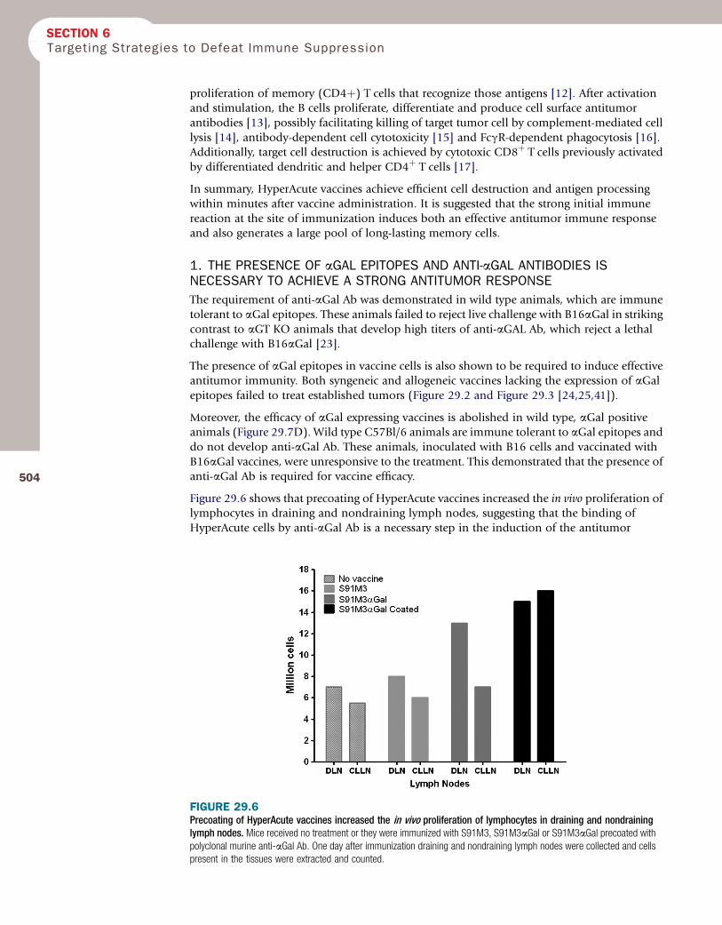
Figure 29.6 shows that precoating of HyperAcute vaccines increased the in vivo proliferation oflymphocytes in draining and nondraining lymph nodes, suggesting that the binding of
HyperAcute cells by anti-aGal Ab is a necessary step in the induction of the antitumor
FIGURE 29.6Precoating of HyperAcute vaccines increased the in vivo proliferation of lymphocytes in draining and nondraininglymph nodes. Mice received no treatment or they were immunized with S91M3, S91M3aGal or S91M3aGal precoated withpolyclonal murine anti-aGal Ab. One day after immunization draining and nondraining lymph nodes were collected and cellspresent in the tissues were extracted and counted.

CHAPTER 29HyperAcute Vaccines: A Novel Cancer Immunotherapy
response. This result is supported by Abdel-Motal et al. [42]. The in vivo formation of immunecomplexes with anti-aGal and the effective internalization of immune complexes by APC, via
Fc/FcgR interaction, resulted in effective activation of vaccine specific CD4þ and CD8þ T cells,
and high cellular and humoral immune response [42].
505
2. BINDING OF ANTI-aGAL AB TO aGAL EPITOPES INDUCES THE RECRUITMENTOF APC, NEUTROPHILS NK AND EOSINOPHILS, AND THE ACTIVATION OF THESECELLS PLAYS A ROLE IN ANTITUMOR IMMUNITY
Using aGal(þ) liposomes for wound healing it was demonstrated that macrophages andneutrophils are recruited and activated accelerating wound healing in aGT KO mice [43].
Similarly, intratumor inoculation of glycolipids induced the recruitment of APC and the
induction of antitumor immunity [44]. The role of aGal expressing glycolipids and NKTcells was evaluated in a double knockout animal lacking the expression of aGal epitopes
and CD1-restricted T cells (aGTCD1 DKO animals). NKT cells are first responder T cells
that serve as a bridge between the innate and adaptive immune system, recognizing lipidand glycolipid antigens in the context of the nonclassical class I MHC molecule CD1d.
NKT cells can become involved in infectious diseases and induce allergy, or mediate
a protective role in certain autoimmune diseases due to their suppressor activities,depending on their cytokine profile. In cancer, they can play opposite roles, contributing
to antitumor immunity or suppressing it [45e48]. Our experiments indicate that in
both the subcutaneous model and in the pulmonary model, NKT-cell ablation abolishesaGal-expressing vaccine efficacy (Figure 29.7 and Figure 29.8). These results demonstrate
that NKT cells play an important role in the induction of antitumor immunity induced
by HyperAcute vaccines. Further, many patients enrolled in a HyperAcute vaccine studyfor pancreatic cancer have shown increased eosinophils in peripheral blood counts
(Figure 29.9). Thus, in accordance with previous observations in the ovarian clinical trials,
increased activity of eosinophils may also be related to the biological activity of Hyper-Acute immunotherapy.
3. CELL INTEGRITY IS IMPORTANT FOR THE POTENCY OF HYPERACUTEIMMUNOTHERAPY
HyperAcute vaccines are irradiated whole tumor cell vaccines. The importance of cell
integrity for the potency of the vaccine was evaluated in animal models. The conventionalvaccine was prepared by rapidly thawing of cryopreserved and irradiated vaccine cells, with
aliquots of this preparation lysed by two cycles of rapid freezing and thawing using liquid
nitrogen. This preparation was shown to be 100% lysed (loss of cell integrity) by flowcytometry. Equal volumes of these two preparations were mixed to prepare a 50% lysed
vaccine.
Vaccine preparations were confirmed by flow cytometry for each of the three injections used
to treat animals with B16 melanoma tumors. Figure 29.10 shows that cell integrity is
important for vaccine efficacy (potency), since complete lysis of the whole cell componentsabrogated antitumor efficacy. As shown previously, administration of the conventional
S91M3aGal whole cell vaccine induces significant tumor retardation as previously
shown [25]. However, while vaccines that lost 100% of cell integrity (100% lysis) were unableto retard tumor growth, 50% lysed vaccine preparations induced tumor retardation similar to
the conventional whole cell vaccine (Figure 29.10A). Vaccines that lost cell integrity (100%
lysis) were also unable to increase the survival of animals bearing melanoma tumors. On thecontrary, vaccine cells that had only 50% viability were as efficient as the conventional whole
cell vaccines to increase survival (Figure 29.10B). In conclusion, these results establish that
the antitumor efficacy (potency) of HyperAcute vaccine preparations rely upon whole cellintegrity.

FIGURE 29.7NKT cells are required in the mechanism for HyperAcute vaccines efficacy in the subcutaneous tumor model. The efficacy of HyperAcute vaccines wascompared in 4 strains of mice. (A) aGT knockout (KO) animals; (B) aGTCD1 double knockout (DKO) animals; (C) CD1 KO animals; and (D) the wild type
C57Bl/6 mice. The number of animals in each group and treatment is indicated. Mice were injected with B16F0 and 4 days later they received either
no treatment or three doses of B16aGal(þ) HyperAcute vaccine. (A) Significant improved survival is observed in aGT KO receiving HyperAcute vaccines. The
lack of NKT cells in the aGTCD1 DKO abolished the efficacy of HyperAcute vaccines (B) as well as the expression of aGT in either CD1 KO and wild
type animals (C and D, respectively). Log-rank test p values are depicted.
SECTION 6Targeting Strategies to Defeat Immune Suppression
506
4. HYPERACUTE VACCINE INDUCES THE EXPANSION OF T CELLS WITHANTITUMOR ACTIVITY IN NON-SMALL LUNG CANCER PATIENTS
B16 melanoma-bearing mice vaccinated with aGal(þ) cells show greater T-cell proliferation,enhanced reactivity to melanoma peptides (as evidenced by intracellular TNFa staining) and
cytotoxic T lymphocytes (detected by IFN-g release and T-cell transfer experiments) than
animals vaccinated with aGal(�) cells. These characters are transferable by T cells from theseanimals and capable of mediating an antitumor response [24,25].
We extended these preclinical observations in clinical trials of a HyperAcute vaccine developed
for treatment of lung cancer. In the HyperAcute-Lung clinical study, 18 evaluable patientswere tested for the production of interferon-gamma (IFN-g) by peripheral blood lymphocytes
by ELISPOT. Notably, 11 out of 18 tested patients responded with 10-fold increased IFN-g afterimmunization. Notably, patients that responded with IFN-g exhibited significant increased
overall survival (Figure 29.11).
The human lung cancer cell lines used in this study to generate the HyperAcute-Lungvaccine were nonmodified parental (wild type) cell lines. However, in testing responses to this

FIGURE 29.8NKT cells are required in the mechanism for HyperAcutevaccines efficacy in the pulmonary metastatic tumormodel. The efficacy of HyperAcute vaccines was compared inaGT knockout (KO) animals and aGTCD1 double knockout
(DKO) animals. Mice were injected intravenously with B16FO
and subsequently they were vaccinated with of B16aGal(þ)
HyperAcute vaccines or received no vaccine. Lung pulmonary
burden was determined 4 weeks later. ANOVA and t test
p values are shown.
CHAPTER 29HyperAcute Vaccines: A Novel Cancer Immunotherapy
507
vaccine we also examined responses against a human lung adenocarcinoma cell line that was
not a component of the irradiated whole cell HyperAcute-Lung vaccine that has been used forimmunization of patients. Interestingly, high responder patients also developed reactivity to
this noncomponent cell line, illustrating the ability of the vaccine to generate cross-reactivity
to other lung cancer cell lines. This result was highly encouraging, because it supported the
FIGURE 29.9Increased eosinophils in patients receiving HyperAcute-Pancreas (Algenpantucel-L) vaccines. Eosinophils counts in patients enrolled in HyperAcute-Pancreas (Algenpantucel-L) clinical trial. Normal range is depicted.

FIGURE 29.10Cell integrity is required for potency of HyperAcute vaccine. Mice received a single subcutaneous injection with B16F0 and subsequently they were
vaccinated with conventional S91M3aGal HyperAcute vaccine or with preparation of vaccines that were 40e50% or 100% lyzed. (A) Tumor kinetics of all
groups; (B) Kaplan-Meir survival analysis. Lon-rank test p value is shown.
FIGURE 29.11IFN-gamma production by peripheral blood lymphocytes after vaccination correlated with better overall survivalin patients treated with HyperAcute-Lung. IFN-g production was measured by ELISPOT. The reactivity of vaccinated
patient’s effector PBMC cultured with tumor-loaded autologous DC pulsed with irradiated parental cell lines was measured
before and after immunization. Kaplan-Meier curve for the overall survival of responder and nonresponder patients in the IFN-gELISPOT assay. Responders were defined as those patients producing 10-fold or more increased IFN-g after vaccination
on half of the cell lines tested. The difference among responders and nonresponders is statistically significant (p< 0.0444).
SECTION 6Targeting Strategies to Defeat Immune Suppression
508
notion that HyperAcute immunotherapy can induce cross-reactivity to patients’ own tumorcells. In summary, these findings strongly suggested that HyperAcute immunotherapy can
activate lymphocytes to eradicate tumor cells in a manner that is sufficiently effective to
increase patient survival, providing a significant clinical benefit.
III. CLINICAL DEVELOPMENT OF HYPERACUTE IMMUNOTHERAPYA. Introduction
HyperAcute immunotherapies are composed of an irradiated mixture of human allogeneic
whole cancer cell lines that were genetically modified to add aGal residues to cell-surface lipidsand proteins. This preparation therefore constitutes an off-the-shelf immunotherapy for cancer
treatment. The aGal epitopes in this irradiated whole cell vaccine essentially function as
a molecular adjuvant, harnessing the mechanism responsible for hyperacute rejection ofxenotransplants. The immunostimulatory mechanism provided by the aGal is geared to break

CHAPTER 29HyperAcute Vaccines: A Novel Cancer Immunotherapy
509
tolerance and enable longer duration of antitumor effects, while avoiding the risk of immu-nosuppressive counter-response posed by certain other immunoregulatory agents (e.g.,
GM-CSF).
HyperAcute immunotherapies now have been tested in multiple Phase 2 trials for various
indications, including advanced non-small lung cancer, (Phase 1-2, n¼ 54), stage III/IV
melanoma (Phase 1-2, n¼ 31), prostate (Phase 1, n¼ 8), pancreas (Phase 1-2, n¼ 86).Currently, the HyperAcute Pancreas immunotherapy is being tested in a 1:1 randomized Phase
3 study for treatment of resected pancreatic cancer. In these studies, a broad range of responses
have been observed ranging from local to systemic skin reactions, objective tumor responsesand immunological responses. Below is a brief description of indications, safety and efficacy
data reported thus far for HyperAcute clinical trials.
B. Composition and indications
HyperAcute cancer immunotherapies are human tumor cell lines genetically modified to
express aGal epitopes. After genetic modification these cells are subsequently selected forexpression of aGal epitopes by fluorescence activated cell sorting. After growth HyperAcute
cells are irradiated and resuspended in a pharmacologically acceptable carrier that preserves
cell integrity during cryopreservation. The tumor cell lines used in the composition of vaccinesof a particular cancer type are derived from cancers of the same tissue type and selected based
on their expression profile of cell surface tumor-associated antigens.
HyperAcute immunotherapy is administered to human patients as a whole cell product. No
specific safety precautions are necessary because administered cells have been lethally irradi-
ated before freezing. Frozen vials of each vaccine cell line are carefully thawed, and equalamounts of each cell line component of the immunotherapy are injected into the patient
intradermally at several sites in each treatment session. Dose escalation studies determined
optimal dosing and scheduling for each indication for the treatment of solid tumors.
C. Safety data
The collective safety data from the four clinical studies, including a dose escalation study forthe treatment of lung cancer, indicates that HyperAcute Immunotherapy has a favorable safety
profile. No dose-limiting toxicities were reported and the maximum tolerated dose was not
reached [49e51]. The most frequently reported adverse events were injection site reactions.Most patients responded with self-resolving skin reactions that are localized at the site of
immunization. These skin manifestations vary from rapid-acute sensitization that occurs
within minutes to hours after injection to delayed type of reactions that occur within days to
(A) (B)
FIGURE 29.12Skin reactions frequently observed after HyperAcute immunotherapy. Picture (A) shows a commonly observed rapid-acute inflammatory reaction at thesite of immunization. Picture (B) shows a delayed-type hypersensitivity-like reaction that occurs days to weeks after immunization. These skin reactions
resolve without intervention in the vast majority of the cases reported.

SECTION 6Targeting Strategies to Defeat Immune Suppression
510
one week after immunization (Figure 29.12A, B respectively). Skin reactions associated withredness, swelling induration and itchiness are common adverse events attributed to the
HyperAcute immunotherapy.
Interestingly, skin reactions have been described where the vaccine injection site “flared up”
under random circumstances, i.e., during a common cold up to one year after immunization
was completed. Furthermore, later vaccinations also have been observed to produce flare-typeskin reactions at a previous vaccine site, frequently in the opposite limb. These observations are
intriguing since they strongly suggest the persistence of immunological effector cells that
respond upon recall at the initial vaccination site [52e56]. Further, these responses suggestthat HyperAcute immunotherapy can elicit immunological memory. Memory T cells in
nonlymphoid tissue, like skin, have been reported to enhance local and systemic immunity
during infection [52]. Thus, flare reactions at the original injection site in patients receivingHyperAcute immunotherapy might be explained by re-induction of long-lasting antitumor
memory cells. Notably, favorable clinical responses have been documented in some patients
experiencing these responses. Ongoing clinical trials might provide further information aboutthese intriguing observations.
D. Clinical responses
1. HYPERACUTE-PANCREAS (ALGENPANTUCEL-L [NLG0205])
After Phase I safety testingwas completed, a Phase II, single arm, open labelmulticentered study
was opened to enroll patients with newly diagnosed Stage 1 or 2 pancreatic carcinomawho hadundergone surgical tumor resection within 6 weeks prior to enrollment (NCT00569387).
Patients received a gemcitabine þ chemoradiation regimen plus the HyperAcute-Pancreas
(algenpantucel-L) immunotherapy for up to 14 intradermal immunizations.
The primary objective of this Phase II study was to assess disease-free survival (DFS), with DFS
at one year following initiation of treatment as the primary endpoint. Since eligiblepatients had no evidence of disease at the time of enrollment in the study, appearance of new
lesions constituted progression of disease. The one year DFS rate was calculated as the
proportion of patients who show no evidence of disease 12 months from the date of studyentry. This study enrolled 73 subjects with 69 evaluable patients.
This trial added HyperAcute-pancreas (algenpantucel-L) immunotherapy to the standard-of-
care treatment regimen defined in RTOG 9704(57) as adjuvant 5-FU chemoradiotherapy plusgemcitabine. It also provided an opportunity to perform a dose-finding analysis of cohorts
receiving biweekly algenpantucel-L injections of 100 million or 300 million cells. As of
September 1, 2010, all patients had reached at least 20 months of follow-up with a medianfollow-up period of 26.6 months for the 69 evaluated patents.
The Kaplan-Meier estimated overall survival in this Phase 2 clinical trial, referred to asNLG0205, compares favorably to data from RTOG 97-04, a trial used to help define the current
standard of care.
At 12 months after surgery overall survival for the combined 69 evaluable patient populationin NLG0205 is 86%. Patients treated at both dose levels in NLG0205 compare favorably to the
63% one-year overall survival calculated by the Memorial Sloan Kettering Cancer Center
nomogram for the NLG0205 patient population [58]. Furthermore, both dose levels inNLG0205 compare favorably to the 69% one-year overall survival observed for the 221
patients who received gemcitabine plus 5-FU based chemoradiotherapy in the RTOG 97-04
study [57] (Table 29.1).
The Kaplan-Meier estimate of median overall survival was 24.4 months for the combined
patient population in NLG0205 versus 16.6 months predicted by the Memorial SloanKettering Cancer Center nomogram [58], for NLG0205 patients based on their study entry

TABLE 29.1 Comparative Results for HyperAcute-Pancreas NLG0205 at One Year
Disease-Free Survival Overall Survival
Brennan et al., 2005 nomogram [58] Not Applicable 63%RTOG 97-04 (221 ptients) [57] <50% 69%NLG-0205-100 million cell dose group 52% 80%NLG-0205-300 million cell dose group 81% 96%
CHAPTER 29HyperAcute Vaccines: A Novel Cancer Immunotherapy
511
characteristics and 18.8 months for RTOG 97-04 patients (based on 221 patients receiving
gemcitabine plus 5-FU based chemoradiotherapy).
This encouraging data prompted the initiation of a multicentered phase 3 study currently
recruiting patients (NCT01072981).
2. HYPERACUTE-LUNG (NLG0101)
This study was a single-arm, open-label clinical trial conducted at the National Cancer Institute
for the treatment of refractory, recurrent or metastatic non-small lung cancer (NSCLC,NCT00073398). Its primary endpoint, besides safety, was to assess tumor response rate after
administration of HyperAcute-Lung, with a secondary endpoint to assess overall survival. For
the Phase 1 portion of this clinical trial, in addition to safety, a positive response includedstable disease for 16 weeks in patients who had enrolled after having previously shown
progressive disease. A total of 17 patients in the Phase 1 portion and 37 patients in the Phase 2
portion were injected with the HyperAcute-Lung immunotherapy. Of 37 patients in the Phase2 portion, only 28 were evaluated for clinical response. In the Phase 1 portion, four cohorts of
patients each received injections of 3 million, 10 million, 30 million, or 100 million cells every
four weeks for four doses, and one cohort of three patients received an initial dose of 500million cells, followed by injections of 300 million cells every two weeks for up to seven doses.
In the Phase 2 portion, the 28 patients evaluated received injections of 300 million cells every
two weeks for up to eight doses.
The Phase 1 portion of this clinical trial demonstrated a favorable safety profile, with no dose-
limiting toxicities at any of the five escalating dose levels. There have been no reported grade
four adverse events attributed to HyperAcute-Lung. The most common treatment-relatedadverse reactions for HyperAcute-Lung were injection site reaction [49].
The interim results of the Phase 2 clinical trial for HyperAcute Lung were encouraging. As of2011, results for the 28 patients evaluated in the Phase 2 clinical trial group showed a median
overall survival of 11.3 months, and a one-year survival rate of 46%.
Prior Phase 3 studies showed that in the refractory, recurrent or metastatic NSCLC setting(second line therapy), the median overall survival of patients receiving best supportive care
was 4.6 months and the median overall survival of patients receiving pemetrexed or docetaxel(Taxotere) therapy was approximately eight months [59,60]. Results from the Phase 2 study
indicated that HyperAcute-Lung immunotherapy compared favorably to current standard-of-
care cytotoxic chemotherapy. Table 29.2 summarizes comparative results for second-linetreatment in advanced NSCLC with pemetrexed, docetaxel and HyperAcute-Lung, including
safety data. HyperAcute-Lung immunotherapy produced an encouraging 11.3 month median
overall survival in the Phase 2 study, with a favorable safety profile, prompting an ongoingevaluation of alternative designs for a Phase 2B/3 clinical trial in NSCLC.
3. HYPERACUTE-MELANOMA IN COMBINATION WITH PEG-INTRON
The HyperAcute-Melanoma immunotherapy was combined in a collaborative investigator-initiated Phase 2 clinical study for the treatment of advanced melanoma. The study treatment

TABLE 29.2 Comparative Results for the Treatment of NSCLC with HyperAcute-Lung
Serious Adverse Events (CTC Grade 3 or 4)Attributed to Therapy
TherapyOverall Survival(months)
12 monthSurvival Nausea Fatigue Anemia Neutrope nia
Best Supportive Care [59] 4.6 11% – – – –Docetaxel [59] 7.5 37% 1.80% 5.40% 4.30% 40.20%Pemetrexed [60] 8.3 30% 2.60% 5.30% 4.20% 5.30%HyperAcute-Lung 11.3 46% 0% 0% 0% 0%
SECTION 6Targeting Strategies to Defeat Immune Suppression
512
consisted of 12 weekly injections of HyperAcute-Melanoma with PEG-Intron being co-
administered in weeks five through 12. This was the first instance of a HyperAcute immuno-therapy product tested in combination with another approved immunomodulator
(PEG-Intron). The primary objective of this clinical trial was to conduct correlative scientific
studies of patient tumor and peripheral blood samples to evaluate the mechanism of anyobserved anti-tumor effect involving the innate or cell-mediated host immune responses to
HyperAcute immunotherapy when combined with PEG-Intron. In this clinical trial, 25
patients were treated.
HyperAcute Melanoma demonstrated good tolerability and a favorable safety profile, with
no systemic, drug-related serious adverse events characterized by the investigators as possibly
or probably attributable to the vaccine. The most common nonserious adverse eventsreported were injection site reactions as before [51].
HyperAcute-Melanoma appeared to produce immunologic effects that are known to impactclinical outcomes. Vitiligo is an autoimmune condition implicated in clinical trials of other
immunotherapy products as correlated with a favorable response to therapy in melanoma
patients [61e63]. Interestingly, vitiligo was observed in 4 out of 25 (16%) patients andcorrelated with clinical response. Furthermore, all patients evaluated developed autoimmune
antibodies. Durable clinical responses were observed in this trial, encouraging evaluation of
further clinical trial designs using HyperAcute-Melanoma alone or in combination in newsettings.
E. Response evaluation in immunotherapy trials
Similar to what has been reported in other high-profile immunotherapy studies, multiplepatients showed either delayed tumor responses or long-term stable disease despite initial
progression by Response Evaluation Criteria in Solid Tumors (RECIST) criteria. In the
HyperAcute prostate study, one patient characterized as a nonresponder according to RECISTremained alive with a stable bone metastasis for over 70 months. Likewise, similar observa-
tions have been documented in HyperAcute lung, melanoma, and pancreas clinical studies.
Recently four different response patterns have been recognized in large immunotherapy trials:(a) shrinkage in baseline lesions, without new lesions; (b) durable stable disease (which in
some patients is followed by a slow, steady decline in total tumor burden); (c) response after
an initial increase in total tumor burden; and (d) response in the presence of new lesions. Allthese patterns were associated with favorable survival [64,65]. Overall, the clinical responses
patterns observed in the HyperAcute immunotherapy trials concur with the systematic criteria
for Immune-Related Response Criteria as a way to measure beneficial clinical responses inimmunotherapy trials [64,66,67].
Immunologic laboratory parameters have been evaluated in an attempt to find surrogatemarkers to evaluate efficacy in these trials. Some of these studies, briefly described below, seem

CHAPTER 29HyperAcute Vaccines: A Novel Cancer Immunotherapy
513
to suggest that immunological parameters as well as delayed and complex clinical tumorresponses might be a more accurate way to determine efficacy in immunotherapy trials.
F. Overview of immunological responses to hyperacute immunotherapy
The effects of HyperAcute immunotherapy on immunological parameters in patients on trialhave been assessed using a panel of in vitro assays. The measurement of immune responses
after immunization allows the association of immunogenicity with eventual clinical outcome.
Ideally, some of such measurements may potentially be used to monitor the immunotherapyand act as supporting or surrogate endpoints for efficacy immunotherapy clinical trials.
Analysis of immunological responses in patients vaccinated with HyperAcute immunotherapyin clinical trials of lung, prostate, melanoma and pancreatic cancer showed the following.
l Baseline levels of IgG anti-aGal antibodies were quite variable. The vast majority of testedpatients responded with elevated IgG anti-aGal antibody levels after immunization (2 to
over 100 fold), demonstrating a clear biological effect of HyperAcute immunotherapy.
l HyperAcute immunotherapy can elicit the production of antitumor antibodies (Ab) andcan overcome tolerance by inducing antibodies (IgG) recognizing peptides derived from
several proteins commonly overexpressed in prostate cancer.
l Anti-Mesothelin Ab anti-CEA Ab were increased post immunization in the HyperAcutePancreas study.
l Antitumor Ab were detected in the HyperAcute-Melanoma (anti-tyrosinase Ab) and
HyperAcute-Lung trials (anti-CEA Ab).l Importantly, the production of interferon gamma by T cells obtained from immunized
patients was associated with better survival in the HyperAcute-Lung study.
l Cross-priming to nonrelated lung cell lines was demonstrated in patients receivingHyperAcute-Lung immunotherapy.
l The majority of patients tested have elevated IL-5 as measured by ELISPOT after
immunization, suggesting that this cytokine may play a role in the biologic properties ofHyperAcute immunotherapy.
l In the HyperAcute Pancreas study a considerable number of patients develop eosinophilia
suggesting that these cells might play a role in the mechanism for this immunotherapyactivity.
In summary HyperAcute immunotherapy is a novel approach that shows signs of clinicalefficacy and immune activation in a number of trials and disease settings. The immunologic
manifestations of HyperAcute immunizations are multifaceted, as evidenced by the
provocative skin reactions and activation of both cellular and humoral arms of the immunesystem. Future clinical trials and combination trials will help elucidate further this novel
immunotherapy.
IV. CONCLUSIONSThe hyperacute rejection of a xenotransplant is characterized by a complement mediatedantibody immune response dependent on aGal epitopes. Based on these immunologic
reactions, we hypothesized that the hyperacute rejection mechanism could be exploited to
alter antigen processing resulting in a novel therapeutic approach to treat human malig-nancies. In animal models aGal epitopes expressed on genetically engineered allogeneic
tumor vaccines elicit a potent T-cell dependent antitumor immunity. In clinical studies of
patients with malignancies, the immunologic manifestations of HyperAcute immunizationare multifaceted with activation of both cellular and humoral arms of the immune system.
Ongoing studies aim to confirm the therapeutic benefit of this class of drug including
laboratory studies to identify mechanisms of action and surrogate markers of activity. Thisinnovative immunotherapy was designed to break immune tolerance of tumor and to

SECTION 6Targeting Strategies to Defeat Immune Suppression
514
stimulate effective antitumor immunity. The data obtained in ongoing clinical studiesindicates that HyperAcute immunotherapy can use a straightforward genetic engineering
approach to develop potent, cost-effective “off the shelf” human cancer vaccines with
a highly tolerable toxicity profile. Continuation and expansion of clinical studies, includingextension to new diseases and combinations with existing modalities, are indicated.
ACKNOWLEDGMENTSThe authors wish to acknowledge our colleagues at NewLink Genetics for their numerous
contributions to the design and execution of the studies reported here. We also wish torecognize the contributions made by the numerous investigators, their institutions, and the
patients participating in these clinical studies that contribute to our advancement in
knowledge.
References[1] Lee JJ, Jacobsen EA, McGarry MP, Schleimer RP, Lee NA. Eosinophils in health and disease: the LIAR
hypothesis. Clin Exp Allergy 2010 Apr;40(4):563e75.
[2] Link Jr CJ, Seregina T, Atchison R, Hall A, Muldoon R, Levy JP. Eliciting hyperacute xenograft response to treat
human cancer: alpha(1,3) galactosyltransferase gene therapy. Anticancer Res. 1998;18(4A):2301e8.
[3] Link Jr CJ, Seregina T, Levy JP, Martin M, Ackermann M, Moorman DW. Murine retroviral vector producer cellssurvival and toxicity in the dog liver. In Vivo 2000;14(5):643e9.
[4] Link CJ, Seregina T, Traynor A, Burt RK. Cellular suicide therapy of malignant disease. Stem Cells
2000;18(3):220e6.
[5] Link Jr CJ, Moorman D, Seregina T, Levy JP, Schabold KJ. A phase I trial of in vivo gene therapy with the herpes
simplex thymidine kinase/ganciclovir system for the treatment of refractory or recurrent ovarian cancer. HumGene Ther 1996;7(9):1161e79.
[6] Joziasse DH, Oriol R. Xenotransplantation: the importance of the Galalpha1,3Gal epitope in hyperacute
vascular rejection. Biochim Biophys Acta 1999;1455(2-3):403e18.
[7] Baumann BC, Forte P, Hawley RJ, Rieben R, Schneider MK, Seebach JD. Lack of galactose-alpha-1,3-galactose
expression on porcine endothelial cells prevents complement-induced lysis but not direct xenogeneic NK
cytotoxicity. J Immunol 2004 May 15;172(10):6460e7.
[8] Schaapherder AF, Daha MR, te Bulte MT, van der Woude FJ, Gooszen HG. Antibody-dependent cell-mediated
cytotoxicity against porcine endothelium induced by a majority of human sera. Transplantation 1994 May15;57(9):1376e82.
[9] Watier H, Guillaumin JM, Vallee I, Thibault G, Gruel Y, Lebranchu Y, et al. Human NK cell-mediated direct and
IgG-dependent cytotoxicity against xenogeneic porcine endothelial cells. Transpl Immunol 1996 Dec;4(4):293e9.
[10] Watier H, Guillaumin JM, Piller F, Lacord M, Thibault G, Lebranchu Y, et al. Removal of terminal alpha-
galactosyl residues from xenogeneic porcine endothelial cells. Decrease in complement-mediated cytotoxicity
but persistence of IgG1-mediated antibody-dependent cell-mediated cytotoxicity. Transplantation 1996 Jul15;62(1):105e13.
[11] Joziasse DH, Shaper JH, Jabs EW, Shaper NL. Characterization of an alpha 1e–3-galactosyltransferasehomologue on human chromosome 12 that is organized as a processed pseudogene. J Biol Chem 1991 Apr
15;266(11):6991e8.
[12] Larsen RD, Rivera-Marrero CA, Ernst LK, Cummings RD, Lowe JB. Frameshift and nonsense mutations ina human genomic sequence homologous to a murine UDP-Gal:beta-D-Gal(1,4)-D-GlcNAc alpha(1,3)-
galactosyltransferase cDNA. J Biol Chem 1990;265(12):7055e61.
[13] Galili U, Shohet SB, Kobrin E, Stults CL, Macher BA. Man, apes, and Old World monkeys differ from othermammals in the expression of alpha-galactosyl epitopes on nucleated cells. J Biol Chem
1988;263(33):17755e62.
[14] Posekany KJ, Pittman HK, Bradfield JF, Haisch CE, Verbanac KM. Induction of cytolytic anti-Gal antibodies in
alpha-1,3-galactosyltransferase gene knockout mice by oral inoculation with Escherichia coli O86:B7 bacteria.
Infect Immun 2002 Nov;70(11):6215e22.
[15] Galili U, Anaraki F, Thall A, Hill-Black C, Radic M. One percent of human circulating B lymphocytes are
capable of producing the natural anti-Gal antibody. Blood 1993 Oct 15;82(8):2485e93.
[16] Galili U, LaTemple DC. Natural anti-Gal antibody as a universal augmenter of autologous tumor vaccineimmunogenicity. Immunol Today 1997 Jun;18(6):281e5.

CHAPTER 29HyperAcute Vaccines: A Novel Cancer Immunotherapy
515
[17] Posekany KJ, Pittman HK, Swanson MS, Haisch CE, Verbanac KM. Suppression of Lewis lung tumor devel-opment in alpha 1,3 galactosyltransferase knock-out mice. Anticancer Res 2004 Mar-Apr;24(2B):605e12.
[18] Galili U. Autologous tumor vaccines processed to express alpha-gal epitopes: a practical approach to immu-
notherapy in cancer. Cancer Immunol Immunother 2004 Nov;53(11):935e45.
[19] Jager U, Takeuchi Y, Porter C. Induction of complement attack on human cells by Gal(alpha1,3)Gal
xenoantigen expression as a gene therapy approach to cancer. Gene Ther 1999 Jun;6(6):1073e83.
[20] Thall AD, Maly P, Lowe JB. Oocyte Gal alpha 1,3Gal epitopes implicated in sperm adhesion to the zona
pellucida glycoprotein ZP3 are not required for fertilization in the mouse. J Biol Chem
1995;270(37):21437e40.
[21] Thall AD, Murphy HS, Lowe JB. alpha 1,3-Galactosyltransferase-deficient mice produce naturally occurring
cytotoxic anti-Gal antibodies. Transplant Proc 1996;28(2):556e7.
[22] Unfer RC, Hellrung D, Link Jr CJ. Immunity to the alpha(1,3)galactosyl epitope provides protection in mice
challenged with colon cancer cells expressing alpha(1,3)galactosyl-transferase: a novel suicide gene for cancer
gene therapy. Cancer Res 2003 Mar 1;63(5):987e93.
[23] Rossi GR, Unfer RC, Seregina T, Link CJ. Complete protection against melanoma in absence of autoimmune
depigmentation after rejection of melanoma cells expressing alpha(1,3)galactosyl epitopes. Cancer Immunol
Immunother 2005 May;54:999e1009.
[24] RossiGR,MautinoMR,UnferRC, SereginaTM,VahanianN, LinkCJ. Effective treatmentof preexistingmelanoma
with whole cell vaccines expressing A(1,3)-galactosyl epitopes. Cancer Res 2005;65(22):10555e61. 2005.
[25] Rossi G, Mautino MR, Awwad D, Husske K, Lejukole H, Koenigsfeld M, et al. Allogeneic melanoma vaccine
expressing aGal epitopes induces antitumor immunity to autologous antigens in mice without signs of toxicity.
J Immunother 2008;31(6):545e54.
[26] Stager S, Alexander J, Kirby AC, Botto M, Rooijen NV, Smith DF, et al. Natural antibodies and complement are
endogenous adjuvants for vaccine-induced CD8þ T-cell responses. Nat Med 2003 Oct;9(10):1287e92.
[27] Galili U, Wigglesworth K, Abdel-Motal UM. Accelerated healing of skin burns by anti-Gal/alpha-gal liposomes
interaction. Burns 2009 Mar;36(2):239e51.
[28] Matzinger P. The danger model: a renewed sense of self. Science 2002 Apr 12;296(5566):301e5.
[29] Foell D, Wittkowski H, Vogl T, Roth J. S100 proteins expressed in phagocytes: a novel group of damage-
associated molecular pattern molecules. J Leukoc Biol. 2007 Jan;81(1):28e37.
[30] BianchiME. DAMPs, PAMPs and alarmins: all we need to know about danger. J Leukoc Biol 2007 Jan;81(1):1e5.
[31] Takeda K, Kaisho T, Akira S. Toll-like receptors. Annu Rev Immunol 2003;21:335e76.
[32] Akira S, Takeda K, Kaisho T. Toll-like receptors: critical proteins linking innate and acquired immunity.
Nat Immunol 2001 Aug;2(8):675e80.
[33] Lotze MT, Zeh HJ, Rubartelli A, Sparvero LJ, Amoscato AA, Washburn NR, et al. The grateful dead: damage-
associated molecular pattern molecules and reduction/oxidation regulate immunity. Immunol Rev 2007
Dec;220:60e81.
[34] Ellerman JE, Brown CK, de Vera M, Zeh HJ, Billiar T, Rubartelli A, et al. Masquerader: high mobility group box-
1 and cancer. Clin Cancer Res 2007 May 15;13(10):2836e48.
[35] Lotze MT, Deisseroth A, Rubartelli A. Damage associated molecular pattern molecules. Clin Immunol 2007
Jul;124(1):1e4.
[36] Honey K, Rudensky AY. Lysosomal cysteine proteases regulate antigen presentation. Nat Rev Immunol 2003Jun;3(6):472e82.
[37] Schuurhuis DH, Ioan-Facsinay A, Nagelkerken B, van Schip JJ, Sedlik C, Melief CJ, et al. Antigen-antibody
immune complexes empower dendritic cells to efficiently prime specific CD8þ CTL responses in vivo.J Immunol 2002 Mar 1;168(5):2240e6.
[38] Heath WR, Carbone FR. Cross-presentation, dendritic cells, tolerance and immunity. Annu Rev Immunol2001;19:47e64.
[39] Li M, Davey GM, Sutherland RM, Kurts C, Lew AM, Hirst C, et al. Cell-associated ovalbumin is cross-presented
much more efficiently than soluble ovalbumin in vivo. J Immunol 2001 May 15;166(10):6099e103.
[40] Rafiq K, Bergtold A, Clynes R. Immune complex-mediated antigen presentation induces tumor immunity.
J Clin Invest 2002 Jul;110(1):71e9.
[41] LaTemple DC, Abrams JT, Zhang SY, Galili U. Increased immunogenicity of tumor vaccines complexed with
anti-Gal: studies in knockout mice for alpha1,3galactosyltransferase. Cancer Res 1999;59(14):3417e23.
[42] Abdel-Motal UM, Wigglesworth K, Galili U. Mechanism for increased immunogenicity of vaccines that form in
vivo immune complexes with the natural anti-Gal antibody. Vaccine 2009 May 18;27(23):3072e82.
[43] Wigglesworth KM, Racki WJ, Mishra R, Szomolanyi-Tsuda E, Greiner DL, Galili U. Rapid recruitment andactivation of macrophages by anti-Gal/alpha-Gal liposome interaction accelerates wound healing. J Immunol
2011 Apr 1;186(7):4422e32.

SECTION 6Targeting Strategies to Defeat Immune Suppression
516
[44] Galili U, Wigglesworth K, Abdel-Motal UM. Intratumoral injection of alpha-gal glycolipids induces xenograft-like destruction and conversion of lesions into endogenous vaccines. J Immunol 2007 Apr 1;178(7):4676e87.
[45] Kinjo Y, Ueno K. iNKT cells in microbial immunity: recognition of microbial glycolipids. Microbiol Immunol
2011 Jul;55(7):472e82.
[46] Terabe M, Berzofsky JA. The role of NKT cells in tumor immunity. Adv Cancer Res 2008;101:277e348.
[47] Berzofsky JA, Terabe M. A novel immunoregulatory axis of NKT cell subsets regulating tumor immunity.Cancer Immunol Immunother 2008 Nov;57(11):1679e83.
[48] Berzofsky JA, Terabe M. The contrasting roles of NKT cells in tumor immunity. Curr Mol Med 2009Aug;9(6):667e72.
[49] Morris JC, Rossi GR, Vahanian NN, Janik JE, Tennant L, Ramsey WJ, et al. Phase I/II study of antitumor
vaccination using lung cancer cells expressing murine a(1,3)galactosyltransferase (aGT) in non-small cell lungcancer (NSCLC). AACR Annual Meeting abstract number 2010;2423.
[50] Hardacre JM, Mulcahy MF, Talamoni M, Obel JC, Rocha Lima CMS, Safran H, et al. Effect of hyperacute
immunotherapy in addition to standard adjuvant therapy for resected pancreatic cancer on disease-free andoverall survival: Preliminary analysis of phase II data. J Clin Oncol 2010;(Suppl. 4059):abstr.
[51] Riker AI, Alsfeld M, Harrison D, Foxworth Q, Lee G, Rossi R, et al. A phase II clinical trial of a novelcombinatorial antitumor immunotherapy for patients with high-risk resected stage III and metastatic mela-
noma. J Clin Oncol 2010;(Suppl. 4059):abstr.
[52] Gebhardt T, Wakim LM, Eidsmo L, Reading PC, Heath WR, Carbone FR. Memory T cells in nonlymphoidtissue that provide enhanced local immunity during infection with herpes simplex virus. Nat Immunol 2009
May;10(5):524e30.
[53] Wakim LM, Gebhardt T, Heath WR, Carbone FR. Cutting edge: local recall responses by memory T cells newlyrecruited to peripheral nonlymphoid tissues. J Immunol 2008 Nov 1;181(9):5837e41.
[54] Wakim LM, Waithman J, van Rooijen N, Heath WR, Carbone FR. Dendritic cell-induced memory T cellactivation in nonlymphoid tissues. Science 2008 Jan 11;319(5860):198e202.
[55] Obar JJ, Jellison ER, Sheridan BS, Blair DA, Pham QM, Zickovich JM, et al. Pathogen-induced inflammatory
environment controls effector and memory CD8þ T cell differentiation. J Immunol 2011 Nov15;187(10):4967e78.
[56] Sheridan BS, Lefrancois L. Regional and mucosal memory T cells. Nat Immunol 2011 Jun;12(6):485e91.
[57] Regine WF, Winter KA, Abrams RA, Safran H, Hoffman JP, Konski A, et al. Fluorouracil vs gemcitabine
chemotherapy before and after fluorouracil-based chemoradiation following resection of pancreatic adeno-
carcinoma: a randomized controlled trial. JAMA 2008 Mar 5;299(9):1019e26.
[58] Brennan MF, Kattan MW, Klimstra D, Conlon K. Prognostic nomogram for patients undergoing resection for
adenocarcinoma of the pancreas. Ann Surg 2004 Aug;240(2):293e8.
[59] Shepherd FA, Dancey J, Ramlau R, Mattson K, Gralla R, O’Rourke M, et al. Prospective randomized trial of
docetaxel versus best supportive care in patients with non-small-cell lung cancer previously treated with
platinum-based chemotherapy. J Clin Oncol 2000 May;18(10):2095e103.
[60] Hanna N, Shepherd FA, Fossella FV, Pereira JR, De Marinis F, von Pawel J, et al. Randomized phase III trial of
pemetrexed versus docetaxel in patients with non-small-cell lung cancer previously treated with chemotherapy.
J Clin Oncol 2004 May 1;22(9):1589e97.
[61] Byrne KT, Cote AL, Zhang P, Steinberg SM, Guo Y, Allie R, et al. Autoimmune melanocyte destruction is
required for robust CD8þ memory T cell responses to mouse melanoma. J Clin Invest 2011 May
2;121(5):1797e809.
[62] Quaglino P, Marenco F, Osella-Abate S, Cappello N, Ortoncelli M, Salomone B, et al. Vitiligo is an inde-
pendent favourable prognostic factor in stage III and IV metastatic melanoma patients: results from a single-
institution hospital-based observational cohort study. Ann Oncol 2009 Feb;21(2):409e14.
[63] Gogas H, Ioannovich J, Dafni U, Stavropoulou-Giokas C, Frangia K, Tsoutsos D, et al. Prognostic significance
of autoimmunity during treatment of melanoma with interferon. N Engl J Med 2006 Feb 16;354(7):709e18.
[64] Wolchok JD, Hoos A, O’Day S, Weber JS, Hamid O, Lebbe C, et al. Guidelines for the evaluation of immune
therapy activity in solid tumors: immune-related response criteria. Clin Cancer Res 2009 Dec
1;15(23):7412e20.
[65] Small EJ, Schellhammer PF, Higano CS, Redfern CH, Nemunaitis JJ, Valone FH, et al. Placebo-controlled phase
III trial of immunologic therapy with sipuleucel-T (APC8015) in patients with metastatic, asymptomatic
hormone refractory prostate cancer. J Clin Oncol 2006 Jul 1;24(19):3089e94.
[66] Hoos A, Eggermont AM, Janetzki S, Hodi FS, Ibrahim R, Anderson A, et al. Improved endpoints for cancer
immunotherapy trials. J Natl Cancer Inst 2010 Sep 22;102(18):1388e97.
[67] FDA, S. USDHH. Guidance for Industry. Clinical Considerations for Therapeutic Cancer Vaccines 2009.


![mRNA vaccine for cancer immunotherapy...vaccines, viral vector-based vaccines, and nucleic acid-based vaccines [7]. Nucleic acid (DNA- or RNA-) based vaccine is a promising vaccine](https://static.fdocuments.in/doc/165x107/6121f11dd9483105bb0a6c2f/mrna-vaccine-for-cancer-immunotherapy-vaccines-viral-vector-based-vaccines.jpg)


