Cancer and cell biology research concepts
18
-
Upload
angela-alexander -
Category
Education
-
view
689 -
download
0
Transcript of Cancer and cell biology research concepts

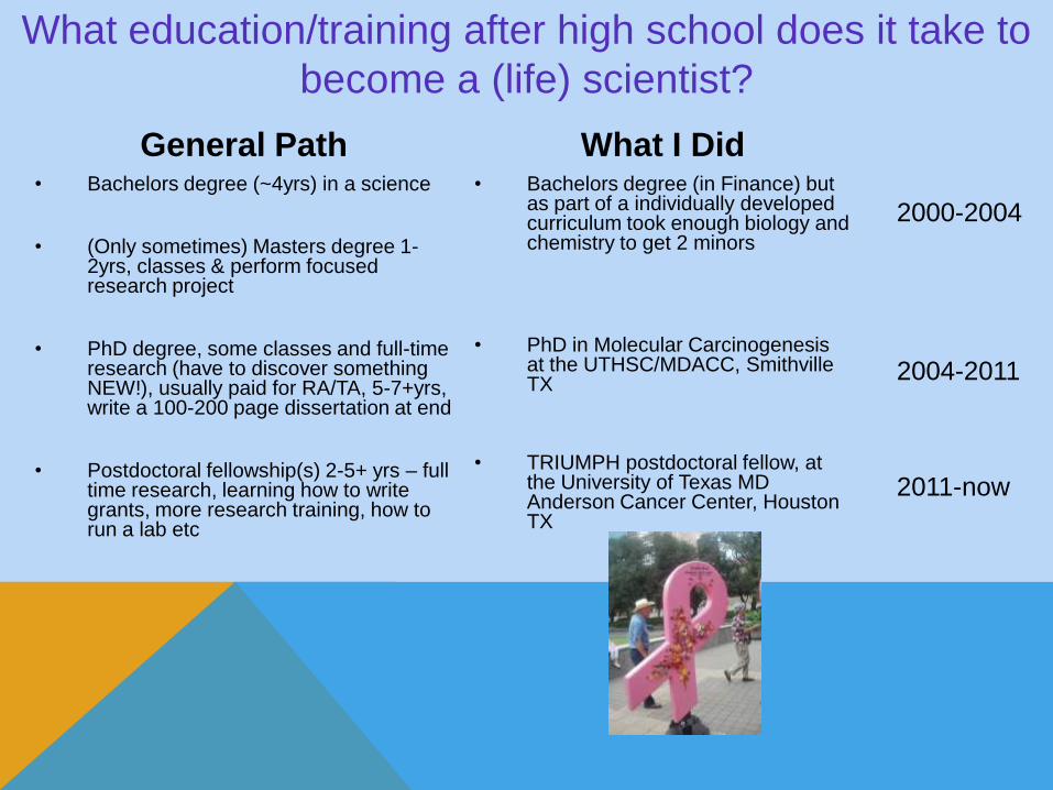
General Path• Bachelors degree (~4yrs) in a science
• (Only sometimes) Masters degree 1-2yrs, classes & perform focused research project
• PhD degree, some classes and full-time research (have to discover something NEW!), usually paid for RA/TA, 5-7+yrs, write a 100-200 page dissertation at end
• Postdoctoral fellowship(s) 2-5+ yrs – full time research, learning how to write grants, more research training, how to run a lab etc
What I Did• Bachelors degree (in Finance) but
as part of a individually developed curriculum took enough biology and chemistry to get 2 minors
• PhD in Molecular Carcinogenesis at the UTHSC/MDACC, Smithville TX
• TRIUMPH postdoctoral fellow, at the University of Texas MD Anderson Cancer Center, Houston TX
What education/training after high school does it take to
become a (life) scientist?
2000-2004
2004-2011
2011-now
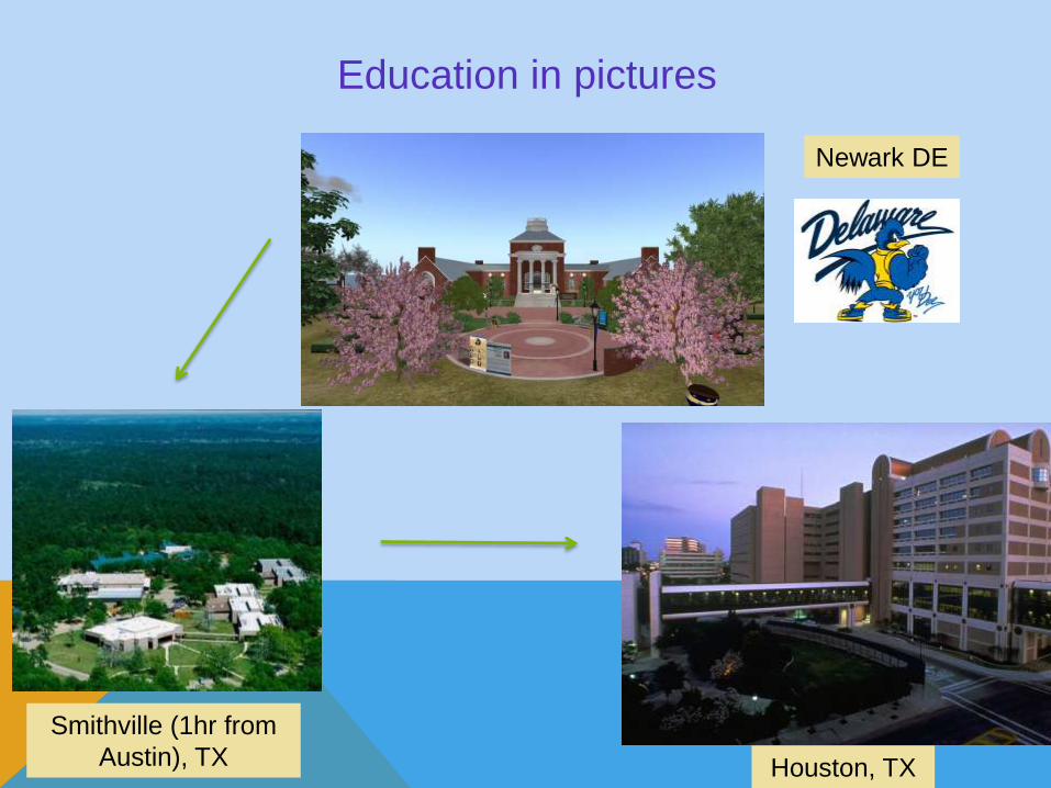
Education in pictures
Smithville (1hr from
Austin), TX Houston, TX
Newark DE
Cancer – the disease which affects nearly 2M
Americans, and causes >500K deaths/year
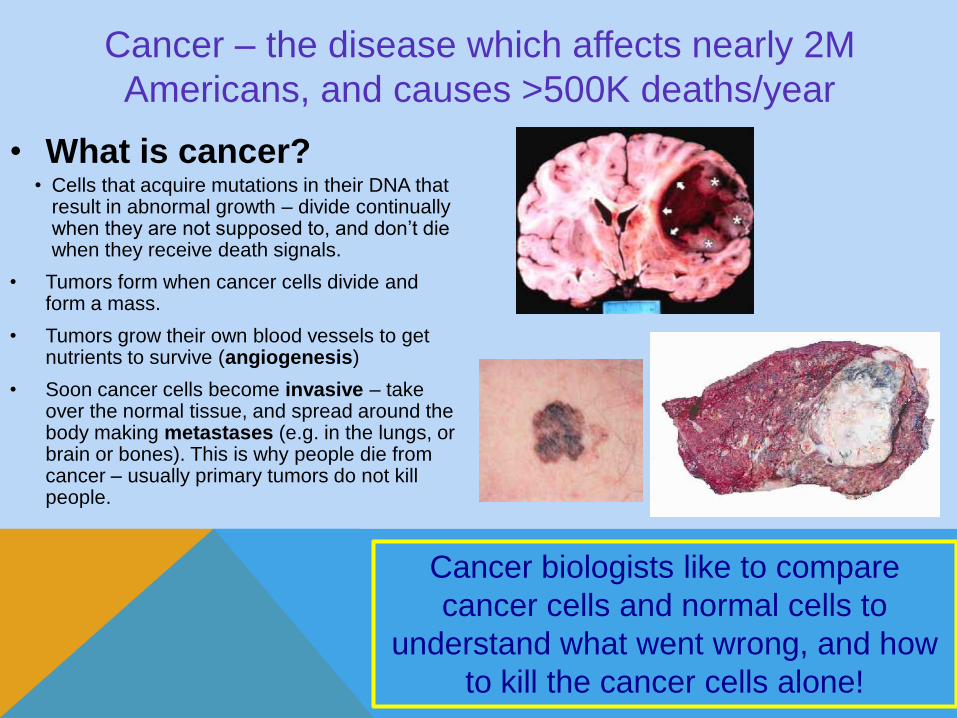
• What is cancer?• Cells that acquire mutations in their DNA that
result in abnormal growth – divide continually when they are not supposed to, and don’t die when they receive death signals.
• Tumors form when cancer cells divide and form a mass.
• Tumors grow their own blood vessels to get nutrients to survive (angiogenesis)
• Soon cancer cells become invasive – take over the normal tissue, and spread around the body making metastases (e.g. in the lungs, or brain or bones). This is why people die from cancer – usually primary tumors do not kill people.
Cancer biologists like to compare
cancer cells and normal cells to
understand what went wrong, and how
to kill the cancer cells alone!

Tools of the trade – examples of different breast cancer cells taken
from human patients growing on plastic plates in lab• Cells can be
cultured from
humans, mice,
other rodents,
insects etc….
• We can culture
cancer cells
indefinitely in
solutions of
glucose and
amino acids (to
make proteins)!
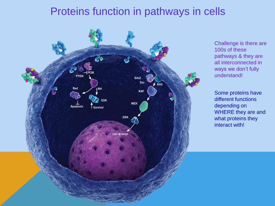
Proteins function in pathways in cells
Challenge is there are
100s of these
pathways & they are
all interconnected in
ways we don’t fully
understand!
Some proteins have
different functions
depending on
WHERE they are and
what proteins they
interact with!

PhD Thesis: “ATM signaling to TSC2: Mechanisms and
Implications for Cancer Therapy”
Key questions:
• How do cells (including cancer cells) detect and respond to DNA and oxidative damage?
• Are these protein pathways linked to cell survival or death?
• Can we target these survival pathways to improve cancer therapy (chemotherapy, radiation, big surgeries)?
Methods overview:
• Study protein localization in the cell and their function (for example whether they are active or not) using fluorescence microscopy and cell fractionation
• Used genetic approaches to study whether a process depends on a single protein – used cells that either lack the protein, or used a technique called RNA interference to reduce the amount of the protein I was interested in.
• Used drugs to inhibit pathways – such as protein export from the nucleus
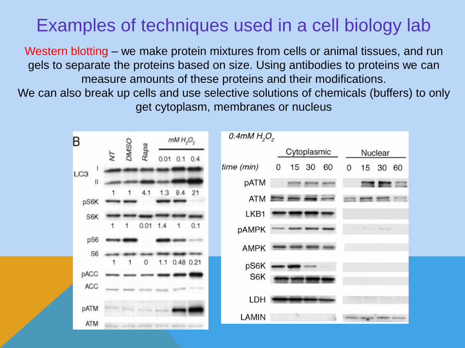
Examples of techniques used in a cell biology lab
Western blotting – we make protein mixtures from cells or animal tissues, and run
gels to separate the proteins based on size. Using antibodies to proteins we can
measure amounts of these proteins and their modifications.
We can also break up cells and use selective solutions of chemicals (buffers) to only
get cytoplasm, membranes or nucleus

Techniques in a cell biology lab (cont.)
Fluorescence and confocal microscopy – use this to observe
protein localization (what organelles are important?)
Supplementary Figure S3
Flag-TSC2
PEX5IP: PEX5
IgG WT RQ RW RG
e
a
HEK 293 cells
IP: Myc
Input
Flag-TSC2
Flag-TSC2
Myc-TSC1
Myc-TSC1
IgG WT RQ RG RW
L1624P
G294E
HEK 293 cells
IP: Flag
PEX5 (light)
Flag-TSC2
IgG WT RQ RW RG
PEX5 (dark)
HEK 293 cellsb
c
WT
PMP70 Merge/DAPI
RQ
PMP70 Merge/DAPI
RG
PMP70 Merge/DAPI
RW
PMP70 Merge/DAPI
HeL
a c
ells
Flag
Flag
Flag
Flag
d
Calnexin Merge/DAPI
RQ
RQ
HeL
a c
ells
Flag
VDAC Merge/DAPIFlag

More cell biology techniques
Electron microscopy – observe structural
information inside cells without using
antibodies to detect individual proteins
Animal/human tissue studies
– immunohistochemistry
staining for proteins using
antibodies then chemical
reactions that generate
brown color
Major findings of my PhD work (team project)
• Key discovery – a very important protein (called ATM) plays different
functions depending on where it is localized in the cell
• Before my work, most people thought it mainly functioned to
sense DNA damage in the nucleus, and recruit other proteins to
REPAIR the damage or trigger cell death if too much damage.
• I showed convincingly that a separate pool of this protein that
didn’t have to leave the nucleus could respond to a oxidative
damage and trigger autophagy
• Autophagy is a recycling process cells use to degrade damaged
organelles and certain proteins
• Often used by cancer cells to survive stresses such as
chemotherapy and radiation.
• Understanding how and when this applies can help us design
ways to block cancer cell survival.


Cancer and the cell cycle
• Cell cycle deregulation is virtually
universal feature in cancer
• Cancer cells both lose the
“brakes” (eg Rb and p53 tumor
suppressor genes) & often have
a “stuck gas pedal”
Key question – how can we target
this defect to selectively kill cancer
cells?
Once we do it in cells and mice, can
we design a human clinical trial…
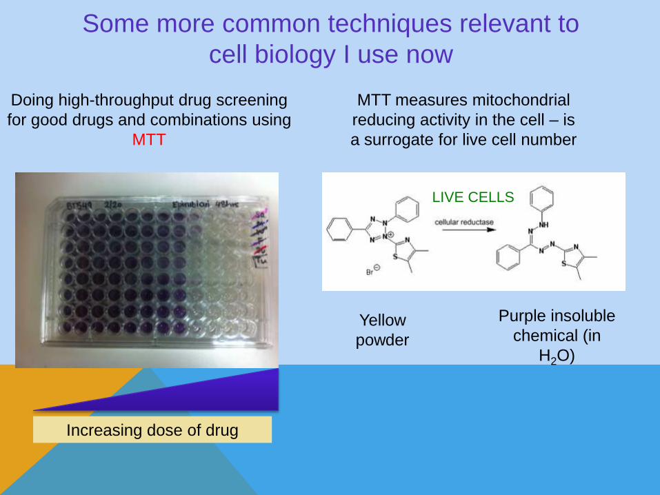
Some more common techniques relevant to
cell biology I use now
Doing high-throughput drug screening
for good drugs and combinations using
MTT
MTT measures mitochondrial
reducing activity in the cell – is
a surrogate for live cell number
Yellow
powder
Purple insoluble
chemical (in
H2O)
Increasing dose of drug
LIVE CELLS
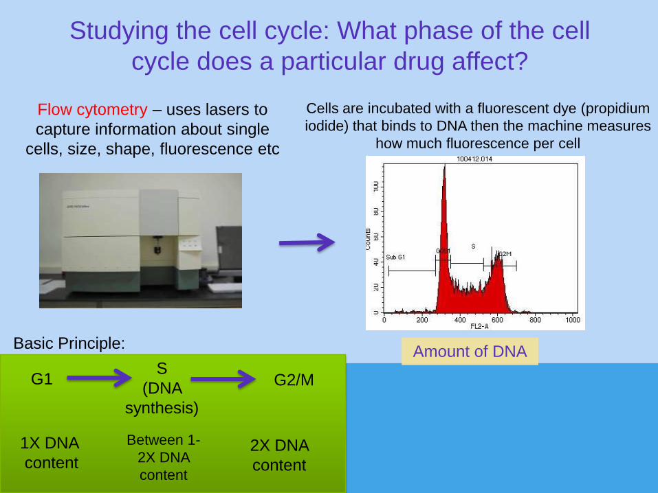
Studying the cell cycle: What phase of the cell
cycle does a particular drug affect?
Flow cytometry – uses lasers to
capture information about single
cells, size, shape, fluorescence etc
Amount of DNA
Cells are incubated with a fluorescent dye (propidium
iodide) that binds to DNA then the machine measures
how much fluorescence per cell
Basic Principle:
G1S
(DNA
synthesis)
G2/M
2X DNA
content
1X DNA
content
Between 1-
2X DNA
content
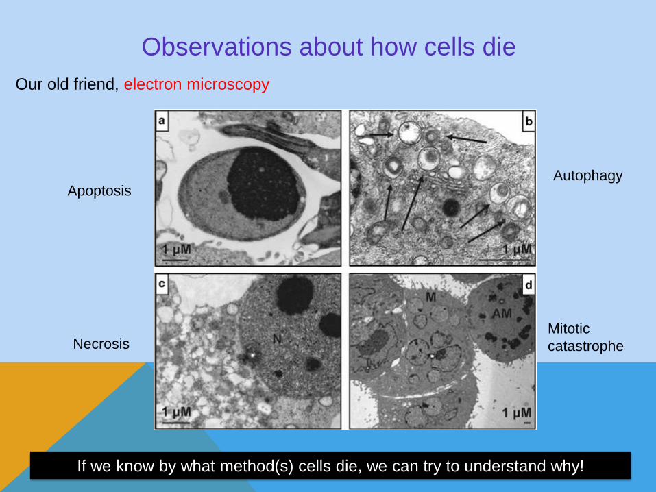
Observations about how cells die
Autophagy
Mitotic
catastrophe
Apoptosis
Necrosis
Our old friend, electron microscopy
If we know by what method(s) cells die, we can try to understand why!
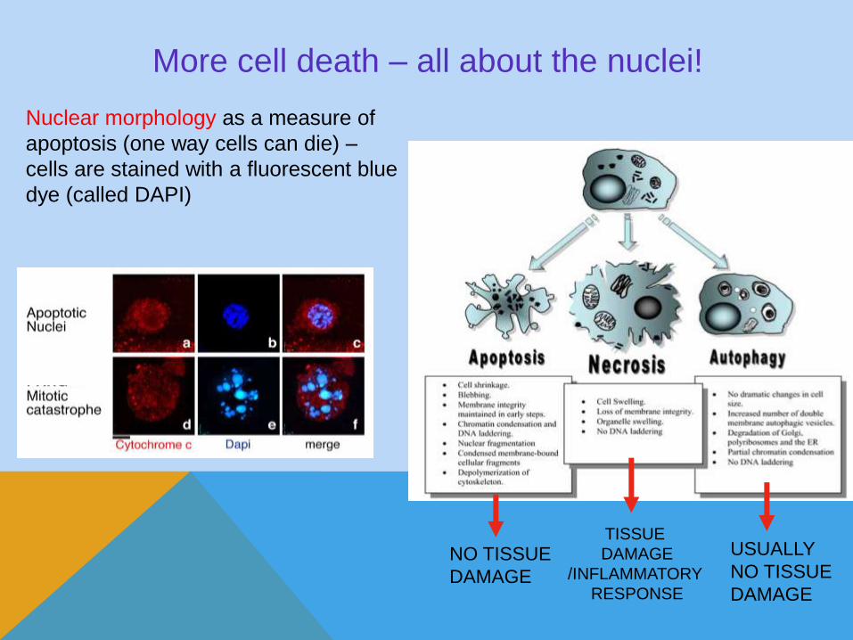
More cell death – all about the nuclei!
Nuclear morphology as a measure of
apoptosis (one way cells can die) –
cells are stained with a fluorescent blue
dye (called DAPI)
TISSUE
DAMAGE
/INFLAMMATORY
RESPONSE
NO TISSUE
DAMAGE
USUALLY
NO TISSUE
DAMAGE