Canaadian Jtouhrnal ofolog y Volume 6, Issue 4 Winter...
48
Canadian Journal of Pathology Publications Agreement Number 40025049 • ISSN 1918-915X Official Publication of the Canadian Association of Pathologists www.cap-acp.org Volume 6, Issue 4 • Winter 2014 O fficia l Pu b l ica t ion of t h e Canadian Ass o cia t ion o f P a t hol o gis t s Challenging Areas in GI Biopsy Diagnosis Hospital Autopsy Quality Assurance
Transcript of Canaadian Jtouhrnal ofolog y Volume 6, Issue 4 Winter...

Canadian Journal of
Pathology
Publications Agreement Number 40025049 • ISSN 1918-915X
Official Publication of the Canadian Association of Pathologists
www.cap-acp.org
Volume 6, Issue 4 • Winter 2014
OOfficiafficiall PuPubbllicaicattionion ofof tthhee CanadianCanadian AssAssoociaciattionion ooff PPaattholholoogisgisttss
Challenging Areas in GI Biopsy Diagnosis
Hospital Autopsy Quality Assurance

References: 1. Liverani CA, et al. Am J Transl Res 2012; 4(4): 452-457
Cervical cancer screening has evolvedThe Aptima® HPV assay is now available on the Panther® system. The Aptima® HPV assay detects HPV mRNA. Studies show HPV mRNA is indicative of infections that may lead to disease.1
With the Panther® system, labs of all sizes can leverage the power of a highly flexible, fully automated test platform, along with the most comprehensive women’s health menu. In addition to HPV, laboratories can obtain results for CT/GC and Trichomonas vaginalis, all from a 1-ml ThinPrep® Pap test sample.
ADS-001016 ©2014 Hologic, Inc. All rights reserved. Hologic, Aptima, Panther, ThinPrep and associated logos are trademarks of Hologic, Inc. and/or its subsidiaries in the U.S. and/or other countries. For information on specific products available for sale in a particular country, please contact your local Hologic representative or email [email protected].
CERVICAL CANCER SCREENING
Canadian Immunohistochemistry Quality Control (cIQc) Symposium
May 29-30, 2015 British Columbia Cancer Research Centre
Vancouver, BC
Immunohistochemistry &
Cancer Genetics
Go to www.ciqc.ca for more details
Interior Health has a number of exci ng opportuni es for Pathologists in the communi es of Cranbrook, Trail, Vernon, Pen cton, and Kelowna.
The Southern Interior of BC is a year-round paradise for nature lovers, adventurers, and recrea onalists. The uniquely varied landscape and moderate, sunny climate provide the perfect se ng for fi rst class golfi ng, water sports, hiking, climbing, and world-renowned skiing and snowboarding. Where else in the world can you go skiing, play a round of golf, mountain bike, and visit a winery all in one day?
If you are a talented pathologist and you are planning to live and work in BC’s Southern Interior then go to our Be er Here website and check out our ads!
For more informa on:
visit our website www.be erhere.ca or
email [email protected]
PathologistCranbrook, Trail, Vernon, Penticton, & Kelowna

Return undeliverable Canadian Addresses to:
115 King Street West, Suite 220, Dundas, ON L9H 1V1
For Instructions to Authors, please visit http://www.andrewjohnpublishing.com/CJP/instructionstoauthors.html
Canadian Journal of P athology 103Winter 2014
Editorial/ÉditorialManon Auger, MD FRCP(C)
Original Articles Hospital Autopsy Quality Control and Assurance: The LondonHealth Sciences Centre Experience Rebekah Jacques, MD, Michael J Shkrum, MD, FRCPC
Recommendations on Quality Assurance Practices Stemming fromthe Findings of an Audit of Randomly Selected Negative BreastCore BiopsiesEsther Ravinsky, MD, CM, FRCP(C), John G. Gartner, MD,CM, FRCP(C), Dan Chateau, PhD
Diagnosis of Pulmonary Echinococcosis by BronchoalveolarLavage CytologyFarshid Razaghi, MD, FRCPC, Kristin Y. Popiel, MD, Ken M Flegel, MD, CM, MSc,Richard Fraser, MD, CM, Manon Auger, MD, CM, FRCPC
Retrospective Comparison of Institutional Cytological Accuracy inWarthin Tumour DiagnosisAshley Stueck, MD, S. Mark Taylor, MD, FRCSC, Robert D. Hart, MD, FRCSC, Jonathan R. Trites, MD, FRCSC, Martin J. Bullock, MD, FRCPC
Current ReviewTen Challenging Areas in Gastrointestinal Biopsy DiagnosisJeremy R. Parfitt, MD, FRCPC, and David K. Driman, MBChB, FRCPC
Book ReviewAccurate Results in the Clinical Laboratory: A Guide to ErrorDetection and Correction
Professional Development/Employment OpportunitiesHologicCanadian Immunohistochemistry Quality Control SymposiumInterior Health
Canadian Journal of Pathology • Volume 6, Issue 4, 2014
Canadian Journal of Pathology is published four times0 annually byAndrew John Publishing Inc., with offices at 115 King Street West,Dundas, ON, Canada L9H 1V1.
We welcome editorial submissions but cannot assume responsibility orcommitment for unsolicited material. Any editorial material, includingphotographs that are accepted from an unsolicited contributor, willbecome the property of Andrew John Publishing Inc.
FEEDBACKWe welcome your views and comments. Please send them to AndrewJohn Publishing Inc., 115 King Street West, Dundas, ON, Canada L9H 1V1.
Copyright 2014 by Andrew John Publishing Inc. All rights reserved.Reprinting in part or in whole is forbidden without express writtenconsent from the publisher.
Publications Agreement Number 40025049 • ISSN 1918-915X
EDITOR-IN-CHIEFJ. Godfrey Heathcote, MA, MB BChir, PhD, FRCPC
EDITORIAL BOARDManon Auger, MD, FRCPC, Cytopathology;
Calvino Cheng, BSc, MD, FRCPC, Pathology Informaticsand Quality Management;
Pierre Douville, MD, FRCPC, Medical Biochemistry;David K. Driman, MB ChB, FRCPC, Anatomical Pathology;
Lawrence Haley, MD, FRCPC, Hematopathology;Todd F. Hatchette, BSc, MD, FRCPC, Medical Microbiology;
Michael J. Shkrum, MD, FRCPC, Forensic Pathology;
FOUNDING EDITORJagdish Butany, MBBS, MS, FRCPC
MANAGING EDITORPaula Mucci
COPY EDITORMichael Peebles
PROOFRE ADERScott Bryant
ART DIRECTORAndrea Mulholland, [email protected]
TRAN SL ATORMarie Dumont
SALES AND CIRCUL ATION COORDINATORBrenda Robinson, [email protected]
ACCOUNTINGSusan McClung
GROUP PUBLI SHERJohn D. Birkby, [email protected]
VOLUME 6, ISSUE 4, 2014
Contents
Echinococcal scolices with hooklets in a bronchoalveolarlavage specimen.
About the Cover

Winter 2014104 Canadian Journal of P athology
INVITED EDITORIAL
Rapid On-Site Evaluation (ROSE) is a procedure by which
a cytopathologist and/or a cytotechnologist provide on-
site assessment of fine needle aspiration biopsies (FNAs)
performed by other physicians. These physicians have
traditionally been radiologists, but now include respirologists
and gastroenterologists among others. The main purpose of
ROSE is to ensure that the FNA specimen is adequate for
diagnosis and therefore to guide the operator to continue to
do more passes if the material is deemed insufficient. Another
important aim is to triage the specimen for various ancillary
studies (e.g., flow cytometry, immunocytochemistry or
microbiology) if necessary to ultimately reach an accurate
definitive diagnosis.
Many articles have touted the advantages of ROSE, most
notably an improved diagnostic yield of the FNA specimens
obtained.1-3 However, others have failed to show a benefit of
ROSE, probably depending on a variety of factors, including
the body site targeted and the skills of the operator
performing the FNA.4,5 Probably the biggest disadvantage of
ROSE is that it is very time-consuming for the
cytotechnologist and/or cytopathologist involved in the
procedure. For institutions in Canada where limited budgets
have resulted in reduced numbers of both cytotechnologists
and pathologists, this extra task can be difficult to
accommodate. The problem is compounded by the
expanding trend of hospital mergers resulting in
cytopathology laboratories often providing service for several
physical sites that may be far apart. Where various FNA
procedures may require ROSE, this adds signicant travelling
time for the cytotechnologist or cytopathologist covering the
ROSE service. In teaching institutions where cytopathology
fellowships exist, fellows can provide welcome help; another
option is telecytopathology. These options are, however, not
widely available, even in most large institutions.
With the increasing popularity and the more widespread use
of endobronchial ultrasound-guided FNA (EBUS-FNA) for
the staging of lung cancer and of endoscopic ultrasound-
guided FNA (EUS-FNA) for sampling lesions in various
abdominal organs, most notably pancreas, cytopathology
laboratories are under the pressure of a rapidly increasing
demand for ROSE. It is always difficult to say “no” to a clinical
request, but medical directors of cytopathology laboratories
may have to do so or say “yes” to only a portion of the
requests, depending on available resources. Directors of
cytopathology laboratories should use their influence on
hospital administrators to request additional resources to
meet new demands such as ROSE. However, in the current
economic climate of the health care system in Canada, this is
a daunting task, at least in some provinces. This situation
could be the spark that sets off a turf war for ROSE, spreading
eventually to Canada from other countries where it has
already started, such as Italy and Japan. Respirologists,
radiologists, and gastroenterologists, frustrated at having been
denied ROSE by their cytopathology laboratories, have
decided to take matters into their own hands (or eyes!). For
example, there are reports of ultrasonographers performing
ROSE themselves on their EUS-guided FNA of solid masses
of the pancreas, leading to a significant increase of 21% in
diagnostic accuracy on final diagnoses.6 In another report,
respirologists, who had received only basic training in
cytopathology, performed ROSE on transbronchial FNA, and
reached an 81% overall agreement with their cytopathologist
colleagues.7 Is this turf war looming for Canada? Will
cytopathologists be willing to give training to non-laboratory
physicians to cover the territory that has traditionally been
theirs? There is certainly a precedent in the opposite
direction. Indeed, at least in the USA, many cytopathologists,
sometimes after attending only a short course in ultrasound
technique, are performing FNAs of superfical body sites, in
particular of the thyroid, under ultrasound guidance, basically
invading what has traditionally been a radiologist’s terrain.
Although ideally cytopathology teams should do their best to
keep ROSE in their own purview, each institution will have
to address the requests for ROSE in their own way, depending
on human resources, the physical layout of their institution
and the philosophy of their department.
Manon Auger, MD FRCP(C), Department of Pathology, McGill University, Montreal, QC, CanadaSection Editor, Cytopathology
Rapid On-Site Evaluation (ROSE): Is a Turf War Looming in Canada?
Competing interests: None declared.
References on Page 106

Canadian Journal of P athology 105Winter 2014
ÉDITORIAL
Conflits d’intérêts : aucun
L’évaluation rapide sur place désigne l’évaluation sur placedu spécimen prélevé à la cytoponction par un médecin,
effectuée par un pathologiste spécialisé en cytopathologie ouun cytotechnologiste. Par le passé, le médecin effectuant lacytoponction était en général un radiologiste, maisaujourd’hui ce peut être un pneumologue ou ungastroentérologue, entre autres. L’évaluation rapide sur placea pour but principal de déterminer si le spécimen prélevé à lacytoponction est optimal en vue du diagnostic et, sinon, deguider l’exécutant dans la poursuite de la cytoponction afinde prélever suffisamment de matériel. L’évaluation rapide surplace a également pour but important le triage du spécimenen prévision de divers examens auxiliaires (cytométrie en flux,immunohistochimie ou microbiologie, par exemple)nécessaires le cas échéant pour établir un diagnostic définitifexact. De nombreux articles font valoir les avantages de l’évaluationrapide sur place, le plus notable étant l’amélioration durendement diagnostique des spécimens prélevés à lacytoponction1-3. D’autres cependant ne sont pas convaincus,en raison sans doute de divers facteurs, dont la région ducorps et les aptitudes pratiques de la personne effectuant lacytoponction4,5. Le plus grand désavantage de l’évaluationrapide sur place est sans doute le temps que doit y mettre lecytotechnologiste ou le pathologiste spécialisé. Dans lesétablissements de santé du Canada où les compressionsbudgétaires ont entraîné la réduction du nombre decytotechnologistes et de pathologistes, il est difficile de prévoirdes ressources pour cette tâche chronophage. Le problème secomplique au vu de la tendance croissante des fusionshospitalières alors que le laboratoire de cytopathologie doitoffrir ses services dans plusieurs établissements qui peuventêtre éloignés les uns des autres. Comme diverses biopsies parcytoponction peuvent nécessiter une évaluation rapide surplace, le cytotechnologiste ou le pathologiste spécialisé offrantle service consacrera beaucoup de temps à se déplacer,ajoutant ainsi à la durée de la tâche. Dans les établissementsd’enseignement accueillant des stagiaires en cytopathologie,ces stagiaires peuvent apporter leur concours à l’exécution decette tâche; une autre option serait de recourir à latélécytopathologie. Toutefois, ces deux options ne sont pasrépandues, même dans les grands établissements de santé.Dans le contexte de la popularité grandissante et del’utilisation de plus en plus répandue de la cytoponction
transbronchique sous guidage échographiqueendobronchique dans la stadification du cancer du poumonet de la cytoponction sous écho-endoscopie pour obtenir desprélèvements de lésions dans des organes abdominaux,surtout au pancréas, les laboratoires de cytopathologiecroulent sous la demande d’évaluation rapide sur place enforte expansion. Il est toujours difficile de répondre par lanégative à une demande de service clinique, mais il se peutque les médecins à la tête des laboratoires de cytopathologien’aient pas d’autre choix ou alors ne pourront qu’accéder àcertaines de ces demandes, en raison des ressourcesdisponibles. Les directeurs de laboratoire devraient user deleur influence auprès des administrateurs de l’hôpital afind’obtenir les ressources supplémentaires nécessaires pourrépondre aux nouvelles demandes, notamment cellesd’évaluation rapide sur place. Mais, voilà qui est loin d’êtrechose aisée, dans certaines provinces du moins, dans laconjoncture du système de santé au Canada. Et ce pourraitêtre l’étincelle qui mettra le feu aux poudres et déclencheraune guerre intestine à propos de cette intervention, une guerrequi se propagerait au Canada en provenance d’autres pays oùelle fait rage déjà, comme en Italie et au Japon. Despneumologues, des radiologistes et des gastroentérologuesfrustrés de se voir refuser l’évaluation rapide sur place par leurlaboratoire de cytopathologie ont pris les choses en main (ouy voient eux-mêmes!). Ainsi, des échographistes effectuentl’évaluation rapide sur place des spécimens prélevés à lacytoponction sous écho-endoscopie de tumeur solide aupancréas, ce qui a amené une hausse notable de 21 % del’exactitude du diagnostic définitif6. Selon un autre rapport,des pneumologues avec une formation élémentaire encytopathologie ont obtenu un taux de concordance globalede 81 % avec leurs collègues pathologistes dans leur propreévaluation rapide sur place de spécimens prélevés à lacytoponction transbronchique7. Assisterons-nous à une telleguerre intestine au Canada? Les pathologistes spécialisésseront-ils disposés à former des médecins pour effectuer unetâche qui a toujours été de leur ressort? Il se pourrait que lasolution soit tout autre à l’instar de ce qui se fait aux États-Unis notamment où de nombreux pathologistes spécialisés,après une formation brève en échographie, effectuent descytoponctions échoguidées à des sites superficiels, enparticulier la thyroïde, et exécutent ainsi une tâche relevantjusque-là du radiologiste. Bien que, en théorie, les équipes de
L’évaluation rapide sur place : une guerre intestine en vue au Canada?

Winter 2014106 Canadian Journal of P athology
ÉDITORIAL
cytopathologie devraient tout mettre en œuvre pourconserver l’évaluation rapide sur place, chaque établissementrépondra aux demandes d’évaluation rapide à sa manière, enfonction de ses ressources humaines, de l’aménagementphysique du centre hospitalier et l’orientation du service.
Manon Auger, M.D., FRCP(C) Département de pathologie, Université McGill, Montréal (Québec), CanadaÉditrice scientifique, cytopathologie
References/Références 1. Andrew H. Fischer, MD; Cynthia C. Benedict, MD; Mojgan Amrikachi, MD.
Five Top Stories in Cytopathology. Arch Pathol Lab Med 2013 July 2013, Vol.
137, No. 7, pp. 894-906.
2. da Cunha Santos G, Ko HM, Saieg MA, Geddie WR. “The petals and thorns” of
ROSE (Rapid on-site evaluation). Cancer (Cancer Cytopathol) 2013;121: 4-8
3. Collins BT, Chen AC, Wang JF, Bernadt CT, Sanati S. Improved laboratory
resource utilization and patient care with the use of rapid on-site evaluation for
endobronchial ultrasound fine-needle aspiration biopsy. Cancer (Cancer
Cytopathology) 2013;121:544-551
4. Cermak TS, Wang B, DeBrito P, Carroll J, Haddad N, Sidawi MK. Does on-site
adequacy evaluation reduce the nondiagnostic rate in endoscopic ultrasound-
guided fine-needle aspiration of pancreatic lesions? Cancer (Cancer Cytopathol)
2012;120:319-325
5. Trisolini R, Cancellieri A, Tinelli C, et al. Rapid on-site evaluation of
transbronchial aspirates in the diagnosis of hilar and mediastinal adenopathy:
a randomized trial. Chest 2011;139:395-401
6. Hayashi T, Ishiwatari H, Yoshida M, Ono M, Sato T, Miyanishi K, et al. Rapid
on-site evaluation by endosonographer during endoscopic ultrasound-guided
fine needle aspiration for pancreatic solid masses. J Gastroenter Hepatol
2013;28:656-663
7. Binfazi M, Sediari M, Ferretti M, Poidomani G, Tramacere I, Mei F, et al. The
role of the pneumologist in rapid on-site cytologic evaluation of transbronchial
needle aspiration. A prospective study. Chest 2014;145:60-65

Canadian Journal of P athology 107Winter 2014
ORIGINAL ARTICLE
Hospital Autopsy Quality Control and Assurance: The London Health Sciences
Centre Experience
Michael J. Shkrum, MD, FRCPC, Department of Pathology and Laboratory Medicine, London Health Sciences Centre and St. Joseph’s Health Care London, University Hospital, 339 Windermere Road, London, ON, N6A 5A5. Correspondence maybe directed to Michael J. Shkrum at [email protected].
This article has been peer reviewed.Competing interests: None declared
Rebekah Jacques, MD, Michael J Shkrum, MD, FRCPC
ABSTRACTPurpose: Few guidelines exist for ensuring quality assurance (QA) and control for hospitalautopsy. We examined QA parameters at our institution in order to make clinically useful
recommendations.
Design: This study, which is a retrospective review of all hospital autopsies performed in 2009,examined QA parameters in the preanalytical, analytical, and postanalytical phases in the
production of an autopsy report.
Results: The study reviewed 166 autopsies. In the pre-analytical phase, 34 (21%) autopsy
authorization issues were identified, half of which required clarification of the next of kin and
autopsy procedure restrictions. In the analytical phase, 67 (41%) of the reports did not have a
complete microscopic blocking code, and clinico-pathologic correlations were absent in 27
(16%) autopsy reports. Photographic images were taken in 99 (60%) of the cases; however, this
was mentioned in only 2 (1%) reports. The postanalytical phase identified 6 (4%) reports in
which formatting, typographical, or grammatical errors were corrected. Overall, the completion
times for provisional anatomical diagnoses and final reports did not conform to professional
association guidelines.
Conclusions: Recommendations from this study include creating a clinician-friendly autopsyauthorization form, incorporating a clinico-pathologic discrepancy classification, monitoring
turnaround times, and auditing randomly selected autopsies.
RÉSUMÉ But : Les lignes directrices en matière de contrôle et d’assurance de la qualité de l’autopsie
médicale ou médicoscientifique sont rares. Nous examinons les paramètres d’assurance de la
qualité à notre établissement pour formuler des recommandations utiles sur le plan clinique.
Devis : L’étude consiste en l’examen rétrospectif de toutes les autopsies effectuées à l’hôpital en2009, portant sur les paramètres d’assurance de la qualité de la production du rapport d’autopsie
dans les phases avant, pendant et après les analyses.
Résultats : Dans le cadre de l’étude, nous avons examiné 166 autopsies. Pour ce qui est de laphase précédant les analyses, nous avons relevé 34 problèmes (21 %) ayant trait à l’autorisation

Winter 2014108 Canadian Journal of P athology
CANADIAN JOURNAL OF PATHOLOGY
Many reports describe how the autopsy is an effective
audit of clinical practice, but there are few guidelines
for autopsy quality assurance (QA).1 These guidelines
primarily focus on the narrative of the report, the timeliness
of its completion, and the recommendations to use autopsy
results in clinical quality control programs.2-6 Diagnostic
accuracy and the procedural standards of the autopsy also
need to be addressed if the autopsy is to continue to function
as an effective medical audit.7,8 The autopsy is a complex
process of macroscopic, microscopic, and data interpretations
that lead to post-mortem diagnoses. This process is not error-
free and post-mortem diagnoses are subject to variation.9,10
Methods and MaterialFollowing ethics approval, all hospital autopsies performed at
London Health Sciences Centre (LHSC) from January 1 to
December 31, 2009, were retrospectively reviewed.
Assessment was divided into four phases: (1) the preanalytical
phase assessed autopsy authorization issues and clinical post-
mortem consultation(s); (2) the analytical phase reviewed
various aspects of the autopsy report, which included
descriptions (gross and microscopic), availability of digital
images of salient gross findings, microscopic blocking code,
internal pathology consultation(s), ancillary pathology, and
other laboratory investigations and the clinico-pathologic
correlation (CPC); (3) the postanalytical phase examined
corrected reports and completed risk management reports, if
applicable; and (4) turnaround time (TAT) parameters
included completion of the preliminary report of provisional
anatomical diagnoses (PAD) and the final report. TAT was
measured in calendar days without adjusting for weekends or
holidays, since services continue to be provided on these days.
Factors that may influence TAT, including the number of cases
per pathologist, resident involvement, pathology consultation
timeliness, and slide preparation times, were examined.
ResultsDuring the 1-year study period, 166 hospital autopsies (5.7% of
in-hospital deaths) that were conducted at the LHSC were
reviewed. Thirty-five of these involved pediatric and stillbirth
cases. One case was excluded, in accordance with consent
restrictions. Autopsies were completed by 25 pathologists and
were categorized by their focus of practice (19 surgical
pathologists, 3 forensic pathologists, and 3 neuropathologists).
de l’autopsie, qui, pour la moitié, ont nécessité l’identification du plus proche parent et la
précision des restrictions à l’intervention. Quant à la phase analytique, 67 rapports (41 %) ne
comportent pas de codification complète des spécimens examinés au microscope et 27 rapports
(16 %) ne font pas état des corrélations clinicopathologiques. Des photographies ont été prises
dans 99 cas (60 %), mais cela n’est mentionné que dans 2 rapports (1 %). En ce qui a trait à la
phase subséquente, notons 6 (4 %) rapports où des erreurs de présentation, typographiques ou
grammaticales ont été corrigées. En général, le délai de production du diagnostic anatomique
provisoire et du rapport final n’est pas conforme à ce que préconisent les lignes directrices de
pratique d’associations professionnelles.
Conclusion : Du nombre des recommandations issues de l’étude, citons la création d’un
formulaire d’autorisation d’autopsie d’utilisation conviviale pour le clinicien, l’incorporation
d’une classification des divergences clinicopathologiques, la surveillance des délais d’exécution
et la vérification d’autopsies choisies au hasard.
!
! ! ! ! ! ! !!!
!
!"#"$"%"&"'"(")"*"+"#!"
Figure 1. Summary of consent form issues.

Canadian Journal of P athology 109Winter 2014
JACQUES AND SHKRUM
The preanalytical phase identified 34 autopsy authorization
issues. Half of these issues required clear identification of
either the next-of-kin or autopsy restrictions (Figure 1). In
four cases, identification of a stillbirth or neonate required
confirmation from the clinical team because the mother’s
name on the hospital addressograph was imprinted on the
autopsy authorization form. Consent issues resulted in
considerable delays (2 and 5 days) in 2 (1%) cases. Despite
issues with consent, risk management reports were not
completed. There were 9 (5%) clinical consultations;
however, only 5 used the formal post-mortem consultation
form available in the LHSC death package. This form should
be completed by the physician requesting the autopsy and has
sections for “Clinical Information” and “Questions to be
Directed to the Pathologist.” Clinicians interested in the
autopsy results are also listed. Informal communications
included a telephone conversation with a clinical team
member, issues mentioned in a death summary report or a
written note on the autopsy authorization form.
In the analytical phase, photographs were taken in 99 (60%)
of cases but were mentioned in only 2 (1%) reports. This was
likely a reflection of the current autopsy report template,
which does not have a heading to specify that photography
was performed. In 4 (2%) cases, the image’s ruler label had
the wrong case number designation. The report template has
a heading for microscopic description. There is no specific
requirement in the template for a complete microscopic
blocking code which accounted for its absence in about half
of the cases—67 (41%) cases. The template does have a
heading for a CPC, but there were still 27 (16%) reports
without one, the majority being authored by the
neuropathology group. There were 116 intradepartmental
pathology consultations, with the majority involving the
neuropathology and cardiovascular pathology services.
Reports from these services were appended to the final
autopsy report. Other consultations were attributed in the
microscopic description section in the final report. The
ancillary investigations included immunostains, special stains,
Table 1. Summary of turnaround times for the provisionalanatomical diagnosis (PAD) and final report with respectto College of American Pathologist guidelines
Average (Range) Completed withinin days Guidelines
PAD 5(0-37) 96 (58%)Final Report 146(1 to 614) 57 (35%)
!
!
! ! ! ! ! ! ! ! ! ! ! ! ! !! !
!
! !
!
!
! ! ! ! ! ! ! ! ! ! !!
!
!
!
!
! ! ! ! ! ! ! ! ! ! !!
!
!
!
! ! ! ! ! ! ! ! ! ! !!
!
!
!
!
! ! ! ! ! ! ! ! ! ! !!
!
!
!
! ! ! ! ! ! ! ! ! ! !!
!
!
!
! ! ! ! ! ! ! ! ! ! !!
!
!
!
! ! ! ! ! ! ! ! ! ! !!
!
!
!
! ! ! ! ! ! ! ! ! ! !!
!
!
!
! ! ! ! ! ! ! ! ! ! !!
!
!
!
! ! ! ! ! ! ! ! ! ! !!
!
!
!
! ! ! ! ! ! ! ! ! ! !!
!
!
!
! ! ! ! ! ! ! ! ! ! !!
!
!
!
! ! ! ! ! ! ! ! ! ! !!
!
!
!
! ! ! ! ! ! ! ! ! ! !!
!
Figure 3. Lack of correlation between number of autopsies performed by individualpathologists and timeliness of reports

and cytogenetic studies.
The postanalytical phase identified 6 (4%) reports in which
formatting, typographical, or grammatical errors had been
corrected. In the TAT phase, the average sign-out times for the
provisional anatomical diagnoses and final report were 5 and
146 days, respectively (Table 1). There was no association
between the timeliness and the number of autopsies
performed by each pathologist (Figure 2). Resident
involvement (118 [72%] of cases) increased turnaround time
for the final report (159 versus116 days; p < 0.05, Mann-
Whitney U-test). Slides were available 10 days, on average,
following the autopsy.
DiscussionPreanalytical PhaseThe preanalytical phase of an autopsy includes obtaining a
properly completed autopsy authorization form that is signed
by the appropriate next-of-kin and which indicates any
autopsy restrictions. The autopsy is unusual in medical
practice because consent is usually authorized by a person not
receiving the procedure and is obtained by a doctor who will
not perform the procedure.11 There were 34 (20%)
problematic hospital consents, some of which resulted in
delays. The most common consent issues involved clear
identification of the next-of-kin and dissection restrictions.
An improperly filled autopsy authorization form is a risk
management issue, but formal documentation through
hospital risk management was limited. To minimize these
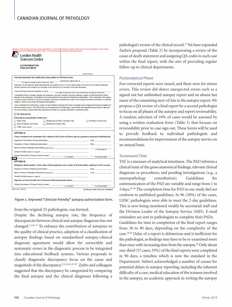
issues the autopsy authorization form was modified by
incorporating clinical input. These changes included placing
the identification of the next-of-kin at the top of the form and
creating a checklist format for restrictions (Figure 3).
Clinical post-mortem consultations were completed in only
8 (5%) of cases. Previous studies report frequencies of up to
72.7%.12,13 This difference may reflect the recent introduction
of these forms at our institution. When questions were
actually posed by clinicians, they were always addressed by the
pathologist. A similar observation was noted in previous
studies.12,14
Analytical PhaseThe analytical phase used reviewability as a means of assessing
diagnostic accuracy. Reviewability is defined as the ability of
a peer to be able to reach a conclusion, similar or different,
upon assessment of sufficient information. Reviewable
information available or indicated in an autopsy report
includes photographic documentation of gross findings,
appropriate tissue sampling for microscopy with orientation
of the microscopic slides, a record of intradepartmental
consultations and ancillary investigations, and a CPC. The
absence of reviewable information compromises
interobserver diagnostic accuracy. Reviewability allows
transparency in how pathological diagnoses were achieved.
Photographs enhance a reviewer’s understanding of a case,
ensure transparency, and confirm completeness.15
Photographs were frequently taken and yet were only
mentioned in 2 (1%) reports. A compact disc of images was
available with the hard copy of the narrative in the autopsy
folder, so the need for documentation in the report may not
have been deemed necessary; however, a heading in a revised
autopsy report template will resolve this problem.
A microscopic block list catalogues the specific organ or tissue
site of each paraffin block and slide. In 67 (41%) of cases, a
complete microscopic section code was present. Incomplete
section codes could reflect a pathologist’s practice of
consistently sampling tissues in the same order. The presence
of a blocking code facilitates reviewability and is a professional
guideline.6,16
Much of the educational function of the autopsy occurs
through the CPC. A CPC is described as an objective
correlation of clinical findings with autopsy gross and
microscopic findings, including results of other ancillary
investigations, to assist clinicians in determining the cause
of death and elucidate the sequence of events leading to
death. 2,16 There were 27 (16%) cases without a CPC; the
majority were authored by the neuropathology group. This
group performs autopsies restricted to examination of the
central nervous system (CNS). The primary indication for
autopsy in these cases is usually to confirm an already
known clinical diagnosis and obtain tissue for research.
The overall autopsy rate was 5.7%, comparable with the
average rate for teaching hospitals.12,13,17 A low autopsy rate
can diminish a pathologist’s autopsy expertise, which may
affect the quality of the autopsy. To ensure maintenance of
autopsy competence, a dedicated autopsy team of 13
pathologists (including the 3 neuropathologists), reduced
Winter 2014110 Canadian Journal of P athology
CANADIAN JOURNAL OF PATHOLOGY

Canadian Journal of P athology 111Winter 2014
JACQUES AND SHKRUM
Table 2. Proposed classification of clinico-pathological discrepancy quality assurance codes
QA Code DescriptionQAA 00 No significant discrepant diagnosis identified at autopsyQAA 01 Major diagnostic clinico-pathologic discrepancy:QAA 02 Minor diagnostic clinico-pathologic discrepancy:
Not applicable:QAA 03a Sudden death/expected death at home with no significant/recent antemortem clinical
care/evaluation QAA 03b No anatomical/biochemical/microbiological/molecular/placental diagnosis was found following
autopsy to explain either the clinical course or death or bothQAA 03c Limited/restricted autopsy in which a pathologic diagnosis was not found to explain the clinical
course or death or both
Table 3. Audit form for preanalytical, analytical, and postanalytical factors and turnaround time in hospital autopsy
Autopsy Number: ________________________________________Date: __________________________________________________Name of Deceased: _________________________________________________________________________________________________Date of Postmortem: _____________________________________ Date of Review: __________________________________________Originating Pathologist: ___________________________________ Reviewing Pathologist: ____________________________________
Preanalytical Factors Yes Not Applicable/NoProperly filled consent formAutopsy restrictions followedPatient identificationClinical postmortem consultation forms completed
Analytical Factors Yes Not Applicable/NoMacroscopic interpretation consistent with descriptionPhotograph of gross relevant findingsMicroscopic interpretation/description supports macroscopic findings Complete microscopic blocking codeAppropriate use of pathology consultsAppropriate use of ancillary investigationsFree of major language errorsClinico-pathologic correlation is reasonableClinico-pathologic discrepancy code completed
Postanalytical Factors Yes Not Applicable/NoCorrelation between provisional and final diagnosesCorrected reports
Turnaround Time Total number of daysPost-mortem to provisional anatomical diagnosisPost-mortem to final anatomical diagnosis
Disposition:Case referred for consultation by another pathologist o yes o noFeedback provided to originating pathologist o yes o no
Comments/suggestions:
Winter 2014112 Canadian Journal of P athology
CANADIAN JOURNAL OF PATHOLOGY
from the original 25 pathologists, was formed.
Despite the declining autopsy rate, the frequency of
discrepancies between clinical and autopsy diagnoses has not
changed.12,18-21 To enhance the contribution of autopsies to
the quality of clinical practice, adoption of a classification of
autopsy findings based on standardized autopsy–clinical
diagnosis agreement would allow for correctible and
systematic errors in the diagnostic process to be integrated
into educational feedback systems. Various proposals to
classify diagnostic discrepancy focus on the cause and
magnitude of the discrepancy.7,12,14,21,22-25 Zarbo and colleagues
suggested that the discrepancy be categorized by comparing
the final autopsy and the clinical diagnoses following a
pathologist’s review of the clinical record.13We have expanded
Zarbo’s proposal (Table 2) by incorporating a review of the
cause of death statement and assigning QA codes to each case
within the final report, with the aim of providing regular
follow-up to clinical departments.
Postanalytical PhaseFew corrected reports were issued, and these were for minor
errors. This review did detect unexpected errors such as a
signed out but unfinished autopsy report and an absent last
name of the consenting next-of-kin in the autopsy report. We
propose a QA review of a final report by a second pathologist
to focus on all phases of the autopsy and report reviewability.
A random selection of 10% of cases would be assessed by
using a written evaluation form (Table 3) that focuses on
reviewability prior to case sign out. These forms will be used
to provide feedback to individual pathologists and
recommendations for improvement of the autopsy service on
an annual basis.
Turnaround TimeTAT is a measure of analytical timeliness. The PAD informs a
clinical team of the gross anatomical findings, relevant clinical
diagnoses or procedures, and pending investigations (e.g., a
neuropathology consultation). Guidelines for
communication of the PAD are variable and range from 1 to
3 days.26-28 The completion time for PAD in our study did not
conform to published guidelines. In 96 (58%) of the cases,
LHSC pathologists were able to meet the 2-day guidelines.
This is now being monitored weekly by secretarial staff and
the Division Leader of the Autopsy Service (MJS). E-mail
reminders are sent to pathologists to complete their PADs.
Guidelines for time to completion of the final report ranges
from 30 to 90 days, depending on the complexity of the
case.28,29 Delay of a report is deleterious and is inefficient for
the pathologist, as findings may have to be re-examined more
than once with increasing time from the autopsy.30 Only about
one-third (57 cases; 35%) of the final reports were completed
in 90 days, a timeline which is now the standard in the
Department. Siebert acknowledged a number of causes for
potential delays in autopsy reporting, including the inherent
difficulty of a case, medical education of the trainees involved
in the autopsy, an academic approach in writing the autopsy
!
! ! ! ! ! ! !
!
Figure 2. Improved “clinician-friendly” autopsy authorization form.

Canadian Journal of P athology 113Winter 2014
JACQUES AND SHKRUM
report, and the need for additional ancillary tests.31 Our study
reinforced Siebert’s observation that resident involvement
increased TAT. Autopsies with resident involvement added an
average of 6 weeks to the final TAT. Increased TAT in resident
teaching institutions has been described by others as an
outcome of the educational process.32 Many of the LHSC
pathologists who performed autopsies at the time of the study
also have a surgical pathology focus, which demands
significant time, since surgical pathology reporting has
become more complex.33 As a result, autopsy report
completion can become a lesser priority.
Timeliness is monitored by providing Autopsy Team
pathologists with a monthly list of all the active cases,
highlighting cases that are more than 3-months-old. This is
reinforced by letters to individual pathologists from the
Division Leader of the autopsy service.
Formal orientation sessions, particularly for new residents, to
familiarize them with autopsy protocols and reporting
enhance their involvement during autopsy rotations. To
improve resident TAT, a pathologist can proactively guide and
monitor a resident’s effort in completing a case by providing
sample autopsy reports to reduce editing time and
incorporate TAT as a competency in a resident’s evaluation.1,33
At LHSC, anatomical pathology residents rotate on the
autopsy service during 1-month blocks. Neuropathology
residents’ involvement is longitudinal and focuses on cases
restricted to the examination of the central nervous system
CNS. Of interest, the turnaround time for final autopsy
reports was similar between neuropathology residents
involved in CNS only cases and anatomical pathology
residents. The Royal College initiative for competency-based
medical education may provide future guidance regarding
block versus longitudinal rotations to indicate which will be
better in allowing residents to achieve their competency
milestones.
In conclusion, current regulations require that pathology
departments have a structured and active program of QA,
with the goals of enhancing patient safety, minimizing error,
ensuring timelines of reports, and functioning as a medical
audit. We have integrated an autopsy QA program as a subset
of our Department QA program as a means to improving
timeliness of service and quality of reports. An annual review
of data derived from the QA monitors will be completed by
the Division Leader of the autopsy service. Some
recommendations from this study that have already been
initiated include a “clinician-friendly” autopsy consent form
(see Figure 3), creation of a dedicated autopsy team,
guidelines for monitoring reporting timelines, and
incorporating TAT as part of the resident evaluation process.
Starting an autopsy–clinical discrepancy classification,
feedback to clinical teams regarding this and other service
issues (e.g., issues with authorization forms) and in-depth
audit of autopsy reports focusing on reviewability are the next
steps.
AcknowledgementsWe wish to thank Ms. Paula Miller and Ms. Laurie Poynter for
their assistance with the preparation of this article.
References 1. Young NA, Naryshkin S. An implementation plan for autopsy quality control
and quality assurance. Arch Pathol Lab Med 1993;117:531–4.
2. Hutchins GM, Berman JJ, Moore WM. Hanzlick and the autopsy committee of
the College American Pathologists. Practice guidelines for autopsy pathology:
autopsy reporting. Arch Pathol Lab Med 1999;123:1085–92.
3. Schned AR, Mogielnicki RP, Stauffer ME. A comprehensive quality assessment
program in the autopsy service. Am J Clin Pathol 1986;86:133–8.
4. Penner DW. Cost effectiveness of the autopsy in maintaining and improving the
standard of patient care. Am J Clin Pathol 1978;69:250–2.
5. Friederici HHR, Sebastian M. The concordance score correlation of clinical and
autopsy findings. Arch Pathol Lab Med 1984;108:515–7.
6. Hanzlick RL. The autopsy lexicon: suggested headings for the autopsy report.
Arch Pathol Lab Med 2000;124:594–603.
7. Anderson RE, Hill RB, Gorstein F. A model for the autopsy-based quality
assessment of medical diagnostics. Hum Pathol 1990;21:174–81.
8. Saracci R. Is necropsy a valid monitor of clinical diagnosis performance? BMJ
1991;303:898–900.
9. Uemura K, Sternby N, Vanӗčk R, Vihert A, Kagan A. Grading atherosclerosis inaorta and coronary arteries obtained at autopsy. Bull W H O 1964;31:297–320.
10. Veres B, Gadaleanu V, Nennesmo I, et al. The reliability of autopsy diagnostics:
inter-observer variation between pathologists, a preliminary report. Qual Assur
Health Care 1993;5:333–7.
11. Williams AT, Morris D, Patel NK. Pathologists’ view on consent for autopsy. Soc
Med 2002;95:547–8.
12. Bayer-Garner I, Fink L, Lamps L. Pathologists in a teaching institution assess
the value of the autopsy. Arch Pathol Lab Med 2002;126:442–7.
13. Chariot P, Witt K, Pautot V, et al. Declining autopsy rate in a French hospital:
physicians’ attitudes to the autopsy and use of autopsy material in research
publications. Arch Pathol Lab Med 2000;124:739–45.
14. Zarbo R, Baker P, Howanitz P. The autopsy as a performance measurement tool-
diagnostic discrepancies and unresolved clinical questions: a College of
American Pathologists Q-Probes study of 2749 autopsies from 248 institutions.
Arch Pathol Lab Med 1999;123:191–8.
15. Pritt B, Gibson P, Kumarasen C, et al. What is a picture worth?: digital imaging
application in autopsy reports. Arch Pathol Lab Med 2004;128:1247–50.
16. Collins KA, Hutchins GM, eds. Autopsy performance and reporting. 2nd ed.

Winter 2014114 Canadian Journal of P athology
CANADIAN JOURNAL OF PATHOLOGY
Northfield, IL: The Laboratory Accreditation Program (LAP) of the College
of American Pathologists (CAP), 2003.
17. Hooper JE, Geller SA. Relevance of the autopsy as a medical tool: a large
database of physician attitudes. Arch Pathol Lab Med 2007;131:268–74.
18. Kirch W, Schafii C. Misdiagnosis at a university hospital in 4 medical eras:
report of 400 cases. Medicine 1996;75:29–39.
19. McPhee S. The autopsy: an antidote to misdiagnosis. Medicine 1996;76:41–
3.
20. Cabot RC. Diagnostic pitfalls identified during a study of three thousand
autopsies. JAMA 1912;59:2295–8.
21. Shojania KG, Burton EC, McDonald KM, Goldman L. Changes in rates of
autopsy-detected diagnostic errors over time: a systematic review. JAMA
2003;289:2849–56.
22. Hill R, Anderson R. An autopsy-based quality assessment program for
improvement of diagnostic accuracy. Qual Assur Health Care 1993;5:351–9.
23. Hall P. Do we really need a higher necropsy rate? Lancet 1999;354:2004.
24. Goldman L, Sayson R, Robbins S, et al. The value of the autopsy in three medical
eras. N Engl J Med 1983;308:1000–15.
25. Battle RM, Pathak D, Humble GC, et al. Factors influencing discrepancies
between premortem and post-mortem diagnoses. JAMA 1987;258:339–44.
26. Association of Directors of Anatomic and Surgical Pathology; Nakhleh R, Coffin
C, Cooper K. Recommendations for quality assurance and improvement in
surgical and autopsy pathology. Hum Pathol 2006;37:985–8.
27. College of American Pathologists. 1992 Inspection Checklist Section VIII:
Anatomic Pathology and Cytopathology. Northfield, IL: College of American
Pathologists, 1992.
28. Joint Commission on Accreditation of Healthcare Organizations. 1993 Joint
Commission on Accreditation Manual for Hospitals. Oakbrook Terrace, IL: Joint
Commission on Accreditation of Healthcare Organizations, 1992.
29. College of American Pathologists, Commission on Laboratory Accreditation.
1997 Inspection Checklist Section VIII: Anatomic Pathology and Cytopathology.
Northfield, IL: College of American Pathologists, 1997.
30. Siebert JR. Increasing the efficiency of autopsy reporting. Arch Pathol Lab Med
2009;133:1932–7.
31. Baker P, Zarbo R, Howanitz P. Quality Assurance of Autopsy face sheet reporting,
final autopsy report turnaround time and autopsy rates. Arch Pathol Lab Med
1996;120:1003–8.
32. Hall P. Do we really need a higher necropsy rate? Lancet 1999;354:2004.
33. Sinard J, Blood D. Quality improvement on an academic autopsy service. Arch
Pathol Lab Med 2001;125:237–45.

Canadian Journal of P athology 115Winter 2014
Recommendations on Quality AssurancePractices Stemming from the Findings of anAudit of Randomly Selected Negative Breast
Core Biopsies
Departments of Pathology (ER and JGG) and Department of Community Health Sciences (DC), Faculty of Medicine, Universityof Manitoba, Winnipeg, Manitoba, CanadaCorrespondence may be directed to Esther Ravinsky at [email protected] article has been peer reviewed.Competing interests: None declared.
Esther Ravinsky, MD, CM, FRCP(C), John G. Gartner, MD,CM, FRCP(C), Dan Chateau, PhD
ABSTRACTIn 2011, Diagnostic Services Manitoba (DSM), the nonprofit corporation operating public
laboratories in the province of Manitoba, implemented a number of quality measures related
to reporting of breast core biopsies obtained under radiological guidance. These measures
included mandatory double sign-out of all breast cores with a diagnosis of atypical duct or
atypical lobular hyperplasia or higher. These measures did not fully address the issue of potential
false-negative reports for which double reporting was not mandated due to workload
considerations. Instead, a 10% random review of negative breast core biopsies was proposed.
This audit was undertaken to both assess the accuracy of negative breast core biopsy reports
and to suggest ways to reduce false-negative rates. The audit included review of 220 negative
breast cores, and the diagnosis was correlated with the original. Cases with a review diagnosis
of atypical duct hyperplasia (ADH) or higher were reviewed by a second pathologist.
A benign diagnosis was confirmed in 98.2% of the cases. A review diagnosis of “unsatisfactory”
was made in 0.5% and missed ADH was seen in 1.4%, with two cases originally diagnosed as
florid duct hyperplasia. This false-negative rate was comparable with that seen in other studies.
Actions resulting from our audit included presentation of the missed ADH to pathology staff,
a recommendation to stain for cytokeratin 5 or 6 (CK5/6) in questionable cases, and mandatory
double reporting of cases of florid duct hyperplasia. A follow-up audit of 189 randomly selected
negative core biopsies, which was performed approximately 2 years after the implementation
of these changes in practice, showed a 0% false-negative rate.
The results of our audit clearly indicated that a random review of 10% of negative core biopsy
reports is not an effective way to improve accuracy and mitigate patient risk. Due to the low
risk of error and the workload implications, we do not recommend double sign-out of negative
breast core biopsies. Targeted review is an effective approach to any quality assurance program
in surgical pathology.
ORIGINAL ARTICLE

Winter 2014116 Canadian Journal of P athology
CANADIAN JOURNAL OF PATHOLOGY
In 2011, the Diagnostic Services of Manitoba (DSM), the
non-profit corporation that runs public laboratories in
the province of Manitoba, implemented a number of quality
measures related to the reporting of breast core biopsies
obtained under radiological guidance. Pathologists signing
out these biopsies are required to report at least 200 cases a
year and to enrol in continuing education related to breast
pathology at least once every 2 years. All breast core biopsies
with a diagnosis of atypia or higher must have the diagnosis
confirmed by a second pathologist with subspecialty interest
in breast pathology. Pathologists reporting breast core
biopsies are also provided with online access to radiological
images and reports, and a special requisition for breast cases,
which required the radiologist to give a complete clinical
history, including the BI-RADS score, was developed.
Finally, regular radiological–pathological rounds were
initiated for adjudication of cases with radiological–
histological discordance.
As stringent as these measures were, they did not fully
address the issue of potential false-negative biopsy reports.
RÉSUMÉ En 2011, Services diagnostiques Manitoba, société sans but lucratif chapeautant les laboratoires
publics de la province, a précisé les exigences de qualité du rapport de biopsie mammaire au
trocart stéréotaxique. La confirmation obligatoire du diagnostic d’hyperplasie canalaire
atypique, d’hyperplasie lobulaire atypique ou de lésions à haut risque par un second pathologiste
est l’une de ces exigences. Néanmoins, la question des faux négatifs n’est pas entièrement réglée,
car la confirmation d’un autre médecin n’est pas obligatoire en raison de la surcharge de travail.
L’organisme propose plutôt la révision d’un échantillon aléatoire de 10 % des biopsies négatives.
Nous avons entrepris la présente vérification pour évaluer l’exactitude des rapports de biopsie
mammaire négatifs et suggérer des moyens de réduire le taux de faux négatif. La vérification
consiste en l’examen de 200 rapports de biopsie mammaire au trocart négative et en la
comparaison entre le diagnostic établi à la vérification et le diagnostic initial. En cas de
diagnostic révisé d’hyperplasie canalaire atypique ou de lésions à plus haut risque, un autre
pathologiste devait se prononcer.
Dans 98,2 % des cas, le diagnostic de lésions bénignes est confirmé. Dans 0,5 % des cas, le
diagnostic révisé est « insatisfaisant », et l’hyperplasie canalaire atypique est passée inaperçue
dans 1,4 % des cas, dont deux cas pour lesquels le diagnostic initial est celui d’hyperplasie
canalaire de forme floride. Ce taux de faux négatif est comparable à celui observé par d’autres
études. Par suite de notre vérification, nous avons présenté un exposé sur les cas d’hyperplasie
passés inaperçus à l’effectif en pathologie et nous avons recommandé la coloration de détection
de la cytokératine 5 ou 6 dans les cas douteux et la confirmation obligatoire de l’hyperplasie
canalaire de forme floride par un autre pathologiste. Nous avons procédé à une vérification de
suivi portant sur 189 biopsies au trocart négatives choisies au hasard deux ans après la mise en
œuvre de ces modifications de la pratique; elle s’est soldée par la constatation d’un taux de faux
négatif de 0 %.
Les résultats de notre vérification indiquent clairement que la révision d’un échantillon aléatoire
de 10 % de biopsies négatives n’est pas une façon efficiente d’améliorer la précision diagnostique
ou d’atténuer les risques pour le patient. En raison du faible risque d’erreur et des répercussions
sur la charge de travail, nous ne recommandons pas la double confirmation de la biopsie
mammaire négative. La révision sélective est une méthode efficiente dans le cadre du
programme d’assurance de la qualité en pathologie chirurgicale.

Canadian Journal of P athology 117Winter 2014
RAVINSKY ET AL.
Double reporting of negative breast cores was not mandated
due to workload considerations. Instead, a 10% random
review of negative breast core biopsies was proposed. To
address the efficacy of such a review, an audit was initiated
to assess the histological reliability of a negative biopsy
report. For the purposes of this study, a negative breast core
biopsy was defined as negative for high-risk lesions,
including atypical duct hyperplasia (ADH), atypical lobular
hyperplasia (ALH), lobular carcinoma in situ, duct
carcinoma in situ (DCIS), and invasive carcinoma.
MethodsThe review included 220 sequential negative breast core
biopsies from the months of June, July, and August, 2011.
The biopsies were taken under stereoscopic guidance from
lesions identified with mammographic screening, as well as
from palpable masses. Biopsies of large palpable masses were
more likely to be taken under ultrasound guidance.
Indications for biopsy included calcifications, architectural
distortion, and the presence of a mass. Large vacuum cores
were not utilized for any of the biopsies. Pathological–
radiological correlation was performed in all cases. A single
senior pathologist with experience and expertise in breast
pathology reviewed all of the cases. The reviewing
pathologist was aware that all of the biopsies had been
reported as negative but was blinded to all other histologic,
clinical, and demographic information. For each biopsy, the
reviewing pathologist gave a report of either “negative
diagnosis confirmed” or “negative diagnosis not confirmed,”
with the corrected diagnosis given. Diagnosis of precursor
lesions is prone to interobserver variation. Therefore, all
cases which the reviewer identified as missed precursor
lesions were reviewed by DSM’s four subspecialty breast
pathologists. The diagnosis was only changed if there was
consensus among all four pathologists. Immuno-
histochemical staining for CK5/6 was also performed on
cases of missed ADH in order to support the diagnosis.
At the conclusion of this study, we implemented several
changes in practice to address the findings of the audit. This
was followed 2 years later by a second audit of approximately
200 randomly selected negative core biopsy reports to
determine the effectiveness of these actions.
ResultsThe diagnosis was confirmed as benign in 216 out of the 220
negative cases (98.2%) (Table 1). The negative breast cores
showed a range of diagnoses, including normal breast tissue,
stromal fibrosis, usual duct hyperplasia, sclerosing adenosis,
fibroadenomatoid changes, fibroadenoma, and duct ectasia.
In one case, the diagnosis after review was “unsatisfactory”
(0.5%). Three cases of missed ADH (Figure 1A and Figure
2) were found on review (1.4%) (Table 2). Two of these cases
had originally been diagnosed as florid duct hyperplasia.
The atypical ducts in these cases showed decreased CK5/6
expression on immunohistochemical staining (Figure 1B).
The third case showed ADH of columnar cell type (see
Figure 2). In all cases, the radiologists who had performed
the biopsies were given the relevant information.
Two of the reclassified cases, one unsatisfactory and one
missed ADH, were radiologically benign. The biopsy
reclassified as ADH had been performed for a fibroadenoma
and the ADH was an incidental finding. No further action
was taken in either case, and repeat mammography was
scheduled for 1 year later for both patients. In the other two
cases, biopsy had been performed because of calcifications,
and the clinical information stated “rule out DCIS.” In both
cases, the calcifications were associated with ADH, and both
patients had a subsequent lumpectomy. One lumpectomy
Table 1. Confirmation of Negative Diagnoses
Review Report Number of Cases1 Percentage of CasesNegative diagnosis 216 98.2%confirmedNegative diagnosis 4 1.8%not confirmed1A follow-up audit of 189 negative breast core biopsies was done 24 months afterchanges in practice as a result of the first audit being implemented. It showed an errorrate of 0%.
Table 2. Misclassified Lesions
Review Diagnosis Number of Cases Percentage of CasesUnsatisfactory 1 0.5%Atypical duct hyperplasia 3 1.4%

Winter 2014118 Canadian Journal of P athology
CANADIAN JOURNAL OF PATHOLOGY
showed residual ADH, but the other showed no evidence of
residual disease. Neither of the lumpectomies contained
DCIS or invasive carcinoma. The radiologist who had
performed the biopsy on the patient with incidental ADH
adjacent to a fibroadenoma decided to monitor the patient
with repeat mammography in 1 year’s time.
Statistical analysis was performed on the data to assess the
potential value a 10% random rescreening might have to
reduce the false-negative rate. False negatives of 3 out of 220
would give a proportion of 0.0136 with a 95% confidence
interval (CI) of 0.0046 to 0.0393. It would, therefore, be
necessary to review, on average, 73 negative tests to identify 1
false negative (95% CI 26–217). A random review of 10% of
negative tests would, by definition, identify only 10% of false
negatives, thus reducing the overall false-negative rate by only
one-tenth. Assuming the error rate of 1.36% in this study, the
random review would have reduced this only to 1.22%.
The follow-up audit that was done 24 months after
implementation of the recommendations stemming from
the initial audit showed a 0% false-negative rate.
ConclusionBreast core biopsy performed under radiological guidance
is an alternative to surgical excision of a breast lesion.1-7 It is
faster, is less invasive, and has fewer complications. Because
the number of surgeries is reduced, there is a significant
reduction in cost to the health care system. False-negative
biopsies are a known risk that can delay diagnosis and
treatment of a malignancy. A number of studies have
evaluated false-negative diagnoses, defining a “false
negative” as a core biopsy with a histological benign
diagnosis followed by a high-risk or malignant diagnosis on
a repeat core biopsy or subsequent surgical excision.2,4-6,8-11
The false-negative rate is the number of false negatives
Figure 2. Ducts lined by columnar cells in atypical duct hyperplasia,columnar cell type. Some of the ducts show a proliferation ofepithelial cells with a cribriform architecture. Cells are perpendicularto the microlumina; Roman arches and bridging of ducts areidentified. (Hematoxylin and eosin)
Figure 1. A, Atypical duct hyperplasia showing ducts filled with a monotonous population of cells. Microlumina are round, and the cells areperpendicular to the microlumina. Nuclei are smaller and more hyperchromatic at the centre than at the periphery. (Hematoxylin and eosin)B, Reduced imunohistochemical staining for cytokeratin 5/6 (CK5/6) in atypical duct hyperplasia. (Immunoperoxidase)

Canadian Journal of P athology 119Winter 2014
RAVINSKY ET AL.
divided by the number of patients diagnosed with cancer.
This rate varies between 0.2 and 8%, depending on the
study. Most of the studies addressing this issue have
concentrated on the technical reasons, in particular
sampling error, for false negatives.5 The choices of the
imaging method and the biopsy system have also been
evaluated.8,9 Sampling errors have been related to poor
visualization of the lesion or needle visualization, small or
deep-seated lesions, lesions in dense breasts, and
heterogeneity of the lesions.11 Biopsies with fewer than 4 or
5 cores have been shown to have a higher proportion of
false-negative diagnoses.10 The value of radiological–
pathological correlation has been demonstrated.7 Patients
with false-negative core biopsies in which radiological and
pathological findings do not correlate require a repeat
biopsy in order to avoid a delay in diagnosis.11 A few studies
have assessed the accuracy of the pathological diagnosis in
false-negative biopsies. In a study by Boba et al., the
histopathology of all false-negative biopsies was reviewed.8
Of the 988 biopsies, 22 (2.2%) were false negatives. In 14 of
the 22 false-negative diagnoses, the histopathological
diagnosis was changed from benign to atypical or malignant
on review. False-negative diagnosis due to incorrect
histopathological assessment comprised 1.5% of all
histologically diagnosed cancers and atypias. The majority
of the false-negative cores (12 of 14) showed atypia on
review, and carcinoma was found in the other 2 cores.
Our study assessed the histopathological accuracy of
negative core biopsies in our program. We defined a “false-
negative diagnosis” as a diagnosis that had been changed
from benign to atypical or malignant on review, and the
“false-negative rate” was defined for this purpose as the
number of false negatives divided by the number of negative
breast cores reviewed. By this calculation, our false-negative
rate was 3 or 220, or 1.4%. Like Boba et al., we found that
false-negative core biopsies were most likely to be due to
failure to recognize ADH.
ADH is defined as a proliferative intraductal lesion with
some, but not all, of the features of DCIS.12 The subsequent
risk of invasive ductal carcinoma in a patient with ADH is 4
to 5 times that of the general population. The carcinoma
could be situated elsewhere in the breast or even in the
contralateral breast. Some lesions may show a combination
of ADH and DCIS, or ADH, DCIS, and invasive carcinoma.
A needle biopsy for a presumed carcinoma may retrieve only
ADH. “Underestimated ADH” is defined as a lesion that
yields ADH at needle biopsy but cancer at excision. The
incidence of underestimated ADH is in the range of 20% to
60%.7,13-16 Therefore, a diagnosis of ADH in a core biopsy
requires surgical excision of the lesion, and nonrecognition
of ADH in a core biopsy can lead to a delay in diagnosis and
treatment.
Figure 3. A, Usual duct hyperplasia. There is proliferation of duct epithelial cells forming solid masses predominantly in the terminal ducts.Slit-like spaces are identified with the epithelial cells lying parallel to the spaces. The epithelial cells show a streaming pattern. (Hematoxylinand eosin) B, Strong, diffuse imunohistochemical staining for cytokeratin 5/6 (CK5/6) in usual duct hyperplasia. (Immunoperoxidase)
Winter 2014120 Canadian Journal of P athology
CANADIAN JOURNAL OF PATHOLOGY
There is, however, controversy over the need for surgery for
nonobligate precursors of malignancy, including ADH. It is
recommended that the decision to follow-up or proceed to
surgery should depend on a combination of pathological,
radiological, and clinical factors. These include, but are not
limited to, degree of radiological or clinical suspicion, size
of the lesion, percentage of lesion removed by the core
biopsy, additional risk factors (family history, history of
breast cancer), and other conditions that may increase the
risk of surgery.17 Clinical trials could clarify the impact of
surgical intervention in ADH, but these trials would require
accuracy in the diagnosis of ADH, since evaluation of
outcome depends on a consistently correct diagnosis.
The distinction between usual duct hyperplasia and atypical
duct hyperplasia can present a diagnostic problem. The
criteria for a diagnosis of ADH are loosely defined and are
both quantitative and qualitative. The use of only rigid
qualitative criteria (features of DCIS involving 2 or fewer
ductal cross-sections or less than 2 mm) is controversial, but
lesion size can be used together with other criteria. ADH can
be best defined as a lesion with partial involvement of ducts
with structural and/or cytological changes seen in DCIS (see
Figures 1A and 2). In the usual duct hyperplasia, these
features are absent (Figure 3A). Immunohistochemical
staining for CK5/6 can be a valuable adjunct in the
differential diagnosis between usual duct hyperplasia and
atypical duct hyperplasia.18,19 Whereas the usual duct
hyperplasia shows strong staining for CK5/6, ADH shows
reduced expression of this marker (see Figure 1B; Figure
3B).
Because our data indicated that a random review of 10% of
negative breast core biopsies would have had a very low
probability of identifying the false negatives, we rejected this
approach as a means of improving the diagnostic accuracy
of negative breast core biopsies. This conclusion is
congruent with those of other studies that also concluded
that a 10% random review is costly, is time consuming, and
detects few errors.20 On the other hand, as we found in our
study, a focused review of negative breast cores was effective
in identifying the problem in the recognition of ADH. If we
had solely relied on a 10% random review because the error
rate was low, we would have been unlikely to detect this
important diagnostic pitfall. The only reason to conduct a
10% random review of negative tests would be to monitor
the false-negative rate of the test on an ongoing basis. Any
actual identification of false negatives would be a collateral
benefit of this process, rather than its intended purpose.
The quality assurance policies of some institutions mandate
double sign-out for all breast core biopsies, both positive and
negative. As the result of a critical incident related to a false-
positive breast core biopsy, the DSM mandates double
sign-out for breast core biopsies with a diagnosis of ADH or
higher. Due to the low risk of error identified in our audit
and the workload implications, we do not recommend
double sign-out for negative breast cores.
As a result of this study, we implemented the following
changes in our practice to increase the recognition of ADH
and thereby reduce the false-negative rate: (1) The cases of
missed ADH were presented to all DSM pathologists in a
mandatory quality assurance conference; (2) pathologists
were encouraged to use CK5/6 in difficult cases; and
(3) pathologists were encouraged to consult on cases of
florid duct hyperplasia with a second pathologist with
subspecialty expertise in breast pathology. With the
implementation of these changes in practice, it is
noteworthy that a follow-up audit 2 years later found 0%
false-negative errors. This clearly demonstrated the
effectiveness of our approach to addressing the issues
identified by our audit and underscores the value of targeted
audits as part of any quality assurance process in surgical
pathology.
References1. Bauer RL, Sung J, Eckhert KH, Jr., et al. Comparison of histologic diagnosis
between stereotactic core needle biopsy and open surgical biopsy. Ann Surg
Oncol 1997;4:316–20.
2. Burns RP, Brown JP, Roe SM, et al. Stereotactic core-needle breast biopsy by
surgeons: minimum 2-year follow-up of benign lesions. Ann Surg
2000;232:542–8.
3. Masroor I, Afzal S, Shafqat G, et al. Comparison of stereotactic core breast
biopsy and open surgical biopsy results at a tertiary care hospital in Pakistan.
Int J Women Health 2011;3:193–6.
4. Pijnappel RM, van den Donk M, Holland R, et al. PH: Diagnostic accuracy for
different strategies of image-guided breast intervention in cases of nonpalpable
breast lesions. Br J Cancer 2004;90:595–600.
5. Schueller G, Jaromi S, Ponhold L, et al. US-guided 14-gauge core-needle breast
biopsy: results of a validation study in 1352 cases. Radiology 2008;248:406–
13.
6. Verkooijen HM, Peeters PH, Buskens E, et al. Diagnostic accuracy of large-
core needle biopsy for nonpalpable breast disease: a meta-analysis. Br J Cancer
2000;82:1017–21.

Canadian Journal of P athology 121Winter 2014
RAVINSKY ET AL.
7. Wiratkapun C, Treesit T, Wibulpolprasert B, Lertsithichai P. Diagnostic
accuracy of ultrasonography-guided core needle biopsy for breast lesions.
Singapore Med J 2012;53:40–5.
8. Boba M, Koltun U, Bobek-Billewicz B, et al. False-negative results of breast
core needle biopsies – retrospective analysis of 988 biopsies. Polish J Radiol
Polish Med Soc Radiol 2011;76:25–9.
9. Dillon MF, Hill AD, Quinn CM, et al. The accuracy of ultrasound, stereotactic,
and clinical core biopsies in the diagnosis of breast cancer, with an analysis of
false-negative cases. Ann Surg 2005;242:701–7.
10. Fishman JE, Milikowski C, Ramsinghani R, et al. US-guided core-needle biopsy
of the breast: how many specimens are necessary? Radiology 2003;226:779–
82.
11. Youk JH, Kim EK, Kim MJ, et al. Missed breast cancers at US-guided core
needle biopsy: how to reduce them. Radiographics 2007;27:79–94.
12. Rosen PP. Rosen’s breast pathology. Philadephia, PA: Lippincott Williams &
Wilkins, 2001:213–215.
13. Flegg KM, Flaherty JJ, Bicknell AM, et al. Surgical outcomes of borderline
breast lesions detected by needle biopsy in a breast screening program. World
J Surg Oncol 2010;8:78.
14. Jang M, Cho N, Moon WK, et al. Underestimation of atypical ductal
hyperplasia at sonographically guided core biopsy of the breast. AJR Am J
Roentgenol 2008;191:1347–51.
15. Liberman L, Holland AE, Marjan D, et al. Underestimation of atypical ductal
hyperplasia at MRI-guided 9-gauge vacuum-assisted breast biopsy. AJR Am J
Roentgenol 2007;188:684–90.
16. Moore MM, Hargett CW, Hanks JB, et al. Association of breast cancer with
the finding of atypical ductal hyperplasia at core breast biopsy. Ann Surg
1997;225:726–31; discussion 731–33.
17. Heywang-Kobrunner SH, Nahrig J, Hacker A, Sedlacek S, Hofler H. B3
Lesions: radiological assessment and multi-disciplinary aspects. Breast Care
(Basel) 2010;5:209–17.
18. Ding Y, Ruan Q. The value of p63 and CK5/6 expression in the differential
diagnosis of ductal lesions of breast. J Huazhong Univ Sci Technol
2006;26:405–7.
19. Otterbach F, Bankfalvi A, Bergner S, et al. Cytokeratin 5/6
immunohistochemistry assists the differential diagnosis of atypical
proliferations of the breast. Histopathology 2000;37:232–40.
20. Raab SS, Grzybicki DM, Mahood LK, et al. Effectiveness of random and
focused review in detecting surgical pathology error. Am J Clin Pathol
2008;130:905–12.

Winter 2014122 Canadian Journal of P athology
Diagnosis of Pulmonary Echinococcosis byBronchoalveolar Lavage Cytology
1 Department of Pathology, 2Department of Medicine, McGill University Health Centre and McGill University, Montreal, Quebec, CanadaCorrespondence may be addressed to Manon Auger at [email protected] article has been peer reviewed.Competing interests: Dr. Auger is the Section Editor for Cytopathology but took no part in the review of this article.
Farshid Razaghi, MD, FRCPC,1 Kristin Y. Popiel, MD,2 Ken M Flegel, MD, CM, MSc,2 Richard Fraser, MD, CM,1
Manon Auger, MD, CM, FRCPC1
ABSTRACTIn Canada, most cases of echinococcosis are caused by the sylvatic form of Echinococcus
granulosus. Although several cases of pulmonary cystic echinococcosis diagnosed by fine-needle
aspiration (FNA) cytology exist in the literature, such diagnoses have only rarely been made on
bronchoalveolar lavage (BAL) samples; we describe such a case here.
RÉSUMÉ Au Canada, la plupart des cas d’échinococcose sont dus à la forme sylvatique d’Echinococcus
granulosus. Bien que la littérature fasse état de plusieurs cas de kystes hydatiques pulmonaires
diagnostiqués à l’examen cytologique du spécimen prélevé par cytoponction, ce diagnostic est
rarement posé par la technique de lavage bronchoalvéolaire; nous décrivons un tel cas ici.
ORIGINAL ARTICLE
Echinococcosis, also known as hydatid disease, is a
zoonotic infection caused by the larval stage of
Echinococcus spp. tapeworms. Human infection is most
common in cattle- and sheep-raising countries, including
Australia, New Zealand, Mediterranean and Middle Eastern
countries, Central Europe, Russia, Northern China, and
Japan. In North America, the disease is especially prevalent
in Alaska and northern Canada.1
Most cases of human echinococcosis are caused by
E. granulosus and E. multilocularis, causing (unilocular) cystic
echinococcosis and (multilocular) alveolar echinococcosis,
respectively. The infrequent polycystic echinococcosis,
restricted to Central and South America, is caused by
E. vogeli or E. oligoarthrus.1,2 Infection with E. granulosus
typically results in the formation of unilocular cysts that can
be located in various organs, most commonly the liver (70%)
or lungs (20%), with the remainder involving other organs
such as kidney, spleen, brain, heart, and bone.1-4
The diagnosis of echinococcosis is generally based on a
combination of radiographic findings and confirmatory
serologic tests; in some cases, the diagnosis is made only at
surgery. A few cases such as those in which no
antiechinococcal antibodies are detected or in which other
conditions cannot be excluded based on the imaging studies,
have been diagnosed by FNA cytology.2,5 However, because
of the possibility of cyst rupture with dissemination of cyst
contents and anaphylaxis, FNA is generally considered risky
and contraindicated. Diagnosis of pulmonary echinococcosis
by bronchoalveolar lavage (BAL) cytology has only been
reported rarely;6 here we report another such case.
Case ReportA 50-year-old First Nations woman from a small town
located 300 kilometres (km) northwest of Montreal, in the
province of Quebec, Canada, presented to a regional hospital
with chest pain that was suggestive of coronary artery

Canadian Journal of P athology 123Winter 2014
RAZAGHI ET AL.
disease. Her past medical history was significant for type II
diabetes mellitus and dyslipidemia. She was a former
smoker, and her mother had been treated for pulmonary
tuberculosis. Clinical assessment did not confirm an acute
coronary syndrome, but chest radiography revealed a focus
of consolidation in the left upper lobe. Moxifloxacin was
given for presumed pneumonia, and the patient was
discharged home.
She returned the following day with worsening fever,
dyspnea, pleuritic chest pain, and a cough productive of
copious amounts of white sputum. She was started on
piperacillin–tazobactam and ciprofloxacin, but there was no
improvement in symptoms. Computed tomography (CT)
of the thorax demonstrated a 5 cm, thin-walled, fluid-filled
cyst, with surrounding consolidation occupying most of the
left upper lobe. Laboratory testing showed mild anemia and
persistent leukocytosis of 20 to 24 × 109/L (normal range:
4.00–1.00 × 10⁹/L) with a striking eosinophilia (absolute
count of 10 × 109/L; normal range: 0.00–0.04 × 10⁹/L).
Kidney and liver function tests were normal. Serum titres of
hepatitis antibodies and human immunodeficiency virus
(HIV) were negative. A purified protein derivative (PPD)
test was negative. Initial BAL did not yield any further
diagnostic information.
Three weeks after the initial presentation, the patient was
transferred to our tertiary care centre. On arrival, she was
stable but still had a fever up to 39.4°C and was short of
breath, with an oxygen saturation of 96% in room air. Her
physical examination was unremarkable. Imaging of the
abdomen was negative. CT of the thorax showed a 5.5 × 2.8
cm necrotic lesion in the anterior segment of the left upper
lobe and a “tree-in-bud” pattern in the left lower lobe,
suggesting aspiration of the contents of the mass. The
differential diagnosis, based on radiologic findings, included
atypical and/or necrotizing pneumonia, tuberculosis,
pulmonary vasculitis, and aspergillosis or other fungal
infection. Induced sputum was negative for acid-fast bacilli
and other organisms, but cytologic examination of a second
BAL fluid revealed numerous echinococcal scolices that
consisted of three-dimensional structures with a stellate
arrangement of hooklets at one of their extremities (Figure
1).The fluid contained a moderate amount of mixed
inflammatory cells with debris and occasional degenerated
macrophages. Upon questioning, the patient acknowledged
contact with a dog that occasionally ventured into a wooded
area.
She eventually underwent a left upper lobe lobectomy.
Sections through the lung showed a well-circumscribed cyst
measuring 3.5 cm in maximal dimension (Figure 2). The
wall of the cyst was about 2 to 3 mm thick. The inner surface
of the cyst was smooth and white. Proximally, the cyst
appeared to be communicating with a bronchus, which also
had a thickened white wall over a distance of 2 cm. The
adjacent lung parenchyma was focally consolidated with a
whitish, necrotic appearance. Histology revealed a necrotic
cystic lesion containing partially degenerated ectocyst
Figure 1. Numerous echinococcal scolices with hooklets are seen in the bronchoalveolar lavage (BAL) specimen. (Papanicolaou stain)

Winter 2014124 Canadian Journal of P athology
CANADIAN JOURNAL OF PATHOLOGY
fragments and scolices diagnostic of echinococcosis.
Necrotizing pneumonia was present adjacent to the cyst.
The patient completed approximately 5 months (including
10 days before lobectomy) of therapy with albendazole. She
discontinued treatment herself due to problems with
alopecia and disabling dizziness, both of which appeared
about 3 months into treatment. Additionally, after 4 months
of treatment, the patient became severely depressed without
any obvious reason or previous history of depression. All
these side effects resolved spontaneously within 4 weeks of
discontinuing the drug. Two years after the lobectomy, the
patient remains well and free of any respiratory symptoms.
DiscussionThe life cycle of Echinococcus organisms involves two hosts,
a definitive carnivore and an intermediate herbivore.
E. granulosus, the most frequent agent of human
echinococcosis, is subdivided into pastural (or European)
and sylvatic variants. In the pastural form, the predominant
life cycle occurs in domestic dogs as definitive hosts, with
livestock animals (e.g., sheep, horses, cattle, pigs, goats, and
camels) as intermediate hosts. In the sylvatic form, the
intermediate hosts are wild animals (e.g., moose, deer, or
caribou), whereas the definitive host is a canine (dog, fox,
or wolf) that harbors the tapeworm after ingestion of tissue
from a dead infected intermediate host. Humans act as
accidental hosts and typically become infected with food or
water contaminated with dogs’ feces containing the eggs of
parasites or through direct contact with dogs.1
In Canada, the most common variant of echinococcosis is
the sylvatic form because of infection with the “north
sylvatic” or “cervid” genotype of E. granulosus; the case
presented here fits that particular profile. The patient, who
lived in a northern community, was presumably exposed to
Echinococcus through her dog, which had likely fed on the
carcass of an infected moose or deer.1 Indeed, the most
important factor identified as increasing the risk of canine
echinococcosis is the access that dogs have to uncooked
infected offal. It has been shown that dogs allowed to roam
have a higher risk of infection because they are more likely
to find and eat raw carcass meat and offal of fallen
livestock.1,7 It is estimated that between 28 and 50% of dogs
in the Northwest Territories are infected with E. granulosus,
and in recent studies in Saskatchewan, 19% to 50% of fecal
samples from the dogs in northern and indigenous
communities were positive for at least one helminth or
protozoan parasite, compared with only 4.4% of dogs in an
urban centre in Saskatchewan.8 The risk of infection after
ingestion of raw moose meat is high; it is estimated that 50%
of moose in Ontario and British Columbia are infected with
the parasite.7 Hydatid cysts have also been found in wapiti,
elk, reindeer, coastal deer, white tail deer, and bison. In the
Northwest Territories and the northern parts of Alberta,
Saskatchewan, and Manitoba, the caribou is the most
common intermediate host.1
Echinococcosis caused by the “north sylvatic” or “cervid”
genotype continues to be relatively commonly diagnosed in
most Canadian provinces.7 In contrast to the other forms,
this variant is characterized by a predominantly pulmonary
localization, a more benign course, and less frequent clinical
complications.7 Most patients with pulmonary
echinococcosis remain asymptomatic and are diagnosed
incidentally while undergoing radiologic investigations for
other reasons. Symptoms develop when the cyst enlarges
sufficiently to cause a mass or pressure on surrounding
tissues or when the cyst ruptures into the adjacent lung,
bronchus, or pleural cavity.9 The most common symptoms
described in the literature are cough (55–60%), chest pain,
dyspnea, and hemoptysis.
The diagnosis of echinococcosis is only infrequently made
through cytology, since percutaneous FNA of a hydatid cyst
has been considered risky for fear of spillage of the cyst
Figure 2. Macroscopic examination of the surgical specimenrevealed a 5.5-cm, well-circumscribed cyst with a smooth, whiteinner surface and focal consolidation of the adjacent lungparenchyma.

Canadian Journal of P athology 125Winter 2014
RAZAGHI ET AL.
contents and anaphylaxis. Degenerated hooklets can
sometimes be demonstrated in the sputum, bronchial
washings, and BAL or pleural fluid following cyst rupture,
but only a few cases have been described in the literature.2,10
Our case represents a rare case of pulmonary echinococcosis
diagnosed in a BAL cytology specimen, presumably
following rupture of the cyst into the bronchial tree. This
case report raises awareness of an unusual presentation of
Echinococcus in an appropriate clinical context as a cystic
lung lesion and of the diagnostic role of exfoliative cytology.
References1. Otero-Abad B, Torgerson PR. A systematic review of the epidemiology of
echinococcosis in domestic and wild animals. PLoS Negl Trop Dis
2013;7(6):e2249.
2. Eckert J, Deplazes P. Biological, epidemiological, and clinical aspects of
echinococcosis, a zoonosis of increasing concern. Clin Microbiol Rev
2004;17:107–35.
3. Somily A, Robinson JL, Miedzinski LJ, Bhargava R, et al. Echinococcal disease
in Alberta, Canada: more than a calcified opacity. BMC Infect Dis
2005;17(5):34.
4. Zhang W, Wen H, Li J, Lin R, McManus DP. Review article- Immunology and
immunodiagnosis of cystic ecchinococcosis: an update. Clin Dev Immunol
2012;2012:101895.
5. Das DK, El-Sharawy M, Ayyash EH, et al. Primary hydatid cyst of the
supraspinus muscle: complete removal of the germinal layer and cytodiagnosis
by fine-needle aspiration. Diagn Cytopathol 2014;42:268–72.
6. Fanne RA, Khamaisi M, Mevorach D, et al. Spontaneous rupture of lung
echinococcal cyst causing anaphylactic shock and respiratory distress
syndrome. Thorax 2006;61:550.
7. Moro P, Schantz PM. Cystic echinococcosis in the Americas. Parasitol Int
2006;55:5181–6.
8. Jenkins EJ, Schurer JM, Gesy KM. Old problems on a new playing field:
helminth zoonosis transmitted among dogs, wildlife, and people in a changing
northern climate. Vet Parasitol 2011;182:154–69.
9. Santivanez S, Garcia HH. Pulmonary cystic ecchinococcosis. Curr Opin Pulm
Med 2010;16:257–61.
10. Morar R, Feldman C. Pulmonary ecchinococcosis. Eur Respir J 2003;21:1069–
77.

Winter 2014126 Canadian Journal of P athology
Retrospective Comparison of Institutional Cytological Accuracy in Warthin Tumour
Diagnosis
1Department of Pathology & Laboratory Medicine, Division of Anatomical Pathology, Capital District Health Authority & Dalhousie University, 5788 University Avenue, Halifax, Nova Scotia, Canada2Department of Surgery, Division of Otolaryngology-Head & Neck Surgery, Capital District Health Authority & Dalhousie University, 5820 University Avenue, Halifax, Nova Scotia, CanadaCorrespondence may be directed to Martin J Bullock at [email protected] article has been peer reviewed.Competing interests: None declared.
Ashley Stueck, MD,1 S. Mark Taylor, MD, FRCSC ,2 Robert D. Hart, MD, FRCSC ,2
Jonathan R. Trites, MD, FRCSC,2 Martin J. Bullock, MD, FRCPC1
ORIGINAL ARTICLE
ABSTRACTPurpose: We hypothesize that cytological accuracy in diagnosis of Warthin tumour (WT) at
our institution has improved over time.
Study Design and Methods: A retrospective review of patients with a WT diagnosis and both
fine-needle aspirate (FNA) and surgical specimens was completed for the past 8 years. All FNA
specimens were prepared using a thin-layer technique. For the review, 82 patients were identified
and divided into two groups based on date of FNA. True-positive (TP), false-positive (FP), and
false-negative (FN) rates were calculated. Parameter of FN versus TP parameters and TP versus
FP results were compared.
Results: Our institution has improved TP rates over time (p ≤.046). FN FNAs were more likelyto have other cellular components (p ≤10-5), less likely to have oncocytes (p ≤10-5) andlymphocytes (p ≤10-5), and had a smaller tumour size at resection (p ≤0.03), compared withTP cases. FP for WT cases were more likely to occur in nonparotid locations (p ≤.0006). ParotidFNAs FP for WT were acinic cell carcinoma and low-grade mucoepidermoid carcinoma.
Conclusions: Cytological diagnosis of WT has improved at our institution. Associated factors
include educational initiatives and changes in FNA practices by the head and neck surgeons.
Mimics and key differentiating factors are reviewed.
RÉSUMÉ But : Vérifier notre hypothèse voulant que l’exactitude du diagnostic cytologique ducystadénolymphome se soit améliorée à notre établissement.
Devis et méthodologie de l’étude : Il s’agit de l’examen rétrospectif de dossiers de patients chezqui l’on a diagnostiqué un cystadénolymphome et pour qui l’on a prélevé des spécimens à la
cytoponction et à la chirurgie dans les huit dernières années. Les spécimens prélevés à la
cytoponction ont été préparés selon la technique de la couche mince. Pour les besoins de l’étude,
les 82 patients examinés sont répartis en deux groupes en fonction de la date de la cytoponction.

Canadian Journal of P athology 127Winter 2014
STUECK ET AL
Warthin tumour (WT) is the second most common
tumour of the parotid gland.1-8 WT is an indolent
neoplasm, primarily treated with surgical excision; it
occasionally recurs and rarely undergoes malignant
transformation.6,8 Males are more commonly affected, with
male-to-female ratios ranging from 1.1:1 to 9:1 in the
literature.2,4-5,9,10 Most patients are diagnosed between ages
50 and 80 years. 2-4,7,9-11 Multifocality and bilaterality are not
uncommon.1,6-8 Fine-needle aspiration (FNA) of salivary
gland masses and cytological examination of the material is
commonly used to attempt diagnosis. A typical aspirate
from a WT contains oncocytes, lymphocytes, and
background debris; however, inflammation, infarction,
sampling error, and pathologist interpretation error are just
some factors that may lead to an unclear or missed
diagnosis.7 This can lead to increased procedures, longer
wait times to surgery, and increased emotional stress
experienced by the patient. Occasionally, metaplastic
changes can lead to a misdiagnosis of malignancy, which
may result in unnecessarily extensive surgery.
On average, 11 WT diagnoses are rendered at the Queen
Elizabeth II Health Sciences Centre (QEII HSC) per year. In
2003, the laboratory switched from conventional-based
cytology to liquid-based preparation for all
nongynecological specimens. Rapid on-site evaluation of
cytology specimens for adequacy is not routine. Some
pathologists perceived that the accuracy of WT diagnosis
had improved over time while using liquid-based cytology;
however, this had neither been confirmed nor any potential
reasons identified. As well, it was felt that the diagnostic
accuracy for WT may reflect accuracy of salivary gland FNA
in the institution. The goal of this study was to determine if
there had been improved diagnostic accuracy for WT on
cytological specimens over the past 4 years compared with
the previous 4 years; if so, possible reasons for improvement
were to be sought.
Materials and MethodsThe study was a retrospective chart review of all patients
with a cytological or surgical pathology diagnosis of WT
rendered at the QEII HSC over the past 8 years. The
laboratory information system of the Anatomical Pathology
Division was searched from November 1, 2003, to November
1, 2011, to identify all patients with a cytological diagnosis
of WT or an oncocytic lesion. Next, all patients with an
excision specimen diagnosed as WT from November 1,
2003, to September 28, 2012, were identified. Cytological
specimens with a diagnosis of WT without a following
surgical excision, as well as surgical excisions without
previous cytology or cytology outside the study timeframe,
were excluded.
From the reports, the following information was recorded:
Nous avons calculé les taux de vrai positif (VP), de faux positif (FP) et de faux négatif (FN),
puis nous avons comparé les paramètres des groupes FN et VP et ceux des VP et des FP.
Résultats : Le taux de VP a augmenté à notre établissement dans cette période (p ≤0 0,046). Lesanalyses statistiques illustrent que, question de probabilité, les spécimens FN ont tendance à
avoir d’autres composants cellulaires (p ≤ 10-5), à ne pas avoir d’oncocytes (p ≤ 10-5) et delymphocytes (p ≤ 10-5), et la tumeur est de plus petite taille à la résection (p ≤ 0,03), que lesspécimens VP. Dans les cas de cystadénolymphome, les FP sont principalement des spécimens
provenant d’autres sites que la glande parotide (p ≤ 00,0006). Les spécimens issus de la glandeparotide FP pour ce qui est du cystadénolymphome sont en fait un carcinome à cellules
acineuses ou un carcinome mucoépidermoïde bien différencié.
Conclusion : Le diagnostic cytologique du cystadénolymphome s’est amélioré à notre
établissement. Des initiatives éducatives et la modification de la technique de cytoponction des
chirurgiens de la tête et du cou comptent au nombre des facteurs à l’origine de cette
amélioration. Nous passons en revue des pathologies où les constats cytologiques sont
semblables et les principaux traits distinctifs.

Winter 2014128 Canadian Journal of P athology
CANADIAN JOURNAL OF PATHOLOGY
surgical diagnosis, cytological diagnosis, dates of diagnoses,
age and sex of the patient, site and size of tumour, and
presence or absence of multifocality. Cytology reports of
patients with known or suspected WT were evaluated for
degree of cellularity and constituent cell types. Patients were
divided into two groups based on date of FNA (November
1, 2003, to July 18, 2007; and July 19, 2007, to November 1,
2011). Cytology specimens were categorized as true positive
(TP) if WT was the cytological diagnosis and the final
surgical resection demonstrated WT. Cytology specimens
were classified as false negative (FN) if the resection
demonstrated a WT but WT was not diagnosed
cytologically. This included cases where oncocytes were
noted, but the possibility of a WT was not specifically
mentioned, as understanding the implication of the
presence of oncocytes could not be assumed on the part of
the physician reading the report. Cytological specimens in
which the diagnosis was indeterminate (n = 5), and included
WT and other oncocytic neoplasms, were considered
nondiagnostic and within the FN category. For the purposes
of the study, unsatisfactory FNAs were deemed FN. Cytology
specimens were deemed false positive (FP) for WT if the
resection specimen did not demonstrate WT but the
cytology had a diagnosis or favoured a diagnosis of WT.
Descriptive statistics were used to describe the patient
demographics and tumour characteristics (site, size, and
multifocality). Cellularity and constituent cell types
(lymphocytes, oncocytes, other) were evaluated qualitatively.
Standard statistical methodology was used to calculate FP,
FN, and TP rates, sensitivity, and positive predictive value
(PPV) in each group. Comparison of FN, FP, and TP rates
between groups, FN and TP cases in terms of cellularity and
cell type, and site differences between TP and FP cases were
completed using the chi-square test. The Fisher Exact test
was utilized in place of the chi-square test where any cell
contained n = <10. The student t test was used to determine
if tumour size was significantly different between the TP and
FN cases.
This project was reviewed and approved by the Capital
Health Research Ethics Board.
ResultsFor the review, 184 patients who had a surgical or cytological
diagnosis of WT were chosen. Seventy-three patients were
excluded because of lack of surgical excision specimens; 29
patients were excluded because of lack of cytology
specimens. Overall, 82 patients met the study criteria, and
there were 99 cytology specimens.
The study population consisted of 36 females and 46 males,
with an average age of 64.1 years (range 29–92 years). There
were no significant age differences between groups (p ≤.18)or between the male and female subgroups (p ≤.14 and 0.56,respectively). Most (n = 68) patients had one salivary gland
FNA, some (n = 11) had two FNAs, and a few had three
FNAs (n = 3). The study population was divided into two
groups, A (FNA obtained between November 1, 2003, and
July 18, 2007) and B (FNA obtained between July 19, 2007,
and November 1, 2011). This represented approximately the
halfway point of the study period, which began when
ThinPrep tissue processing of cytological specimens was
introduced at our institution. The group of seven
cytopathologists reading salivary gland FNAs had been
relatively stable during the entire period and included one
of the authors (MJB). Group A had 49 patients (20 females
and 29 males; mean age 66.1); group B had 50 patients (22
females and 28 males; mean age 61.3).
Results are summarized in Table 1. TP rates were 28.6% for
Group A, and 48% for Group B (p ≤.046). FN rates weredifferent between the groups: Group A had an FN rate of
65.3%, whereas it dropped to 48% in Group B. However, this
difference did not reach statistical significance (p ≤.17). FPrates for WT were 6.1% and 4% for Group A and Group B,
respectively (p ≤.68). Sensitivity improved in Group B (50%)
versus Group A (30.4%). PPV also improved in Group B
(92.3%) compared with Group A (82.4%). True negative
rates and specificity could not be calculated, as capture of
true negative cases was not in the scope of this project. The
FN for malignancy rate was 4% for both groups. The FP for
malignancy rate was 0% in both groups. The percentage of
unsatisfactory FNAs was 8% for both groups.
A detailed comparison of FN and TP cytological diagnoses
of WT was completed. All specimens came from the parotid
gland. Tumours correctly diagnosed as WT on cytology were
larger (on average 0.64 cm larger) compared with their
undetected counterparts (p ≤.03). Oncocytes were more
likely to have been detected in cases diagnosed as WT, versus

Canadian Journal of P athology 129Winter 2014
STUECK ET AL
those called negative (p ≤10-5). Interestingly, in a subset ofsix FN cases, the presence of oncocytes was recorded, but the
possible diagnosis of WT was not mentioned in the report.
TP cases were significantly more likely to have lymphocytes
(p ≤10-5) compared with FN cases. In contrast, other cells(acinar cells, squamous cells, macrophages) were more likely
to be seen in FN for WT cases than in their TP counterparts
(p ≤10-5). Cellularity, presence of debris, and multifocalitydid not differ significantly.
Five FP diagnoses of WT were made (Table 2). In three of
these cases, the aspirate was not from parotid tissue.
However, all cytology specimens falsely diagnosed as WT
were reported to have oncocytes and lymphocytes. The
misdiagnoses in the parotid gland were low-grade
mucoepidermoid carcinoma and acinic cell carcinoma. FP
cases of WT were significantly more likely to be of
nonparotid origin (p ≤.0007).
DiscussionThe accuracy of cytological diagnosis of WT at our
institution has improved since 2003. All factors examined
(TP, FP, FN, sensitivity, PPV) have improved when Group A
(November 1, 2003, to July 18, 2007) was compared with
Group B (July 19, 2007, to November 1, 2011), although only
the improvement of the TP rate was statistically significant.
When attempting to discover the possible reason(s) for
improvement, two specific events were brought to light, in
addition to the increased familiarity of our cytopathologists
with the appearance of WT in ThinPrep specimens. First, in
November 2006, a didactic lecture on cytological features of
WT was presented to cytotechnologists, residents, and other
pathologists by one staff pathologist (MJB) interested in
head and neck pathology. Second, FNA practice by the
surgeons from the Division of Otolaryngology – Head &
Neck Surgery (from whom the vast majority of the
specimens were received) changed in 2007. Prior to this
time, the FNA would be done routinely, with each specimen
containing the aspirated material from one FNA.
Subsequently, two separate FNAs, using two separate
needles, were taken, with the combined specimens
submitted in the same container. Unfortunately, exact data
on when this practice began and the degree of consistency
among the surgeons were not available for further analysis.
This particular technique has not been evaluated in the
literature, although studies of repeat FNA may provide some
insight. Brennan et al. (2010), who reviewed surgical salivary
tumour specimens and their previous FNAs, found that
repeat FNA, where the previous FNA was adequate but
nondiagnostic, provided diagnostic material in 82% of
patients.12 Similarly, simulations by Schmidt et al. (2012)
showed increased adequacy with increased needle pass
rates.13 We hypothesize that improved familiarity with
ThinPrep appearance, specific educational initiatives, and
altered FNA practice may all have contributed to our
improved cytological accuracy.
Our FP rates and PPV are within the reported range;
however, our institution’s sensitivity (Sn) and TP rates are
below the values reported in the literature, greater than 95%
and greater than 88%, respectively.1,10-11,14 One explanation
for the discrepancy is that literature rates are based on
conventional cytology, whereas our study used liquid-based
preparations. Studies comparing liquid-based to
conventional preparations in salivary gland FNAs have
Table 1. Parameters of Accuracy of Diagnosis over Time
Group A Group B(November 1, 2003 – (July 19, 2007 –July 18, 2007) November 1, 2011) p ≤
TP 28.6% 48.0% .046FN 65.3% 48.0% .17FP 6.1% 4.0% NSSn 30.4% 50.0% NAPPV 82.4 % 92.3% NATP = true positive; FN = false negative; FP = false positive; Sn = sensitivity; PPV = positive predictive value; NS =not significant; NA = not applicable
Table 2. Sites of False-Positive Diagnoses of Warthin Tumour
Cytologic Final Surgical Diagnosis SiteDiagnosis
Acinic cell carcinoma ParotidLow-grade mucoepidermoid carcinoma ParotidBenign salivary gland Minor
WT salivary glandAcinic cell carcinoma Submandibular glandSquamous cell carcinoma Level II lymph
nodeWT = Warthin tumour

Winter 2014130 Canadian Journal of P athology
CANADIAN JOURNAL OF PATHOLOGY
identified that the former have decreased blood,
inflammatory cells, and extracellular material.15,16
Conventional preparations for salivary gland FNAs appear
to be more accurate, with higher specificity, Sn and TP
rates.16,17 In reference to WT, studies show that liquid-based
preparations have small aggregates of oncocytes, rather than
sheets, and fewer lymphocytes than conventional smears.15,17
We hypothesize that the sole use of liquid-based cytology
may sometimes deprive pathologists of the diagnostic cues
or materials of WT. Our Sn and TP rate have increased
throughout the liquid-based preparation era, perhaps
indicating adaptation to the liquid-based alterations. The
differences in conventional and liquid-based cytology,
especially dehiscence of oncocytes and removal of
lymphocytes, may have affected our Sn, TP, and FN rates.
A contrast study of the TP and FN cases were then
performed. In TP cases, the tumours were significantly
larger, and the cytology specimens had significantly more
oncocytes, lymphocytes, and debris, and fewer cells of other
types. Although most TP cases contained more diagnostic
Figure 1. A, Oncocytes in cytology preparation. Note centrally placed nuclei, abundant cytoplasm, and fine green-blue cytoplasmicgranules. B, Warthin tumour excision shows cysts lined by bilayered oncocytes supported by lymphocytic stroma. (A, Papanicolaoustain; B, hematoxylin and eosin)
A B
Figure 2. A, Acinic cell carcinoma cytology. Note slightly eccentric nucleus, variable chromocentres, and vacuolated cytoplasm withbasophilic granules. B, Acinic cell carcinoma in surgical resection. Sheets of acinar cells are present. (A, Papanicolaou stain; B,hematoxylin and eosin)
A B

Canadian Journal of P athology 131Winter 2014
STUECK ET AL
material than FN ones, a small subset (n = 6) of FN
specimens were noted to have oncocytes in the cytology
report, but WT was either not considered or not mentioned
in the differential diagnosis. The cytology preparations were
reviewed by two authors (AES, MJB) and could be grouped
into two categories: (1) those with only rare oncocytes (n = 3)
and (2) those lacking lymphocytes (n = 3). In the former,
oncocytes were scant and admixed with other cells,
suggesting inadequate sampling of the lesion. In the latter,
oncocytes were noted and were adequate in number;
however, lymphocytes were not prominent. It is important
to remember that absence of lymphocytes does not exclude
WT from the differential diagnosis.
The site of the specimen is another key factor to consider.
All surgical WT diagnoses in this study were from the
parotid gland. Indeed, one WT was originally diagnosed in
a submandibular location; however, on review, portions of
residual, serous salivary gland, consistent with parotid tail,
were identified. There was a significant difference in tumour
site in TP and FP cases. Three of the five lesions diagnosed
on cytology as WT, but which represented other non-
neoplastic or malignant lesions, were FNAs from nonparotid
sites (submandibular gland, minor salivary gland, and a level
II lymph node). The cytology of these cases was reviewed by
two authors (AES, MJB). In the submandibular gland, acinic
cell carcinoma cells were misinterpreted as oncocytes, which
is a well-recognized diagnostic pitfall.7,11 In the minor
salivary gland resection, benign acinar cells were mistaken
for oncocytes. In the level II lymph node resection,
neoplastic squamous cells and enlarged lymphocytes were
likely mistaken for oncocytes. It is thought that WT arise
from salivary ductal inclusions in intraparotid lymph
nodes.6 A WT may arise in proximity to these other
structures but likely represents origin from the parotid tail.
Extreme caution should be used in suggesting a WT
diagnosis in a nonparotid site. Indeed, our FN malignancy
rate decreased from 4 to 2% when nonparotid sites were
excluded.
The two cases falsely diagnosed as WT on cytology in the
parotid received final diagnoses of low-grade
mucoepidermoid carcinoma and acinic cell carcinoma on
surgical resection. Upon review of the cytology specimens
by two of the authors (AES, MJB), we believe that
intermediate and acinar cells, respectively, were mistaken for
oncocytes. Indeed, these are two of the most common
cytological mimics of WT.2,10-11
Oncocytes are recognized on Papanicolaou cytology
preparations as sheets or clusters of well-spaced, cyanophilic
cells with centrally located nuclei, no atypia, and a cytoplasm
with fine green-blue cytoplasmic granules.7 To consider the
diagnosis of WT or oncocytic neoplasm, a cytological
preparation must include oncocytes. Lymphocytes and
cellular debris may be seen in the background but are not
required to raise the diagnosis. On surgical excision, there is
a well-circumscribed, thinly encapsulated neoplasm
composed of cystic spaces lined by bilayered, benign
oncocytic epithelium with supporting lymphoid stroma.6
Figure 1 emphasizes the cytological and histological features.
Oncocytic metaplasia occurs with age and is not commonly
associated with mass formation; however, nodular
hyperplastic forms do exist.1,8,18 Cytologically, there are fewer
oncocytes and more normal salivary cells in the background
compared with WT, although this is not always the case.
Oncocytomas and WT may present similarly on cytology.
Oncocytomas are rarer and have no sex predilection.7,18 The
parotid is the most common salivary gland involved.1,7,18 The
background in a cytological preparation is often clean.7
Histologically, oncocytomas are composed of sheets or
trabeculae of oncocytes separated by thin fibrovascular
strands.18 Clinical management is similar to that of WT.8
Mucoepidermoid carcinoma (MEC) is the most common
salivary gland malignancy and can be a cytological mimic
of WT.7,19,20 Females are more commonly affected.19 The
parotid gland is affected in approximately 45% of cases.8,19
The age range is broad, with the mean in the 4th decade.20
Low-grade MEC is the most difficult to distinguish from
oncocytic neoplasms. It is composed of rare squamoid cells,
variable proportions of mucous cells, and abundant
intermediate cells, which may mimic oncocytes.7,11,19
Mucinous material may be present in the background.20
Recognition of mucous cells, which can have a goblet cell or
macrophage-like appearance, is critical to the diagnosis.
Histologically, the tumour is composed predominantly of
bland intermediate cells with cystic spaces lined by mucous
cells.19 To further confuse matters, an oncocytic variant of
mucoepidermoid carcinoma exists. Careful examination for

Winter 2014132 Canadian Journal of P athology
CANADIAN JOURNAL OF PATHOLOGY
mucous cells and cytological atypia is required.7 Goblet cell
metaplasia may be seen in WT; however, these mucous cells
are often less frequent than is found in low-grade
mucoepidermoid carcinoma.7,10,19 In our case, intermediate
and epidermoid cells were likely misinterpreted as
oncocytes. A careful review of the cytology preparation
identified mild cytological atypia, a minor mucinous
background component, and extremely rare cells suspicious
for mucous cells. The resection correlated with predominant
intermediate cell presence, rare mucous cells, and a
prominent lymphoproliferative response.
Acinic cell carcinoma is the second most common salivary
gland malignancy.7 There is a female and parotid
preponderance.7,21 Patients with acinic cell carcinoma
usually present in the 3rd to 4th decade, earlier than the WT
cohort.1,8 The smear is classically cellular with a clean
background.7,11,18 The serous acinar cells, which mimic
oncocytes, are often bland, with abundant, vacuolated
cytoplasm and fine, basophilic cytoplasmic granules on
Papanicolaou-stained preparations.7,11 Cells often adopt a
syncytial pattern and may demonstrate overlapping and
crowding.7 Apart from morphology, cell block preparation
and use of special stains may aid in diagnosis if a neoplasm
is suspected. Oncocytes will stain positively with
phosphotungstic acid–hematoxylin because of their
abundant mitochondria. Alternatively, cytoplasmic zymogen
granules in acinar cells will stain positively with the Periodic
acid-Schiff reagent, and are not digested by diastase.7,21
Histologically, the tumour is composed of polygonal acinar
cells with basophilic cytoplasm and round to oval,
eccentrically placed nuclei, in various architectural growth
patterns.21 A review of our case revealed that acinic cells, in
three-dimensional clusters of varying sizes, were mistaken
for oncocytes. Figure 2 highlights the cytology and histology
of acinic cell carcinoma. Careful morphologic analysis and
use of ancillary studies may help to differentiate WT from
acinic cell carcinoma in a cytological specimen.
In addition, a variety of salivary gland tumours may have
oncocytic metaplastic change, and/or tumour associated
lymphoproliferative response, which could lead to a
misdiagnosis of WT when another benign or malignant
tumour is present.6,8,18,19,21
ConclusionsOur institution has improved accuracy in cytological
diagnosis of WT, likely secondary to changes in FNA
practices, educational initiatives, and improved familiarity
with the diagnosis in liquid-based preparations. FN
cytological diagnoses may be secondary to sampling error
and small tumour size and are less likely to contain
diagnostic, lesional material. Oncocytes are necessary to
consider WT or an oncocytic neoplasm in the differential
diagnosis; lymphocytes are not required to suggest the
diagnosis but should be present for a definitive diagnosis.
The site of the cytological specimen is exceedingly
important, and we recommend great caution when
considering WT as a diagnosis in a nonparotid location.
Indeed, all of our surgical WT diagnoses were from the
parotid. In our cohort, and in the literature, low-grade
mucoepidermoid and acinic cell carcinomas may mimic
oncocytic neoplasms on cytological specimens. Careful
morphological examination and use of ancillary studies may
guide the pathologist to the correct diagnosis.
References1. Torske KR. Benign neoplasms of the salivary glands. In: Thompson L, editor. Head
and neck pathology: a volume in foundations in diagnostic pathology series.
Philadelphia, PA: Churchill-Livingstone, 2006: 295–320.
2. Jafari A, Royer B, Lefevre M, et al. Value of the cytological diagnosis in the
treatment of parotid tumors. J Otolaryngol Head Neck Surg 2009;140:381–5.
3. Jones AV, Craig GT, Speight PM, Franklin CD. The range and demographics of
salivary gland tumours diagnosed in a UK population. Oral Oncol 2008;44:407–
17.
4. Lin CC, Tsai MH, Huang CC, et al. Parotid tumors: a 10-year experience.
Otolaryngol Head Neck Surg 2008;29:94-100.
5. Tian Z, Li L, Wang L, et al. Salivary gland neoplasms in oral and maxillofacial
regions: A 23-year retrospective study of 6982 cases in an eastern Chinese
population. Int J Oral Maxillofac Surg 2010;39:235–42.
6. Simpson RHW, Eveson JW. Warthin tumour. In: Barnes L, Eveson J, Reichart P,
Sidransky D, editors. Pathology and genetics of head and neck tumours: IARC
WHO classification of tumours. Lyon, France: IARC Press, 2005, 263–5.
7. Krane JF, Faquin WC. Salivary gland. In: Cibas E, Ducatman B, editors. Cytology,
3rd ed. Philadelphia, PA: Elsevier, 2009, 285–318.
8. Rosai J. Major and minor salivary glands. In: Rosai J, editor. Rosai and Ackerman’s
surgical pathology. New York: Elsevier, 2011, 873–916.
9. Cardoso SV, do Nascimento Souza KC, de Faria PR, et al. Warthin’s tumor at the
Brazilian National Cancer Institute: additional evidence of homogenous sex
prevalence and association with other neoplasms. J Otolaryngol Head Neck Surg
2008;70:339–43.
10. Viguer JM, Vicandi B, Jimenez-Heffernan JA, et al. Role of fine needle aspiration
cytology in diagnosis and management of Warthin’s tumour of the salivary glands.
Cytopathology 2009;21:164–9.
11. Veder LL, Kerrebijn JDF, Smedts FM, den Bakker MA. Diagnostic accuracy of
fine-needle aspiration cytology in Warthin tumors. Head Neck 2010;32:1635–40.

Canadian Journal of P athology 133Winter 2014
STUECK ET AL
12. Brennan PA, Davies B, Poller D, et al. Fine needle aspiration cytology (FNAC)
of salivary gland tumours: repeat aspiration provides further information in
cases with an unclear initial cytological diagnosis. Br J Maxillofac Surg
2010;48:26–9.
13. Schmidt RL, Howard K, Hall BJ, Layfield LJ. The comparative effectiveness of
fine-needle aspiration cytology sampling policies: a simulation study. Am J
Clin Pathol 2012;138:823–30.
14. Inohara H, Akahani S, Yamamoto Y, et al. The role of fine-needle aspiration
cytology and magnetic resonance imaging in the management of parotid mass
lesions. Acta Otolaryngol 2008;128:1152–8.
15. Michael CW, Hunter B. Interpretation of fine-needle aspirates processed by
the ThinPrep (R) technique: cytologic artifacts and diagnostic pitfalls. Diagn
Cytopathol 2000;23:6–13.
16. Al-Khafaji BM, Afify AM. Salivary gland fine needle aspiration using the
ThinPrep (R) Technique. Diagnostic accuracy, cytologic artifacts and pitfalls.
Acta Cytol 2001;45:567–74.
17. Parfitt JR, McLachlin CM, Weir MM. Comparison of ThinPrep and
conventional smears in salivary gland fine-needle aspiration biopsies. Cancer
Cytopathol 2007;111:123–9.
18. Huvos AG. Oncocytoma. In: Barnes L, Eveson J, Reichart P, Sidransky D,
editors. Pathology and genetics of head and neck tumours: IARC WHO
classification of tumours. Lyon, France: IARC Press, 2005, 266.
19. Goode RK, El-Naggar AK. Mucoepidermoid carcinoma. In: Barnes L, Eveson
J, Reichart P, Sidransky D, editors. Pathology and genetics of head and neck
tumours: IARC WHO classification of tumours. Lyon, France: IARC Press,
2005,219–20.
20. Eveson JW, Thompson LDR. Malignant neoplasms of the salivary glands. In:
Thompson L, editor. Head and neck pathology: a volume in foundations in
diagnostic pathology series. Philadelphia, PA: Churchill-Livingstone, 2006,
321–70.
21. Ellis G, Simpson RHW. Acinic cell carcinoma. In: Barnes L, Eveson J, Reichart
P, Sidransky D, editors. Pathology and genetics of head and neck tumours:
IARC WHO classification of tumours. Lyon, France: IARC Press, 2005,216–8.

Winter 2014134 Canadian Journal of P athology
Ten Challenging Areas in GastrointestinalBiopsy Diagnosis
Department of Pathology, London Health Sciences Centre, Schulich School of Medicine and Dentistry, Western University,London, Ontario, CanadaCorrespondence may be directed to David Driman at [email protected] interests: None declared.This article has been peer reviewed.
Jeremy R. Parfitt, MD, FRCPC, and David K. Driman, MBChB, FRCPC
ABSTRACTIn this review article, we discuss 10 areas that provide a challenge to surgical pathologists,
because of difficulties with diagnosis, terminology, or clinical associations and importance.
These are all entities and issues that have been encountered in a gastrointestinal (GI) pathology
consultation service. We acknowledge that the required scope and length of this article has
necessarily required the omission of several other entities that may also prove difficult for
surgical pathologists. Selected references only are provided to allow further review of the issues
discussed.
RÉSUMÉ Dans le présent article de fond, nous examinons 10 entités complexes en pathologie chirurgicale
en raison des difficultés que posent le diagnostic, la terminologie et les manifestations ou
l’importance clinique. Ce sont des entités et des problèmes auxquels un service de consultation
en pathologie gastro-intestinale a été confronté. À souligner que, pour des motifs de longueur
et de portée, le présent article fait abstraction de plusieurs autres entités susceptibles de poser
certaines difficultés en pathologie chirurgicale. L’article se termine par une sélection de
références qui permettront d’approfondir les aspects abordés ici.
CURRENT REVIEW
Esophagitis with EosinophilsAny number of eosinophils may be abnormal in the
esophageal squamous mucosa. There are several causes, the
most common of which are gastroesophageal reflux disease
(GERD) and eosinophilic esophagitis (EE).1-3 Less common
causes include generalized GI eosinophilia, celiac disease,
Crohn's disease, drugs, achalasia, pemphigus, and a variety
of collagen vascular diseases. Clinical history is important:
EE is more common in males than in females and tends to
present in the 2nd decade to the 4th decade with solid-food
dysphagia; patients frequently have other allergic conditions
such as asthma, eczema, or food intolerances. Endoscopy also
provides clues: a variety of findings such as corrugation,
trachealization, white exudates or plaques, and longitudinal
furrows have been described in EE. Obtaining a sufficient
number of biopsies is important; eosinophilia is often
patchy, and it is recommended that two to four biopsies be
obtained from at least two different locations in the
esophagus, usually in the distal and proximal halves of the
esophagus. We encourage our endoscopists to biopsy the
distal and the proximal or mid-esophagus, as finding more
than a few eosinophils in the proximal or mid-esophagus or
finding more eosinophils here than in the distal esophagus
points away from a diagnosis of GERD. It may also be helpful

Canadian Journal of P athology 135Winter 2014
PARFITT AND DRIMAN
to perform biopsies of the stomach and duodenum to
exclude a generalized GI eosinophilic disorder, although this
would be uncommon.
In terms of the number of eosinophils required to make the
diagnosis, there is an emerging consensus that 15 or more
eosinophils in at least one high-power field (HPF) is
consistent with the diagnosis of EE in the appropriate
clinical setting. However, in a typical case of EE, there will
be many more eosinophils than this, often greater than 30
to 40 per HPF somewhere in the biopsies. There are several
other findings that point toward a diagnosis of EE over
GERD, including eosinophilic microabscesses (clusters of ≥4eosinophils), superficial layering of eosinophils, numerous
degranulated eosinophils, marked squamous hyperplasia,
and lamina propria fibrosis (Figure 1A). These findings are
much less likely to be seen in GERD than in EE.
Furthermore, eosinophil counts in GERD are usually less
than 10 per HPF and are highest in the distal esophagus.
Sometimes, there are borderline cases with eosinophil
counts hovering around 15 per HPF, potentially without
typical clinical or endoscopic features of EE or with a clinical
picture more in keeping with GERD. In such cases, it may
be prudent to diagnose “esophagitis with prominent
eosinophils” with a comment, for example, “Although there
are more eosinophils than are usually seen with GERD, the
differential diagnosis includes both EE and severe GERD (or
concurrent EE and GERD); additional biopsies from both
the proximal and distal esophagus, after a trial of proton
pump inhibitors (PPIs), may help resolve this differential.”
Barrett Esophagus and Goblet CellsPathologists are often confounded by goblet cells. Either they
are absent when the endoscopist makes a diagnosis of
Barrett esophagus (BE) or they are present when
unexpected. BE is defined as the presence of metaplastic
columnar mucosa within the esophagus, and the American
College of Gastroenterology (ACG) requires both
endoscopic evidence of columnar mucosa within the
esophagus and the presence of intestinal metaplasia
histologically for a diagnosis of BE. A couple of awkward
situations arise from the differences between these
pathological and clinical definitions.4-7
The first is when the endoscopist submits samples from what
appears to be BE, but the pathologist sees columnar mucosa
without goblet cells. Notwithstanding the ACG guidelines,
we agree with many other GI pathologists, especially those
in Europe (and with the recent British Society of
Gastroenterologists guidelines), that metaplastic columnar
mucosa with an intestinal phenotype but without goblet
cells represents a type of BE that has neoplastic potential. In
such cases, the endoscopist’s determination and
communication to the pathologist of the site of the biopsies
is important, but there may be clues in the biopsy to
corroborate that the columnar mucosa is from the
esophagus. These include intermingling of squamous islands
and columnar mucosa within the same biopsy fragment, an
esophageal duct underlying the columnar mucosa, and the
presence of multilayered epithelium (a transitional or
immature metaplastic type of epithelium) (Figures 1B and
1C). In such a situation, an appropriate diagnosis may be
“columnar mucosa; non–goblet cell type” with a comment:
“Goblet cells are not identified; this may represent non–
goblet cell intestinalized epithelium of BE; the lack of goblet
cells could be due to sampling error.”
Another problematic situation arises when biopsies are
submitted from the gastroesophageal junction without a
clear endoscopic description of BE but intestinal metaplasia
is seen histologically. BE has neoplastic potential and
requires regular surveillance, but the significance of
intestinal metaplasia within the gastric cardia is unclear;
however, surveillance is not warranted. Intestinal metaplasia
in this location, without an endoscopic Barrett segment, may
be related to chronic gastritis, in which case biopsies of the
distal stomach can be informative. Here, it is helpful to look
for the above-listed clues to determine whether the intestinal
metaplasia is esophageal, and for squamous epithelium
overlying the crypts with intestinal metaplasia and hybrid
glands (mucinous glands that are only partly replaced by
intestinal metaplasia), all of which likely indicate true BE.
In the absence of these, a diagnosis of “intestinal metaplasia
present” may be rendered with the comment: “The
significance of isolated intestinal metaplasia around the
gastroesophageal junction is uncertain. Whether or not this
represents BE will depend on whether features of BE were
seen endoscopically.”
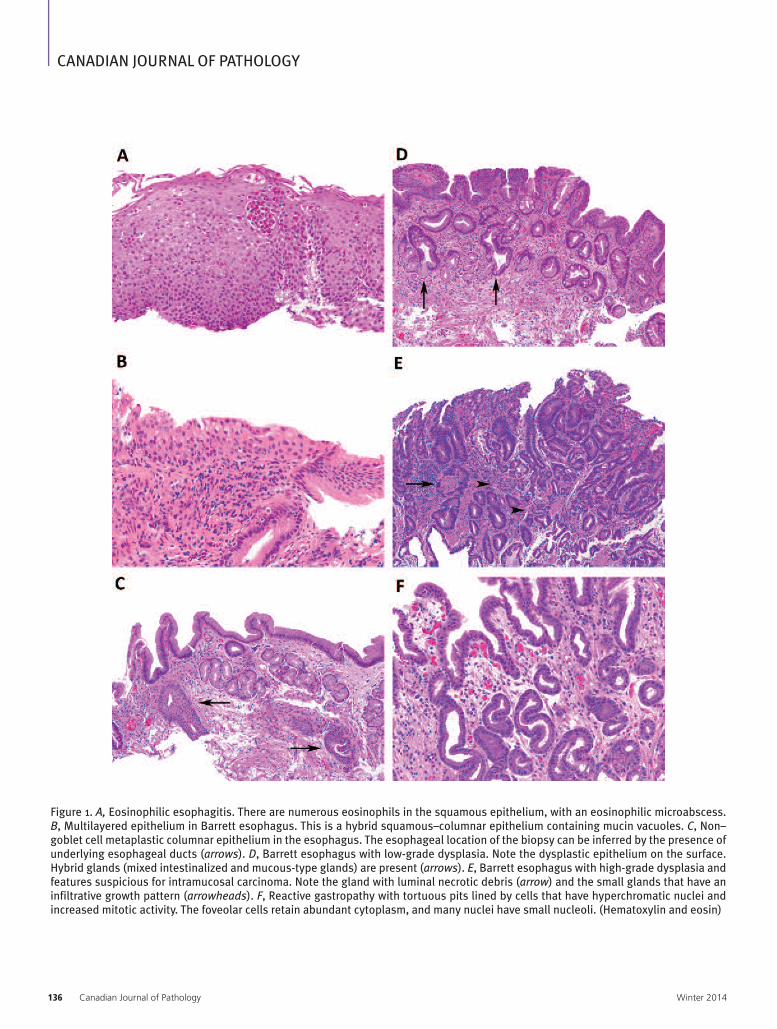
Winter 2014136 Canadian Journal of P athology
CANADIAN JOURNAL OF PATHOLOGY
Figure 1. A, Eosinophilic esophagitis. There are numerous eosinophils in the squamous epithelium, with an eosinophilic microabscess.B, Multilayered epithelium in Barrett esophagus. This is a hybrid squamous–columnar epithelium containing mucin vacuoles. C, Non–goblet cell metaplastic columnar epithelium in the esophagus. The esophageal location of the biopsy can be inferred by the presence ofunderlying esophageal ducts (arrows). D, Barrett esophagus with low-grade dysplasia. Note the dysplastic epithelium on the surface.Hybrid glands (mixed intestinalized and mucous-type glands) are present (arrows). E, Barrett esophagus with high-grade dysplasia andfeatures suspicious for intramucosal carcinoma. Note the gland with luminal necrotic debris (arrow) and the small glands that have aninfiltrative growth pattern (arrowheads). F, Reactive gastropathy with tortuous pits lined by cells that have hyperchromatic nuclei andincreased mitotic activity. The foveolar cells retain abundant cytoplasm, and many nuclei have small nucleoli. (Hematoxylin and eosin)

Canadian Journal of P athology 137Winter 2014
PARFITT AND DRIMAN
Barrett Esophagus –Dysplasia and CancerDysplasia in BE is a common cause of diagnostic problems
for pathologists, be it differentiating low-grade dysplasia
(LGD) from reactive atypia or high-grade dysplasia
(HGD) or differentiating HGD from early invasive
adenocarcinoma.8-11 Interobserver reproducibility for LGD,
even among GI pathologists, is poor. LGD should be
recognizable at low magnification; the features include
nuclear enlargement and hyperchromasia, with or without
mitoses; nucleoli are generally inconspicuous. Some degree
of atypia is always present within intestinalized crypt bases
but this dissipates toward the mucosal surface, and LGD is
diagnosed when there is pan-crypt atypia (i.e., atypia that
extends to the surface) (Figure 1D). An additional clue is
that dysplastic epithelium typically shows an abrupt change
to nondysplastic epithelium, whereas reactive or
inflammatory atypia has a diffuse appearance and fades in
and out within a biopsy. We have also noted that tufted
epithelium is more often reactive (personal observations).
The term “indefinite for dysplasia” should be used when
there is atypia beyond what may be readily recognized as
reactive epithelium but falling short of what is obviously
dysplastic. This situation usually arises when there is
abundant neutrophilic inflammation. Traditionally, the term
“indefinite for dysplasia” has also been used when dysplasia-
like atypia is present in crypt bases but absent on the surface,
because of either maturation or artifactual absence of
surface epithelium. Recently, however, the entity “crypt
dysplasia” has been reported; proponents suggest the use of
this term when atypia within crypt bases is greater than
expected with intestinal metaplasia alone but surface
maturation is present. Some authors advocate including
“crypt dysplasia” within the traditional category of LGD.
Although the evidence for crypt dysplasia is convincing, we
have difficulty and reservations about reporting this as LGD,
since it seems likely that the interobserver reproducibility of
LGD, which is already poor, will be worse. We prefer to
report such cases as “indefinite for dysplasia” with a
comment such as: “Although this case is interpreted as
indefinite for dysplasia, there are features that may represent
so-called ‘crypt dysplasia.’ Repeat short interval biopsies are
suggested to further assess the presence and degree of
dysplasia.”
HGD is more easily recognized, with better interobserver
reproducibility, being characterized by epithelium with
severe nuclear atypia, often with prominent nucleoli, loss of
nuclear polarity, and (often) architectural complexity.
Nevertheless, discomfort arises because of the importance
of this diagnosis, since, given the risk of malignancy of up
to 50% in these patients, it leads in most cases to definitive
therapy. Thus, the recommendation is that a second opinion
from an “expert” pathologist should be sought to confirm
HGD. There is a similar risk of malignancy with any grade
of dysplasia that is endoscopically polypoid, and such cases
should also have mandatory second review. What constitutes
an “expert” in these circumstances is open to debate; we
contend that review by a competent pathologist experienced
in assessing GI biopsies would be appropriate.
Another challenging diagnosis is early invasive
adenocarcinoma, especially distinguishing it from HGD.
Features that distinguish intramucosal adenocarcinoma
from HGD include effacement of the lamina propria by
confluent glands (cribriforming without intervening
stroma) and invasion of single cells or small cell clusters; the
presence of neoplastic glands with intraluminal necrotic
debris also most often indicates intramucosal invasion
(Figure 1E). If only the latter feature is seen, we report “at
least high-grade dysplasia; suspicious for intramucosal
adenocarcinoma.” In cases of unequivocal invasion, we
report “adenocarcinoma, at least intramucosal invasion
present,” given that even lamina propria invasion in the
esophagus signifies a T1 cancer with the potential for lymph
node metastases. It is unwise to diagnose submucosal
invasion in mucosal biopsies from the esophagus, even if
there is apparent traverse of muscularis mucosae, since
duplication of the muscularis mucosae is often present with
BE. A diagnosis of submucosal invasion may preclude
endoscopic mucosal resection, given the high risk of lymph
node metastases associated with submucosal invasion.
Pseudodysplastic Changes in Reactive GastropathyReactive gastropathy, also known as chemical gastropathy, is
a form of injury to the gastric mucosa induced by chemicals;
the chemicals are most commonly drugs such as
nonsteroidal anti-inflammatory drugs (NSAIDs), aspirin, or
steroids but also include alcohol and bile, when there is bile

Winter 2014138 Canadian Journal of P athology
CANADIAN JOURNAL OF PATHOLOGY
reflux. This gives rise to a characteristic constellation of
histological changes that include foveolar hyperplasia, mucin
depletion, lamina propria edema, congestion, and
proliferation of smooth muscle bundles in the lamina propria.
In cases that have an acute component, with or without
erosive changes, the foveolar epithelium can look markedly
atypical and may be confused with dysplasia (either
conventional adenomatous or foveolar types) (Figure 1F). In
reactive gastropathy, both cytoplasmic basophilia and nuclear
hyperchromasia within mucin-depleted foveolar epithelium
may stand out against the pale, edematous lamina propria to
give an initially alarming appearance. However, in our
experience, the atypia associated with reactive gastropathy has
a characteristic morphology and looks different from
dysplasia.
First, the characteristic elon gation and tortuosity of pits, as
well as the lamina propria edema and smooth muscle
proliferation of reactive gastropathy, are typically lacking with
dysplasia. Second, the cytology of the foveolar epithelium in
reactive gastropathy is different from dysplasia. The foveolar
epithelial cells in reactive gastropathy are cuboidal and have
hyperchromatic nuclei, typically devoid of much nuclear
detail, taking on a smudged appearance. Nuclear size and
shape vary from cell to cell more than in dysplasia; however,
on average, the nuclear size is smaller than with dysplasia, and
significant nuclear crowding or pseudostratification is lacking.
Also, in reactive gastropathy, there is an increase in nuclear-
to-cytoplasmic ratio, which is mainly due to decreased
cytoplasm or mucin depletion. With dysplasia, the increase in
nuclear to cytoplasmic ratio is mainly due to increased nuclear
size. Finally, the atypia of reactive gastropathy is typically
diffuse, possibly fading in and out within biopsies. By contrast,
a sharp demarcation between atypical and non-atypical
epithelium is feature of dysplasia. Once these features are
recognized, the atypia of reactive gastropathy can usually be
readily distinguished from true dysplasia.
Duodenal Intraepithelial LymphocytosisThe number of duodenal biopsies with normal or near-
normal villous architecture and increased numbers of
intraepithelial lymphocytes (IELs) appears to be increasing.
Such cases can be a conundrum for pathologists (and treating
physicians) because the causes are protean, and the way in
which they should be reported is not always clear. 12,13 Normal
duodenal mucosa has a villus-to-crypt ratio of around 3:1,
but pathologists should be aware that in the proximal
duodenum (especially the duodenal bulb), villi are often
shorter and appear somewhat distorted. IELs are normally
present, but there should be less than 20 to 25 lymphocytes
per 100 enterocytes. However, we count IELs rarely and
typically rely on a gestalt approach for assessing their number.
In rare borderline cases, a simple procedure to determine if
there are increased IELs is to count lymphocytes per 20
enterocytes at the tip of a few villi; greater than 5 lymphocytes
per 20 enterocytes indicates intraepithelial lymphocytosis.
Biopsies with normal villous architecture and increased IELs
arise in several circumstances. The three most common causes
are early celiac disease, gastric Helicobacter pylori infection,
and medications, especially NSAIDs, but it is also reported
with PPI use. Other causes include viral infection, food allergy,
giardiasis, small intestine bacterial overgrowth, tropical sprue,
Crohn's disease, and various immunological or autoimmune
disorders. Gastroenterologists are typically most interested in
whether any changes present reflect celiac disease. Several
studies have investigated whether the location of the IELs
(villous tip versus mid-villus or base) or the level of IEL
increase can distinguish celiac disease from other causes. The
results of these studies are variable, and we do not find this
reliable. Correlation with serology, namely, tissue
transglutaminase (TTG), is the most helpful approach, and
intraepithelial lymphocytosis associated with an elevated TTG
is highly likely to represent celiac disease.
In terms of reporting, we diagnose these cases as
“intraepithelial lymphocytosis with normal villous
architecture” and we add a comment to address the most
common associations: “Intraepithelial lymphocytosis with
normal villous architecture is a nonspecific finding that may
be associated with several causes, including celiac disease,
gastric H. pylori infection, and medication effects, particularly
NSAIDs. Correlation with serology is recommended.” In cases
where there are gastric biopsies that allow the presence or
absence of H. pylori infection to be assessed, we change the
comment appropriately.
Serrated Colorectal PolypsThe spectrum of serrated colorectal polyps includes
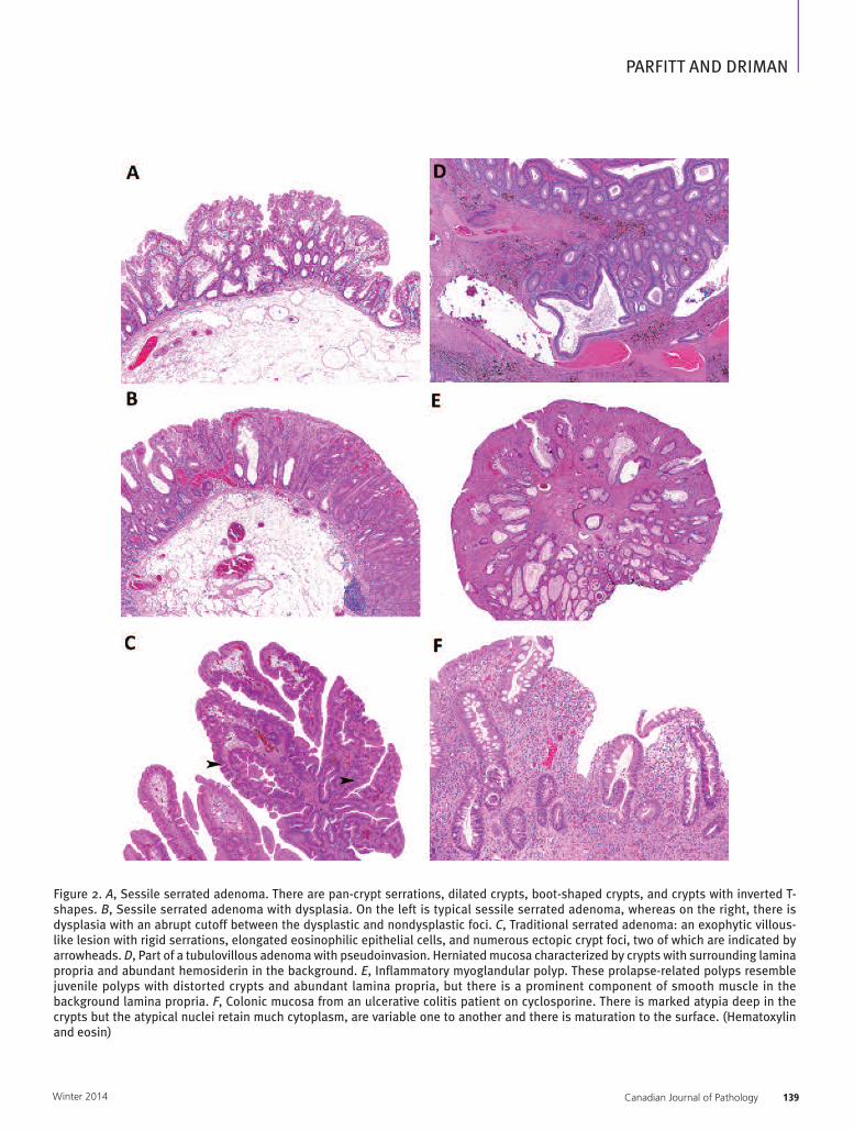
Canadian Journal of P athology 139Winter 2014
PARFITT AND DRIMAN
Figure 2. A, Sessile serrated adenoma. There are pan-crypt serrations, dilated crypts, boot-shaped crypts, and crypts with inverted T-shapes. B, Sessile serrated adenoma with dysplasia. On the left is typical sessile serrated adenoma, whereas on the right, there isdysplasia with an abrupt cutoff between the dysplastic and nondysplastic foci. C, Traditional serrated adenoma: an exophytic villous-like lesion with rigid serrations, elongated eosinophilic epithelial cells, and numerous ectopic crypt foci, two of which are indicated byarrowheads. D, Part of a tubulovillous adenoma with pseudoinvasion. Herniated mucosa characterized by crypts with surrounding laminapropria and abundant hemosiderin in the background. E, Inflammatory myoglandular polyp. These prolapse-related polyps resemblejuvenile polyps with distorted crypts and abundant lamina propria, but there is a prominent component of smooth muscle in thebackground lamina propria. F, Colonic mucosa from an ulcerative colitis patient on cyclosporine. There is marked atypia deep in thecrypts but the atypical nuclei retain much cytoplasm, are variable one to another and there is maturation to the surface. (Hematoxylinand eosin)

Winter 2014140 Canadian Journal of P athology
CANADIAN JOURNAL OF PATHOLOGY
hyperplastic polyps (HPs), sessile serrated adenomas (SSAs),
and traditional serrated adenomas (TSAs).14-17
HPs are typically left sided, small (less than 5 mm), and often
multiple. Serrations are present in the luminal half of the
crypt, and the crypts tend to be straight with narrow bases.
In the sigmoid colon and rectum, basal crypt distortion with
splaying of the muscularis mucosae may occur due to
prolapse. In the lower-third to one-half of the crypt, the
crypt lining cells appear immature, and proliferative cells
and mitoses are seen here but not in the upper halves of the
crypt. Crypt endocrine cells are often readily identified, and
many HPs have a thickened subepithelial collagen band.
Three types of HP have been described: (1) the commonest
is the microvesicular type in which the enterocytes contain
bubbly cytoplasmic mucin; (2) the second commonest type
is the goblet cell type, in which there are few enterocytes and
most crypt lining cells are goblet cells; these are more subtle
in their appearance, having a lesser degree of serration and
surface tufting; and (3) the mucin-poor variant, in which
there are fewer goblet cells than normal and little
microvesicular mucin; these are rare. It is recommended that
the particular type of HP not be reported, but it is important
to be aware of the variations so that the lesions can be
recognized. In our practice, it has become unusual to
diagnose an HP on the right side of the colon, and we would
suggest that pathologists be wary of making a diagnosis of
HP for any polyp that is larger than 0.5 to 1.0 cm or has a
flat, sessile, or otherwise unusual endoscopic appearance, as
the polyp is probably an SSA. Cutting deeper levels may
reveal an SSA that is initially less than obvious.
SSAs occur throughout the large intestine but are commoner
on the right side, where they appear as sessile, flat lesions
that may be rather inconspicuous at endoscopy. Polyps are
often larger than 1 cm in size and have a characteristic
constellation of features. Crypts typically show basal
architectural changes, with serrations that span the full
length of the crypt; crypt bases are usually dilated or show
complex serrated architecture with lateral extensions (boot-
shaped crypts) (Figure 2A). Abnormally differentiated cells
such as gastric epithelium may be seen in the deeper aspect
of crypts. Endocrine cells are typically sparse or absent.
There is often fat in the submucosa, and occasional SSAs
show foci of nonspecific stromal proliferation within the
lamina propria. The cells lining the upper aspects of the
crypts may have vesicular nuclei with prominent nucleoli;
mitoses may be found. It is possible to find hyperplastic
polyp-like foci and traditional serrated adenoma-like foci
(eosinophilic, pencillate cells) in an SSA; these are typically
focal findings and do not deter us from making the
diagnosis of SSA. There are published proposals for
minimum diagnostic criteria for SSAs such as the presence
of one unequivocal, architecturally distorted, dilated and/or
horizontally branched crypt, especially with inverted
maturation, or greater than two or three contiguous crypts
that demonstrate features of SSA (crypts that are dilated and
assume abnormal shapes including L-shapes and inverted
T-shapes with prominent serrations at the base of crypts).
In our practice, we prefer the former, that is, the presence of
one classical crypt being sufficient for a diagnosis of SSA.
The pathologist must exclude dysplasia when examining
SSAs. Dysplastic foci may resemble that found in
conventional adenomas but are sometimes qualitatively
different and more subtle (Figure 2B). The presence of
dysplasia indicates that the SSA is advanced, that is, further
along the pathway to malignancy than those without
dysplasia. Following the Canadian consensus guidelines, we
report SSAs without dysplasia as “sessile serrated adenoma;
negative for dysplasia and malignancy.” We report those
with dysplasia as “sessile serrated adenoma with dysplasia
(low or high grade); negative for malignancy.” SSAs with
dysplasia should not be reported as mixed SSA-tubular (or
tubulovillous) adenoma (TVA), as these are not mixed
polyps but rather advanced SSAs. For advanced SSAs, we
would add the comment: “Advanced SSAs (with either low-
or high-grade dysplasia) are high-risk premalignant lesions
that may have a more rapid transformation to malignancy
than conventional adenomas. Complete removal of this
polyp is recommended.”
TSAs are the most enigmatic of the serrated polyps, and we
know the least about them. They are typically exophytic and
somewhat villiform lesions that sometimes can be difficult
to distinguish from TVAs. TSAs occur more commonly on
the left side. A curious but useful diagnostic feature of these
lesions, and seen in virtually all examples, are crypt buds
that appear as small neocrypts forming within the surface
epithelium (Figure 2C). Other features such as

Canadian Journal of P athology 141Winter 2014
PARFITT AND DRIMAN
hypereosinophilic cells with elongated, pencillate nuclei are
seen in most cases. All TSAs have dysplasia; this can be subtle
and mild or more obvious and similar to that seen in other
adenomas. There is often a patchwork of typical low-grade
dysplasia and hypereosinophilic cells with pencillate nuclei
in these lesions. The pathologist must exclude high-grade
dysplasia, which, in our practice, has not been encountered
frequently. Following the Canadian consensus guidelines,
we report TSAs without high-grade dysplasia as “traditional
serrated adenoma; negative for high-grade dysplasia and
malignancy.”
Unfortunately, the spectrum of serrated polyps does not end
entirely with the above lesions. Not infrequently, we come
across polyps that defy accurate classification. We tend to
find these more on the left side, where they are more often
exophytic. The differential diagnoses usually include TSA
versus TVA, or TSA versus SSA. In such cases, we resort to
calling these “serrated adenoma, unclassified” with a
description of the various features and possible diagnoses.
Hopefully, as we learn more about these polyps, this group
will shrink in number.
Pseudoinvasion versus Invasion in Colorectal AdenomasThe differentiation of pseudoinvasion from true invasion in
adenomas is a frequent reason for consultation because it
can be challenging in nonclassic cases. Typical
pseudoinvasion occurs in pedunculated sigmoid colon
polyps and is identified by herniation of mucosa into the
submucosa. As the process is one of herniation of mucosa,
the herniated structures represent adenomatous mucosa and
therefore consist of circumscribed groups of crypts with
their surrounding lamina propria, rather than naked or
haphazardly arranged separate glands, as seen with invasive
adenocarcinoma. In addition, the degree of atypia present,
that is, grade of dysplasia, within the herniated mucosa
should match that within the mucosa proper; it generally
should not be of a higher grade or look more atypical. The
herniation is due to polyp torsion, which also causes
hemorrhage, so this or the presence of hemosiderin-laden
macrophages is often identified in the submucosa of these
polyps (Figure 2D). There may also be hemorrhage at the
polyp surface with erosion and exudate, which is further
evidence of mechanical disruption to the polyp’s blood
supply. Acellular mucin pools due to crypt disruption and
liberation of mucin into the surrounding tissue are frequent
with pseudoinvasion. Problems arise when these classic
features are not all present or when there has been some
degree of chronicity to the process, resulting in fibrosis
within the submucosa. The fibrosis may isolate single crypts
and obscure the lamina propria, resulting in an appearance
that suggests true invasion. In such cases, we look carefully
for hemosiderin (using a histochemical stain, if necessary)
and for remnants of lamina propria around the crypts,
which would favour pseudoinvasion. We have observed that
cutting several extra levels resolves many initially challenging
cases.
Mucosal Prolapse – a MimicMucosal prolapse is significant because it may mimic
numerous more important diseases in the large intestine.18-22
Most importantly, it may mimic colorectal cancer both
endoscopically, appearing as an ulcerated or polypoid lesion,
and histologically. Although mucosal prolapse occurs most
often in the rectosigmoid and may be associated with
ostomies, anastomoses, and diverticular disease, we have
observed that it may occur anywhere in the large intestine.
The histological features of mucosal prolapse include
fibromuscular proliferation within the lamina propria, crypt
elongation and distortion, variable epithelial serration and
regenerative changes, surface erosion, and vascular ectasia.
There is often a considerable degree of regenerative
epithelial atypia, and this, particularly when combined with
a fibromuscular stroma, may mimic invasive
adenocarcinoma with desmoplasia. The presence of
misplaced glands within the submucosa with associated
pools of extruded (but acellular) mucin, as seen with the
colitis cystica profunda variant of mucosal prolapse,
completes the potential diagnostic trap. Awareness of
mucosal prolapse and its variants, recognition of the
constellation of characteristic features, and knowledge of the
potential associated pitfalls are the best defence against
erroneous diagnoses.
On the benign side, mucosal prolapse may show overlapping
features with acute ischemic colitis, mainly because
repetitive mechanical ischemia plays a likely role in the
pathogenesis of prolapse. The term “polypoid mucosal

Winter 2014142 Canadian Journal of P athology
CANADIAN JOURNAL OF PATHOLOGY
prolapse” may be used for lesions submitted as polyps.
These may be misdiagnosed as adenomatous polyps, again
due to the degree of regenerative atypia present in some
prolapse lesions. Other erroneous diagnoses that may be
rendered when faced with a prolapse polyp include (1) SSA,
given that epithelial serration is often seen with mucosal
prolapse, (2) hamartomatous polyp (either juvenile or
Peutz-Jeghers), given that inflammatory myoglandular
polyps (variants of mucosal prolapse found in the
rectosigmoid) resemble juvenile polyps, but with prominent
stromal smooth muscle proliferation (Figure 2E), and (3)
perineurioma or fibroblastic polyp, another rectosigmoid
lesion with lamina propria fibroplasia and epithelial
serrations.
Finally, it must be remembered that although the problem
of prolapse is primarily one of overcalling neoplasia, some
neoplastic lesions, especially low rectal adenomas, may be
associated with secondary mucosal prolapse changes. If care
is not taken in this latter situation, diagnostic errors may
range from inflammatory cloacogenic polyp (undercalling
of dysplasia) to invasive adenocarcinoma (overcalling of
muscularis mucosae or submucosal invasion).
Inflammatory Bowel Disease – Pouches and StumpsQuestions often arise about reporting biopsies of ileal
pouches in patients previously diagnosed with ulcerative
colitis. The problem is often related to a treating physician’s
question such as “Query pouchitis?“ or “Pouchitis versus
Crohn disease?”23-27 Because performance of an ileal pouch–
anal anastomosis is generally (but not always)
contraindicated for patients with Crohn's disease, the latter
is a loaded question, since reassignment of a diagnosis from
ulcerative colitis to Crohn's disease could imply prior
diagnostic and/or management errors and also potentially
lead to surgical removal of the pouch. Ileal pouches almost
always have at least mild inflammation, along with mild
villous blunting, which is nonspecific and likely related to
changes in the anatomy and intestinal flora.
Pouchitis is an idiopathic clinicopathological condition,
generally diagnosed in patients with diarrhea and
endoscopic and/or histological evidence of inflammation,
which often responds to antibiotics. However, correlation
among symptoms, endoscopic findings, and histology is
poor. Since pouchitis occurs in up to half the patients with
ulcerative colitis, and endoscopists generally biopsy pouches
only when something is amiss clinically, it is generally safe
to assume that pouch biopsies that appear more than mildly
inflamed represent “pouchitis,” in the absence of a more
specific etiology, for example, Clostridium difficile or
cytomegalovirus (CMV) infection. The decision as to
whether pouchitis requires antibiotic treatment will then
depend on correlation with clinical and endoscopic findings.
We report pouchitis in a manner similar to how we report
inflammatory bowel disease (IBD), including the chronicity
and degree of activity of the inflammation. Evidence of
chronicity includes villous blunting, crypt distortion, and
pseudopyloric metaplasia. The presence of erosions or ulcers
may be worth stating explicitly and should always prompt a
careful search for CMV inclusions. As an example, “chronic
active pouchitis (severe) with ulceration,” may be the
diagnosis for cases of severe pouchitis. As mentioned above,
many pouches will show mild nonspecific inflammatory and
architectural changes; we report these descriptively and do
not diagnose as “pouchitis.”
It is unwise to entertain a diagnosis of Crohn's disease in
ileal pouch biopsies from a patient previously diagnosed
with ulcerative colitis, as Crohn's-like features, including
granulomas and even fistulas, may be seen with pouchitis,
although the latter become less common after the first year.
When asked to assess for Crohn's disease in this situation,
we suggest the comment: “There are no reliable histological
criteria for diagnosing Crohn's disease in ileal pouch
biopsies from a patient previously diagnosed with ulcerative
colitis,” in order to inform endoscopists that pouch biopsies
performed to find the answer to such a question are
invariably unhelpful.
Similar questions often arise about reporting on biopsies
from colorectal stumps in patients with IBD. It is generally
safe to assume that stump biopsies that appear chronically
inflamed represent diversion proctocolitis. Never reassign a
previous diagnosis of ulcerative colitis to Crohn's disease
based on stump biopsies, since Crohn's-like features may be
seen with diversion proctocolitis. When faced with the
question “Diversion versus IBD?” we suggest reporting it as
“chronic active proctocolitis (mild/moderate/severe)” with
the comment: “Although the changes likely represent

Canadian Journal of P athology 143Winter 2014
PARFITT AND DRIMAN
diversion proctocolitis, it is not possible to absolutely
exclude the possibility of superimposed IBD within biopsies
from a colorectal stump.”
Inflammatory Bowel Disease – DysplasiaThe challenges related to the recognition of dysplasia in IBD,
as well as correct grade assignment, are similar to those
described for BE and occur mainly with regard to low-grade
dysplasia and cases indefinite for dysplasia.28-31 An additional
confounder in the setting of IBD is drug-related
pseudodysplasia. This has been best characterized with
cyclosporine therapy but may also be seen with other
immunosuppressive agents with antimetabolite action and
should especially be considered in patients who have
undergone liver transplantation for primary sclerosing
cholangitis (Figure 2F).
However, what has become more important for the
pathological reporting of dysplasia in IBD is the correct
interpretation of the clinical significance of such lesions in
the context of their endoscopic or macroscopic appearance.
Older patients with IBD may develop sporadic adenomas,
which require no further management beyond the routine
surveillance schedule, and such a diagnosis may be made in
an older patient with a discrete lesion outside the colitic
zone. However, lesions in younger patients and/or within
the colitic zone are correctly considered to be an IBD-
related, dysplasia-associated lesion or mass (i.e., DALM
lesions), but it is no longer the case that high-grade dysplasia
in any DALM lesion warrants a colectomy. What matters
now for an IBD-related dysplastic lesion is whether it is flat
dysplasia or raised dysplasia and, in the case of raised
dysplasia, whether it is endoscopically “adenoma-like” or
not. Flat dysplasia of any grade carries a high risk of
associated malignancy. The risk of coincidental cancer with
flat low-grade dysplasia is 20% (with a 50% risk of
progression to a higher grade lesion), whereas the risk with
high-grade dysplasia is greater than 40%. Thus, colectomy
should be considered for patients with flat dysplasia,
regardless of the grade. In the case of raised dysplasia, the
endoscopic appearance of the lesion is paramount. An
“adenoma-like” dysplastic lesion is discrete, completely
removable by endoscopy, and, by definition, shows no
malignancy. Patients with such a lesion, irrespective of the
grade of dysplasia, should be followed up in the short term
because of frequent polyp recurrence but otherwise may be
spared colectomy and managed with surveillance, since the
risk of malignancy is no greater than baseline. However, a
raised dysplastic lesion that is not discrete or is not readily
removable by endoscopy, irrespective of the grade of
dysplasia, carries a risk of associated malignancy (occult or
visible) of greater than 40%, and a colectomy should then
be recommended to the patients.
Biopsies of any polypoid lesion should be accompanied by
biopsies of the surrounding flat mucosa in order to exclude
flat dysplasia. Although it is still recommended that the
grade of dysplasia be reported, it is usually not particularly
relevant to the management. The endoscopic or
macroscopic appearance of what was biopsied, coupled with
a definite diagnosis of “dysplasia” should provide the
clinician with sufficient information for appropriate
management. Further, it is recommended that a second
opinion from an expert pathologist be sought to confirm
any diagnosis of dysplasia in the setting of IBD, with the
exception of an obviously sporadic adenoma. Finally, since
the pathologist may not have a reliable description of the
endoscopic appearance of such lesions, it may be useful to
use the comment: “This biopsy shows (low-grade or high-
grade) dysplasia. The appropriate management will depend
on the endoscopic appearance of the lesion. Colectomy
should be considered in the case of flat dysplasia or raised
dysplasia that is not discrete or is not readily removable by
endoscopy, given the high risk of associated malignancy.”
References1. Dellon ES, Gonsalves N, Hirano I, et al. ACG clinical guideline: evidenced
based approach to the diagnosis and management of esophageal eosinophilia
and eosinophilic esophagitis (EoE). Am J Gastroenterol 2013;108:679–92.
2. Maguire A, Sheahan K. Pathology of oesophagitis. Histopathology
2012;60:864–79.
3. Parfitt JR, Gregor JC, Suskin NG, et al. Eosinophilic esophagitis in adults:
distinguishing features from gastroesophageal reflux disease: a study of 41
patients. Mod Pathol 2006;19:90–6.
4. Fitzgerald RC, di Pietro M, Ragunath K, et al. British Society of
Gastroenterology guidelines on the diagnosis and management of Barrett’s
oesophagus. Gut 2014;63:7–42.
5. Vietha M, Langnerb C, Neumannc H, Takubod K. Barrett’s esophagus.
Practical issues for daily routine diagnosis. Pathol Res Pract 2012;208:261–8.
6. Odze RD. What the gastroenterologist needs to know about the histology of
Barrett’s esophagus. Curr Opin Gastroenterol 2011;27:389–96.
7. Riddell RH, Odze RD. Definition of Barrett ’s esophagus: time for a rethink
— is intestinal metaplasia dead? Am J Gastroenterol 2009;104:2588–94.

Winter 2014144 Canadian Journal of P athology
CANADIAN JOURNAL OF PATHOLOGY
8. Patil DT, Goldblum JR, Rybicki L, et al. Prediction of adenocarcinoma in
esophagectomy specimens based upon analysis of preresection biopsies of
Barrett esophagus with at least high-grade dysplasia: a comparison of 2
systems. Am J Surg Pathol 2012;36:134–41.
9. Voltaggio L, Montgomery EA, Lam-Himlin D. A clinical and histopathologic
focus on Barrett esophagus and Barrett-related dysplasia. Arch Pathol Lab Med
2011;135:1249–60.
10. Maru DM. Barrett’s esophagus: diagnostic challenges and recent
developments. Ann Diagn Pathol 2009;13:212–21.
11. Lomo LC, Blount PL, Sanchez CA, et al. Crypt dysplasia with surface
maturation: a clinical, pathologic, and molecular study of a Barrett’s esophagus
cohort. Am J Surg Pathol 2006;30:423–35.
12. Hammer STG, Greenson JK. The clinical significance of duodenal
lymphocytosis with normal villus architecture. Arch Pathol Lab Med
2013;137:1216–9.
13. Walker MM, Murray JA. An update in the diagnosis of coeliac disease.
Histopathology 2011; 59:166–79.
14. Bettington M, Walker N, Clouston A. The serrated pathway to colorectal
carcinoma: current concepts and challenges. Histopathology 2013;62:367–86.
15. Driman DK, Marcus VA, Hilsden RJ, Owen DA. Pathological reporting of
colorectal polyps: pan-Canadian consensus guidelines. Can J Pathol
2012;4:81–90.
16. Huang CS, Farraye FA, Yang S, O’ Brien MJ. The clinical significance of serrated
polyps. Am J Gastroenterol 2011;106:229–40.
17. Rex DK, Ahnen DJ, Baron JA, et al. Serrated lesions of the colorectum: review
and recommendations from an expert panel. Am J Gastroenterol
2012;107:1315–29.
18. Parfitt JR, Shepherd NA. Polypoid mucosal prolapse complicating low rectal
adenomas: beware the inflammatory cloacogenic polyp! Histopathology
2008;53:91–6.
19. Singh B, Mortensen NJ, Warren BF. Histopathological mimicry in mucosal
prolapse. Histopathology 2007;50:97–102.
20. Ball CG, Dupre MP, Falck V, et al. Sessile serrated polyp mimicry in patients
with solitary rectal ulcer syndrome. Is there evidence of preneoplastic change?
Arch Pathol Lab Med 2005;129:1037–40.
21. Li SC, Hamilton SR. Malignant tumors in the rectum simulating solitary rectal
ulcer syndrome in endoscopic biopsy specimens. Am J Surg Pathol
1998;22:106–12.
22. Groisman GM, Polak-Charcon S, Appelman HD. Fibroblastic polyp of the
colon: clinicopathological analysis of 10 cases with emphasis on its common
association with serrated crypts. Histopathology 2006;48:431–7.
23. Weber CR, Rubin DT. Chronic pouchitis versus recurrent Crohn’s disease: a
diagnostic challenge. Dig Dis Sci 2013;58:2748–50.
24. McCurdy JD, Loftus EV, Jr., Tremaine WJ, et al. Cytomegalovirus infection of
the ileoanal pouch: clinical characteristics and outcomes. Inflamm Bowel Dis
2013;19:2394–9.
25. Liu ZX, Deroche T, Remzi FH, et al. Transmural inflammation is not
pathognomonic for Crohn’s disease of the pouch. Surg Endosc 2011;25:3509–
17.
26. Navaneethan U, Shen B. Diagnosis and management of pouchitis and ileoanal
pouch dysfunction. Curr Gastroenterol Rep 2010;12:485–94.
27. Warren BF, Shepherd NA, Bartolo DC, Bradfield JW. Pathology of the
defunctioned rectum in ulcerative colitis. Gut 1993;34:514.
28. Harpaz N, Polydorides AD. Colorectal dysplasia in chronic inflammatory
bowel disease: pathology, clinical implications, and pathogenesis. Arch Pathol
Lab Med 2010;134:876–95.
29. Ullman T, Odze R, Farraye FA. Diagnosis and management of dysplasia in
patients with ulcerative colitis and Crohn’s disease of the colon. Inflamm
Bowel Dis 2009;15:630–8.
30. Parfitt JR, Driman DK. Pathological effects of drugs on the gastrointestinal
tract: a review. Hum Pathol 2007;38:527–36.
31. Hyde GM, Jewell DP, Warren BF. Histological changes associated with the use
of intravenous cyclosporin in the treatment of severe ulcerative colitis may
mimic dysplasia. Colorectal Dis 2002;4:455–8.

Canadian Journal of P athology 145Winter 2014
BOOK REVIEW
Accurate Results in the Clinical Laboratory: A Guide to Error Detection and Correction
Laboratories produce important information for clinicians, and a good quality
assurance program is expected to be implemented to guarantee accuracy. In
Accurate Results in the Clinical Laboratory: A Guide to Error Detection and
Correction, the quality assurance issues are examined in reverse mode, with
the focus on what can go wrong. The reader will not find a systematic
approach to implement a quality assurance program here. Rather, the book
is intended as a guide to increase the awareness of both clinicians and
laboratory professionals with regard to the various sources of errors in
laboratory tests and how to minimize them.
The two editors were assisted by 28 experienced contributors from the United
States and Canada. One positive aspect of the book is the case studies given
throughout the 22 chapters. This makes reading more interesting by
highlighting the concepts and the clinical impact of laboratory errors, which
may be devastating. The authors not only provide a list of potential errors
and interferences but also explain the mechanisms involved. In many cases,
errors are related to specific interferences in a particular specimen. Contrary
to other textbooks, many chapters are sufficiently detailed to provide
discussions of specific errors on a test-by-test basis.
Preanalytical issues are covered in four chapters, and 11 chapters are devoted
to chemistry, endocrinology, immunoassays, therapeutic drug monitoring
(TDM), drugs of abuse, and tumour markers. TDM and toxicology are
especially well covered with many interesting case reports. One chapter focuses
on problems with alcohol testing and how to identify false positives or
negatives. Another explains the impact of herbal medicines that lead to
unexpected laboratory results. Organ toxicity may be the first clue to exposure
to herbal medicine.
Hematology, coagulation, and transfusion are discussed in two chapters. Two
other chapters are also devoted to challenges in serologic and microbiologic
testing. False negative or positives, misidentification of organisms, and errors
in antimicrobial susceptibility testing are examined. Issues in molecular
biology and pharmacogenomics are developed in two specific chapters.
Unfortunately, anatomic pathology is not part of this textbook.
I noticed several minor errors, for example, the listing of hydrogen ions at 40
millimoles per litre (mmol/L) in Table 3.1 “Principal components of plasma”.
(Hydrogen ion [H+] concentration is 40 nanomoles, one million-fold less,
and does not belong in that table). However, the authors have done a good
job, as the book is easy to read and quite comprehensive with ample references.
Overall, the book is definitely worth reading, as it fulfills its objective of
A. Dasgupta and J. L. SepulvedaElsevier Inc., St. Louis, MO, 2013ISBN 978-0-12-415783-5382 pagesList price: US$99.95

Winter 2014146 Canadian Journal of P athology
BOOK REVIEW
providing a guide to sources of laboratory errors and how
to minimize them. I would recommend it to all professionals
involved in laboratory medicine, especially clinical
pathologists, medical biochemists, microbiologists,
hematopathologists, residents, and their teachers! Error-free
laboratory results are essential for patient safety. The book
well help laboratory workers develop for the ability to
identify and be aware of discordant specimen results. I guess
we could call this experience!
Pierre Douville, MD, FRCPCMedical BiochemistDépartement de biologie médicaleCHU de Québec- L’Hôtel-Dieu de Québec

CANADIAN ASSOCIATION OF PATHOLOGISTSASSOCIATION CANADIENNE DES PATHOLOGISTES
Edelman was engaged to develop recommendations for a public relations campaign aimed at building the reputation of pathology in Canada. Through industry research and various consultations, recommendations for a communications campaign were developed based on the strategy of “Putting a face on pathology”.
The goal of the proposed ‘MyPathologist.ca’ campaign is to identify and personalize pathologists, and elevate their role as important members of a patient’s healthcare team. So that as the public, we understand that “my diagnosis and treatment depends on my pathologist”.
Putting a Face on Pathology
Pathology is at the heart of medical treatment in Canada. Every day, pathologists across the country analyze hundreds of thousands of test results and make critical diagnoses that determine how patients are treated. Yet, research shows that less than 10% of the public understands the role pathologists play in making diagnosis that drive patient treatment and care.
Building the reputation of pathology in Canada
MyPathologist.caAnalyze. Diagnose. Care.
MyPathologist.caAnalyze. Diagnose. Care.
55% 76%
6% 5%
Responsibility for Determining Diagnosis From the Lab Test
Responsibility for Determining Treatment
Most of us have a GP we refer to as My Doctor.
If we were to have an operation, we’d talk about My Surgeon.
Shouldn’t someone diagnosed with cancer, talk about My Pathologist?
As doctors who work behind the scenes, pathologists have remained largely invisible to the public. A series of incidents of pathology error in recent years have shone a light on the profession, but in the worst way possible. Pathologists are now grappling with signi� cant public scrutiny, worsened by low public awareness and understanding. Insu� cient funding, increasing workloads and an inability to recruit enough young, talented doctors add to the challenges. The result is a profession struggling to earn the reputation and respect it deserves.
Changing technology and advances in personalized medicine are opening up new areas for pathologists and innovative ways to improve patient care. In order for pathologists to take proper leadership in these areas, the profession needs to strengthen its reputation and build pro� le among patients, other medical professionals, government and hospital administration. This begins with building public awareness.
The doctor who ordered the test
The doctor who ordered the test
Pathologists Pathologists
Public Awareness Levels

Targeted therapy depends on reliable test results
For in vitro diagnostic use
Sample & Assay Technologies
KRA
S091
4CA
only
New therascreen® companion diagnostic
Benefits of the therascreen KRAS RGQ PCR Kit:
New Health Canada approved KRAS test for IVD use
Helps to identify CRC patients who may not benefit from anti-EGFR therapies
Treatment effective in patients with KRAS mutation-negative (wild-type) tumors
Standardized assay for reproducible results
Easy workflow with automated reporting
Visit www.qiagen.com for more information.
Trademarks: QIAGEN®, therascreen® (QIAGEN Group). 08/2014