cAMP-dependent Dynorphin. Natl. Acad. Sci. USA Vol. 87, pp. 7025-7029, September 1990...
5
Proc. Natl. Acad. Sci. USA Vol. 87, pp. 7025-7029, September 1990 Physiology/Pharmacology Dynorphin A and cAMP-dependent protein kinase independently regulate neuronal calcium currents ROBERT A. GROSS*, HYLAN C. MOISESt, MICHAEL D. UHLERt§, AND ROBERT L. MACDONALD*t Departments of *Neurology, tPhysiology, and tBiochemistry and WMental Health Research Institute, University of Michigan Medical Center, Ann Arbor, MI 48104 Communicated by Avram Goldstein, June 18, 1990 (received for review April 6, 1990) ABSTRACT The c-selective opioid peptide dynorphin A (DYN) inhibits neuronal adenylate cyclase activity and reduces neuronal voltage-dependent calcium currents. It is not yet known, however, whether the regulation of calcium channel activity is dependent on or independent of the adenylate cyclase/cAMP system. We used the whole-cell variation of the patch clamp technique to show that DYN reversibly reduced, in a naloxone-sensitive manner, calcium currents in acutely dissociated rat nodose ganglion neurons. DYN slowed the rate of current activation and had a greater effect on currents evoked from relatively negative holding potentials. These ac- tions were mimicked by guanosine 5'-+y-thioltriphosphate, which activates GTP-binding proteins (G proteins), and were blocked by pretreatment with pertussis toxin, which inactivates Gj- and G.-type G proteins. In contrast, calcium currents recorded in the presence of the catalytic subunit of the cAMP- dependent protein kinase (AK-C), included in the recording pipette, increased in magnitude throughout the recording. DYN was applied to neurons before and after the effect of AK-C became apparent; the reduction of calcium currents by DYN was greater in the presence of AK-C than in its absence. We conclude that the acute reduction of neuronal calcium currents by DYN occurred by means of activation of pertussis toxin- sensitive G1- or G.-type G proteins. The persistence of the action of DYN in the presence of AK-C indicates, however, that this effect was independent of a reduction of the activity of the adenylate cyclase/cAMP system and suggests in addition that phosphorylated channels may be preferentially inhibited by DYN. The K-selective opioid peptide dynorphin A (DYN) reduces neuronal calcium currents (1-3) and inhibits neuronal ade- nylate cyclase activity (4, 5), but the pathway by which it alters channel activity is not known. Several neurotransmit- ters reduce voltage-dependent neuronal calcium currents by means of GTP-binding proteins (G proteins) (for example, see refs. 6-9), effects that can be mimicked by guanosine 5'- [y-thio]triphosphate (GTP[yS]) (6), an activator of G proteins (10). In addition, pertussis toxin (PTX), which inactivates Gj- and Go-type G proteins, can block neurotransmitter action on calcium currents (6-9). The G protein subtypes that couple neurotransmitter receptors to calcium channels have been identified by using specific antibodies to block a neurotrans- mitter response or exogenous G proteins to restore a re- sponse previously blocked with PTX (8, 11, 12). The coupling of K-opioid receptors to calcium channels may thus require G proteins, but this has not yet been demonstrated directly. Furthermore, the identity of the G protein-dependent path- way(s) that mediates neurotransmitter effects on calcium channels is not entirely known. Neurotransmitter-activated G proteins might affect calcium channels directly, as G. does in heart (13), or indirectly, by activating a second messenger system. For example, elevated intracellular levels of cAMP or increased activity of the cAMP-dependent protein kinase (AK) increases calcium channel activity in heart cells, inver- tebrate neurons, and clonal pituitary cells (14-16). Taken together, the results of these studies suggest that DYN might reduce calcium currents by inhibition of adenyl- ate cyclase, a reduction of intracellular cAMP levels, and a reduction in the activity of AK. It is also possible that DYN-induced changes in calcium currents could be depen- dent on G proteins but independent of the adenylate cyclase/ cAMP system. To distinguish between these possibilities, we examined the effects of DYN on calcium currents in acutely dissociated rat nodose ganglion neurons by using the whole- cell variation of the patch clamp technique. The effects of DYN were tested after PTX pretreatment and in the presence of GTP[yS] to assess the role of G proteins in the coupling of K receptors to calcium channels. In addition, we determined the effect of DYN in the absence and presence of the catalytic subunit of AK (AK-C). We reasoned that if a reduction in adenylate cyclase activity were the sole mechanism for the DYN-induced reduction in calcium current, then the effect of DYN would not be apparent in the presence of exogenous AK-C. MATERIALS AND METHODS Preparation of Acutely Dissociated Neurons. Nodose gan- glion neurons were prepared from 6- to 10-day-old rats, using a procedure similar to that already described (17). Nodose ganglion neurons were isolated after enzymatic treatment and trituration and were plated in minimum essential medium (GIBCO) supplemented with NaHCO3 (16.5 mM), glucose (28.2 mM), nerve growth factor (10 ng/ml; Boehringer Mann- heim), penicillin (50 units/ml), streptomycin (50 mg/ml), and fetal calf serum (10%; GIBCO). Cultures were incubated at 37°C in a 93% air/7% CO2 atmosphere and used for record- ings within 1-24 hr. Preparation of Solutions. DYN (Peninsula Laboratories) was dissolved in distilled water and stored frozen as a lyophilized powder. Just before the experiment, DYN was reconstituted in bath solution (see below) containing 0.1% bovine serum albumin (Sigma) at a final concentration of 3 ,uM. Naloxone (Sigma) was dissolved in bath solution, also at a concentration of 3 ,tM. Purified AK-C was prepared within 24 hr of the experiment as described (18) and stored at 4°C as a 1 mg/ml stock. AK-C was diluted into the recording pipette solution (see below) to a final concentration of 50 ,ug/ml. This solution, stored on ice, retained full activity for several hours (assayed as described in ref. 19). PTX (Sigma) was prepared as described (17). Whole-Cell Patch Clamp Recordings. Voltage clamp re- cordings were obtained using the whole-cell variation of the Abbreviations: DYN, dynorphin A; AK, cAMP-dependent protein kinase; AK-C, AK catalytic subunit; PTX, pertussis toxin; G protein, GTP-binding protein; GTP[yS], guanosine 5'-[L-thio]triphosphate. 7025 The publication costs of this article were defrayed in part by page charge payment. This article must therefore be hereby marked "advertisement" in accordance with 18 U.S.C. §1734 solely to indicate this fact.
-
Upload
duongtuong -
Category
Documents
-
view
215 -
download
3
Transcript of cAMP-dependent Dynorphin. Natl. Acad. Sci. USA Vol. 87, pp. 7025-7029, September 1990...

Proc. Natl. Acad. Sci. USAVol. 87, pp. 7025-7029, September 1990Physiology/Pharmacology
Dynorphin A and cAMP-dependent protein kinase independentlyregulate neuronal calcium currentsROBERT A. GROSS*, HYLAN C. MOISESt, MICHAEL D. UHLERt§, AND ROBERT L. MACDONALD*tDepartments of *Neurology, tPhysiology, and tBiochemistry and WMental Health Research Institute, University of Michigan Medical Center,Ann Arbor, MI 48104
Communicated by Avram Goldstein, June 18, 1990 (received for review April 6, 1990)
ABSTRACT The c-selective opioid peptide dynorphin A(DYN) inhibits neuronal adenylate cyclase activity and reducesneuronal voltage-dependent calcium currents. It is not yetknown, however, whether the regulation of calcium channelactivity is dependent on or independent of the adenylatecyclase/cAMP system. We used the whole-cell variation of thepatch clamp technique to show that DYN reversibly reduced,in a naloxone-sensitive manner, calcium currents in acutelydissociated rat nodose ganglion neurons. DYN slowed the rateof current activation and had a greater effect on currentsevoked from relatively negative holding potentials. These ac-tions were mimicked by guanosine 5'-+y-thioltriphosphate,which activates GTP-binding proteins (G proteins), and wereblocked by pretreatment with pertussis toxin, which inactivatesGj- and G.-type G proteins. In contrast, calcium currentsrecorded in the presence of the catalytic subunit of the cAMP-dependent protein kinase (AK-C), included in the recordingpipette, increased in magnitude throughout the recording.DYN was applied to neurons before and after the effect ofAK-Cbecame apparent; the reduction of calcium currents by DYNwas greater in the presence of AK-C than in its absence. Weconclude that the acute reduction of neuronal calcium currentsby DYN occurred by means of activation of pertussis toxin-sensitive G1- or G.-type G proteins. The persistence of theaction ofDYN in the presence ofAK-C indicates, however, thatthis effect was independent of a reduction of the activity of theadenylate cyclase/cAMP system and suggests in addition thatphosphorylated channels may be preferentially inhibited byDYN.
The K-selective opioid peptide dynorphin A (DYN) reducesneuronal calcium currents (1-3) and inhibits neuronal ade-nylate cyclase activity (4, 5), but the pathway by which italters channel activity is not known. Several neurotransmit-ters reduce voltage-dependent neuronal calcium currents bymeans ofGTP-binding proteins (G proteins) (for example, seerefs. 6-9), effects that can be mimicked by guanosine 5'-[y-thio]triphosphate (GTP[yS]) (6), an activator ofG proteins(10). In addition, pertussis toxin (PTX), which inactivates Gj-and Go-type G proteins, can block neurotransmitter action oncalcium currents (6-9). The G protein subtypes that coupleneurotransmitter receptors to calcium channels have beenidentified by using specific antibodies to block a neurotrans-mitter response or exogenous G proteins to restore a re-sponse previously blocked with PTX (8, 11, 12). The couplingof K-opioid receptors to calcium channels may thus require Gproteins, but this has not yet been demonstrated directly.
Furthermore, the identity of the G protein-dependent path-way(s) that mediates neurotransmitter effects on calciumchannels is not entirely known. Neurotransmitter-activatedG proteins might affect calcium channels directly, as G. doesin heart (13), or indirectly, by activating a second messenger
system. For example, elevated intracellular levels of cAMPor increased activity of the cAMP-dependent protein kinase(AK) increases calcium channel activity in heart cells, inver-tebrate neurons, and clonal pituitary cells (14-16).Taken together, the results of these studies suggest that
DYN might reduce calcium currents by inhibition of adenyl-ate cyclase, a reduction of intracellular cAMP levels, and areduction in the activity of AK. It is also possible thatDYN-induced changes in calcium currents could be depen-dent on G proteins but independent of the adenylate cyclase/cAMP system. To distinguish between these possibilities, weexamined the effects of DYN on calcium currents in acutelydissociated rat nodose ganglion neurons by using the whole-cell variation of the patch clamp technique. The effects ofDYN were tested after PTX pretreatment and in the presenceof GTP[yS] to assess the role ofG proteins in the coupling ofK receptors to calcium channels. In addition, we determinedthe effect ofDYN in the absence and presence ofthe catalyticsubunit of AK (AK-C). We reasoned that if a reduction inadenylate cyclase activity were the sole mechanism for theDYN-induced reduction in calcium current, then the effect ofDYN would not be apparent in the presence of exogenousAK-C.
MATERIALS AND METHODSPreparation of Acutely Dissociated Neurons. Nodose gan-
glion neurons were prepared from 6- to 10-day-old rats, usinga procedure similar to that already described (17). Nodoseganglion neurons were isolated after enzymatic treatment andtrituration and were plated in minimum essential medium(GIBCO) supplemented with NaHCO3 (16.5 mM), glucose(28.2 mM), nerve growth factor (10 ng/ml; Boehringer Mann-heim), penicillin (50 units/ml), streptomycin (50 mg/ml), andfetal calf serum (10%; GIBCO). Cultures were incubated at37°C in a 93% air/7% CO2 atmosphere and used for record-ings within 1-24 hr.
Preparation of Solutions. DYN (Peninsula Laboratories)was dissolved in distilled water and stored frozen as alyophilized powder. Just before the experiment, DYN wasreconstituted in bath solution (see below) containing 0.1%bovine serum albumin (Sigma) at a final concentration of 3,uM. Naloxone (Sigma) was dissolved in bath solution, also ata concentration of 3 ,tM. Purified AK-C was prepared within24 hr of the experiment as described (18) and stored at 4°C asa 1 mg/ml stock. AK-C was diluted into the recording pipettesolution (see below) to a final concentration of 50 ,ug/ml. Thissolution, stored on ice, retained full activity for several hours(assayed as described in ref. 19). PTX (Sigma) was preparedas described (17).
Whole-Cell Patch Clamp Recordings. Voltage clamp re-cordings were obtained using the whole-cell variation of the
Abbreviations: DYN, dynorphin A; AK, cAMP-dependent proteinkinase; AK-C, AK catalytic subunit; PTX, pertussis toxin; G protein,GTP-binding protein; GTP[yS], guanosine 5'-[L-thio]triphosphate.
7025
The publication costs of this article were defrayed in part by page chargepayment. This article must therefore be hereby marked "advertisement"in accordance with 18 U.S.C. §1734 solely to indicate this fact.

7026 Physiology/Pharmacology: Gross et al.
patch clamp technique. Cells were bathed in a solution of 67mM choline chloride, 100mM tetraethylammonium chloride,5.6 mM glucose, 5.3 mM KCl, 5.0mM CaCl2, 0.8 mM MgCl2,and 10 mM Hepes (pH 7.3-7.4, 310-330 milliosmolar; allreagents from Sigma). Glass recording patch pipettes (Fisherbrand microhematocrit tubes) with resistances of 0.5-1.25M1t were filled with recording solution consisting of 140 mMCsCl, 10 mM Hepes, 10 mM EGTA, 5 mM ATP (magnesiumsalt), and 0.1 mM GTP (lithium salt) or 0.1 mM GTP[yS](lithium salt) (all reagents from Sigma). The pH (7.3-7.4) wasadjusted with 1 M CsOH after the addition of ATP, and theosmolality was 10-15% below that of the bath solution(280-300 milliosmolar).
Recordings were made at room temperature by using anAxopatch 1-B patch clamp amplifier (Axon Instruments,Burlingame, CA). Pipette and whole-cell capacitance andseries resistance were corrected by compensation circuitryon the patch clamp amplifier. Typically, initial input resis-tances were 500 Mfl-1 Gil, and the series resistance was 1-4M. Voltage step commands were generated, and currentswere digitized (5 kHz), stored, and analyzed by a microcom-puter (IBM AT or equivalent) using the program pClamp(Axon Instruments). The current traces were filtered with aBessel filter at 10 kHz (-3 decibels).DYN and naloxone were applied to the cell by using
pressure ejection (6-10 kPa) from blunt-tipped glass micropi-pettes positioned :'50 ,um from the cell. Applications ofDYNor naloxone were 2-5 s in duration, just before currents wereevoked. Neither diluent nor naloxone (usually applied beforeDYN) had any effect on evoked currents. The pressureejection technique allowed application of compounds toindividual neurons but did not allow determination of theexact concentration of test compounds at the cell understudy, a concentration that could be less than the concen-tration in the pressure-ejection micropipette. The pipetteswere removed from the bath when not in use.DYN is a K-selective opioid agonist that can bind to , or
6 receptors in vitro at concentrations in the micromolar range(20). In our preparation DYN reduced calcium currents atconcentrations as low as 300 nM (data not shown). Inaddition, we have shown that ,u- and 6-selective agonistsincreased potassium conductance of dorsal root ganglionneurons (21), an effect also noted in locus coeruleus andmyenteric neurons (22, 23). DYN, in contrast, reducedcalcium conductance in dorsal root ganglion neurons (1, 2,21, 24) and in myenteric (25) and spinal cord neurons (26). Inthe present experiments, there was no effect of p,- and6-receptor ligands, morphiceptin, and [2-D-penicillamine, 5-D-penicillamine]enkephalin, respectively, on calcium cur-rents in neurons responsive to DYN (unpublished observa-tions). We have assumed, therefore, that the effects ofDYNon nodose ganglion neurons were likely mediated by Kreceptors.Some neurons were pretreated with PTX (150-200 ng/ml)
for 18-24 hr before use, as described (17). The recordingpipette solution also contained PTX (150 ng/ml) when re-cording from pretreated neurons.For all recordings in the presence of AK-C, the tip (0.5-1
mm) of the recording pipette was filled with standard pipettesolution and the pipette was "back-filled" with the experi-mental solution. For the experiments illustrated in Fig. 3, thetip was filled to 2 mm with standard solution, therebydelaying the onset of the actions of AK-C. The effects ofAK-C on calcium currents were reduced by boiling or elim-inated by incubation ofAK-C with a specific peptide inhibitor(27).
Analysis of Current Components. Leak current was esti-mated as the inverse of the current evoked with hyperpolar-izing voltage commands of equal magnitude to the depolar-izing commands used to evoke the inward currents. This
current was digitally subtracted from the relevant inwardcurrent to obtain the calcium current. Leak currents wereunaffected by DYN, AK-C, or naloxone.Three calcium current components were present in nodose
ganglion neurons (17), similar to the T, N, and L currentsdescribed in dorsal root ganglion neurons (2, 28). The Tcurrent was evoked at clamp potentials (Vc) at or positive to-50 mV, whereas at Vc positive to -20 mV the N and Lcurrent components were evoked. In order to distinguishbetween N and L components, we evoked currents at 1-minintervals after patch rupture, alternating between holdingpotentials (Vh) of -80 and -40 mV. We used the currentevoked from Vh = -40 mV as an estimate of the L currentcomponent, and the additional more rapidly inactivatingcurrent component evoked from Vh = -80 mV as an estimateof the N current component. The L current component wasslightly underestimated, and the N current component wassomewhat overestimated by using this protocol because therewas steady-state inactivation of the L current component atVh = -40 mV. The results are described in terms of theevoked currents unless we refer to a particular currentcomponent. The T/N/L nomenclature is used, with thecaveat that the whole-cell technique does not allow unequiv-ocal identification of currents carried by channel subtypes.
Statistical Comparisons. Statistical comparisons weremade by using the Student two-tailed t test.
RESULTSIn the first series of experiments, we compared the effects ofDYN and AK-C on the calcium current components ofnodose ganglion neurons (Fig. 1). Stabilization ofthe currentsoccurred within the first 5 min after patch rupture. Thereaf-ter, T currents remained constant (not shown; see refs. 17 and27), but currents containing the N and L components de-clined slowly to about 60% of their maximal value by 20 minafter patch rupture (Fig. 1A, compare early and late cur-rents). Application of 3 p.M DYN had no effect on T currents(data not shown; see below) but reduced calcium currentsconsisting of the N and L components (Fig. lA). DYNreduced currents in 22 out of 25 neurons, with a meanreduction ofpeak current (Ip), evoked from Vh = -80 mV, of43 ± 3% (mean ± SEM; P < 0.05). DYN also slowed the rateof current activation. For example, the time to Ip in currentsevoked from Vh = -80 mV increased from 10-15 ms to 30-80ms in currents evoked in the presence of DYN. Calciumcurrents returned to control values within 2-5 min afterDYNapplication.
All effects of DYN were reduced or prevented by priorapplication of 3 p.M naloxone (data not shown). In 1 out of 5neurons, naloxone reduced the effect ofDYN 60%, and in theremaining 4 neurons, naloxone application resulted in a>90% block.The reduction of calcium currents by DYN was voltage-
dependent in two respects. First, the action of DYN wasgreater on the current evoked from Vh = -80 mV than on thecurrent evoked from Vh = -40 mV. This was seen most easilyby comparing the Ip values of currents evoked from the twoVh; in the presence of DYN, this difference was virtuallyeliminated. Second, the effect ofDYN was not present at allV,. We evoked a series of currents from Vh = -90 mV at Vcranging from -120 to +80 mV and used Ip values to constructcurrent-voltage plots (Fig. 1C). The T current component,evoked at Vc between -50 and -20 mV, was unaffected byDYN, whereas currents evoked at Vc between -10 and +40mV were reduced by DYN. Outward currents, evoked at Vcpositive to +50 mV, were also unaffected by DYN.AK-C (50 jig/ml), included in the recording pipette, also
had no effect on T currents (data not shown; see ref. 27).Currents containing the N and L components, recorded in the
Proc. Natl. Acad. Sci. USA 87 (1990)

Proc. Natl. Acad. Sci. USA 87 (1990) 7027
A CONTROL+ DYNORPHIN 3pM
3-4 min 1 56 min
7 ~~~0~~~~~~
* *0Vh=.8OmV° Vh=-4omV Vc = +10 mV
B A KINASE 50 pg/mI
11-12 min 15-16 min
0
CVh
-80 -40
D2 Vh
-80 -40
I1|I?________ ,_y
* CONTROL
DYNORPHIN
V
* A INASE
t = 15 min
presence ofAK-C, were similar to control currents in the first5 min of the recording but increased in magnitude thereafter(Fig. 1B). This effect of AK-C was maximal 12-15 min afterpatch rupture. In one experiment, for example, Ip values ofcurrents evoked from Vh = -80 mV increased 33 + 8% overinitial Ip values by 15 min (n = 9 neurons, P < 0.05).AK-C also had a greater effect on the additional current
component evoked from Vh = -80 mV compared to thatevoked from Vh = -40 mV. Compared to the 33% increasein Ip of currents evoked from Vh = -80 mV, the Ip of currentsevoked from Vh = -40 mV increased only 17 ± 18% over 15min (different than control values, P < 0.05). In addition, therate of current inactivation was more rapid in the presence ofAK-C, particularly in those currents evoked from the more
negative Vh. The voltage range of current activation was
similar to that in control neurons (compare Fig. 1 C and D).We next tested the effect ofDYN on currents recorded in
the presence ofPTX or GTP[yS] (Fig. 2). First, we tested theeffect of the peptide on currents in neurons that had beenpretreated with PTX (150-200 ng/ml) for 18-24 hr. Whereas88% ofcontrol neurons (22 out of 25) responded to DYN, only20% of PTX-treated neurons (2 out of 10) responded to DYNwith an average current reduction of 14% (Fig. 2 A and B).Thus, the effect ofDYN was blocked by PTX, an inhibitor ofG1- and GO-type G proteins. Inclusion in the recording pipetteof GTP[yS], an activator of G proteins, mimicked the DYNeffect (compare Fig. 2 A and C) within the first minutes afterpatch rupture (see ref. 17). The reduction of calcium currentsby GTP[yS] was irreversible, however, and application ofDYN in the presence of GTP[vyS] was without effect. Takentogether, these results indicate that the reduction of calciumcurrents by DYN was mediated by activation of Gi- and/orGO-type G proteins.
Finally, as a direct test of the hypothesis that the action ofDYN on calcium currents was dependent on the activity ofthe adenylate cyclase/cAMP system, we applied DYN in thepresence of AK-C. We reasoned that if the DYN-inducedreduction of calcium currents required G protein-mediatedinhibition of adenylate cyclase activity, an effect of DYNwould not be apparent in the presence of exogenous AK-C.The effect ofDYN on calcium currents was determined twice
FIG. 1. DYN and AK-C effects on calciumcurrents in nodose ganglion neurons. (A) Cur-rents were evoked at Vc = + 10 mV from Vh of-80 (e) and -40 mV (o) (the position of the
-2 symbol indicates 4p) in the absence and presenceof 3 j.M DYN. (B) Currents recorded, in a
+40 different neuron, as in A except that the record-V ing pipette contained AK-C (50 pg/ml). Calibra-
tion bars: 1 nA vertical; 20 ms horizontal. (C and-2 D) Peak current-voltage plots derived from con-
trol currents recorded in the absence and pres-
ence of 3 ,uM DYN (C) or from currents re-t = 25 min corded in the presence of AK-C (D).
in each neuron, before and after the effect of AK-C was
apparent (Fig. 3). The initial application ofDYN reduced Ip46 + 8% (Vh = -80 mV; n = 7 neurons, P < 0.05). AK-Cincreased Ip 32 + 9% (P < 0.05), and when DYN was
reapplied, the reduction in current was greater than the initial
A CONTROLPRE DYNORPHIN 3pM
- - H 23 nsecHVh =-800
Vh =-8O mV 0 Vh =-4OrrnV Vc =-1O mV
PRE DYN
100 _.
R 50
N = 22
B PERTUSSIS TOXINPRE I DYNORPHIN 3pM PRE DYN
i 1 100
,@ N=1
20
N 1O
C GTP-y-S
|PRE DYNORPHIN 3pM
20 rsec
PRE DYN
100
-, 50l
N = 6
FIG. 2. DYN effects on calcium currents recorded from neuronspretreated with PTX or recorded in the presence of GTP~yS].Currents recorded alternately from Vh of -80 (e) and -40 mV (o) inthe absence (PRE) and presence of 3 ,uM DYN. The bar graphs showthe responses of the stated number of neurons to DYN as a percentof the PRE Ip value (Vh = -80 mV). The height of each bar is themean SEM (error bars shown in one direction only). Currentsrecorded from control neurons (A), from neurons pretreated withPTX (B), and from neurons in the presence of GTP[yS] (C) areshown.
Physiology/Pharmacology: Gross et al.
+lr
7028 Physiology/Pharmacology: Gross et al.
A A KINASE 50pg/ml
EARLY L 3-4 min
15-16 min
0
DYN18 DYN20
-V-- --0-
Vh = -80 MV
300 10 20
TIME (min)
+ DYNORPHIN 3pM17-18 mi
o Vh = -40 mV Vc = +20 mV
cVh
-80 40
* AIQNASEO A IONASE + DYNORPHIN
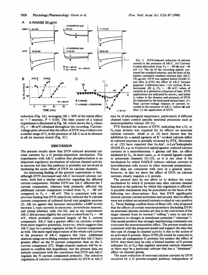
L FIG. 3. DYN-induced reduction of calciumcurrent in the presence of AK-C. (A) Currentsevoked alternately from Vh = -80 (e) and -40mV (o). The tip of the recording pipette con-tained the standard solution, and the body ofthepipette contained standard solution plus AK-C(50 ,ug/ml). DYN was applied before (EARLY)and after (LATE) the effect of AK-C became
-2 apparent. Calibration bars: 1 nA vertical; 20 msa! horizontal. (B) Ip (Vh = -80 mV) values of
,+4,0 2 currents inA plotted as a function of time. DYNV applications are indicated by arrows, and initial
Ip values in the absence and presence of DYNt-2 ! are indicated by the horizontal dashed lines. (C)
Peak current-voltage relation of currents re-corded in the presence of AK-C, before (a) and
t= 32 min after (o) the application of DYN.
reduction (Fig. 3A), averaging 168 + 30% of the initial effect(n = 7 neurons, P < 0.05). The time course of a typicalexperiment is illustrated in Fig. 3B, which shows the lp values(Vh = -80 mV) obtained throughout the recording. Current-voltage plots showed that the effect ofDYN was evident overa similar range of Vc in the presence ofAK-C as in its absencein all six neurons tested (Fig. 3C).
DISCUSSIONThe present results show that DYN reduced neuronal cal-cium currents by a G protein-dependent mechanism. Theexperiments with AK-C confirm that phosphorylation is an
important regulatory mechanism of calcium channel activityin neurons but that this pathway is probably not involved inmediating the acute effect of DYN on calcium channels.An interesting finding of the present experiments is that,
although DYN decreased and AK-C increased calcium cur-
rents, both had a similar selectivity regarding the affectedcurrent components. Neither DYN nor AK-C affected the Tcurrent component, whereas both primarily affected theadditional current component evoked from Vh = -80 mVcompared to Vh = -40 mV. This is consistent with our
previous finding that DYN selectively reduced the N calciumcurrent component of cultured dorsal root ganglion neurons
(2). AK (or agents that increase intracellular cAMP levels)increase L-type currents in myocytes, invertebrate neurons,and clonal pituitary cells (14-16), and in our experimentsAK-C did increase slightly the current evoked from Vh = -40mV, which probably consisted largely of the L currentcomponent. AK-C had a greater effect on currents evokedfrom more negative potentials, however, which suggests thatAK-C may be a potent regulator of the N current componentas well. The more rapid inactivation of the whole-cell currentin the presence of AK-C supports this view. Additionalanalysis using curve-fitting routines showed that AK-C had a
greater effect on the N current component than on the Lcurrent component (27). Single-channel analysis will be re-
quired to confirm this hypothesis, but the present data are
entirely consistent with the conclusion that DYN and AK-Cregulate the N current component primarily. The selectiveregulation of calcium current components by DYN or AK-C
may be of physiological importance, particularly if differentchannel types control specific neuronal processes such as
neurotransmitter release (29-31).PTX blocked the actions of DYN, indicating that a G,- or
Go-type protein was required for its effects on neuronalcalcium currents. Attali et al. (3) have shown that theinhibition by K-opioid agonists of K+-evoked calcium influxin cultured neurons is partially reversed by PTX. Hescheleret al. (32) have reported that [D-Ala2, D-Leu5]enkephalin(DADLE), a u- or 6-selective opioid agonist, reduced calciumcurrents in a neuroblastoma x glioma cell line, an effectmediated by Go. In most cases, ,- or 8-opioid ligands coupleto potassium channels (21-23), so it is not clear if themechanism by which DADLE reduces calcium currents inneuroblastoma cells occurs in other neuronal preparations.These data are consistent with ours in a general sense,
however, in that we show the effect of DYN on calciumcurrents clearly requires a G protein.The present data do not allow us to deduce the exact
mechanism by which G proteins may alter calcium channelfunction or the pathway by which this regulation is effected.A possible mechanism may be postulated on the basis of thefollowing two observations: first, that DYN significantlyslowed calcium current activation and second, that its effectswere not evident on outward currents evoked at very positiveVc. These findings confirm those of Bean (24), who proposedthat the effects of certain neurotransmitters (including DYN)on neuronal calcium currents are due to the conversion of thetarget channel from its normal ("willing") state to one lessresponsive to changes in membrane potential ("reluctant").This model predicts that strongly depolarizing commands canovercome the neurotransmitter effect. The present results areconsistent with the proposed model and support the idea thatthis type of change in channel activity is due to the action ofan activated G protein. Since GTP[yS] would be expected toactivate all endogenous G proteins but mimics the effect ofDYN, then there may be only a limited number of G proteinsubtypes (Gi or GO) that regulate neuronal calcium channelsor there may be a particular subtype that has a "dominant"effect on channel gating.The acute reduction of neuronal calcium currents by DYN
occurred by a G protein-coupled pathway independent of
LATE
BI
0° 1000
- 50Q.
mr-EtMA NM Am4--
Proc. Natl. Acad. Sci. USA 87 (1990)
0
Proc. Natl. Acad. Sci. USA 87 (1990) 7029
Q DYN
cAMP GTPGDP
AK AK-C ____::
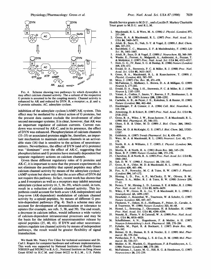
FIG. 4. Scheme showing two pathways by which dynorphin Amay affect calcium channel activity. The a subunit of the respectiveG protein is assumed to be the coupling moiety. Channel activity isenhanced by AK and reduced by DYN. R, K receptor; a, 8, and y,G protein subunits; AC, adenylate cyclase.
inhibition of the adenylate cyclase/cAMP/AK system. Thiseffect may be mediated by a direct action of G proteins, butthe present data cannot exclude the involvement of othersecond messenger systems. It is clear, however, that AK wasan important regulator of calcium currents. Current run-
down was reversed by AK-C, and in its presence, the effectofDYN was enhanced. Phosphorylation of calcium channels(33-35) or associated proteins might be, therefore, an impor-tant mechanism to maintain calcium channels in an activat-able state (16) that is sensitive to the actions of neurotrans-mitters. Nevertheless, the effect ofDYN (and ofG proteins)was "dominant" over the effect of AK-C, suggesting thatphosphorylation and G proteins have mutually dependent butseparate regulatory actions on calcium channels.Given these different regulatory roles of G proteins and
AK-C, it is important to note that the present experiments donot eliminate the possibility that opioid peptides may affectcalcium channel activity by means of the adenylate cyclase/cAMP system but show only that the acute effect ofDYN didnot require this pathway. In fact, recent work has shown that,u and 8 receptors as well as K receptors may inhibit neuronaladenylate cyclase activity (4, 5, 36-39), which could, in turn,result in a reduction of calcium channel activity. This hy-pothesis could account for both acute (AK-independent) andlong-term (AK-dependent) modulation of calcium channelactivity by K-opioid peptides, by means of different G pro-tein-dependent pathways (Fig. 4). Such a scheme may alsoaccount for development of desensitization or tolerance toopiates (for example, see refs. 5, 38, and 39). The end result,a decrease in calcium influx, would influence a wide varietyof calcium-dependent intraneuronal processes and may bethe basis for the inhibition of neurotransmitter release byopioid peptides (25, 40, 41). If DYN and other neurotrans-mitters regulate ion channel activity by means of independentpathways, the result would be greater flexibility of signaltransduction.
We thank Ms. Nancy Fox Ciliax for technical assistance and Dr.Carl J. Rogers for computer hardware and software implementation.This work was supported by National Institutes of Health GrantsNS01019 and NS1%13 to R.A.G., National Institute on Drug AbuseGrant 03365 to H.C.M. and Grant 04122 to R.L.M., U.S. Public
Health Service grants to M.D.U., and a Lucille P. Markey CharitableTrust grant to M.D.U. and R.L.M.
1. Macdonald, R. L. & Werz, M. A. (1986) J. Physiol. (London) 377,237-249.
2. Gross, R. A. & Macdonald, R. L. (1987) Proc. Nat!. Acad. Sci.USA 84, 5469-5473.
3. Attali, B., Saya, D., Nah, S.-Y. & Vogel, Z. (1989) J. Biol. Chem.264, 347-353.
4. Barchfield, C. C., Maassen, Z. F. & Medzihradsky, F. (1982) LifeSci. 31, 1661-1665.
5. Attali, B., Saya, D. & Vogel, Z. (1989) J. Neurochem. 52, 360-369.6. Wanke, E., Ferroni, A., Malgaroli, A., Ambrosini, A., Pozzan, T.
& Meldolesi, J. (1987) Proc. Nati. Acad. Sci. USA 84, 4313-4317.7. Holz, G. G., IV, Rane, S. G. & Dunlap, K. (1986) Nature (London)
319, 670-672.8. Ewald, D. A., Sternweis, P. C. & Miller, R. J. (1988) Proc. Nat!.
Acad. Sci. USA 85, 3633-3637.9. Gross, R. A., Macdonald, R. L. & Ryan-Jastrow, T. (1989) J.
Physiol. (London) 411, 585-595.10. Moss, J. (1987) Clin. Res. 35, 451-458.11. McFadzean, I., Mullaney, I., Brown, D. A. & Milligan, G. (1989)
Neuron 3, 177-182.12. Ewald, D. A., Pang. I.-H., Sternweis, P. C. & Miller, R. J. (1989)
Neuron 2, 1185-1193.13. Yatani, A., Codina, J., Imoto, Y., Reeves, J. P., Birnbaumer, L. &
Brown, A. M. (1987) Science 238, 1288-1292.14. Cachelin, A. B., de Peyer, J. E., Kokubun, S. & Reuter, H. (1983)
Nature (London) 304, 462-464.15. Hockberger, P. & Connor, J. A. (1984) Cell. Mol. Neurobiol. 4,
319-338.16. Armstrong, D. & Eckert, R. (1987) Proc. Nat!. Acad. Sci. USA 84,
2518-2522.17. Gross, R. A., Wiley, J. W., Ryan-Jastrow, T. & Macdonald, R. L.
(1990) Mol. Pharmacol. 37, 546-553.18. Olsen, S. R. & Uhler, M. D. (1989) J. Biol. Chem. 264, 18662-
18666.19. Uhler, M. D. & McKnight, G. S. (1987) J. Biol. Chem. 262, 15202-
15207.20. Goldstein, A. (1987) Trends Pharmacol. Sci. 8, 456-459.21. Werz, M. A. & Macdonald, R. L. (1983) Neurosci. Lett. 42, 173-
178.22. North, R. A. & Williams, J. T. (1985) J. Physiol. (London) 364,
265-280.23. Morita, K. & North, R. A. (1981) Brain Res. 242, 145-150.24. Bean, B. P. (1989) Nature (London) 340, 153-156.25. Cherubini, E. & North, R. A. (1985) Proc. Nat!. Acad. Sci. USA 82,
1860-1863.26. Sah, D. W. Y. (1990) J. Neurosci. 10, 136-141.27. Gross, R. A., Uhler, M. D. & Macdonald, R. L. (1990) J. Physiol.
(London) 429, 483-4%.28. Fox, A. P., Nowycky, M. C. & Tsien, R. W. (1987) J. Physiol.
(London) 394, 147-172.29. Hirning, L. D., Fox, A. P., McClesky, E. W., Olivera, B. M.,
Thayer, S. A., Miller, R. J. & Tsien, R. W. (1988) Science 239,57-61.
30. Perney, T. M., Hirning, L. D., Leeman, S. E. & Miller, R. J. (1986)Proc. Nat!. Acad. Sci. USA 83, 6656-6659.
31. Wiley, J. W., Gross, R. A., Lu, Y. & Macdonald, R. L. (1990) J.Neurophysiol. 63, 1499-1507.
32. Hescheler, J., Rosenthal, W., Trautwein, W. & Schultz, G. (1987)Nature (London) 325, 445-447.
33. Flockerzi, V., Oeken, H.-J., Hoffmann, F., Pelzer, D., Cavalie, A.& Trautwein, W. (1986) Nature (London) 323, 66-68.
34. Hymel, L., Striessing, J., Glossmann, H. & Schindler, H. (1988)Proc. Nat!. Acad. Sci. USA 85, 4290-4294.
35. Nunoki, K., Florio, V. & Catterall, W. A. (1989) Proc. Nat!. Acad.Sci. USA 86, 6816-6820.
36. Schoffelmeer, A. N., Hogenboom, F. & Mulder, A. H. (1987)Naunyn-Schmiedeberg's Arch. Pharmacol. 335, 278-284.
37. Eybalin, M., Pujol, R. & Bockaert, J. (1987) Brain Res. 421,336-342.
38. Beitner, D. B., Duman, R. S. & Nestler, E. J. (1989) Mol. Phar-macol. 35, 559-564.
39. Puttfarcken, P. S., Werling, L. S. & Cox, B. M. (1988) Mol. Phar-macol. 33, 520-527.
40. Mulder, A. H., Wardeh, G., Hogenboom, F. & Frankhuyzen, A. L.(1984) Nature (London) 308, 278-280.
41. McFadzean, I., Lacey, M. G., Hill, R. G. & Henderson, G. (1987)Neuroscience 20, 231-239.
Physiology/Pharmacology: Gross et al.