
Ca2+-Induced Rigidity Change of the Myosin VIIa IQ …ias.ust.hk/ias/files/pdf/1506416155_b2.pdf ·...
18
Article Ca 2+ -Induced Rigidity Change of the Myosin VIIa IQ Motif-Single a Helix Lever Arm Extension Graphical Abstract Highlights d Myo7a contains a stable single a helix (SAH) following its IQ motifs d Apo-CaM stabilizes the conformational rigidity of the IQ5- SAH lever arm extension d Ca 2+ increases flexibility of the IQ5-SAH lever arm extension of Myo7a d Ca 2+ -induced lever arm flexibility change may also occur in other myosins Authors Jianchao Li, Yiyun Chen, Yisong Deng, Ilona Christy Unarta, Qing Lu, Xuhui Huang, Mingjie Zhang Correspondence [email protected] (J.L.), [email protected] (M.Z.) In Brief The structures of Myo7a IQ5-SAH lever arm extension in complex with apo- and Ca 2+ -CaM, respectively, reveal that the motor contains an extended and rigid lever arm at low Ca 2+ concentration conditions. Increased cellular Ca 2+ concentration induces conformational flexibility of the motor and thus regulates its activity. Li et al., 2017, Structure 25, 579–591 April 4, 2017 ª 2017 Elsevier Ltd. http://dx.doi.org/10.1016/j.str.2017.02.002
Transcript of Ca2+-Induced Rigidity Change of the Myosin VIIa IQ …ias.ust.hk/ias/files/pdf/1506416155_b2.pdf ·...

Article
Ca2+-Induced Rigidity Cha
nge of the Myosin VIIa IQMotif-Single a Helix Lever Arm ExtensionGraphical Abstract
Highlights
d Myo7a contains a stable single a helix (SAH) following its IQ
motifs
d Apo-CaM stabilizes the conformational rigidity of the IQ5-
SAH lever arm extension
d Ca2+ increases flexibility of the IQ5-SAH lever arm extension
of Myo7a
d Ca2+-induced lever arm flexibility change may also occur in
other myosins
Li et al., 2017, Structure 25, 579–591April 4, 2017 ª 2017 Elsevier Ltd.http://dx.doi.org/10.1016/j.str.2017.02.002
Authors
Jianchao Li, Yiyun Chen, Yisong Deng,
Ilona Christy Unarta, Qing Lu,
Xuhui Huang, Mingjie Zhang
[email protected] (J.L.),[email protected] (M.Z.)
In Brief
The structures of Myo7a IQ5-SAH lever
arm extension in complex with apo- and
Ca2+-CaM, respectively, reveal that the
motor contains an extended and rigid
lever arm at low Ca2+ concentration
conditions. Increased cellular Ca2+
concentration induces conformational
flexibility of the motor and thus regulates
its activity.

Structure
Article
Ca2+-Induced Rigidity Change of the Myosin VIIaIQ Motif-Single a Helix Lever Arm ExtensionJianchao Li,1,5,* Yiyun Chen,1,5 Yisong Deng,1,6 Ilona Christy Unarta,2 Qing Lu,1,3,4 Xuhui Huang,2,3
and Mingjie Zhang1,3,4,7,*1Division of Life Science, State Key Laboratory of Molecular Neuroscience, Hong Kong University of Science and Technology,
Clear Water Bay, Kowloon, Hong Kong, China2Department of Chemistry, Hong Kong University of Science and Technology, Clear Water Bay, Kowloon, Hong Kong, China3Center of Systems Biology and Human Health, Hong Kong University of Science and Technology, Clear Water Bay, Kowloon,
Hong Kong, China4Institute for Advanced Study, Hong Kong University of Science and Technology, Clear Water Bay, Kowloon, Hong Kong, China5Co-first author6Present address: Department of Integrative Computational and Structural Biology, The Scripps Research Institute, La Jolla, CA 92037, USA7Lead contact
*Correspondence: [email protected] (J.L.), [email protected] (M.Z.)http://dx.doi.org/10.1016/j.str.2017.02.002
SUMMARY
Several unconventional myosins contain a highlycharged single a helix (SAH) immediately followingthe calmodulin (CaM) binding IQ motifs, functioningto extend lever arms of these myosins. How suchSAH is connected to the IQ motifs and whether theconformation of the IQ motifs-SAH segments areregulated by Ca2+ fluctuations are not known. Here,we demonstrate by solving its crystal structurethat the predicted SAH of myosin VIIa (Myo7a) formsa stable SAH. The structure of Myo7a IQ5-SAHsegment in complex with apo-CaM reveals that theSAH sequence can extend the length of the Myo7alever arm. Although Ca2+-CaM remains bound toIQ5-SAH, the Ca2+-induced CaM binding modechange softens the conformation of the IQ5-SAHjunction, revealing aCa2+-induced lever armflexibilitychange for Myo7a. We further demonstrate that thelast IQ motif of several other myosins also bindsto both apo- and Ca2+-CaM, suggesting a commonCa2+-induced conformational regulationmechanism.
INTRODUCTION
Unconventional myosins are actin-based molecular motors that
function in cell motility and intracellular transportation (Hartman
et al., 2011). Myosin VIIa (Myo7a), a member of the class VII un-
conventional myosins (Myo7) encoded bymyo7a, plays essential
roles in both the development and physiological functions of the
auditory and visual systems in mammals. In the auditory system,
Myo7a, together with its associated proteins, is essential for the
development and structural maintenance of stereocilia, which
are actin-based protrusions at the apical surface of cochlear
and vestibular hair cells of inner ears responsible for sound per-
ceptions and spatial balancing (Gillespie and Muller, 2009; Lefe-
vre et al., 2008; Pan and Zhang, 2012; Richardson et al., 2011;
Tilney et al., 1992). In the visual system, Myo7a is expressed in
photoreceptor cells and pigmented epithelial cells, and is essen-
tial for proper connecting cilium formation as well as melano-
some transportation in photoreceptors (Liu et al., 1997a, 1998).
Mutations ofmyo7a can cause various human genetic disorders
such as Usher syndrome type IB (USH1B), characterized by
combined early-onset deafness and blindness (Gibson et al.,
1995; Well et al., 1995), and non-syndromic deafness (DFNA11
and DFNB2) (Liu et al., 1997b; Tamagawa et al., 1996; Weil
et al., 1997). Numerous Myo7a mutations have been identified
in patients, and many of these mutations can alter its structural
stability or affect its interactions with binding partners (Pan and
Zhang, 2012; Wu et al., 2011).
From its N to C terminus, Myo7a contains a motor domain,
five calmodulin (CaM) binding IQ motifs, a highly charged post-
IQ region predicted to form a single a helix (SAH), and two
MyTH4-FERM tandems separated by an SH3 domain (Fig-
ure 1A). In vertebrates, there are two isoforms of Myo7, namely
Myo7a and Myo7b, which share a highly similar structural archi-
tecture because of their >50% amino acid sequence identity
(Chen et al., 2001). Analogous to Myo7a, Myo7b is essential for
the formation and maintenance of microvilli in intestinal and kid-
ney brush borders, another type of actin-rich protrusion analo-
gous to stereocilia (Crawley et al., 2014, 2016; Li et al., 2016).
Myo7b differs from Myo7a by possessing a short insertion in
its motor domain, which may promote its actin binding (Henn
and De La Cruz, 2005). Myo7b also lacks a highly charged
SAH fragment in its post-IQ region (Figure 1A).
Initially, the highly charged post-IQ region in Myo7a was
predicted to be a coiled coil and thought to mediate Myo7a
dimerization (Chen et al., 2001). However, a study published a
few years later showed that a similar highly charged region
directly after the last IQ motif in Myosin X (Myo10) forms a stable
SAH and functions to elongate the lever arm of the motor (Knight
et al., 2005). Subsequently, it was discovered that Myosin VI
(Myo6) also contains a similar charged SAH in its lever arm
extension region (Peckham, 2011; Spink et al., 2008). Myo7a
is also predicted to contain an SAH-like structure, like a num-
ber of non-myosin proteins such as inner centromere protein
and caldesmon-1 (Peckham and Knight, 2009; Swanson and
Structure 25, 579–591, April 4, 2017 ª 2017 Elsevier Ltd. 579

Figure 1. Myo7a Post-IQ Region Forms a Stable SAH
(A) Domain organization of Myo7a and sequence alignment of the post-IQ regions of Myo7a and Myo7b. The 3-residue in-frame deletion found in DFNA11
patients is highlighted with a box in magenta.
(B) FPLC-SLS showing that Myo7a post-IQ is monomeric in solution. The experimentally determined molar masses of Trx-Myo7a-857-1000, Trx-Myo7a-866-
935, and Myo7a-866-935 were 31.4, 23.0, and 9.2 kDa, which fitted well with their respective theoretical molar masses of 31.8, 23.4, and 9.2 kDa.
(C) 15N-HSQC spectrum of Myo7a-866-935.
(D) Circular dichroism spectrum of Myo7a-866-935.
(E) Urea-induced denaturation profile of Myo7a-866-935. The change of ellipticity value of the protein at 222 nm was used to calculate the relative fraction of
protein denaturation at each urea concentration. Values are means ± SD from three independent measurements.
(F) Ribbon diagram of the crystal structure of Myo7a-866-935 with ionic interactions between charged residues labeled. Themagenta box highlights K887, which
is deleted in the DFNA11 mutation.
(G) Helical net diagrams of Myo7a-866-935. The residues involved in the ionic interactions are highlighted with filled circles. The 3-residue in-frame deletion in
DFNA11 patients is highlighted by magenta circles. The C-terminal three residues missing in the electron density are shown in dashed circles.
Sivaramakrishnan, 2014). However, whether the post-IQ region
of Myo7a indeed forms an SAH is still being debated. It was
recently suggested that part of the Myo7a post-IQ forms a
coiled-coil dimer (Sakai et al., 2015).
CaM, as a common light chain of unconventional myosins, is
widely known to be critical for the motors to function properly
in a Ca2+-regulated manner (Bahler and Rhoads, 2002; Batters
and Veigel, 2016; Cheney and Mooseker, 1992; Heissler and
Sellers, 2014; Trybus, 1994). It is commonly accepted that
CaM, in its Ca2+-free state, specifically binds to and thereby
maintains the conformational rigidity of the IQ motifs (Houdusse
et al., 2006; Lu et al., 2015). The apo-CaM-bound IQmotifs serve
as lever arms of myosins both in their dimeric transport compe-
tent forms and in their monomeric mechanical tethering forms
(Heissler and Sellers, 2014; Houdusse et al., 2006). It is also
generally perceived that an increase in cellular Ca2+ concentra-
tion will weaken the interaction between CaM and IQ motifs
and thereby cause CaM to dissociate frommyosins, with subse-
quent softening of the lever arms of the motors (Heissler and
Sellers, 2014; Lu et al., 2015). It should be pointed out that a
few IQ motifs in several myosins have been shown to bind to
580 Structure 25, 579–591, April 4, 2017
apo- and Ca2+-CaM (Lu et al., 2012b, 2015; Sakai et al., 2015;
Zhu et al., 1998). However, structural studies of IQ/Ca2+-CaM
have been limited to IQ motifs from non-myosin proteins (Chen
et al., 2014; Kim et al., 2008; Mori et al., 2008; Van Petegem
et al., 2005; Wang et al., 2014).
In this work we solved the crystal structure of the Myo7a post-
IQ region, which reveals that it indeed forms a stable SAH. We
further determined the crystal structure of IQ5-SAH in complex
with apo-CaM. The complex structure, together with biochem-
ical experiments, reveals that the bound apo-CaMplays a critical
role in maintaining the conformational rigidity in the junctional re-
gion connecting IQ5 and SAH of Myo7a. We discovered that
Ca2+-CaM remains stably bound to IQ5. The crystal structure
of IQ5/Ca2+-CaM complex reveals that, upon binding to Ca2+,
CaM undergoes a translocation along the IQ motifs toward
the N terminus of Myo7a by �2.5 helical turns, a movement
that causes the IQ5-SAH junction to be completely solvent
accessible. As a result, the conformational rigidity of IQ5-SAH
(i.e., the lever arm extension) of Myo7a decreases. We further
showed that IQmotifs situated at the distal end of several myosin
lever arms also bind to both apo- and Ca2+-CaM. Finally, we

demonstrated that the missense DFNA11 R853C mutation of
Myo7a disrupts the IQ5/apo-CaM interaction and causes func-
tional impairments of the motor.
RESULTS
Myo7a Post-IQ Region Forms a Stable SAHThe post-IQ region of Myo7a is evolutionarily conserved (Fig-
ure 1A) and shares a charge distribution pattern similar to that
of Myo10 SAH. To test whether the post-IQ region of Myo7a
may also form a stable SAH instead of a coiled-coil dimer, we
first purified a Trx-tagged fragment of mouse Myo7a spanning
Arg857 (directly after the last IQ motif) to Lys1000 (the beginning
of the first MyTH4 domain). Analytical gel filtration chromatog-
raphy coupled with static light-scattering (FPLC-SLS) analysis
showed that the entire Myo7a post-IQ forms a stable monomer
in solution (Figure 1B, fitted molecular weight 31.4 kDa versus
theoretical molecular weight 31.8 kDa). We mapped the Myo7a
post-IQ to a shorter and stable monomer spanning residues
866–935 (denoted Myo7a-866-935) based on its sequence
conservation and secondary structure prediction (Figure 1B). It
is noted that the 9.2-kDa Myo7a-866-935 after Trx-tag cleavage
was eluted at a smaller volume than the 14-kDa Trx tag,
indicating that Myo7a-866-935 adopts an elongated shape
instead of the globular Trx (Figure 1B). The 15N-heteronuclear
single-quantum coherence (HSQC) spectrum of the 15N-labeled
Myo7a-866-935 showed that themajority of the backbone peaks
are distributed in a narrow chemical-shift window typical for
a-helical conformation (Figure 1C). Consistently, the circular
dichroism (CD) spectrum of Myo7a-866-935 showed that the
protein is largely a-helical (Figure 1D). We next performed a
denaturation experiment of Myo7a-866-935 by monitoring the
ellipticity of the protein at 222 nm as a function of increasing
urea concentrations. It is noteworthy that the decreasing of the
helical content of the protein is linearly proportional to increasing
urea concentration (R2 = 0.99), indicating that Myo7a-866-935
is stably folded but does not contain any tertiary structure (Fig-
ure 1E). Taken together, the above biophysical characterizations
strongly indicate that Myo7a-866-935 forms a stable monomer
enriched in a-helical conformation.
To gain further insight, we determined the crystal structure
of Myo7a-866-935 at 2.1-A resolution (Table 1 and Figure 1F).
The structure was solved by a combination of single-wavelength
anomalous dispersion of iodide derivatives of sample crystals
and molecular replacement methods. All of Myo7a-866-935
except for the C-terminal three residues (933–935) are visible in
the electron density, and the protein forms a straight, rigid SAH.
A total of eight pairs of E-R/K charge-charge interactions are
distributed throughout the entire helix (Figures 1F and 1G), which
presumably are the key determinants for the stable a-helix forma-
tionofSAH inMyo7aaswell asMyo10 (Knight et al., 2005).Human
genetics studies have identified a 9-bp deletion within exon 22
of myo7a, resulting an in-frame deletion of Ala886, Lys887, and
Lys888 fromMyo7a, and themutation is known to causenon-syn-
dromic deafness DFNA11 in patients (Liu et al., 1997c). From the
Myo7a-866-935 structure, Lys887 forms a charge-charge inter-
action with Glu891 (Figures 1F and 1G). The deletion of the three
residues containing Lys887 is likely to destabilize SAH, which
might impair the function of Myo7a in the patients.
IQ5 and SAH Form a Continuous Helix in the Presence ofApo-CaMSingle-molecule experiments have shown that by replacing part
of the IQ motif-based lever arm with a similar length of SAH,
Myosin-Va (Myo5a) can still processively move on actin fila-
ments (Baboolal et al., 2009), indicating that SAH can function
to extend the lever arm length of myosins. To function as a lever
arm extension of a myosin, the SAH segment of the motor must
be connected to its IQ motif helix, forming a rigid structural en-
tity (i.e., a continuous and stable a helix), so that the mechanical
force produced by the motor head can be transmitted through
the entire lever arm. To test this hypothesis, we set out to deter-
mine the crystal structure of IQ5-SAH in complex with apo-
CaM. Using a boundary with residues 834–935 of mouse
Myo7a (referred to as Myo7a-IQ5-SAH) and by co-expressing
with CaM, we succeeded in purifying a stable Myo7a-IQ5-
SAH/CaM complex in the presence of EDTA in the sample
buffer and obtained complex crystals that diffracted to 3.0-A
resolution (Table 1). The structure of the Myo7a-IQ5-SAH/CaM
complex was solved by the molecular replacement method
using the Myo5a-IQ1/apo-CaM complex structure (PDB: 2IX7)
and the Myo7a SAH structure determined above as the search
models. Indeed, IQ5 is bound by apo-CaM in a canonical
fashion, with the C-terminal lobe of CaM adopting a semi-
open conformation and the N lobe adopting a closed conforma-
tion (Figure S1) (Houdusse et al., 2006). Importantly, SAH fol-
lows directly after IQ5, together forming one continuous and
rigid a helix (Figure 2A). The electron density (2Fo-Fc map, con-
toured at 1s) of the junctional region between IQ5 and SAH
clearly showed that the C-terminal tail of IQ5 and the beginning
of SAH form a continuous and regular a-helical structure, and
the N-terminal lobe of apo-CaM intimately contacts with several
residues from the junctional region between IQ5 and SAH
(Figure 2B). The interaction between very negatively charged
CaM and the positively charged IQ5/SAH junction is likely to
be important for the structural stability of the IQ5-SAH helix,
as otherwise unfavorable repulsions resulted from two pairs of
positively charged residues in the junctional region (Arg854-
Arg858 and Arg853-His856, Figure 2B) would distort the helix
formation (see below for more details). Supporting this, in the
recently solved crystal structure of Myo10 SAH together with
the antiparallel coiled coil (Myo10-SAH-antiCC), although half
of the IQ3 was included in the crystallization construct, the
absence of apo-CaM binding made it partially disordered (Ro-
pars et al., 2016). It is generally accepted that tandem IQ motifs
can form a continuous helix in the presence of apo-CaM in un-
conventional myosins (Houdusse et al., 2006; Lu et al., 2015). As
such, the five IQ motifs of Myo7a contribute to a lever arm
length of �19 nm and the SAH further extends the lever arm
by �9 nm, rendering the motor to have a total lever arm length
of �28 nm (Figure 2A).
The continuous helix formation in Myo7a-IQ5-SAH provides a
mechanistic basis for SAH to act as a lever arm extension of the
motor. The regular distribution of charged residues is the charac-
teristic of SAH, and an algorithm to predict the existence of SAH
has been developed (Gaspari et al., 2012). We set out to search
for the possible presence of SAH in all myosins using this
method. For all myosins in the mouse proteome deposited in
Swiss-Prot (UniProt Consortium, 2015), only Myo7a, Myo10,
Structure 25, 579–591, April 4, 2017 581

Table 1. X-Ray Crystallographic Data Collection and Model Refinement
Data Collection
Datasets 866-935-Native 866-935-KI IQ5-SAH/Apo-CaM IQ5/Ca2+-CaM
Space group P21 C2 P41 P21
Wavelength (A) 0.9793 1.5418 1.0300 0.9791
Unit cell parameters (A) a = 38.81, b = 42.61,
c = 59.68
a = g = 90�, b = 90.07�
a = 49.06, b = 38.79,
c = 45.44
a = g = 90�, b = 99.78�
a = b = 80.93,
c = 175.29
a = b = g = 90�
a = 38.68, b = 89.76,
c = 45.72
a = g = 90�, b = 106.03�
Resolution range (A) 50–2.10 (2.21–2.10) 50–2.70 (2.85–2.70) 50–3.0 (3.16–3.00) 50–2.35 (2.39–2.35)
No. of unique reflections 11,455 (1,683) 2,393 (356) 22,489 (3,279) 12,459 (622)
Redundancy 3.4 (3.5) 6.6 (6.5) 6.7 (6.9) 2.8 (2.7)
I/s 8.3 (2.0) 10.6 (3.9) 9.3 (2.0) 23.0 (4.1)
Completeness (%) 99.0 (99.5) 100 (100) 99.6 (100) 97.5 (98.9)
Rmergea (%) 8.8 (61.8) 12.2 (43.6) 13.1 (96.7) 6.2 (36.0)
Structure Refinement
Resolution (A) 50–2.1 (2.31–2.10) 50–3.0 (3.08–3.00) 50–2.35 (2.56–2.35)
Rcrystb/Rfree
c (%) 19.46/23.85 (27.87/29.95) 22.37/29.01 (30.62/34.11) 22.73/27.06 (29.40/32.66)
Rmsd bonds (A)/angles (�) 0.010/1.071 0.006/0.939 0.013/1.153
Average B factord 40.4 102.3 45.4
No. of atoms
Protein atoms 1,198 3,104 2,472
Other molecules 7 0 35
No. of reflections
Working set 10,868 20,428 11,838
Test set 548 2002 598
Ramachandran plot regionsd
Favored (%) 98.6 97.2 98.8
Allowed (%) 1.4 2.6 1.2
Outliers (%) 0 0.2 0
Numbers in parentheses represent the value for the highest-resolution shell. Rmsd, root-mean-square deviation.aRmerge = SjIi � <I>j/SIi, where Ii is the intensity of measured reflection and <I> is the mean intensity of all symmetry-related reflections.bRcryst = SjjFcalcj � jFobsjj/SFobs, where Fobs and Fcalc are observed and calculated structure factors.cRfree = STjjFcalcj � jFobsjj/SFobs, where T is a test dataset of about 5% or 10% of the total unique reflections randomly chosen and set aside prior to
refinement.dB factors and Ramachandran plot statistics are calculated using MolProbity (Chen et al., 2010).
and Myo6 have SAH prediction scores above a critical threshold
(Figure 2C), a result consistent with a previous prediction (Peck-
ham, 2011). We believe that myosins with predicted SAH score
below 2.0 do not contain SAH, as the regions predicted to be
SAH in these myosins overlap with their coiled-coil regions
(e.g., the known coiled-coil dimerization domain of Myo5a). For
Myo7a and Myo10, the predicted SAHs partially overlap with
or begin directly after the last IQ motif. For Myo6, the predicted
SAH partially overlap with the last helix (a3) of the three-helix
bundle lever arm extension (Mukherjea et al., 2009; Yu et al.,
2012) (Figure 2D). Taken together, the structure of the IQ5-
SAH/apo-CaM complex and the amino acid sequence analysis
discussed above strongly suggest that, in addition to Myo7a,
Myo10 also contains a rigid lever arm extending SAH smoothly
connected to their respective last IQ motif.
Myo7a IQ5 Also Binds Ca2+-CaMThe generally accepted paradigm of myosin IQ motifs is that IQ
motifs can specifically bind to apo-CaM, and a rise in cellular
582 Structure 25, 579–591, April 4, 2017
Ca2+ concentration leads to dissociation or at least weakened
binding of Ca2+-CaM. However, there exist examples that
certain IQ motifs from myosins can bind to apo-CaM as well as
to Ca2+-CaM (Lu et al., 2012b, 2015; Zhu et al., 1998). We thus
asked whether Myo7a IQ5 may also bind to Ca2+-CaM. For the
IQ5-SAH and CaM mixture, in buffer containing either EDTA or
CaCl2, the molar masses of the complex measured by the
light-scattering experiments are essentially identical and fit well
with a 1:1 stoichiometry (Figure 3A), indicating that one molecule
of Ca2+-CaM can stably bind to IQ5-SAH. To rule out the possi-
bility that SAH instead of IQ5might be the Ca2+-CaM binding site
of IQ5-SAH, we truncated SAH from the IQ5-SAH and tested its
binding to CaM. FPLC-SLS showed that IQ5 alone can still form
a stable 1:1 complex with CaM in buffers containing either EDTA
or CaCl2 (Figure 3B), indicating that IQ5 can bind to both apo-
and Ca2+-CaM. The different elution volumes of the IQ5/CaM
complexes in the two buffer conditions indicated that the con-
formations of the complex in the Ca2+-free and Ca2+-saturated
conditions are different.

Figure 2. Structure of Myo7a IQ5-SAH in Complex with apo-CaM
(A) Ribbon diagram of Myo7a IQ5-SAH in complex with apo-CaM. IQ5 is colored in gray and SAH is in green. The N lobe and C lobe of CaM are colored blue and
light blue, respectively. This color-coding scheme is used throughout subsequent figures.
(B) Electron density of the IQ5-SAH junctional region contoured at 1s.
(C) SAHs found by the program scan4csah in all myosins in the mouse proteome (indicated with green stars). The SAHs with score below 2 were proved to be
coiled coil. The score of 2.0 was chosen as the threshold for SAH, as the predicted SAH with scores below 2.0 are actually part of the coiled coils of myosins.
(D) Secondary structure analysis of Myo7a, Myo10, and Myo6 showed continuous helix formation of SAH following the distal IQ motifs (or a3 of Myo6 LAE). The
boundaries of SAHs are based on the predictions by the scan4csah program. The boundaries for the segments proceeding SAHs are from the IQ/apo-CaM
structures and the structure of Myo6 LAE (PDB: 2LD3).
See also Figure S1.
To understand the structural basis governing the interaction
between IQ5 andCa2+-CaM,we determined the crystal structure
of Myo7a-IQ5/Ca2+-CaM complex at 2.35-A resolution, using a
strategy of co-expressing IQ5 (amino acids 828–870) with
Ca2+-CaM to obtain a high-quality complex sample (Table 1).
Ca2+-CaM binds to IQ5 in parallel (i.e., with the N lobe of CaM in-
teracting with the N-terminal half of IQ5 and the C lobe interact-
ing with the C-terminal half of IQ5, Figure 3C, left). In the pres-
ence of Ca2+, the EF hands in both lobes of CaM adopt a
typical open conformation. Phe833 and Leu837 from IQ5 insert
into the N-lobe hydrophobic pocket, and Tyr846 and Met850
of IQ5 anchor into the C-lobe hydrophobic pocket of CaM (Fig-
ure 3C, left), resulting in a highly compact Ca2+-CaM/IQ5 com-
plex structure similar to many well-studied high-affinity Ca2+-
CaM/target complexes (Hoeflich and Ikura, 2002; Tidow and
Nissen, 2013). The solubility of the isolated IQ5 peptide is
extremely low, which has prevented us from measuring the
quantitative binding constant of the Ca2+-CaM/IQ5 complex.
Comparison of the apo-CaM/IQ5-SAH and Ca2+-CaM/IQ5
complex structures reveals several important insights. For easy
viewing, we have aligned the IQ5 helix from the two structures
into the exact same position (Figure 3C). The binding of Ca2+
to CaM flips the overall orientation of CaM in binding to IQ5. In
the apo-CaM/IQ5-SAH complex, the CaM C lobe is anchored
by the Val843 of IQ5 (a residue equivalent to the signature Ile res-
idue of the ‘‘IQ’’ motif) and the CaM N lobe weakly engages
several residues at the C-terminal half of the IQ motif (Figure 3C,
left), adopting a so-called antiparallel binding mode. Upon Ca2+
binding, the CaM N lobe moves along the IQ5 helix by �4 nm (or
eight helix turns) toward IQ4 and anchors by the two hydropho-
bic residues (Phe833 and Leu837) at the beginning of IQ5, and
the C lobe moves slightly toward C-terminal end of the IQ5 helix
by �0.5 nm (or one helix turn), resulting in a parallel and more
extended Ca2+-CaM/target binding mode (Figure 3C, left). This
Ca2+-induced CaM binding change leads to the exposure of
�2.5 helix turns (�9 residues) located exactly at the junctional re-
gion between IQ5 and SAH to solvent (Figure 3C). We also noted
that the N lobe of Ca2+-CaM covers part of the IQ4 sequence,
indicating that the concatenated IQ45 together instead of IQ5
alone serves as the high-affinity Ca2+-CaM binding sequence.
This phenomenon has also been observed inMyo5a, where pairs
of tandemly connected IQ motifs bind to Ca2+-CaM with higher
affinity than to apo-CaM (Martin and Bayley, 2004).
Ca2+-Induced Softening of the Myo7a IQ5-SAH LeverArm ExtensionOur structural analysis has shown that the junctional region be-
tween IQ5 and SAH is not protected by CaM when Ca2+ is pre-
sent, and this Ca2+-induced change in binding mode may affect
the conformational flexibility of the IQ5-SAH helix (Figure 4A). To
test this hypothesis, we used trypsin partial digestion to probe
possible flexibility changes of IQ5-SAH induced by the binding
of Ca2+ to CaM. For digestion performed in the presence of
EDTA in the reaction buffer, the IQ5-SAH/CaM complex is stable
Structure 25, 579–591, April 4, 2017 583
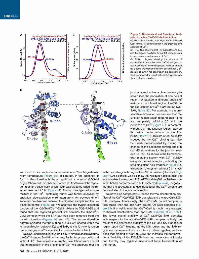
Figure 3. Biochemical and Structural Anal-
ysis of the Myo7a-IQ5/CaM Interaction
(A) FPLC-SLS showing that Myo7a-IQ5-SAH and
CaM form a 1:1 complex both in the presence and
absence of Ca2+.
(B) FPLC-SLS showing that Trx-taggedMyo7a-IQ5
and Trx-tagged CaM also form a 1:1 complex both
in the presence and absence of Ca2+.
(C) Ribbon diagram showing the structure of
Myo7a-IQ5 in complex with Ca2+-CaM (left) or
apo-CaM (right). The hydrophobic residues critical
for binding are highlighted by the stick model. Ca2+
ions are shown in red spheres. In this comparison,
the IQ5motifs in the two structures are alignedwith
the exact same position.
andmost of the complex remained intact after 2 hr of digestion at
room temperature (Figure 4A). In contrast, in the presence of
Ca2+ in the digestion buffer a significant amount of IQ5-SAH
degradation could be observedwithin the first 5min of the diges-
tion reaction. Essentially all IQ5-SAH was digested when the re-
action reached 1.5 hr (Figure 4A). The trypsin-digested sample
mixture in the Ca2+-containing buffer was further analyzed by
analytical size-exclusion chromatography. An obvious differ-
ence can be observed between the digested sample and the un-
digested control (Figure 4B). We analyzed the trypsin digestion
product of the IQ5-SAH/Ca2+-CaM mixture by SDS-PAGE and
found that the digested product still contains the IQ5/Ca2+-
CaM complex while the SAH part has been removed from the
trypsin digestion (Figures 4C and 4D). The trypsin digestion
pattern indicated that the cutting site is likely located within the
junctional region between IQ5 and SAH, as this is the only region
that undergoes Ca2+-dependent exposure to the solvent.
Wealso usedmolecular dynamics (MD) simulations to evaluate
the Ca2+-induced flexibility changes. For both systems, with and
without Ca2+, five individual 40-ns MD simulations were carried
out. Interestingly, in the presence of Ca2+ we observed that the
584 Structure 25, 579–591, April 4, 2017
junctional region has a clear tendency to
unfold (see the population at non-helical
region for backbone dihedral angles of
residue at junctional region, Leu859, in
the simulations of Ca2+-CaM-bound IQ5-
SAH, Figure S2). For example, in a repre-
sentative simulation we can see that the
junction region began to bend after 14 ns
and completely unfold at 20 ns in the
presence of Ca2+ (Figure 4E). In contrast,
without Ca2+ the junction region retained
its helical conformational in the first
20 ns (Figure 4E). This structural flexibility
induced by the Ca2+ binding can also
be clearly demonstrated by tracing the
change of the backbone torsion angle in
our MD simulations for the junction resi-
due Leu859. As shown in the Ramachan-
dran plot, the system with Ca2+ quickly
escapes the helical region, indicating the
unfolding of the helix (red line in Figure 4F).
In contrast, the systemwithout Ca2+ stays
in the helical region throughout theMDsimulation (blue line in Fig-
ure 4F). As a control, we also show that residues not located in the
junctional region (e.g., Arg848 on IQ5 andArg881 onSAH) remain
in the helical conformation in both systems (Figure 4F), suggest-
ing that the structural changes induced by the Ca2+ binding are
concentrated in the junctional region.
We have also compared CD-based thermal denaturation pro-
files of the Ca2+-CaM/IQ5-SAH complex and the apo-CaM/IQ5-
SAH complex. Interestingly, the Ca2+-CaM bound complex is
less stable than the apo-CaM bound IQ5-SAH complex (Fig-
ure S3). It is well known that Ca2+-CaM is much more resistant
to thermal denaturation than apo-CaM (Brzeska et al., 1983).
The lower overall stability of Ca2+-CaM/IQ5-SAH complex
with respect to the apo-CaM/IQ5-SAH complex is likely the
result of the decreased stability of the IQ5 and SAH junctional
region upon Ca2+ binding, as the IQ5 region and the SAH re-
gion are the same in both complexes. Taken together, we pro-
pose that binding of Ca2+ to CaM can increase the conforma-
tional flexibility of the IQ5-SAH lever arm extension of Myo7a
and thereby may regulate mechanical force transduction of
the motor.
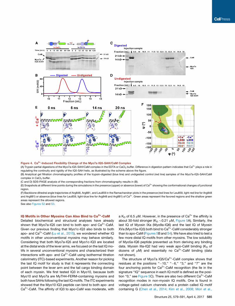
Figure 4. Ca2+-Induced Flexibility Change of the Myo7a IQ5-SAH/CaM Complex
(A) Trypsin partial digestions of the Myo7a-IQ5-SAH/CaM complex in the EDTA or CaCl2 buffer. Difference in digestion pattern indicates that Ca2+ plays a role in
regulating the continuity and rigidity of the IQ5-SAH helix, as illustrated by the scheme above the figure.
(B) Analytical gel filtration chromatography profiles of the trypsin-digested (blue line) and undigested control (red line) samples of the Myo7a-IQ5-SAH/CaM
complex in CaCl2 buffer.
(C and D) SDS-PAGE analysis of the corresponding fractions from chromatography results in (B).
(E) Snapshots at different time points during the simulations in the presence (upper) or absence (lower) of Ca2+ showing the conformational changes of junctional
region.
(F) Backbone dihedral angle trajectories of Arg848, Arg881, and Leu859 in the Ramachandran plots in the presence (red lines for Leu859, light-red line for Arg848
and Arg881) or absence (blue lines for Leu859, light-blue line for Arg848 and Arg881) of Ca2+. Green areas represent the favored regions and the shallow green
areas represent the allowed regions.
See also Figures S2 and S3.
IQ Motifs in Other Myosins Can Also Bind to Ca2+-CaMDetailed biochemical and structural analyses have already
shown that Myo7a-IQ5 can bind to both apo- and Ca2+-CaM.
Given our previous finding that Myo1c-IQ3 also binds to both
apo- and Ca2+-CaM (Lu et al., 2015), we wondered whether IQ
motifs in other unconventional myosins may behave similarly.
Considering that both Myo7a-IQ5 and Myo1c-IQ3 are located
at the distal ends of the lever arms, we focused on the last IQmo-
tifs in several unconventional myosins and characterized their
interactions with apo- and Ca2+-CaM using isothermal titration
calorimetry (ITC)-based experiments. Another reason for picking
the last IQ motif for study is that it represents the connecting
point between the lever arm and the tail cargo binding domain
of each myosin. We first tested IQ3 in Myo10, because both
Myo10 and Myo7a are MyTH4-FERM-containing myosins and
both have SAHs following the last IQmotifs. The ITC experiments
showed that the Myo10 IQ3 peptide can bind to both apo- and
Ca2+-CaM. The affinity of IQ3 to apo-CaM was moderate, with
a KD of 6.5 mM. However, in the presence of Ca2+ the affinity is
about 30-fold stronger (KD �0.21 mM, Figure 5A). Similarly, the
last IQ of Myosin IXa (Myo9a-IQ6) and the last IQ of Myosin
XVa (Myo15a-IQ3) both bind to Ca2+-CaM considerably stronger
than to apo-CaM (Figures 5B and S4).We have also tried to test a
few more distal IQ motifs from other myosins. The low solubility
of Myo5a-IQ6 peptide prevented us from deriving any binding
data. Myosin IIIa-IQ2 had very weak apo-CaM binding (KD of
dozens of mM) and essentially no Ca2+-CaM binding (data
not shown).
The structure of Myo7a IQ5/Ca2+-CaM complex shows that
residues at the positions ‘‘�10,’’ ‘‘�6,’’ ‘‘3,’’ and ‘‘7’’ are the
four anchoring points for Ca2+-CaM recognition (the Ile in the
signature ‘‘IQ’’ sequence in each IQ motif is defined as the posi-
tion ‘‘0,’’ see Figure 5C). There are also two different Ca2+-CaM
recognition modes in non-myosin IQ motifs. One is found in
voltage-gated calcium channels and a protein called IQ motif
containing G (Chen et al., 2014; Kim et al., 2008; Mori et al.,
Structure 25, 579–591, April 4, 2017 585

Figure 5. IQ Motifs from Other Myosins Can Bind to Both apo- and
Ca2+-CaM
(A) ITC titration curves showing the binding affinities of Ca2+-CaM (left) and
apo-CaM (right) to the Myo10-IQ3 peptide.
(B) Summary of the ITC-derived binding results of several distal IQ motifs of
myosins tested in this study. Myo10-IQ3, Myo9a-IQ6, and Myo15a-IQ3,
together with the previously reported Myo1c-IQ3, can bind to both apo- and
Ca2+-CaM. In all these cases, Ca2+-CaM binds to these IQ motifs with higher
affinities than does apo-CaM.
(C) Sequence alignment of the IQ motifs that can interact with both apo- and
Ca2+-CaM tested in this study. The red box highlights the positions of the
residues critical for anchoring Myo7a-IQ5 to Ca2+-CaM. The Myo5a-IQ1
sequence and the IQ consensus sequence are included for reference.
See also Figure S4.
2008; Van Petegem et al., 2005), in which residues at the
positions ‘‘�6’’ and ‘‘3’’ are responsible for anchoring to CaM.
Another binding mode is found in voltage-gated sodium chan-
nels (Wang et al., 2014), in which residues at the positions ‘‘0’’
and ‘‘9’’ are two anchoring points for CaM. Alignment of the
Ca2+-CaM binding IQ motifs from myosins characterized in this
study shows that except for Myo1c-IQ3, the other three IQ
motifs listed in Figure 5B cannot be fitted into any of the known
Ca2+-CaM/IQ binding patterns (Figure 5C), illustrating the
remarkable capacity of CaM to bind to diverse target proteins
586 Structure 25, 579–591, April 4, 2017
(Hoeflich and Ikura, 2002; Tidow and Nissen, 2013). The exact
binding modes of these myosin IQ motifs will need further struc-
tural characterization.
The R853C Mutation of Myo7a Found in DFNA11 IsFunctionally Defective Due to Its Loss of Apo-CaMBindingHuman genetic studies have identified a single-point missense
mutation, R853C, in Myo7a IQ5, which can cause autosomal
dominant hearing loss DFNA11 (Bolz et al., 2004). The structure
of the IQ5/apo-CaM complex shows that Arg853, which is
located at position ‘‘10’’ of the IQ motif, forms several hydrogen
bonds with both backbone and side chain of residues from both
lobes of apo-CaM (Figure S5A). Mutation of this residue to a
small and uncharged Cys will disrupt these hydrogen bonds,
thus decreasing the binding affinity between IQ5 and apo-
CaM. However, in the structure of IQ5/Ca2+-CaM complex, the
side chain of Arg853 is exposed to solvent and electron density
is missing, indicating that this Arg is not involved in the interac-
tion with Ca2+-CaM (Figure S5B). We used FPLC-SLS to eval-
uate the effect of the mutation. Consistent with our analysis,
essentially no IQ5/apo-CaM complex peak was observed
when mixing R853C-IQ5-SAH with apo-CaM (Figure 6A). In
contrast, in buffer containing CaCl2, both the elution profile
and the fitted molar mass of the mutant IQ5-SAH/Ca2+-CaM
complex are almost identical to those of the wild-type IQ5-
SAH/Ca2+-CaM complex (Figure 6B). Based on the structures
of Myo7a IQ5 bound to apo- and Ca2+-CaM, Val843 in the ‘‘0’’
position of the IQ motif is critical for its binding to apo-CaM,
but this residue is not in contact with Ca2+-CaM (Figure 3C).
Therefore, substitution of Val843 with Glu is expected to
eliminate the IQ5’s binding to apo-CaM but has no impact on
Ca2+-CaM binding. Indeed, FPLC-SLS analysis showed that
V843E-IQ5-SAH has no detectable binding to apo-CaM, and
the mutant behaves the same as the wild-type counterpart in
binding to Ca2+-CaM (Figures 6A and 6B). This analysis suggests
that the DNFA11 missense mutation specifically affects the apo-
CaM binding but not the Ca2+-CaM binding of Myo7a.
Next, we tried to set up a cell-based assay to evaluate the
impact of the DFNA11 missense mutation. It has been reported
that when forced into a dimer, Myo7a can target to the tips of fi-
lopodia when overexpressed in heterologous cells (Sakai et al.,
2011, 2015). However, in our hands neither the full-length wild-
typeMyo7a nor themotor-SAH construct fused to aGCN4 dimer
can localize to the tip of filopodia in HeLa cells. Another MyTH4-
FERM-containing myosin (Myo10), when overexpressed, can
robustly induce filopodia formation as well as target the motor
to the tips of filopodia (Kerber and Cheney, 2011; Lu et al.,
2012a). We used a chimera strategy by replacing Myo10-IQ3
with Myo7a-IQ5 to evaluate the structural/functional relationship
of Myo7-IQ5, as both IQ motifs can bind to apo- and Ca2+-CaM
as well as form a continuous helix with the immediately following
SAH. The position selected for the IQ motif swap is carefully de-
signed and based on the structure shown in Figure 3 and the
amino acid sequence alignment of the IQ motifs (Figure 6C). As
expected, when overexpressed in HeLa cells, the GFP-tagged
full-length Myo10 are enriched at the tips of numerous elongated
filopodia (Figure 6D1). By replacing its IQ3 with Myo7a IQ5 (de-
noted as Myo10-(IQ5)7a, Figure 6C), the filopodia tip localization

Figure 6. Biochemical and Functional Effect
of the R853C DFNA11 Mutation of Myo7a
(A and B) FPLC-SLS results showing that the
R853C mutation and the V843E mutation affect
their respective Myo7a-IQ5/apo-CaM binding (A)
but not their Myo7a-IQ5/Ca2+-CaM binding (B).
(C) The design of the Myo10 chimera constructs.
The black box highlights the sequences of Myo7a-
IQ5 and Myo1c-IQ2 used to replace Myo10-IQ3.
(D) Representative images of HeLa cells trans-
fected with various GFP-tagged constructs and
with actin filament stained with phalloidin (red),
and nuclei stained with DAPI (blue): D1, Myo10;
D2, Myo10-(IQ5)7a; D3, Myo10-R853C-(IQ5)7a; D4,
Myo10-V843E-(IQ5)7a; D5, Myo10-(IQ2)1c. Scale
bar, 20 mm.
(E) Quantitative analysis of GFP puncta number
per cell at the tips of filopodia. Values are means ±
SD from three independent experiments with >10
cells per experiment, using an unpaired t test.
***p < 0.001.
See also Figure S5.
of the chimera Myo10 is essentially the same as that of the wild-
type motor (Figures 6D2 and 6E). In total contrast, the Myo10
chimera with its IQ3 replaced by the Myo7a R853C-IQ5 showed
essentially no elongated filopodia and no filopodia tip localiza-
tion (Figures 6D3 and 6E), indicating that the R853C mutation
causing loss of apo-CaM binding compromised the function of
chimera Myo10. Using the same chimera strategy, we further
demonstrated that loss of apo-CaM binding caused by the
V843E mutation of Myo7a-IQ5 also eliminated filopodia induc-
tion capacity of the chimera Myo10 (Figures 6D4 and 6E). Since
processive walking on bundled actin filaments is essential for the
filopodia induction and tip localization of Myo10 (Lu et al.,
2012a), the failed filopodia induction and tip localization of the
two Myo10 chimeras bearing the Myo7a IQ5 mutants are pre-
sumably caused by loss of the lever arm rigidity of the mutant
chimeras. We also wanted to test the role of Ca2+-CaM binding
to IQ5 using the same chimera approach. However, this effort
was not successful, as we were unable to find a single-point mu-
tation either in Myo7a IQ5 or Myo10 IQ3 that can specifically
disrupt their Ca2+-CaM binding but retain the apo-CaM binding.
As an alternative, we designed another chimera by replacing
Myo10-IQ3 with Myo1c-IQ2 (denoted
Myo10-(IQ2)1c, Figure 6C), which we
have previously shown to interact only
with apo-CaM but not with Ca2+-CaM
(Lu et al., 2015). The Myo10 chimera car-
rying the Myo1c IQ2 can induce filopodia
elongation and target to the tips of filopo-
dia as well as the wild-type motor (Figures
6D5 and 6E).
DISCUSSION
Possible Roles of SAH in theFunction of Myo7aThe crystal structure of Myo7a-IQ5-SAH/
apo-CaM complex provided direct struc-
tural evidence supporting the view that an SAH following a distal
IQmotif can indeed function as a lever arm extension of amyosin
motor. Myo7a contains five IQmotifs forming the canonical lever
arm with a length of �19 nm. The SAH further extends the lever
arm length by �9 nm, thereby granting Myo7a the longest lever
arm among all vertebrate unconventional myosins. The exact
cellular function of having such a long lever arm for Myo7a will
require further study, as whether the motor functions as a cargo
transporter or mechanical tether (or both) is still not clear. Intui-
tively, the SAH-mediated extension of the rigid lever arm may
allow monomeric Myo7a to tether motor head-attached actin fil-
aments with tail-bound membrane cargoes with a wider space
and to transduce mechanical force between a motor head and
cargo binding tail. If Myo7a, analogous to Myo6, forms a cargo
binding-induced dimer (Sakai et al., 2011; Yu et al., 2009), the
dimerized motor with an extended lever arm containing rigid
SAH may increase the step size of the motor on actin filaments.
Importantly, the conformational rigidity of the Myo7a lever arm
extension can be regulated by cellular Ca2+ concentration
changes. Increasing the cellular Ca2+ concentration softens the
rigidity of the Myo7a lever arm extension.
Structure 25, 579–591, April 4, 2017 587
Possible Roles of IQ/Ca2+-CaM BindingIn Myo7a, the Ca2+-induced CaM binding mode change can
cause a breakage/softening of the continuous helix formed be-
tween IQ5 and SAH. This will lead to the loss of rigidity in the lever
arm, meaning that the motor cannot function properly in the
presence of Ca2+. However, this mechanism does not explain
why Ca2+-CaM needs to remain associated with the IQ motif
with an even higher affinity. One could argue that a simple
Ca2+-induced dissociation of CaM can achieve the same lever
arm-softening effect. Additionally we showed that high-affinity
binding of Ca2+-CaM to the most distal IQ motif seems to be a
rather common property for a number of myosins. It is possible
that retained Ca2+-CaM binding to IQ motifs may maintain the
lever arm to be partially structured even when myosins are in
their off-state upon elevation of Ca2+ concentration. When cells
return to their resting state, formation of the rigid lever arms of
myosins does not require a complete refolding process, thereby
shortening the recovery time required for motors to return to their
working state. Second, the retained binding of Ca2+-CaM to IQ
motifs provides a mechanism for CaM to be a continuous resi-
dent protein of myosins. With such a CaM molecule residing at
an IQ motif, this IQ motif can rapidly bind to the same CaM
when cellular concentration drops, as otherwise the binding
has to wait for diffusion-mediated encounter of another CaM
molecule in cytosol. Such resident CaM on IQ motifs may
significantly speed up the functional recovery of myosins in
response to changes in cellular Ca2+ concentration. Similar resi-
dent CaM-facilitated rapid IQ motif rebinding by apo-CaM has
been demonstrated to be critical for kinetics of Ca2+-dependent
activations of Ca2+ andNa+ channels (Ben-Johny and Yue, 2014;
DeMaria et al., 2001).
Recently, it was shown that CaM in Myo6 can also undergo
a Ca2+-induced binding rearrangement in the motor’s lever arm
region (Batters et al., 2016). Combined with low-resolution elec-
tron microscopy-based study, the authors proposed a model
describing Ca2+-mediated regulation of Myo6 activity (Batters
et al., 2016). In this model, Myo6 adopts a tail-backfolded,
auto-inhibited conformation at low Ca2+ concentration state.
Upon rise of Ca2+ concentration, Ca2+-induced CaM binding re-
arrangement leads to release of Myo6’s auto-inhibition and con-
verts the motor into a non-motile but primed state capable of
cargo binding. Interestingly, binding of Ca2+-CaM to the distal
IQ motif of Myo6 also appears to increase the conformational
flexibility of the motor lever arm, with the motor thus adopting
a non-motile state (Batters et al., 2016). The cargo-loaded motor
will further convert into the active, apo-CaM binding state when
Ca2+ concentration returns to low level. A similar mechanism
may also apply to Myo7a (Batters and Veigel, 2016). Myo7a
also adopts an auto-inhibited conformation in its apo-CaM
bound, cargo-free form (Umeki et al., 2009; Yang et al., 2009).
Myo7a can undergo Ca2+-induced CaM binding rearrangement,
as shown in the current study. Finally, cargo binding to the tail of
Myo7a can convert the motor into a processive dimer (Sakai
et al., 2011).
Our studies on chimeric Myo10 in filopodia inductions in heter-
ologous cells provided a convenient assay for us to investigate
roles of apo-CaM binding to Myo7a’s IQ motifs in the motor’s
cellular function. However, it should be pointed out that as this
chimera approach is indirect in nature, the derived conclusion
588 Structure 25, 579–591, April 4, 2017
should be viewed as tentative. Further studies will be required
to address this question. For example, in vitro single-molecule-
based assays can be used to assess the impact of Ca2+ on
Myo7a’s stepping velocity or stride size and processivity on actin
filaments. Alternatively, one might compare functional changes
of photoreceptors or hair cells containing the wild-type Myo7a
or its CaM binding mutants to obtain more direct evidence per-
taining to the roles of apo-CaM and Ca2+-CaM on the motor’s
physiological functions.
Potential Uses of SAH as a Research ToolAlthough the prevalent existence of SAH has been recognized
for years, the only structural information is the recently reported
crystal structure of Myo10-SAH-antiCC at 3.5-A resolution (Ro-
pars et al., 2016). However, the low resolution in that study did
not allow confident resolution of the side chains and thus cannot
provide any details of the structural determinants of the SAH. The
crystal structure of Myo7a SAH presented in the current study
provides the high-resolution atomic picture of how an isolated,
stable SAH is formed. The inter-turn charge-charge interactions
contribute to the high stability of the SAH. Given the extremely
simple interaction mode observed in the Myo7a SAH structure,
it will be feasible to design SAHs with different lengths. With their
composition of alternative E and K/R, SAHs are biologically inert,
genetically encodable, and length programmable. These unique
features, together with their high stability and conformational ri-
gidity, make SAHs attractive molecular rulers to physically posi-
tion two biomolecules with precise distances (Sivaramakrishnan
and Spudich, 2011; Swanson and Sivaramakrishnan, 2014). In
addition, a combined use of IQ motifs together with SAH (e.g.,
based on the Myo7a-IQ5-SAH segment demonstrated in this
study) not only can provide genetically encodable spacers for
precisely positioning two proteins apart but also allows such
spacers (or, more precisely, the conformational flexibility of the
spaces) to be regulated by Ca2+.
In the construct used for crystallization (residues 866–935),
around 60% of the residues are charged residues E/R/K. To
the best of our knowledge, Myo7a-866-935 is the protein with
the highest percentage of charged residues that has ever
been crystallized. To our surprise, this highly charged protein is
extremely easy to crystallize. In our initial crystallization screen
experiments, many conditions containing polyethylene glycol
(PEG) and low concentration of salts yielded high-diffraction-
quality crystals. The crystallization of Myo7a SAH is facilitated
by the packing of a small stretch of hydrophobic residues
(Leu880/Met884 and Leu925/Leu926/Met929). Since it is biolog-
ically inert and very easy to crystallize, Myo7a-SAH or its deriva-
tives might be used as a protein crystallization tag for proteins
that are difficult to crystallize.
STAR+METHODS
Detailed methods are provided in the online version of this paper
and include the following:
d KEY RESOURCES TABLE
d CONTACT FOR REAGENT AND RESOURCE SHARING
d EXPERIMENTAL MODEL AND SUBJECT DETAILS
B HeLa Cells Culture

d METHOD DETAILS
B Protein Expression and Purification
B Static Light Scattering Analysis
B Circular Dichroism Spectroscopy
B NMR Spectroscopy
B Crystallography
B Sequence Analysis
B Trypsin Partial Digestion Studies
B Molecular Dynamics Simulations
B Isothermal Titration Calorimetry Assay
B Cellular Localization and Filopodia Quantification
d QUANTIFICATION AND STATISTICAL ANALYSIS
d DATA AND SOFTWARE AVAILABILITY
SUPPLEMENTAL INFORMATION
Supplemental Information includes five figures and can be found with this
article online at http://dx.doi.org/10.1016/j.str.2017.02.002.
AUTHOR CONTRIBUTIONS
Conceptualization, J.L., Q.L., and M.Z.; Methodology, J.L., Q.L., Y.C., and
I.C.U.; Investigation, J.L., Y.C., Y.D., and I.C.U.; Writing – Original Draft, J.L.
and M.Z.; Writing – Review & Editing, J.L., Y.C., Y.D., I.C.U., Q.L., X.H.,
and M.Z.; Funding Acquisition, Q.L., X.H., and M.Z.; Supervision and Coordi-
nation, M.Z.
ACKNOWLEDGMENTS
We thank the Shanghai Synchrotron Radiation Facility (SSRF) BL19U1 and
BL17U1 for X-ray beam time and Dr. Quan Hao for accessing the in-house
X-ray diffraction machine. This work was supported by grants from RGC
of Hong Kong (664113, 16103614, AoE-M09-12, and T13-607/12R), and a
973 program grant from the Minister of Science and Technology of China
(2014CB910204) to M.Z.; a grant from RGC of Hong Kong (16149516) to Q.L.;
and a grant from RGC of Hong Kong (C6009-15G) to X.H. and M.Z.. M.Z. is a
Kerry Holdings Professor in Science and a Senior Fellow of IAS at HKUST.
Received: September 14, 2016
Revised: December 8, 2016
Accepted: February 9, 2017
Published: March 2, 2017
REFERENCES
Adams, P.D., Afonine, P.V., Bunkoczi, G., Chen, V.B., Davis, I.W., Echols, N.,
Headd, J.J., Hung, L.W., Kapral, G.J., Grosse-Kunstleve, R.W., et al. (2010).
PHENIX: a comprehensive Python-based system for macromolecular struc-
ture solution. Acta Crystallogr. D Biol. Crystallogr. 66, 213–221.
Baboolal, T.G., Sakamoto, T., Forgacs, E., White, H.D., Jackson, S.M., Takagi,
Y., Farrow, R.E., Molloy, J.E., Knight, P.J., Sellers, J.R., et al. (2009). The SAH
domain extends the functional length of themyosin lever. Proc. Natl. Acad. Sci.
USA 106, 22193–22198.
Bahler, M., and Rhoads, A. (2002). Calmodulin signaling via the IQ motif. FEBS
Lett. 513, 107–113.
Batters, C., and Veigel, C. (2016). Mechanics and activation of unconventional
myosins. Traffic 17, 860–871.
Batters, C., Brack, D., Ellrich, H., Averbeck, B., and Veigel, C. (2016). Calcium
can mobilize and activate myosin-VI. Proc. Natl. Acad. Sci. USA 113,
E1162–E1169.
Ben-Johny, M., and Yue, D.T. (2014). Calmodulin regulation (calmodulation) of
voltage-gated calcium channels. J. Gen. Physiol. 143, 679–692.
Bolz, H., Bolz, S.S., Schade, G., Kothe, C., Mohrmann, G., Hess, M., and Gal,
A. (2004). Impaired calmodulin binding of myosin-7A causes autosomal domi-
nant hearing loss (DFNA11). Hum. Mutat. 24, 274–275.
Brzeska, H., Venyaminov, S.Vu., Grabarek, Z., and Drabikowski, W. (1983).
Comparative studies on thermostability of calmodulin, skeletal muscle
troponin C and their tryptic fragments. FEBS Lett. 153, 169–173.
Bussi, G., Donadio, D., and Parrinello, M. (2007). Canonical sampling through
velocity rescaling. J. Chem. Phys. 126, 014101.
Chen, Z.Y., Hasson, T., Zhang, D.S., Schwender, B.J., Derfler, B.H.,
Mooseker, M.S., and Corey, D.P. (2001). Myosin-VIIb, a novel unconventional
myosin, is a constituent of microvilli in transporting epithelia. Genomics 72,
285–296.
Chen, V.B., Arendall, W.B., 3rd, Headd, J.J., Keedy, D.A., Immormino, R.M.,
Kapral, G.J., Murray, L.W., Richardson, J.S., and Richardson, D.C. (2010).
MolProbity: all-atom structure validation for macromolecular crystallography.
Acta Crystallogr. D Biol. Crystallogr. 66, 12–21.
Chen, L.T., Liang, W.X., Chen, S., Li, R.K., Tan, J.L., Xu, P.F., Luo, L.F., Wang,
L., Yu, S.H., Meng, G., et al. (2014). Functional and molecular features of the
calmodulin-interacting protein IQCG required for haematopoiesis in zebrafish.
Nat. Commun. 5, 3811.
Cheney, R.E., and Mooseker, M.S. (1992). Unconventional myosins. Curr.
Opin. Cell Biol. 4, 27–35.
Cowtan, K. (2006). The Buccaneer software for automated model building. 1.
Tracing protein chains. Acta Crystallogr. D Biol. Crystallogr. 62, 1002–1011.
Crawley, S.W., Shifrin, D.A., Jr., Grega-Larson, N.E., McConnell, R.E., Benesh,
A.E., Mao, S., Zheng, Y., Zheng, Q.Y., Nam, K.T., Millis, B.A., et al. (2014).
Intestinal brush border assembly driven by protocadherin-based intermicrovil-
lar adhesion. Cell 157, 433–446.
Crawley, S.W., Weck, M.L., Grega-Larson, N.E., Shifrin, D.A., Jr., and Tyska,
M.J. (2016). ANKS4B is essential for intermicrovillar adhesion complex forma-
tion. Dev. Cell 36, 190–200.
Darden, T., York, D., and Pedersen, L. (1993). Particle Mesh Ewald - an
N.Log(N) method for ewald sums in large systems. J. Chem. Phys. 98,
10089–10092.
DeMaria, C.D., Soong, T.W., Alseikhan, B.A., Alvania, R.S., and Yue, D.T.
(2001). Calmodulin bifurcates the local Ca2+ signal that modulates P/Q-type
Ca2+ channels. Nature 411, 484–489.
Emsley, P., Lohkamp, B., Scott, W.G., and Cowtan, K. (2010). Features and
development of Coot. Acta Crystallogr. D Biol. Crystallogr. 66, 486–501.
Gaspari, Z., Suveges, D., Perczel, A., Nyitray, L., and Toth, G. (2012). Charged
single alpha-helices in proteomes revealed by a consensus prediction
approach. Biochim. Biophys. Acta 1824, 637–646.
Gibson, F., Walsh, J., Mburu, P., Varela, A., Brown, K.A., Antonio, M., Beisel,
K.W., Steel, K.P., and Brown, S.D.M. (1995). A type-VII myosin encoded by
the mouse deafness gene shaker-1. Nature 374, 62–64.
Gillespie, P.G., and Muller, U. (2009). Mechanotransduction by hair cells:
models, molecules, and mechanisms. Cell 139, 33–44.
Hartman, M.A., Finan, D., Sivaramakrishnan, S., and Spudich, J.A. (2011).
Principles of unconventional myosin function and targeting. Annu. Rev. Cell
Dev. Biol. 27, 133–155.
Heissler, S.M., and Sellers, J.R. (2014). Myosin light chains: teaching old dogs
new tricks. Bioarchitecture 4, 169–188.
Henn, A., and De La Cruz, E.M. (2005). Vertebrate myosin VIIb is a high duty
ratio motor adapted for generating and maintaining tension. J. Biol. Chem.
280, 39665–39676.
Hess, B., Bekker, H., Berendsen, H.J.C., and Fraaije, J.G.E.M. (1997). LINCS:
a linear constraint solver for molecular simulations. J. Comput. Chem. 18,
1463–1472.
Hoeflich, K.P., and Ikura, M. (2002). Calmodulin in action: diversity in target
recognition and activation mechanisms. Cell 108, 739–742.
Houdusse, A., Gaucher, J.F., Krementsova, E., Mui, S., Trybus, K.M., and
Cohen, C. (2006). Crystal structure of apo-calmodulin bound to the first
two IQ motifs of myosin V reveals essential recognition features. Proc. Natl.
Acad. Sci. USA 103, 19326–19331.
Structure 25, 579–591, April 4, 2017 589

Jorgensen, W.L., and Madura, J.D. (1983). Quantum and statistical studies of
liquids. 25. Solvation and conformation of methanol in water. J. Am. Chem.
Soc. 105, 1407–1413.
Kerber, M.L., and Cheney, R.E. (2011). Myosin-X: aMyTH-FERMmyosin at the
tips of filopodia. J. Cell Sci. 124, 3733–3741.
Kim, E.Y., Rumpf, C.H., Fujiwara, Y., Cooley, E.S., Van Petegem, F., andMinor,
D.L., Jr. (2008). Structures of CaV2 Ca2+/CaM-IQ domain complexes reveal
binding modes that underlie calcium-dependent inactivation and facilitation.
Structure 16, 1455–1467.
Knight, P.J., Thirumurugan, K., Xu, Y.H., Wang, F., Kalverda, A.P., Stafford,
W.F., Sellers, J.R., and Peckham, M. (2005). The predicted coiled-coil domain
of myosin 10 forms a novel elongated domain that lengthens the head. J. Biol.
Chem. 280, 34702–34708.
Lefevre, G., Michel, V., Weil, D., Lepelletier, L., Bizard, E., Wolfrum, U.,
Hardelin, J.P., and Petit, C. (2008). A core cochlear phenotype in USH1 mouse
mutants implicates fibrous links of the hair bundle in its cohesion, orientation
and differential growth. Development 135, 1427–1437.
Li, J., He, Y., Lu, Q., and Zhang, M. (2016). Mechanistic basis of organization of
the harmonin/USH1C-mediated brush border microvilli tip-link complex. Dev.
Cell 36, 179–189.
Lindorff-Larsen, K., Piana, S., Palmo, K., Maragakis, P., Klepeis, J.L., Dror,
R.O., and Shaw, D.E. (2010). Improved side-chain torsion potentials for the
Amber ff99SB protein force field. Proteins 78, 1950–1958.
Liu, X., Ondek, B., andWilliams, D.S. (1998). Mutantmyosin VIIa causes defec-
tive melanosome distribution in the RPE of shaker-1 mice. Nat. Genet. 19,
117–118.
Liu, X., Vansant, G., Udovichenko, I.P., Wolfrum, U., and Williams, D.S.
(1997a). Myosin VIIa, the product of the Usher 1B syndrome gene, is concen-
trated in the connecting cilia of photoreceptor cells. Cell Motil. Cytoskeleton
37, 240–252.
Liu, X.Z., Walsh, J., Mburu, P., Kendrick-Jones, J., Cope, M.J., Steel, K.P., and
Brown, S.D.M. (1997b). Mutations in the myosin VIIA gene cause non-syn-
dromic recessive deafness. Nat. Genet. 16, 188–190.
Liu, X.Z., Walsh, J., Tamagawa, Y., Kitamura, K., Nishizawa, M., Steel, K.P.,
and Brown, S.D. (1997c). Autosomal dominant non-syndromic deafness
caused by a mutation in the myosin VIIA gene. Nat. Genet. 17, 268–269.
Lu, Q., Ye, F., Wei, Z.Y., Wen, Z., and Zhang, M.J. (2012a). Antiparallel coiled-
coil-mediated dimerization of myosin X. Proc. Natl. Acad. Sci. USA 109,
17388–17393.
Lu, Z., Shen, M., Cao, Y., Zhang, H.M., Yao, L.L., and Li, X.D. (2012b).
Calmodulin bound to the first IQ motif is responsible for calcium-dependent
regulation of myosin 5a. J. Biol. Chem. 287, 16530–16540.
Lu, Q., Li, J., Ye, F., and Zhang, M. (2015). Structure of myosin-1c tail bound to
calmodulin provides insights into calcium-mediated conformational coupling.
Nat. Struct. Mol. Biol. 22, 81–88.
Martin, S.R., and Bayley, P.M. (2004). Calmodulin bridging of IQ motifs in
myosin-V. FEBS Lett. 567, 166–170.
Mccoy, A.J., Grosse-Kunstleve, R.W., Adams, P.D., Winn, M.D., Storoni, L.C.,
and Read, R.J. (2007). Phaser crystallographic software. J. Appl. Crystallogr.
40, 658–674.
Mori, M.X., Vander Kooi, C.W., Leahy, D.J., and Yue, D.T. (2008). Crystal
structure of the CaV2 IQ domain in complex with Ca2+/calmodulin: high-reso-
lution mechanistic implications for channel regulation by Ca2+. Structure 16,
607–620.
Mukherjea, M., Llinas, P., Kim, H., Travaglia, M., Safer, D., Menetrey, J.,
Franzini-Armstrong, C., Selvin, P.R., Houdusse, A., and Sweeney, H.L.
(2009). Myosin VI dimerization triggers an unfolding of a three-helix bundle in
order to extend its reach. Mol. Cell 35, 305–315.
Murshudov, G.N., Skubak, P., Lebedev, A.A., Pannu, N.S., Steiner, R.A.,
Nicholls, R.A., Winn, M.D., Long, F., and Vagin, A.A. (2011). REFMAC5
for the refinement of macromolecular crystal structures. Acta Crystallogr.
D Biol. Crystallogr. 67, 355–367.
Otwinowski, Z., and Minor, W. (1997). Processing of X-ray diffraction data
collected in oscillation mode. Methods Enzymol. 276, 307–326.
590 Structure 25, 579–591, April 4, 2017
Pan, L., and Zhang, M. (2012). Structures of usher syndrome 1 proteins and
their complexes. Physiology (Bethesda) 27, 25–42.
Parrinello, M., and Rahman, A. (1981). Polymorphic transitions in single-crys-
tals—a new molecular-dynamics method. J. App. Phys. 52, 7182–7190.
Peckham, M. (2011). Coiled coils and SAH domains in cytoskeletal molecular
motors. Biochem. Soc. Trans. 39, 1142–1148.
Peckham, M., and Knight, P.J. (2009). When a predicted coiled coil is really a
single alpha-helix, in myosins and other proteins. Soft Matter 5, 2493–2503.
Pronk, S., Pall, S., Schulz, R., Larsson, P., Bjelkmar, P., Apostolov, R., Shirts,
M.R., Smith, J.C., Kasson, P.M., van der Spoel, D., et al. (2013). GROMACS
4.5: a high-throughput and highly parallel open source molecular simulation
toolkit. Bioinformatics 29, 845–854.
Richardson, G.P., de Monvel, J.B., and Petit, C. (2011). How the genetics of
deafness illuminates auditory physiology. Annu. Rev. Physiol. 73, 311–334.
Ropars, V., Yang, Z., Isabet, T., Blanc, F., Zhou, K., Lin, T., Liu, X., Hissier, P.,
Samazan, F., Amigues, B., et al. (2016). The myosin X motor is optimized for
movement on actin bundles. Nat. Commun. 7, 12456.
Sakai, T., Umeki, N., Ikebe, R., and Ikebe, M. (2011). Cargo binding activates
myosin VIIA motor function in cells. Proc. Natl. Acad. Sci. USA 108,
7028–7033.
Sakai, T., Jung, H.S., Sato, O., Yamada, M.D., You, D.J., Ikebe, R., and Ikebe,
M. (2015). Structure and regulation of the movement of human myosin VIIA.
J. Biol. Chem. 290, 17587–17598.
Sivaramakrishnan, S., and Spudich, J.A. (2011). Systematic control of protein
interaction using a modular ER/K alpha-helix linker. Proc. Natl. Acad. Sci. USA
108, 20467–20472.
Spink, B.J., Sivaramakrishnan, S., Lipfert, J., Doniach, S., and Spudich, J.A.
(2008). Long single alpha-helical tail domains bridge the gap between struc-
ture and function of myosin VI. Nat. Struct. Mol. Biol. 15, 591–597.
Swanson, C.J., and Sivaramakrishnan, S. (2014). Harnessing the unique
structural properties of isolated alpha-helices. J. Biol. Chem. 289, 25460–
25467.
Tamagawa, Y., Kitamura, K., Ishida, T., Ishikawa, K., Tanaka, H., Tsuji, S., and
Nishizawa, M. (1996). A gene for a dominant form of non-syndromic sensori-
neural deafness (DFNA11) maps within the region containing the DFNB2
recessive deafness gene. Hum. Mol. Genet. 5, 849–852.
Terwilliger, T.C., Grosse-Kunstleve, R.W., Afonine, P.V., Moriarty, N.W., Zwart,
P.H., Hung, L.W., Read, R.J., and Adams, P.D. (2008). Iterative model building,
structure refinement and density modification with the PHENIX AutoBuild
wizard. Acta Crystallogr. D Biol. Crystallogr. 64, 61–69.
Tidow, H., and Nissen, P. (2013). Structural diversity of calmodulin binding to
its target sites. FEBS J. 280, 5551–5565.
Tilney, L.G., Tilney, M.S., and DeRosier, D.J. (1992). Actin filaments, stereoci-
lia, and hair cells: how cells count and measure. Annu. Rev. Cell Biol. 8,
257–274.
Trybus, K.M. (1994). Role of myosin light-chains. J. Muscle Res. Cell Motil 15,
587–594.
Umeki, N., Jung, H.S., Watanabe, S., Sakai, T., Li, X.D., Ikebe, R., Craig, R.,
and Ikebe, M. (2009). The tail binds to the head-neck domain, inhibiting
ATPase activity of myosin VIIA. Proc. Natl. Acad. Sci. USA 106, 8483–8488.
UniProt Consortium. (2015). UniProt: a hub for protein information. Nucleic
Acids Res. 43, D204–D212.
Vagin, A., and Teplyakov, A. (1997). MOLREP: an automated program for
molecular replacement. J. Appl. Crystallogr. 30, 1022–1025.
Van Petegem, F., Chatelain, F.C., and Minor, D.L., Jr. (2005). Insights into
voltage-gated calcium channel regulation from the structure of the CaV1.2
IQ domain-Ca2+/calmodulin complex. Nat. Struct. Mol. Biol. 12, 1108–1115.
Wang, C., Chung, B.C., Yan, H., Wang, H.G., Lee, S.Y., and Pitt, G.S. (2014).
Structural analyses of Ca(2)(+)/CaM interaction with NaV channel C-termini
reveal mechanisms of calcium-dependent regulation. Nat. Commun. 5, 4896.
Weil, D., Kussel, P., Blanchard, S., Levy, G., LeviAcobas, F., Drira, M., Ayadi,
H., and Petit, C. (1997). The autosomal recessive isolated deafness, DFNB2,

and the Usher 1B syndrome are allelic defects of the myosin-VIIA gene. Nat.
Genet. 16, 191–193.
Well, D., Blanchard, S., Kaplan, J., Guilford, P., Gibson, F., Walsh, J., Mburu,
P., Varela, A., Levillers, J., Weston, M.D., et al. (1995). Defective myosin VIIA
gene responsible for usher syndrome type 1b. Nature 374, 60–61.
Winn, M.D., Ballard, C.C., Cowtan, K.D., Dodson, E.J., Emsley, P., Evans,
P.R., Keegan, R.M., Krissinel, E.B., Leslie, A.G., McCoy, A., et al. (2011).
Overview of the CCP4 suite and current developments. Acta Crystallogr.
D Biol. Crystallogr. 67, 235–242.
Wu, L., Pan, L., Wei, Z., and Zhang, M. (2011). Structure of MyTH4-FERM do-
mains in myosin VIIa tail bound to cargo. Science 331, 757–760.
Yang, Y., Baboolal, T.G., Siththanandan, V., Chen, M., Walker, M.L., Knight,
P.J., Peckham, M., and Sellers, J.R. (2009). A FERM domain autoregulates
Drosophila myosin 7a activity. Proc. Natl. Acad. Sci. USA 106, 4189–4194.
Yu, C., Feng,W.,Wei, Z., Miyanoiri, Y., Wen,W.Y., Zhao, Y.X., and Zhang, M.J.
(2009). Myosin VI undergoes cargo-mediated dimerization. Cell 138, 537–548.
Yu, C., Lou, J., Wu, J., Pan, L., Feng, W., and Zhang, M. (2012). Membrane-
induced lever arm expansion allows myosin VI to walk with large and variable
step sizes. J. Biol. Chem. 287, 35021–35035.
Zhu, T., Beckingham, K., and Ikebe, M. (1998). High affinity Ca2+ binding sites
of calmodulin are critical for the regulation of myosin I beta motor function.
J. Biol. Chem. 273, 20481–20486.
Structure 25, 579–591, April 4, 2017 591

STAR+METHODS
KEY RESOURCES TABLE
REAGENT or RESOURCE SOURCE IDENTIFIER
Chemicals, Peptides, and Recombinant Proteins
rhodamine-conjugated phalloidin Invitrogen Cat#R415
DMEM Life Technologies Cat#12800-017
FBS Hyclone Cat#SV30160.03
Trypsin Sigma Cat#T4665
Synthetic Myo10 IQ3 peptide
(LLRRRFLHLKKAAIVFQKQLRGQIARRVY)
Synthesized by ChinaPeptides N/A
Synthetic Myo9a IQ6 peptide
(RKKYKEQRNKIILLQSIYRGFRARQRC)
Synthesized by ChinaPeptides N/A
Synthetic Myo15a IQ3 peptide
(RQRYQQMRQSLLKFRSLVHTYVNRRRY)
Synthesized by ChinaPeptides N/A
Recombinant protein: mouse Myo7a SAH
(aa 866-935, ref# NP_001243010.1)
This paper N/A
Recombinant protein: mouse Myo7a IQ5
(aa 828-870, ref# NP_001243010.1)
This paper N/A
Recombinant protein: mouse Myo7a IQ5-SAH
(aa 828-935, ref# NP_001243010.1)
This paper N/A
Recombinant protein: human Calmodulin
(full length, ref# P62158)
This paper N/A
Critical Commercial Assays
NanoJuice transfection kit Millipore Cat#71902-4
Deposited Data
Myo7a 866-935 structure This paper PDB: 5WST
Myo7a IQ5-SAH/apo-CaM complex structure This paper PDB: 5WSU
Myo7a IQ5/Ca2+-CaM complex structure This paper PDB: 5WSV
Experimental Models: Cell Lines
Human: HeLa cells ATCC CCL-2
Experimental Models: Organisms/Strains
Escherichia coli: BL21-CodonPlus Invitrogen #230240
Recombinant DNA
Plasmid: GFP-Myo10 This paper N/A
Plasmid: GFP-Myo10-(IQ5)7a This paper N/A
Plasmid: GFP-Myo10-(IQ2)1c This paper N/A
Software and Algorithms
Origin7.0 OriginLab http://www.originlab.com/
GraphPad Prism GraphPad Software Inc. http://www.graphpad.com/scientific-software/prism/
HKL2000 (Otwinowski and Minor, 1997) http://www.hkl-xray.com/
iMosflm & SCALA (Winn et al., 2011) www.ccp4.ac.uk/
MOLREP (Vagin and Teplyakov, 1997) http://www.ccp4.ac.uk/
PHASER (Mccoy et al., 2007) http://www.phaser.cimr.cam.ac.uk/
PHENIX (Adams et al., 2010) http://www.phenix-online.org/
Buccaneer (Cowtan, 2006) http://www.ccp4.ac.uk/
Refmac (Murshudov et al., 2011) http://www.ccp4.ac.uk/
Coot (Emsley et al., 2010) http://www2.mrc-lmb.cam.ac.uk/Personal/pemsley/coot/
GROMACS 4.5 (Pronk et al., 2013) http://www.gromacs.org/
PyMOL DeLano Scientific LLC http://www.pymol.org/
scan4sah (Gaspari et al., 2012) http://csahserver.chem.elte.hu/
e1 Structure 25, 579–591.e1–e4, April 4, 2017

CONTACT FOR REAGENT AND RESOURCE SHARING
Further information and requests for reagents may be directed to, and will be fulfilled by the Lead Contact Mingjie Zhang (mzhang@
ust.hk).
EXPERIMENTAL MODEL AND SUBJECT DETAILS
HeLa Cells CultureHeLa cells (from ATCC) were cultured in DMEM media containing 10% fetal bovine serum (FBS).
METHOD DETAILS
Protein Expression and PurificationVarious constructs for Myo7a IQ and post-IQ region were created by cloning the corresponding mouse Myo7a (NCBI:
NP_001243010.1) cDNA into a pET-32m3C vector that contains an N-terminal Trx-tag and a (His)6-tag. All truncations and point mu-
tations used in the current study were created with the standard PCR-based mutagenesis method and confirmed by DNA
sequencing. For heterologous cell expressions, the full-length human Myo10 (NCBI: NP_036466.2) and its chimeras were cloned
into a modified EGFP vector.
Recombinant proteins were expressed in Escherichia coli BL21-CodonPlus and protein expression was induced by addition of
IPTG at 16 �C. The His-tagged proteins were purified by Ni2+-NTA agarose (QIAGEN) affinity chromatography followed by size-exclu-
sion chromatography using the column buffer of 50 mM Tris-HCl, pH 7.8, containing 100 mM NaCl, 1 mM DTT, and 5 mM EDTA
(EDTA buffer) or 50 mM Tris-HCl, pH 7.8, containing 100 mM NaCl, 1 mM DTT, and 10 mM CaCl2 (CaCl2 buffer).
Static Light Scattering AnalysisProteins were loaded on to a Superose 12 (10/300 GL) column (GE Healthcare) equilibrated with EDTA buffer or CaCl2 buffer. The
AKTA FPLC system was coupled to a three-angle light-scattering detector (miniDAWN TREOS, Wyatt technology) and a refractive
index detector (Optilab T-rEX, Wyatt technology). The data were recorded and processed by the ASTRA 6.1.2 software (Wyatt
Technology).
Circular Dichroism SpectroscopyCD spectra of Myo7a-866-935 at a concentration of 1.8 mg/ml in buffer containing 50mM Tris-HCl, pH 7.8, 100mMNaCl, 1 mMDTT
and 1 mM EDTA were acquired on a JASCO J-815 CD spectropolarimeter at 20 �C using a cell path length of 1 mm. Each spectrum
was collected with three scans spanning a spectral window of 200�250 nm. To investigate urea-induced denaturation, CD spectra of
Myo7a-866-935 solutions were measured in the presence of different concentrations of urea. Thermal denaturation experiments of
Myo7a-IQ5-SAH/CaM complex (concentration at 26 mM) in the presence or absence of Ca2+ were performed on Chirascan CD spec-
trometer (Applied Photophysics) from 16�60 �C at a 0.5 �C stepped temperature ramping with a setting time of 30 sec. In both dena-
turation experiments, the ellipticities at 222 nmwere used to calculate the unfolded fraction and plotted as a function of temperature.
NMR SpectroscopyProtein samples for NMR experiments were concentrated to�0.6 mM in 20 mM Tris buffer containing 100 mMNaCl, 1 mM DTT and
5 mM EDTA at pH 7.0 in 90% H2O/10% D2O. The 1H-15N HSQC spectrum was acquired at 35 �C on a Varian Inova 750 MHz
spectrometer.
CrystallographyAll the crystals were obtained by hanging drop or sitting drop vapor diffusion methods at 16 �C. Crystals of Myo7a-866-935 were
grown in solution containing 0.2 M ammonium acetate, 0.1 M sodium acetate (pH 4.6) and 30% w/v PEG 4,000. Crystals of
Myo7a-IQ5-SAH/apo-CaM complex were grown in solution containing 0.1 M sodium citrate tribasic (pH 5.6), 20% v/v isopropanol
and 20% w/v PEG 4,000. Crystals of IQ5/Ca2+-CaM were grown in solution containing 0.2 M ammonium sulfate, 0.1 M Bis-Tris
(pH 6.5) and 25% w/v PEG 3,500. To obtain KI-derivative, crystals were soaked in crystallization solution with additional 200 mM
KI for several minutes. Crystals were soaked in crystallization solution containing additional 25% glycerol or 25% PEG 400 for
cryoprotection. All native datasets were collected at the Shanghai Synchrotron Radiation Facility at 100 K at a wavelength at
0.9793 A. The iodide derivative dataset was collected on a Rigaku RAXIS IV++ imaging-plate system with a MicroMax-007 copper
rotating-anode generator. Data were processed and scaled using the MOSFLM and SCALA in the CCP4 suite (Winn et al., 2011) or
HKL2000 (Otwinowski and Minor, 1997).
Structure of Myo7a-866-935 was solved by combining molecular replacement and single anomalous dispersion methods. First,
molecular replacementwasperformedwith themodel of a long helix (1L2P) against the native dataset usingprogramPHASER (Mccoy
et al., 2007). Themodel gavehigh z-scoresof only a fewsolutions in the rotation function search (>6) but lowz-scoresofmany solutions
in translation function search (around 5). Initial refinement was performed with PHENIX (Adams et al., 2010) but failed to improve
beyond theR-free value of 0.42. Attempts to locate the position of heavy atoms failed using variousmethods for the derivative dataset.
Structure 25, 579–591.e1–e4, April 4, 2017 e2

However, refinement of the model against the derivative dataset showed several strong densities in the difference map around the
helix. Then several iodide atomswereplaced in these strongdensities. Thecoordinates of theseplaced iodideswere input intoAutosol
as heavy atom positions (Adams et al., 2010) for sites refinement and phasing, which resulted in an improved electron density. Model
was further improved by Autobuild in PHENIX (Terwilliger et al., 2008). Molecular replacement with the rebuilt model was carried out
against the native dataset using PHASER (Mccoy et al., 2007). Further manual model building and refinement were completed itera-
tively using COOT (Emsley et al., 2010) and PHENIX with twin law found using Xtriage in PHENIX (Adams et al., 2010).
The structure of IQ5-SAH/apo-CaM complex was solved by molecular replacement using MOLREP (Vagin and Teplyakov, 1997)
and PHASER (Mccoy et al., 2007) with models Myo5a-IQ1/CaM (PDB code: 2IX7) and Myo7a-866-935, respectively. Further manual
model building and refinement were completed iteratively using COOT (Emsley et al., 2010) and PHENIX with twin law found using
Xtriage in PHENIX (Adams et al., 2010).
The structure of Ca2+-CaM in complex with IQ5 (828-870) was solved by molecular replacement using PHASER (Mccoy et al.,
2007) with themodel of Ca2+-CaMC-lobe (PDB code: 3G43). Initial model improvement was performed by autobuilding using Bucca-
neer (Cowtan, 2006). Further manual model building and refinement were completed iteratively using COOT (Emsley et al., 2010) and
Refmac (Murshudov et al., 2011). The final refinement statistics are summarized in Table 1. All structure figures were prepared by
PyMOL (http://www.pymol.org).
Sequence AnalysisProtein sequences of all found mouse myosins were extracted from Swiss-Prot. Only sequences located at the post-IQ regions
(�400 residues following the motor domains) were used for analysis by the scan4csah program (http://csahserver.chem.elte.hu).
Relative scores of best found SAH from each myosin were extracted to construct the bar chart. Score of 2.0 was chosen as the
threshold for SAH since SAHs with score lower than 2 turned out to be false positive (i.e. overlapping with the coiled-coil region).
Trypsin Partial Digestion StudiesMyo7a-IQ5-SAH/CaM complex solution (1 mg/ml) was treated with trypsin (from Sigma) at an enzyme/substrate molar ratio of 1:800
at room temperature in EDTA buffer or CaCl2 buffer. Aliquots were taken at various time points and stopped by boiling with the sam-
ple buffer, and the reaction products were analyzed by SDS/PAGE with Coomassie blue staining.
Similar experimental setting was applied for the analytical gel chromatography analysis. Myo7a-IQ5-SAH/CaM complex (1 mg/ml)
was treated with trypsin with the same ratio at room temperature in CaCl2 buffer. After 45 minutes digestion, both the digested so-
lution and the undigested control were analyzed by analytical size exclusion chromatography with the digestion buffer as the column
buffer. Fractionations were also analyzed by SDS/PAGE with Coomassie blue staining.
Molecular Dynamics SimulationsTwo models were built, Myo7a IQ5-SAH/apo-CaM and Myo7a IQ5-SAH/CaM-Ca2+. The IQ5/CaM parts were built based on the two
complex structure. The backbones of the junctional region and SAH were built based on the IQ5-SAH/apo-CaM structure while the
sidechains of the SAH were built based on the 2.1 A SAH structure. These two models were used as initial structures for MD simu-
lations. For Ca2+-CaM system, the structure was solvated in a dodecahedron box with 104,849 TIP3P water molecules (Jorgensen
and Madura, 1983). Then, we added 296 Na+ and 289 Cl- ions, to neutralize the system and create a 0.15 M salt concentration envi-
ronment. For apo-CaM system, the model was solvated in a dodecahedron box with 104,982 TIP3P water molecules, followed by
addition of 305 Na+ and 290 Cl- ions to neutralize the system and create a 0.15 M salt concentration environment. For all succeeding
steps, GROMACS 4.5 simulation package (Pronk et al., 2013) and AMBER 99SB-ILDN force field (Lindorff-Larsen et al., 2010) were
used. Long-range electrostatic interactions were treated by Particle-Mesh Ewald (PME) method (Darden et al., 1993). Electrostatic
and van der Waals short-range interactions were calculated using a cutoff of 10 A. All bonds in protein molecule and water were con-
strained using LINCS algorithm (Hess et al., 1997).
Following the solvation step, for both systems, energy minimization was performed using steepest descent method. For equilibra-
tion, we performed a 100 ps NVT simulation at 300 K using the V-rescale thermostat with a coupling constant of 0.1 ps (Bussi et al.,
2007). This is followed by 100 ps NPT equilibration simulation using Parrinello-Rahman (Parrinello and Rahman, 1981) barostat with a
coupling constant of 2.0 ps at pressure of 1 bar at 300 K. For these two equilibration simulations, position restraints (with force con-
stant of 1000 kJ mol-1 nm-1) were applied to heavy atoms. For the production, we performed five independent 40 ns NPT simulations
for each system starting from different velocities.
Isothermal Titration Calorimetry AssayIsothermal titration calorimetry (ITC) measurements were carried out on a VP-ITC Microcal calorimeter (Microcal) at 25 �C. Titrationbuffer contained 50 mM Tris-HCl, pH 7.8, 100 mM NaCl, 1 mM DTT, containing 5 mM EDTA or 5 mM CaCl2. Each titration point was
performed by injecting a 10 mL 100 mMCaM into various peptides in the cell at a time interval of 120 seconds to ensure that the titra-
tion peak returned to the baseline. The titration data were analyzed by Origin7.0 (Microcal).
Cellular Localization and Filopodia QuantificationHeLa cells were transiently transfected with 0.5 mg of each plasmid (12-well plate) per well using a NanoJuice transfection kit (Milli-
pore), and cells were cultured for 24 h in DMEM containing 10% FBS in 10% CO2 before fixation. Cells were imaged with a Nikon
e3 Structure 25, 579–591.e1–e4, April 4, 2017

TE2000E inverted fluorescentmicroscope. Actin was stainedwith rhodamine-conjugated phalloidin (Invitrogen) to visualize filopodia.
The number of GFP puncta at the tips of filopodia were tracked and quantified.
QUANTIFICATION AND STATISTICAL ANALYSIS
Data of filopodia assays were expressed as means ± SD from three independent experiments and analyzed using a Student’s t test,
***p < 0.001. Typically, at least 10 cells were quantified for each construct at each experiment. Statistical analysis was performed in
GraphPad Prism.
DATA AND SOFTWARE AVAILABILITY
The atomic coordinates of Myo7a-866-935, Myo7a-IQ5-SAH/apo-CaM complex and Myo7a-IQ5/Ca2+-CaM complex reported in
this paper are deposited to the Protein Data Bank under accession codes of PDB: 5WST, 5WSU, and 5WSV, respectively.
Structure 25, 579–591.e1–e4, April 4, 2017 e4


