c3bm00197k 589..602 - RSC Publishing Home
15
ISSN2047-4830 Biomaterials Science 2047-4830(2013)1:6;1-E www.rsc.org/biomaterialsscience Volume1 | Number6 | June2013 | Pages549–680 PAPER PhillipB. Messersmithetal. Hydrogelsformedbyoxo-ester mediatednativechemical ligation www.rsc.org/biomaterialsscience Registered Charity Number 207890 Showcasing joint research from Department of Chemistry and Department of Physics at Umeå University, Sweden, as well as School of Engineering and Materials Science at Queen Mary University of London, UK The surface charge of anti-bacterial coatings alters motility and biofilm architecture This paper represents a systematic study of biofilm formation onto a range of polymer brush surfaces with the model organism Pseudomonas aeruginosa. The influence of physicochemical properties of both the bacterial cell surface and the substrate surface is investigated. The image above shows the start of biofilm formation onto a polycationic polymer brush surface. As featured in: See Madeleine Ramstedt et al., Biomater. Sci., 2013, 1, 589.
Transcript of c3bm00197k 589..602 - RSC Publishing Home

ISSN 2047-4830
BiomaterialsScience
2047-4830(2013)1:6;1-E
www.rsc.org/biomaterialsscience Volume 1 | Number 6 | June 2013 | Pages 549–680
PAPER Phillip B. Messersmith et al.Hydrogels formed by oxo-ester mediated native chemical ligation
www.rsc.org/biomaterialsscienceRegistered Charity Number 207890
Showcasing joint research from Department of Chemistry and Department of Physics at Umeå University, Sweden, as well as School of Engineering and Materials Science at Queen Mary University of London, UK
The surface charge of anti-bacterial coatings alters motility and biofi lm architecture
This paper represents a systematic study of biofi lm formation onto a range of polymer brush surfaces with the model organism Pseudomonas aeruginosa. The infl uence of physicochemical properties of both the bacterial cell surface and the substrate surface is investigated. The image above shows the start of biofi lm formation onto a polycationic polymer brush surface.
As featured in:
See Madeleine Ramstedt et al., Biomater. Sci., 2013, 1, 589.
BiomaterialsScience
PAPER
Cite this: Biomater. Sci., 2013, 1, 589
Received 21st December 2012,Accepted 15th February 2013
DOI: 10.1039/c3bm00197k
www.rsc.org/biomaterialsscience
The surface charge of anti-bacterial coatings altersmotility and biofilm architecture†
Olena Rzhepishevska,a Shoghik Hakobyan,a Rohit Ruhal,a Julien Gautrot,b
David Barberoc and Madeleine Ramstedt*a
Bacterial biofilms affect many areas of human activity including food processing, transportation, public
infrastructure, and most importantly healthcare. This study addresses the prevention of biofilms and
shows that the surface charge of an abiotic substrate influences bacterial motility as well as the mor-
phology and physiology of the biofilm. Grafting-from polymerisation was used to create polymer brush
surfaces with different characteristics, and the development of Pseudomonas aeruginosa biofilms was fol-
lowed using confocal microscopy. Interestingly, two types of biofilms developed on these surfaces: mush-
room structures with high levels of cyclic diguanylate (c-di-GMP) were found on negatively charged poly
(3-sulphopropylmethacrylate) (SPM) and zwitterionic poly (2-(methacryloyloxy)ethyl)dimethyl-3-sulpho-
proyl) ammonium hydroxide) (MEDSAH), while flat biofilms developed on glass, positively charged poly
(2-(methacryloyloxy)-ethyl trimethyl ammonium chloride) (METAC), protein-repellent poly oligo(ethylene
glycol methyl ether methacrylate) (POEGMA) and hydrophobic polymethylmethacrylate (PMMA). The
results show that of all the surfaces studied, overall the negatively charged polymer brushes were most
efficient in reducing bacterial adhesion and biofilm formation. However, the increased level of regulatory
c-di-GMP in mushroom structures suggests that bacteria are capable of a quick physiological response
when exposed to surfaces with varying physicochemical characteristics enabling some bacterial coloniza-
tion also on negatively charged surfaces.
Introduction
Bacterial biofilms are organized clusters of bacterial cells thatare attached to a surface and produce extracellular substances,also called “matrix”, that mediate bacterial attachment andprotect the cells.1 Pathogenic bacteria create highly resistantbiofilms on medical devices and implants, resulting in chronicinfections in humans.2 Biofilms are responsible for approxi-mately 80% of all microbial infections, and cause 100 000deaths annually in the USA alone.3,4 Consequently, controllingbiofilms is of high importance and several methods are beingdeveloped to inhibit their formation. One approach is tomodify the properties of surfaces that bacteria attach to, forexample, by coating the surface with a layer of polymerbrushes. Polymer brushes are thin polymer films where the
polymer chains are tethered to the surface of an underlyingsubstrate. These films are convenient model substrates forattachment and biofilm studies since their chemistry can betailored and controlled easily through monomer compositionand polymerisation conditions.5 Surface charge and hydro-phobicity are two properties of the substrate that are activelydiscussed in the literature.6–10 Positively charged polymersurfaces have been reported to be bactericidal supposedlybecause the positive charge can disrupt membrane potential ofthe cell or damage the membrane structure.7,8,11 Therefore,polycationic surfaces are often suggested to be efficient antibac-terial coatings that bind (due to electrostatic interactions) andkill bacteria. Negatively charged surfaces on the other hand canbe expected to repel bacterial adhesion due to electrostaticrepulsion between the often negatively charged bacterial surfaceand the negatively charged polymer surface.9 Polymers andpolymer brushes containing oligoethylene glycol subunits havebeen shown to be efficient in reducing bacterial attachment.5
Jiang and co-workers observed that zwitterionic brushes andbrushes with oligoethylene glycol units both prevented biofilmformation and attachment of Pseudomonas aeruginosa PAO1 to alarger extent than monolayers of molecules carrying the samefunctionalities.10 It is, thus, clear that differences in substratechemistry will influence bacterial attachment.
†Electronic supplementary information (ESI) available: Three films related toFig. 6a–c are available free of charge. See DOI: 10.1039/c3bm00197k
aDepartment of Chemistry, Umeå University, Umeå, SE-901 87, Sweden.
E-mail: [email protected]; Fax: +0046907867655;
Tel: +0046907866328bSchool of Engineering and Materials Science, Queen Mary University of London,
London E1 4NS, UKcDepartment of Physics, Umeå University, Umeå, SE-901 87, Sweden
This journal is © The Royal Society of Chemistry 2013 Biomater. Sci., 2013, 1, 589–602 | 589
Ope
n A
cces
s A
rtic
le. P
ublis
hed
on 0
4 M
arch
201
3. D
ownl
oade
d on
10/
3/20
21 1
1:40
:08
PM.
Thi
s ar
ticle
is li
cens
ed u
nder
a C
reat
ive
Com
mon
s A
ttrib
utio
n 3.
0 U
npor
ted
Lic
ence
.
View Article OnlineView Journal | View Issue

Bacterial attachment to surfaces is governed by bothspecific receptor–ligand interactions (adhesins) as well asmore general physicochemical interactions. The physicochem-ical interactions play an important role allowing bacteria tocome close enough to a surface to make adhesin binding poss-ible.12,13 Gram-negative bacteria have the possibility to modifyand alter the physicochemical properties of their outer mem-brane and thereby influence these physicochemical inter-actions, for example by changing the composition andstructure of their lipopolysaccharide (LPS) layer.14 Alteredcomposition of LPS has been reported for different strains ofP. aeruginosa grown in biofilms compared to planktoniccells.15 Previous studies have shown that mutations in thepathways for LPS synthesis in Escherichia coli result in alteredadhesion and biofilm formation on polystyrene. This adhesionappeared to be increased for strains with shorter LPS that weremore hydrophobic.16 Similar observations have also beenreported for LPS mutants of P. aeruginosa.17
P. aeruginosa is one of the most important Gram-negativebacteria causing biofilm-associated infections and at the sametime it is a well-established model organism to study biofilmdevelopment. It has been shown that biofilm development iscontrolled by complex molecular mechanisms and influencedby environmental conditions, i.e. temperature, nutrients.2,18
Switching between sessile growth (biofilm) and free swim-ming in P. aeruginosa and other Gram-negative bacteria isregulated by the signaling molecule cyclic diguanylate (c-di-GMP).19 Low intracellular levels of c-di-GMP in P. aeruginosaincrease motility and inhibit production of extracellular matrixcomponents, while high levels of c-di-GMP inhibit motilityand increase the production of adhesin CdrA20 and extracellu-lar polysaccharide Pel.21 Though motility appears to be inhib-ited in biofilms, type IV pili-dependent (surface-associatedtwitching) motility is needed for the progress of biofilmdevelopment.18,22
Despite the growing research on antibacterial and anti-fouling surfaces, on the one hand, and biofilm biology, on theother hand, there is a gap in understanding the mechanismsbehind decreased or increased biofilm formation on materialswith different properties. One reason for this is the complexityof bacterial attachment and biofilm formation processeswhere many parameters contribute to the final result. Thismakes comparisons of the literature studies very difficult, ifnot impossible, and there is a need for studies where we cancorrectly judge whether one surface is superior to another, forexample, in reducing biofilm formation. In order to accom-plish this, well designed approaches are needed where severalsurfaces can be studied under the same conditions, for longertime periods and with well-defined bacterial strains.
In this work we have used several different approaches tocarefully investigate the interaction between P. aeruginosa anda range of model surfaces, as well as to study how the surfacephysicochemical properties of the bacterium influence biofilmformation. We report on large differences in surface associatedmotility, attachment, biofilm formation and biofilm physi-ology following physicochemical changes in the model
surface, as well as changes in biofilm properties due to differ-ences in bacterial cell surface properties.
Results and discussionCharacterization of polymer brushes
The different model surfaces (Fig. 1A) had dry thicknessesaround 100 nm and their chemical composition was con-firmed using XPS (data not shown). In the hydrated state, thehydrophilic brushes have been reported to have up to doublethickness depending on conditions such as ionic strength andbrush charge.23–25 The surfaces used in this study spanned awide range of hydrophilicity, as measured by the contact angleformed by a sessile drop of water placed on the various sur-faces (Fig. 1C). The contact angles were <10° for METAC andSPM, 39° ± 1 for MEDSAH, 49° ± 1 for POEGMA and 76° ± 2 forPMMA. The charge density was 0.30 ± 0.16 μmol cm−2 and0.09 ± 0.01 μmol cm−2 for METAC and SPM brushes,respectively.
Exposure of the model surfaces to pure bacterial growthmedia with differences in medium composition (Fig. 1B) wasperformed to investigate whether medium componentsproduce a conditioning film that could enhance bacterialattachment. Such conditioning films have been shown to formon most types of medical devices and serve as attachment sitesfor colonising bacteria.26 Consequently, if reduction of bac-terial attachment is desired, it is important to use surfacesthat are subject to low or, ideally, no surface conditioningfrom surrounding fluids. SPR experiments showed that of thehydrophilic brushes only METAC adsorbed elevated amountsof substances from the growth media tested especially fromthe rich tryptic soy broth (TSB). POEGMA brushes adsorbedvery low levels, whereas SPM and MEDSAH did not adsorb sub-stances from the media. Consequently, the METAC and tosome extent POEGMA surfaces can be expected to have dis-played an altered surface chemistry due to surface condition-ing from the medium in our bacterial experiments. Theadsorption of media components onto the hydrophilicbrushes can be explained by the electrostatic interactionsbetween medium components and the polymer brush, and bepredicted by the zeta potential of the brush. (The zeta poten-tial of a charged particle will be influenced by the surfacecharge as well as ionic strength of the surrounding medium.)The zeta potential for METAC brushes was positive whereasthe zeta potential for SPM was negative (approximately +27 mVand −35 mV, respectively, in PBS), correlating to the charge ofthe polymer brushes. Both MEDSAH and POEGMA displayed aslightly negative zeta potential (approximately −16 mV and−2 mV, respectively, in PBS) which should result in repulsionof negatively charged proteins and substances from the growthmedium, although more so in the case of MEDSAH.
Attachment and biofilm formation on different surfaces
Attachment of P. aeruginosa PAO1 to different polymer brusheswas first studied under shear force condition in a flow
Paper Biomaterials Science
590 | Biomater. Sci., 2013, 1, 589–602 This journal is © The Royal Society of Chemistry 2013
Ope
n A
cces
s A
rtic
le. P
ublis
hed
on 0
4 M
arch
201
3. D
ownl
oade
d on
10/
3/20
21 1
1:40
:08
PM.
Thi
s ar
ticle
is li
cens
ed u
nder
a C
reat
ive
Com
mon
s A
ttrib
utio
n 3.
0 U
npor
ted
Lic
ence
.View Article Online

chamber. POEGMA and MEDSAH were previously shown tohave low bacteria binding properties10 and this was alsoobserved in our experimental conditions as less than 10 cells/4.03 × 105 μm2 were bound to these surfaces (Fig. 2A). In con-trast, many more bacteria (9100 ± 3000 cells/4.03 × 105 μm2)were bound to untreated glass, and binding to positivelycharged METAC was the most extensive (Fig. 2). Despite lowcell binding to POEGMA and PMMA, flat biofilms covering thewhole surface with occasional mushroom structures (type I)developed on these polymers after 72 hours (Fig. 3A). Anothertype of biofilm (type II) developed on SPM and MEDSAH,where the biofilm consisted of occasional mushroom struc-tures with a regular shape, but without any trace of a flatbiofilm (Fig. 3A and B). These two distinct types of biofilmarchitecture (flat and mushroom) could already be observed18 h post inoculation and were maintained up to 96 hours,i.e. the longest experimental period in our study (Fig. 4).However, the amount of biomass was found to be larger fortype I biofilms in all experiments (Fig. 3C). Interestingly, flatbiofilms displayed approximately the same thickness (about20 μm) in all experiments, while mushroom structures rangedin size with the largest being about 60 μm in diameter and thesmallest consisting of only a group of several cells (Fig. 3A and7B). This range of sizes probably reflects the “seeding process”when single cells from mushroom structures can be released,manage to attach to the surface and start a new colony. Theseresults agree with a recently published study where biofilms ofanother organism, E. coli, formed sparse, heterogeneous andmushroom-like colonies on a negatively charged surface in aflow chamber6 indicating that this behaviour might be acommon trait for Gram-negative bacteria. However, the longterm biofilm result, presented here, contradict previouslypublished data for POEGMA and MEDSAH,10 which showedlow deposition of biofilm for both surfaces in flow chamberexperiments. This could be explained by differences inexperimental conditions such as the culture medium andflow speed between our study and previous work.11 SPRanalysis on POEGMA surfaces (Fig. 1B) indicated that a lowlevel of surface conditioning occurred from iso-sensitestmedium, which perhaps facilitated biofilm formation in thepresent study. However, neither changes nor additional sub-stances could be detected using XPS on POEGMA surfacesimmersed for three days in isosensitest medium (data notshown). This could indicate that if there are changes occurringin the surface composition of the POEGMA, they are below thedetection limit of XPS or they require the presence of bacteriato occur.
Positively charged surfaces have been suggested to be anti-bacterial and it has previously been shown that polycationicsurfaces like poly-lysine11 and poly(2-(dimethylamino)ethylmethacrylate (pDMAEMA) modified with alkyl bromides7 arebactericidal and disrupt the proton motive force of bacteria.11
Hence, it was interesting that polycationic METAC surfaces inour study were covered with biofilm similarly to untreatedglass. Live–dead staining of cells attached 1 h post inoculationshowed that most of the cells in direct contact with the surface
Fig. 1 Polymer brush surfaces used in the study and their characteristics;A – chemical structure; polymerisation was initiated from self-assembled mono-layers of initiator molecules (a) on cleaned glass slides. R represents crosslinkingto other initiator molecules or the glass surface. The polymerisation resulted inpolymer brush films of (b) POEGMA, (c) METAC, (d) SPM, (e) PMMA, and(f ) MEDSAH. B – adsorption from medium components to different brushes (theblack bar represents tryptic soy broth and the grey bar isosensitest medium);C – contact angles for polymer brushes; drops on METAC spread in the sameway as on SPM (not shown).
Biomaterials Science Paper
This journal is © The Royal Society of Chemistry 2013 Biomater. Sci., 2013, 1, 589–602 | 591
Ope
n A
cces
s A
rtic
le. P
ublis
hed
on 0
4 M
arch
201
3. D
ownl
oade
d on
10/
3/20
21 1
1:40
:08
PM.
Thi
s ar
ticle
is li
cens
ed u
nder
a C
reat
ive
Com
mon
s A
ttrib
utio
n 3.
0 U
npor
ted
Lic
ence
.View Article Online

were, in fact, non-viable while the majority of the cells at thetop of the non-viable cell layer were undamaged and viable(Fig. 2). Moreover, 5 hours after the beginning of the experi-ment, clusters of actively dividing cells were detected on theMETAC surface (data not shown), which suggests that the cellssurviving during the attachment form a biofilm on top of thelayer of dead attached cells. This implies that polycationic sur-faces may not form long-term bactericidal surfaces unless theyare constantly cleaned or refreshed.
Surface associated motility
A biofilm pattern similar to the one observed for SPM andMEDSAH (mushroom structures) has previously beendescribed for P. aeruginosa associated with surfaces coatedwith mucin,27 a negatively charged glycoprotein,28 while flatbiofilm developed on glass.27 Mucin was shown to affectsurface-associated motility of bacteria through the proposedmucin-FilD specific interaction,27 thereby promoting the for-mation of a biofilm with mushroom structures. In our study,bacterial motility was measured as spreading of bacteria
between agar and a glass slide with or without polymer. P. aeru-ginosa is classified as an aerobic bacterium29 and, hence, therationale of the motility test was that the bacteria would movefrom a relatively oxygen poor environment under the glassslide, towards the edge of the slide where oxygen was abun-dant. Indeed, we saw that bacteria covered with a glass slideon an agar plate moved along the surface of the glass towardsthe edge and rich growth was seen around the slide (Fig. 5).Bacteria behaved similarly in the case of POEGMA or PMMA-coated surfaces. In contrast, for METAC, SPM and MEDSAH,growth only occurred in a small circular area where a drop ofthe bacterial suspension was initially placed indicating thatbacterial motility was inhibited by these surfaces. Anadditional explanation for the absence of motility on METAC isthat the cells were killed by the dense positive charge at thesurface and that progression of surviving bacteria was con-siderably slowed down or restricted by the toxicity of the brushsurrounding the initial colony.
To better understand the attachment, the dynamics ofP. aeruginosa interactions with the polymer surfaces was
Fig. 2 Attachment of P. aeruginosa PAO1 to different surfaces; A – cell counts of bacteria attached to five surfaces in the flow chamber 1 h post inoculation; B, C, D– P. aeruginosa cells attached to the METAC surface, stained by Live–Dead staining and measured by confocal microscopy 1 h post inoculation. It was difficult toperform accurate counting of cells attached to METAC due to their high number density. Representative images are shown for attachment to METAC (B, C, D). B –
side view of a confocal image (the METAC surface is at the bottom of image B); C and D – slice images of the same group of attached cells as B at different distancesfrom the surface. C shows the layer of cells most distant from the METAC surface where most of the cells are stained green and hence viable. D shows a layer of cellsclosest to the METAC surface where most cells are stained red and hence non-viable. Scale bar represents 10 μm in C and D.
Paper Biomaterials Science
592 | Biomater. Sci., 2013, 1, 589–602 This journal is © The Royal Society of Chemistry 2013
Ope
n A
cces
s A
rtic
le. P
ublis
hed
on 0
4 M
arch
201
3. D
ownl
oade
d on
10/
3/20
21 1
1:40
:08
PM.
Thi
s ar
ticle
is li
cens
ed u
nder
a C
reat
ive
Com
mon
s A
ttrib
utio
n 3.
0 U
npor
ted
Lic
ence
.View Article Online

studied using live-cell microscopy in the absence of shear force.The micrographs (Fig. 6) and the movies (ESI†) show that bac-teria attach transiently to the glass, under these conditions, andare constantly moving. Fewer cells seemed to be in contact withthe glass under these conditions compared to the flow-chamber(Fig. 6A). This corresponds to the previous observationsshowing increased bacterial attachment to glass surfaces underflow conditions compared to static conditions.30 In the case ofMETAC and SPM, bacteria displayed a more stable contact withthe surface in static conditions. The whole bacterial cellappeared to be in contact with the METAC surface for themajority of cells (Fig. 6C). For SPM, on the other hand, bacteriawere mainly seen as “dots” i.e. oriented perpendicular to thesurface (Fig. 6B). These data illustrate that positively chargedMETAC surfaces attract negatively charged bacterial cells whilenegatively charged SPM surfaces repel cells that have the samecharge and make them change their spatial orientation to mini-mize the exposure to the polymer. The cells on SPM alsoappeared to be retained at the surface during static conditionsand did not exhibit the same dynamic movement as bacteria onuntreated glass (ESI†). In other words, motility of P. aeruginosaappears to be disrupted when associated with highly charged
surfaces under static conditions, as was also seen in the motilityassay on agar plates. The reason for this immobilization couldbe electrostatics or, in the case of negatively charge polymers,presence of mechanosensitive bonds on some part of the bac-terium that are disrupted by the force from the flow in the flowchamber experiments.31
Previous reports have shown that flagella of P. aeruginosacan bind to negatively but not positively charged substances,32
and that the FliD protein of P. aeruginosa, located at the distalend of a flagellum, plays a key role in binding to the negativelycharged glycoprotein mucin.27 Pili have also been shown toenable bacteria to orient vertically33 and enhance bacterialattachment to charged surfaces.34 Moreover, it has beenshown that curli of E. coli can overcome repulsion forcesbetween bacterial cells and negatively charged particles allow-ing cells to associate with these particles.35 Hence, it is poss-ible that cell appendices could loosely anchor P. aeruginosa tothe SPM surface.
Bacterial cell polarity has also been suggested to orient bac-teria with respect to a surface. Jones et al.35 showed that cellsof E. coli devoid of both flagella and pili could attach to nega-tively charged polystyrene particles in an oriented manner.
Fig. 3 Biofilm of P. aeruginosa PAO1 on different polymer surfaces formed after 72 h in the flow cell. A – confocal microscopy images; although flat biofilms aredominating on glass, POEGMA, PMMA and METAC, occasional mushroom structures are seen; B – zoomed side view of a typical mushroom structure formed on SPMand MEDSAH; C – bacterial biomass of P. aeruginosa PAO1 attached to the surface; the height of the 3D-box in panel A represents 60 μm for METAC, 18 μm forPOEGMA, 20 μm for PMMA, 23 μm for glass, 50 μm for SPM and 63 μm for MEDSAH.
Biomaterials Science Paper
This journal is © The Royal Society of Chemistry 2013 Biomater. Sci., 2013, 1, 589–602 | 593
Ope
n A
cces
s A
rtic
le. P
ublis
hed
on 0
4 M
arch
201
3. D
ownl
oade
d on
10/
3/20
21 1
1:40
:08
PM.
Thi
s ar
ticle
is li
cens
ed u
nder
a C
reat
ive
Com
mon
s A
ttrib
utio
n 3.
0 U
npor
ted
Lic
ence
.View Article Online

To test whether cell appendices were involved in verticalorientation of P. aeruginosa on SPM surfaces, we performedlive-cell microscopy with a ΔfliCΔpilA double mutant of P. aeru-ginosa PAO1.36 Mutants at the surface of SPM did not behavelike wild type bacteria; instead most of the cells were seen asrods e.g. cells were oriented parallel to the surface. Thissuggests that cell appendices such as flagella and pili may beinvolved in the vertical orientation of the cells with respect tothe SPM surface rather than cell polarity. However, anchoringthrough flagella or pili does not appear to be sufficient underflow conditions as few cells were found attaching to SPM inthe flow chamber experiments.
Results from the motility experiments provide a clue whymushroom structures were found on SPM and MEDSAHinstead of flat biofilms. It has previously been shown that flatand mushroom-like biofilm phenotypes of P. aeruginosa inflow cells develop under distinct conditions (such as presence/absence of particular nutrients or mucin).18,27 Here we observethat cells attached to SPM and MEDSAH display poor surface
Fig. 5 Motility of P. aeruginosa PAO1 associated with polymer surfaces; a dropof bacterial culture was applied onto an agar plate, dried and covered with apolymer-coated glass slip. Untreated glass was used as a control. Panel A showsthe diameter of the ring of bacteria spreading under the coated glass slip after18 h. Panel B shows a similar experiment after two days. In the case of untreatedglass, POEGMA and PMMA, the bacteria placed in the center of the surfacereached the edge of the glass slip and started growing around it. In the case ofSPM, METAC and MEDSAH, the bacteria only spread in a small circle under thesurface. 1 – POEGMA, 2 – SPM, 3 – MEDSAH, 4 – METAC, and 5 – untreatedglass.
Fig. 4 P. aeruginosa PAO1 biofilm development on glass (1) and MEDSAH (2);A – after 1 h, B – 18 h, C – 72 h, and D – 96 h. It is evident that flat biofilms andmushroom structures are present from the beginning of the experiment onglass and MEDSAH; the height of the 3D-boxes in the figure represents 1B –
20 μm, 1C – 36 μm, 1D – 49 μm, 2B – 49 μm, 2C – 63 μm and 2D – 48 μm; scalebar in A represents 10 μm. Note that the images for different time points rep-resent different fields of view.
Paper Biomaterials Science
594 | Biomater. Sci., 2013, 1, 589–602 This journal is © The Royal Society of Chemistry 2013
Ope
n A
cces
s A
rtic
le. P
ublis
hed
on 0
4 M
arch
201
3. D
ownl
oade
d on
10/
3/20
21 1
1:40
:08
PM.
Thi
s ar
ticle
is li
cens
ed u
nder
a C
reat
ive
Com
mon
s A
ttrib
utio
n 3.
0 U
npor
ted
Lic
ence
.View Article Online

motility, and probably cannot move over the surface to create aflat biofilm but instead develop mushroom structures.
Flat and mushroom biofilms represent two physiologicallydifferent phenotypes of P. aeruginosa
Since the bacterial motility and biofilm architecture on SPM/MEDSAH and glass/POEGMA/PMMA/METAC were different, wetested whether these effects could be mediated through c-di-
GMP signaling. Production of c-di-GMP was detected in mush-room-like biofilm (Fig. 7) but not in the flat biofilms. Thisindicates that biofilms formed on SPM and MEDSAH aredifferent from flat biofilms not only in their appearance andamount of biomass, but also in their physiology. It is wellestablished that high c-di-GMP levels in bacteria are respon-sible for extensive secretion of exopolymers.20,21,37 Conse-quently, it is possible that secretion of these exopolymersallows bacteria to alter their surface properties and attach alsoto negatively charged surfaces.
Biofilm formation by P. aeruginosa LPS mutants
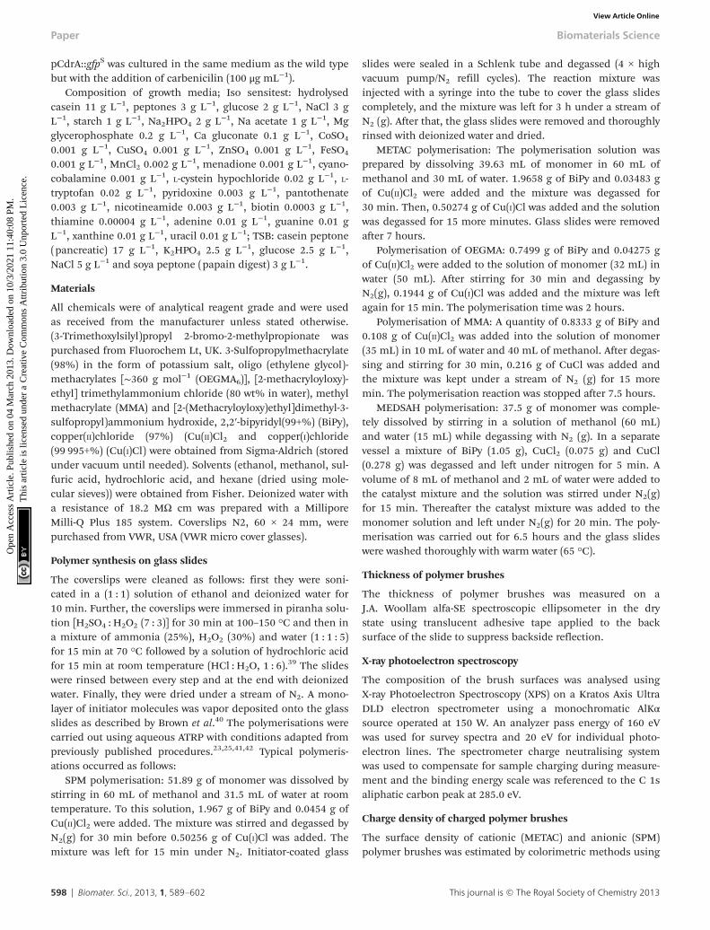
LPS-mutants (with and without o-antigen) exhibit alteredsurface physicochemical properties, which have been shown toinfluence biofilm formation.16 Wild type P. aeruginosa PAO1produces both A-band and B-band o-antigen, while the ΔwbpAmutant produces only A-band. PAO1ΔrmlC mutant does notproduce A-band but instead produces B-band (Fig. 8E and F).However, due to the truncated LPS core, the o-antigen ofPAO1ΔrmlC is not attached to the LPS core38 and is notexpected to be exposed at the surface of the bacterium.Measurements of zeta potential and hydrophobicity of thesestrains show that PAO1ΔrmlC had more negative zeta potentialthan the wild type but a similar cell hydrophobicity. In con-trast, the zeta potential of PAO1ΔwbpA was similar to wild typewhile cell hydrophobicity for this mutant was higher (Fig. 8Aand B) probably due to the presence of the relatively hydro-phobic A-band. The differences in zeta potential and hydro-phobicity in the wild type and mutants were mainly due todistinct LPS phenotypes as the outer membrane proteins ofthese strains did not differ significantly apart from a smallintensity variation in a protein band identified as outer mem-brane lipoprotein OprI precursor around 10 kDa (Fig. 8D).
Biofilms formed on glass by PAO1ΔwbpA and PAO1ΔrmlCmutants differed from the wild type. The PAO1ΔwbpA mutantformed flat but grainy biofilms and PAO1ΔrmlC formed thickbiofilms (about 50 μm thick) resembling merged mushroomstructures with uneven edges (Fig. 9). Such a mushroombiofilm of PAO1ΔrmlC may be due to less efficient swimmingand twitching motility (Fig. 8C), properties that previouslyhave been shown to influence biofilm phenotype in P. aerugi-nosa.18,27 When grown on SPM, PAO1ΔrmlC produced scarcebiofilm while PAO1ΔwbpA developed multiple mushroom-likestructures (Fig. 9). This scarce biofilm can be explained by thephysicochemical properties of the bacterial cell. ThePAO1ΔrmlC mutant exhibits high zeta potentials and can, con-sequently, be more strongly repelled by a negatively chargedpolymer brush surface in comparison to the wild type or thePAO1ΔwbpA mutant. The increase in the number of mushroomcolonies for the PAO1ΔwbpA mutant could be a result of thehigher hydrophobicity and relatively low zeta potential of thePAO1ΔwbpA mutant. This would result in lower repulsiveforces between the bacterium and the negatively chargedpolymer brush, and perhaps some increased interactions withthe polymer backbone of the surface, enabling the bacteriumto better attach to the film or at pin-hole defects or scratches
Fig. 6 DIC images of P. aeruginosa PAO1 (A, B, C) and P. aeruginosa PAO1 Δpi-lAΔfliC double mutant (D) at the surface of glass (A), SPM (B and D), and METAC(C); P. aeruginosa PAO1 bacteria associated with SPM (B) are seen as “dots” asthey are perpendicularly oriented with respect to the SPM brush surface. This isnot seen in P. aeruginosa PAO1 associated with METAC or in the P. aeruginosaPAO1 ΔpilAΔfliC double mutant on SPM. Note that cells of the double mutanthave a larger size than the wild type, are non-motile, and did not come incontact with the SPM surface (making focusing difficult and giving rise to theless sharp image in D).
Biomaterials Science Paper
This journal is © The Royal Society of Chemistry 2013 Biomater. Sci., 2013, 1, 589–602 | 595
Ope
n A
cces
s A
rtic
le. P
ublis
hed
on 0
4 M
arch
201
3. D
ownl
oade
d on
10/
3/20
21 1
1:40
:08
PM.
Thi
s ar
ticle
is li
cens
ed u
nder
a C
reat
ive
Com
mon
s A
ttrib
utio
n 3.
0 U
npor
ted
Lic
ence
.View Article Online
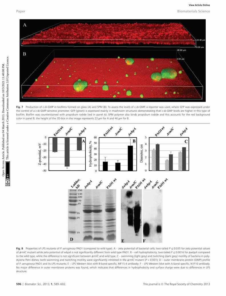
Fig. 7 Production of c-di-GMP in biofilms formed on glass (A) and SPM (B). To assess the levels of c-di-GMP, a reporter was used, where GFP was expressed underthe control of a c-di-GMP sensitive promoter; GFP (green) is expressed mainly in mushroom structures demonstrating that c-di-GMP levels are higher in this type ofbiofilm. Biofilm was counterstained with propidium iodide (red in panel A). SPM polymer also binds propidium iodide and this accounts for the red backgroundcolor in panel B; the height of the 3D-box in the image represents 23 μm for A and 46 μm for B.
Fig. 8 Properties of LPS mutants of P. aeruginosa PAO1 (compared to wild type). A – zeta potential of bacterial cells; two-tailed P ≤ 0.035 for zeta potential valuesof ΔrmlC mutant while zeta potential of wbpA is not significantly different from wild type PAO1. B – cell hydrophobicity; two-tailed P ≤ 0.0016 for ΔwbpA comparedto the wild type, while the difference is not significant between ΔrmlC and wild type. C – swimming (light grey) and twitching (dark grey) motility of bacteria in poly-styrene Petri dishes; both swimming and twitching motility were significantly inhibited in the ΔrmlC mutant (P < 0.001); D – outer membrane protein (OMP) profileof P. aeruginosa PAO1 and its LPS mutants; E – LPS Western blot with B-band specific, MF15-4 antibody; F – LPS Western blot with A-band specific, N1F10 antibody.No major difference in outer membrane proteins was found, which indicates that differences in hydrophobicity and surface charge were due to differences in LPSstructure.
Paper Biomaterials Science
596 | Biomater. Sci., 2013, 1, 589–602 This journal is © The Royal Society of Chemistry 2013
Ope
n A
cces
s A
rtic
le. P
ublis
hed
on 0
4 M
arch
201
3. D
ownl
oade
d on
10/
3/20
21 1
1:40
:08
PM.
Thi
s ar
ticle
is li
cens
ed u
nder
a C
reat
ive
Com
mon
s A
ttrib
utio
n 3.
0 U
npor
ted
Lic
ence
.View Article Online

in the surface film. However, the number of colonies attachingin the different experiments was most probably not only afunction of the amount of defects in the polymer film, sinceidentical SPM films were used in the experiments withdifferent strains. The amount of pin-holes also seems to below. XPS data from the brush surfaces showed no or less than0.1 atom% of Si at the surface. The exact mechanism for theincreased interactions with the negative surface in the case ofthe PAO1ΔwbpA mutant, as well as the influence of extracellu-lar substances, is a subject for further studies.
ExperimentalStrains and growth conditions
Strains and plasmids used in this study are described inTable 1. Bacteria were cultured either on blood agar plates orin Isosensitest broth (Oxoid LTD, Hampshire, England)
supplemented with antibiotics when needed. pCdrA::gfpS
reporter plasmid was introduced into P. aeruginosa PAO1(J. Lam group) by electroporation. P. aeruginosa PAO1 expressing
Table 1 Bacterial strains and plasmids
Strain/plasmid Relevant characteristics Source
P. aeruginosaPAO1
Wild type Joseph Lamlab14
PAO1ΔwbpA Deficient in B-band LPS biosynthesis Joseph Lamlab14
PAO1ΔrmlC Truncated LPS core Joseph Lamlab14
PAO1ΔfliCΔpilA
Deficient in flagella and pili Alain Fillouxlab36
pJBA129 pME6030 PA1/04/03-gfp-T0–T1, Tcr;
constitutive GFP expressionMichaelGivskov lab1
pCdrA::gfpS pUCP22Not-PcdrARBS-CDS-RNaseIIIgfp(Mut3)-T0–T1, Ampr
MatthewParsek lab49
Fig. 9 Biofilm architecture of P. aeruginosa PAO1 LPS mutants on SPM and on glass, 72 hours post inoculation. A – biofilm of PAO1 ΔrmlC on glass and SPM, 3Dview, height of the 3D-box represents 63 μm for glass and 18 μm for SPM; B – biofilm of PAO1 ΔrmlC on glass, top and side view, scale bar represents 50 μm; C –
biofilm of PAO1 ΔwbpA on glass and SPM, 3D view, height of the 3D-box represents 17 μm for glass and 30 μm for SPM; D – PAO1 (wild type) biofilm on glass, topand side view, scale bar represents 50 μm.
Biomaterials Science Paper
This journal is © The Royal Society of Chemistry 2013 Biomater. Sci., 2013, 1, 589–602 | 597
Ope
n A
cces
s A
rtic
le. P
ublis
hed
on 0
4 M
arch
201
3. D
ownl
oade
d on
10/
3/20
21 1
1:40
:08
PM.
Thi
s ar
ticle
is li
cens
ed u
nder
a C
reat
ive
Com
mon
s A
ttrib
utio
n 3.
0 U
npor
ted
Lic
ence
.View Article Online
pCdrA::gfpS was cultured in the same medium as the wild typebut with the addition of carbenicilin (100 μg mL−1).
Composition of growth media; Iso sensitest: hydrolysedcasein 11 g L−1, peptones 3 g L−1, glucose 2 g L−1, NaCl 3 gL−1, starch 1 g L−1, Na2HPO4 2 g L−1, Na acetate 1 g L−1, Mgglycerophosphate 0.2 g L−1, Ca gluconate 0.1 g L−1, CoSO4
0.001 g L−1, CuSO4 0.001 g L−1, ZnSO4 0.001 g L−1, FeSO4
0.001 g L−1, MnCl2 0.002 g L−1, menadione 0.001 g L−1, cyano-cobalamine 0.001 g L−1, L-cystein hypochloride 0.02 g L−1, L-tryptofan 0.02 g L−1, pyridoxine 0.003 g L−1, pantothenate0.003 g L−1, nicotineamide 0.003 g L−1, biotin 0.0003 g L−1,thiamine 0.00004 g L−1, adenine 0.01 g L−1, guanine 0.01 gL−1, xanthine 0.01 g L−1, uracil 0.01 g L−1; TSB: casein peptone(pancreatic) 17 g L−1, K2HPO4 2.5 g L−1, glucose 2.5 g L−1,NaCl 5 g L−1 and soya peptone (papain digest) 3 g L−1.
Materials
All chemicals were of analytical reagent grade and were usedas received from the manufacturer unless stated otherwise.(3-Trimethoxylsilyl)propyl 2-bromo-2-methylpropionate waspurchased from Fluorochem Lt, UK. 3-Sulfopropylmethacrylate(98%) in the form of potassium salt, oligo (ethylene glycol)-methacrylates [∼360 g mol−1 (OEGMA6)], [2-methacryloyloxy)-ethyl] trimethylammonium chloride (80 wt% in water), methylmethacrylate (MMA) and [2-(Methacryloyloxy)ethyl]dimethyl-3-sulfopropyl)ammonium hydroxide, 2,2′-bipyridyl(99+%) (BiPy),copper(II)chloride (97%) (Cu(II)Cl2 and copper(I)chloride(99 995+%) (Cu(I)Cl) were obtained from Sigma-Aldrich (storedunder vacuum until needed). Solvents (ethanol, methanol, sul-furic acid, hydrochloric acid, and hexane (dried using mole-cular sieves)) were obtained from Fisher. Deionized water witha resistance of 18.2 MΩ cm was prepared with a MilliporeMilli-Q Plus 185 system. Coverslips N2, 60 × 24 mm, werepurchased from VWR, USA (VWR micro cover glasses).
Polymer synthesis on glass slides
The coverslips were cleaned as follows: first they were soni-cated in a (1 : 1) solution of ethanol and deionized water for10 min. Further, the coverslips were immersed in piranha solu-tion [H2SO4 : H2O2 (7 : 3)] for 30 min at 100–150 °C and then ina mixture of ammonia (25%), H2O2 (30%) and water (1 : 1 : 5)for 15 min at 70 °C followed by a solution of hydrochloric acidfor 15 min at room temperature (HCl : H2O, 1 : 6).
39 The slideswere rinsed between every step and at the end with deionizedwater. Finally, they were dried under a stream of N2. A mono-layer of initiator molecules was vapor deposited onto the glassslides as described by Brown et al.40 The polymerisations werecarried out using aqueous ATRP with conditions adapted frompreviously published procedures.23,25,41,42 Typical polymeris-ations occurred as follows:
SPM polymerisation: 51.89 g of monomer was dissolved bystirring in 60 mL of methanol and 31.5 mL of water at roomtemperature. To this solution, 1.967 g of BiPy and 0.0454 g ofCu(II)Cl2 were added. The mixture was stirred and degassed byN2(g) for 30 min before 0.50256 g of Cu(I)Cl was added. Themixture was left for 15 min under N2. Initiator-coated glass
slides were sealed in a Schlenk tube and degassed (4 × highvacuum pump/N2 refill cycles). The reaction mixture wasinjected with a syringe into the tube to cover the glass slidescompletely, and the mixture was left for 3 h under a stream ofN2 (g). After that, the glass slides were removed and thoroughlyrinsed with deionized water and dried.
METAC polymerisation: The polymerisation solution wasprepared by dissolving 39.63 mL of monomer in 60 mL ofmethanol and 30 mL of water. 1.9658 g of BiPy and 0.03483 gof Cu(II)Cl2 were added and the mixture was degassed for30 min. Then, 0.50274 g of Cu(I)Cl was added and the solutionwas degassed for 15 more minutes. Glass slides were removedafter 7 hours.
Polymerisation of OEGMA: 0.7499 g of BiPy and 0.04275 gof Cu(II)Cl2 were added to the solution of monomer (32 mL) inwater (50 mL). After stirring for 30 min and degassing byN2(g), 0.1944 g of Cu(I)Cl was added and the mixture was leftagain for 15 min. The polymerisation time was 2 hours.
Polymerisation of MMA: A quantity of 0.8333 g of BiPy and0.108 g of Cu(II)Cl2 was added into the solution of monomer(35 mL) in 10 mL of water and 40 mL of methanol. After degas-sing and stirring for 30 min, 0.216 g of CuCl was added andthe mixture was kept under a stream of N2 (g) for 15 moremin. The polymerisation reaction was stopped after 7.5 hours.
MEDSAH polymerisation: 37.5 g of monomer was comple-tely dissolved by stirring in a solution of methanol (60 mL)and water (15 mL) while degassing with N2 (g). In a separatevessel a mixture of BiPy (1.05 g), CuCl2 (0.075 g) and CuCl(0.278 g) was degassed and left under nitrogen for 5 min. Avolume of 8 mL of methanol and 2 mL of water were added tothe catalyst mixture and the solution was stirred under N2(g)for 15 min. Thereafter the catalyst mixture was added to themonomer solution and left under N2(g) for 20 min. The poly-merisation was carried out for 6.5 hours and the glass slideswere washed thoroughly with warm water (65 °C).
Thickness of polymer brushes
The thickness of polymer brushes was measured on aJ.A. Woollam alfa-SE spectroscopic ellipsometer in the drystate using translucent adhesive tape applied to the backsurface of the slide to suppress backside reflection.
X-ray photoelectron spectroscopy
The composition of the brush surfaces was analysed usingX-ray Photoelectron Spectroscopy (XPS) on a Kratos Axis UltraDLD electron spectrometer using a monochromatic AlKαsource operated at 150 W. An analyzer pass energy of 160 eVwas used for survey spectra and 20 eV for individual photo-electron lines. The spectrometer charge neutralising systemwas used to compensate for sample charging during measure-ment and the binding energy scale was referenced to the C 1saliphatic carbon peak at 285.0 eV.
Charge density of charged polymer brushes
The surface density of cationic (METAC) and anionic (SPM)polymer brushes was estimated by colorimetric methods using
Paper Biomaterials Science
598 | Biomater. Sci., 2013, 1, 589–602 This journal is © The Royal Society of Chemistry 2013
Ope
n A
cces
s A
rtic
le. P
ublis
hed
on 0
4 M
arch
201
3. D
ownl
oade
d on
10/
3/20
21 1
1:40
:08
PM.
Thi
s ar
ticle
is li
cens
ed u
nder
a C
reat
ive
Com
mon
s A
ttrib
utio
n 3.
0 U
npor
ted
Lic
ence
.View Article Online

UV-Vis spectrophotometry adapted to our system.43,44 The pro-cedure assumes that counter ions from the charged brush getreplaced by charged dye molecules to give a 1 : 1 complexbetween one charged dye molecule and one charged functionalgroup. Orange II dye was used for cationic brushes and Tolui-dine Blue O (TB) dye for anionic brushes. At pH 3 Orange IIhas an absorption peak at 485 nm, with an extinction coeffi-cient of 19 476 L mol−1 cm−1, while TB has an absorption peakat 633 nm and an extinction coefficient of 50 000 L mol−1
cm−1.METAC: A volume of 50 mL of 0.5 mM aqueous Orange II
solution was prepared and adjusted to pH 3 with a 1 mM solu-tion of HCl (solution 1). Samples were placed in the solutionand left overnight at 30 °C. Thereafter each sample was rinsedwith water and immersed in 100 mL of 1 mM NaOH solutionunder stirring, to remove physically adsorbed dye from thebrush (solution 2). After 24 h the pH of solution 2 was adjustedto pH 3 with 100 mM HCl and colorimetric analyses werecarried out on both solutions. The amount of dye bound to thebrush is deduced from the difference between the two solu-tions. SPM: A volume of 50 mL of 0.5 mM aqueous TB dyesolution was prepared and adjusted to pH 10 using a buffer(Na2CO3/NaHCO3). Samples were placed in this solution andkept at 30 °C for 6 h. After washing with NaOH, 0.5 mM, eachsample was placed in a 50% aqueous solution of acetic acidfor 24 h to dissociate the dye from the brush. By analyzing thelatter solution with UV-Vis the amount of absorbed dye wasobtained.
SPR measurements
In order to monitor the conditioning of substrates duringincubation in culture media, adsorption was followed viasurface plasmon resonance (SPR), using a Biacore 3000. SPRsensor chips (Ssens), on which polymer brushes had beengrown, were mounted onto a substrate holder and docked inthe instrument before priming twice with a buffer (PBS). Thesubstrates were allowed to equilibrate for 30 min at 20 μLmin−1 in PBS, until a stable baseline was obtained. To monitoradsorption from culture media, the substrates were washedand equilibrated for 5 min whilst recording the baseline signaldetected. The surfaces were then exposed to culture media for5 min before washing with PBS for 30 min. The reading of theamount of material deposited on the surface was carried outafter 30 min equilibration. The flow rate was 20 μL min−1 andmeasurements were carried out in triplicate.
Contact angle measurement
Sessile drop contact angles were measured on the substrateswith an optical tensiometer (KSV Instruments). A drop of3–4 μL of deionized water was deposited on the surface of thesubstrate from an automated syringe and pictures of the waterdrop on the surface were taken during and after deposition at250 ms intervals. The contact angle of the drop at each timewas measured to assess the evolution of the drop shape and apossible change in the surface properties of the substrate overtime. The Young Laplace method (Attension software) was
used to fit the shape of the drops and to measure the contactangles. The values reported are averages of 3 to 4 drops depos-ited at different locations on one substrate. All samples withcontact angles above ≈15° had stable drop shapes over time(without taking into account slow evaporation of the waterdroplet). The error associated with each value was low andindicates that the surfaces were homogeneous and the contactangles were reproducible on each substrate.
Flow chamber biofilm and confocal laser scanning microscopy(CLSM)
Bacteria were cultured in Isosensitest broth diluted 10 timeswith MQ water. Flow chamber BST FC270, glass flow breakFB50, and bubble trap FC34, were all purchased from Biosur-face Technology Corporation, USA. These parts were connectedwith silicon tubing, 2 mm bore diameter; Marprene tubing,0.8 mm bore diameter (Alitea, Sweden), was used in the placeof contact with a pump head. After the flow chamber was auto-claved, functionalized glass slides sterilized in 70% ethanolwere inserted in sterile conditions. For inoculation, bacteriafrom the late exponential growth phase were pelleted and re-suspended in NaCl 0.9% to the concentration of 2 × 10⁹ cellmL−1. 2 mL of this bacterial suspension was injected to eachchannel of the flow chamber with a syringe. The flow in thechamber was stopped for 30 min to allow bacterial attachment.All through the experiment the system was operated at 1.2 mLmin−1 using a 405U/L2 double-channel pump (WatsonMarlow, Alitea, Sweden). Bacteria were visualized eitherthrough expression of green fluorescent protein (GFP) or bystaining with Syto-9 fluorescent dye (Invitrogen, MolecularProbes, USA). When Syto-9 was used, 1 mL of media contain-ing 5 nM Syto-9 was injected into each channel and the flowwas stopped for 10 min to allow staining. Flow chamber experi-ments were performed at least in duplicate and images werecaptured at several positions on each slide.
Confocal (3-D) biofilm images were captured with a NikonEclipse90i fluorescent microscope equipped with a NikonD-eclipse C1+ laser system (Nikon Corporation, Japan). Imageswere acquired and the intensity of the signal from the biofilmwas measured at 510–530 nm wavelength using EZ-C1 ver.3.80and NIS-Elements Advanced Research ver.3.2 software (NikonCorporation). Biomass was measured through the intensity ofsyto-9 in confocal 3D images; the same settings were usedduring image acquisition. Attached bacteria were countedusing free ImageJ software (rsb.info.nih.gov/ij/) and presentedas the number of cells in a field of view, which was 4.03 × 10⁵
μm2.
Live cell microscopy
To monitor more precisely the interaction between polymersurfaces and bacteria, live cell microscopy was performed asfollows: Petri dishes 3.5 cm in diameter were completely filledwith tryptic soy agar (TSA) medium and after solidification1 μL of bacterial suspension (8 × 10¹⁰ cell mL−1) in NaCl 0.9%was applied on the top of the agar. Bacteria were covered witha glass slide with or without polymer coating and directly
Biomaterials Science Paper
This journal is © The Royal Society of Chemistry 2013 Biomater. Sci., 2013, 1, 589–602 | 599
Ope
n A
cces
s A
rtic
le. P
ublis
hed
on 0
4 M
arch
201
3. D
ownl
oade
d on
10/
3/20
21 1
1:40
:08
PM.
Thi
s ar
ticle
is li
cens
ed u
nder
a C
reat
ive
Com
mon
s A
ttrib
utio
n 3.
0 U
npor
ted
Lic
ence
.View Article Online

observed using a Nikon Eclipse Ti-E inverted microscopeequipped with ×100 objective. Differential interference con-trast (DIC) images were captured using an Andor iXon+EMCCD camera. Sequences of images taken with 5 s intervalswere assembled in a movie (130 ms interval) using NIS-Ele-ments 3.2 Software.
Surface associated motility
25 mL of TSA-agar (BD, BLL™) medium was poured into aPetri dish with an inner diameter of 88 mm. After solidifica-tion plates were dried at 37 °C for 20 min. Bacterial suspension(2 μL, 1 × 10⁹ cell mL−1) was dropped on the surface of agarand allowed to dry. Glass slides coated with polymer were steri-lized in 70% ethanol for 10 min, rinsed in MQ water anddried. The glass slides were placed on the top of a dried dropof bacterial suspension. After incubation at 37 °C the growthassociated with glass slides was observed.
Isolation and analysis of outer membrane proteins
Outer membrane proteins (OMPs) were isolated as describedpreviously with some modifications.45 In brief, 25 mL of over-night grown cultures were centrifuged at 10 000 rpm for15 minutes at 4 °C, re-suspended in 25 mL of milliQ water andsonicated on ice. Cell debris and intact cells were removed bycentrifugation at 7000 rpm for 10 minutes at 4 °C. The super-natant fraction was treated with 2% N-lauryl sarcosyl at roomtemperature. This solution was ultra-centrifuged twice at29 000 rpm for 1.5 hours in a 45Ti Beckmann rotor and thepellet containing OMPs was resuspended in milliQ water.OMPs were separated by 15% polyacrylamide gel electro-phoresis.
Cell hydrophobicity
Hydrophobicity was measured as previously described.16
Briefly: an overnight culture was centrifuged at 10 000 rpm for15 min and re-suspended in phosphate buffered saline (PBS)to give an OD595 of approximately 1(A0). Then, hexadecane wasadded to the suspension in the ratio of 4 : 1 (bacterialsuspension : hexadecane). The optical density of the aqueousphase was measured at 595 nm (A). The hydrophobicity wascalculated according to the equation:
Hydrophobicity% ¼ A0� AA0
� 100
Zeta potential
Zeta potential was measured by dynamic light scattering usingMalvern Nano ZS zetasiser and clear disposable zeta cells(Malvern). Before measuring, bacteria were cultured overnight,washed and re-suspended in phosphate buffer with an ionicstrength of 20 mM (1 × 10⁹ cell mL−1).
Swimming and twitching motility
The swimming and twitching of P. aeruginosa PAO1 and itsLPS mutants were evaluated in the Isosensitest medium com-plemented with 0.3% agar as described elsewhere.46 Briefly:
12 mL of the medium was poured into 53 mm diameter Petridishes and 5 μL of bacteria suspension with ABS600 = 1 wasstabbed into the agar in the middle of the plate. Swimmingwas represented by a cloudy ring within the agar, while twitch-ing was represented by a ring of thin film between the agarand the plastic bottom of the Petri dish. The diameter of bothkinds of motility rings was measured 15 h after theinoculation.
Polyacrylamide gel electrophoresis (PAGE) and Westernimmunoblotting
Cultures of P. aeruginosa PAO1 and respective mutants weregrown to ABS600 = 1. Cells from 1 mL of the culture were col-lected by centrifugation and LPS preparation and staining wasdone as previously described.47 LPS was resolved on gradient 4to 12% gel. LPS differences in mutants were confirmed bystaining with an Emerald Green LPS Kit (Invitrogen, MolecularProbes, USA) and Western blot analysis with N1F10 (A-bandspecific) and MF15-4 (B-band specific) monoclonal antibodies.
Live–dead staining
Live–dead staining (a combination of syto-9 and propidiumiodide staining) was performed to discriminate between viableand non-viable cells attached to the glass/brush in the flowcell. Syto-9 (Invitrogen, Molecular Probes, USA) was usedaccording to manufacturer’s instructions. Viable cells werestained green with Syto-9 dye and cells with compromisedmembrane were stained red.
Conclusions
Using P. aeruginosa as a model bacterium, this work illustratessome of the complexity of bacterial attachment and biofilmformation and how it is dependent on many interlocking para-meters of both the abiotic and the bacterial surface. Expectedantifouling and antibacterial surfaces such as brushes withethylene glycol subunits or cationic brushes were found tobecome covered with biofilm in the same way as glass refer-ence surfaces. However, on negatively charged surfaces thebiofilm formation was strongly reduced. Both attachment andmotility were found to be inhibited for bacterial cells associ-ated with negative SPM and zwitterionic MEDSAH surfaces.Additionally, bacteria that were attached to SPM and MEDSAHshowed high levels of c-di-GMP, which indicates increased pro-duction of biofilm matrix components (exopolysaccharides).This suggests that Gram-negative bacteria, such as P. aerugi-nosa, can modify their cell surfaces when in contact with nega-tively charged substrates and this, in turn, influences thedevelopment and architecture of the biofilm. This work alsoshows that LPS structures that confer high zeta potential onthe bacterial cell decrease biofilm formation on negativelycharged surfaces. On the other hand, LPS structures thatincrease cell hydrophobicity could facilitate the formation ofbiofilm on polymer surfaces with negative charge. Biofilms onSPM and MEDSAH consisted of characteristic mushroom
Paper Biomaterials Science
600 | Biomater. Sci., 2013, 1, 589–602 This journal is © The Royal Society of Chemistry 2013
Ope
n A
cces
s A
rtic
le. P
ublis
hed
on 0
4 M
arch
201
3. D
ownl
oade
d on
10/
3/20
21 1
1:40
:08
PM.
Thi
s ar
ticle
is li
cens
ed u
nder
a C
reat
ive
Com
mon
s A
ttrib
utio
n 3.
0 U
npor
ted
Lic
ence
.View Article Online

structures, but the quantity of biofilm was much lower than onglass, POEGMA and PMMA. Similar mushroom structures werepreviously reported by other authors to be more resistant toantibiotics than flat biofilms,27,48 which should be investigatedfurther and taken into consideration in the development ofantifouling surfaces. Taken together, this study shows that tosuccessfully design antifouling surfaces, emphasis needs to beplaced on understanding the dynamics of the bacterial cellsurface in relation to the abiotic material in question.
Abbreviations
SPM poly (3-sulphopropylmethacrylate)MEDSAH poly (2-(methacryloyloxy)ethyl)dimethyl-3-sulpho-
proyl) ammonium hydroxide)METAC poly (2-(methacryloyloxy)-ethyl trimethyl
ammonium chloride)POEGMA poly oligo(ethylene glycol methyl ether
methacrylate)PMMA polymethylmethacrylatec-di-GMP cyclic diguanylate
Acknowledgements
P. aeruginosa PAO1 strain expressing the pJBA129 plasmid waskindly provided by Jens Bo Andersen and Michael Givskov,University of Copenhagen. We are grateful to Joseph Lam, Uni-versity of Guelph and Mathew Parsek, University of Washing-ton for generously providing us with P. aeruginosa LPSmutants, anti-LPS antibodies and c-di-GMP reporter plasmid,respectively. Alain Filloux, Imperial College London isacknowledged for the gift of P. aeruginosa PAO1 ΔfliCΔpilAdouble mutant. We thank Joseph D. Mougous, University ofWashington and Helena Lindgren, Umeå University for fruitfuldiscussions. The Curth Nilsson Foundation for StrategicResearch, the Carl Trygger Foundation for Scientific Research,the Swedish Research Council and the Swedish Foundation forInternational Cooperation in Research and Higher Education(STINT) are acknowledged for funding.
Notes and references
1 A.P. Stapper, G. Narasimhan, D. E. Ohman, J. Barakat,M. Hentzer, S. Molin, A. Kharazmi, N. Høiby andK. Mathee, J. Med. Microbiol., 2004, 53, 679–690.
2 J. W. Costerton, P. S. Stewart and E. P. Greenberg, Science,1999, 284, 1318–1322.
3 D. Davies, Nat. Rev. Drug Discov., 2003, 2, 114–122.4 R. M. Klevens, J. R. Edwards, C. L. Richards Jr.,
T. C. Horan, R. P. Gaynes, D. A. Pollock and D. M. Cardo,Public Health Rep., 2007, 122, 160–166.
5 N. Ayres, Polym. Chem., 2010, 1, 769–777.6 A. Terada, K. Okuyama, M. Nishikawa, S. Tsuneda and
M. Hosomi, Biotechnol. Bioeng., 2012, 109, 1745–1754.
7 H. Murata, R. R. Koepsel, K. Matyjaszewski andA. J. Russell, Biomaterials, 2007, 28, 4870–4879.
8 B. Zdyrko, V. Klep, X. W. Li, Q. Kang, S. Minko, X. J. Wenand I. Luzinov, Mater. Sci. Eng., C, 2009, 29, 680–684.
9 D. Montag, M. Frant, H. Horn and K. Liefeith, Biofouling,2012, 28, 315–327.
10 G. Cheng, Z. Zhang, S. F. Chen, J. D. Bryers and S. Y. Jiang,Biomaterials, 2007, 28, 4192–4199.
11 H. Strahl and L. W. Hamoen, Proc. Natl. Acad. Sci. U. S. A.,2010, 107, 12281–12286.
12 A. Clements, F. Gaboriaud, J. F. Duval, J. L. Farn,A. W. Jenney, T. Lithgow, O. L. Wijburg, E. L. Hartland andR. A. Strugnell, PLoS One, 2008, 3, e3817.
13 F. Gosselin, J. F. L. Duval, J. Simonet, C. Ginevra,F. Gaboriaud, S. Jarraud and L. Mathieu, Colloids Surf., B,2011, 82, 283–290.
14 J. S. Lam, V. L. Taylor, S. T. Islam, Y. Hao and D. Kocincova,Front. Microbiol., 2011, 2, 118.
15 C. D. Ciornei, A. Novikov, C. Beloin, C. Fitting, M. Caroff,J. M. Ghigo, J. M. Cavaillon and M. Adib-Conquy, Innate.Immun., 2010, 16, 288–301.
16 R. Nakao, M. Ramstedt, S. N. Wai and B. E. Uhlin, PLoSOne, 2012, 7, e51241.
17 C. A. Flemming, R. J. Palmer, A. A. Arrage, H. C. Van derMei and D. C. White, Biofouling, 1999, 13, 213–231.
18 M. Klausen, A. Heydorn, P. Ragas, L. Lambertsen, A. Aaes-Jorgensen, S. Molin and T. Tolker-Nielsen, Mol. Microbiol.,2003, 48, 1511–1524.
19 R. Simm, M. Morr, A. Kader, M. Nimtz and U. Romling,Mol. Microbiol., 2004, 53, 1123–1134.
20 B. R. Borlee, A. D. Goldman, K. Murakami, R. Samudrala,D. J. Wozniak and M. R. Parsek, Mol. Microbiol., 2010, 75,827–842.
21 V. T. Lee, J. M. Matewish, J. L. Kessler, M. Hyodo,Y. Hayakawa and S. Lory, Mol. Microbiol., 2007, 65,1474–1484.
22 G. A. O’Toole and R. Kolter, Mol. Microbiol., 1998, 30,295–304.
23 O. Azzaroni, S. Moya, T. Farhan, A. A. Brown andW. T. S. Huck, Macromolecules, 2005, 38, 10192–10199.
24 O. Azzaroni, A. A. Brown and W. T. S. Huck, Angew. Chem.,Int. Ed., 2006, 45, 1770–1774.
25 J. E. Gautrot, W. T. S. Huck, M. Welch and M. Ramstedt,ACS Appl. Mater. Inter., 2010, 2, 193–202.
26 L. Ploux, A. Ponche and K. Anselme, J. Adhes. Sci. Technol.,2010, 24, 2165–2201.
27 R. M. Landry, D. An, J. T. Hupp, P. K. Singh andM. R. Parsek, Mol. Microbiol., 2006, 59, 142–151.
28 R. Bansil and B. S. Turner, Curr. Opin. Colloid Interface,2006, 11, 164–170.
29 Medical Microbiology, ed. P. Murray, K. Rosenthal andM. Pfaller, Elsevier MOSBY, Philadelphia, 5th edn, 2005,p. 357.
30 H. H. Rijnaarts, W. Norde, E. J. Bouwer, J. Lyklema andA. J. Zehnder, Appl. Environ. Microbiol., 1993, 59,3255–3265.
Biomaterials Science Paper
This journal is © The Royal Society of Chemistry 2013 Biomater. Sci., 2013, 1, 589–602 | 601
Ope
n A
cces
s A
rtic
le. P
ublis
hed
on 0
4 M
arch
201
3. D
ownl
oade
d on
10/
3/20
21 1
1:40
:08
PM.
Thi
s ar
ticle
is li
cens
ed u
nder
a C
reat
ive
Com
mon
s A
ttrib
utio
n 3.
0 U
npor
ted
Lic
ence
.View Article Online

31 M. Chabria, S. Hertig, M. L. Smith and V. Vogel, Nat.Commun., 2010, 1.
32 L. D. Hazlett and X. L. Rudner, Ophthalmic Res., 1994, 26,375–379.
33 J. C. Conrad, M. L. Gibiansky, F. Jin, V. D. Gordon, D. A. Motto,M. A. Mathewson, W. G. Stopka, D. C. Zelasko, J. D. Shroutand G. C. L. Wong, Biophys. J., 2011, 100, 1608–1616.
34 B. Pidhatika, J. Moller, E. M. Benetti, R. Konradi,E. Rakhmatullina, A. Muhlebach, R. Zimmermann, C. Werner,V. Vogel and M. Textor, Biomaterials, 2010, 31, 9462–9472.
35 J. F. Jones, J. D. Feick, D. Imoudu, N. Chukwumah,M. Vigeant and D. Velegol, Appl. Environ. Microbiol., 2003,69, 6515–6519.
36 S. de Bentzmann, M. Aurouze, G. Ball and A. Filloux, J. Bac-teriol., 2006, 188, 4851–4860.
37 E. Karatan and P. Watnick, Microbiol. Mol. Biol. Rev., 2009,73, 310–347.
38 R. Rahim, L. L. Burrows, M. A. Monteiro, M. B. Perry andJ. S. Lam, Microbiology, 2000, 146, 2803–2814.
39 S. Tugulu, A. Arnold, I. Sielaff, K. Johnsson and H. A. Klok,Biomacromolecules, 2005, 6, 1602–1607.
40 A. A. Brown, N. S. Khan, L. Steinbock and W. T. S. Huck,Eur. Polym. J., 2005, 41, 1757–1765.
41 M. Ramstedt, N. Cheng, O. Azzaroni, D. Mossialos,H. J. Mathieu and W. T. S. Huck, Langmuir, 2007, 23,3314–3321.
42 N. Cheng, A. A. Brown, O. Azzaroni and W. T. S. Huck,Macromolecules, 2008, 41, 6317–6321.
43 E. Uchida, Y. Uyama and Y. Ikada, Langmuir, 1993, 9,1121–1124.
44 M. Ciobanu, A. Siove, V. Gueguen, L. J. Gamble,D. G. Castner and V. Migonney, Biomacromolecules, 2006, 7,755–760.
45 T. Song, F. Mika, B. Lindmark, Z. Liu, S. Schild, A. Bishop,J. Zhu, A. Camilli, J. Johansson, J. Vogel and S. Wai, Mol.Microbiol., 2008, 70, 100–111.
46 M. H. Rashid and A. Kornberg, Proc. Natl. Acad.Sci. U. S. A., 2000, 97, 4885–4890.
47 X. H. Lai, R. L. Shirley, L. Crosa, D. Kanistanon, R. Tempel,R. K. Ernst, L. A. Gallagher, C. Manoil and F. Heffron, PLoSOne, 2010, 5, e11857.
48 Y. Kaneko, M. Thoendel, O. Olakanmi, B. E. Britigan andP. K. Singh, J. Clin. Invest., 2007, 117, 877–888.
49 M. T. Rybtke, B. R. Borlee, K. Murakami, Y. Irie,M. Hentzer, T. E. Nielsen, M. Givskov and M. R. Parsek,Appl Environ Microbiol, 2012, 78, 5060–5069.
Paper Biomaterials Science
602 | Biomater. Sci., 2013, 1, 589–602 This journal is © The Royal Society of Chemistry 2013
Ope
n A
cces
s A
rtic
le. P
ublis
hed
on 0
4 M
arch
201
3. D
ownl
oade
d on
10/
3/20
21 1
1:40
:08
PM.
Thi
s ar
ticle
is li
cens
ed u
nder
a C
reat
ive
Com
mon
s A
ttrib
utio
n 3.
0 U
npor
ted
Lic
ence
.View Article Online