C o pyrig t en u i ADVANCES IN ntess n z RESTORATIVE · PDF file · 2013-02-15This...
18
A DVANCES IN R ESTORATIVE D ENTISTRY Edited by Adrian Lussi and Markus Schaffner With contributions from: Adrian Lussi, Brigitte Zimmerli, Klaus Neuhaus, Matthias Strub, Stefan Hänni, Markus Schaffner, Svante Twetman, Martina Eichenberger, Simon Flury, Philippe Perrin, Rainer Seemann, Philip Ciucchi, Anne Grüninger, Daniel Jacky, Thomas Jaeggi, Franziska Jeger, Karin Kislig, Domenico Di Rocco, Jonas Rodrigues, Benjamin Schüz, and Beat Suter London, Berlin, Chicago, Tokyo, Barcelona, Beijing, Istanbul, Milan, Moscow, New Delhi, Paris, Prague, São Paulo, Seoul, Singapore and Warsaw C o p y r i g h t b y Q u i n t e s s e n z Alle Rechte vorbehalten
Transcript of C o pyrig t en u i ADVANCES IN ntess n z RESTORATIVE · PDF file · 2013-02-15This...

ADVANCES IN RESTORATIVE
DENTISTRY Edited by
Adrian Lussi and Markus Schaffner
With contributions from:
Adrian Lussi, Brigitte Zimmerli, Klaus Neuhaus,
Matthias Strub, Stefan Hänni, Markus Schaffner, Svante Twetman,
Martina Eichenberger, Simon Flury, Philippe Perrin, Rainer Seemann,
Philip Ciucchi, Anne Grüninger, Daniel Jacky, Thomas Jaeggi,
Franziska Jeger, Karin Kislig, Domenico Di Rocco, Jonas Rodrigues,
Benjamin Schüz, and Beat Suter
London, Berlin, Chicago, Tokyo, Barcelona, Beijing, Istanbul, Milan,
Moscow, New Delhi, Paris, Prague, São Paulo, Seoul, Singapore and Warsaw
CopyrightbyQ
uintessenz
Alle Rechte vorbehalten
IV
British Library Cataloguing in Publication Data
Lussi, Adrian.
Advances in restorative dentistry.
1. Dentistry, Operative. 2. Dentistry, Operative--
Technological innovations.
I. Title II. Schaffner, Markus.
617.6‘05-dc23
ISBN-13: 978-1-85097-228-0
Quintessence Publishing Co. Ltd,
Grafton Road, New Malden, Surrey KT3 3AB,
United Kingdom
www.quintpub.co.uk
Copyright © 2012 Quintessence Publishing Co. Ltd
All rights reserved. This book or any part thereof may not be reproduced, stored in a retrie-
val system, or transmitted in any form or by any means, electronic, mechanical, photocopy-
ing, or otherwise, without prior written permission of the publisher.
Editing: Quintessence Publishing Co. Ltd, London, UK
Layout and Production: Walburga Rothenhagen, Falkensee, Germany and
Quintessenz Verlags-GmbH, Berlin, Germany
Printed and bound in Germany
AcknowledgementsThanks go to Ueli Iff and Anne Seeger (Department of Multimedia and Computer Science,
University of Bern) for the production of the graphical figures, as well as to Herrmann Stich
(Department of Preventive, Restorative and Pediatric Dentistry, University of Bern) for the
histology pictures.
CopyrightbyQ
uintessenz
Alle Rechte vorbehalten
V
PrefaceDentistry has undergone a major transformation over recent years and decades. New tech-
nologies have been developed and a better understanding of biological principles and pro-
cesses has been gained. This book sheds light on these new aspects in preventive dentistry
and restorative dentistry.
Advances in Restorative Dentistry gives an overview of current trends in this diverse and
important specialist field for dental practitioners. The broad scope of restorative and preven-
tive dentistry is examined in 25 chapters. The following subjects are discussed:
The wealth of illustrations and highlighted key sentences make it easy to incorporate current
knowledge into daily practice as well as into teaching and study activities.
Adrian Lussi
Markus Schaffner
CopyrightbyQ
uintessenz
Alle Rechte vorbehalten

VI
Authors and contributorsProf. Dr. med. dent. Adrian LussiExecutive Director and Head of DepartmentDepartment of Preventive, Restorative and Pediatric Dentistry University of BernE-Mail: [email protected]
Dr. med. dent. Markus SchaffnerSenior LecturerDepartment of Preventive, Restorative and Pediatric DentistryUniversity of BernE-Mail: [email protected]
Dr. med. dent. Philip CiucchiResearch AssociateDepartment of Preventive, Restorative and Pediatric DentistryUniversity of BernE-Mail: [email protected]
Dr. med. dent. Martina EichenbergerLecturerDepartment of Preventive, Restorative and Pediatric DentistryUniversity of BernE-Mail: [email protected]
Dr. med. dent. Simon FluryResearch AssociateDepartment of Preventive, Restorative and Pediatric DentistryUniversity of BernE-Mail: [email protected]
Dr. med. dent. Anne GrüningerSenior LecturerDepartment of Preventive, Restorative and Pediatric DentistryUniversity of BernE-Mail: [email protected]
Dr. med. dent. Stefan HänniSenior LecturerDepartment of Preventive, Restorative and Pediatric DentistryUniversity of BernE-Mail: [email protected]
Dr. med. dent. Daniel JackyLecturer Department of Preventive, Restorative and Pediatric DentistryUniversity of Bern
Dr. med. dent. Thomas JaeggiSenior LecturerDepartment of Preventive, Restorative and Pediatric DentistryUniversity of BernE-Mail: [email protected]
Dr. med. dent. Franziska JegerSenior LecturerDepartment of Preventive, Restorative and Pediatric DentistryUniversity of BernE-Mail: [email protected]
Dr. med. dent. Karin KisligSenior LecturerDepartment of Preventive, Restorative and Pediatric DentistryUniversity of BernE-Mail: [email protected]
Dr. med. dent. Klaus W. NeuhausSenior LecturerDepartment of Preventive, Restorative and Pediatric DentistryUniversity of BernE-Mail: [email protected]
Dr. med. dent. Philippe PerrinSenior LecturerDepartment of Preventive, Restorative and Pediatric DentistryUniversity of BernE-Mail: [email protected]
Dr. med. dent. Domenico Di RoccoSenior LecturerDepartment of Preventive, Restorative and Pediatric DentistryUniversity of BernE-Mail: [email protected]
Dr. med. dent. Jonas de Almeida Rodrigues MSc, PhDResearch AssociateDepartment of Preventive, Restorative and Pediatric DentistryUniversity of BernE-Mail: [email protected]
PD. Dr. med. dent. Rainer Seemann Senior LecturerDepartment of Preventive, Restorative and Pediatric DentistryUniversity of BernE-Mail: [email protected]
Benjamin Schüz, Dipl.-Psych.LecturerSchool of PsychiatryUniversity of TasmaniaE-Mail: [email protected]
Dr. med. dent. Matthias StrubSenior LecturerDepartment of Preventive, Restorative and Pediatric DentistryUniversity of BernE-Mail: [email protected]
Dr. med. dent. Beat SuterSenior LecturerDepartment of Preventive, Restorative and Pediatric DentistryUniversity of BernE-Mail: bs@endodontic-bern-ch
Prof. Svante TwetmanHead of DepartmentDepartment of Cariology and EndodonticsFaculty of Health SciencesUniversity of CopenhagenE-mail: [email protected]
Dr. med. dent. Brigitte ZimmerliSenior LecturerDepartment of Preventive, Restorative and Pediatric DentistryUniversity of BernE-mail: [email protected]
CopyrightbyQ
uintessenz
Alle Rechte vorbehalten

VII
Contents
I Structure and pathology of the tooth 11 Structure and pathology of the tooth 3
Markus Schaffner and Adrian Lussi
II Aspects of prevention 172 Motivation and action – two aspects of oral hygiene at home 19 Benjamin Schüz and Rainer Seemann3 Cariostatic mechanisms of action of fluorides 25
Adrian Lussi
4 The role of xylitol in caries prevention 33Svante Twetman
5 Probiotics – a new approach in caries prevention? 39Svante Twetman
6 Novel methods of promoting remineralization 45Klaus Neuhaus and Adrian Lussi
7 Antibacterial agents for the prevention of caries 53Svante Twetman and Klaus Neuhaus
III Caries 638 Diagnosing caries and caries activity 65
Adrian Lussi, Markus Schaffner, Jonas Rodrigues, and Klaus Neuhaus
9 Sealing and infiltration of caries – is this the future? 79
Brigitte Zimmerli and Simon Flury
IV Magnification aids in restorative dentistry 8510 Utility and futility of magnification aids in restorative dentistry 87
Martina Eichenberger, Philippe Perrin, Daniel Jacky, and Adrian Lussi
V Damage to adjacent teeth and minimally invasive preparation 95
11 Damage to adjacent teeth and minimally invasive preparation 97
Martina Eichenberger, Philippe Perrin, and Adrian Lussi
12 Novel preparation and excavation methods 105
Klaus Neuhaus, Franziska Jeger, Philip Ciucchi, and Adrian Lussi
CopyrightbyQ
uintessenz
Alle Rechte vorbehalten

VIII
VI Yesterday retention – today adhesion? 11313 Adhesive techniques for dental restorations 115 Brigitte Zimmerli and Matthias Strub14 Direct restorative technology 123 Brigitte Zimmerli, Matthias Strub, and Simon Flury15 Restoration repairs 137 Brigitte Zimmerli and Matthias Strub16 Post systems 143 Brigitte Zimmerli and Matthias Strub17 The CEREC system 151 Domenico Di Rocco and Adrian Lussi
VII Bleaching 16118 Bleaching 163 Brigitte Zimmerli and Anne Grüninger
VIII Dental erosion 17319 Dental erosion 175 Adrian Lussi and Thomas Jaeggi
IX Endodontology 19120 Root canal preparation 193 Beat Suter21 Root canal irrigation 207 Stefan Hänni22 Root canal filling 215 Stefan Hänni23 Cracked tooth syndrome 223 Stefan Hänni and Adrian Lussi24 Endodontology in the primary dentition 233 Markus Schaffner, Klaus Neuhaus, and Adrian Lussi
X Halitosis 24325 Halitosis 245 Rainer Seemann and Karin Kislig
Index 261
CopyrightbyQ
uintessenz
Alle Rechte vorbehalten

I Structure and pathology of the tooth
6
Fig 1-9 Enamel tuft Enamel tufts are hypomineralized areas of enamel which look like tufts of grass under light micros-copy. Enamel tufts can provide a location favourable to bacteria in the event of carious attack. Caries is clearly visible in the histologic image.
Fig 1-10 Enamel pearl Left: radiograph of an enamel pearl in the interproximal area of a maxillary molar. Right: enamel pearl in the bifurca-tion of a molar.
Fig 1-7 Structural characteristics – enamel The periodic laying down of enamel is expressed in the lines of Retzius. Where these lines reach the surface, the perikymata are visible. Viewing the longitudinal and transverse sections of enamel by light microscopy reveals light and dark striae in the inner two-thirds. These Hunter-Schreger bands are caused by the wavelike path of the enamel prisms.
Fig 1-8 Perikymata under scanning electron microscope (SEM) The magnification clearly shows not only the perikymata but also the lines of imbrication running between them.
CopyrightbyQ
uintessenz
Alle Rechte vorbehalten

1Structure and pathology of the tooth
7
Structural defects and paraplasias of the enamel In most teeth, enamel structural defects can be identified by light microscopy. A large pro-
portion of these defects arise during amelogenesis. These include enamel tufts (Fig 1-9) and
enamel lamellae. Enamel tufts and lamellae can prove to be the line of least resistance in
respect of the spread of caries.
The enamel pearl is a paraplasia of the enamel. This means the formation of enamel
in an atypical localization. Enamel pearls can cause isolated periodontitis in the area of the
furcation (Fig 1-10).
Dysplasias of the enamel (and dentin)Dysplasia of enamel and/or dentin can be caused by defects of genes that are responsible
for odontogenesis. However, traumatic, inflammatory, and chemical processes as well as
metabolic disorders and systemic diseases can also cause malformations of the enamel and/
or dentin.
In enamel and/or dentin dysplasias of genetic origin, all the teeth of one or both den-
titions are usually affected to a varying degree. They can be inherited from generation to
generation, so that similar disorders of odontogenesis can be found in siblings, parents, and
grandparents (Figs 1-11 to 1-13, see also Fig 1-19).
Fig 1-11 Amelogenesis imperfecta, hypoplastic form (pitting type) Deposition of exogenous dyes makes the enamel pits in the area of the vestibular surface clearly visible.
Fig 1-12 Amelogenesis imperfecta, hypomatured form Left and right: the enamel is incompletely mineralized. White, opaque enamel areas are visible in the area of the cusp tips and incisal margins.
CopyrightbyQ
uintessenz
Alle Rechte vorbehalten
III Caries
66
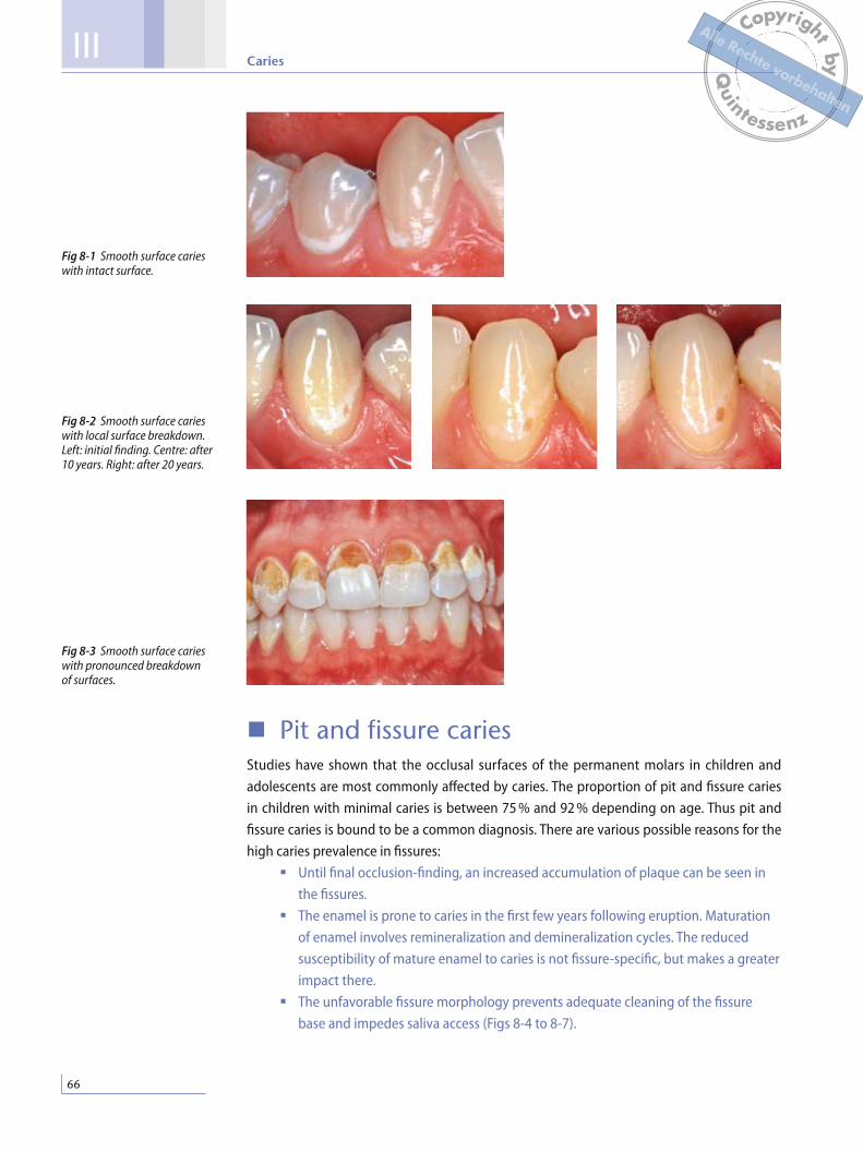
Pit and fissure cariesStudies have shown that the occlusal surfaces of the permanent molars in children and
adolescents are most commonly affected by caries. The proportion of pit and fissure caries
in children with minimal caries is between 75 % and 92 % depending on age. Thus pit and
fissure caries is bound to be a common diagnosis. There are various possible reasons for the
high caries prevalence in fissures:
Until final occlusion-finding, an increased accumulation of plaque can be seen in
the fissures.
The enamel is prone to caries in the first few years following eruption. Maturation
of enamel involves remineralization and demineralization cycles. The reduced
susceptibility of mature enamel to caries is not fissure-specific, but makes a greater
impact there.
The unfavorable fissure morphology prevents adequate cleaning of the fissure
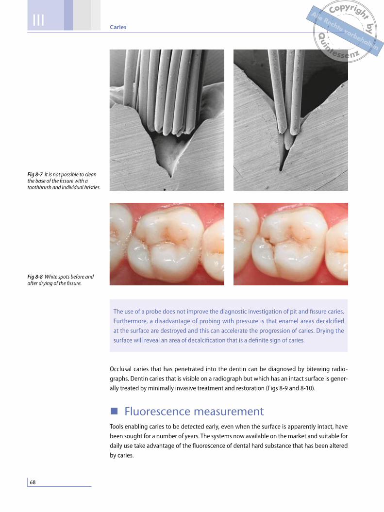
base and impedes saliva access (Figs 8-4 to 8-7).
Fig 8-2 Smooth surface caries with local surface breakdown. Left: initial finding. Centre: after 10 years. Right: after 20 years.
Fig 8-3 Smooth surface caries with pronounced breakdown of surfaces.
Fig 8-1 Smooth surface caries with intact surface.
CopyrightbyQ
uintessenz
Alle Rechte vorbehalten

8Diagnosing caries and caries activity
67
It is important for the teeth to be cleaned before diagnosis so that white spots at the fis-
sure entrance can be identified (Fig 8-8). If a white spot is already visible before drying, it
is reasonable to assume that the caries is more advanced than in a white spot which needs
to be dried before it can be detected. This long-known fact was recently systematized with
the ICDAS system, one of the aims being to publicize comparable diagnostic criteria in all
countries.5
Diagnosis is difficult because dentin caries can exist underneath an apparently intact
surface. In most cases, however, drying and close inspection will reveal an area of decalcifica-
tion at the fissure entrance. The frequency of the so called “hidden caries” in molars varies
between 10 % and 50 %. It appears to be a direct consequence of suboptimal technique in
clinical diagnostics.
Fig 8-4 to 8-6 Different types of fissure morphology.
CopyrightbyQ
uintessenz
Alle Rechte vorbehalten
III Caries
68
The use of a probe does not improve the diagnostic investigation of pit and fissure caries.
Furthermore, a disadvantage of probing with pressure is that enamel areas decalcified
at the surface are destroyed and this can accelerate the progression of caries. Drying the
surface will reveal an area of decalcification that is a definite sign of caries.
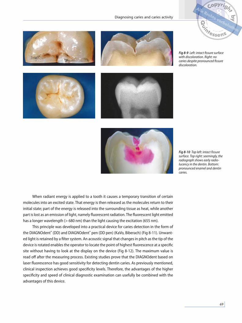
Occlusal caries that has penetrated into the dentin can be diagnosed by bitewing radio-
graphs. Dentin caries that is visible on a radiograph but which has an intact surface is gener-
ally treated by minimally invasive treatment and restoration (Figs 8-9 and 8-10).
Fluorescence measurementTools enabling caries to be detected early, even when the surface is apparently intact, have
been sought for a number of years. The systems now available on the market and suitable for
daily use take advantage of the fluorescence of dental hard substance that has been altered
by caries.
Fig 8-7 It is not possible to clean the base of the fissure with a toothbrush and individual bristles.
Fig 8-8 White spots before and after drying of the fissure.
CopyrightbyQ
uintessenz
Alle Rechte vorbehalten
8Diagnosing caries and caries activity
69
Fig 8-9 Left: intact fissure surface with discoloration. Right: no caries despite pronounced fissure discoloration.
Fig 8-10 Top left: intact fissure surface. Top right: seemingly, the radio graph shows early radio-lucency in the dentin. Bottom: pronounced enamel and dentin caries.
When radiant energy is applied to a tooth it causes a temporary transition of certain
molecules into an excited state. That energy is then released as the molecules return to their
initial state; part of the energy is released into the surrounding tissue as heat, while another
part is lost as an emission of light, namely fluorescent radiation. The fluorescent light emitted
has a longer wavelength (> 680 nm) than the light causing the excitation (655 nm).
This principle was developed into a practical device for caries detection in the form of
the DIAGNOdent® (DD) and DIAGNOdent® pen (DD pen) (KaVo, Biberach) (Fig 8-11). Unwant-
ed light is retained by a filter system. An acoustic signal that changes in pitch as the tip of the
device is rotated enables the operator to locate the point of highest fluorescence at a specific
site without having to look at the display on the device (Fig 8-12). The maximum value is
read off after the measuring process. Existing studies prove that the DIAGNOdent based on
laser fluorescence has good sensitivity for detecting dentin caries. As previously mentioned,
clinical inspection achieves good specificity levels. Therefore, the advantages of the higher
specificity and speed of clinical diagnostic examination can usefully be combined with the
advantages of this device.
CopyrightbyQ
uintessenz
Alle Rechte vorbehalten

IX Endodontology
234
Reversible and irreversible pulpitis due to caries
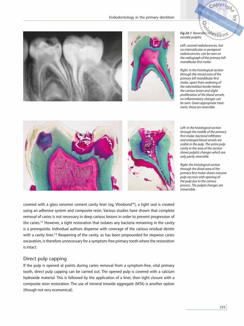
Owing to the large pulp cavity and the minimal thickness of enamel and dentin, caries reach-
es the dentin close to the pulp after only a short time. Initial signs of inflammation in primary
tooth pulp can be observed histologically soon after first contact of caries with dentin. At the
beginning, this process is still reversible (reversible pulpitis). However, if the caries advances
further, it will result in irreversible spread of the inflammation (irreversible pulpitis). These
changes do not always involve severe pain. However, if a primary tooth causes persistent
pain and/or pain in response to heat, this means the inflammation has spread to the entire
pulp of the primary tooth. Sensitivity to percussion means the inflammation has reached
the apical or interradicular periodontium. Clinically, it is often very difficult to distinguish
between reversible and irreversible pulpitis, especially because the sensitivity test with cold
is not very informative in children. In the same primary tooth, healthy, vital areas of pulp can
be observed alongside severely inflamed to necrotic pulp segments (Fig 24-1).
Treatments for reversible pulpitis
Incomplete (stepwise) caries excavationIn the case of a vital, symptom-free primary tooth with profound caries, pulp opening can
be prevented by incomplete caries excavation. Preparation and thorough excavation of car-
ies close to the pulp are first performed. The carious residual dentin close to the pulp is left
in place. The dentin wound is then cleaned and disinfected (eg, with Tubulicid or chlorhex-
idine). If disinfecting the cavity with hydrogen peroxide, it is important to make sure that
polymerization of acrylic resin can be inhibited. After the carious residual dentin has been
Table 24-2 Special features of the clinical examination
Caution is advisable during clinical examination because of particular aspects of some children’s behavior. The following points should be looked for.
Is there any swelling of the face and/or lymph nodes?
Are there any intraoral swellings? Do these swellings fluctuate on palpation?
Can fistulous tracts be probed? Does any pus exude?
Is the painful tooth sensitive to percussion? What is the tap note like?
Are there any sore mucosal areas on palpation?
Is there mobility of the aching tooth?
If caries is present, the consistency and color of the carious material are of interest.
If pulp is opened, the size and location of the opened site as well as the extent and nature of the bleed-ing can provide valuable information about inflammation of the pulp.
The informative value of the CO2 test is limited in small children because of the lack of cooperation and, for psychological reasons, it should therefore be used sparingly.
CopyrightbyQ
uintessenz
Alle Rechte vorbehalten
24Endodontology in the primary dentition
235
Left: in the histological section through the middle of the primary first molar, bacterial infiltrates and enlarged blood vessels are visible in the pulp. The entire pulp cavity in the area of the section shows pulpitis changes which are only partly reversible. Right: the histological section through the distal area of the primary first molar shows massive pulp necrosis with opening of the pulp due to the carious process. The pulpal changes are irreversible.
Fig 24-1 Reversible versus irre-versible pulpitis Left: coronal radiolucencies, but no interradicular or periapical radiolucencies, can be seen on the radiograph of the primary left mandibular first molar. Right: in the histological section through the mesial area of the primary left mandibular first molar, apart from widening of the odontoblast border below the carious lesion and slight proliferation of the blood vessels, no inflammatory changes can be seen. Given appropriate treat-ment, these are reversible.
covered with a glass ionomer cement cavity liner (eg, Vitrebond™), a tight seal is created
using an adhesive system and composite resin. Various studies have shown that complete
removal of caries is not necessary in deep carious lesions in order to prevent progression of
the caries.17 However, a tight restoration that isolates any bacteria remaining in the cavity
is a prerequisite. Individual authors dispense with coverage of the carious residual dentin
with a cavity liner.13 Reopening of the cavity, as has been propounded for stepwise caries
excavation, is therefore unnecessary for a symptom-free primary tooth where the restoration
is intact.
Direct pulp cappingIf the pulp is opened at points during caries removal from a symptom-free, vital primary
tooth, direct pulp capping can be carried out. The opened pulp is covered with a calcium
hydroxide material. This is followed by the application of a liner, then tight closure with a
composite resin restoration. The use of mineral trioxide aggregate (MTA) is another option
(though not very economical).
CopyrightbyQ
uintessenz
Alle Rechte vorbehalten

261
Index
Aabrasion 176, 185accessory canals 12–13acellular afibrillar cementum
(AAC) 11acellular extrinsic fiber cemen-
tum (AEFC) 11acid attack 26–27acidic drinks 185, 186adhesive luting agents, classifica-
tion 12–121adhesive systems 115–121adjacent tooth damage 97–13
aids to prevent 99–12consequences 98origin 97–98
air abrasion 15–17air-polishing technique 16aluminum oxide 15–17, 13amalgam restoration repairs 139amelogenesis 5anorexia nervosa 183, 184antibacterial agents, topical
application 53–57antibacterial approaches 53
limitations 59apical periodontitis, etiology 27aseptic treatment approach 28attrition 176, 185
Bbacteria
metabolism 15, 33, 59, 246where found 27–28
balanced force technique 21Basic Erosive Wear Examination
(BEWE) 177–178Bevelshape file 1–11bifidobacteria 4, 41bioactive glass 5, 16bleaching 163–171
home 165–167, 17–171household products 17
in-office 169mechanism 164–165over-the-counter products 17procedure 165walking bleach technique
133, 167–168, 17bulimia nervosa 183, 184
Cc-factor 127calcium content, of drink or food
185calcium fluoride (CaF2) 3–31calcium hydroxide 79, 235–24canal inlay 155, 156carbon posts 144caries 14–15, 65–83
activity 76–77antibacterial agents for pre-
vention 53–6approximal 71–74CPP-ACP use in prevention 47diagnosis 65–78fluorescence measurement
68–7incomplete (stepwise) exca-
vation 234–235measures to combat vertical
transmission 57–58minimally invasive treatment
9–92noninvasive treatment tech-
niques for initial lesions 8–83
pit and fissure 66–68, 7–71, 79
prevention of progression 79–83
in primary dentition 233–24probiotics and 42–43protective and promoting
factors 25risk assessment (CRA) 76–78root 74–76
selective removal of dentin 19–11
smooth surface 65–66understanding 2
Carisolv 19–11carrier-based filling systems 22,
221caseins 45–46
see also CPP-ACPCavishape file 1cellular intrinsic fiber cementum
(CIFC) 11cellular mixed stratified cemen-
tum (CMSC) 11cementum 11
types 11ceramic restoration repairs
138CEREC system 151–159
case studies 153–159aftercare 159amalgam replacement
153–155anterior rehabilitation with
veneers 157–158endo-crown 155–157
preparation guidelines for ceramic restoration fabric-ation 152–153
cheek retractors 125chemochemical excavation
19–11chlorhexidine (CHX)
in caries prevention 54–55, 57–58
in root canal irrigation 21in tongue cleaning 255
Clearfil SE Bond 117CO2 laser 17, 18CoJet system 137, 138, 139compomers 124composite resin materials
123–124 see also direct restorative
technology
CopyrightbyQ
uintessenz
Alle Rechte vorbehalten
Index
262
composite resin restoration repairs 137–138
condenser-based technique 22, 221
continuous-wave technique 219core materials 217coronal reconstruction, impor-
tance 215–216correlation method 158CPP-ACP 46–49, 185cracked tooth syndrome
223–23clinical examination 228–229clinical picture 227definition 224diagnosis 227–229distribution 225epidemiology 225etiology 224radiologic examination 229restoration and 226, 229symptoms 226–227tooth type 225–226treatment 229–23trial cavity 229
Ddemineralization 118
inhibition by condensed phosphates
49by fluoride 29by metallic ions 49
dental erosion 175–188Basic Erosive Wear Examina-
tion (BEWE) 177–178case studies of progression
181CPP-ACP for 48–49, 185diagnosis 175–176etiology 182–185incidence 178localization in dentition
179–18nutritional factors 185occupation and leisure activi-
ties and 186patient-related factors
182–185prevalence 178prevention 186–188risk assessment 186
denticles 12–13dentin 8–11
chemical properties 26dysplasias 7–8selective removal 19–11
dentin hypersensitivity 223development of teeth 3–5DIAGNOdent pen (DD pen)
69–7, 71, 73, 74diet, and halitosis 25diode laser 17, 18direct pulp capping 235–236direct restorative technology
123–135clinical application 125–131
auxiliary instruments 126–127
final finishing and polishing 13–131, 134–135
layering techniques 127–129light polymerization
129–13optimal operating field
creation 125–126composite resin materials
123–124composite resin restora-
tion fabrication procedure 132–135
discoloration, etiology 164disinfection
alternative approaches 212see also root canal irrigation
Duraphat 29, 31, 238
Eeating and drinking habits
182–183ecological plaque hypothesis 39electronic apex locator 25enamel 5–8
chemical properties 26dysplasias 7–8paraplasias 7structural defects 7
Er:YAG laser 17–19Er,Cr:YSGG laser 17, 18erosion see dental erosionetch-and-rinse luting agents
12, 147etch-and-rinse systems
115–117, 118, 132, 147–148
etching, enamel 117ethylene diamine tetra-acetic
acid (EDTA) 21extension for prevention princi-
ple 97
Ffetor ex ore 245fiber-optic transillumination
(FOTI) 71, 73fiber posts 143, 144, 145, 146Filtek Silorane 124finishing 13–131, 134–135fluorapatite (FAP) 25, 26, 29fluorescence measurement
68–7fluoridated hydroxyapatite
(FHAP) 26, 27–28, 29fluorides 25–31
adsorbed 3demineralization inhibition
by 29and dental erosion 187incorporated 3remineralization promotion
by 27–28usage recommendations 31see also calcium fluoride
formaldehyde 24formocresol 24four-step adhesive systems 116
GGalilean loupes 88gastroesophageal reflux 181,
183–184, 187glide path 2gold-cast cores 143, 144, 145GT hand files 25GTX system 25–26gutta-percha 217
HHalimeter 252–253halitophobia 259halitosis 245–259
causes of odor 246–248cofactors 25diagnostic procedure
248–254
CopyrightbyQ
uintessenz
Alle Rechte vorbehalten

Index
263
forms 245–246informing patient 259patient history 249prevalence 245pseudohalitosis 245, 248,
249, 258–259sources of odor 246–248, 252treatment in dental practice
254–258hand instruments 12HEMA 119, 128Hertwig’s epithelial root sheath
3–5hybrid layer 118, 119hydrofluoric acid 138, 157, 158hydroxyapatite (HAP) 26, 27hypersalivation 184hyposalivation 188
Iiatrogenic damage 97–98ICDAS system 67incomplete caries excavation
234–235infiltration technique 8–81, 83Intensiv Margin Shaper 1–11iodoform paste 29, 236IPS e.max CAD ceramic 155, 156,
158, 159irreversible pulpitis 234, 235
treatment 236–237
KK-files 21, 22, 26Kepler loupes 88
Llactobacilli 4, 41, 42–43, 75laser preparation 17–19lateral condensation technique
219, 221layering techniques 127–129light polymerization 129–13loupes 87, 88
MM wire 25magnification aids 87–92,
12
manual dynamic irrigation (MDI) 212
matrices 99, 126–127, 132metal matrices 99metal points 217metal posts 143, 145metal restoration repairs 139microbial homeostatis 59microscopes, operating 87, 89MicroSeal System 22, 221milk proteins 45mineral trioxide aggregate (MTA)
235, 236, 24minimally invasive preparation
97minimally invasive restorations
9–92
Oodontogenesis 3odor diagnostics, instrumental
252–253one-step adhesive systems 116,
117operating microscopes 87, 89Optibond FL 117, 132oral hygiene
action 21–22and halitosis 248, 249,
255–256motivation 19–21overcoming implementation
deficits 22–24practical implications for
dental team 24see also teeth cleaning
oral infections 39organoleptic examination
251–252ormocers 124oscillating instruments 99–12Oswald maturation 156Owen, lines of 8
Ppassive ultrasonic irrigation (PUI)
212paste fillings 218, 221patency file 2, 211PathFiles 2pellicles 185
periodontitisetiology 27probiotics and 42–43understanding 2
phosphate content, of drink or food 185
photodynamic therapy (PDT) 212
planning, oral hygiene 22–23polishing 13–131, 134–135post-and-core restoration 143,
148post systems 143–149povidone iodine (PI), in caries
prevention 55–56, 57–58Prepcontrol system 1–11prevention of extension
approach 97primary dentition, endodontol-
ogy in 233–24clinical examination 234history taking 233materials for endodontic
measures 24see also irreversible pulpitis;
pulp necrosis; reversiblepulpitis
primary-primary prevention 57–58
prismatic loupes 88probiotics 39–43ProTaper instruments 21proton pump inhibitors 187Proxoshape file 1pseudohalitosis 245, 248, 249,
258–259pulp 12–13
devitalized, with periradicular radiolucency 27
vital 27zones 12
pulp necrosis, treatment 238–239
pulpectomy 236–237pulpitis 15–16pulpotomy 236
Qquantitative laser fluorescence
(QLF) 7
CopyrightbyQ
uintessenz
Alle Rechte vorbehalten

Index
264
Rradiography, in caries diagnosis
71–73, 75Raschkow, nerve plexius of 12remineralization 45
promotionby bioactive glass 5by CPP-ACP 46–48by fluoride 27–28by milk proteins 45
replacement therapy 41Resilon 217resin patch 82, 83restoration repairs 137–14Retzius, lines of 5reversible pulpitis 234, 235
treatments 234–236rinsing 31root canal filling 215–221
importance 215materials 216–217quality 215rating of methods 221requirements 216 techniques 218–22
root canal irrigation 27–213alternate rinsing 21, 211choice of irrigant 29–21disinfection strategies 28–29efficacy 21heating of irrigant 212importance 27manual 211–212protocol 213requirements for irrigants 29
root canal preparation 193–26anatomy of root canals
196–197chemomechanical 28modern principles 198–23
apical gauging 22apical patency 2apical resistance form 22,
218crown down preparation
199deep shape 23delayed length measure-
ment 2glide path 2inlay-shaped access cavity
198
instrumentation 21straight line access 198–199
Ni-Ti instrument properties 23–24
for post placement 145–147risk analysis 193–195techniques 25–26
root filling cements 217root filling materials 216rotary Ni-Ti instruments 199,
2, 21, 22–23fractures 24material properties 23–24
rubber dams 125–126
Ssaliva, protective actions 185sealants 217sealing 79–83
conventional 8, 83self-adhesive luting agents
12–121, 147self-etch luting agents 12, 147self-etch systems 116, 117,
118–119, 147–148self-observation 23separating rings 126–127, 133silanization 18, 138, 139, 146,
147silicatization 147siloranes 124silver diamine fluoride (SDF) 53,
57, 61, 238single-cone technique 218, 221sodium hexametaphosphate 49sodium hypochlorite (NaOCl)
146, 29, 212–213SONICflex airscaler 11–12stannous fluoride 187stepwise caries excavation
234–235streptococci 33, 34, 36, 4–42,
57, 75subjective perception, scientific
evidence vs. 87–88Syntac classic 117
Tteeth cleaning
and dental erosion 185, 187and rinsing 31
see also oral hygienetetracycline 9, 11Thermafil 22, 221thermoplastic filling methods
219Thomas spanner key 21TiF4 187tongue
assessment 254cleaning 255–258morphology 246
triclosan, in caries prevention 56two-step adhesive systems 119
Vvisual gauging 26volatile sulfur compounds (VSCs)
246, 252–253, 256vomiting 183, 184, 187von Ebner, lines of 8
Wwalking bleach technique 133,
167–168, 17warm vertical condensation
technique 219–22, 221wedges 88, 89, 126, 132Weil, zone of 12white spot lesions, CPP-ACP in
treatment 47–48white teeth 163Winkel tongue coating index
254
Xxerostomia 188xylitol, role in caries prevention
33–37, 57–58clinical evidence 34clinical guidelines 37cost-benefit perspective
35–36doses 34, 35patients benefiting 36products 36–37side effects 34
Zzirconium posts 143, 144–145
CopyrightbyQ
uintessenz
Alle Rechte vorbehalten


![[1][m] minimally invasive restorative dentistry](https://static.fdocuments.in/doc/165x107/587254011a28ab852f8b7e5b/1m-minimally-invasive-restorative-dentistry.jpg)