C M Order Euphausiacea - Sociedad Entomológica...
20
Revista IDE@ - SEA, nº 86B (30-05-2015): 1–20. ISSN 2386-7183 1 Ibero Diversidad Entomológica @ccesible www.sea-entomologia.org/IDE@ Class: Malacostraca Order EUPHAUSIACEA Manual Versión en español CLASS MALACOSTRACA Order Euphausiacea Letterio Guglielmo 1 , Antonia Granata 1 & Rosanna Guglielmo 2 1 Dipartimento di Scienze Biologiche e Ambientali, Università di Messina. Viale Guglielmo Stagno d’Alcontres, 31. 98166 Messina, Italy 2 Laboratorio di Ecologia del Benthos, Stazione Zoologica “Anton Dohrn”, Punta San Pietro, 80077 Ischia (NA), Italy [email protected] 1. Brief definition of the group and the main diagnostic characters Euphausiids are small holoplanktonic shrimp-like crustaceans. The term "euphausiid" is commonly used for all members of the crustaceans superorder Eucarida, order Euphausiacea, distributed in two families (Euphausiidae and the mono-specific Bentheuphausiidae, the single genus and species Bentheuphausia amblyops ). There are 86 known species of Euphausiacea and amongst 11 genera (Baker et al., 1990). The word Euphausia derives from Greek eu for good or true, combined with –phausia for shining or light emitting. Another term, krill, has become synonymous with euphausiid. Krill was first used in this sense by Norwegian whalers who applied it to the swarming little fish (krill) which signaled whale feeding grounds. 1.1. Morphology (Text sources from: Einarsson, 1945; Mauchline, 1980, 1984; Boden et al., 1955; Mauchline & Fisher, 1969; Brinton, 1975; Baker et al., 1990; Gibbons et al., 1999; Brinton et al., 2000). Nevertheless, to identify characters clearly and for the sake of brevity, specialized terms were necessary. A diagram of a generalised euphausiid and details of some parts are shown in Figure 1. Like other eucarids, euphausiids have the body divided in 5 cephalic, 8 thoracic, and 6 abdominal segments. The first 2 body regions are fused as a cephalothorax, covered by a carapace fused to the entire thorax and which extends laterally on each side of the body, but does not cover the gills. So, the body is composed of two main parts, the cephalothorax and the abdomen. The cephalothorax includes the carapace dorsally and laterally. Features of the carapace useful in identifications include the length and shape of the rostrum or frontal plate (which lies between the eyes), as well as the number of various lateral and post-ocular spines. A mid-dorsal keel and accompanying cervical groove may be present in some species. Other diagnostic feature are the two pairs of antennae and the eyes anteriorly, and the mouthparts and thoracic limbs ventrally. The abdomen comprises six articulating segments which carry the pleopods ventrally and the telson and uropods posteriorly. Euphausiids lack statocysts. The paired uppermost antennae (Fig. 2), the antennules (first antennae), each consist of a three- segmented antennular peduncle and a pair of many segmented antennular flagella. The anterior-dorsal margin of the first segment is often elongated to form a plate which overlaps the base of the second seg- ment. The plate is known as the antennular lappet and its size and shape serve as very useful diagnostic characters, particularly for Euphausia species. It may be rounded or have one (simple), or two (bifid) points or many marginal spines (pectinate). The second segment of the antennular peduncle may have variously shaped spines or rounded tubercles near the distal margin, while the third segment often has a raised keel. These spines, tubercles and keels are also useful for identification, again particularly for Euphausia species.
Transcript of C M Order Euphausiacea - Sociedad Entomológica...

Revista IDE@ - SEA, nº 86B (30-05-2015): 1–20. ISSN 2386-7183 1 Ibero Diversidad Entomológica @ccesible www.sea-entomologia.org/IDE@ Class: Malacostraca Order EUPHAUSIACEA Manual Versión en español
CLASS MALACOSTRACA
Order Euphausiacea
Letterio Guglielmo1, Antonia Granata1 & Rosanna Guglielmo2
1 Dipartimento di Scienze Biologiche e Ambientali, Università di Messina. Viale Guglielmo Stagno d’Alcontres, 31. 98166 Messina, Italy
2 Laboratorio di Ecologia del Benthos, Stazione Zoologica “Anton Dohrn”, Punta San Pietro, 80077 Ischia (NA), Italy
1. Brief definition of the group and the main diagnostic characters Euphausiids are small holoplanktonic shrimp-like crustaceans. The term "euphausiid" is commonly used for all members of the crustaceans superorder Eucarida, order Euphausiacea, distributed in two families (Euphausiidae and the mono-specific Bentheuphausiidae, the single genus and species Bentheuphausia amblyops ). There are 86 known species of Euphausiacea and amongst 11 genera (Baker et al., 1990). The word Euphausia derives from Greek eu for good or true, combined with –phausia for shining or light emitting. Another term, krill, has become synonymous with euphausiid. Krill was first used in this sense by Norwegian whalers who applied it to the swarming little fish (krill) which signaled whale feeding grounds. 1.1. Morphology (Text sources from: Einarsson, 1945; Mauchline, 1980, 1984; Boden et al., 1955; Mauchline & Fisher, 1969; Brinton, 1975; Baker et al., 1990; Gibbons et al., 1999; Brinton et al., 2000). Nevertheless, to identify characters clearly and for the sake of brevity, specialized terms were necessary. A diagram of a generalised euphausiid and details of some parts are shown in Figure 1.
Like other eucarids, euphausiids have the body divided in 5 cephalic, 8 thoracic, and 6 abdominal segments. The first 2 body regions are fused as a cephalothorax, covered by a carapace fused to the entire thorax and which extends laterally on each side of the body, but does not cover the gills. So, the body is composed of two main parts, the cephalothorax and the abdomen. The cephalothorax includes the carapace dorsally and laterally. Features of the carapace useful in identifications include the length and shape of the rostrum or frontal plate (which lies between the eyes), as well as the number of various lateral and post-ocular spines. A mid-dorsal keel and accompanying cervical groove may be present in some species. Other diagnostic feature are the two pairs of antennae and the eyes anteriorly, and the mouthparts and thoracic limbs ventrally. The abdomen comprises six articulating segments which carry the pleopods ventrally and the telson and uropods posteriorly. Euphausiids lack statocysts. The paired uppermost antennae (Fig. 2), the antennules (first antennae), each consist of a three- segmented antennular peduncle and a pair of many segmented antennular flagella. The anterior-dorsal margin of the first segment is often elongated to form a plate which overlaps the base of the second seg-ment. The plate is known as the antennular lappet and its size and shape serve as very useful diagnostic characters, particularly for Euphausia species. It may be rounded or have one (simple), or two (bifid) points or many marginal spines (pectinate). The second segment of the antennular peduncle may have variously shaped spines or rounded tubercles near the distal margin, while the third segment often has a raised keel. These spines, tubercles and keels are also useful for identification, again particularly for Euphausia species.

Revista IDE@ - SEA, nº 86B (30-05-2015): 1–20. ISSN 2386-7183 2 Ibero Diversidad Entomológica @ccesible www.sea-entomologia.org/IDE@ Class: Malacostraca Order EUPHAUSIACEA Manual
Fig. 1. Generalized euphausiid morphology (after Brinton, 1975). Fig. 2. Lateral and dorsal views of antennular peduncle. Sources from Baker et al., 1990: b, d, e, h; Boden et al., 1955: f, g; Brinton, 1975: a, c, i.
The paired lower antennae (second antennae) each consist of a basal segment bearing an antennal scale and a two segmented antennal peduncle ending in a long flagellum. There is generally little specif-ic variation in the shape of the peduncle or the antennal scale except in Stylocheiron in which the scale can be useful for distinguishing groups of species.
The Euphausiidae can be separated roughly into two groups according to the general shape of the eyes (Fig. 3). With the exception of the genus Thysanoessa in which the eye-types are mixed, all species of a genus have either round (Thysanopoda, Meganyctiphanes, Nyctiphanes, Pseudeuphausia and Euphausia) or bilobed (Stylocheiron, Nematoscelis, Nematobrachion, Tessarabrachion) eyes divided by a constriction into clearly distinguishable upper and lower lobes. The shape and size of the eyes can be an important specific character, particularly in Stylocheiron and to a lesser extent in Nematoscelis species. It

Revista IDE@ - SEA, nº 86B (30-05-2015): 1–20. ISSN 2386-7183 3 Ibero Diversidad Entomológica @ccesible www.sea-entomologia.org/IDE@ Class: Malacostraca Order EUPHAUSIACEA Manual
Fig. 3. Differences in the form of the eyes. Sources, from Baker et al., 1990: b, d, f; Brinton et al., 2000 a, c, e.
is worth noting that some species, which are clearly round-eyed as juveniles and adults, have quite marked bilobed eyes in the larval stages. Genera with bilobed eyes also tend to have elongated second or third thoracic legs.
The mouthparts consist of the labrum followed by the paired mandibles, labia, maxillules (first maxillae) and the maxillae (second maxillae). These are used very little in routine identification but can be invaluable in the specialized task of identifying euphausiid remains in the stomach contents of predators. A very detailed account of mouthpart structure in most euphausiid species has been given by Mauchline (1967). The morphology and number of thoracic limbs can provide valuable clues to genus identification (Fig. 4). There are eight pairs of thoracic limbs, the distal one of which bears a two segmented exopodite and a 5 segmented endopodite. In numbering the limbs, the most anterior is counted as the first. Only Bentheuphausia has the full complement of eight typical thoracic legs; in all the other genera the eighth legs are reduced to simple inconspicuous lobes with a few setae. The length and number of segments present on the seventh leg varies with genus, and the position of this limb can be identified by the pres-ence of a photophore on the coxa. There is a consistent relationship between the eye shape and the form of the thoracic legs. In round-eyed species the unreduced thoracic legs are all similar, whereas in those with bilobed eyes one or two pairs are greatly elongated. For genera which display eloganted second and/or third legs (Stylocheiron, Nematoscelis, Thysanoessa, Nematobrachion) the arrangement of setae on their more distal segments can be diagnostic. The coxa bears obvious gills on its posterior margin and these are characteristic of euphausiids and can be used to immediately separate them from decapods. It should be noted, however, that the legs, particularly the elongated ones, are delicate and prone to damage in nets. As a result they are frequently broken off and elongated limbs are frequently lost.
The carapace (Fig.5), which covers the thoracic region, has a number of features that are useful for identifying species or, more often, groups of species. The rostrum, which lies centrally between the eyes, can be long and sharply pointed, reduced to a slightly raised obtuse protrusion, or may be absent as in Pseudeuphausia in which the frontal plate is extended forward and has a concave anterior margin. In a few species there is a post-ocular spine immediately posterior to the eye. Mid-dorsally, just posterior to the frontal plate, there is often a longitudinal carapace keel (see Fig.1). In addition to the post-ocular spines the carapace may have spines in three other positions. Mid-laterally and approximately above the base of the first thoracic limb, there may be an hepatic spine and on or near the lateral margin there can be one or two lateral denticles. In the genus Nematoscelis the presence or absence of a lateral denticle can be sex dependent. Variations in the morphology of the abdomen (Fig. 6) are used to separate many euphausiids at the species level. The six abdominal segments are numbered from anterior to posterior. The dorsal surface of segments three to five may be raised to form a longitudinal keel (see Fig.1), the posterior edge of which may be extended mid-dorsally to form as a spine of variable length. When spines occur on all three seg-ments that on the third is nearly always the largest. The exoskeleton plates covering segments one to five are known as pleurae and the sculpturing of their posterior and ventral margins varies with species. Alt-hough the sixth segment is cylindrical and lacks pleura, its relative dimensions can be used in the identifi-cation of some species.

Revista IDE@ - SEA, nº 86B (30-05-2015): 1–20. ISSN 2386-7183 4 Ibero Diversidad Entomológica @ccesible www.sea-entomologia.org/IDE@ Class: Malacostraca Order EUPHAUSIACEA Manual
▲ Fig. 4. Differences in the arrangement and number of thoracic limbs. Sources, from Mauchline, 1967: a; Brinton, 1975: b; Mathew, 1980b: c; Baker et al., 1990: d, e, f. ◄ Fig. 5. Differences in the carapace structure and form. Sources from Brinton, 1975: a, b; Baker et al. 1990: c, d, e, f, g.

Revista IDE@ - SEA, nº 86B (30-05-2015): 1–20. ISSN 2386-7183 5 Ibero Diversidad Entomológica @ccesible www.sea-entomologia.org/IDE@ Class: Malacostraca Order EUPHAUSIACEA Manual
Fig. 6. Subdorsal keels (after Brinton, 1975). Fig. 7. Photophores position. Sources from Knight, 1975: a; Brin-ton et al., 2000: b, c.
The pre-anal spine (see Fig.1) lies mid-ventrally and just forward of the posterior margin of the sixth
segment, is rarely useful for identification purposes. The paired biramous uropods are inserted on either side of the pre-anal spine and between, but dorsal to them, is the telson. Neither are used for identification purposes in adults, but the number of terminal spines on the telson is an essential character in determining the developmental stage of larvae.
The name Euphausia is derived from the luminescence produced by large light organs or photophores (Fig. 7). With the exception of Bentheuphausia ambylops and Thysanopoda minyops (Brin-ton, 1987), photophores are present in all species of euphausiids and occur in reduced numbers in Thysanopoda spinicaudata and the Stylocheiron species. Their distribution on the body is generally con-servative: one at the base of each eye, a pair on the coxae of thoracic segments three and seven, and one mid-ventrally on abdominal segments one to four. In Thysanopoda spinicaudata the only photophores present are those on the eyes whereas in Stylocheiron they occur on the eyes, the seventh thoracic limbs and the first abdominal segment. The photophores are of little value as an aid to identification except that their distribution serves to distinguish Stylocheiron from other genera, which can be useful when working with damaged specimens. Abdominal segments one to five each carry a pair of pleopods used for swimming. These are of uniform structure and are composed of a very muscular basal segment bearing an outer exopodite and an inner endopodite. Except in Bentheuphausia, the endopodites of the first and second pleopods of adult males are modified as sexual organs The various lobes, hooks and processes that develop on the endopodite of first pleopods are modified in males to form petasma or copulatory organ (Fig. 8). The form of the mature petasma is of diagnostic value at the species level. However, the disadvantage is that it can only be used to identify adult males and also in the genera Thysanoessa and Stylocheiron the petasma is particularly small and difficult to prepare for examination. The modified second pleopod of the male has never been used as an aid in species identification (e.g. Bargmann, 1937). The petasma is composed of four main lobes (setiferous, auxiliary, median and inner), some of which may be enlarged or lost. The rela-tive size, shape and arrangement of the different processes on the lobes of the petasma are also variable, and it is here that differences should be sought when separating species. So, sexes can be readily distin-guished by the presence or absence of petasma.
The external reproductive organ of the female is called the thelycum and is situated on the ventral surface of the sixth and seventh thoracic segments (Fig.9a) It is the external opening to the female repro-ductive tract, and it consists of various outgrowths from the ventral body wall and the coxal plates (Fig.9b). Points of difference to notice when examining thelyca include the relative shape and sizes of the coxal and sternal plates. Mated females can be readily identified by the presence of one or more spermatophores attached to the thelycum (Fig. 9c). The spermatophore is located in a central depression in the thelycum

Revista IDE@ - SEA, nº 86B (30-05-2015): 1–20. ISSN 2386-7183 6 Ibero Diversidad Entomológica @ccesible www.sea-entomologia.org/IDE@ Class: Malacostraca Order EUPHAUSIACEA Manual
Fig. 8. Petasma structure in some euphausiid species. Sources, from after Baker et al., 1990: a; Brinton, 1975: b, c, d, e, f.
where it is held in place by a cementing substance. Some thelyca morphology by SEM micrograph, in different species, are showed in Figure 9d. Until relatively recently, descriptions of thelyca were available for almost known species. This situation has changed following the publication of detailed accounts by Sebastian (1966), James (1977), Costanzo & Guglielmo (1976a, b, 1977, 1980, 1981, 1991) and Guglielmo & Costanzo (1977, 1978, 1983). Although it has been shown that it has a strong diagnostic value, examination of the thelycum is not easy and usually requires staining and some dissection, and so has not been used as a routine method. However, it is used to differentiate some species (particularly the "gibba" group of Euphausia and the "atlantica/microps" group of Nematoscelis), in which the females can be rather difficult to identify, especially if damaged, and in these cases the thelycum can be a helpful diag-nostic character. However, this character should be used for identifying only fully mature females because the immature specimens of some species can have thelyca resembling the mature state of others.

Revista IDE@ - SEA, nº 86B (30-05-2015): 1–20. ISSN 2386-7183 7 Ibero Diversidad Entomológica @ccesible www.sea-entomologia.org/IDE@ Class: Malacostraca Order EUPHAUSIACEA Manual
Fig. 9. Thelycum (female). Sources, from: a) schematic side view of thelycum, Brinton, 1975; b) schematic drawing, Einarsson, 1942; c) Thyanopoda longicaudata, Einarsson, 1945; d-m) SEM micrographs in different euphausiid species (Costanzo & Guglielmo, 1976a, b; 1980; Guglielmo & Costanzo, 1983): d) Thysanopoda aequalis, Hansen 1905, 18.5 mm, SEM (X 120); e) Meganyctiphanes norvegica, M. Sars, 1857, 9.35 mm, SEM (X 175); f) Euphausia krohnii, Brandt, 1851,16 mm, SEM (X 150); g) Nematoscelis megalops, G.O. Sars, 1883, 22 mm, SEM (X 210); h) Stylocheiron longicorne G.O. Sars, 1883, 9.5 mm, SEM (X 280); i) Nematoscelis tenella, 18.5 mm; l) Bentheuphausia amblyops, G.O. Sars, 1885, SEM micrograph, arrow indicating spermatic mass. Bar = 0.1 mm; m) Nematobrachion sexspinosum, Hansen 1911, SEM micrograph. Bar = 0.1 mm
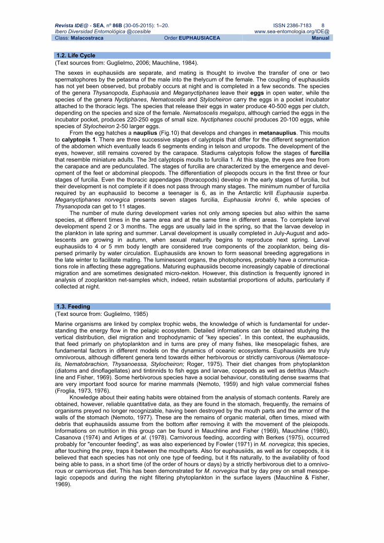
Revista IDE@ - SEA, nº 86B (30-05-2015): 1–20. ISSN 2386-7183 8 Ibero Diversidad Entomológica @ccesible www.sea-entomologia.org/IDE@ Class: Malacostraca Order EUPHAUSIACEA Manual 1.2. Life Cycle (Text sources from: Guglielmo, 2006; Mauchline, 1984). The sexes in euphausiids are separate, and mating is thought to involve the transfer of one or two spermatophores by the petasma of the male into the thelycum of the female. The coupling of euphausiids has not yet been observed, but probably occurs at night and is completed in a few seconds. The species of the genera Thysanopoda, Euphausia and Meganyctiphanes leave their eggs in open water, while the species of the genera Nyctiphanes, Nematoscelis and Stylocheiron carry the eggs in a pocket incubator attached to the thoracic legs. The species that release their eggs in water produce 40-500 eggs per clutch, depending on the species and size of the female. Nematoscelis megalops, although carried the eggs in the incubator pocket, produces 220-250 eggs of small size. Nyctiphanes couchii produces 20-100 eggs, while species of Stylocheiron 2-50 larger eggs.
From the egg hatches a nauplius (Fig.10) that develops and changes in metanauplius. This moults to calyptopis 1. There are three successive stages of calyptopis that differ for the different segmentation of the abdomen which eventually leads 6 segments ending in telson and uropods. The development of the eyes, however, still remains covered by the carapace. Stadiums calyptopis follow the stages of furcilia that resemble miniature adults. The 3rd calyptopis moults to furcilia 1. At this stage, the eyes are free from the carapace and are pedunculated. The stages of furcilia are characterized by the emergence and devel-opment of the feet or abdominal pleopods. The differentiation of pleopods occurs in the first three or four stages of furcilia. Even the thoracic appendages (thoracopods) develop in the early stages of furcilia, but their development is not complete if it does not pass through many stages. The minimum number of furcilia required by an euphausiid to become a teenager is 6, as in the Antarctic krill Euphausia superba. Meganyctiphanes norvegica presents seven stages furcilia, Euphausia krohni 6, while species of Thysanopoda can get to 11 stages. The number of mute during development varies not only among species but also within the same species, at different times in the same area and at the same time in different areas. To complete larval development spend 2 or 3 months. The eggs are usually laid in the spring, so that the larvae develop in the plankton in late spring and summer. Larval development is usually completed in July-August and ado-lescents are growing in autumn, when sexual maturity begins to reproduce next spring. Larval euphausiids to 4 or 5 mm body length are considered true components of the zooplankton, being dis-persed primarily by water circulation. Euphausiids are known to form seasonal breeding aggregations in the late winter to facilitate mating. The luminescent organs, the photophores, probably have a communica-tions role in affecting these aggregations. Maturing euphausiids become increasingly capable of directional migration and are sometimes designated micro-nekton. However, this distinction is frequently ignored in analysis of zooplankton net-samples which, indeed, retain substantial proportions of adults, particularly if collected at night. 1.3. Feeding (Text source from: Guglielmo, 1985) Marine organisms are linked by complex trophic webs, the knowledge of which is fundamental for under-standing the energy flow in the pelagic ecosystem. Detailed informations can be obtained studying the vertical distribution, diel migration and trophodynamic of “key species”. In this context, the euphausiids, that feed primarly on phytoplankton and in turns are prey of many fishes, like mesopelagic fishes, are fundamental factors in different models on the dynamics of oceanic ecosystems. Euphausiids are truly omnivorous, although different genera tend towards either herbivorous or strictly carnivorous (Nematosce-lis, Nematobrachion, Thysanoessa, Stylocheiron; Roger, 1975). Their diet changes from phytoplankton (diatoms and dinoflagellates) and tintinnids to fish eggs and larvae, copepods as well as detritus (Mauch-line and Fisher, 1969). Some herbivorous species have a social behaviour, constituting dense swarms that are very important food source for marine mammals (Nemoto, 1959) and high value commercial fishes (Froglia, 1973, 1976).
Knowledge about their eating habits were obtained from the analysis of stomach contents. Rarely are obtained, however, reliable quantitative data, as they are found in the stomach, frequently, the remains of organisms preyed no longer recognizable, having been destroyed by the mouth parts and the armor of the walls of the stomach (Nemoto, 1977). These are the remains of organic material, often times, mixed with debris that euphausiids assume from the bottom after removing it with the movement of the pleiopods. Informations on nutrition in this group can be found in Mauchline and Fisher (1969), Mauchline (1980), Casanova (1974) and Artiges et al. (1978). Carnivorous feeding, according with Berkes (1975), occurred probably for "encounter feeding", as was also experienced by Fowler (1971) in M. norvegica; this species, after touching the prey, traps it between the mouthparts. Also for euphausiids, as well as for copepods, it is believed that each species has not only one type of feeding, but it fits naturally, to the availability of food being able to pass, in a short time (of the order of hours or days) by a strictly herbivorous diet to a omnivo-rous or carnivorous diet. This has been demonstrated for M. norvegica that by day prey on small mesope-lagic copepods and during the night filtering phytoplankton in the surface layers (Mauchline & Fisher, 1969).

Revista IDE@ - SEA, nº 86B (30-05-2015): 1–20. ISSN 2386-7183 9 Ibero Diversidad Entomológica @ccesible www.sea-entomologia.org/IDE@ Class: Malacostraca Order EUPHAUSIACEA Manual
Fig. 10. Life cycle. Source from Mauchline, 1984; Brinton, 2000 1.4. Distribution (Text source from: Brinton et al., 2000) Euphausiids are exclusively marine, distributed throughout the coastal seas and oceans of the world, from epi-to bathypelagic zone. Their distribution is frequently associated to horizontal or vertical thermal charac-teristics, such as the thermocline, oxycline or with particular water masses, such as to frontal zones and upwelling systems, characterized by high productivity values (Dadon and Boltovskoy, 1982; Sameoto et al., 1987; Gibbons et al., 1995; Tarling et al., 1995). Euphausiids area often a dominant component of net-caught zooplankton, particularly at high latitudes such as the antarctic and the subarctic Pacific and Atlan-tic Ocean basins. Three-dimensional aggregations of euphausiids, usually a daytime phenomenon, have been observed in many species. In some aggregation, they form tight balls or layers, whereas in others aggregations form schools (like Euphausia superba under sea ice). In reason of both their pelagic nature and high capability to performed greater vertical distances, euphausiids are an active energy flux from surface layers to deep waters (Casanova 1970). In fact, essentially for trophic purposes, many species display pronounced diel vertical migration and frequently traverse distances in excess of 200 m at night (Mauchline, 1980; Sameoto, 1980a; Sameoto et al., 1987). Thus, detailed informations on their DVM could also provide elements for better understanding their role in the biological pump (Longhurst and Harrison 1988) and in the vertical flux of POM (Minutoli & Guglielmo, 2009, 2012).

Revista IDE@ - SEA, nº 86B (30-05-2015): 1–20. ISSN 2386-7183 10 Ibero Diversidad Entomológica @ccesible www.sea-entomologia.org/IDE@ Class: Malacostraca Order EUPHAUSIACEA Manual
Fig. 11. a) BIONESS (Bedford Institute Oceanography Net Environmental Sampling System); b) MOCNESS (Multiple Opening/Closing Net and Environmental Sampling System; c) RMT25 Rectangular Midwater Trawl; d) IKMT (Isaacs-Kidd Midwater Trawl).
1.5. Collection and Preservation (Text sources from: Mauchline, 1984; Gibbons et al., 1999) The equipment required to collect euphausiids is of necessity complex because of their offshore habitat. The animals are active swimmers. Because of their large size and well developed eyes, most adult euphausiids are able to detect and avoid nets, especially during the day (Pillar, 1984). If nets are to be used to estimate euphausiid abundance or biomass, they should be of large mouth area and wide mesh diameter (> 0.5 mm) and be towed obliquely at night. To avoid underestimating abundance, therefore, nets should be towed from as close to the bottom as possible. Multiple opening-closing nets, like to RMT8 (Baker et al., 1973), MOCNESS (Wiebe et al., 1976) and BIONESS (Sameoto et al., 1980b) should be used where possible because these not only yield information on biomass but also on vertical distribution (Fig. 11). To collect large quantities of krill are used pelagic net as PHN (Plankton Hamburg Net) and IKMT (Isaacs-Kidd Midwater Traw). The younger development stages can be collected using standard vertical mesozooplankton hauls with nets of smaller mouth area and narrower mesh diameter (e.g. WP2 57cm diameter, 200 um mesh size). Various books provide background information on how to construct the nets and use them. References should be made to Wimpenny (1966), UNESCO (1968), Wiebe & Benfield (2003), Sameoto et al. (2000).
Adult euphausiids are liable to be very conspicuous in the samples because no other planktonic crustaceans are likely to be so large unless the samples are taken well offshore. Adult euphausiids can therefore be picked out at sea if the sample is decanted, from the collecting jar on the end of the net, into a white plastic tray. The simplest way of preserving a sample of euphausiids or mixed plankton is in 5-8% neutral formalin (depending from individual abundance and size) made up in seawater.

Revista IDE@ - SEA, nº 86B (30-05-2015): 1–20. ISSN 2386-7183 11 Ibero Diversidad Entomológica @ccesible www.sea-entomologia.org/IDE@ Class: Malacostraca Order EUPHAUSIACEA Manual 1.6. Scientific and applied interest (Text sources from: Mauchline, 1980; Baker et al., 1990) Although this is a much smaller number than in most other crustacean orders, the euphausiids probably constitute the most economically important component of the oceanic planktonic biomass because of their relatively large size compared with other zooplankton and the great abundance of many of their species. The euphausiids usually constitute 5-10% of the total biomass of plankton and about 30% of the biomass of the crustacean plankton (Mauchline & Fisher, 1969; Mauchline, 1980). Popularly known as krill, they form an important part of the diet of many animals including whales, seals, fishes, birds and, to a lesser extent, man. Euphausiids are important in the diets of the micronektonic fish which in turn are fed on by the tunas (Legand et al., 1972; Roger, 1973, 1975; Roger & Grandperring, 1976; Scotto di Carlo et al., 1982; Guglielmo et al., 1995; Granata et al., 2001). Euphausia pacifica is caught commercially in several region, especially off northen Japan where it is dried and marketed for bait and feed for farmed fish (Fulton, 1976; Mason, 1976; Koops et al., 1977). A bibliography on krill as a human food resource is provided by Gran-tham (1976, 1977) and Taylor (1976). Estimates of the possible annual yeld of a commercial fishery for Euphausia superba range 30 to 200 million tons (Everson, 1977). The krill contain large amounts of trace elements, vitamin A, “B” vitamins and important fatty acids (Mauchline, 1980). Krill pastes and meal (Lyubimova et al., 1973; Bulycheva et al., 1977; Fedotova et al., 1977; Leinemann & Crhristians, 1977; Jahn et al., 1978) can be manufactured and used as animal feed and in the therapeutic diets of patients suffering from stomach ulcers and arteriosclerosis. Lyubimova et al. (1973) and Grantham (1977) discuss the production of various manufactured products such as krill sausages, stuffed eggs and shrimp butter. The krill species both in the northern Atlantic and in the Mediterranean Sea is Meganyctiphanes norvegica. The presence of birds feeding on surface swarms of euphausiids often aids fishermen in locating schools of fish. They are of interest to oceanographers because species distributions are readily definable and, on a large scale, can be regarded as good indicators of water masses. In the Mediterranean Sea, the two species can play this role are Thysanoessa gregaria for the waters of the Atlantic origin and Stylocheiron suhmi for the waters of eastern origin (Casanova, 1974). A whale may consume many tons of North Atlan-tic or antarctic krill in a day. Euphausia species are fished commercially in Japanese, Canadian, and Ant-arctic waters. They are a high protein food for aquarium and farm animals as well as humans. They can be a delicacy, particularly when freshly caught (Brinton et al., 2000). 1.7. Key to families and genera (Text sources from: Mauchline, 1984; Baker et al., 1990; Gibbons et al., 1999; Miller, 2004). This illustrated key is intended to help those who may wish to identify euphausiids but are unfamiliar with the Order, either because they are new to it, or simply because specimen identification is peripheral to their main interest. In compiling this key every attempt has been made to keep the terminology simple.
The arrangement used in the key is very common. It must be emphasised that this key is intended for identifying adult or nearly mature specimens. Caution is necessary when examining juveniles because some of the key characters appear at different stages of development. The more obvious characters used for identification of adult euphausiids can be readily observed with the aid of a low power stereo-microscope. A number of guide have been published to aid in the identification of the euphausiids of the world oceans.
Those examining euphausiids for the first time may find the following general account of their mor-phology helpful. More detailed accounts of euphausiid morphology and anatomy is given by Sars, 1885; Hansen, 1910, 1911, 1912; Einarsson, 1945; Boden et al., 1955; Mauchline & Fisher, 1969; Casanova, 1974; Brinton, 1975; Mauchline, 1980; Baker et al., 1990; Gibbons et al., 1999. A comprehensive, cross- indexed bibliography has been prepared by McWhinnie et al. (1981). The most recent systematic work on euphausiids of the world is a CD-ROM expert system for identification (including larval stages) by Brinton et al. (2000). It included references to virtually the entire literature on Euphausiacea and a wealth of sys-tematic and biological information.
Key to families:
A. Without photophores, with all 8 pairs of thoracic legs well developed ...................... Bentheuphausiidae – Photophores are present and 8th or the 7th and 8th pairs of thoracic legs are rudimentary ........................ ........................................................................................................................................... Euphausiidae Key to genera: 1. Photophores present on abdominal segments one to four ..................................................................... 2 – Photophores present only on abdominal segment one ...................................................... Stylocheiron
2. Eyes round; thoracic legs of approximately equal length ........................................................................ 3 – Eyes divided into two lobes; one or two pairs of thoracic legs very elongated ........................................ 6
3. Seventh pair of thoracic leg smaller than sixth, with six segments ..................................... Thysanopoda – Seventh pair of thoracic leg rudimentary. Anterior margin of frontal plate produced to form a sharp or
gently rounded rostrum ........................................................................................................................... 4

Revista IDE@ - SEA, nº 86B (30-05-2015): 1–20. ISSN 2386-7183 12 Ibero Diversidad Entomológica @ccesible www.sea-entomologia.org/IDE@ Class: Malacostraca Order EUPHAUSIACEA Manual 4. A denticle at the mid-point or on the posterior half of the lateral margin of the carapace. An anterior
lateral denticle also may be present ....................................................................................................... 5 – There is no lateral denticle or, if one is present, it is well anterior to the mid-point of the lateral margin of
the carapace. A strong recurved lappet on first segment of antennular peduncle ................ Nyctiphanes
5. Seventh thoracic leg consisting of two elongated joints and the seventh thoracic exopod present. Strong post-ocular spines and long recurved antennular lappets present .................................................
................................................................................................................. Meganyctiphanes norvegica – Seventh thoracic leg consisting of only a minute process and seventh thoracic exopod absent ............... ................................................................................................................................................ Euphausia
6. Second thoracic leg elongated ............................................................................................................... 7 – Third thoracic leg elongated .......................................................................................... Nematobrachion
7. Second thoracic legs very slender and naked, with only a tuft of apical bristles. Terminal segment of first thoracic leg with short, robust comb-like spines ......................................................... Nematoscelis
– Second pair of thoracic legs rather strong and last two segments armed with spiniform bristles. Dactyus of 1th pair of thoracic legs not pectinate ............................................................................. Thysanoessa
2. Diversity of Macaronesia area (Text Sources from: Mauchline, 1980; Gibbons et al., 1999) The order Euphausiacea is divided into 2 families (Bentheuphausiidae and Euphausiidae), 11 genera and 86 species. The genus Euphausia is the most represented with 31 species, followed by Thysanopoda with 14 species, Stylocheiron with 12 species, Thysanoessa with 10 species, Nematoscelis with 7 species, Nyctiphanes with 4 species, Nematobrachion with 3 species, Pseudoeuphausia with 2 species and 3 monospecific genera (Bentheuphausia, Meganyctiphanes and Tessarabrachion). Of the 86 species of euphausiids found worldwide, 37 have been recovered from Macaronesia Area (14°30’-39°40’ N, 11°25’E-31°30’W), North Atlantic (Table I). The absentees all display limited distribution patterns and are endemic to the Southern Hemisphere or are confined northern.
One of the most detailed analyses on euphausiid assemblages around Canary Islands, is that pro-duced by the “Discovery” SOND cruise of 1965 in the region of Fuertaventura. Baker (1970) found 28 species in this area, the commonest of which are Euphausia krohni, E. hemigibba, Thysanoessa parva, Nematoscelis microps/atlantica, Stylocheiron longicorne and S. elongatum. Other common species are Thysanopoda aequalis, Euphausia brevis, E. gibboides, Nematoscelis tenella, N. megalops, Nemato-brachion flexipes, Stylocheiron suhmi and S. affine. The great Meteor Seamount lies west of the Canaries at approximately 30°N, 28°30’W. Weigmann (1974) found 20 species of euphausiids in the region around the plateau of the seamount, the dominant species being Thysanopoda aequalis, Euphausia brevis, E. hemigibba, Stylocheiron suhmi and S. longicorne. Only 7 of these 20 species occurred on the plateau region, dominated by Euphausia brevis and Stylocheiron suhmi, the depth of water being considered insuf-ficient to allow colonization by the other 13 species. The euphausiid fauna in the area south of the Canar-ies, off Cape Blanc, north-west Africa, has been studied by Weigmann-Haass (1976) and Andreu (1976). This is a region of coastal upwelling. The species composition of the fauna was similar to that of the Ca-naries and Meteor Seamount. Meira (1970) recorded 7 species, including the boreal Thysanoessa longi-caudata, present among the Cape Verde Islands.
Referring to the list of 28 euphausiid species found by Baker (1970), Stylocheiron affine can be con-sidered dubious after the worldwide distribution known for this species (Brinton, 1975; Brinton et al., 2000), while Thysanopoda subaequalis is now considered a synonym of T. aequalis (Brinton et al., 2000). So, to the list of 26 species of euphausiids found by Baker (1970), must be added: one family (Bentheu-phausiidae), two monospecific genera (Bentheuphausia and Meganyctiphanes) and ten species (Bentheu-phausia amblyops, Meganyctiphanes norvegica, Euphausia hanseni, E. mutica, E. pseudogibba, E. tenera, Stylocheiron robustum, Thysanopoda acutifrons, T. cornuta and T. egregia). Two other species may be considered dubious: Thysanopoda tricuspidata and Thysanoessa longicaudata found by Meira (1970) around the Cape Verde Islands.

Revista IDE@ - SEA, nº 86B (30-05-2015): 1–20. ISSN 2386-7183 13 Ibero Diversidad Entomológica @ccesible www.sea-entomologia.org/IDE@ Class: Malacostraca Order EUPHAUSIACEA Manual
Table I. Euphausiid species found in the Macaronesian area, from: Brinton et al., 2000 (CD Room); Mauchline & Fisher, 1969; Brinton, 1975; Gibbons et al., 1999. CAN: Canary Islands, Baker, 1970; MED: Medi-teranean, Mauchline & Fisher, 1969; DD: Day Depth (m), Mauchline, 1980; M: migrates, Mauchline, 1980;
Species CAN MED DD M References on the horizontal distribution in the Macaronesian
BENTHEUPHAUSIDAE Bentheuphausia G.O. Sars, 1885 ● B. ambylops G.O. Sars, 1885 >1000 James (1983, 1987) EUPHAUSIIDAE Euphausia Dana, 1852 ● E. americana Hansen, 1911 ● 500-700 M ● E. brevis Hansen, 1905 ● ● 200-400 M ● E. gibboides Ortmann, 1893 ● 200-500 M ● E. hanseni Zimmer,1915 <300 Mauchline & Fisher (1969) ● E. hemigibba Hansen, 1910 ● ● 400-500 M ● E. krohni Brandt, 1851 ● ● 400-600 M ● E. mutica Hansen, 1905 300-600 M Hong (1969), Brinton (1975) ● E. pseudogibba Ortmann, 1893 25-100 M Brinton (1975), Tsetlin (1981b) ● E. tenera Hansen, 1905 300-500 M Hong (1969), Brinton (1975), James
(1983, 1987) Meganyctiphanes Holt & Tattersal, 1905 ● M. norvegica M. Sars, 1857 ● 100-400 M Mauchline & Fisher (1969) Nematobrachion Calman, 1905 ● N. boopis Calman, 1896 ● >300 ● N. flexipes Ortmann, 1893 ● 100-600 M ● N. sexspinosum Hansen, 1911 ● 100-200 M Nematoscelis G.O. Sars, 1883 ● N. atlantica Hansen, 1910 ● ● >250 M ● N. megalops G.O. Sars, 1883 ● ● >140 M? ● N. microps G.O. Sars, 1883 ● 100-400 M ● N. tenella G.O. Sars, 1883 ● 100-450 M Nyctiphanes G.O. Sars, 1883 ● N. couchi Bell, 1853 ● ● 0-200 M Stylocheiron G.O. Sars, 1883 ● S. abbreviatum G.O. Sars, 1883 ● ● 50-300 M? ● S. carinatum G.O. Sars, 1883 ● 100-300 M? ● S. elongatum G.O. Sars, 1883 ● 200-500 ● S. longicorne G.O. Sars, 1883 ● ● 100-300 ● S. maximum Hansen, 1908 ● ● >400 M? ● S. robustum Brinton, 1962 >450 M? James (1983, 1987), Brinton (1975) ● S. suhmi G.O. Sars, 1883 ● ● <200 Thysanoessa Brandt, 1851 ● T. gregaria G.O. Sars, 1883 ● ● 0-200 M? ● T. parva Hansen, 1905 ● 400-1600 Thysanopoda Milne-Edwards, 1830 ● T. acutifrons Holt & Tattersal, 1905 700-900 Mauchline & Fisher (1969), Brinton (inédito) ● T. aequalis Hanse, 1905 ● ● 500-700 M ● T. cornuta Illig, 1905 >1000 M Mauchline & Fisher (1969); James (1983,
1987), Tsetlin (1981b) ● T. cristata G.O. Sars, 1883 ● 400-800 ● T. egregia Hansen, 1905 >1000 Mauchline & Fisher (1969); James (1983,
1987), Tsetlin (1981b) ● T. microphtalma G.O. Sars, 1885 ● >500 ● T. monacantha Ortmann, 1893 ● 400-800 M ● T. obtusifrons G.O. Sars, 1883 ● 300-600 M ● T. pectinata Ortmann, 1893 ● 500-700 M

Revista IDE@ - SEA, nº 86B (30-05-2015): 1–20. ISSN 2386-7183 14 Ibero Diversidad Entomológica @ccesible www.sea-entomologia.org/IDE@ Class: Malacostraca Order EUPHAUSIACEA Manual
3. Suggested References
3.1. General.
ANTEZANA, T., N. AGUIRRE & R. BUSTAMANTE 1976. Clave illustrada y distribucion latitudinal de los eufausi-dos del Oceano Antarctico (Crustacea, zooplancton). Serv. Cient. Inst. Antárt. Chileno, 4: 53-69.
BANNER, A. H. 1950. A taxonomic study of the Mysidacea and Euphausiacea (Crustacea) of the northeas-tern Pacific. Part III. Euphausiacea. Trans. R. Can. Inst., 28: 1-62.
BANNER, A. H. 1954. New records of Mysidacea and Euphausiacea from the northeastern Pacific and adjacent areas. Pacif. Sci., 8: 125-139.
BARY, B. M. 1956. Notes on ecology, systematics, and development of some Mysidacea and Euphausia-cea (Crustacea) from New Zealand. Pacif. Sci., 10: 431-467.
BODEN, B. P. 1954. The euphausiid crustaceans of southern African waters. Trans. R. Soc. S. Afr., 34: 181-243.
BODEN, B.P. 1961. Euphausiacea (Crustacea) from tropical West Africa. Atlantide Rep., 6: 251-262. BRINTON, E. 1962. The distribution of Pacific euphausiids. Bull. Scripps Instn Oceanogr., 8: 51-270. CASANOVA-SOULIER, B. 1968. Les Euphausiacés de la Mediterranée. Comm. int. Explor. Scient. Mer
Mediterr., Comité du Plancton, Monaco, September, 1-62. COLOSI, G. 1917. Crostacei. Parte II. Eufausiacei. Raccolte planctoniche fatte dalla R. Nave “Liguria”.
Pubblicazioni del R. Istituto di Studi superiori Pratici e di perfezionamento, Firenze, sezione di Scienze fisiche e naturali, 2, 165-205.
COLOSI, G. 1922a. Eufausiacei e Misidacei raccolti nella campagna del 1920. Memorie R. Com. talassogr. ital., 96: 1-12.
COLOSI, G. 1922b - Eufausiacei e Misidacei dello Stretto di Messina. Memorie R. Com. talassogr. ital., 98: 1-22.
DANA, J. D. 1850. Synopsis generum crustaceorum ordinis Schizopoda. Am. J. Sci., 9: 129-133. DANA, J. D. 1852. Crustacea. U.S. Exploring Expedition under the command of Charles Wilkes, U.S.N.,
13, pp. 685. GORDON, I. 1955. Systematic position of the Euphausiacea. Nature, 176: 934. HANSEN, H. J. 1905a. Preliminary report on the Schipozoda collected by H.S.H. Prince Albert of Monaco
during the cruise of the “Princess Alice” in the year 1904. Bull. Mus. océanogr. Monaco, 30: 1-32. HANSEN, H. J. 1905b.- Further notes on the Schizopoda. Bull. Mus. océanogr. Monaco, 42: 1-32. HANSEN, H. J. 1908a. Crustacea Malacostraca. Dan. Ingolf-Exped., 3: 1-120. HANSEN, H. J. 1908b. Schizopoda and Cumacea. Result. Voyage S.Y. Belgica, Rapp. Sci., Zool., 1-20. HANSEN, H. J. 1908c. Sur quelques Crustacés pélagiques dAmboine. Revue suisse Zool, 16: 157-159. HANSEN, H. J. 1913. Crustacea Schizopoda. Rep. Swed.Antarct. Exped. 1901 -1903, 1-56. HANSEN, H. J. 1915. The Crustacea Euphausiacea of the United States Musem. Proc. U.S. natn. Mus., 48:
59-114. HANSEN, H. J. 1916. The euphausiacean crustaceans of the “Albatross” Expedition to the Philippines.
Proc. U.S. natn. Mus., 49: 635-654. HANSEN, H. J. 1927. Sergestides et Schizopodes. Exped. Sci. du “Travailleur” et du “Talisman” 1880-
1883,1-27. HOLT, E. W. L. & W. I. BEAUMONT 1902. Report on the Crustacea Schizopoda of Ireland. Scient. Trans. R.
Dubl. Soc., Ser. II, 7, 221-252. HOLT, E.W. & W.M. TATTERSALL 1905a. Schizopodous Crustacea from the northeast Atlantic slope. Scient.
Invest. Fish. Brch Ire., 1902-03, Pt. II, App. IV, 99-152. HOLT, E. W. & W. M. TATTERSALL 1905b. Report on the schizopods collected by Mr George Murray, F.R.S.,
during the cruise of the “Oceana” in 1898. Ann. Mag. nat. Hist., Ser.7, 16: 1-10. HOLT, E. W. & W. M. TATTERSALL 1905c. Biscayan plankton. Part V. The Schizopoda. Trans. Linn. Soc.
Lond., (Zool.), Ser. 2, 10: 103-129. HOLT, E. W. & W. M. TATTERSALL 1906a - Schizopodous Crustacea from the northeast Atlantic slope.
Scient. Invest. Fish. Brch Ire., 1904, (suppl.), App. V, 1-50. HOLT, E. W. & W. M. TATTERSALL 1906b - Preliminary notice of the Schizopoda collected by H.M.S. “Dis-
covery” in the Antarctic region. Ann. Mag. nat. Hist., Ser. 1, 17: 1-11. ILLIG, G. 1930. Die Schizopoden der Deutschen Tiefsee-Expedition. Rep. Valdivia Exped., 22: 397-625. JAMES, B. M. 1970. Euphausiacean Crustacea. Oceanogr. Stud, of Texas A. & M. University, 1: 205-229. LOMAKINA, N. B. 1978. Euphausiids of the world oceans (Euphausiacea). In: “Determinations of the Fauna
of the SSSR”, Academy Nauk, SSSR, 118: 1-222. MAUCHLINE, J. 1971. Crustacea, Euphausiacea: adults. Conseil International pour l’Exploration de la Mer,
Zooplankton, Sheet, 134: 1-8. MAUCHLINE J. & T. NEMOTO 1977. Integumental sensilla of diagnostic value in euphausiids. J. oceanogr.
Soc. Japan, 33: 283-289. ORTMANN, A. E. 1893. Decapoden und Schizopoden. Ergebn. Atlant. Ozean Planktonexped. Humboldt-Stift,
2, 1-120. ORTMANN, A. E. 1894. Reports on the dredging operations off the west coast of Central America to the
Galapagos to the west coast of Mexico, and the Gulf of California, in charge of Alexander Agassiz, by the U.S. Fish Commission steamer “Albatross” during 1891. XIV. The pelagic Schizopoda. Bull. Mus. comp. Zool. Harv., 25: 99-111.
ORTMANN, A. E. 1905. Schizopods of the Hawaiian Islands collected by the steamer “Albatross” in 1902. Bull. U.S. Fish Commn, 1903, 961-973.

Revista IDE@ - SEA, nº 86B (30-05-2015): 1–20. ISSN 2386-7183 15 Ibero Diversidad Entomológica @ccesible www.sea-entomologia.org/IDE@ Class: Malacostraca Order EUPHAUSIACEA Manual PONOMAREVA, L. A. 1956. Methods of studying euphausiids. Trudy Inst. Okeanol., 19: 334-339. PONOMAREVA, L. A. 1959. Euphausiids of the Okhotsk and Bering Seas. Trudy Inst. Okeanol., 30: 115-147. PONOMAREVA, L. A. 1963. The euphausiids of the North Pacific, their distribution and ecology. Dolci. Akad.
Nauk SSSR, 1-142. (Israel programme for Science Translation, 1966). PONOMAREVA, L. A. 1975. The Euphausiids of the Indian Ocean and Red Sea. Academy Nauk, SSSR,
Moscow, pp. 83. RIGGIO, G. 1905. Contributo alla Carcinologia del Mediterraneo. I. Nota sopra alquanti crostacei del Mare
di Messina. Naturalista sicil., 17: 1-56. RUSTAD, D. 1930. Euphausiacea with notes on their biógeography and development. Scient. Results
Norw. Antarct. Exped., 5: 1-82. RUSTAD, D. 1934. On the Antarctic euphausiids from the “Norvegia” expeditions, 1929-30 and 1930-31.
Scient. Results Norw.Antarct. Exped., 12: 1-53. RUUD, J. T. 1932. On the biology of southern Euphausiidae. Hvalràd. Skr., 2, 1-105. RUUD, J. T. 1936. Euphausiacea. Rep. Dan. oceanogr. Exped. Mediterr., 2, 1-86. SARS, G. O. 1883. Preliminary notices on the Schizopoda of H.M.S. “Challenger” Expedition. Forh.
VidenskSelsk. Krist., 7, 1-43. SARS, G. O. 1898. On the propagation and early development of Euphausiidae. Arch. Math. Naturv., 20:
1-41. SHEARD, K. 1953. Taxonomy, distribution and development of the Euphausiacea (Crustacea). Report of
the British and New Zealand Antarctic Research Expedition, Ser. B (Zoology and Botany), 8, 1-72. STEBBING, T. R. R. 1893. A History of Crustacea. Recent Malacostraca. Internat. Science, Ser. 74,1-466. STEBBING, T. R. R. 1900. On some crustaceans from the Falkland Islands collected by Mr Rupert Vallen-
tin. Proc. Zool. Soc. Lond., 1: 517-568. STEBBING, T. R. R. 1905. General catalogue of South African Crustacea, Part III. Mar. Invest. S. Afr., 4:
21-123. STEBBING, T.R.R. 1910. General catalogue of South African Crustacea, Part V. Ann. S. Afr. Mus., 6: 281-
593. STEPHENSEN, K. 1913. Report on the Malacostraca collected by the “Tjalfe” expedition, under the direction
of Ad. S. Jensen, especially at W. Greenland. Vidensk. Meddr dansk naturh. Foren., 64: 57-134. STEPHENSEN, K. 1933. The Godthaab Expedition 1928. Schipozoda. Meddr Gronland, 79: 1-20. STEPHENSEN, K. 1938. Euphausiacea, Mysidacea, Cumacea, and Nebaliacea. Zoology Iceland, 3: 1-24. STEPHENSEN, K. 1943. The zoology of East Greenland. Leptostraca, Mysidacea, Cumacea, Tanaidacea,
Isopoda, and Euphausiacea. Meddr. Gronland, 121: 1-82. TATTERSALL, W. M. 1908a. Crustacea. VII. Schizopoda. National Antarct. Exped. 1901-1904, Nat. Hist.
Rep. Br. Antarct. Terra Nova Exped. (zool.), 4: 1-42. TATTERSALL, W. M. 1908b. The Schizopoda and Isopoda collected by the “Huxley” from the north side of
the Bay of Biscay in August, 1906. J. mar. biol. Ass. U.K., 8: 189-196. TATTERSALL, W. M. 1909. The Schizopoda collected by the Maia and Puritan in the Mediterranean. Mitt.
zool. Stn. Neapel, 19: 117-143. TATTERSALL, W. M. 1911. Schizopodous Crustacea from the north-east Atlantic Slope. Scient. Invest. Fish.
Brch. Ire., 1910, 2 (suppl.), 1-77. TATTERSALL, W. M. 1912a. On the Mysidacea and Euphausiacea collected in the Indian Ocean during
1905. Trans. Linn. Soc. Lond., Ser.II, (Zool.), 15: 119-136. TATTERSALL, W. M. 1912b. Clare Island survey. Nebaliacea, Cumacea, Schizopoda, Stomatopoda. Proc.
R. Ir. Acad., 31: 1-10. TATTERSALL, W. M. 1913. The Schizopoda, Stomatopoda, and non Antarctic Isopoda of the Scottish Na-
tional Antarctic Expedition. Trans. R. Soc. Edinb., 49: 865-894. TATTERSALL, W. M. 1918. Euphausiacea and Mysidacea. Scient. Rep. Australas. Antarct. Exped., Ser. C-
Zool. Bot., 5: 5-15. TATTERSALL, W. M. 1924. Crustacea. VIII. Euphausiacea. Nat. Hist. Rep. Br. Antarct. Terra Nova Exped.,
1910, (Zool.), 8: 1-36. TATTERSALL, W. M. 1925. Mysidacea and Euphausiacea of marine survey, South Africa. Rep. Fish. mar.
biol. Surv. Un. S. Afr., 4, (1924), Spec. Rep., No. 5, 1-12. TATTERSALL, W. M. 1927. Crustaceans of the orders Euphausiacea and Mysidacea from the western
Atlantic. Proc. U.S. natn. Mus., 69: 1-28. TATTERSALL, W. M. 1936a. The zooplankton. V. The occurrence and seasonal distribution of the Mysida-
cea and Euphausiacea. Scient. Rep. Gt Barrier Reef Exped., 2: 277-289. TATTERSALL, W. M. 1936b. Mysidacea and Euphausiacea. Scient. Rep. Gt Barrier Reef Exped., 5: 143-176. THIELE, J. 1905. Uber einige stielaugige Krebse von Messina. Zool. Jber. Neapel, S. (suppl.), 443-474. TORELLI, B. 1934. Eufausiacei del Mar Rosso. Memorie R. Com. talassogr. ital., 208: 1-17. ZIMMER, C. 1912. Sudwestafrikanische Schizopoden. Denkschr. med. -naturw. Ges. Jena, 17: 1-11. ZIMMER, C. 1914. Die Schizopoden der Deutschen Sudpolar-Exped., 1901-1903. Dt. SiXdpol-Exped., 15:
377-445. ZIMMER, C. 1915. Schizopoden des Hamburger Naturhistorischen (Zoologischen) Museum. Mitt, naturh.
Mus. Hamb., 32: 159-182. ZIMMER, C. & H. E. GRÙNER 1956. Euphausiacea. Dr H.G. Bronns Klassen und Ordnungen des Tierreichs, 6,
1-286.

Revista IDE@ - SEA, nº 86B (30-05-2015): 1–20. ISSN 2386-7183 16 Ibero Diversidad Entomológica @ccesible www.sea-entomologia.org/IDE@ Class: Malacostraca Order EUPHAUSIACEA Manual 2. Euphausia BRINTON, E. 1962. Two new species of Euphausiacea, Euphausia nana and Stylocheiron robustum from
the Pacific. Crustaceana, 4: 167-179. 3. Nematoscelis
GOPALAKRISHNAN, K. 1973. Developmental and growth studies of the euphausiid Nematoscelis difficilis (Crustacea) based on rearing. Bull. Scripps Instn Oceanogr., 20: 1-87.
GOPALAKRISHNAN, K. 1974. Zoogeography of the genus Nematoscelis (Crustacea, Euphausiacea). Fishery Bulletin, National Oceanic Atmospheric Administration of the United States, 72: 1039-1074.
GOPALAKRISHNAN, K. 1975. Biology and taxonomy of the genus Nematoscelis (Crustacea, Euphausiacea). Fishery Bulletin Rational Oceanic Atmospheric Administration of the United States, 73: 797-814.
JAMES. P. T. 1973. Distribution of dimorphic males of three species of Nematoscelis (Euphausiacea). Mar. Biol., 19: 341-347.
KAREDIN, E.P. 1971. About identity between Nematoscelis megalops G.O. Sars, 1885, N. difficilis Hansen, 1911 (Euphausiacea, Crustacea) and validity of distinguishing N. difficilis Hansen. Izv. tikhookean. nauchnoissled. Inst. ryb. Khoz. Okeanogr., 75: 121-129.
MCLAUGHLIN, P. A. 1965. A redescription of the euphausiid crustacean, Nematoscelis difficilis Hansen, 1911. Crustaceana, 9: 41-44.
4. Thysanoessa
JONES, L.T., D. C. T. FORSYTH & G. A. COOPER 1967. The occurrence of the two- spined form of Thysanoessa inermis (Crustacea: Euphausiacea) in the North Sea. Bull. Mar. Ecol., 6: 181-184.
NEMOTO, T. 1963. A new species of Euphausiacea, Thysanoessa inspinata, from the North Pacific. J. oceanogr. Soc. Japan, 19: 41-47.
NEMOTO, T. 1966. Thysanoessa euphausiids, comparative morphology, allomorphosis and ecology. Scient. Rep. Wales Res. Inst., Tokyo, 20: 109-155.
5. Thysanopoda
BODEN B. P. & E. BRINTON 1957. The euphausiid crustaceans Thysanopoda aequalis Hansen and Thysanopoda subaequalis Boden, their taxonomy and distribution in the Pacific. Limnol. Oceanogr., 2: 337-341.
BRINTON, E. 1953. Thysanopoda spinicaudata, a new bathypelagic giant euphausiids crustacean, with comparative notes on T. cornuta and T. egregia. J. Wash. Acad. Sci., 43: 408-412.
ILLIG, G. 1905. Eine neune Art der Gattung Thysanopoda. Zool.Anz., 28: 663-664. ILLIG, G. 1908a. Thysanopoda megalops spec. nov. Erbeutet auf der Deutschen Tiefsee-Expedition, 1898-
99. Zool. Anz., 33: 54-55. ILLIG, G. 1908b. Ein weiterer Bericht iiber die Schizopoden der Deutschen Tiefsee- Exped. 1898-1899.
Gattung Thysanopoda (Fortsetzung). Zool.Anz., 33: 112- 115. 6. Meganyctiphanes
COLOSI, G. 1918. Sul genere Meganyctiphanes (Eufausiacei). Monitore zool. ital., 29: 178-181. MAUCHLINE, J. 1959. The development of the Euphausiacea (Crustacea) especially that of Mega-
nyctiphanes norvegica (M. Sars). Proc. Zool. Soc. Lond., 132: 627- 639. 7. Stylocheiron
SILAS, E. G. & K. J. MATHEW 1967. Stylocheiron indicus, a new euphausiid (Crustacea: Euphausiacea) from Indian Seas. Cun. Sci., 36: 169-172.
GUGLIELMO, L. & G. COSTANZO 1990. Diagnostic value of the thelycum in euphausiids, II. Oceanic species, Genus Stylocheiron.]. Crust. Biol. (In press/en prensa).
8. Other Genera ILLIG, G. 1908. Ein weiterer Bericht iiber die Schizopoden der Deutschen Tiefsee- Expedition 1898-1899.
Nyctiphanes latifrons n. sp. Zool. Anz., 33: 252-253. WANG, R. & K CHEN 1963. Description of a new species of the genus Pseudeuphausia (Crustacea)
Pseudeuphausia sinica sp. nov. Oceanologia Limnol. sin., 5: 353-358. 9. Larval stages BODEN, B.P. 1950. The post naupliar stages of the crustacean Euphausia pacifica. Trans. Amer. Microsc.
Soc., 69: 373-386. BODEN, B. P. 1951. The egg and larval stages of Nyctiphanes simplex, a euphausiid crustacean from
California. Proc. Zool. Soc. Lond., 121: 515-527. CASANOVA-SOULIER, B. 1968. Una sèrie larvaire dans le genre Nematoscelis (Euphausiacés). Cah. Biol,
mar., 9: 1-12. CASANOVA, B. 1972. Clé de détermination des larves calyptopis des euphausiacés de Méditerranée.
Crustaceana, 22: 178-180.

Revista IDE@ - SEA, nº 86B (30-05-2015): 1–20. ISSN 2386-7183 17 Ibero Diversidad Entomológica @ccesible www.sea-entomologia.org/IDE@ Class: Malacostraca Order EUPHAUSIACEA Manual ENDO, Y. & Y. KOMAKI 1979. Larval stages of euphausiids with description of those of Thysanoessa
longipes Brandt. Bull. Japan Sea reg. Fish. Res. Lab., 30: 97-110. ENDO, Y. 1980. Larval stages of a euphausiid Tessarabrachion oculatum Hansen. Bull. Plankton Soc. Ja-
pan, 27: 113-122. FROST, W. E. 1935. Larval stages of the euphausiids Nematoscelis megalops (G.O. Sars) and Stylocheiron
longicorne (G.O. Sars) taken off the south-west coast of Ireland. Proc. R. Ir. Acad., 42B: 443-458. FROST, W. E. 1939. Larval stages of the euphausiid Thysanopoda acutifrons (Holt and Tattersall) taken off
of the south-west coast of Ireland. Proc. R. Ir. Acad., 45B: 301-319. KNIGHT, M. D. 1973. The nauplius II, metanauplius and calyptopis stage of Thysanopoda tricuspidata Mil-
ne-Edwards (Euphausiacea). Fishery Bulletin, National Oceanic Atmospheric Administration of the United States, 71: 53-67.
KNIGHT, M. D. 1976. The larval development of Euphausia sanzoi Torelli (Crustacea, Euphausiacea). Bull. Mar. Sci., 26: 538-557.
KNIGHT, M. D. 1978. Larval development of Euphausia fallax Hansen (Crustacean: Euphausiacea) with a comparison of larval morphology within the E. gibboides species group. Bull. Mar. Sci., 28: 255-281.
LEBOUR, M. V. 1926a. A general survey of larval euphausiids, with a scheme for their identification. J. mar. biol. Ass. U.K., 14: 519-527.
LEBOUR, M. V. 1926b. The young of Stylocheiron suhmii G.O. Sars and Stylocheiron abbreviatum G.O. Sars (Crustacea) from Mediterranean plankton collected by Mr F.S. Russell in the neighbourhood of Alexandria, Egypt. Proc. Zool. Soc. Lond., Pt. 1, 203-211.
LEBOUR, M. V. 1926c. On some larval euphausiids from Mediterranean in the neighbourhood of Alexan-dria, Egypt, collected by Mr F.S. Russell. Proc. Zool. Soc. Lond., Pt. 3, 765-776.
LEWIS, J. B. 1955. Some larval euphausiids of the genus Stylocheiron from the Rorida Current. Bull. Mar. Sci., 5: 190-202.
LE ROUX, A. 1973. Observations sur le développement larvaire de Nyctiphanes couchii (Crustacea: Eu-phausiacea) au laboratoire. Mar. Biol., 22: 159-166.
LE ROUX, A. 1974. Observations sur le développement larvaire de Meganyctiphanes norvegica (Crustacea: Euphausiacea) au laboratoire. Mar. Biol., 26: 45-56.
LE ROUX, A. 1976. Observations sur les larves de Nyctiphanes couchii et de Meganyctiphanes norvegica (Crustacea: Euphausiacea) recoltées dans le Golfe du Morbihan. Cah. Biol, mar., 17: 375-386.
MAUCHLINE, J. 1965. The larval development of the euphausiid Thysanoessa raschii (M. Sars). Crustaceana, 99: 31-40.
SILAS, E.G. & K. J. MATHEW 1977. A critique to the study of larval development in Euphausiacea. In: “Pro-ceedings of the Symposium on Warm Water Zooplankton”. Special Publication of National Institute of Oceanography, Goa, India, 571- 582.
SOULIER, B. 1965. Essai d’harmonisation de la nomenclature des larves d’Euphausiacès. Rev. Trav. Inst. Pèches marit., 29: 191-195.
WANG, R. 1965. On the larval stages of Pseudeuphausia sinica Wang and Chen (Euphausiacea). Oceanologia Limnol. sin., 7: 35-53.
WIEGMANN-HAASS, R. 1977. Dei calyptopis und furcilia-stadien von Euphausia hanseni (Crustacea: Eu-phausiacea). Helgolander wiss. Meeresunters., 29: 315-327.
4. Text References
ANDREU, P. 1976. Contribucion al estudio de los eufausiaceos de los alrededores de Cabo Blanco (NW de Africa). Reesultatos Expediciones Cientificas del Buque Oceanografico “Cornde de Saavedra”, 5: 77-111.
ARTIGES, J. M., M. PAGANO & A. THIRIOT 1978. Morphologie functionnelle des appendices nutritionnels de Meganyctiphanes norvegica (M. Sars, 1856) et Euphausia krohnii (Brandt, 1851) (Crustacea Eup-hausiacea). Arch. Zool. Exp. Gkn., 119: 95-106.
BAKER, A. de C. 1970. The vertical distribution of euphausiids near Fuertaventura Canary Islands (“Disco-very” Sond Cruise, 1965). Journal of the Marine Biological Association of the United Kingdom, 50: 301-342.
BAKER, A. DE C., M. R. CLARKE & M. J. HARRIS 1973. The N.I.O. combination net (RMT 1 + 8) and further developments of rectangular midwater trawls. Journal of the Marine Biological Association of the United Kingdom, 53: 167-184.
BAKER, A. DE C., B. P. BODEN & E. BRINTON 1990. A practical guide to the euphausiids of the world. British Museum (Natural History), London. 96 pp.
BARGMANN. H. E. 1937. The reproductive system of Euphausia superba. "Discovery" Reports, 14: 325-350.
BERKES, F. 1975. Some aspects of feeding mechanisms of euphausiid crustaceans. Crustaceana, 29: 266-270.
BODEN, B. P. 1955. Euphausiacea of the Benguela Current. ‘Discovery’ Rep., 27: 337-376. BODEN B. P., M. W. JOHNSON & E. BRINTON 1955. The Euphausiacea (Crustacea) of the North Pacific. Bull.
Scripps Inst. Oceangr., 6: 287-400. BRINTON, E. 1975. Euphausiids of the southeast Asian waters. Naga Rep., 4: 1-287. BRINTON, E. 1987. A new abyssal euphausiid, Thysanopoda minyops, with comparisons of eye size, pho-
tophores, and associated structures among deep-living species. Journal of Crustacean Biology, 7: 636-666.

Revista IDE@ - SEA, nº 86B (30-05-2015): 1–20. ISSN 2386-7183 18 Ibero Diversidad Entomológica @ccesible www.sea-entomologia.org/IDE@ Class: Malacostraca Order EUPHAUSIACEA Manual BRINTON, E., M. D. OHMAN. A. W. TOWNSEND, M. D. KNIGHT & A. L. BRIDGEMAN 2000. Euphausiids of the
World Ocean (cd-room Expert System). Springer-Verlag, Heildeberg. BULYCHEVA, N. P., L.I. LOZBIN, P. N. MAJSTRUK & M. S. MAROVA 1977. Microflora of the “Ocean” krill paste
as a quality index. Gigiena i sanitariya, 3: 102-104. CASANOVA, B. 1970. Répartition bathymétrique des euphausiacés dans le bassin occidental de la Médite-
rranée. Revue de Travaux de L’Institut des Pêches Maritimes, 34: 205-219. CASANOVA, B. 1974. Les Euphausiacés de Mediterranée (Systematique et Développement larvaire. Bio-
geographie et Biologie). Thèse pour Docteur Es. Sciences Naturelles, l’Université de Provence, Aix-Marseille, pp. 380.
COSTANZO G. & L. GUGLIELMO 1976a. Diagnostic value of the thelycum in euphausiids, I. Mediterranean species (first note). Crustaceana, 31: 45-52.
COSTANZO G. & L. GUGLIELMO 1976b. Diagnostic value of the thelycum in euphausiids, I. Mediterranean species (second note). Crustaceana, 31: 178-180.
COSTANZO G. & L. GUGLIELMO 1977. Sur l’importance du thelycum dans la systematique des espèces voisines du genre Thysanoessa (Euphausiacea). Rapp. P.-v. Réun. Comm. int. Explor. scient. Mer Méditerr., 24: 129-131
COSTANZO G. & L. GUGLIELMO 1980. Diagnostic value of the thelycum in euphausiids. II Oceanic species, Genus Nematoscelis. Mar. Biol., 56: 311-317.
COSTANZO G. & L. GUGLIELMO 1981. Valore diagnostico del thelycum negli eufausiacei, II. Specie oceani-che. Genere Thysanoessa. Cah. Biol, mar., 22: 221-229.
COSTANZO G. & L. GUGLIELMO 1991. Diagnostic value of the thelycum in euphausiids. II. Oceanic species. Genus Stylocheiron. Journal of Crustacean Biology, 11: 437-450.
DADON J.R. & D. BOLTOVSKOY 1982. Zooplanktonic recurrent groups (Pteropoda, Euphausiacea, Chaetog-natha) in the Southwestern Atlantic Ocean. Physis (Buenos Aires), A, 41: 63-83.
EINARSSON, H. 1942. Notes on Euphausiacea I-III. On the systematic value of the spermatheca, on sexual dimorphism in Nematoscelis and on the male in Bentheuphausia. Vidensk. Meddr dansk naturh. Foren., 106: 263-286.
EINARSSON, H. 1945. Euphausiacea I. North Atlantic species. Dana Rep., 27: 1-185. EVERSON, I. 1977. The living resources of the Southern Ocean. Food and agricultural organization of the
United Nations, United Nations development programme, Rome, Report GLO/SO/77/1: 1-156. FOWLER, S.W., G. BENAYOUN & L.F. SMALL 1971. Esperimental studies on feeding, growth and assimilation
in Mediterranean euphausiid. Thalassia jugosl., 7: 35-47. FROGLIA, C. 1973. Osservazioni sull’alimentazione del merluzzo (Merluccius merluccius L.) del medio
Adriatico. Atti V Congr. Soc. It. Biol. Mar., Edit. Salentina: 327-341. FROGLIA, C. 1976. Observations on the feeding of Helicolenus dactylopterus (Delaroche) (Pisces, Scor-
paenidae) in the Mediterranean Sea. Rapp. Comm. Int. Expl. Scien. Medit., 23: 47-48. FEDOTOVA N.I., V.S. BARANOV, S.H. MIKHAILOV & I.M. SKURIKHIN 1977. The amino acid composition of the
“Ocean Krill Paste” as affected by heat treatment. Viprosy Pitaniya, 3: 84-88. FULTON, J. 1976. Frozen marine zooplankton as food for larval and juvenile fishes. Manuscript Report
Series of the Fisheries Research Board of Canada, 1390: 1-23. GIBBONS, M. J., M. BARANGE & L. HUTCHINGS 1995. The zoogeography and diversity of euphausiids around
southern Africa. Marine Biology, 123: 257-268. GIBBONS, M. J., V. A. SPIRIDONOV & G. A. TARLING 1999. Euphausiacea. In: South Atlantic Zooplankton. Ed
by Boltovskoy D., Leiden, Netherlands, 1241-1279. GRANATA, A., G. BRANCATO, O. SIDOTI & L. GUGLIELMO 2001. Energy flux in the South Tyrrhenian deep-sea
ecosystem: role of mesopelagic fishes and squids. In: “Mediterranean Ecosystem: Structures and Processes”. Faranda F.M., Guglielmo L. and G. Spezie (Eds), Springer Verlag: 197-207.
GRANTHAM, G. J. 1976. The Antarctic Krill. Manuscript published by Unilever Research, Aberdeen, Sco-tland, 88 pp.
GRANTHAM. G. J. 1977. The utilization of krill. Food and Agricultural Organization of the United Nations, United Nations Development Programme, Report GLO/SO/77/3: 1-61.
GUGLIELMO, L. 1985. Il grazing nelle comunità planctoniche marine. Atti II Congresso Nazionale S.IT.E. Padova 25-28 giugno 1984. S.IT.E. Atti, 5: 223-228.
GUGLIELMO, L. 2006. Larve Pelagiche: Eufausiacei, Decapodi. In: Avancini M., Cicero A.M., Di Girolamo I., Innamorati M., Magaletti T., Sertorio Zunini T. (Eds.) Guida al riconoscimento del plancton nei mari italiani. Vol. II, Zooplancton neritico, Ministero dell’Ambiente e della Tutela del Territorio e del Mare. ICRAM, Roma: 167-168, Tav. 154-155.
GUGLIELMO L. & G. COSTANZO 1977. Diagnostic value of the thelycum in euphausiids, II. Oceanic species. Genus Thysanopoda Milne-Edward, 1830. Crustaceana, 33: 275-283.
GUGLIELMO L. & G. COSTANZO 1978. Diagnostic value of the thelycum in euphausiids. H Oceanic species. Genus Euphausia Dana, 1852. Arch. Oceanogr. Limnol., 19: 143-155.
GUGLIELMO L. & G. COSTANZO 1983. Diagnostic value of the thelycum in euphausiids, II. Oceanic species, Genera Bentheuphausia, Nyctiphanes, Pseudoeuphausia, Tessarabrachion and Nematobrachion. J. Crust. Biol., 3: 278- 292.
GUGLIELMO L., F. MARABELLO & S. VANUCCI 1995. The role of the mesopelagic fishes in the pelagic food web of the Strait of Messina. In: Proceedings of Symposium held in Messina, Guglielmo L., Manga-naro A. & De Domenico E., (eds), Messina 4-6 April 1991: 223-246.
HANSEN, H. J. 1910. The Schizopoda of the Siboga Expedition. Siboga Exped., 37, 1-123. HANSEN, H. J. 1911. The genera and species of the order Euphausiacea, with account of remarkable
variation. Bull. Inst, océanogr. Monaco, 210: 1-54.

Revista IDE@ - SEA, nº 86B (30-05-2015): 1–20. ISSN 2386-7183 19 Ibero Diversidad Entomológica @ccesible www.sea-entomologia.org/IDE@ Class: Malacostraca Order EUPHAUSIACEA Manual HANSEN, H. J. 1912. Reports on the scientific results of the expedition to the eastern tropical Pacific, in
charge of Alexander Agassiz, by the U.S. Fish Commission steamer “Albatross”, from October 1904 to March 1905, Lieut. Commander L.M. Garret, U.S.N. commanding. 27. The Schizopoda. Mem. Mus. comp. Zool. Harv., 35: 175-296.
HONG, S. Y. 1969. The euphausiid crustaceans of Korean waters. In: J. C. Marr (ed), The Kuroshio (a Symposium on the Japan Current), University of Hawaii Press, Hawaii: 291-300.
JAHN, F., M. LUCOVICZ & R. LOERZ 1978. Erfahrungen mit Krillmhel im Forellenfutter. Prktischer Fuette-rungsversuch Fischerei Teichwirt, 29: 67-68.
JAMES, P.T. 1977. The comparative morphology of the thelycum in the Euphausia gibba group. Deep-Sea Research, 24: 103-147.
JAMES, P.T. 1983 - The distribution of euphausiids along 32°N in the Atlantic Ocean. Institute of Oceano-graphic Sciences Report, 171: 1-47.
JAMES, P.T. 1987. Euphausiids of the north-east Atlantic. Institute of Oceanographic Sciences Report 240: 1-103.
KNIGHT, M.D. 1975. The larval development of Pacific Euphausia gibboides (Euphausiacea). Fishery Bulletin of the National Oceanic and Atmospheric Administration. Washington, D.C., 73: 145-168.
KOOPS, H., K. TIEWS, H. BECK & J. GROPP 1977. Ersatz von Fischmel durch Alkanhefe und Krillmehl im Forellenfutter. Informations Fischwirtschaft, 24: 21-22.
LEGAND, M.P., P. BOURRET, R. FOURMANOIR, J.A. GRANDPERRIN, A. GUEREDRAT, P. MICHEL, R. RANCUREL, R. REPELIN & C. ROGER 1972. Relations trophique dans I’ocean Pacifique intertropical. Cah. O.R.S.T.O.M., ser. Oceanogr., X(4): 303-393.
LEINEMANN, M. & O. CRHRISTIANS 1977. Methods zur Pruefung der Gewebesaft-laessigkeit in Kochkrillfarce (KKF). Informations Fishwirtschaft, 24: 124-126.
LONGHURST, A. R. & W. G. HARRISON 1988. Vertical nitrogen flux from the oceanic photic zone by diel migrant zooplankton and nekton. Deep-Sea Research I, 35: 881-889.
LYUBIMOVA, T. G., A. G. NAUMOV & L. L. LAGUNOV 1973. Prospects of the utilization of krill and other non-conventional resources of the word ocean. Journal of the fisheries research board of Canada, 30: 2196-2201.
MASON, J.C. 1976. Response of underyearling coho salmon to supplemental feeding in a natural stream. Journal of Wildlife Management, 40: 775-788.
MATHEW, K.J. 1980. Taxonomic status of Stylocheiron armatum Colosi 1917 (Crustacea: Euphausiacea) - A critical evaluation. Journal of the Marine Biological Association of India, 22: 50-56.
MAUCHLINE, J. 1967. The feeding appendages of the Euphausiacea (Crustacea). Journal of Zoology, London, 153: 1-43.
MAUCHLINE, J. 1980. The biology of mysids and euphausiids. Adv. Mar. Biol., 18: 1-681. MAUCHLINE, J. 1984. Euphausiid, stomatopod and leptostracan crustaceans: In: Synopses of the british
Fauna (new series), 30. Eds by Kermack DM and Barnes RSK, London, 1-91. MAUCHLINE J. & L.R. FISHER 1969. The Biology Of Euphausiids. Adv. Mar. Biol., 7: 1-454. MCWHINNIE, M. A. M, C. J. DENYS & P. V. ANGIONE 1981. Euphausiacea Bibliography: a world literature
survey. Pergamon Press: USA. MEIRA, C. 1970. Contribuicao para o estudo dos eufausiaceos do arquipelago de Cabo Verde. Notas do
Centro de Biologia Aquatica Tropical, Lisboa, 19: 1-25. MILLER, C.B. 2004. Biological Oceanographic. Blackwell Publishing, Oxford UK: 402 pp. MINUTOLI, R. & L. GUGLIELMO 2009. Zooplankton respiratory Electron Transport System (ETS) activity in
the Mediterranean Sea: spatial and diel variability. MEPS, 381: 199-211 MINUTOLI, R. & L. GUGLIELMO 2012. Mesozooplankton carbon requirement in the Tyrrhenian Sea: its verti-
cal distribution, diel variability and its relation to particle flux. MEPS, 446: 91-105. NEMOTO, T. 1959. Food of baleen whales with references to whale movements. Scientific Reports of the
Whales Research Institute of Tokyo, 14: 149-290 NEMOTO, T. 1977. Food and feeding structures of deep sea Thysanopoda euphausiids. In: N. R. Ander-
sen and B. J. Zahuranec (eds), Oceanic Sound Scattering Prediction, Marine Science, 5: 457-480. Plenum Press, New York and London.
PILLAR, S.C. 1984. A comparison of the performances of four zooplankton samplers. South African J. Mar. Sci., 2: 1-18
ROGER, C. 1973. Recherches sur la situation trophique d'un groupe d'organismes pelagigues (Euphau-siacea). 11. Comportements nutritionnels. Mar. Biol., 18: 317-320
ROGER, C. 1975. Rythmes nutritionnels et organization d’une population de crustaces pelagiques (Eu-phausiacea). Mar. Biol., 32: 365-378
ROGER, C. & R. GRANDPERRING 1976. Pelagic food webs in the tropical Pacific. Limnol. Oceanogr., 21: 731-746.
SAMEOTO, D. 1980. Quantitative measurements of euphausiids using a 120 kHz sounder and their in siti orientation. Journal Fisheries Research Board of Canada, 37: 693-702.
SAMEOTO, D., L. O. JAROSZYNSKI & W. B. FRASER 1980b. BIONESS, a new design in multiple net zooplank-ton samplers, Can. J. Fish. Aquat. Sci., 37: 722-724.
SAMEOTO, D., L. GUGLIELMO & M.K. LEWIS 1987. Day/night vertical distribution of euphausiids in the Eas-tern Tropical Pacific. Mar. Biol., 96: 235-245.
SAMEOTO, D., P. WIEBE, J. RUNGE, L. POSTEL, J. DUNN, C. MILLER & C. COOMBS 2000. Collecting zooplank-ton. In: Harris, R. et al. (Ed.), ICES Zooplankton Methodology Manual. Academic Press: London: 55-81.

Revista IDE@ - SEA, nº 86B (30-05-2015): 1–20. ISSN 2386-7183 20 Ibero Diversidad Entomológica @ccesible www.sea-entomologia.org/IDE@ Class: Malacostraca Order EUPHAUSIACEA Manual SARS, G.O. 1885. Report on the Schizopoda collected by H.M.S. “Challenger” during the years 1873-76.
The Voyage of HM.S. “Challenger”, 13: 1-228. SARS, G.O. 1886. Crustacea II. Norw. North Atlantic Exped., 1876-1878, 1-96. SCOTTO DI CARLO, B., G. COSTANZO, E. FRESI, L. GUGLIELMO & A. IANORA 1982. Feeding ecology and stran-
ding mechanisms in two lanternfishes, Hygophum benoiti and Myctophum punctatum. Mar. Ecol. Prog. Ser., 9: 13-24.
SEBASTIAN, M.J. 1966. Euphausiacea from Indian Seas: systematics and general considerations. In: ’’Symposium on Crustacea”, Vol. 1, Marine Biological Association of India, 233-254.
TARLING, G.A., P. WARD, M. SHEADER, J.A. WILLIAMS & C. SYMON 1995. Distribution patterns of macrozoo-plancton assemblages in the southwest Atlantic. Marine Ecology Progress Series, 120: 29-40.
TAYLOR, P.M. 1976. Bibliography on krill as a human food source, 1-9. Ministry of Agriculture, Fisheries and Food, Marine Laboratory, Torry, Aberden.
TSETLIN, N.N. 1981. New finds of euphausiid (Gen. Thysanopoda) Milne-Edwards 1830 in the Atlantic Ocean. (From the collections of R/V Ac. Kurchatov, Cruise XI). Moscow Universiteta Vestnik Mos-kovskogo Universiteta Serria Biology, 16: 19-25.
UNESCO 1968. Zooplankton sampling. Paris, UNESCO, 2nd impr., 1974: 1- 174 (Monographs on Ocea-nographic Methodology, 2).
WEIGMANN, R. 1974. Untersuchungen zum Vorkmmen der Euphausiaceen (Crustacea) im Bereich der Groben Meteorbank. “Meteor” – Forschungsergebnisse D, 17: 17-32.
WEIGMANN-HAASS, R. 1976. Verbreitung und Verteilung der Euphausiacea (Crustacea) im auftriebsgebiet vor NW-Afrika. “Meteor”- Reise 19 (1970) und 26 (1972). “Meteor” – Forschungsergebnisse D, 23: 62-72.
WIEBE, P.H., K. H. BURT, S. H. BOYD & A. W. MORTON 1976. A multiple opening/closing net and environ-mental sensing system for sampling zooplankton. Journal of Marine Research, 34: 313-326.
WIEBE, P. H. & M. C. BENFIELD 2003. From the Hensen net toward four-dimensional biological oceano-graphy. Progress in Oceanography, 56: 7-136.
WIMPENNY, R. S. 1966. The Plankton of the Sea. Faber and Faber Ltd., London, 426 pp.