c Infantile myofibromatosis: case report and literature revie · Infantile myofibromatosis: case...
5
274 Bol med Hosp Infant mex Infantile myofibromatosis: case report and literature review clinical case Bol Med Hosp Infant Mex 2011;68(4):274-278 María Letizia Fasola Maccari, 1 Lisandro Manfrin, 1 María Laura Galluzo, 2 and Adriana Vilma Scrigni 1 ABSTRACT Background. Infantile myofibromatosis (IM) is a disease characterized by solitary or multiple benign tumors. The etiology is unknown. IM is a benign mesenchymal disorder of early infancy and is more frequent in males. IM may present in two manners: as a solitary lesion most commonly in skin, bone, muscle, subcutaneous tissue, in head, neck and trunk, with good prognosis, or a multicentric form of IM with or without visceral involvement (heart, lung, gastrointestinal tract, kidney) with a poor prognosis. The definitive diagnosis of IM is confirmed by pathology. Treatment may be conservative (observation with close follow-up) or surgery (solitary form) or chemotherapy (visceral form). Case report. We report a case of a 1-year-old male patient with left neck tumor diagnosed as IM. He was admitted into intensive care unit because of respiratory distress with stridor caused by tumor compression. Mechanical ventilation was required by the patient who underwent surgery to resect the tumor. The patient had a favorable postoperative evolution. Conclusions. IM must be considered in the differential diagnosis of tumors in early infancy, despite its low frequency. Treatment and prognosis depend on location, clinical form (solitary or multicentric), with or without visceral involvement. Key words: infantile myofibromatosis, neck tumors. 1 Servicio de Clínica Médica, 2 Servicio de Anatomía Patológica, Hospital Nacional de Pediatría Prof. Dr. Juan P. Garrahan, Buenos Aires, Argentina Correspondence: Dr. Lisandro Manfrin Servicio de Clínica Médica Hospital Nacional de Pediatría Prof. Dr. Juan P. Garrahan Buenos Aires, Argentina E-mail: [email protected] Received for publication: 2-11-10 Accepted for publication: 9-26-10 INTRODuCTION Infantile myofibromatosis (IM) is a disease characterized by the presence of a single or multicentric neoplasm of benign nature. 1 Incidence of the disease is low, although it is considered as the most frequent mesenchymal-type tumor in the neonatal period and primary infancy. 2 The etiology is unknown. These tumors are more common in children <2 years of age. The presentation is sporadic or is autosomal recessive or dominant or polygenic with different expressiveness. IM is an entity first described in 1954 by Stout 3 and ini- tially referred to as “congenital generalized fibromatosis.” Since that publication, several cases were reported in the literature with different names including multiple conge- nital fibromatosis, multiple hamartomas, multiple vascular leiomyomatosis of the newborn, and multiple congenital fibromatosis. In 1981, Chung and Enzinger conducted a review of cases reported up to that time and delineated cer- tain characteristics that remain in effect. They introduced the name “infantile myofibromatosis” instead of congenital generalized fibromatosis, established cell lines from which the tumor arises and noted the prognosis depending on the location and involvement, solitary or multicentric, with or without visceral compromise. 4 Finally, in 1989, Smith et al. 5 and Daimaru et al. 6 coined the terms “myofibromas” and “myofibromatosis.” This nomenclature was adopted by the World Health Organization (WHO) to describe the solitary form (myofibromas) or multicentric form (myo- fibromatosis). 7 This article describes a case of a 16-month-old patient who presented with a cervical tumor in which the diagnosis of IM was confirmed. The aim is to describe the clinical case, its evolution and treatment, and conduct a literature review on this disease entity.
Transcript of c Infantile myofibromatosis: case report and literature revie · Infantile myofibromatosis: case...
274 Bol med Hosp Infant mex
Infantile myofibromatosis: case report and literature review
clinical case
Bol Med Hosp Infant Mex 2011;68(4):274-278
María Letizia Fasola Maccari,1 Lisandro Manfrin,1 María Laura Galluzo,2 and Adriana Vilma Scrigni1
ABSTRACT
Background. Infantile myofibromatosis (IM) is a disease characterized by solitary or multiple benign tumors. The etiology is unknown. IM is a benign mesenchymal disorder of early infancy and is more frequent in males. IM may present in two manners: as a solitary lesion most commonly in skin, bone, muscle, subcutaneous tissue, in head, neck and trunk, with good prognosis, or a multicentric form of IM with or without visceral involvement (heart, lung, gastrointestinal tract, kidney) with a poor prognosis. The definitive diagnosis of IM is confirmed by pathology. Treatment may be conservative (observation with close follow-up) or surgery (solitary form) or chemotherapy (visceral form).Case report. We report a case of a 1-year-old male patient with left neck tumor diagnosed as IM. He was admitted into intensive care unit because of respiratory distress with stridor caused by tumor compression. Mechanical ventilation was required by the patient who underwent surgery to resect the tumor. The patient had a favorable postoperative evolution.Conclusions. IM must be considered in the differential diagnosis of tumors in early infancy, despite its low frequency. Treatment and prognosis depend on location, clinical form (solitary or multicentric), with or without visceral involvement.Key words: infantile myofibromatosis, neck tumors.
1 Servicio de Clínica Médica, 2Servicio de Anatomía Patológica, Hospital Nacional de Pediatría Prof. Dr. Juan P. Garrahan, Buenos Aires, Argentina
Correspondence: Dr. Lisandro ManfrinServicio de Clínica MédicaHospital Nacional de Pediatría Prof. Dr. Juan P. GarrahanBuenos Aires, ArgentinaE-mail: [email protected]
Received for publication: 2-11-10Accepted for publication: 9-26-10
INTRODuCTION
Infantile myofibromatosis (IM) is a disease characterized by the presence of a single or multicentric neoplasm of benign nature.1 Incidence of the disease is low, although it is considered as the most frequent mesenchymal-type tumor in the neonatal period and primary infancy.2 The etiology is unknown. These tumors are more common in children <2 years of age. The presentation is sporadic or is autosomal recessive or dominant or polygenic with different expressiveness.
IM is an entity first described in 1954 by Stout3 and ini-tially referred to as “congenital generalized fibromatosis.” Since that publication, several cases were reported in the literature with different names including multiple conge-nital fibromatosis, multiple hamartomas, multiple vascular leiomyomatosis of the newborn, and multiple congenital fibromatosis. In 1981, Chung and Enzinger conducted a review of cases reported up to that time and delineated cer-tain characteristics that remain in effect. They introduced the name “infantile myofibromatosis” instead of congenital generalized fibromatosis, established cell lines from which the tumor arises and noted the prognosis depending on the location and involvement, solitary or multicentric, with or without visceral compromise.4 Finally, in 1989, Smith et al.5 and Daimaru et al.6 coined the terms “myofibromas” and “myofibromatosis.” This nomenclature was adopted by the World Health Organization (WHO) to describe the solitary form (myofibromas) or multicentric form (myo-fibromatosis).7
This article describes a case of a 16-month-old patient who presented with a cervical tumor in which the diagnosis of IM was confirmed. The aim is to describe the clinical case, its evolution and treatment, and conduct a literature review on this disease entity.
275Vol. 68, July-August 2011
Infantile myofibromatosis: case report and literature review
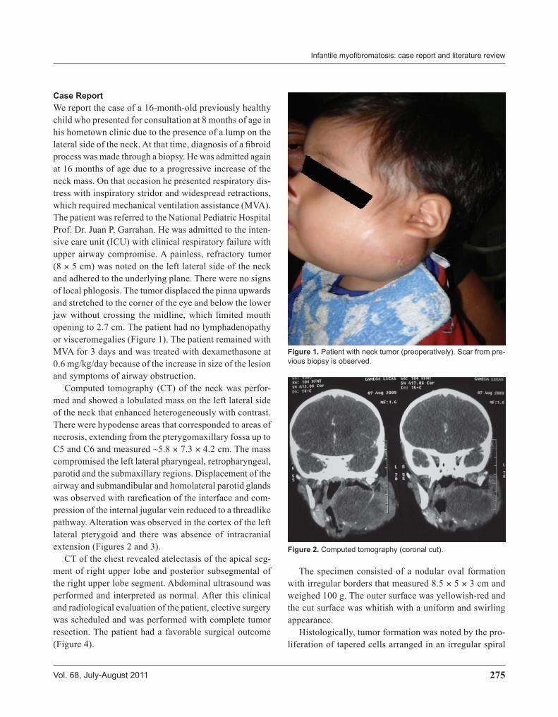
Case ReportWe report the case of a 16-month-old previously healthy child who presented for consultation at 8 months of age in his hometown clinic due to the presence of a lump on the lateral side of the neck. At that time, diagnosis of a fibroid process was made through a biopsy. He was admitted again at 16 months of age due to a progressive increase of the neck mass. On that occasion he presented respiratory dis-tress with inspiratory stridor and widespread retractions, which required mechanical ventilation assistance (MVA). The patient was referred to the National Pediatric Hospital Prof. Dr. Juan P. Garrahan. He was admitted to the inten-sive care unit (ICU) with clinical respiratory failure with upper airway compromise. A painless, refractory tumor (8 × 5 cm) was noted on the left lateral side of the neck and adhered to the underlying plane. There were no signs of local phlogosis. The tumor displaced the pinna upwards and stretched to the corner of the eye and below the lower jaw without crossing the midline, which limited mouth opening to 2.7 cm. The patient had no lymphadenopathy or visceromegalies (Figure 1). The patient remained with MVA for 3 days and was treated with dexamethasone at 0.6 mg/kg/day because of the increase in size of the lesion and symptoms of airway obstruction.
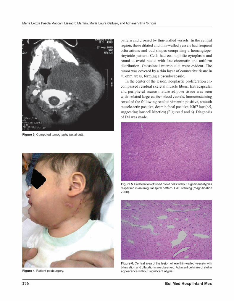
Computed tomography (CT) of the neck was perfor-med and showed a lobulated mass on the left lateral side of the neck that enhanced heterogeneously with contrast. There were hypodense areas that corresponded to areas of necrosis, extending from the pterygomaxillary fossa up to C5 and C6 and measured ~5.8 × 7.3 × 4.2 cm. The mass compromised the left lateral pharyngeal, retropharyngeal, parotid and the submaxillary regions. Displacement of the airway and submandibular and homolateral parotid glands was observed with rarefication of the interface and com-pression of the internal jugular vein reduced to a threadlike pathway. Alteration was observed in the cortex of the left lateral pterygoid and there was absence of intracranial extension (Figures 2 and 3).
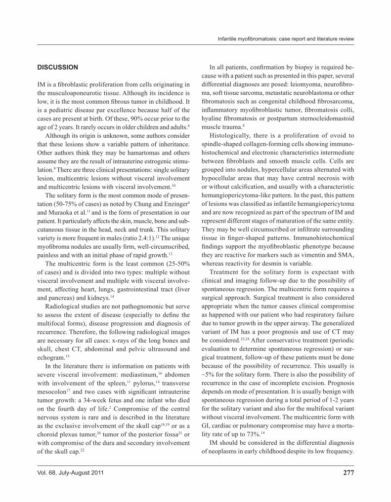
CT of the chest revealed atelectasis of the apical seg-ment of right upper lobe and posterior subsegmental of the right upper lobe segment. Abdominal ultrasound was performed and interpreted as normal. After this clinical and radiological evaluation of the patient, elective surgery was scheduled and was performed with complete tumor resection. The patient had a favorable surgical outcome (Figure 4).
Figure 1. Patient with neck tumor (preoperatively). Scar from pre-vious biopsy is observed.
Figure 2. Computed tomography (coronal cut).
The specimen consisted of a nodular oval formation with irregular borders that measured 8.5 × 5 × 3 cm and weighed 100 g. The outer surface was yellowish-red and the cut surface was whitish with a uniform and swirling appearance.
Histologically, tumor formation was noted by the pro-liferation of tapered cells arranged in an irregular spiral
276 Bol med Hosp Infant mex
María Letizia Fasola Maccari, Lisandro Manfrin, María Laura Galluzo, and Adriana Vilma Scrigni
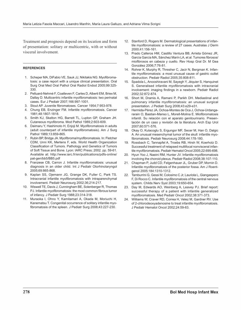
pattern and crossed by thin-walled vessels. In the central region, these dilated and thin-walled vessels had frequent bifurcations and odd shapes comprising a hemangiope-ricytoide pattern. Cells had eosinophilic cytoplasm and round to ovoid nuclei with fine chromatin and uniform distribution. Occasional micronuclei were evident. The tumor was covered by a thin layer of connective tissue in <1-mm areas, forming a pseudocapsule.
In the center of the lesion, neoplastic proliferation en-compassed residual skeletal muscle fibers. Extracapsular and peripheral scarce mature adipose tissue was seen with isolated large-caliber blood vessels. Immunostaining revealed the following results: vimentin positive, smooth muscle actin positive, desmin focal positive, Ki67 low (<5, suggesting low cell kinetics) (Figures 5 and 6). Diagnosis of IM was made.
Figure 6. Central area of the lesion where thin-walled vessels with bifurcation and dilatations are observed. Adjacent cells are of stellar appearance without significant atypia.
Figure 3. Computed tomography (axial cut).
Figure 4. Patient postsurgery.
Figure 5. Proliferation of fused ovoid cells without significant atypias dispersed in an irregular spiral pattern. H&E staining (magnification ×200).
277Vol. 68, July-August 2011
Infantile myofibromatosis: case report and literature review
DISCuSSION
IM is a fibroblastic proliferation from cells originating in the musculoaponeurotic tissue. Although its incidence is low, it is the most common fibrous tumor in childhood. It is a pediatric disease par excellence because half of the cases are present at birth. Of these, 90% occur prior to the age of 2 years. It rarely occurs in older children and adults.8
Although its origin is unknown, some authors consider that these lesions show a variable pattern of inheritance. Other authors think they may be hamartomas and others assume they are the result of intrauterine estrogenic stimu-lation.9 There are three clinical presentations: single solitary lesion, multicentric lesions without visceral involvement and multicentric lesions with visceral involvement.10
The solitary form is the most common mode of presen-tation (50-75% of cases) as noted by Chung and Enzinger4 and Muraoka et al.11 and is the form of presentation in our patient. It particularly affects the skin, muscle, bone and sub-cutaneous tissue in the head, neck and trunk. This solitary variety is more frequent in males (ratio 2.4:1).12 The unique myofibroma nodules are usually firm, well-circumscribed, painless and with an initial phase of rapid growth.13
The multicentric form is the least common (25-50% of cases) and is divided into two types: multiple without visceral involvement and multiple with visceral involve-ment, affecting heart, lungs, gastrointestinal tract (liver and pancreas) and kidneys.14
Radiological studies are not pathognomonic but serve to assess the extent of disease (especially to define the multifocal forms), disease progression and diagnosis of recurrence. Therefore, the following radiological images are necessary for all cases: x-rays of the long bones and skull, chest CT, abdominal and pelvic ultrasound and echogram.15
In the literature there is information on patients with severe visceral involvement: mediastinum,16 abdomen with involvement of the spleen,11 pylorus,14 transverse mesocolon17 and two cases with significant intrauterine tumor growth: a 34-week fetus and one infant who died on the fourth day of life.2 Compromise of the central nervous system is rare and is described in the literature as the exclusive involvement of the skull cap18,19 or as a choroid plexus tumor,20 tumor of the posterior fossa21 or with compromise of the dura and secondary involvement of the skull cap.22
In all patients, confirmation by biopsy is required be-cause with a patient such as presented in this paper, several differential diagnoses are posed: leiomyoma, neurofibro-ma, soft tissue sarcoma, metastatic neuroblastoma or other fibromatosis such as congenital childhood fibrosarcoma, inflammatory myofibroblastic tumor, fibromatosis colli, hyaline fibromatosis or postpartum sternocleidomastoid muscle trauma.8
Histologically, there is a proliferation of ovoid to spindle-shaped collagen-forming cells showing immuno-histochemical and electronic characteristics intermediate between fibroblasts and smooth muscle cells. Cells are grouped into nodules, hypercellular areas alternated with hypocellular areas that may have central necrosis with or without calcification, and usually with a characteristic hemangiopericytoma-like pattern. In the past, this pattern of lesions was classified as infantile hemangiopericytoma and are now recognized as part of the spectrum of IM and represent different stages of maturation of the same entity. They may be well circumscribed or infiltrate surrounding tissue in finger-shaped patterns. Immunohistochemical findings support the myofibroblastic phenotype because they are reactive for markers such as vimentin and SMA, whereas reactivity for desmin is variable.
Treatment for the solitary form is expectant with clinical and imaging follow-up due to the possibility of spontaneous regression. The multicentric form requires a surgical approach. Surgical treatment is also considered appropriate when the tumor causes clinical compromise as happened with our patient who had respiratory failure due to tumor growth in the upper airway. The generalized variant of IM has a poor prognosis and use of CT may be considered.23,24 After conservative treatment (periodic evaluation to determine spontaneous regression) or sur-gical treatment, follow-up of these patients must be done because of the possibility of recurrence. This usually is ~5% for the solitary form. There is also the possibility of recurrence in the case of incomplete excision. Prognosis depends on mode of presentation. It is usually benign with spontaneous regression during a total period of 1-2 years for the solitary variant and also for the multifocal variant without visceral involvement. The multicentric form with GI, cardiac or pulmonary compromise may have a morta-lity rate of up to 73%.14
IM should be considered in the differential diagnosis of neoplasms in early childhood despite its low frequency.
278 Bol med Hosp Infant mex
María Letizia Fasola Maccari, Lisandro Manfrin, María Laura Galluzo, and Adriana Vilma Scrigni
Treatment and prognosis depend on its location and form of presentation: solitary or multicentric, with or without visceral involvement.
REFERENCES
1. Scheper MA, DiFabio VE, Sauk JJ, Nikitakis NG. Myofibroma-tosis: a case report with a unique clinical presentation. Oral Surg Oral Med Oral Pathol Oral Radiol Endod 2005;99:325-330.
2. Pelluard-Nehmé F, Coatleven F, Carles D, Alberti EM, Briex M, Dallay D. Multicentric infantile myofibromatosis: two perinatal cases. Eur J Pediatr 2007;166:997-1001.
3. Stout AP. Juvenile fibromatoses. Cancer 1954;7:953-978. 4. Chung EB, Enzinger FM. Infantile myofibromatosis. Cancer
1981;48:1807-1818. 5. Smith KJ, Skelton HG, Barrett TL, Lupton GP, Graham JH.
Cutaneous myofibroma. Mod Pathol 1989;2:603-609. 6. Daimaru Y, Hashimoto H, Enjoji M. Myofibromatosis in adults
(adult counterpart of infantile myofibromatosis). Am J Surg Pathol 1989;13:859-865.
7. Rubin BP, Bridge JA. Myofibroma/myofibromatosis. In: Fletcher CDM, Unni KK, Mertens F, eds. World Health Organization Classification of Tumors. Pathology and Genetics of Tumors of Soft Tissue and Bone. Lyon: IARC Press; 2002. pp. 59-61. Available at: http://www.iarc.fr/en/publications/pdfs-online/pat-gen/bb5/BB5.pdf
8. Franzese CB, Carron J. Infantile myofibromatosis: unusual diagnosis in an older child. Int J Pediatr Otorhinolaryngol 2005;69:865-868.
9. Kaplan SS, Ojemann JG, Grange DK, Fuller C, Park TS. Intracranial infantile myofibromatosis with intraparenchymal involvement. Pediatr Neurosurg 2002;36;214-217.
10. Wiswell TE, Davis J, Cunningham BE, Solenberger R, Thomas PJ. Infantile myofibromatosis: the most common fibrous tumor of infancy. J Pediatr Surg 1988;23:314-318.
11. Muraoka I, Ohno Y, Kamitamari A, Okada M, Moriuchi H, Kanematsu T. Congenital occurrence of solitary infantile myo-fibromatosis of the spleen. J Pediatr Surg 2008;43:227-230.
12. Stanford D, Rogers M. Dermatological presentations of infan-tile myofibromatosis: a review of 27 cases. Australas J Derm 2000;41:156-161.
13. Prado Calleros HM, Castillo Ventura BB, Arrieta Gómez JR, García García MA, Sánchez Marín LA, et al. Tumores fibrosos/miofibrosos en cabeza y cuello. Rev Hosp Gral Dr. M Gea González 2006;7:76-81.
14. Rohrer K, Murphy R, Thresher C, Jacir N, Bergman K. Infan-tile myofibromatosis: a most unusual cause of gastric outlet obstruction. Pediatr Radiol 2005;35:808-811.
15. Spadola L, Anooshiravani M, Sayegh Y, Jéquier S, Hanquinet S. Generalised infantile myofibromatosis with intracranial involvement: imaging findings in a newborn. Pediatr Radiol 2002;32:872-874.
16. Short M, Dramis A, Ramani P, Parikh DH. Mediastinal and pulmonary infantile myofibromatosis: an unusual surgical presentation. J Pediatr Surg 2008;43:e29-e31.
17. Hermida-Pérez JA, Ochoa-Montes de Oca J, Ochoa-Urdanga-rarain O, Bastian-Manso L, Morell-Molina E. Miofibromatosis infantil. Su relación con el aparato genitourinario. Presen-tación de un caso y revisión de la literatura. Arch Esp Urol 2007;60:571-576.
18. Okay O, Kulacoglu S, Ergungor MF, Secer M, Han O, Dalgic A. An unusual mesenchymal tumor of the skull: infantile myo-fibromatosis. Pediatr Neurosurg 2008;44:178-180.
19. Rossbach C, Tannapfel A, Troebs RB, Hirsh W, Koerholz D. Successful treatment of relapsed multifocal nonvisceral infan-tile myofibromatosis. Pediatr Hematol Oncol 2005;22:695-698.
20. Hyun Yoo J, Naeini RM, Hunter JV. Infantile myofibromatosis involving the choroid plexus. Pediatr Radiol 2008;38:107-110.
21. Chapman P, Judd CD, Felgenhauer JL, Gruber DP, Mornin D. Infantile myofibromatosis of the posterior fossa. Am J Roent-genol 2005;184:1310-1312.
22. Tamburrini G, Gessi M, Colosimo C Jr, Lauriola L, Giangaspero F, Di Rocco C. Infantile myofibromatosis of the central nervous system. Childs Nerv Syst 2003;19:650-654.
23. Day M, Edwards AO, Weinberg A, Leavey PJ. Brief report: successful therapy of a patient with infantile generalized myofibromatosis. Med Pediatr Oncol 2002;38:371-373.
24. Williams W, Craver RD, Correa H, Velez M, Gardner RV. Use of 2-chlorodeoxyadenosine to treat infantile myofibromatosis. J Pediatr Hematol Oncol 2002;24:59-63.