
C HROMOSOMAL ANOMALIES REPLICATION, TRANSCRPITION AND TRANSLATION Dr MOHAMED FAKHRY 1.
56
CHROMOSOMAL ANOMALIES CHROMOSOMAL ANOMALIES REPLICATION, REPLICATION, TRANSCRPITION AND TRANSCRPITION AND TRANSLATION TRANSLATION D r M O H A M E D F A K H R Y 1
-
Upload
juliet-eleanore-robertson -
Category
Documents
-
view
214 -
download
0
Transcript of C HROMOSOMAL ANOMALIES REPLICATION, TRANSCRPITION AND TRANSLATION Dr MOHAMED FAKHRY 1.

CHROMOSOMAL CHROMOSOMAL ANOMALIESANOMALIES
REPLICATION, REPLICATION, TRANSCRPITION AND TRANSCRPITION AND
TRANSLATIONTRANSLATION
Dr M
OH
AM
ED
FA
KH
RY
1

CHROMOSOMAL ANOMALIES CHROMOSOMAL ANOMALIES
2
Dr M
OH
AM
ED
FA
KH
RY

NUMBER OF CHROMOSOMES
Normally, all the individuals of a species have the same number of chromosomes.
Closely related species usually have similar chromosome numbers.
Presence of a whole sets of chromosomes is called euploidy.
It includes haploids, diploids, triploids, tetraploids etc.
Gametes normally contain only one set of chromosome – this number is called Haploid
Somatic cells usually contain two sets of chromosome - 2n : Diploid
3

3n – triploid4n – tetraploidThe condition in which the chromosomes sets
are present in a multiples of “n” is PolyploidyWhen a change in the chromosome number
does not involve entire sets of chromosomes, but only a few of the chromosomes - is Aneuploidy.
Monosomics (2n-1) Trisomics (2n+1) Nullisomics (2n-2) Tetrasomics (2n+2)
4

Organism No. chromosomes
Human 46 Chimpanzee 48 Dog 78 Horse 64 Chicken 78 Goldfish 94 Fruit fly 8 Mosquito 6 Nematode 11(m), 12(f) Horsetail 216 Sequoia 22 Round worm 2
5
Dr M
OH
AM
ED
FA
KH
RY

Organism No. chromosomes
Onion 16 Mold 16 Carrot 20 Tomato 24 Tobacco 48 Rice 24 Maize 20 Haploppus gracilis 4 Crepis capillaris 6
6
Dr M
OH
AM
ED
FA
KH
RY

EUCHROMATIN AND EUCHROMATIN AND HETEROCHROMATINHETEROCHROMATIN
Chromosomes may be identified by regions that stain in a particular manner when treated with various chemicals.
Several different chemical techniques are used to identify certain chromosomal regions by staining then so that they form chromosomal bands.
For example, darker bands are generally found near the centromeres or on the ends (telomeres) of the chromosome, while other regions do not stain as strongly.
The position of the dark-staining are heterochromatic region or heterochromatin.
Light staining are euchromatic region or euchromatin. 7

Heterochromatin is classified into two groups: (i) Constitutive = تأسيسي and (ii) Facultative = أختياري.
Constitutive heterochromatin remains permanently in the heterochromatic stage, i.e., it does not revert to the euchromatic stage.
In contrast, facultative heterochromatin consists of euchromatin that takes on the staining and compactness characteristics of heterochromatin during some phase of development.
8

When the DNA of a prokaryote, such as E.coli, is isolated, fragmented and centrifuged to equilibrium in a Cesium chloride (CsCl) density gradient, the DNA usually forms a single band in the gradient.
On the other hand, CsCl density-gradient analysis of DNA from eukaryotes usually reveals the presence of one large band of DNA (usually called “Mainband” DNA) and one to several small bands.
These small bands are referred to as “Satellite DNAs”.
For e.g., in Drosophila virilis, contain three distinct satellite DNAs.
9
Satellite DNAsSatellite DNAs

CHROMOSOMAL CHROMOSOMAL ABERRATIONSABERRATIONS
The somatic (2n) and gametic (n) chromosome numbers of a species ordinarily remain constant.
This is due to the extremely precise mitotic and meiotic cell division.
Somatic cells of a diploid species contain two copies of each chromosome, which are called homologous chromosome.
Their gametes, therefore contain only one copy of each chromosome, that is they contain one chromosome complement or genome.
Each chromosome of a genome contains a definite numbers and kinds of genes, which are arranged in a definite sequence.
10

CHROMOSOMAL ABERRATIONSCHROMOSOMAL ABERRATIONS Sometime due to mutation or spontaneous (without
any known causal factors), variation in chromosomal number or structure appears in nature: Chromosomal aberrations.
Chromosomal aberration may be grouped into two broad classes:
1. Structural 1. Structural and 2. Numerical2. Numerical
11

STRUCTURAL CHROMOSOMAL STRUCTURAL CHROMOSOMAL ABERRATIONS ABERRATIONS
Chromosome structure variations result from chromosome breakage.
Broken chromosomes tend to re-join; if there is more than one break, rejoining occurs at random and not necessarily with the correct ends.
The result is structural changes in the chromosomes. Chromosome breakage is caused by X-rays, various various
chemicalschemicals, and can also occur spontaneouslyspontaneously. There are four common type of structural aberrations:
1. Deletion or Deficiency 2. Duplication or Repeat3. Inversion, and 4. Translocation.
12

Consider a normal chromosome with genes in alphabetical order: a b c d e f g h i
1. Deletion: part of the chromosome has been removed: a b c g h i
2. Dupliction: part of the chromosome is duplicated: a b c d e f d e f g h i
3. Inversion: part of the chromosome has been re-inserted in reverse order: a b c f e d g h i
ring: the ends of the chromosome are joined together to make a ring
4. translocation: parts of two non-homologous chromosomes are joined:
If one normal chromosome is a b c d e f g h i and the other chromosome is u v w x y z, then a translocation between them would be
a b c d e f x y z and u v w g h i. 13

14
Dr M
OH
AM
ED
FA
KH
RY

Deletion in HumanDeletion in Human:
Chromosome deletions are usually lethal even as heterozygotes, resulting in zygotic loss, stillbirths, or infant death.
Sometimes, infants with small chromosome deficiencies however, survive long enough to permit the abnormal phenotype they express.
15

Cri-du-chat (Cat cry syndrome):The name of the syndrome came from a catlike mewing cry from small weak infants with the disorder.
Other characteristics are microcephaly (small head), broad face and saddle = الفرس صهوة أو الدابة سرجnose, physical and mental retardation.
Cri-du-chat patients die in infancy or early childhood. The chromosome deficiency is in the short arm of
chromosome 5 . Myelocytic leukemiaAnother human disorder that is associated with a
chromosome abnormality is chronic myelocytic leukemia.
A deletion of chromosome 22 was described by P.C.Nowell and Hungerford and was called “Philadelphia” (Ph’) chromosome after the city in which the discovery was made.
16

Duplication:- The presence of an additional chromosome segment, as compared to that normally present in a nucleus is known as Duplication. In a diploid organism, presence of a chromosome segment in more than two copies per nucleus is called duplication. Four types of duplication:
1. Tandem duplication2. Reverse tandem duplication3. Displaced duplication4. Translocation duplication
17

The extra chromosome segment may be located immediately after the normal segment in precisely the same orientation forms the tandem = – ترادفي
األخر تلو واحد When the gene sequence in the extra segment of a
tandem in the reverse order i.e, inverted , it is known as reverse tandem duplication
In some cases, the extra segment may be located in the same chromosome but away from the normal segment – termed as displaced duplication
The additional chromosome segment is located in a non-homologous chromosome is translocation duplication.
18
Dr M
OH
AM
ED
FA
KH
RY

Inversion
When a segment of chromosome is oriented in the reverse direction, such segment said to be inverted and the phenomenon is termed as inversion.
Inversion occur when parts of chromosomes become detached , turn through 1800 and are reinserted in such a way that the genes are in reversed order.
Inversion may be classified into two types: Pericentric - include the centromere Paracentric - do not include the centromere
19

20

Translocation:- Integration of a chromosome segment into a
nonhomologous chromosome is known as translocation.
Three types: 1. simple translocation2. shift3. reciprocal translocation.
21

Simple translocation: In this case, terminal segment of a chromosome is integrated at one end of a non-homologous region. Simple translocations are rather rare.
Shift: In shift, an intercalary segment= not terminal = بيني of a chromosome is integrated جزءwithin a non-homologous chromosome. Such translocations are known in the populations of Drosophila, Neurospora etc.
Reciprocal = متبادل أو translocation: It is تبادليproduced when two non-homologous chromosomes exchange segments – i.e., segments reciprocally transferred.
Translocation of this type is most common
22
Dr M
OH
AM
ED
FA
KH
RY

VARIATION IN CHROMOSOME VARIATION IN CHROMOSOME NUMBERNUMBER
Organism with one complete set of chromosomes is said to be euploid (applies to haploid and diploid organisms).
Aneuploidy - variation in the number of individual chromosomes (but not the total number of sets of chromosomes).
The discovery of aneuploidy dates back to 1916 when Bridges discovered XO male and XXY female Drosophila, which had 7 and 9 chromosomes respectively, instead of normal 8.
23

Nullisomy - loss of one homologous chromosome pair. (e.g., Oat = نبات( الشوفان
Monosomy – loss of a single chromosome (Maize).
Trisomy - one extra chromosome. (Datura)
Tetrasomy - one extra chromosome pair.
More about Aneuploidy
24

TRISOMY IN HUMANSTRISOMY IN HUMANS
Down Syndrome The best known and most common
chromosome related syndrome. Formerly known as “Mongolism” 1866, when a physician named John
Langdon Down published an essay in England in which he described a set of children with common features who were distinct from other children with mental retardation he referred to as “Mongoloids.”
One child in every 800-1000 births has Down syndrome
250,000 in US has Down syndrome. The cost and maintaining Down syndrome
case in US is estimated at $ 1 billion per year.
25

Patients having Down syndrome will Short in stature (four feet tall) and had an epicanthal fold, broad short skulls, wild nostrils, large tongue, stubby= بدين و قصيرhands
Some babies may have short necks, small hands, and short fingers.
They are characterized as low in mentality. Down syndrome results if the extra chromosome is
number 21.
26

Amniocentesis for Detecting AneuploidyAmniocentesis for Detecting Aneuploidy
Chromosomal abnormalities are sufficiently well understood to permit genetic counseling.
A fetus may be checked in early stages of development by karyotyping the cultured cells obtained by a process called amniocentesis.
A sample of fluid will taken from mother and fetal cells are cultured and after a period of two to three weeks, chromosomes in dividing cells can be stained and observed.
If three No.21 chromosomes are present, Down syndrome confirmed.
The risk for mothers less than 25 years of age to have the trisomy is about 1 in 1500 births.
At 40 years of age, 1 in 100 births At 45 years 1 in 40 births. 27

Other SyndromesOther Syndromes
Chromosome Nomenclature: 47, +13Chromosome formula: 2n+1Clinical Syndrome: Trisomy-13Estimated Frequency Birth: 1/20,000Main Phenotypic Characteristics:Mental deficiency and deafness, minor muscle seizures,
cleft lip, cardiac anomalies
28

Other SyndromesOther Syndromes
Chromosome Nomenclature: 45, XChromosome formula: 2n - 1Clinical Syndrome: TurnerEstimated Frequency Birth: 1/2,500 femaleMain Phenotypic Characteristics:
Female with retarded sexual development, usually sterile, short stature, webbing = حزام of skin in neck region, cardiovascular abnormalities, hearing impairment.
29

Other Syndromes
Chromosome Nomenclature: 47, XXY, 48, XXXY, 48,XXYY, 49, XXXXY, 50, XXXXXY
Chromosome formula: 2n+1; 2n+2; 2n+2; 2n+3; 2n+4
Clinical Syndrome: KlinefelterEstimated Frequency Birth: 1/500 male
birthMain Phenotypic Characteristics:
Pitched= كويس ,voice, Male مشsubfertile with small testes, developed breasts, feminine, long limbs.
30

DOSAGE COMPENSATIONDOSAGE COMPENSATION
Sex Chromosomes: females XX, males XY Females have two copies of every X-linked gene;
males have only one. How is this difference in gene dosage compensated
for? OR How to create equal amount of X chromosome gene
products in males and females?
31

Levels of enzymes or proteins encoded by genes on the X chromosome are the same in both males and females
Even though males have 1 X chromosome and females have 2.
G6PD, glucose 6 phosphate dehydrogenase, gene is carried on the X chromosome
This gene codes for an enzyme that breaks down sugar
Females produce the same amount of G6PD enzyme as males
XXY and XXX individuals produce the same about of G6PD as anyone else
32

In cells with more than two X chromosomes, only one X remains genetically active and all the others become inactivated.
In some cells the paternal allele is expressed In other cells the maternal allele is expressed In XXX and XXXX females and XXY males only 1 X is
activated in any given cell the rest are inactivated
33

34
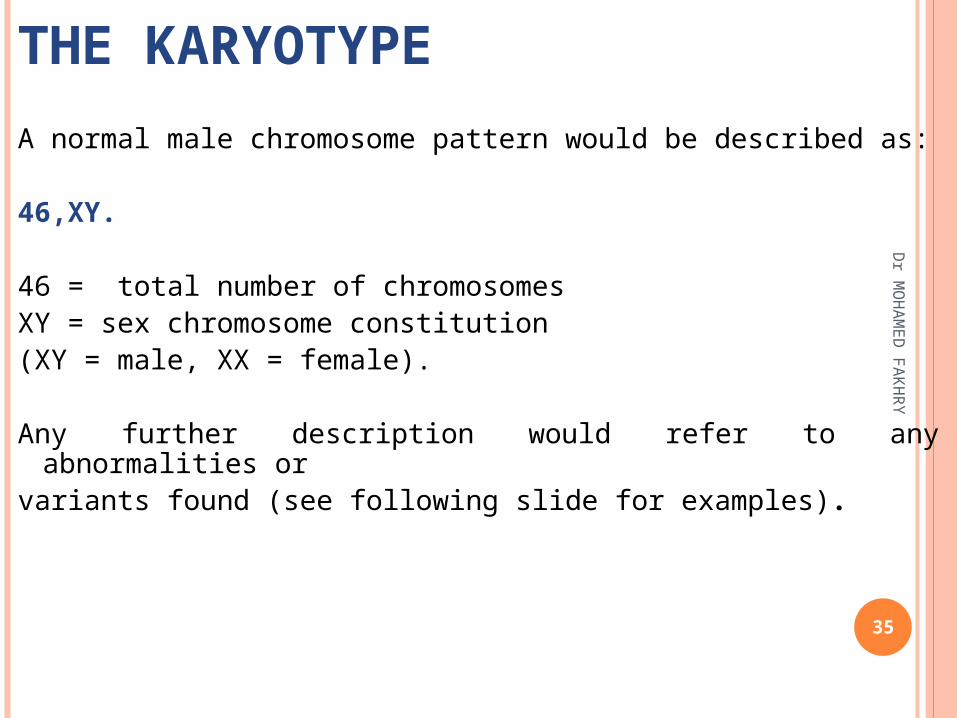
THE KARYOTYPE
A normal male chromosome pattern would be described as:
46,XY.
46 = total number of chromosomesXY = sex chromosome constitution(XY = male, XX = female).
Any further description would refer to any abnormalities orvariants found (see following slide for examples).
35
Dr M
OH
AM
ED
FA
KH
RY

Total number of chromosomes,
Sex chromosome constitution,
Anormalies/variants.
46,XY47,XX,+2147,XXX69,XXY45,XX,der(13;14)(q10;q10)46,XY,t(2;4)(p12;q12)46,XX,del(5)(p25)46,XX,dup(2)(p13p22)46,XY,inv(11)(p15q14)46,XY,fra(X)(q27.3)46,XY/47,XXY
The Karyotype: an international descriptionThe Karyotype: an international descriptionD
r MO
HA
ME
D F
AK
HR
Y
36

Total number of chromosomes,
Sex chromosome constitution,
Anomalies/variants.46,XY47,XX,+21 Trisomy 21 (Down syndrome)47,XXX Triple X syndrome69,XXY Triploidy
45,XX,der(13;14)(p11;q11) Robertsonian translocation46,XY,t(2;4)(p12;q12) Reciprocal translocation
46,XX,del(5)(p25) Deletion tip of chromosome 546,XX,dup(2)(p13p22) Duplication of part of short arm
Chr 246,XY,inv(11)(p15q14) Pericentric inversion
chromosome 1146,XY,fra(X)(q27.3) Fragile X syndrome46,XY/47,XXY Mosaicism normal/Klinefelter syndrome
The Karyotype: an international descriptionThe Karyotype: an international descriptionD
r MO
HA
ME
D F
AK
HR
Y
37

CHROMOSOMAL FINDINGS IN EARLY CHROMOSOMAL FINDINGS IN EARLY MISCARRIAGES = MISCARRIAGES = الاجهاضالاجهاض
Dr MOHAMED FAKHRY 38
40% apparently normal
60% abnormal: Trisomy (47 chromosomes – one extra) 30%
45,X (45 chromosomes – one missing) 10%
Triploidy (69 chromosomes – three sets) 10%
Tetraploidy (92 chromosomes – four sets) 5%
Other chromosome anomalies 5%(e.g. structural anomalies)

The Genetic material-Deoxyribonucleic acid (DNA)
-Double strandof polynucleotide.-Coiled around each other forming double helix.-Strands are anti- Parallel.-Sugar phosphate backbone is outside & bases are inside.-A=T and G=C.-A/T=1 and G/C=1 (Cargaff Ratio)
5’
3’
3’
5’
Dr M
OH
AM
ED
FA
KH
RY
39

Detailed view of DNA StructureD
r MO
HA
ME
D F
AK
HR
Y
40

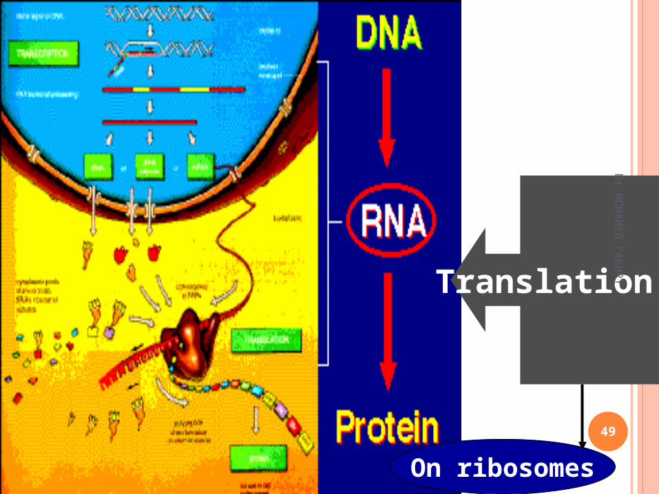
The Central Dogma D
r MO
HA
ME
D F
AK
HR
Y
41

Replication-in nucleus
Transcription-in nucleus
Translation-in cytoplasm on ribosomes
Dr M
OH
AM
ED
FA
KH
RY
42
The Central Dogma

DNA Replication -Replications occurs before cell division. During S Phase of cell cycle.-Entire DNA content is doubled.-Replication is Semi-conservative =محافظ شبه-Requires: -DNA polymerases -dNTPs(N=A,T,C,G) -RNA primer -Mg++ -DNA ligases - Primase - Helicase - SS (single stranded) DNA binding proteins
43

Major Steps in DNA Replication
Leading strand,continuous
Lagging strand
Dr M
OH
AM
ED
FA
KH
RY
44

Dr M
OH
AM
ED
FA
KH
RY
45

Steps in transcription
Initiation
Elongation
• Binding of RNA polymerase causes opening of the DNA strand and synthesis of the RNA
• RNA polymerase continues synthesis of RNA complementary to DNA till termination site
Dr M
OH
AM
ED
FA
KH
RY
46

Elongation -contd
Termination
Steps in transcription (contd)
• Rho factor binds to the termination site and when RNA polymerase reaches this site, termination occurs
47

Dr M
OH
AM
ED
FA
KH
RY
48
Translation
On ribosomes
Dr M
OH
AM
ED
FA
KH
RY
49

Ribosomes- free and attached to endoplasmic reticulum
50

Codons on mRNA
51

Structure of tRNA
52

Steps in Translation
ii. Elongation
i. Initiation
iii. Termination
Dr M
OH
AM
ED
FA
KH
RY
53

Polysomes
54

MITOCHONDRIAL DNA
In the human mitochondria the chromosomes are present as 10 circular double helices of DNA.
They are self replicative. Contain: 16,596 bp, genes for 22 tRNAs and 2 types
of ribosomal RNA required for mitochondrial protein synthesis.
They also have genes for 13 polypeptides, involved in cellular oxidative phosphorylation.
Both strands of DNA are transcribed and translated.
55

MITOCHONDRIAL DNA
The genes on mitochondrial DNA have no introns. The codon recognition pattern for several amino
acids is different from the nuclear DNA. Mitochondria are transmitted in the egg from a
mother to all of her children. Thus mitochondrial DNA is only maternally derived.
56


![[Saza Ahmed Fakhry Boskany] a Contrastive Analysis of Agreement (2)](https://static.fdocuments.in/doc/165x107/563dba5b550346aa9aa4ec5a/saza-ahmed-fakhry-boskany-a-contrastive-analysis-of-agreement-2.jpg)

