C HAPTER 5 Integumentary System Thursday, Sept 19 th.
121
CHAPTER 5 Integumentary System Thursday, Sept 19 th
-
Upload
megan-harvey -
Category
Documents
-
view
221 -
download
1
Transcript of C HAPTER 5 Integumentary System Thursday, Sept 19 th.

CHAPTER 5
Integumentary System
Thursday, Sept 19th

OVERVIEW OF THE INTEGUMENT
Composed of 2 layers: Epidermis Dermis
Hypodermis - lies beneath skin
Thickness varies, normally 1-2 mm
Forms Skin, nails, hair Glands

FUNCTIONS OF THE SKINProtection
Sensory
Maintenance of body temp
Synthesis of Vitamin D
Excretion

FUNCTIONS OF THE SKINProtection
Against traumaFluid lossChemical attackUltraviolet lightInfection
Sensory receptorDetects touchPressurePainTemperature

FUNCTIONS OF THE SKINMaintenance of normal body
temperatureThrough insulation or evaporative cooling
Synthesis of vitamin D Converted to a hormone important to
maintaining Ca++ balance
Excretion SaltsWaterOrganic wastes

THE LAYERS OF SKIN1. Epidermis
Epithelial tissue
2. DermisConnective tissue
HypodermisLoose connective tissue

SKIN STRUCTURE

THE EPIDERMIS

THE EPIDERMIS Keratinized stratified squamous epithelium
Avascular Nourished by diffusion of nutrients from dermis
Cells found in the epidermis include Stem cells Keratinocytes Melanocytes Tactile (Merkel) cells Dendritic (Langerhans) cells

THE EPIDERMIS Cells produced by mitosis in deepest layer
of epidermis
New cells push older cells to surface = desquamate or slough off
Keratinization

LAYERS OF THE EPIDERMIS Stratum basale (or germinativum) – deepest
layer Stratum spinosum Stratum granulosum Stratum lucidum (only in thick skin) Stratum corneum – superficial layer
Cell movement goes from DEEP to superificial


STRATUM BASALE
StructureDeepest layerSingle layer cells attached to basement
membrane
FunctionProtection – Melanocytes (melanin)
Cell types in this layer:Stem cellsKeratinocytesMelanocytes Merkel cells

STRATUM SPINOSUMStructure
Several layers of keratinocytes (8-10 layers)Appear ‘spiny’Called DESMOSOMES (spiny like projections)
FunctionProduces lamellar bodies (lipid filled
organelles)Begin to synthesize protein keratin which
cause cells to flatten

STRATUM GRANULOSUM Structure
3 to 5 layers of flat keratinocytesCells stop dividing here
FunctionContain keratinohyalin granulesForms keratin
Major component of hair and nailsGlycolipids waterproof the skinNucleus degenerate and cell dies

STRATUM LUCIDUM Structure
3 – 5 layers of dead cellsAppears transparentThin translucent zone seen ONLY in thick
skin
LocationFound where skin is hairless and extra
thick

STRATUM CORNEUM
StructureVery thickUp to 15-30 layers of dead, scaly, keratinized
cellsKeratinization (cornification) occurs here
FunctionsOccurs on all exposed skin surfaces except
anterior surface of eyesPrevents water lossResists abrasions

EPIDERMAL LAYERS AND KERATINIZATION

THICK AND THIN SKIN Thick skin
Has all 5 epithelial strata
Stratum corneum has many layers
Found in areas subject to pressure or friction Palms of hands, fingertips, soles of feet
Fingerprints and footprintsPapillae of underlying dermis in parallel rows

THICK AND THIN SKIN Thin skin
More flexible than thick skin
Covers rest of body
Each stratum contains fewer layers
Hair is found only in thin skin

THIN AND THICK SKIN Callus
Found in skin subject to friction or pressure Produces a thickened area Over a bony prominence, a corn forms
Calluses and corns develop in both thin & thick skin

SKIN COLOR: PIGMENTS
Determined by 3 factors: 1. Pigments (melanin, Hb, carotene)
2. Blood circulating through the skin
3. Thickness of stratum corneum

SKIN COLOR: PIGMENTS Pigments
Melanin: provides for protection against UV light
Accounts for color in skin, hair & eyes
Lips, palms and soles contain less melanin
Differences in pigmentation among individuals reflects levels of synthetic activity and NOT numbers of melanocytes

SKIN COLOR: PIGMENTS
PigmentsCarotene: yellow pigment
Acquired from egg yolks and yellow and orange vegetables (corn and carrots)
Source of Vitamin A Results in yellowish tint
Location of pigments affect color produced in skin

ABNORMAL SKIN COLORS
Albinism = a genetic lack of melanin
Erythema = increased redness due to increased blood flow through the skin.
Cyanosis = blueness from deficiency of oxygen in the circulating blood (cold weather)

EPIDERMIS FORMATIVE
Monday, Sept 23rd

FORMATIVE QUIZ – LET’S SEE WHAT YOU KNOW…
1. Name the 2 layers of the skin. What type of tissue is each layer made out of? (Try to be specific)
2. What are 3 of the 5 major functions of the integument?
3. What does keratin do in cells?
4. What is the term for when dead skin cells slough off?

FORMATIVE QUIZ – LET’S SEE WHAT YOU KNOW…
5. Name the 5 layers of epidermis in order from deep to superficial
6. Where do cells START to die off (meaning – no more cell division takes place – cell division has stopped in this layer)

FORMATIVE QUIZ – LET’S SEE WHAT YOU KNOW…
1. Epidermis & DermisEpidermis = Epithelial; Dermis = Connective
2. Protection, Sensory Receptors, Excretion, Synthesis of Vit D and Maintenance of Body Temp
3. Makes the cells hard in order to provide strength
4. Desquamate

FORMATIVE QUIZ – LET’S SEE WHAT YOU KNOW…
5. Stratum basale – deepest layer, Stratum spinosum, Stratum granulosum, Stratum lucidum (only in thick skin), Stratum corneum – superficial layer
6. Stratum granulosum

DERMIS & HYPODERMIS
Quiz on FRIDAY•Functions of integument & hypodermis•Layers of epidermis (list & label)•Melanin•Thick vs thin skin•Nails

DERMIS2nd major layer of the skin
Strong, flexible CONNECTIVE tissueAdipose cells & macrophages
Richly supplied with blood vessels and nerves
CompositionCollagen, elastic and reticular fibers, fibroblasts
Nerve endings, hair follicles, smooth muscles, glands and lymphatic vessels

DERMIS – 2 Layers1. Reticular: Deeper layer; 80% of dermis
Hair follicles, nerves, sweat and oil glands found here
Composed of dense irregular C.T.
Contains collagen and elastic fibers
Stretching of skin (obesity, pregnancy) can tear collagen fibers and produce striae (stretch marks)

DERMIS – 2 LAYERS 2. Papillary - Superficial layer; 20% of dermis
Blood vessels that supply epidermis with nutrients
Removes waste products
Aids in regulating body temperature
Fingertips have a papillary patternAll fingerprints are different (even in twins)


HYPODERMIS AKA subcutaneous tissue or superficial fascia
Mostly adipose tissue (some areolar)½ of the body’s fat deposits is found here
Functions:Binds skin to underlying tissueEnergy reservoir (fat)Thermal insulationPadding/cushioning
Skin rests on the hypodermis which attaches to bone and muscle

ACCESSORY SKIN STRUCTURESHairHair folliclesSmooth muscles (arrector pilli)Sweat and sebaceous (oil) glandsNails

CHARACTERISTICS OF HUMAN HAIR Hair (composed of hard keratin)
Hair is found almost everywhere
75% of the 2.5 million are on body surface, not head
Hair is made by HAIR FOLLICLES
Hair pigment is made by melanocytes

CHARACTERISTICS OF HUMAN HAIR
3 different body hair typesLanugo - fine, unpigmented fetal hair
Vellus - fine, unpigmented hair of children and women
Terminal hair - coarser, longer, pigmented hair Chest, legs and arm hair : 90% males vs. 35%
females

STRUCTURE OF HAIR AND FOLLICLEHair has 3 zones:
Bulb - swelling at base in dermis
Root - remainder of hair with follicle; below the surface of skin
Shaft - exposed portion above skin surface

STRUCTURE OF HAIR FOLLICLE

STRUCTURE OF HAIR FOLLICLE
Follicle - a diagonal tube within the skin
Nerve fibers (hair receptors) encircle follicle; detect motion
Arrector pili muscles stimulate piloerection (goose bumps)
Bulb is where hair originates

HAIR GROWTH AND LOSS Hair growth
Due to mitosis of S. basale cells in epithelial root sheath
Scalp hair - grows 1 mm every 3 days on avg (10-18 cm/yr or about 6 inches)
Pushes out by new hair growing beneath it
Loss of 100 scalp hairs a day is normal

Eumelanin Pheomelanin and little eumelanin
Pheomelanin and little eumelanin
Red HairBlonde HairBrown/Black hair

White hair = air in medulla and lack of pigment in cortex. Gray hair is a mixture of white and pigmented hairs.
GRAY AND WHITE HAIR

HAIR GROWTH AND LOSS Thinning or baldness = alopecia
Pattern baldness - the loss of hair from only some regions of the scalp rather than thinning uniformally
Hirsutism = excessive hair growth in areas not normally hairy in women Hormone imbalance (Ovary or adrenal
cortex problem)


FUNCTIONS OF HAIR Body hair
Alert us to parasites crawling on skin
Scalp hair Heat retention and sunburn cover
Beard, pubic and axillary hair indicate sexual maturity (in some guys, that is) and help distribute sexual scents
Guard hairs and eyelashes Prevent foreign objects from getting into nostrils, ear
canals or eyes
Expression of emotions with eyebrows

DERMIS FORMATIVE

LET’S SEE IF YOU ARE STUDYING!
1. Name the layer of connective tissue that connects the skin to the underlying muscle or bone.
2. Identify the 2 layers of dermis.
3. Which of the 2 layers accounts for 80% of the dermis?
4. What is hair composed of?

LET’S SEE IF YOU ARE STUDYING
5. What gives hair it’s color (pigment)?
6. What are the 3 zones of a piece (or strand) of hair?

LET’S SEE IF YOU ARE STUDYING!
1. Name the layer of connective tissue that connects the skin to the underlying muscle or bone. - HYPODERMIS
2. Identify the 2 layers of dermis. – RETICULAR and PAPILLARY
3. Which of the 2 layers accounts for 80% of the dermis? - RETICULAR
4. What is hair composed of? - KERATIN

LET’S SEE IF YOU ARE STUDYING
5. What gives hair it’s color (pigment)? - MELANIN
6. What are the 3 zones of a piece (or strand) of hair? – BULB, ROOT, SHAFT

DERMIS PART 2

FACT OR FICTION?
Cutting hair causes it to grow back faster? Plucking or waxing causes hair to grow back
thicker or more coarse?

NAILS Derivative of stratum corneum
Densely packed cells filled with hard keratin
Keratin is what makes cells hard
Flat nails allow for fleshy, sensitive fingertips
Growth rate is 1 mm per week
Fingernails vs Toenails

FINGERNAIL STRUCTURE
GLANDS – 2 MAJOR TYPESALL EXOCRINE
2 typesSebaceous (oil)Sweat

SEBACEOUS (OIL) GLANDS Location
Dermis Occur over entire body,
except palms and soles
Function Produces oily secretion
(sebum) Sebum is rich in lipids Collects dirt Soften and lubricate hair Protection

SUDORIFEROUS (SWEAT) GLANDS
Widely distributed on body
Sweat is a filtrate of plasma and some waste products500 ml of insensible
perspiration/daysweating with visible
wetness is diaphoresis

SUDORIFEROUS (SWEAT) GLANDS2 TypesMerocrine (or Eccrine)
Most common Open directly onto surface of skin Have own pores Secretes products with no loss of cellular material
Numerous in palms and soles

SUDORIFEROUS (SWEAT) GLANDS2 TypesApocrine glands
Produce sweat containing fatty acids
Found in underarms and genitalia
Active at puberty, causes body odor
Mammals use this for scent as means of communication
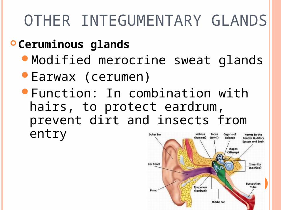
OTHER INTEGUMENTARY GLANDS
Ceruminous glandsModified merocrine sweat glandsEarwax (cerumen)Function: In combination with hairs,
to protect eardrum, prevent dirt and insects from entry

OTHER INTEGUMENTARY GLANDS
Mammary glands
Modified apocrine sweat gland
Breasts and mammary glands not the sameBoth sexes have breastsIn females, breasts are a secondary sexual
characteristic
Mammary gland is a milk-producing gland within the breast Only during lactation and pregnancy

WHAT TO EXPECT ON THE QUIZ FRIDAY
1. List the 5 layers in order 2. List the functions of the
integumentary system 3. Identify the layer of dead squamous
cells 4. List the layers of the dermis 5. Identify what the layers of dermis
are made of 6. List the function of the hypodermis

WHAT TO EXPECT ON THE QUIZ FRIDAY
7. Define keratin 8. List areas where keratin is found on
the body 9. Identify the cells that produce
melanin 10. List factors that stimulate melanin
production 11. List the function of nails 12. Compare & contrast thick vs thin
skin

CANCER

WHAT IS CANCER?
Cancer - Malignant, spreading tumor
Tumor – any swelling
Oncology – study of tumors and their problems
Neoplasm – abnormal tissue growth resulting in rapid cellular proliferation which continues after growth of normal tissue has stopped or slowed

WHAT ARE NEOPLASMS?
Benign – not inclined to spread or get worse
Less dangerous than malignant tumors
Can compress surrounding tissues Impair tissue functions as a result of
enlargement

WHAT ARE NEOPLASMS?
Malignant – able to spread and become worseMore embryonic, less mature, than normal tissue
Invasive, ability to squeeze into spaces and enter surrounding tissues
Secrete enzymes that cut paths through healthy tissue

WHAT ARE NEOPLASMS?
MalignantGrow irregularly, sending processes in
every direction
Ability to dislodge, enter blood vessels, lymphatic vessels or body cavities
Travel to distant sites, attach and invade tissues

THE GENETICS OF CANCER
Mutations of genes within somatic cells
Occurs during DNA replication
Radiation, chemicals, toxins & viruses cause mutations by damaging or altering DNA
Usually develop in tissues that undergo frequent cell division

THE GENETICS OF CANCER
Neoplasm can become cancerous when cell’s structure and functions are changedIncrease ability of cancer cells to
invade and destroy surrounding tissue
Metastasize
Resistant to drug treatments

THE GENETICS OF CANCER
Prevention of development in cancer cells:DNA repair enzymes detect and
correct errors during replication
Self-destruction mechanism destroys cells with abnormal DNA (Apoptosis)

THE GENETICS OF CANCER
Proto-oncogenes – promote cell division
Oncogenes – abnormal regulatory genes, increases rate of cell division
Tumor suppressor genes – normal genes that slow down or stop cell division

HOW ARE CANCER CELLS DIFFERENT?

TYPES OF CANCER
Metastasis – process by which cancer cells spread to distant sites
Secrete substances that cause blood vessels to grow into the tumor and supply oxygen and nutrients.

TYPES OF CANCER
Carcinoma – cancer of epithelial tissueBasal cell & Squamous cell
carcinomas
Adenocarcinoma – glandular epitheliumBreast cancer

TYPES OF CANCER
Sarcoma – connective tissue
Osteosarcoma – bones
Chondrosarcoma - cartilage

CANCER THERAPY
Concentrates on confining and killing malignant cells
X-rays, lasers, surgical removal, drugs
Problem: Some cancers can’t be removed completely by surgery or killed Affects normal tissues

CANCER THERAPY
Newer drugs prevent blood vessel development
Anticancer therapies – cells that can recognize tumor cells and destroy them

CHEMOTHERAPY
Any treatment involving chemical agents to stop cancer cells from growing
Eliminate cancer cells at sites great distances from original cancer
More than half receive chemotherapy

SKIN CANCER
Most common type of cancer
Caused by UV rays, chemicals, x-rays, inflammation, genetics
Amount of protective melanin affects skin cancerFair-skinned = gen. predisposed to develop skin
cancerIndividuals over 50 yrs = increased risk

SKIN CANCER
Three major types of skin cancer are:
Basal cell carcinomaSquamous cell carcinomaMelanoma

NO. 1 BASAL CELL CARCINOMA
Least malignant and most common skin cancer
Stratum basale cells proliferate and invade the dermis and hypodermis
Slow growing and do not often metastasize
Can be cured by surgical excision in 99% of the cases

NO. 2SQUAMOUS CELL CARCINOMA
Arises from keratinocytes of stratum spinosum
Arise most often on scalp, ears, and lower lip; bleeding can occur
Grows rapidly and metastasizes if not removed; if travels to lymph nodes, can be fatal
Can be treated by radiation therapy or removed surgically

NO. 3MELANOMA
Cancer of melanocytes is the most dangerous type of skin cancer because it is:
Highly metastatic
Resistant to chemotherapy
77% of skin cancer deaths in U.S.
40% develop in preexisting moles

MELANOMA (ABCDE RULE) Melanomas have the following characteristics
A: Asymmetry; the two sides of the pigmented area do not match
B: Border is irregular and exhibits indentationsC: Color (pigmented area) is not uniform: black,
brown, tan, and sometimes red or blueD: Diameter is larger than 6 mm (size of a pencil
eraser)E: Evolving (lesions change over time) Change size,
shape, elevation or color, bleed, crust or become tender

UVA VS UVB
The sun has 2 types of UV (ultraviolet), rays:
1. UVA2. UVB


UVA
UVA - long-wave solar rays of 320-400 nm. Goes through windows, light clothing and
even windshields
Responsible for agingPenetrates the skin more deeply, causing
wrinkling and leathering of the skin
May also directly induce some skin cancers, including melanomas.

UVB
Short-wave solar rays of 290-320 nm
More potent than UVA in producing sunburn
Main cause of skin cancers
The"tanning ray", UVB stimulate the melanocyte cell, producing a suntan as a defense against UV radiation


SKIN CANCER PREVENTION
NOT FOOLPROOF BUT IMPORTANT….wear SUNCREEN!

CANCER FORMATIVE ASSESSMENT

LET’S SEE IF YOU CAN ANSWER THESE QUESTIONS….
1. What is the difference between benign and malignant tumors?
2.What does the term “metastasis” mean?
3. What is a neoplasm?

LET’S SEE IF YOU CAN ANSWER THESE QUESTIONS….
4. How does the body defend itself against cancer?
5. What are 2 ways we can treat cancer?

LET’S SEE IF YOU CAN ANSWER THESE QUESTIONS….
1. What is the difference between benign and malignant tumors? Benign not inclined to spread or get worse,
less dangerous, can compress surrounding tissue, cannot grow into other tissue
Malignant able to spread, invasive, secretes enzymes that cut paths through healthy tissue, grow irregularly, can dislodge and travel to distant sites

LET’S SEE IF YOU CAN ANSWER THESE QUESTIONS….
2.What does the term “metastasis” mean? process by which cancer cells spread to distant sites through the bloodstream or lymph vessels
3. What is a neoplasm? Abnormal tissue growth resulting in rapid cellular
division; continues after normal tissue growth has slowed or stopped

LET’S SEE IF YOU CAN ANSWER THESE QUESTIONS….
4. How does the body defend itself against cancer? Cells detect and correct errors within mutated
DNA; can self destruct if correction cannot occur (Apoptosis)
5. What are 2 ways we can treat cancer? surgical removal, x-rays, lasers, drugs

CANCER FORMATIVE #2
Cancer Quiz is TOMORROW - Friday

LET’S SEE WHAT YOU DO KNOW….
1. List 2 ways that cancer cells can differ from normal cells.
2. Describe one of the disadvantages (or problems) with current cancer treatments.
3. Identify the most common type of skin cancer.
4. List the 3 types of skin cancer
5. Using the ABCDE rule, identify what 3 of the letters stand for.

LET’S SEE WHAT YOU DO KNOW….
6. Name 2 ways in which people develop skin cancer.
7. What individuals are most likely to develop skin cancer?
8. What is the best way to prevent skin cancer?

ANSWERS
1. List 2 ways that cancer cells can differ from normal cells. Invasive (can squeeze into spaces and enter
surrounding tissue) Dislodge (can travel to distant sites)
2. Describe one of the disadvantages (or problems) with current cancer treatments. Kills good cells with the bad

LET’S SEE WHAT YOU DO KNOW….
3. Identify the most common type of skin cancer. Basal cell carcinoma
4. List the 3 types of skin cancer Basal cell carcinoma Squamous cell carcinoma Melanoma

5. USING THE ABCDE RULE, IDENTIFY WHAT 3 OF THE LETTERS STAND FOR.
Melanomas have the following characteristicsA: Asymmetry; the two sides of the pigmented area
do not match B: Border is irregular and exhibits indentationsC: Color (pigmented area) is not uniform: black,
brown, tan, and sometimes red or blueD: Diameter is larger than 6 mm (size of a pencil
eraser)E: Evolving (lesions change over time) Change size,
shape, elevation or color, bleed, crust or become tender

LET’S SEE WHAT YOU DO KNOW…. 6. Name 2 ways in which people develop skin cancer.
UV rays Chemicals Genetics X rays
7. What individuals are most likely to develop skin cancer? Fair skinned and individuals over 50
8. What is the best way to prevent skin cancer? Sunscreen
OLD AGE, DISORDERS AND BURNS

DEVELOPMENTAL ASPECTS OF THE INTEGUMENT: OLD AGE
Epidermal replacement of cells slows and skin becomes thinner
Skin becomes dry and itchy
Subcutaneous fat layer diminishes, leading to intolerance of cold

DEVELOPMENTAL ASPECTS OF THE INTEGUMENT: OLD AGE Decreased elasticity and loss of
subcutaneous tissue leads to wrinkles
Decreased numbers of melanocytes and Langerhans’ cells increase the risk of skin cancer
Decrease in blood supply causes poor ability to regulate body temperature
Functioning melanocytes decrease or increase; age spots

DISORDERS
AcneDisease of the hair follicles of the face,
chest and back Is NOT caused by bacteria – bacteria plays a
roleCaused by elevated sebum production and
the maturation of skin cells
Psoriasis Inflammatory skin disease – no known causeScaly skin thought to result from abnormal
lymphocytes

PSORIASIS

DISORDERS
SeborrheaCommon form of eczema
DandruffOccurs in areas of high sebum productionNo known cause, but the yeast that lives
on the skin may play a role
MolesBrown or black growthsOccur when melanocytes in the skin grow
in a cluster instead of being spread out

BURNS First-degree – only the epidermis is damaged
Least seriousLocalized rednessSwellingpain
Second-degree – epidermis and upper regions of dermis are damagedSkin becomes intensely redBlisters also appear and swelling

1ST DEGREE

2ND DEGREE

1ST & 2ND DEGREE BURNS TREATMENT Cool the burn
Hold burn under cool water for 10 to 15 minutesCool compress also works
Cover the burn with a gauze bandageWrap it loosely to avoid pressureDon’t use fluffy cotton to avoid getting lint into
the woundOver the counter pain reliever
Usually heal without further treatment

BURNS
Third-degree – entire thickness of the skin is damagedPermanent damageFat, muscle and bone may be affectedBurned area appears gray-white, cherry red,
or blackThere is no initial edema or pain (since nerve
endings are destroyed)
Treatment - IV nutrition and fluid replacement, de-bridement, and infection control

3RD DEGREE

RULE OF NINES
You can estimate the body surface area on an adult that has been burned by using multiples of 9.

RULE OF NINES
Estimates the severity of burns
Burns considered critical if:Over 25% of the body has second-
degree burnsOver 10% of the body has third-
degree burnsThere are third-degree burns on face,
hands, or feet