c-fos mRNA, Fos, and Fos-related Antigen Induction by ... · The Journal of Neuroscience, August...
11
The Journal of Neuroscience, August 1991, 7 7(8): 2321-2331 c-fos mRNA, Fos, and Fos-related Antigen Induction by Hypertonic Saline and Stress Frank Ft. Sharp, Stephen M. Sagar, Katy Hicks, Daniel Lowenstein, and Kinya Hisanaga Department of Neurology, University of California at Francisco and the San Francisco Veterans Affairs Medical Center, San Francisco, California 94121 The induction of c-fos mRNA was assessed using Northern blots and in situ hybridization in adult rats administered hy- pertonic saline (HS) and isotonic saline (IS). HS induced c-fos mRNA in magnocellular paraventricular nucleus (PVNm), parvocellular paraventricular nucleus (PVNp), supraoptic nu- cleus (SON), and lamina terminalis (LMT). This occurred within 5 min, peaked at 30-60 min, and disappeared by 160 min. Fos protein, detected using a specific monoclonal antibody, was maximal at l-2 hr and disappeared 4-6 hr after HS administration. This confirms observations showing that the c-fos gene response is transient even in the presence of a continuing stimulus. In contrast, Fos-like immunoreactivity (FLI), detected us- ing two polyclonal antisera, was observed in PVNm, PVNp, SON, and LMT for l-24 hr during continuous osmotic stim- ulation. Moreover, FLI was obsenrable in these structures for 7 d in rats administered HS and allowed to drink water ad libitum beginning 24 hr later. At times greater than 6 hr, FLI presumably represents Fos-related antigens (FRA), pro- teins immunologically and functionally related to Fos, whose expression is much more prolonged than authentic Fos fol- lowing the osmotic stimulus. In addition to induction of c-fos expression in regions spe- cifically involved in osmotic regulation, HS injections also induced c-fos in many other forebrain regions. In order to assess the induction of c-fos mRNA due to the “stress” of the injections, rats injected with isotonic saline were com- pared to uninjected controls. Isotonic saline injections in- duced c-fos mRNA in the PVNp, anterior hypothalamus, su- prachiasmatic nucleus, cingulate gyrus, neocortex, ventral lateral septal nucleus, piriform cortex, hippocampal pyra- midal and dentate granule neurons, paraventricular and in- tralaminar thalamic nuclei, bed nuclei of stria terminalis, cor- tical and medial amygdaloid nuclei, and other structures. In accord with other work, we interpret this pattern of c-fos expression to result from the stress of handling and injec- tions. Since Fos and FRA probably bind to the promoters of tar- get genes and regulate their expression, they likely mediate biochemical changes in the cells activated by the osmotic Received Dec. 18, 1990; revised Mar, 5, I99 1; accepted Mar. 12, I99 1. This work was supported by the researchserviceofthe Veterans Administration (F.R.S.and S.M.S.) and NIH Grants NS24666, NS28167 (F.R.S.), and NS27864 (S.M.S.). We thank Dr. Tom Curran and Dr. James Morgan (Roche) for providing a polyclonal antiserum to Fos and the c-fis riboprobe. Correspondence should he addressedto Dr. Sharp, Department of Neurology VI 27, Veterans Affairs Medical Center, 4 150 Clement St., San Francisco, CA 94 I2 I. Copyright 0 I99 1 Society for Neuroscience 0270-6474/9 l/I 1232 1-l 1$03.00/O and stressful stimuli. Whereas the Fos signal is transient, FRA may act on target genes for the duration of the stimulus or longer. Fos is the 380-amino-acid protein product of c-fox Fosand Jun complex with DNA to regulate transcription from specific target genes (Curran and Morgan, 1987; Curran, 1988; Sonnenberg et al., 1989~).Antibodies to somepeptides of Fos recognize both Fos and related proteins termed Fos-related antigens (FRA). FRA are also induced by a variety of extracellular stimuli and have antigenic, structural, and DNA-binding propertiesin com- mon with Fos (Sonnenberg et al., 1989a,b). Fos and FRA are proposed to act as “third messengers” that link extracellular signals to long-term alternations in gene expression (Verma and Sassone-Corsi, 1987; Curran, 1988). Since ions, neurotransmitters, and growth factors induce c-fos, it is not surprising that many stimuli, including seizures, cutaneous stimulation, and cortical injury, induce Fosand FRA in brain (Morgan and Curran, 1986; Morgan et al., 1987; Dra- gunow and Robertson, 1987, 1988; Hunt et al., 1987; White and Gall, 1987; Sagar et al., 1988; Bartel et al., 1989; Draisci and Iadarola, 1989; Sharp et al., 1989a~; Sonnenberg et al., 1989a,b). Induction of the c-fos gene in brain is mediated by many receptors including neuronalNMDA and opiate receptors (Szekely et al., 1987, 1989; Cole et al., 1989; Menetrey et al., 1989; Sonnenberg et al., 1989a; Hisanaga et al., 1991) as well as glial EGF and FGF receptors (Arenander et al., 1989; Con- dorelli et al., 1989; Hisanaga et al., 1990). Water deprivation also inducesFos and possiblyFRA (Sagar et al., 1988) in brain regions known to respond electrophysio- logically to osmotic stimuli (Kalimo, 1975; Brimble and Dyball, 1977; Swansonand Sawchenko, 1983). In this study, we ad- ministered hypertonic salineintraperitoneally in order to define the time course of Fos and Fra induction related to the onset and cessation of the osmotic stimulus. Materials and Methods Sprague-Dawley rats (250-350 gm) were injected intraperitoneally with 4 cc of isotonic (0.9%) NaCl or 4 cc of hvoertonic (1.35%) NaCl. Fol- lowing isotonic saline (IS), three subjects were allowed to survive for 5 mitt, two for 20 min, and seven for 60 min. Following hypertonic saline (HS), three subjects were allowed to survive for 5 mitt, two for 15 min, two for 30 mitt, five for 60 min, three for 120 min, two for 180 min, three for 240 mitt, and three for 24 hr. Opiate analgesics were not administered following HS because these drugs affect Fos and FRA. Subjects were anesthetized with ketamine (80 mg/kg, i.p.) and xylazine (20 mg/kg, i.p.) and perfused with 100 cc of IS and heparin (10 U/cc) and 500 cc of 4% paraformaldehyde (PFA) in 0.1 M, pH 7.4, phosphate buffer. Six controls were anesthetized in the animal room without prior handling and perfused within 5 min. Brains were removed and postfixed
Transcript of c-fos mRNA, Fos, and Fos-related Antigen Induction by ... · The Journal of Neuroscience, August...
The Journal of Neuroscience, August 1991, 7 7(8): 2321-2331
c-fos mRNA, Fos, and Fos-related Antigen Induction by Hypertonic Saline and Stress
Frank Ft. Sharp, Stephen M. Sagar, Katy Hicks, Daniel Lowenstein, and Kinya Hisanaga
Department of Neurology, University of California at Francisco and the San Francisco Veterans Affairs Medical Center, San Francisco, California 94121
The induction of c-fos mRNA was assessed using Northern blots and in situ hybridization in adult rats administered hy- pertonic saline (HS) and isotonic saline (IS). HS induced c-fos mRNA in magnocellular paraventricular nucleus (PVNm), parvocellular paraventricular nucleus (PVNp), supraoptic nu- cleus (SON), and lamina terminalis (LMT). This occurred within 5 min, peaked at 30-60 min, and disappeared by 160 min. Fos protein, detected using a specific monoclonal antibody, was maximal at l-2 hr and disappeared 4-6 hr after HS administration. This confirms observations showing that the c-fos gene response is transient even in the presence of a continuing stimulus.
In contrast, Fos-like immunoreactivity (FLI), detected us- ing two polyclonal antisera, was observed in PVNm, PVNp, SON, and LMT for l-24 hr during continuous osmotic stim- ulation. Moreover, FLI was obsenrable in these structures for 7 d in rats administered HS and allowed to drink water ad libitum beginning 24 hr later. At times greater than 6 hr, FLI presumably represents Fos-related antigens (FRA), pro- teins immunologically and functionally related to Fos, whose expression is much more prolonged than authentic Fos fol- lowing the osmotic stimulus.
In addition to induction of c-fos expression in regions spe- cifically involved in osmotic regulation, HS injections also induced c-fos in many other forebrain regions. In order to assess the induction of c-fos mRNA due to the “stress” of the injections, rats injected with isotonic saline were com- pared to uninjected controls. Isotonic saline injections in- duced c-fos mRNA in the PVNp, anterior hypothalamus, su- prachiasmatic nucleus, cingulate gyrus, neocortex, ventral lateral septal nucleus, piriform cortex, hippocampal pyra- midal and dentate granule neurons, paraventricular and in- tralaminar thalamic nuclei, bed nuclei of stria terminalis, cor- tical and medial amygdaloid nuclei, and other structures. In accord with other work, we interpret this pattern of c-fos expression to result from the stress of handling and injec- tions.
Since Fos and FRA probably bind to the promoters of tar- get genes and regulate their expression, they likely mediate biochemical changes in the cells activated by the osmotic
Received Dec. 18, 1990; revised Mar, 5, I99 1; accepted Mar. 12, I99 1. This work was supported by the research service ofthe Veterans Administration
(F.R.S. and S.M.S.) and NIH Grants NS24666, NS28167 (F.R.S.), and NS27864 (S.M.S.). We thank Dr. Tom Curran and Dr. James Morgan (Roche) for providing a polyclonal antiserum to Fos and the c-fis riboprobe. Correspondence should he addressed to Dr. Sharp, Department of Neurology VI 27, Veterans Affairs Medical Center, 4 150 Clement St., San Francisco, CA 94 I2 I. Copyright 0 I99 1 Society for Neuroscience 0270-6474/9 l/I 1232 1-l 1$03.00/O
and stressful stimuli. Whereas the Fos signal is transient, FRA may act on target genes for the duration of the stimulus or longer.
Fos is the 380-amino-acid protein product of c-fox Fos and Jun complex with DNA to regulate transcription from specific target genes (Curran and Morgan, 1987; Curran, 1988; Sonnenberg et al., 1989~). Antibodies to some peptides of Fos recognize both Fos and related proteins termed Fos-related antigens (FRA). FRA are also induced by a variety of extracellular stimuli and have antigenic, structural, and DNA-binding properties in com- mon with Fos (Sonnenberg et al., 1989a,b). Fos and FRA are proposed to act as “third messengers” that link extracellular signals to long-term alternations in gene expression (Verma and Sassone-Corsi, 1987; Curran, 1988).
Since ions, neurotransmitters, and growth factors induce c-fos, it is not surprising that many stimuli, including seizures, cutaneous stimulation, and cortical injury, induce Fos and FRA in brain (Morgan and Curran, 1986; Morgan et al., 1987; Dra- gunow and Robertson, 1987, 1988; Hunt et al., 1987; White and Gall, 1987; Sagar et al., 1988; Bartel et al., 1989; Draisci and Iadarola, 1989; Sharp et al., 1989a~; Sonnenberg et al., 1989a,b). Induction of the c-fos gene in brain is mediated by many receptors including neuronal NMDA and opiate receptors (Szekely et al., 1987, 1989; Cole et al., 1989; Menetrey et al., 1989; Sonnenberg et al., 1989a; Hisanaga et al., 1991) as well as glial EGF and FGF receptors (Arenander et al., 1989; Con- dorelli et al., 1989; Hisanaga et al., 1990).
Water deprivation also induces Fos and possibly FRA (Sagar et al., 1988) in brain regions known to respond electrophysio- logically to osmotic stimuli (Kalimo, 1975; Brimble and Dyball, 1977; Swanson and Sawchenko, 1983). In this study, we ad- ministered hypertonic saline intraperitoneally in order to define the time course of Fos and Fra induction related to the onset and cessation of the osmotic stimulus.
Materials and Methods Sprague-Dawley rats (250-350 gm) were injected intraperitoneally with 4 cc of isotonic (0.9%) NaCl or 4 cc of hvoertonic (1.35%) NaCl. Fol- lowing isotonic saline (IS), three subjects were allowed to survive for 5 mitt, two for 20 min, and seven for 60 min. Following hypertonic saline (HS), three subjects were allowed to survive for 5 mitt, two for 15 min, two for 30 mitt, five for 60 min, three for 120 min, two for 180 min, three for 240 mitt, and three for 24 hr. Opiate analgesics were not administered following HS because these drugs affect Fos and FRA. Subjects were anesthetized with ketamine (80 mg/kg, i.p.) and xylazine (20 mg/kg, i.p.) and perfused with 100 cc of IS and heparin (10 U/cc) and 500 cc of 4% paraformaldehyde (PFA) in 0.1 M, pH 7.4, phosphate buffer. Six controls were anesthetized in the animal room without prior handling and perfused within 5 min. Brains were removed and postfixed
2322 Sharp et al. l c-fos Induction by Saline and Stress
30 60 120 180
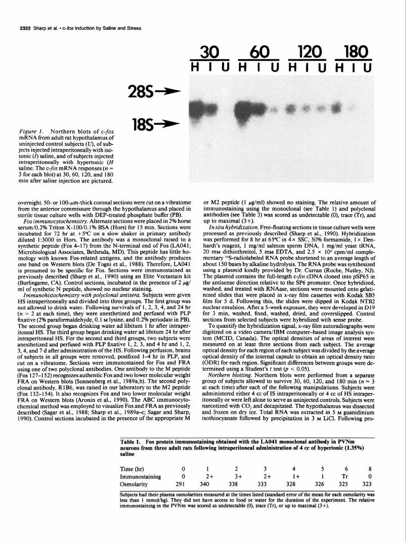
Figure 1. Northern blots of c-fos mRNA from adult rat hypothalamus of uninjected control subjects (U), of sub- jects injected intraperitoneally with iso- tonic (r) saline, and of subjects injected intraperitoneally with hypertonic (H saline. The c-fos mRNA responses (n = 3 for each blot) at 30,60, 120, and 180 min after saline injection are pictured.
HIUHIUHIUHIU
28S+
18S+
overnight. 50- or 100~pm-thick coronal sections were cut on a vibratome from the anterior commissure through the hypothalamus and placed in sterile tissue culture wells with DEP-treated phosphate buffer (PB).
Fos immunocytochemistry. Alternate sections were placed in 2% horse serum/0.2% Triton X-100/0.1% BSA (Hors) for 15 min. Sections were incubated for 72 hr at +YC on a slow shaker in primary antibody diluted 1:3000 in Hors. The antibody was a monoclonal raised to a synthetic peptide (Fos 4-17) from the N-terminal end of Fos (LA041; Microbiological Associates, Bethesda, MD). This peptide has little ho- mology with known Fos-related antigens, and the antibody produces one band on Western blots (De Togni et al., 1988). Therefore, LAO41 is presumed to be specific for Fos. Sections were immunostained as previously described (Sharp et al., 1990) using an Elite Vectastain kit (Burlingame, CA). Control sections, incubated in the presence of 2 pg/ ml of synthetic N peptide, showed no nuclear staining.
Zmmunohistochemistry with polyclonal antisera. Subjects were given HS intraperitoneally and divided into three groups. The first group was not allowed to drink water. Following survivals of 1, 2, 3, 4, and 24 hr (n = 2 at each time), they were anesthetized and perfused with PLP fixative (2% paraformaldehyde, 0.1 M lysine, and 0.2% periodate in PB). The second group began drinking water ad libitum 1 hr after intraper- itoneal HS. The third group began drinking water ad libitum 24 hr after intraperitoneal HS. For the second and third groups, two subjects were anesthetized and perfused with PLP fixative 1, 2, 3, and 4 hr and 1, 2, 3,4, and 7 d after administration of the HS. Following perfusion, brains of subjects in all groups were removed, postfixed 14 hr in PLP, and cut on a vibratome. Sections were immunostained for Fos and FRA using one of two polyclonal antibodies. One antibody to the M peptide (Fos 127-l 52) recognizes authentic Fos and two lower molecular weight FRA on Western blots (Sonnenberg et al., 1989a,b). The second poly- clonal antibody, R 1 B6, was raised in our laboratory to the M2 peptide (Fos 132-l 54). It also recognizes Fos and two lower molecular weight FRA on Western blots (Aronin et al., 1990). The ABC immunocyto- chemical method was employed to visualize Fos and FRA as previously described (Sagar et al., 1988; Sharp et al., 1989a-c; Sagar and Sharp, 1990). Control sections incubated in the presence of the appropriate M
or M2 peptide (1 pg/ml) showed no staining. The relative amount of immunostaining using the monoclonal (see Table 1) and polyclonal antibodies (see Table 3) was scored as undetectable (0), trace (Tr), and up to maximal (3+).
In situ hybridization. Free-floating sections in tissue culture wells were processed as previously described (Sharp et al., 1990). Hybridization was performed for 8 hr at 65°C in 4 x SSC, 50% formamide, 1 x Den- hardt’s reagent, 1 mg/ml salmon sperm DNA, 1 mg/ml yeast tRNA, 20 mM dithiothreitol, 5 mM EDTA, and 2.5 x lo6 cpm/ml comple- mentary 35S-radiolabeled RNA probe shortened to an average length of about 150 bases by alkaline hydrolysis. The RNA probe was synthesized using a plasmid kindly provided by Dr. Cut-ran (Roche, Nutley, NJ). The plasmid contains the full-length c-fos cDNA cloned into pSP65 in the antisense direction relative to the SP6 promoter. Once hybridized, washed, and treated with RNAase, sections were mounted onto gelati- nized slides that were placed in x-ray film cassettes with Kodak SB5 film for 5 d. Following this, the slides were dipped in Kodak NTB2 nuclear emulsion. After a 5-week exposure, they were developed in D 19 for 3 min, washed, fixed, washed, dried, and coverslipped. Control sections from selected subjects were hybridized with sense probe.
To quantify the hybridization signal, x-ray film autoradiographs were digitized on a video camera/IBM computer-based image analysis sys- tem (MCID, Canada). The optical densities of areas of interest were measured on at least three sections from each subject. The average optical density for each region ofeach subject was divided by the average optical density of the internal capsule to obtain an optical density ratio (ODR) for each region. Significant differences between groups were de- termined using a Student’s t test (p < 0.05).
Northern blotting. Northern blots were performed from a separate group of subjects allowed to survive 30, 60, 120, and 180 min (n = 3 at each time) after each of the following manipulations. Subjects were administered either 4 cc of IS intraperitoneally or 4 cc of HS intraper- itoneally or were left alone to serve as uninjected controls. Subjects were narcotized with CO, and decapitated. The hypothalamus was dissected and frozen on dry ice. Total RNA was extracted in 5 M guanidinium isothiocyanate followed by precipitation in 3 M LiCl. Following pro-
Table 1. Fos protein immunostaining obtained with the LAO41 monoclonal antibody in PVNm neurons from three adult rats following intraperitoneal administration of 4 cc of hypertonic (1.35%) saline
Time (hr) 0 1 2 3 4 5 6 8 Immunostaining 0 2+ 3+ 2+ 1+ 1 Tr 0 Osmolarity 291 340 338 333 328 326 325 323
Subjects had their plasma osmolarities measured at the times listed (standard error of the mean for each osmolarity was less than 1 mmol/kg). They did not have access to food or water for the duration of the experiment. The relative immunostaining in the PVNm was scored as undetectable (0), trace err), or up to maximal (3+).
The Journal of Neumsdence, August 1991, II(E) 2323
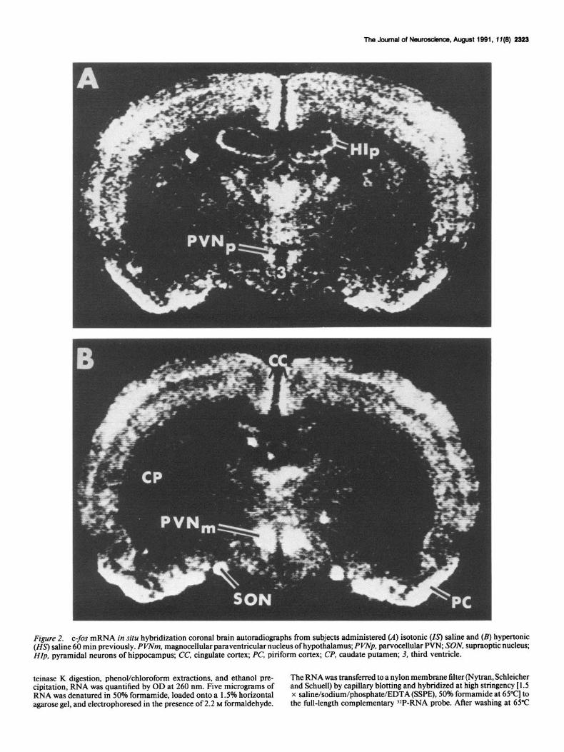
Figure 2. c-fos mRNA in situ hybridization coronal brain autoradiographs from subjects administered (A) isotonic (Is) saline and (B) hypertonic (Z&S) saline 60 min previously. PVNm, magnocellular paraventricular nucleus of hypothalamus; PWp, parvocellular PW, SON, supraoptic nucleus; HZp, pyramidal neurons of hippocampus; CC, cingulate cortex; PC, piriform cortex; CP, caudate putamen; 3, third ventricle.
teinase K digestion, phenolkhloroform extractions, and ethanol pre- cipitation, RNA was quantified by OD at 260 nm. Five micrograms of RNA was denatured in 50% formamide, loaded onto a 1.5% horizontal agarose gel, and electrophoresed in the presence of 2.2 M formaldehyde.
The RNA was transferred to a nylon membrane filter (Nytran, Schleicher and Schuell) by capillary blotting and hybridized at high stringency [ 1.5 x saline/sodium/phosphate/EDTA (SSPE), 50% formamide at 65”c] to the full-length complementary ‘*P-RNA probe. After washing at 65°C
The Journal of Neuroscience, August 1991, 17(B) 2325
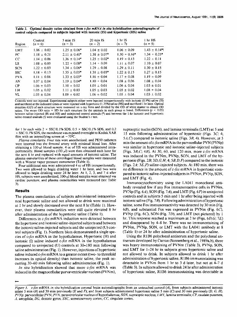
Table 2. Optimal density ratios obtained from c-fis mRNA in situ hybridization autoradiographs of control subjects compared to subjects injected with isotonic (IS) and hypertonic (HS) saline
Control 5 min IS 20 min IS 1 hr IS 1 hrHS Region (n = 6) (n = 3) (n = 2) (n = 7) (n = 5)
LMT 1.06 f 0.02 1.25 k 0.06* 1.04 * 0.02 1.06 f 0.09 1.43 + 0.14*!
PC 1.18 f 0.21 2.11 + 0.40* 1.38 + 0.07* 1.30 + 0.16* 1.34 * 0.25*
cc 1.14 +- 0.06 1.36 f 0.14* 1.25 f 0.02* 1.19 -t 0.13 1.22 k 0.11
LS 1.08 -t 0.05 1.22 + 0.08* 1.14 f 0.09 1.11 -t 0.07 1.10 + 0.07 SCN 1.22 + 0.03 1.34 +- 0.06* 1.29 k 0.08 1.29 + 0.11 1.30 f 0.15
ssc 1.18 f 0.13 1.35 k 0.05* 1.31 -t 0.03* 1.22 f 0.15 1.27 f 0.15
PVA 1.11 + 0.06 1.33 + 0.05* 1.16 k 0.04 1.17 f 0.08 1.19 k 0.09
AN 1.07 f 0.04 1.19 f 0.04* 1.10 + 0.04 1.08 + 0.06 1.08 +- 0.04
CP 1.06 + 0.03 1.10 f 0.02 1.05 5 0.01 1.06 +- 0.04 1.03 + 0.01
LH 1.05 -c 0.02 1.11 f 0.03 1.05 t- 0.03 1.05 * 0.02 1.08 f 0.04
VL 1.05 f 0.04 1.09 +- 0.02 1.06 k 0.02 1.05 + 0.04 1.03 f 0.02
Controls were not injected. Experimental subjects either were injected intraperitoneally with isotonic (0.9%) saline (IS) and sacrificed at the indicated times or were injected with hypertonic (1.35%) saline (HS) and sacrificed 1 hr later. Optical densities (OD) of each structure were measured on x-ray films and divided by the OD of white matter to obtain OD ratios. The mean OD ratio f SD for each structure for the animals in each group is listed. Differences (p < 0.05) between saline-injected (IS and HS) and uninjected control animals (*) and between the I-hr isotonic and hypertonic saline-treated animals (!) were evaluated using the Student’s t test.
for 1 hr each with 2 x SSC/O. 1% SDS, 0.5 x SSC/O. 1% SDS, and 0.2 x SSC/O. 1% SDS, the membrane was exposed overnight to Kodak XAR film with an intensifying screen at - 70°C.
Plasma osmolarity. Four rats were anesthetized, and PESO catheters were inserted into the femoral artery with minimal blood loss. After obtaining a 100~~1 blood sample, 4 cc of HS was administered intra- peritoneally. Blood samples (100 ~1) were then obtained each hour for the next 8 hr and replaced with equal amounts of isotonic saline. The plasma osmolarities of these centrifuged blood samples were measured with a Wescor vapor pressure osmometer (Table 1).
Four additional rats were administered 4 cc of HS intraperitoneally. Two were allowed to begin drinking water 1 hr later, and two were allowed to begin drinking water 24 hr later. At 1, 2, 3, and 7 d after HS, subjects were anesthetized, 1 00-~1 blood samples were obtained via cardiac puncture, and plasma osmolarities were measured (see Table 3).
Results
The plasma osmolarities of subjects administered intraperito- neal hypertonic saline and not allowed to drink were maximal at 1 hr and slowly decreased over the next 8 hr (Table 1). How- ever, their plasma osmolarities were still quite elevated 8 hr after administration of the hypertonic saline (Table 1).
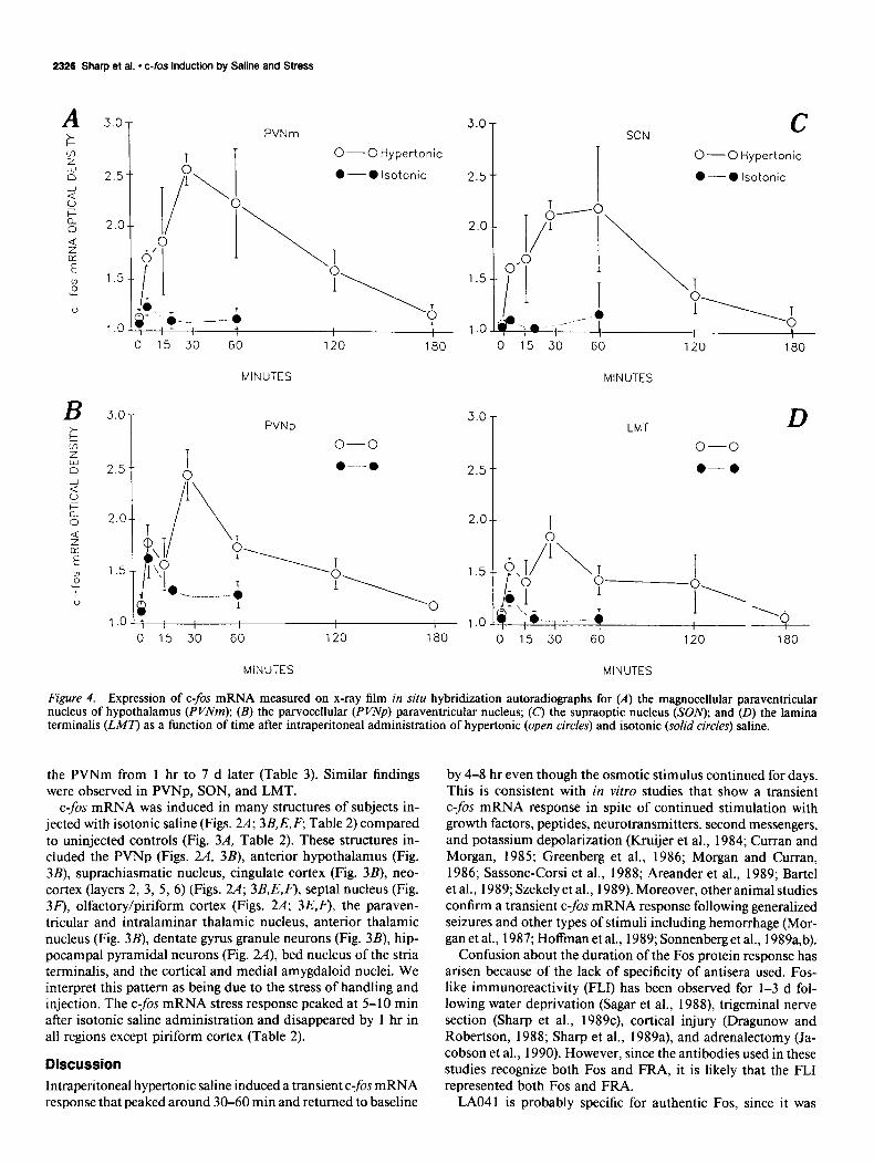
Differences in c-fos mRNA induction were detected between the hypertonic and isotonic saline-injected subjects and between the isotonic saline-injected subjects and the uninjected (U) con- trol subjects (Fig. 1). Northern blots demonstrated a single spe- cies of c-fis mRNA in the hypothalamus. Hypertonic (H) and isotonic (I) saline induced C-$X mRNA in the hypothalamus compared to uninjected (U) controls at 30-180 min following saline administration (Fig. 1). However, injections of hypertonic saline induced c-fos mRNA to a greater extent (two- to threefold increases in optical density) than isotonic saline, the peak oc- curring 30-60 min following saline administration (Fig. 1).
In situ hybridization showed that more c-fos mRNA was induced in the magnocellular parvaventricular nucleus (PVNm),
t
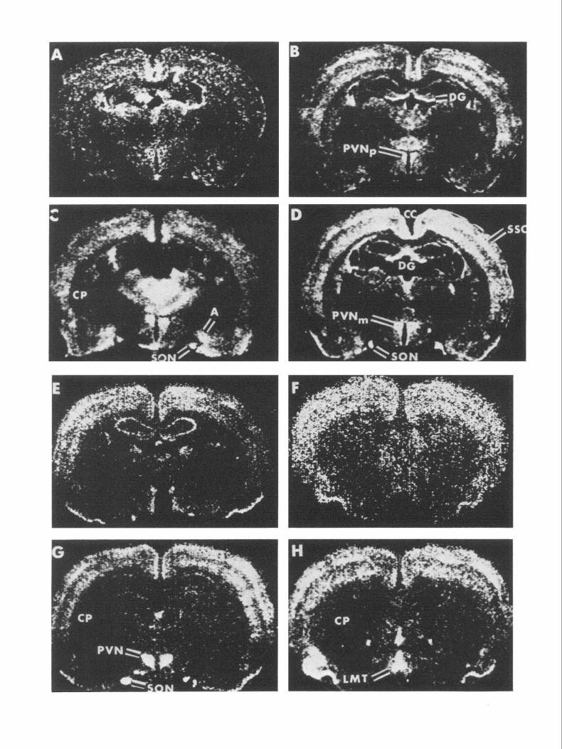
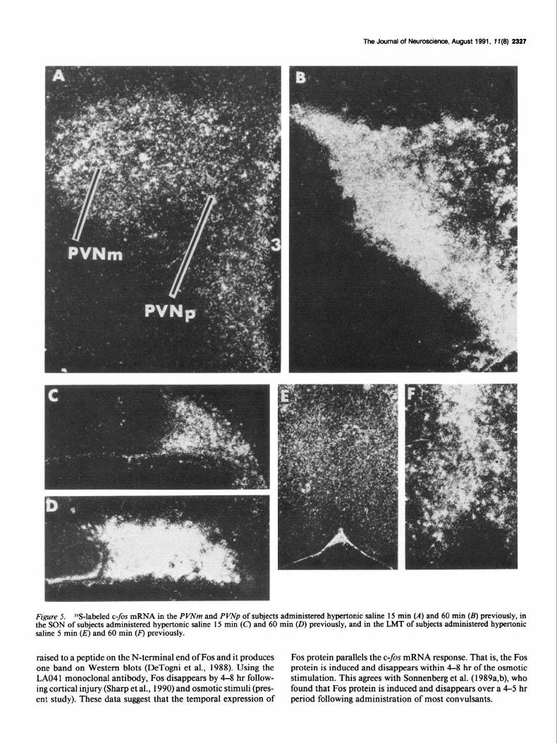
supraoptic nuclei (SON), and lamina terminalis (LMT) at 5 and 15 min following administration of hypertonic (Figs. 3C, 4; 5A,C) compared to isotonic saline (Figs. 3B, 4). However, at 5 min the amount oft-fos mRNA in the parvocellular PVN (PVNp) was similar in hypertonic and isotonic saline-injected subjects (Figs. 3B,C; 4B). At 30, 60, and 120 min, more c-fos mRNA was induced in the PVNm, PVNp, SON, and LMT of the hy- pertonic (Figs. 2B, 3D,G,H, 4; SB,D,F) compared to the isotonic (Figs. 2A; 3E,F') saline-injected subjects. At 180 min, there was no difference in the amount of c-fos mRNA in hypertonic com- pared to isotonic saline-injected subjects in PVNm, PVNp, SON, and LMT (Fig. 4).
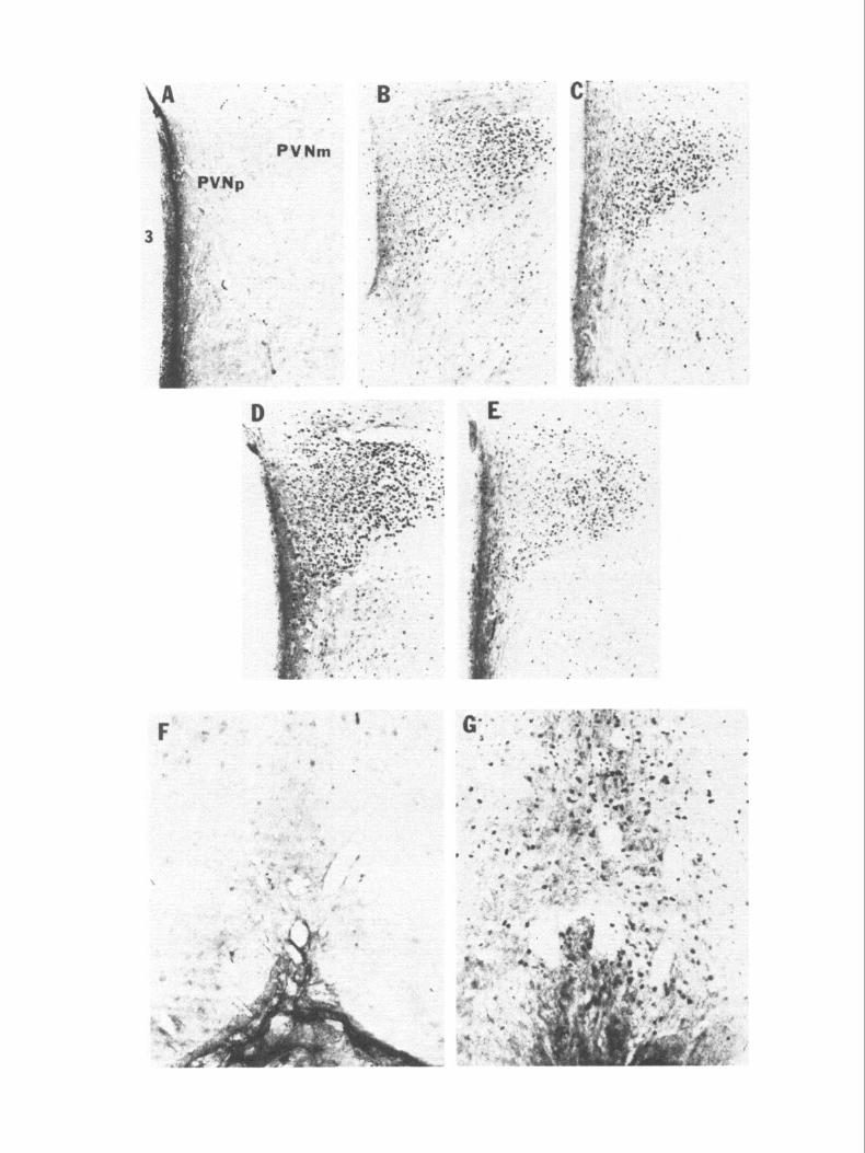
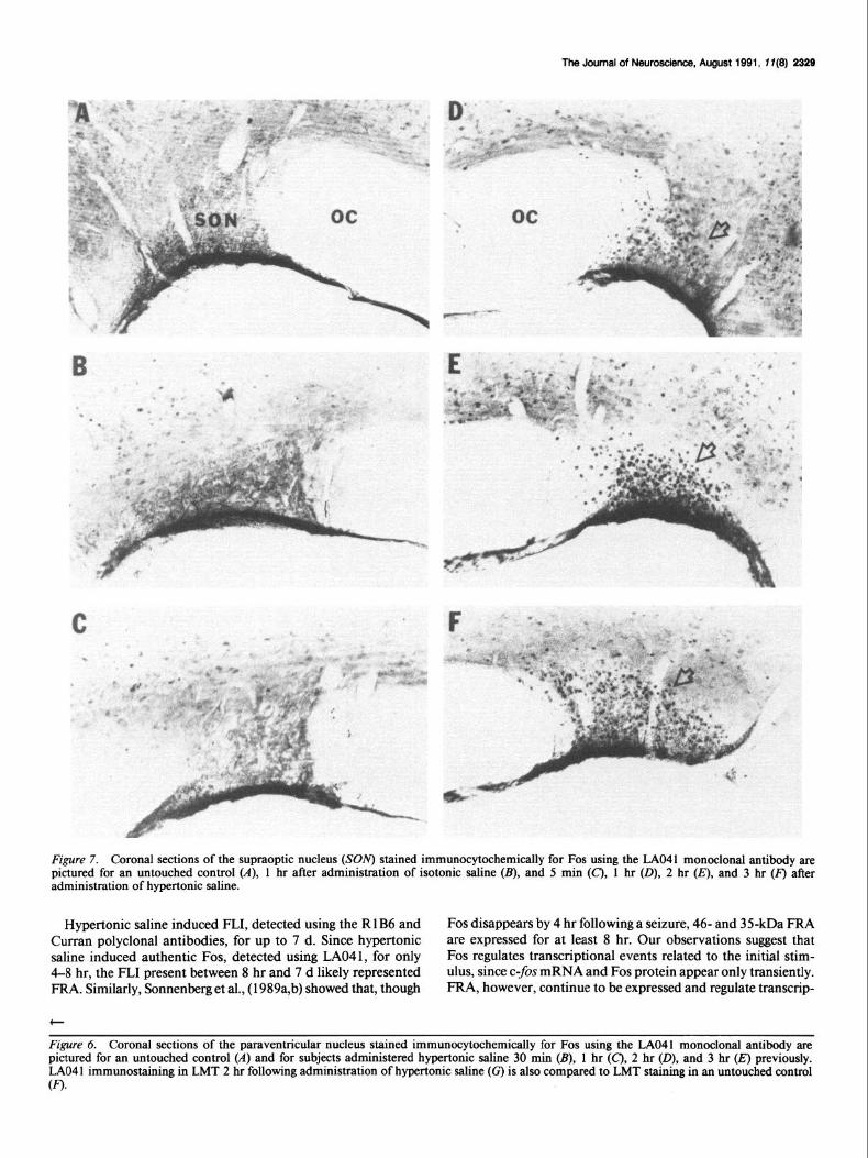
Immunocytochemistry using the LAO4 1 monoclonal anti- body revealed few if any Fos immunoreactive cells in PVNm, PVNp (Fig. 6A), SON (Fig. 7A), and LMT (Fig. 6F) in uninjected controls and in subjects 5 min and 1 hr after being injected with isotonic saline (Fig. 7B). Following administration of hypertonic saline, some Fos immunoreactivity was detected by 30 min (Fig. 6B), and substantial Fos was expressed in PVNm (Fig. 6C), PVNp (Fig. 6C), SON (Fig. 7D), and LMT (not pictured) by 1 hr. This response reached a maximum at 2 hr (Figs. 6D,G; 7E) and disappeared by 4-8 hr. There was no immunostaining of PVNm, PVNp, SON, or LMT with the LA041 antibody at 8 (Table 1) or 24 hr after administration of hypertonic saline.
Using the R 1 B6 polyclonal antiserum and the polyclonal an- tiserum developed by Cm-ran (Sonnenberg et al., 1989a,b), there was heavy immunostaining of PVNm (Table 3), PVNp, SON, and LMT for l-24 hr in subjects given hypertonic saline and not allowed to drink. In subjects allowed to drink 1 hr after administration of hypertonic saline, R 1 B6 immunostaining was detectable in PVNm from 1 hr to 3 d later, but not at 4-7 d (Table 3). In subjects allowed to drink 24 hr after administration of hypertonic saline, RlB6 immunostaining was detectable in
Figure 3. c-fos mRNA in situ hybridization coronal brain autoradiographs from an untouched control (A), from subjects administered isotonic saline 5 min (B) and 30 min previously (E and F); and from subjects administered hypertonic saline 5 min (C) and 30 min previously (D, G, H). PVNp, parvocellular PVN; PVN, paraventricular nucleus ofhypothalamus; SON, supraoptic nucleus; LMT, lamina terminalis; CP, caudate putamen; A, amygdala; DG, dentate gyrus; SSC, somatosensory cortex; CC, cingulate cortex.
2326 Sharp et al. l ofos Induction by Saline and Stress
SON c
01530 60 120 180
MINUTES
PVNp
o-o
2.5 1 9
. ~. .-a
01530 60 120 180
2.5
O-O Hypertonic
l --- 0 Isotonic
01530 60 120 ISO
MINUTES
3.0
I
LMT D o-o
2.5 O-0
, ,o Q ‘,h. .., -- 4 1
I , I I I
01530 60 120 180
MINUTES MINUTES
Figure 4. Expression of C-$X mRNA measured on x-ray film in situ hybridization autoradiographs for (A) the magnocellular paraventricular nucleus of hypothalamus (PVNm); (B) the parvocellular (PVNp) paraventricular nucleus; (c) the supraoptic nucleus (SOlv); and (D) the lamina terminalis @MT) as a function of time after intraperitoneal administration of hypertonic (open circles) and isotonic (solid circles) saline.
the PVNm from 1 hr to 7 d later (Table 3). Similar findings were observed in PVNp, SON, and LMT.
c-fis mRNA was induced in many structures of subjects in- jected with isotonic saline (Figs. 2A; 3B,E,F, Table 2) compared to uninjected controls (Fig. 3A, Table 2). These structures in- cluded the PVNp (Figs. 2A, 3B), anterior hypothalamus (Fig. 3B), suprachiasmatic nucleus, cingulate cortex (Fig. 3B), neo- cortex (layers 2, 3, 5, 6) (Figs. 2A; 3B,E,F), septal nucleus (Fig. 3F), olfactory/piriform cortex (Figs. 2A; 3E,F), the paraven- tricular and intralaminar thalamic nucleus, anterior thalamic nucleus (Fig. 3B), dentate gyrus granule neurons (Fig. 3B), hip- pocampal pyramidal neurons (Fig. 2A), bed nucleus of the stria terminalis, and the cortical and medial amygdaloid nuclei. We interpret this pattern as being due to the stress of handling and injection. The c-fis mRNA stress response peaked at 5-10 min after isotonic saline administration and disappeared by 1 hr in all regions except piriform cortex (Table 2).
Discussion
Intraperitoneal hypertonic saline induced a transient c-fos mRNA response that peaked around 30-60 min and returned to baseline
by 4-8 hr even though the osmotic stimulus continued for days. This is consistent with in vitro studies that show a transient C-$X mRNA response in spite of continued stimulation with growth factors, peptides, neurotransmitters, second messengers, and potassium depolarization (Kruijer et al., 1984; Curran and Morgan, 1985; Greenberg et al., 1986; Morgan and Curran, 1986; Sassone-Corsi et al., 1988; Areander et al., 1989; Bartel et al., 1989; Szekely et al., 1989). Moreover, other animal studies confirm a transient c-&s mRNA response following generalized seizures and other types of stimuli including hemorrhage (Mor- gan et al., 1987; Hoffman et al., 1989; Sonnenberg et al., 1989a,b).
Confusion about the duration of the Fos protein response has arisen because of the lack of specificity of antisera used. Fos- like immunoreactivity (FLI) has been observed for l-3 d fol- lowing water deprivation (Sagar et al., 1988), trigeminal nerve section (Sharp et al., 1989c), cortical injury (Dragunow and Robertson, 1988; Sharp et al., 1989a), and adrenalectomy (Ja- cobson et al., 1990). However, since the antibodies used in these studies recognize both Fos and FRA, it is likely that the FL1 represented both Fos and FRA.
LA041 is probably specific for authentic Fos, since it was
The Journal of New-, August 1991, 1 I(9) 2327
Figure 5. Yj-labeled c-fos mRNA in the PVNm and PVNp of subjects administered hypertonic saline 15 min (A) and 60 min (B) previously, in the SON of subjects administered hypertonic saline 15 min (C) and 60 min (0) previously, and in the LMT of subjects administered hypertonic saline 5 min (E) and 60 min (F) previously.
raised to a peptide on the N-terminal end of Fos and it produces Fos protein parallels the c-$x mRNA response. That is, the Fos one band on Western blots (DeTogni et al., 1988). Using the protein is induced and disappears within 4-8 hr of the osmotic LAO4 1 monoclonal antibody, Fos disappears by 4-8 hr follow- stimulation. This agrees with Sonnenberg et al. (1989a,b), who ing cortical injury (Sharp et al., 1990) and osmotic stimuli (pres- found that Fos protein is induced and disappears over a 4-5 hr ent study). These data suggest that the temporal expression of period following administration of most convulsants.
The Journal of New-, August 1991, 7 T(8) 2329
Figure 7. Coronal sections of the supraoptic nucleus (SON) stained immunocytochemically for Fos using the LAO41 monoclonal antibody are pictured for an untouched control (A), 1 hr after administration of isotonic saline (B), and 5 min (C’), 1 hr (D), 2 hr (IT), and 3 hr (F) after administration of hypertonic saline.
Hypertonic saline induced FLI, detected using the R 1 B6 and Fos disappears by 4 hr following a seizure, 46- and 35kDa FRA Curran polyclonal antibodies, for up to 7 d. Since hypertonic are expressed for at least 8 hr. Our observations suggest that saline induced authentic Fos, detected using LA041, for only Fos regulates transcriptional events related to the initial stim- 4-8 hr, the FL1 present between 8 hr and 7 d likely represented ulus, since c-fos mRNA and Fos protein appear only transiently. FRA. Similarly, Sonnenberg et al., (1989a,b) showed that, though FRA, however, continue to be expressed and regulate transcrip-
t
Figure 6. Coronal sections of the paraventricular nucleus stained immunocytochemically for Fos using the LAO41 monoclonal antibody are pictured for an untouched control (A) and for subjects administered hypertonic saline 30 min (B), 1 hr (C), 2 hr (D), and 3 hr (E) previously. LAO4 1 immunostaining in LMT 2 hr following administration of hypertonic saline (c) is also compared to LMT staining in an untouched control (0.
2330 Sharp et al. * c-fos Induction by Saline and Stress
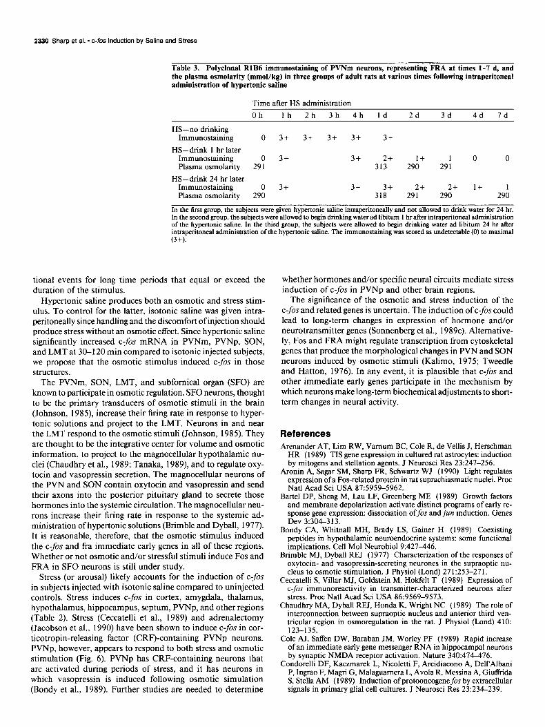
Table 3. Polyclonal RlB6 immunostaining of PVNm neurons, representing FRA at times l-7 d, and the plasma osmolarity (mmol/kg) in three groups of adult rats at various times following intraperitoneal administration of hypertonic saline
Time after HS administration Oh lh 2h 3h 4h Id 2d 3d 4d Id
HS-no drinking Immunostaining 0 3+ 3+ 3+ 3+ 3+
H&drink 1 hr later Immunostaining 0 3+ 3+ 2+ 1+ 1 0 0 Plasma osmolarity 291 313 290 291
HS-drink 24 hr later Immunostaining 0 3+ 3+ 3+ 2+ 2+ 1+ 1 Plasma osmolarity 290 318 291 290 290
In the first group, the subjects were given hypertonic saline intraperitoneally and not allowed to drink water for 24 hr. In the second group, the subjects were allowed to begin drinking water ad libitum 1 hr after intraperitoneal administration of the hypertonic saline. In the third group, the subjects were allowed to begin drinking water ad libitum 24 hr after intraperitoneal administration of the hypertonic saline. The immunostaining was scored as undetectable (0) to maximal t3+1.
tional events for long time periods that equal or exceed the duration of the stimulus.
Hypertonic saline produces both an osmotic and stress stim- ulus. To control for the latter, isotonic saline was given intra- peritoneally since handling and the discomfort of injection should produce stress without an osmotic effect. Since hypertonic saline significantly increased c-&s mRNA in PVNm, PVNp, SON, and LMT at 30-l 20 min compared to isotonic injected subjects, we propose that the osmotic stimulus induced c-fos in those structures.
The PVNm, SON, LMT, and subfomical organ (SFO) are known to participate in osmotic regulation. SF0 neurons, thought to be the primary transducers of osmotic stimuli in the brain (Johnson, 1985), increase their firing rate in response to hyper- tonic solutions and project to the LMT. Neurons in and near the LMT respond to the osmotic stimuli (Johnson, 1985). They are thought to be the integrative center for volume and osmotic information, to project to the magnocellular hypothalamic nu- clei (Chaudhry et al., 1989; Tanaka, 1989), and to regulate oxy- tocin and vasopressin secretion. The magnocellular neurons of the PVN and SON contain oxytocin and vasopressin and send their axons into the posterior pituitary gland to secrete those hormones into the systemic circulation. The magnocellular neu- rons increase their firing rate in response to the systemic ad- ministration of hypertonic solutions (Brimble and Dyball, 1977). It is reasonable, therefore, that the osmotic stimulus induced the czfos and fra immediate early genes in all of these regions. Whether or not osmotic and/or stressful stimuli induce Fos and FRA in SF0 neurons is still under study.
Stress (or arousal) likely accounts for the induction of c-fos in subjects injected with isotonic saline compared to uninjected controls. Stress induces C-$X in cortex, amygdala, thalamus, hypothalamus, hippocampus, septum, PVNp, and other regions (Table 2). Stress (Ceccatelli et al., 1989) and adrenalectomy (Jacobson et al., 1990) have been shown to induce c-fos in cor- ticotropin-releasing factor (CRF)-containing PVNp neurons. PVNp, however, appears to respond to both stress and osmotic stimulation (Fig. 6). PVNp has CRF-containing neurons that are activated during periods of stress, and it has neurons in which vasopressin is induced following osmotic simulation (Bondy et al., 1989). Further studies are needed to determine
whether hormones and/or specific neural circuits mediate stress induction of c-fos in PVNp and other brain regions.
The significance of the osmotic and stress induction of the c-fos and related genes is uncertain. The induction of c-fos could lead to long-term changes in expression of hormone and/or neurotransmitter genes (Sonnenberg et al., 1989~). Altemative- ly, Fos and FRA might regulate transcription from cytoskeletal genes that produce the morphological changes in PVN and SON neurons induced by osmotic stimuli (Kalimo, 1975; Tweedle and Hatton, 1976). In any event, it is plausible that c-fos and other immediate early genes participate in the mechanism by which neurons make long-term biochemical adjustments to short- term changes in neural activity.
References Arenander AT, Lim RW, Vamum BC, Cole R, de Vellis J, Herschman
HR (1989) TIS gene expression in cultured rat astrocytes: induction by mitoge& and stellation agents. J Neurosci Res 23:247-256.
Aronin A. Saaar SM. Sham FR. Schwartz WJ (1990) Light regulates expression of a Fos-related prdtein in rat suprachias&aticnuclei. Proc Nat1 Acad Sci USA 875959-5962.
Bartel DP, Sheng M, Lau LF, Greenberg ME (1989) Growth factors and membrane depolarization activate distinct programs of early re- sponse gene expression: dissociation offos andjun induction. Genes Dev 3:304-313.
Bondy CA, Whitnall MH, Brady LS, Gainer H (1989) Coexisting peptides in hypothalamic neuroendocrine systems: some functional implications. Cell Mol Neurobiol 9:427-446.
Brimble MJ, Dyball REJ (1977) Characterization of the responses of oxytocin- and vasopressin-secreting neurones in the supraoptic nu- cleus to osmotic stimulation. J Physiol (Lond) 27 1:253-27 1.
Ceccatelli S, Villar MJ, Goldstein M, Hokfelt T (1989) Expression of C-$X immunoreactivity in transmitter-characterized neurons after stress. Proc Nat1 Acad Sci USA 86:9569-9573.
Chaudhry MA, Dyball REJ, Honda K, Wright NC (1989) The role of interconnection between supraoptic nucleus and anterior third ven- tricular region in osmoregulation in the rat. J Physiol (Lond) 410: 123-135.
Cole AJ, Saffen DW, Baraban JM, Worley PF (1989) Rapid increase of an immediate early gene messenger RNA in hippocampal neurons by synaptic NMDA receptor activation. Nature 340:474-476.
Condorelli DF, Kaczmarek L, Nicoletti F, Arcidiacono A, Dell’Albani P, Ingrao F, Magri G, Malaguamera L, Avola R, Messina A, Giuffrida S, Stella AM (1989) Induction of protooncogenefos by extracellular signals in primary glial cell cultures. J Neurosci Res 23:234-239.
The Journal of Neuroscience, August 1991, 1 I(9) 2331
Cm-ran T (1988) The fos oncogene. In: The oncogene handbook (Red- dy EP, Skalka AM, Curran T, kds), pp 307-325. Amsterdam: Elsevier.
Curran T. Moraan JI (1987) Memories of fos. Bioessavs 7:255-258. De Togni P, Nilman H,‘ Raymond V, Sawchenko P, Vet-ma IM (1988)
Detection of fos protein during osteogenesis by monoclonal antibod- ies. Mol Cell Biol 8:225 l-2256.
Dragunow M, Robertson HA (1987) Kindling stimulation induces c&s protein(s) in granule cells of the rat dentate gyrus. Nature 329: 441-442.
Dragunow M, Robertson HA (1988) Brain injury induces c-fos pro- tein(s) in nerve and glial-like cells in adult mammalian brain. Brain Res 4551295-299.
Draisci G, Iadarola MJ (1989) Temporal analysis of increases in c&s, preprodynorphin and preproenkephalin mRNAs in rat spinal cord. Mol Brain Res 6:3 l-37.
Greenberg ME, Ziff EB, Greene LA (1986) Stimulation of neuronal acetylcholine receptors induces rapid gene transcription. Science 234: 80-83.
Hisanaga K, Sagar SM, Hicks KJ, Swanson RA, Sharp FR (1990) c-fos protooncogene expression in astrocytes associated with differ- entiation or proliferation but not depolarization. Mol Brain Res 8: 69-75.
Hisanaga K, Sagar SM, Sharp FR (1991) N-methyl-d-aspartate an- tagonists block Fos-like protein expression induced via multiple sig- nalling pathways in cultured neurons. J Neurochem, in press.
Hoffman GE, Roberts MM, Verbalis JG, Robinson AG (1989) Ex- pression of c-fos in magnocellular hypothalamic neurons: specificity for oxytocin and vasopressin. Neurosci Abstr 15: 189.
Hunt SP, Pini A, Evan G (1987) Induction of c-&-like protein in spinal cord neurons following sensory stimulation. Nature 328:632- 634.
Jacobson L, Sharp FR, Dallman MF (1990) Induction of Fos-like immunoreactivity in hypothalamic neurons after adrenalectomy in the rat. Endocrinology 126: 1709-l 7 19.
Johnson AK (1985) The periventricular anteroventricular third ven- tricle (AV3V): its’relationship with the subfomical organ and neural systems involved in maintaining body fluid homeostasis. Brain Res Bull 15:595-601.
Kalimo H (1975) Ultrastructural studies on the hypothalamic neu- rosecretory neurons of the rat. III. Paraventricular and supraoptic neurons during lactation and dehydration. Cell Tissue Res 163: 15 l- 168.
Kruijer W, Cooper JA, Hunter T, Verrna IM (1984) Platelet-derived growth factor induces rapid but transient expression of the c-fis gene and protein. Nature 3 12:7 1 l-7 16.
Menetrey D, Gannon A, Levine JD, Basbaum AI (1989) Expression of c-fi.r protein in intemeurons and projection neurons of the rat spinal cord in response to noxious somatic, articular, and visceral stimulation. J Comp Neuro1285: 177-195.
Morgan JI, Curran T (1986) Role of ion flux in the control of c-fis expression. Nature 322:552-555.
Morgan JI, Cohen DR, Hempstead JL, Curran T (1987) Mapping patterns oft-fos expression in the central nervous system after seizure. Science 2371192-196.
Sagar SM, Sharp FR (1990) Light induces a Fos-like nuclear antigen in retinal neurons. Mol Brain Res 7: 17-2 1.
Sagar SM, Sharp FR, Curran T (1988) Expression of c-fos protein in brain: metabolic mapping at the cellular level. Science 240:1328- 1331.
Sassone-Corsi P, Sisson JC, Verma IM (1988) Transcriptional auto- regulation of the proto-oncogenefos. Nature 334:3 14-319.
Sharu FR. Gonzalez MF. Hisanaaa K. Moblev WC. Saaar SM (1989a) Induction of Fos-like ‘immun&eactivity in rat forebrain following cortical lesions and NGF injections. Neurosci Lett 100: 117-l 22.
Sharp FR, Gonzalez MF, Sharp JW, Sagar SM (1989b) C-fos expres- sion and 14C 2-deoxyglucose uptake in the caudal cerebellum of the rat during motor/sensory cortex stimulation. J Comp Neurol 284: 621-636.
Sharp FR, Griffiths J, Gonzalez MF, Sagar SM (1989~) Trigeminal nerve section induces Fos-like immunoreactivity (FLI) in brainstem and decreases FL1 in sensory cortex. Mol Brain-R& 6.2 17-220.
Sharp JW, Sagar SM, Hisanaga K, Jasper P, Sharp FR (1990) The NMDA receptor mediates cortical induction of Fos and Fos-related antigens following cortical injury. Exp Neurol 109:323-332.
Sonnenberg JL, Mitchelmore C, Macgregor-Leon PF, Hempstead J, Morgan JI, Curran T (1989a) Glutamate receptor agonists increase the expression of Fos, Fra, and AP-1 DNA binding activity in the mammalian brain. J Neurosci Res 24:72-80.
Sonnenberg JL, Macgregor-Leon PF, Cm-ran T, Morgan Jl (1989b) Dynamic alterations occur in the levels and composition of transcrip- tion factor AP-1 complexes after seizure. Neuron 3:359-365.
Sonnenberg JL. Rauscher FJ III. Moraan JI. Curran T (1989~) Ree- ulation if proenkephalin by Fos and Jun. ‘Science 246:‘1622-1625.-
Swanson LW, Sawchenko PE (1983) Hypothalamic integration: or- ganization of the paraventricular and supraoptic nuclei. Annu Rev Neurosci 61269-324.
Szekely AM, Barbaccia ML, Costa E (1987) Activation of specific glutamate receptor subtypes increases czfos proto-oncogene expres- sion in primary cultures of cerebellar granule cells. Neuropharma- cology 26: 1779-1782.
Szekely AM, Barbaccia ML, Alho H, Costa E (1989) In primary cul- tures of cerebellar granule cells the activation of N-methyl-d-aspar- tate-sensitive glutamate receptors induces c-fis mRNA expression. Mol Pharmacol 35:401408.
Tanaka J (1989) Involvement of the median preoptic nucleus in the regulation of paraventricular vasopressin neurons by the subfomical organ in the rat. Exp Brain Res 76~47-54.
Tweedle CD, Hatton GI (1976) Ultrastructural comparisons of neu- rons of supraoptic and circularis nuclei in normal and dehydrated rats. Brain Res Bull 1: 103-l 2 1.
Verrna IM, Sassone-Corsi P (1987) Proto-oncogene fos: complex but versatile regulation. Cell 5 1:5 13-5 14.
White JD, Gall CM (1987) Differential regulation of neuropeptide and proto-oncogene mRNA content in hippocampus following recurrent seizure. Mol Brain Res 3:21-29.