By A. K.^GATUMA B. Sc. (PHARMACY)
116
J '' A PHARMACOGNOSTICAL, PHYTOCHEMICAL AND PHARMACOLOGICAL INVESTIGATION OF THE POISONOUS PRINCIPLE(S) OF ELAEODENDRON BUCHANANII (LOES.) LOES By A. K.^GATUMA B. Sc. (PHARMACY) A thesis submitted for the degree of Master of Science (Pharmacy) of the University of Nairobi.
Transcript of By A. K.^GATUMA B. Sc. (PHARMACY)

J'' A PHARMACOGNOSTICAL, PHYTOCHEMICAL AND PHARMACOLOGICAL INVESTIGATION
OF THE POISONOUS PRINCIPLE(S) OF ELAEODENDRON BUCHANANII (LOES.) LOES
By
A. K.^GATUMA
B. Sc. (PHARMACY)
A thesis submitted for the degree of Master of
Science (Pharmacy) of the University of Nairobi.

D E C L A R A T I O N
I hereby declare that this thesis is niy original work and has not
been submitted for a degree in any other University.
A. K. Gatuma
CANDIDATE
This thesis has been submitted for examination with our approval
as University Supervisors.

A C K N O W L E D G E M E N T S
The author wishes to express his sincere appreciation to Dr.
C. K. Maitai, Chairman of the Department of Pharmacy for his inva
luable guidance and assistance throughout this study and for his
critical review of the manuscript.
The author also wishes to thank Drs. G. S. Ahmed and W. M.
Kofi - Tsekpo for their interest, assistance, and guidance during
this study and also for their comments on the manuscript. The
author is greatly indebted to Dr. P. Speiser of Pharmaceutical Insti
tute, Galenishe, Abteilung for carrying out the elemental and nuclear
magnetic resonance analysis and confirming the heart glycoside stru
cture of the isolated compound. Many thanks are also due to the
technical staff of the Department of Pharmacy of the University of
Nairobi for their invaluable assistance and co-operation. Sincere
thanks are also expressed to the Chief Government Chemist, Mr. N.
Muraguri for granting permission to use infrared spectrophotometer
and melting point determination apparatus.
The author is also grateful to Miss Muthoni Nginya for typing
this thesis. Also the author would like to thank his wife and Helen
Kanyua for helping him collect the plant material and encourangement
throughout this study.
Finally, the author wishes to express sincere thanks to the
University of Nairobi for granting him ?. research grant No. 670 - 371
without which this work would not have been carried out.

D E D I C A T I O N
This thesis is dedicated to my wife and son.

TABLE OF CONTENTS
SUMMARY i
INTRODUCTION iii
LITERATURE REVIEW viii
1 A Pharmacognostical investigation of Elaeodendron
Buchananii (Loes.) Loes 001
Morphological characteristic and distribution
2 Experimental Work 006
Reagents 007
Preparation of Reagents 008
Apparatus 009
Collection of materials for investigation 010
Preliminary investigation of active principles
in Elaeodendron buchananii (Loes.) 010
Investigation of suitable extracting solvents
for cardiac glycosides in Elaeodendron buchananii 012
Extraction and purification procedure of the
cardiac glycoside(s) from the leaves of Elaeode
ndron buchananii 014
Characterisation of the isolated glycosides 024
Estimation of recovery yield of the cardiac
glycosides in the dry leaves of the p1 ant 033
A preliminary investigation of the pharmacolo
gical effects of the isolated compound on intact
and isolated mammalian preparations 034
3 Discussion 038
4 Conclusion 057*
References OSS
CHAPTEP PAGE
v

I L L U S T R A T I O N S

LIST OF TABLES
I Results of tests for possible chemical constitu
ents likely to be present in different morpholo
gical parts of Elaeodendron buchananii (Loes.)
Loes Oil
II Investigation of the best extracting solvent for
the active principles of Elaeodendron buchananii
(Loes.) Loes 060
III Results of purification of the glycosidal solu
tion of chloroform using an alumina column 061
IV Solubility of the isolated compound in different
solvents 062
V TLC analysis of the isolated glycoside(s) of
Elaeodendron compound with digoxin and ouabain
by TLC 063
VI Comparison of infra-red spectra of isolated gly
coside with that of digitoxigenin 065
VII Results of the elemental analysis for the isolated
glycoside of Elaeodendron 030
VIII Empirical formula and molecular formula of the iso
lated glycoside of Elaeodendron as calculated from
the elemental analysis and molecular weight 031
13IX C NMR spectrum data for the isolated glycoside
of Elaeodendron 060
X Comparison of mass spectrum of the isolated gly
coside of Elaeodendron with the m/e values of
digitoxigenin and somalin
TABLE PAGE
0C7

TABLE PAGE
XI Data for the calibration curve of absorbance
against concentration of the isolated glycoside
of Elaeodendron 070
XII Pharmacological response of the isolated glycoside
of Elaeodendron, adrenalin, and digoxin on the
blood pressure of anaesthetised rat 072

LIST OF FIGURES
1
2
3
4
5
6
7
8
9
10
11
a
12
FIGURE
13
Elaeodendron buchananii (Loes.) Loes
a) a branchlet b) fruiting branchlet
c) flower d) leaf
Ultraviolet absorption spectrum of the isolated
glycoside in alcohol
Ultraviolet absorption spectrum of the isolated
glycoside for the colour reaction with 3:5 Kedde
reagent and dinitrobenzoic acid (3:5 DNBA) reagent
Ultraviolet absorption spectrum of the isolated
glycoside for the colour reaction with 1,3 dini
trobenzene (m - DNB) reagent
Infra-red spectrum (KBr disc) of the isolated
glycoside
Infra-red spectrum (KBr disc) of digitoxigenin
1 3C NMR spectrum of the isolated glycoside
Proton NMR of the isolated glycoside
Mass spectrum of the isolated glycoside
A graph of absorbance against concentration of
the isolated glycoside
A condon pressure manometer used to study the
effects of the isolated glycoside on blood pressure
of anaesthetised rat
Effects of adrenalin, isolated glycoside and di«̂ o
xin on blood pressure of anaesthetised rat
Effects of adrenalin, isolated glycoside and
digoxin on isolated rabbit heart on a Kymograph
073
075
076
077
078
079
080
081
082
073
084
085
PAGE
085
FIGURE PAGE
14 Effects of adrenalin, isolated glycoside on
isolated perfused rabbit heart using i Harvard
Smooth Muscle/Heart Apparatus 087
*

•I
SUMMARY
A pharmacognostical investigation of Elaeodendron buchananii
(Loes.) Loes. has been undertaken. Phytochemical and pharmacolo
gical properties of the active (poisonous) principles of the plant
have also been studied.
The pharmacognostical investigation of the plant involved iden
tifying features of the different parts of the plant using photo
graphic and macroscopic methods.
Results of the screening tests of the different parts of the
plant for the active constituents indicated the presence of chemical
compounds v/ith “ , &- unsaturated <r - lactone ring, possibly cardiac
glycosides. Investigation of a suitable solvent system for the ext
raction of these compounds was undertaken. Of the different parts
of the plant examined for active principles, the leaves were found
to contain the highest percentage of the chemical compounds with
3 - unsaturated 3- lactone ring. Isolation and purification of the
active principle(s) from the original crude plant extracts involving
the removal of pigments, tannins, resins and excess lead has been
described. Crystallisation of the isolated glycoside from a suitable
solvent system and the subsequent study of some of the physical and
chemical properties of the isolated compound has been described.
From the elemental analysis and the molecular weight of the
isolated compound the molecular formula of the compound has been
determined as . Using the infra-red, ultraviolet, nuclear

11
magnetic resonance and mass spectra, a partial molecular structure
has been suggested.
The isolated compound has been reacted with Kedde reagent and
the resulting coloured complex has been examined to see whether it
obeys Beer - Lambert law. The calibration curve obtained has been
used to determine the percentage recovery of the isolated compound
in the leaves of the plant.
The pharmacological study of the isolated compound has also
been undertaken. This study involved the investigation of the
effects of the isolated compound on the blood pressure of anaesthe
tised rat and the effect of the compound on the isolated perfused
rabbit heart.
Suggestions for further work as regards pharmacognostical inve
stigation of the plant together with ascertaining the exact structu
ral formula of the compound has been proposed.

Ill
INTRODUCTION
In his intensive search among the plants for food, man found
that certain plants were not only edible but had poisonous and/or
medicinal value as well. As time passed the interest in the medi
cinal properties of plants grew and the findings were passed down
from generation to generation, at first orally and then in writing.
This interest continues today despite the vast range of synthetic
drugs. The majority of synthetic drugs are expensive to manufactu
re, and consequently, in developing countries extensive research is
being carried out into old herbal remedies and also other indige
nous plants in an attempt to discover new, better and cheaper sources
of drugs.
Use of plants as poisons and medicines dates back to the dawn
of recorded history. For example, the Greeks are known to have used
poisonous plants to condemn criminals and philosophers. A case in
print is Socrates who was forced to drink "oil of hemlock" now known
to contain a deadly alkaloid, coniine. As early as five thousand
years ago, the Chinese used the plant Ma Huang, (Ephedra species)
but it was not until 1337 that the active constituent, ephedrine,
was isolated. Since then ephedrine has been used as a reliever of
asthma and hay fever (Waller 1970, Bowman at aj , 1970, Trease and
Evans, 197?). In India, the usefulness of Serpentine rauwolfia root
(snake root) in treating snake bites dates back several centuries.
The alkaloid, reserpine, whose present day medicinal use is in treat
ment of essential hypertension, anu in certain neurcpschiactric dis
orders, was isolated from the plant in 1952. Similarly the use of

opium dates back to time immemorial even though the tincture of opium
was introduced in official monograph in 1670. Meconic acid and mor
phine (some of the constituents of opium) were isolated in 1806 and
1816 respectively. Cinchona bark had been and is still used by the
indigenous people of South America for cure of malaria. Its alka
loids, quinine and cinchonine which are used for the same purpose
today were isolated in 1820 and 1821 respectively. Another of its
alkaloid, quinidine was isolated in 1833. Other plants, for example,
digitalis species have a long traditional use as herbal medicines and
are presently used as sources of cardiac glycosides (Bailey 1976).
The use of arrow poisons by Africans for hunting and in tribal wars
is a legend. Presently, these arrow poisons have been shown to con
tain cardiac glycosides (Mines, 1908; Laidlaw, 1908; Raymond, 1936;
Watt £t al_, 1962). Such cardiac glycosides like ouabain have been
isolated from ouabio tree (Arnand 1888).
Despite the interest shov/n by numerous workers in medicinal and
poisonous plants, there are still many other plant drugs which have
not been investigated and the properties of their chemical constitu
ents still remain unknown. Research organisations all over the
v/orld are now becoming interested in screening plants of East Africa
for possible useful products and the result cf such researches have
been enccuranging. Anti - cancer drugs have been isolated from
Catharanthus roseus, from which the alkaloids viniblistine and vin
cristine have been isolated. A cardiac glycoside whose structure is
yet to be elucidated has been isolated from Ajuga remota Benth (K. A.
iv
M. Kuna, M.Sc. thesis 1975, University of Nairobi).

V
The poisonous plants of East Africa have been and are still
used in small quantities for medicinal purpose by the indigenous
people v/ith varying degrees of efficacy probably due to variation in
the amount of active principles in these plants and also due to vary
ing dosages. Therefore, it is often impossible, without additional
evidence, to state that death was not due to error of dosage or cir
cumstances over which the person administering the medicine had con
trol. The matter is often greatly complicated by the fact that as
many as a dozen plants may be used in thenixture. In other cases,
plants have been reported to be poisonous or of medicinal value, but
when analysed, the plant have been found to be devoid of any known
active principles. In some other cases the active principles present
have been found to have pharmacological actions which have no rela
tion to the medicinal uses the plants are put to.
Plant poisoning is also a frustrating problem. Quite often, the
amount of toxic principle and even its presence or absence depends
on climatic and soil conditions. A plant may be poisonous at one
stage and not at another. A human or animal in poor health may t>uc-
cumb to a certain mild plant poison whereas a healthy one could to
lerate the effect. Therefore, results of screening of such plants
have to be viewed critically. The screening results of many of those
poisonous plants have indicated that those plants could be sources ofl
a) Rat poisons, for example, sodium fluoroacetate and strychnine
from the genus dichspetalum ar.d strychnos species respectively.
b) Important drugs being used in the treatment of such diseases as
cancer, hypertension, and cardiac failure.
c) Raw materials from which other drugs, for example, anti - inflam

VI
matory agents and anti - fertility drugs can be prepared, for
instance, steroids from sisal.
The screening of plants for their active (poisonous) principles
is time consuming, expensive and requires painstaking patience, but
still there are plants with valuable medicinal properties whose che
mical constituents remain unknown. It is, therefore, imperative to
screen them for possible chemicals which could prove beneficial to
the suffering humanity.
Knowledge of poisonous plants is also important espesially in
developing countries, where the imported pedigree cattle have been
known to die after ingesting the poisonous plants. The indigenous
cattle seem to have a sense of caution and instinctly avoids grazing
areas where poisonous plants occur. Imported stock, which is often
more valuable, does not seem to possess this sense of caution and
succumbs more readily. Indigenous stock moved to a completely dif
ferent area is also sometimes unable to distinguish poisonous plants.
Therefore, it would well pay a farmer if he has prior information of
areas where poisonous plants grow. This would enable him to seek
pastures and brownsing grounds free from the same.
Presently, persistent failures to develop new synthetic drugs
for the treatment of diseases like cancer, hypertension and asthma
have given a new impetus to search for new drugs from the plant king
dom. Numerous attempts are now being made to discover the cures from
the plants. As a result emphasis on research in this field is great
and has been intensified in the last few years.

vn
The plant, Elaeodendron buchananii (Loes.) Loes, is found widely
distributed in East Africa and has been responsible for several live
stock deaths. There is no evidence to show that it has been used as
a herbal medicine or as an arrow poison. Literature survey showed
that no work had been undertaken to elucidate the poisonous principles
of this plant. It was therefore, decided to carry out a detailed
research on this plant in an attempt to ascertain the nature of all
or some of the active (poisonous) principles present in the plant.
It was also the intention of the author to do preliminary pharmaco
logical work on the isolated compound(s) to determine if such com
pounds would have some medicinal properties which could be usefully
exploited.

vrn
LITERATURE REVIEW
Elaeodendron buchananii (Loes.) Loes is a very common and poi
sonous plant in East Africa. Verdcourt et al (1969) has reported
that ingestion of the leaves of the plant has resulted in many deaths
to livestock, especially sheep. In one case a large number of rabbits
died on being fed the leaves of the plant. Information obtained from
many indigenous people of Kenya (Kamba, Kikuyu, Kisii etc) indicate
that sheep and cows die when they eat the small shoots growing from
the bases of tree trunks. Verdcourt e_t al_ (1969) report that poi
soning following ingestion of the leaves results in sudden death andj
the post-mortem findings after such deaths reveal congestion of the
liver, oedema of the lungs and haemorrhages of the heart.
Mugera (1970) has reported similar observations when he experi
mented with the leaves of the plant on sheep, goats and cows. He
showed that 5g of dried leaves would kill a goat or sheep in two
days and 25g of leaves would kill a cow in five days. He has also
shown that poisoning by Elaeodendron may be paracute or acute. In
both cases the symptoms are the same except that symptoms iri acute
poisoning appear in 2 - 3 days after eating the leaves of the plant
while in paracute the symptoms appear in about two hours after eat
ing the leaves of the plant. However, Mugera (1970) has not reported
v/hat the poisonous principles might be.
Elaeodendron buchananii (Loss.) Loes. has not been included
by Watt et al̂ (1962) in their book "Medicinal and Poisonous Plants
of Southern and Eastern Africa", but these authors have included

IX
other species of Elaeodendron and have indicated that these might be
poisonous.
Literature survey did not show any contradiction with respect to
symptoms observed following poisoning of animals by the plant. Verd-
court et̂ aj_ (1969) suggest that the poisonous principles of Elaeo-
dendron might be a resin, but none of the other workers have suggested
what the poisonous principles might be. Literature survey also showed
that Elaeodendron buchananii has not been used by Africans for medici
nal purposes. A related plant, Elaeodendron stuhlmanii (Loes.) is a
Tanzanian ankylostomicide and the active principle of this plant has
been suggested to be a glucoside (Watt jet al_ 1962). In Rhodesia,
there is a species of Elaeodendron which is also known to be poiso
nous and the Africans use it for trial by ordeal. The symptoms of
poisoning of this plant are instant unconsciousness and vomiting.
Present work
The present work was undertaken partly to elucidate diagnostic
features of Elaeodendron buchananii (Loes.) Loes. which would help
to identify the plant in the field. For this purpose, photographic
and macroscopical methods have been employed. Similarly standard
methods of extraction, purification and identification of naturally
occuring active compounds were used in an attempt to study the acti
ve constituents of Elaeodendron buchananii. The physical, chemical
and pharmacological properties of the isolated active (poisonous)
principle present in this plant have also been investigated.

001CHAPTER I-
A PHAfHACOGNOSTICAL INVESTIGATION OF ELAEODENDRON
BUCHANAN 11 LOES.
Elaeodendron :.-.:chanani i (Loes) Loes. (Synonyms
Cassine buchanam . ioes., Elaeodendron Keniense loes.)
belongs to the plant family Cel a straceae. The plant
was identified by the Department of Botany ,
University of Nairobi, and a reference sample is pre
served in the Department of Pharmacy, University of
Nairobi. .The plant is widely distributed in Kenya and
is known by the following tribal names:- enkanda(Kisii)
murunda (Kikamba),Mutanga (Kikuyu), mutimweru (Meru),
sawanet (Kipsigis), sunwa (Sebei). The plant is very
common in East and Central Africa and can be encoutered
as a tree of different sizes depending on soil and
climatic conditions. The height ranges from 7 to
80ft but the normal height is about 35 ft with a
compact crown (Plate I).
Description of the leaves- the leaves are moslly
opposite and the lamina of the mature leaves are 2-7
inches long and 1-4 inches wide. They are ovate to
ovate lanceolate in shape. The apex varies in
different leaves being rounded to acute (Fig. 1 d).
The margin is dentate and the base is obtuse to acute,
sometimes unequal. The upper surface is conaceous
glabrous and dark green. The midrib on the upper
surface is more prominent on the basal half. The
lower surface is pale green, slightly conaceous and
glabrous and the midrib is more prominent on the basal
half. The side veins are more conspicuous on the lower
surface than on the upper surface and they leave the
midrib at varying angles showing anastomosis near the
margin. The petiole is short and rounded and up to
1 cm long. At the growing point of some braches, the
young leaves are bright green on both surfaces and the

cost•••001leaves are linear lanceolate in shape and up to 1 cm
long. (Fig. 1 d).
The flowers are small with five yellowish green petals
and five conspicuous stamens (Fig. 1 c ). They occur in
short stalked clusters in the axils of the leaves
(Plate IV). The fruits are green rounded berries when
young and orange when ripe.

ELAEODENDRON BUCHANAN11 (LOES.) LOES. AS ENCOUNTERED IN THE FIELD
Plate I: Photograph of the tree (about 15ft high) growing
among aloes and grass

m

from a cut stump. The tree is growing singly in
dry land.
NB: It has very large and dark green leaves.
Plate II: A photograph of the tree which has coppiced readily


Plate II : A photograph of the tree growing in a bush among
other foliage. The tree is in form of a small
shrubby growth.

004

Plave IV: A photograph of a flowering branch of Elaeodendron
showing the distribution of the leaves and flowers
on the tree.

005

096
CHAPTER 2
EXPERIMENTAL WORK
1. Reagents
2. Preparation of reagents
3. Apparatus
4. Collection of plant material for investigation
5. Preliminary investigations
6. Extraction and purification of the active (poisonous) principle
from the leaves of Elaeodendron buchananii (Loes.) Loes.
7. Characterisation of the isolated compound(s)
8. Estimation of the total content of cardiotonic compound(s)
from the leaves.
9. Preliminary pharmacological investigation of the isolated
compound

007REAGENTS
The reagents used for the present work were obtained from
British Drug House (BDH), Hay and Baker (M & B), and Imperial Che
mical Industries (I Cl).
Reagent Grade Brand
Acetic acid (Glacial) Lab. reagent BDH
Acetone II II II
Benzene II ll II
Chloroform II " and Analar II
Decolourising charcoal Lab. Chemical M & B
m - dinitrobenzene Lab. reagent BDH
3:5 dinitrobenzoic acid II II II
Ethanol absolute II " and Analar ICI
Ethyl acetate II II BDH
Ferric chloride II II II
Formamide II ll II
Lead acetate Lab. Chemical M & B
Petroleum spirit Lab. reagent BDH
Potassium hydroxide II II II
Proplylene glycol II II II
Silica gel (160 - 120 mesh) II II II
Sodium hydroxide II II II
Sodium sulphate (anhydrous) II II II
Sulphuric acid (cone) Lab. Chemicals M & 3
Dig ox*o
Ouabain (G - strophanthin) BDH

0 0 8
PREPARATION OF REAGENTS
3:5 dinitrobenzene solution
A 1% w/v solution of 3:5 dinitrobenzene in uosolute alcohol was
prepared by weighing the required amount of 3:5 dinitrobenzene
and dissolving it in absolute alcohol. The solution was warmed
to enhance dissolution process. The reagent was stored in amber
coloured bottles until required.
3:5 dinitrobenzoic acid (alkaline) Kedde reagent
This reagent was prepared by mixing equal quantities of a 2%
solution of 3:5 dinitrobenzoic acid in methanol and a 5.7% w/v
aqueous solution of potassium hydroxide.
Sodium hydroxide solution
Sodium hydroxide pellets were weighed accurately and dissolved
in freshly distilled water to make the required strength.
Keller - Killiani reagent
A 5% w/v solution of ferric sulphate was prepared by dissolving
the appropriate weight of ferric sulphate in distilled water.
One volume of the ferric sulphate solution was added to 99 volu
mes of glacial acetic acid. This reagent was used with concen
trated sulphuric acid to test for glycosides with 2 - desoxy
sugars.
0 0 9
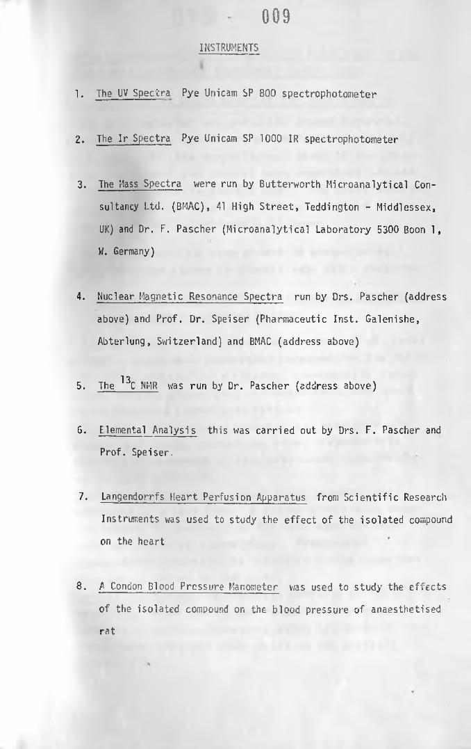
INSTRUMENTS
1. The UV Spectra Pye Unicam SP 800 spectrophotometer
2. The Ir Spectra Pye Unicam SP 1000 IR spectrophotometer
3. The Mass Spectra were run by Butterworth Microanalytical Con
sultancy Ltd. (BMAC), 41 High Street, Teddington - Middlessex,
UK) and Dr. F. Pascher (Microanalytical Laboratory 5300 Boon 1,
W. Germany)
4. Nuclear Magnetic Resonance Spectra run by Drs. Pascher (address
above) and Prof. Dr. Speiser (Pharmaceutic Inst. Galenishe,
Abterlung, Switzerland) and BMAC (address above)
135. The C NMR was run by Dr. Pascher (address above)
6. Elemental Analysis this was carried out by Drs. F. Pascher and
Prof. Speiser.
7. Langendorrfs Heart Perfusion Apparatus from Scientific Research
Instruments was used to study the effect of the isolated compound
on the heart
8. A Condon Blood Pressure Manometer was used to study the effects
of the isolated compound on the blood pressure of anaesthetised
rat

010CHEMICAL INVESTIGATION OF THE ACTIVE PRINCIPLES IN THE
LEAVES OF ELAEODENDRON BUCHANAN 11 (LOES) LOES.
A . Col 1 ecti on of plant material for investigation
The plant material was collected around Dagoretti,
Riruta Satellite, and Uthiru on the outskirts of City
of Nairobi. All the morphological parts of the plant
(i.e. roots, bark and leaves) were separately cut and
collected in separate plastic bags. The plant material
collected were cut into small pieces and dried in the
oven at 60°C for approximately 48 hours.
The dried materials were ground to powder using a
Willey Mill and stored in plastic bags until required.
B. Preliminary investigation of the active principles
of Elaeodendron buchananii L o e s .
The different morphological parts (about 3 gm. each)
of the ' plant were separately screened for the follow
ing active principles, alkaloids, cyanogenetic glyco
sides, cardiac glycosides, anthroquoinones and their
derivatives and saponins as follows-:
Alkaloids - The powdered material was extracted with
ethanol and to the extract was added dragendorff1s
reagent, the presence of alkaloids ^eing indicated by
an orange precipitate.Cyanogenetic glycosides- Fresh plant materia! was
moistened in a test tube. A sodium picric acid paper
was trapped in the mouth of the test tube and test
tube was placed in a warm place. Presence of
cyanooenetic glycosides is indicated by the formation
of a purple colour on the paper.
Cardiac cilvcosides- To alcoholic extracts of the plant— ^ —
materials, Kedde and Raymond reagents were added, the
presence p* cardiac glycosides being indicated by the
formation of pink and blue-violet in the extracts
respecti vely.

011Anthroouinones and their derivatives- The plant material
was hydrolysed with hydrochloric acid and extracted
with toluene. To the toluene extract was added ammonium
hydroxide, the presence of anthroquinone- and their
derivatives beir.- indicated by the formation of a pink
colour.
Saponins- The plant material was mixed with water in
a test tube and shaken vigorously to find out if any
persistent frothing occured.
The results obtained are presented in Table I.
TABLE I
Results of tests for chemical constituents likely to be
present in different morphological parts of Elaeodendron
buchananii Loes.
Active principle Morphological part Result
a. Alkaloids Bark, leaves and no alkaloidsroot
b. Cyanogenetic Bark, leaves and no cyanoge-glycosi des root netic glyco-
sides
c. Cardiac glycos i des
Bark no cardiac glycosides
Leaves cardi ac glycos i des present
Root no cardiac glycosi des
d. Anthroqui- Bark, leaves and no anthro-nones and root q u i n c n e s andtheir der- thei r deri-i.vati ves vati ves
e. Saponins Bark, leaves and no saponinsroot

The results of preliminary investigation of active principle in
Elaeodendron buchananii for different morphological parts of the
plant indicated that the plant contains substances with °=, g- unsa
turated y- lactone ring probably cardiac glycosides. The test for
cardiac glycosides were only positive for extractions of the leaves
showing that only the leaves contain cardiac glycosides in signifi
cant amount. The other active constituents tested for were not de
tected in any part of the plant.
•
Investigation of suitable extracting solvents for cardiac glycosides
from the leaves of Elaeodendron buchananii
The leaves were shown to contain cardiac glycosides (page Oil )
' in the preliminary investigation of active principles of Elaeodend
ron. The other parts of the plant were found to contain negligible
amount of these compounds. Therefore, investigation for suitable
solvents or mixtures of solvents for extraction of cardiac glycosides
from the leaves was carried out.
A comparative study of extraction efficiency of various solvent
systems v/as done by placing equal quantities of dried powdered leaves
(about 250g.) in 500 ml flasks containing different solvent systems.
The extraction v/as carried out using the following solvent systems:-
i) Ether
i i) Methanol
iii) Chloroform
iv) 9535 alcohol
v) 70% alcohol
vi) 50% alcohol/chloroform (1:1)
vii) Chlorofonn/absolute alcohol (1:1)
To each flask containing 250g of powdered leaves v/as added 200 ml of
0 1 2

0 1 3
each of the above solvent system and allowed to macerate on a water
- bath at 3C°C overnight except for ether, where the maceration was
carried out at room temperature.
In case of 50% alcohol/chloroform system, adequate water was
added to the macerate to effect the separation of the mixture into
aqueous and organic layers. The mixture was transferred to a sepa
rating funnel and the two layers v/ere then separated. Each of these
two layers was treated as an extract on its own right.
Each of the extract from the above solvent systems was filtered
and decolourised using charcoal and evaporated at 45 - 50°C to dry
ness under reduced pressure. The residue from each extract was dis
solved in 80 percent alcohol and treated with saturated lead sub
acetate solution to precipitate out tannins. The precipitated tannis
were filtered off and the tannin - free extracts treated with a 10%
solution of sodium sulphate to remove excess lead. The contents
were filtered and decolourised further by passing through a bed of
charcoal held in a filter paper. The extiacts were reduced to 15
ml at 45 - 50°C under reduced pressure.
In each case, the extract obtained was slightly yellow in colour,
but sufficiently clear to allow measurement of absorbance of the co
loured complex formed when reacted with Kedde reagent.
To 5 ml of e*ch extract v/as added 2 ml of Kedde reagent and the
intensity of the coloured complex formed v/as measured by a UV spect
rophotometer. The results obtained are presented in Table II.

0 1 4From these results it was found that powdered leaves macerated
with 50% alcohol/chloroform solvent system gave the highest absorb-C
ance reading for the chloroform portion of the extract. The aqueous
portion contained little or none of cardiac glycosides. The effici
ency of extraction was found to decrease in the order:- 50% alcohol/
chloroform > chloroform/absolute alcohol > chloroform > 95% alcohol >
70% of alcohol » methanol > ether. Therefore, it was concluded that
50% alcohol/chloroform system was the best solvent system for the
extraction of cardiac glycoside(s) using the above procedure. An
added advantage of the above solvent system over the others is that
most of the tannins were left in the aqueous layer, thus giving a
relatively tannin - free extract.
Extraction and purification of cardiac glycosides from the leaves of
Elaeodendron buchananii
Material and Methods
Approximately 2 kg of ground dry leaves were weighed and put into
a 12 litre flask. Adequate 50% alcohol was added until the plant
material was completely covered with the solvent. The material was
left to macerate overnight on a water - bath at 30°C. Enough chloro
form (3 litres) was then added to the material and the extraction was
allowed to continue for another 24 hours at 30°C with occasional sha
king. The material was then removed from the water bath and allowed
to stand for four days at room temperature with occasional shaking.
Adequate v/ater was added to the material to effect the separation of
chloroform and aqueous layers. The mixture was filtered by passing
through cotton wool held in a filter funnel after which it was trans
ferred to a separating funnel and left to separate. The chloroform
layer (at the bottom) was run off and the aqueous layer was returned

to the plant material and the extraction was repeated twice, each
time with 4 litres of chloroform. The chloroform extracts were
combined (about 'll litres) and distilled under reduced pressure at
45 - 50°C using a rotatory evaporator. The residue obtained (about
15g) was a dark green resinous material.
Purification of the extract
The dark green resinous material was defatted by washing se
veral times with petroleum spirit which removed most of the green
colour along with most of the fatty material. The residue obtained
was a dry crumbly material. This was dissolved in alcohol (95%)
and treated with lead subacetate strong solution to precipitate out
tannins (Canback, 1949). The alcoholic extract was then filtered
and the precipitate of tannins was washed with alcohol (3 x 100 cc)
so as to recover as much as possible any glycosides which might have
co-precipitated with tannins (Canback, 1949, Rowson, 1952). The alco
holic washings were added to the mother liquor. The filtrate (com
bined alcoholic extracts) v/as treated with a 10% solution of sodium
sulphate to remove excess lead (Sehandri and Subramanian 1952, Rowson
1952, Rangasv/anii and Subramanian, 1955). The contents were again
filtered and the lead precipitate was washed with alcohol (2 x 100
cc), each time adding the washings to the filtrate.
Ihe above process not only removed tannins and excess lead but
also most of the pigments. The final alcoholic extract (900 cc) had
a light green colouration.

The alcoholic extract was evaporated to dryness under reduced
pressure at 45 - 50°C and the residue was dissolved in 350 ml of
chloroform. This helped to remove any excess sodium sulphate pre
sent in theertract. The chloroform extract was further decolourised
by adding charcoal to the extract and shaking the mixture vigorously
and finally filtering the extract. The charcoal was washed with chlo
roform (300 cc) until the washings were negative for cardenolides
when tested with Raymond reagent. The washings were added to the
filtrate. The charcoal removed most of the pigments. However, the
solution obtained was yellow in colour and gave positive test for
cardenolides when tested with Kedde and Raymond reagents. The volume
of glycosidal solution obtained (about 650 cc) was found to be too
large to v/ork on. Consequently, it was concentrated to 50 ml under
reduced pressure at 45 - 50°C, transferred to a petri - dish and
evaporated to dryness by blowing hot air current over the surface
of the dish. The residue obtained had a syrupy consistency and was
brown in colour. The residue indicated the presence of cardiac
glycosides when tested with Keller - Killiani reagent.
The residue was then dissolved in minimum amount of chloroform
and run down a column of neutral active alumina (2 ft long). Elu
tion was first carried out using alcohol (95%) and ethyl acetate
(200 cc). Alcohol and Ethyl acetate eluates were collected into 100
ml portions. When chloroform v/as used, the eluate collected (first
100 ml and then 50 ml portions) contained the glycoside(s) and little
of the coloured material. The results of the column purification are
presented in Table III.
- S 1 6
The positive eluates were combined and evaporated to dryness
under reduced pressure at 45 - 50°C. The residue obtained was yellow

ft 17
in colour and was dissolved in the minimum amount of ether. Most
of the yellow coloured material did not dissolve in ether. The
ethereal solution was filtered and both the filtrate and the preci
pitate (yellow coloured material) were tested for cardiac glycosides
with Kedde reagent. The precipitate gave a negative test while the
ethereal solution was positive for cardenolides. The glycosidal
solution of ether was evaporated to dryness on a water - bath at 30°C
and a glycosidal residue which was slightly yellow coloured and
amorphous was obtained.
To effect further purification of the amorphous glycoside(s),
the glycosidal residue was dissolved in minimum amount of chloroform
and rerun down a column of neutral active alumina (1ft long). Chlo
roform was used as the eluant and the eluate was collected in 5 ml
portions, each fraction being tested for cardenolides with Kedde
reagent.
It was found that the first few portions were coloured but nega
tive for cardenolides. The rest of the eluates were clear and posi
tive for cardenolides. Elution from the column was carried out until
the eluate was negative with Kedde reagent.
The eluate (230 ml) was distilled to 20 ml under reduced pres
sure at 45 - 50°C. The concentrated extract was transferred to a
petri - dish and the chloroform was evaporated on the water - bath
at 45°C. A slightly yellow coloured crystalline residue v/as obtained.
The residue v/as positive to the glycosidal test with Keller -
Killiani reagent and with Kedde and Raymond for cardenolides. The
glycosidal residue was redissolvcd in minimum amount of ether and the

0 1 8
solution was filtered through a bed of charcoal held in a filter
paper. A clear glycosidal solution of ether was obtained.
C. Crystallisation of the isolated glycoside(s)
The glycosidal solution of ether was warmed on the water - bath
at 30°C to evaporate the ether. As the ether evaporated a white
amorphous precipitate was observed to form. The precipitate was al
lowed to form until no further precipitation took place. The pre
cipitate was filtered off and tested for cardiac glycosides with
Kedde, Raymond and Keller - Killiani reagents. The results were
positive after which attempts were made to obtain the amorphous gly
coside^) in crystalline form.
The amorphous glycoside(s) was sampled into different peteri -
dishes so that different solvents or solvent mixtures could be tried
to find a suitable solvent system for crystallisation. The follow
ing solvent systems were tried:-
i) chloroform
ii) chloroform - ethanol (9:1)
iii) chloroform - ethanol (3:2)
iv) chloroform - ethanol (2:1)
These solvent systems were chosen because they have been used by ot'ne
workers (Rangaswani and Subramanian 1955) with success to crystallise
cardiac glycosides from squill (Indiana maritima).
Attempts to crystallise the glycoside(s) was made by dissolving
the amorphous glycoside(s) in minimum amount of each of the above
solvent system and allowing the glycosidal solutions to cool and eva
porate at 30°C.

01 9
It was found that the best solvent, system for crystallisation
of the isolated glycoside(s) was chloroform - ethanol (9:1) system
The rate of appearance of crystals was faster than in the rest of
the solvent systems. The rate of appearance of crystals was observed
to occur in this order:- chloroform - ethanol (9:1) > chloroform -
ethanol (2:1) > chloroform - ethanol (3:2). The crystals produced by
the three solvent systems (ii), (iii), (iv) above were the same. The
chloroform system (i) above also produced crystals but they were
smaller in size than those formed from the other solvent systems. So
for further work chloroform - ethanol (9:1) system was used to crysta
llise the isolated amorphous glycoside(s).
The 2 kg of the dried leaves of Elaeodendron buchananii (Loes.)
Loes yielded about 300 mg of crystals.
The extraction and purification techniques described above are
summarised in form a flow chart on the next page.

0 20FLOW CHART FOR THE EXTRACTION AND PURIFICATION OF THE ACTIVE PRINCI—
PLE OF ELAEODENDRON BUCHANAN11
ii) treated with lead subacetate strongI
solution to remove tannins and pigments
iii) filtered to separate
v

021
treated with 10% w/v
solution of sodium
sulphate to remove
excess lead
i) evaporated to dryness
under pressure
ii) dissolved in chloroform
decolourised c animal
charcoal
reduced to 50 ml under
reduced pressure at
45 - 50°
evaporated to dryness
using hot air current
___ i ____Ethanolic extract
(tannin free)
_______V ________Ethanolic extract
lead free
------- ^ __________Chloroform extract
_______ ___________Chloroform extract
(yellow)
V

022
i) dissolved in the minimum amount
of chloroform
ii) run down an alumina (neutral)
column
iii) eluated c chloroform
clear eluate of chloroform
■i

evaporated to dryness
dissolved in the
minimum amount of ether
ether evaporated off
i) dissolved in the minimum
amount of chloroform/ethanol
9:1
ii) cooled and allowed to
evaporate at 30°C

024
CHARACTERISATION OF THE ISOLATED GLYCOSIDES
A. Investigation of the general physical properties
i) Solubility - the solubility of the isolated glycoside(s) was
investigated by attempting to dissolve the compound in various sol
vents at room temperature (21°C). About 10 mg of the compound was
placed in a micro - test tube and to it was added the respective
solvent using a dropper pipette. The degree of solubility of the
isolated glycoside(s) was determined by the number of drops of the
solvent required to dissolve the compound. The following solvents
were tried
i) Ether
ii) Methanol
iii) Acetone
iv) Ethyl acetate
v) Absolute alcohol
vi) Chloroform
The results of solubility are presented in Table IV.
ii) Melting point - the melting point of the isolated glycoside(s)
was determined using capillary melting point apparatus and the hot
stage microscope. It was observed that the compound melted at 110
- Ill°C. The narrow melting range indicated that the compound was
pure and probably only one glycoside.
B. Detection and Identification
i) Colour react'!ens - Concentrated sulphuric acid - two drops
of concentrated sulphuric acid were added to a few crystals of the
isolated compound. The crystals dissolved and the solution went

brown in colour. When the same test v/as repeated with digoxin and
ouabain, similar results were obtained.
ii) Reaction with m - dinitrobenzene (Raymond reagent) - A few
crystals were dissolved in 10 ml of alcohol (80%). To 1 ml of this
solution was added 1 ml of Raymond reagent followed by a few drops
of a 20% w/v solution of sodium hydroxide. A blue - violet colour
developed and this was observed to fade fairly rapidly to a pale
greenish colour. Similar results v/ere observed with digoxin and
ouabain. The above test have been used to test for cardenclides
and a positive test with Raymond reagent indicated that the isola
ted compound is a cardenolide.
iii) Reaction with 3:5 dinitrobenzoic acid (3:3 DNBA) Kedde reagent
The Kedde reagent is also used to test for cardenolides. The iso
lated compound, having been positive with. Raymond reagent, it was
tested with Kedde reagent. 1 ml of the ethanolic solution contain
ing the isolated compound (approx. 0.1% w/v) was reacted with Kedde
reagent. A purplish colour developed, and unlike with Raymond rea
gent, the colour was observed to fade very slowly.
iv) Reaction with sodium nitroprusside (Legal's reagent) - A few
crystals of the isolated compound were dissolved in pyridine. To
this was added a drop of a 2% w/v solution of sodium nitroprusside,
followed by a drop of a 2% w/v solution of sodium hydroxide solution.
A deep red colour was produced indicating that the isolated compound
is a cardiac glycoside.
v) Reaction c-f Keller - Killiani reagent - A few crystals of the
isolated compound were dissolved in 1 ml of glacial acetic acid.

G2fi.Two drops of a 5% w/v solution of ferric chloride were added and
the contents were carefully transferred onto the surface of concen
trated sulphuric acid (1 ml) in a test tube. A brown ring developed
at the junction of the two liquids and the upper layer slowly acqu
ired a bluish green colour after about 10 minutes. This constitutes
a positive test for cardiac glycosides containing 2 - desoxy sugars
(Fieser, 1959).
C. Chromatographic analysis of the isolated glycoside(s)
Chromatographic analysis of the purified glycoside(s) was car
ried out to determine the possible number of cardiac glycosides from
the material isolated from the leaves of Elaeodendron buchananii
(Lees.).
Various solvent systems were tried in an attempt to effect the
resolution of the isolated glycoside(s). In all cases some reference
glycosides (digoxin and ouabain) were run against the isolated gly
coside^) of Elaeodendron.
THIN - LAYER CHROMATOGRAPHIC ANALYSIS OF THE ISOLATED GLYCOSIDE(S)
Experimental procedure - TLC plates (20 x 20 cm) were prepared
according to the method described by Stahl, (1965) using Silica gel
G. The plates were air - dried by leaving them exposed to air at
room temperature overnight. Several chromatographic tanks were pre
pared using different solvent systems and the tanks were saturated
with each solvent system. To the silica gel plates about 5 yl of
the test and reference glycosidal solutions were spotted on the
plates which were aeveloped in the tanks. The solvent system was
left to travel to approximately 13 cm after which the plates were

- n ?removed and dried in a current of hot air. The plates were sprayed
with Kedde reagent and visualised in daylight. The results obtained
are presented in Table V.
The TLC results shew that both the isolated glycosidal fraction
and a crude extract of the leaves of the plant produced one spot with
the same Rf value and same colour for all the solvent systems tried.
The Rf value of the spot produced by the isolated glycoside and cru
de extract of the plant was different from that of ouabain and digo-
xin. Literature survey also showed that no known cardiac glycoside
has similar Rf values in the solvent systems used. The colour, on
spraying with Kedde reagent was the same for the isolated glycoside,
crude extract of the leaves, ouabain and digoxin and for unknown
glycoside from Ajuga remota Benth.
D. Spectroscopy of the isolated compound
i) Examination of UV absorption soectrum of the isolated compound
Method: Approximately 100 mg of the isolated crystals were accura
tely weighed and dissolved in 100 ml of absolute alcohol in a volu
metric flask. This solution was used as a stock solution for the
study of UV absorption spectra of the isolated compound.
2 ml of the stock solution were transferred to a 10 ml volume
tric flask and made up to volume with absolute alcohol. This solu
tion was used for UV spectrophometrie analysis of the 'solated com
pound.
The isolated compound was found to have an absorption maximum
at ?C3 nm. The spectrum is presented in Figure 2.

028i i ) Examination of the absorption spectrum of the
by the isolated compound and 3:5 dinitrobenzoic
acid (Kedde reagent)
One millilitre of a lOOmg % solution (st.i .v solution)
was added to 1 ml of a 2%w/v solution of dinitrobenzoic
acid (3:5 DNBA) and 1 ml of 1 N sodium hydroxide solut
ion and the volume was made up to 5 ml with ethanol in
a volumetric flask. The mixture was allowed to stand
at room temperature for four minutes. A blank was
prepared in the same way as the reaction mixture
except that 1 ml of alcohol was substituted for the
test solution.
The maximum intensity of the purple colour which
developed in the reaction mixture was not attained
immediately. The purple colour has a steady value
approximately 8 minutes after the addition of 1 N
sodium hydrcxide solution (C.K. Maitai, Msc thesis,
Otago University, New Zealand). This time interval
was employed in the intial spectroscopic studies of
the isolated compound.
After the full purple colour had developed in the
reaction mixture, the absorption spectrum (190-700 nm)
was determined. The absorption spectrum showed two
maxima, a sharp peak at 355 nm and a broad peak at
540 nm (Figure 3).
The change in colour intensity of the reaction mix
ture was investigated over a period by running spectra
of the reaction mixture after 15, 20, 30 and 45
minutes. No change could be detected at the absorpt
ion maximum 3t 355 nm wavelength but because of the
broad nature of the peak at 540 nm it was impossible
to discern the change in the absorption maximum. The
optical density at both wavelengths decreased. This
indicated that the two wavelengths, can be used to

029plot a graph of absobance against concentration to find
out if Beer-l.ambert law is obeyed under specified
experimental conditions.
i i i ) Examination of the absorption spectri m of the
coloured complex formed by the isolated compound and
m-dinitrobenzene (Raymond reagent)
The coloured complex of the reaction mixture of the
isolated compound and 1, 3 DNB was developed by taking
1 ml of lOOmg % solution of the isolated compound, 1 ml
of 1, 3 DNB and 1 ml of 1 N sodium hydroxide. The
solution was made up to 5ml in a volumetric flask. A
blank was prepared in the same way except that 1 ml of
absolute alcohol was substituted for the test solution.
The violet colour which appeared on adding sodium
hydroxide was found to change to blue and eventually to
light brown. This change in colour has been found not
only to affect the height of the peak but also the
position of the absorption maximum (C.K. Maitai, Msc.
thesis, Otago University, New Zealand). Therefore, it
was decided to examine the absorption spectrum in the
blue colour of the reaction mixture because the colour
is intense and the peak height is at its maximum
(Hassel e_t 1 953 thro' C.K, Maitai, MSc. thesis,
Otago University, New Zealand). The spectrum is
presented in Figure 4.
The spectrum was found to have a broad peak with
maximum at 590 nm. The spectrum is disorted due to
colour change during the time of run.

820
iv) Examination of the infra-red spectrum of the isolated compound
A potassium bromide disc of the iso'ated compound was prepared using
the hydraulic press. A whole spectrum of the compound was run and
the main peaks noted.
The spectrum obtained is presented in Figure 5 and the main
peaks arepresented in Table VI where those of digitoxigenin have been
included for comparison purposes only.
v) Elemental analysis of the isolated compound - An elemental
analysis of the isolated compound was done by two independent labo
ratories
1) Dr. F. Pascher, Microanalytical Laboratory, Boon, West Germany
2) Prof. Speiser, Pharmaceutical Institute, Abteilung, Switzerland
The results are presented in Table VII.
TABLE VII
RESULTS OF ELEMENTAL ANALYSIS '
Element Dr. Pascher's laboratory Prof. Speiser's laboratory
Carbon 63.67 63.9
Hydrogen 7.87 5.32
Oxygen
128.01 28.3

031From these elemental analysis the empirical formula was determined.
The molecular weight of the compound was determined as 603 and from
the empirical formula and the molecular weight, the molecular for
mula was calculated. The results are presented in Table below.
TABLE VIII
EMPIRICAL FORMULA AND MOLECULAR FORMULA OF THE ISOLATED GLYCOSIDE AS
CALCULATED FROM THE ELEMENTAL ANALYSIS AND MOLECULAR WEIGHT
Laboratory Empiricalformula
Molecularweight
Molecularformula
Dr. Pascher's C3H4.5°1 603"4 C32H47°11
Prof. Speiser's C3H4.43°1 C32H47°11
*Molecular weight v/as determined by Dr. Pascher.
13vi) Examination of the C NMR spectrum of the isolated compound
13The C NMR of the isolated compound is presented in Figure 7 and the
data is presented in Table IX. CDC1^ was used as the solvent and
TMS as the internal standard.
Only a partial interpretation of the spectrum has been carried
out by attempting to estimate the number of carbon atoms ti the iso
lated compound. This has been dono using the intensity of the peaks
and with reference to the internal standard (TMS).

- 032 .
vii) Examination of the proton NMR spectrum of the isolated compound
The proton NMR spectrum of the isolated compound is presented in Fig.
8. The spectrum was not properly integrated and so it was found dif
ficult to determine the number of protons in the compound.
viii) Examination of the mass spectrum of the isolated compound
The mass spectrum of the isolated compound is presented in Fig. 9 and
the m/e values of its fragment ions are presented in Table X. For
comparison purposes, the m/e values of two steroids, somalin and di-
gitoxigenin are included in the same table. The m/e values of soma
lin and digitoxigenin was obtained from Ardene (1964).
SPECTROPHOTOMETRIC DETERMINATION OF THE PERCENTAGE RECOVERY OF THE
ISOLATED GLYCOSIDES
To determine the percentage recovery of the isolated glycoside,
the relationship between absorbance of the coloured complex formed
by reacting the isolated glycoside with Kedde reagent and the concen
tration of the drug was investigated first.
A 0.1% w/v solution of the isolated glycoside was made by dis
solving 100 mg of the glycoside in 95% alcohol and making the solu
tion to 100 mi in a volumetric flask. From this solution, a series
of dilutions were made to give the following standard concentrations:-
0.06, 0.043, 0.04, 0.032, 0.020, 0.012 and C.004% w/v. To 2 ml of
each of these concentrations was added 1 ml of 2% solution of dini-
trobenzoic acid (3:5 DNBA) and 1 mi of sodium hydroxide. The volume
was made up to 5 ml in a volumetric flask and the mixture was allowed
to stand at room temperature for four minutes.

The abosrbance of the coloured complex from each standard con
centration was determined and a calibration curve of absorbance
against concentration was plotted. The results obtained are pre
sented in Table Xi and Fig. 10. A straight line passing through the
origin was obtained indicating that Beer - Lambert law was obeyed
under the prevailing conditions.
DETERMINATION OF THE PERCENTAGE RECOVERY
Two samples of 50 g of dry powdered leaves were accurately wei
ghed into 1 litre flasks and extracted with a mixture of 50% alcohol
and chloroform as described previously. After the decolourising sta
ge, the chloroform extracts were adjusted to 250 ml. From each of
the 250 ml extract, 10 ml were evaporated to dryness. Each of the
residue was dissolved in 3 ml of alcohol (95%) and 1 ml of a 2% so
lution of 3:5 DNBA and 1 ml of 1 N. Sodium hydroxide solution were
added.
A blank was prepared in the same way except the extract was
amitted.
The absorbances of the coloured complexes formed from each
extract was read from the calibration curve (Fig. 10).
CALCULATION OF THE PERCENTAGE RECOVERY
From the absorbances of the two extracts (referred to as T̂
and T^) their concentrations from the calibration curve were found
to be 25 x 1C'3% and 29.5 x 10'3% respectively.
_3i) 10ml of the extract were found to contain 26 x 10 g.

03A250 ml of the extract contain:-
26 x 10~3 x 250
10o 0.65 g.
The percentage recovery is
0.65 x 100
250
= 0.26%
ii) Using the second extract, the percentage recovery was found to
be 0.295%.
A PRELIMINARY INVESTIGATION OF THE PHARMACOLOGICAL EFFECTS OF THE
ISOLATED COMPOUND ON IKTACT AND ISOLATED MAM/iALIAN PREPARATIONS
The chemical screening of the plant for active principles
showed the presence of cardiac glycoside(s) in the leaves of the
plant. Subsequent extraction, isolation and purification gave a
chemical compound which on investigation proved to be a cardiac
glycoside.
Cardiac glycosides have a direct stimulating effect on the myo
cardium with consequent increase in the force of contraction and
hence an increase in efficiency of a failing heart. This effect of
cardiac glycosides has secondary effects such as diuresis due to
improved circulation. Cardiac glycosides may also have a direct
effect on renal tubules (Sim, 1967). Digoxin, for example is used
to treat oedema in addition to its use to treat heart diseases
(Bowman et a^, 1970).

Since the isolated compound was shown to be a cardiac glycoside
pharmacological investigations were carried out to find out whether
the isolated compound has any effects on the blood pressure of an
intact rat and its effects on an isolated perfused rabbit heart.
The above two pharmacological effects would help to confirm the che
mical and spectroscopical findings which indicated that the isolated
compound is a cardiac glycoside.
INVESTIGATION OF THE EFFECTS OF THE ISOLATED COMPOUND ON THE BLOOD
PRESSURE OF AN ANAESTHETISED RAT
Material and methods
Adult rats (200 - 300g) were anaesthetised with a 25% w/v aqu-
ecus solution of urethane by injecting 0.3 ml/I kg of the anaesthe
tic intraperitoneally. The trachea, common carotid artery and exter
nal jugular vein were exposed and cannulated according to the method
described by Mcleod L. J. et a^ 1970 in his book "Pharmacological
experiments on intact preprations". The arterial cannula was con
nected to a condon blood pressure manometer and the drug injections
were given through the venous cannula. The condon blood pressure
manometer was in turn connected to a kymograph (see Fig. 11).
Preparation of the drug mixture
TOO mg of the isolated compound were dissolved in 10 ml of pro
pylene glycol by warming on a wafer - bath until ail the compound
dissolved. The solution was cooled arid made tc 100 ml with normal
saline. A control was also prepared containing a 10% w/v propylene
glycol in normal saline.

036Injections of adrenalin 100 mg/ml and digoxin 1 mg/ml were also
prepared for comparison.
Test with the dru"j
0.1 ml of the control was injected at point A and the effect
of the control is represented by section AB (Fig. 12). The blood
pressure was allowed to settle and adrenalin (10 yg) was injected
at point C. After the effect of adrenalin had subsided and the
blood pressure was back to normal the test material (0.4 mg) was
injected at point E after mean blood pressure had returned to normal.
RESULTS
The results (Fig. 12 and Table XII) show that the control caused
a transient fall in blood pressure and that adrenalin had an imme
diate increase in blood pressure with a quick recovery. The drug
mixture first showed a fall in blood pressure due to propylene gly
col then a gradual rise above the mean blood pressure. The effect
was sustained. Digoxin was also found to have a sustained pressor
effect. The response of the isolated compound compared very wellv
with that of digoxin (Fig. 12).
INVESTIGATION OF THE EFFECTS OF THE ISOLATED COMPOUND ON THE ISOLATED
PERFUSED RABBIT HEART
The isolated perfused rabbit heart preparation was carried out
as described by Mcleod et aj_, 1970. The heart response i.e. both
chronotropic and inotropic effects, were recorded in a kymograph and
also by a Harvard smooth muscle/heart recorder. Solutions of the
isolated glycoside, adrenalin, control and digoxin were used on the
isolated heart each at a time.

0 3 ?RESULTS
The results (Fig. 13 and 14) show that propylene glycol did
not have any significant effects on the isolated perfused heart.
Adrenalin had an immediate increase in heart rate arid force of con
traction while the isolated compound increased the force of contract
ion while lowering the heart rate (Fig. 14). Digoxin had an effect
similar to that of the isolated compound (Fig. 13). They both had a
gradual increase in force of contraction, by far the most important
property of cardiac glycosides on the heart.

038
CHAPTER 3
DISCUSSION
A literature survey of Elaeodendron buchananii (Loes.) Loes.
showed that there is no record of the plant having been used as a
medicinal herb by the Africans. The plant, however, is known to
have caused deaths to stock. Most of the poisonous plants of East
Africa are used in small doses either in the treatment of various
ailments in form of decoctions prepared by herbalists or for trial
by ordeal or as arrow poisons by poachers.
Pharmacognostical investigation on Elaeodendron showed that the
plant grows in different habitats and the height of the tree varies
depending on soil and climatic conditions. The plant can always be
distinguished by the characteristic features of the leaves and flo
wers. No other plant growing in the same habitats was found to have
similar features. Even those growing among grass and aloes (Plate
I) can easily be distinguished. Animals hardly have any access to
the leaves and consequently few deaths have been reported. Where
a tree has coppiced from a cut stump (Plate II) young shoots appear
around the tree trunk and these shoots have been responsible for
numerous deaths that have been reported. Where the tree is growing
among other foliage in fora of a shrubby growth (Plate III) it is
difficult even for man co distinguish it from other trees. Such
shrubby growths of Elaeodendron have beer, responsible for deaths to
stock, especially goats which feed very much on twigs. Human fata
lities have not been reported although the tree is used as a fire
wood in the homes in rural areas. This would probably be due to the

fact that is the dry trunk which is used and not the leaves.
Giraffes have been known to brownse on the tree with punitive im
munity.
Preliminary work using standard chemical tests on ethanolic
extracts of the leaves of Elaeoaendron buchananii indicated the pre
sence of compounds <=, 8- unsaturated 3- lactone ring which were shown
to be cardiac glycosides by Keller - KiVliani reagent. Investigation
of other active principles, for example, alkaloids, cyanogenetic gly
cosides et centra in different morphological parts of the plant re
vealed that only the leaves contain cardiac glycosides in signifi
cant amount. Test on different morphological parts of the plant for
the active principles gave negative result.
Investigation for the best extracting solvent for the cardiac
glycoside(s) present in the leaves of the plant showed that the suit
able solvent and conditions under which the extraction of the glyco
side^) could be carried out was as follows:- first, digesting the
powdered leaves with 50% alcohol overnight at 50°C and then adding
chloroform to the macerate and adding enough v/ater to effect the
separation of the aqueous and chloroform layers. The chloroform was
found to extract most of the cardiac glycoside(s) and after exhau
sting the aqueous layer of the glycoside(s) by washing several times
with small portions of chloroform the 3queous layer was discarded.
The solvent systems chosen for this investigation were those which
have been used by other workers (Rangaswani and Subramanian, 1355,
Canback 1949, Raymond 1S32, Rcwson 1952) with success to extract
different cardiac glycosides from different plant sources. Also
these solvent systems have been given as general solvent systems
for extraction of medicinal plant glycosides (S. K. Sim, 1967).

040
Chloroform and alcohol (absolute alcohol), 95%, 70%) v/ere found to
extract the glycoside(s) from the plant and the differences in the
amount extracted could be due to differences in the degree of selec
tivity and solubi-' .ty of each solvent system. Alcohol being non -
selective extracted a lot of other unwanted material (e.g. pigments,
tannins etc) than chloroform. The differences in the amount of gly
coside extracted by different alcohol percentages could also be due
to differences in selectivity and differences in the degree of solu
bility of the cardiac glycoside(s). From the results, it seems that
the cardiac glycoside(s) of Elaeodendron buchananii are more soluble
in chloroform than in absolute alcohol. The solubility in alcohol
decreases with decrease in the percentage of alcohol. This fact was
confirmed when the solubility of the isolated glycoside was investi
gated. The advantage of the above extraction procedure over the other
solvent systems is that almost all the tannins were extracted in the
aqueous layer and this on its own v/as a stage of purification in the
isolation process. Maceration by 50% alcohol seemed to help the extrac
tion of the glycoside(s) by chloroform probably by removing a lot of
tannins, pigments and resins which would otherwise have interferrcd.
Von Eew et al (1951), Stoll and Kreis (1951), Rangaswani and
Subramanian (1955), Fieser and Fieser (1959), and many other workers
have indicated the use of petroleum spirit to remove resinous fatty
materials from plant extracts. In this case too the unwanted mate
rial was removed by washing with petroleum spirit the syrupy resi
due obtained on evaporating off the chloroform.
The petroleum spirit only removed part of the unwanted mate
rial. Canback (1949), Sehandri and Subramanian (195C). P.owson (1552),
Maitai (1969) and many other workers have shown that plant tannins

041(and pigments) could be removed successfully from alcoholic extracts
of the plant by use of lead subacetate solution. Canback (1949) and
Rowson (1952) also have shown that during the precipitation of tan
nins co-precipitation of active principles occur-. Therefore, it
was necessary to dissolve the glycosidal residue in alcohol (95%)
and on precipitation of tannins, to v.'ash the precipitate with etha
nol several times arid to add the ethanolic washings to the mother
liquor to recover as much of the co-precipitated glycosides as pos
sible. The tannin - free extract contained excess lead which needed
to be removed before further purification was attempted. A 10% solu
tion of sodium sulphate was used to remove excess lead (Rangaswani
and Subramanian, 1955). The precipitation of tannins enhanced the
decolourisation of the extract but still the extract was green in
colour and had excess sodium sulphate. By evaporating the alcoholic
extract to dryness and redissolving the residue in chloroform, excess
sodium sulphate was got rid off. Animal charcoal was used to deco
lourise the extract and by first saturating the charcoal with chlo
roform the loss of the glycoside(s) was minimised. Also saturation
of the charcoal hastened the filtration.
In the present work, charcoal failed to remove the coloured
material completely. This probably may be due to the fact that the
yellow colouring matter of the extract was not adsorbed to the char
coal or if any adsorption occured, the desorption in the chloroform
was favoured. The alumina column was found to be successful in
removing most of the coloured matter. Choice of the eluent was an
important factor. When alcohol and ethyl acetate were used as elu
ants, both the glycoside(s) and a lot cf the colouring matter were
eluated simultaneously from the column but when ncn-polar solvents,
for example, chloroform was used most of the colouring matter was

042
left adsorbed to the column. The fact that the first column did not
decolourise the extract completely could be attributed to the short
length of the column used.
Final purification of the isolated glycoside(s) was done by
crystallising the glycoside from chlorofonn/ethanol (9:1), a system
which was found to give pure crystals at a reasonably good time as
compared to the other solvent systems tried.
CHARACTERISATION AND IDENTIFICATION OF THE ISOLATED GLYCOSIDE
The solubility and the melting point of the isolated glycoside
v/ere determined as described under physical properties (page 0i<*-).
The compound v/as found to be insoluble in water but soluble in orga
nic solvents. However, the degree of solubility in different orga
nic solvents v/as found to vary, being extremely soluble in chloro
form, soluble in alcohol and fairly soluble in the others. The iso
lated glycoside melted at 110 - 111°C v/hich indicated that the com
pound was pure. Literature survey showed that no knov/n cardiac gly
coside m-lts within this range, an indication that the isolated gly
coside could be a new cardiac glycoside.
The isolated glycoside was reacted with various colour reagents
to determine the best chemical tests for the purpose of detection.
The colour reactions (given by the isolated glycoside when reacted
with various colour reagents) while not being specific for cardiac
glycosides and their aglycones, are nevertheless characteristic of
the compounds having a c3 rdenclide type of aglycones. Such reagents
included concentrated sulphuric, Raymond, Kedde and Keller - Killiani
reagents. When the isolated glycoside gave these characteristic colour

043
reactions, they constituted a positive test for cardiac glycosides
indicating that the isolated compound is a cardiac glycoside.
Cardiac glycos'des undergo colour reactions with the above rea
gents based on the different parts of the molecule.
Steroid nucleus
Sugar moiety [ r- o 0!(
<s £5- unsaturated
lactone ring
The B- unsaturated lactone ring gives reactions with polynitrophenyl
derivatives (e.g. Kedde and Raymond reagents), the steroid nucleus
reacts with concentrated acids and oxidising agents, while the sugar
moiety reacts with ferric salts. When the isolated compound was rea
cted with concentrated sulphuric acid, a reddish brown colour was pro
duced. This colour with concentrated sulphuric acid is known to be
produced by cardiac glycosides like ouabain, digoxin, strophanthidin
etc. and can be attributed to the steroidal nucleus. Not all cardiac
glycosides give this colour and impurities too could give a positive
test. However, since other chemical tests with the isolated compound
were positive for cardiac glycosides, this test could be considered
positive for a cardiac glycoside for the isolated compound.

Testing v.’ith polynitrophenyl derivatives, the isolated compound
gave positive colour reactions as would be expected with other car
diac glycosides. The observed colour complexes with Kedde and Ray-
mod reagents for the isolated compound are given by cardenolides.
Buch and Taylor (1952), Zoller and Tamm (1953), Shoppe (1964),
Mathound (1935) pointed out that m - dinitrobenzene (Raymond rea
gent) and cardiac glycosides produce the characteristic blue-violet
colour. Kedde (1947) investigated the use of 3:5 dinitrobenzoic acid
for the determination of digoxin and other cardiac glycosides. Lange-
jam (1947), Rowson (1952 and 1955) and Tattje (1957) all have used
Kedde reagent in the determination of the glycosides and aglycones
of digitalis.
- 04 4 .
The reactions of cardiac glycosides with the polynitrophenyl
derivatives is thought to be due to the presence of an active methyl -
lene (-CH^) groups C - 21. These colour reactions are given by other
Methylene __ _
(-CH2~) group
compounds e.g. acetone and the 17 keto-stroids will give a positivei
Raymond test (Stahl, 1959). So these tests are not on their own
conclusive for the butenolide group. The Keller - Killiani test
for 2 - desoxy sugars was positive indicating that the isolated com
pound was a. glycoside and not an aglycone. The identity of the sugar
is not known end whether the glycoside has one or more sugar units
has as yet to be determined.

0 4 5
The results of the TLC analysis showed that the isolated com
pound was one glycoside. The solvent systems used produced one spot
for a crude extract of the leaves whose Rf value was the same as that
of the isolated giycoside. This showed that no other glycoside was
lost during the extraction and purification process. The solvent
system, chloroform - methanol - formamide (80:19:1) has been used
with success (Planta Medica, 1973) to resolve the glycosides and ag1 -
ycones of squill (Indiana maritima) which has over 14 glycosides and
has been indicated to be a good solvent system for TLC analysis of
other plant glycosides. These results indicate that there is one
glycoside in the plant. The Rf values of the isolated compound for
the solvent systems and adsorbent used do not compare with any known
cardiac glycoside.
In general very rarely do cardiac glycosides occur singly in
plants but the TLC analysis show that only one glycoside is present
in Elaeodendron. Use of more than one solvent system would normally
help to overcome the incomplete resolution of several compounds into
individual glycosides, and since they all produced one spot for the
isolated compound, the only conclusion which can be arrived at from
the TLC analysis is that only one glycoside is present in the leaves
of Elaeodendron buchanar.ii (Loes.) Loes.
Since the TLC analysis showed the presence of only one glyco
side in the plant, further analytical techniques were employed to
confirm this.
The ultraviolet absorption of the isolated glycoside in alcohol
showed an absorption maximum at 208 nm. This absorption maximum was
not in the normal range 211 - 220 nm (Canback 1949) which is characte-

ristic for compounds with = , $- unsaturated carbonyl group. The
absorption spectrum (Fig. 2) gave a symmetrical peak and the absence
of other absorption peaks in both the UV and visible regions shov/ed
absence of impurities.
The absorption spectrum of the isolated compound for the colour
reaction with Kedde reagent was observed to have two absorption maxi
ma , one at 540 nm and the other at 360 nm. The 540 nm peak was bro
ad and less intense and symmetrical whereas the 360 nm peak was sharpi
and unsymmetrical. The 540 nm peak would be recommended for assay
of the isolated compound. With Raymond reagent the isolated compound
was observed to have a broad absorption maximum at 590 nm. Tho spec
trum is slightly distorted and this could be due to the fact that the
blue colour produced with Raymond reagent was fading rapidly while
the spectrum was being recorded. Repeated determination of the absor
ption spectrum while the colour was rapidly fading revealed that the
wavelength of maximum absorption at 590 nm remained unaffected.
Apart from the colour reactions produ'eri by the polynitrcphenyl
derivatives, the physical and chemical properties of the isolated com
pound indicate that it is a cardiac glycoside. Examination of the
infra-red spectrum (Fig. 5) show features characteristic to cardiac
glycosides. The spectrum shows good resolved peaks indicating that
the compound was pure.
A broad peak at 3450 cnf^ indicates the presence of associated
hydroxyl (OH) groups. A strong absorption at 2950 cm~^ suggests
C-H stretch of a liphatic features. Strong absorption at 1750 cm ̂
indicates a stretch vibration of an 3- unsaturated ketone (C=0).
A weak absorption at 1630 cnf ̂ suggests a stretch vibration of an

olefinic (C=C). A peak at 1760 cm 1 and 1790 cm ̂ indicates the
presence of a five membered ring compound having a carbonyl i.e. a
lactone. A strong absorption band at 1260 cm ̂ indicates the pre
sence of a C-0 link, the carbon atom of which is jnsaturated.
The infra-red spectrum of the isolated compound compares very
well with that of digitoxigenin in the finger-print region (CF Fig.
5 C Fig. 6 ). The principal peaks of both the isolated compound and
digitoxigenin have been compared and are shown in Table V.
The above main peaks noted are those which might be expected
from an i.r. spectrum of a typical cardiac glycoside because cardiac
glycosides have all these features which contribute to the above
peaks from their general structure.
0 4 7
The finger-print region (1400 - 600 cm of the isolated compound
is complicated and no attempt was made to assign the absorption
peaks to functional groups. The finger-print region is characteri
stic of each molecular species and is commonly used for identifying
unknown compounds by matching the i.r. spectra of the unknown com
pounds with those of known compounds. An attempt was made to com

04S
pare the infra-red spectrum of the isolated compound with those of
digoxin, somalin, echujin and ouabain but the spectrum of the iso
lated glycoside did not resemble any of them.
The elemental analysis from two laboratories (Micro Analytical
Laboratory, Boon, West Germany and Pharm. Inst. Ethz. Switzerland)
were in close agreement. From the molecular weight (mwt - 603) the
molecular formula of the compound was worked out to be C3 2 ,!4 7 0n •
This molecular formula indicates that the isolated compound is a gly
coside and not an aglycone. The aglycones of cardiac glycosides
have the molecular formula C23H34°(4-8)• is difficult to state
whether the isolated glycoside is found in the same form in the
plant because one or more sugars might have split off during the
isolation procedure.
The 13C NMR and the proton NMR were only partially interpreted
and detailed interpretation was considered to be beyond the scope
of the present work. From the intensities of the peaks and with re
ference to the internal standard (TMS), the total number of carbon
atoms present in the isolated compound could be estimated from the
^3C NMR spectrum (Fig. 7). The data for same is presented in Table.
VI. The peaks at 79.34b, 77.299 and 75.113 parts per million are
due to the solvent CDC1, and the intensity at 0.000 ppm is due to the
internal standard. The rest of the peak intensities are due to the
carbon atoms in the molecule. When these intensities due to carbon
atoms in the isolated glycoside are added up they come to 10387.
Assuming there is no spin - spin coupling, the following peaks would
be considered to be due to one carbon atom.
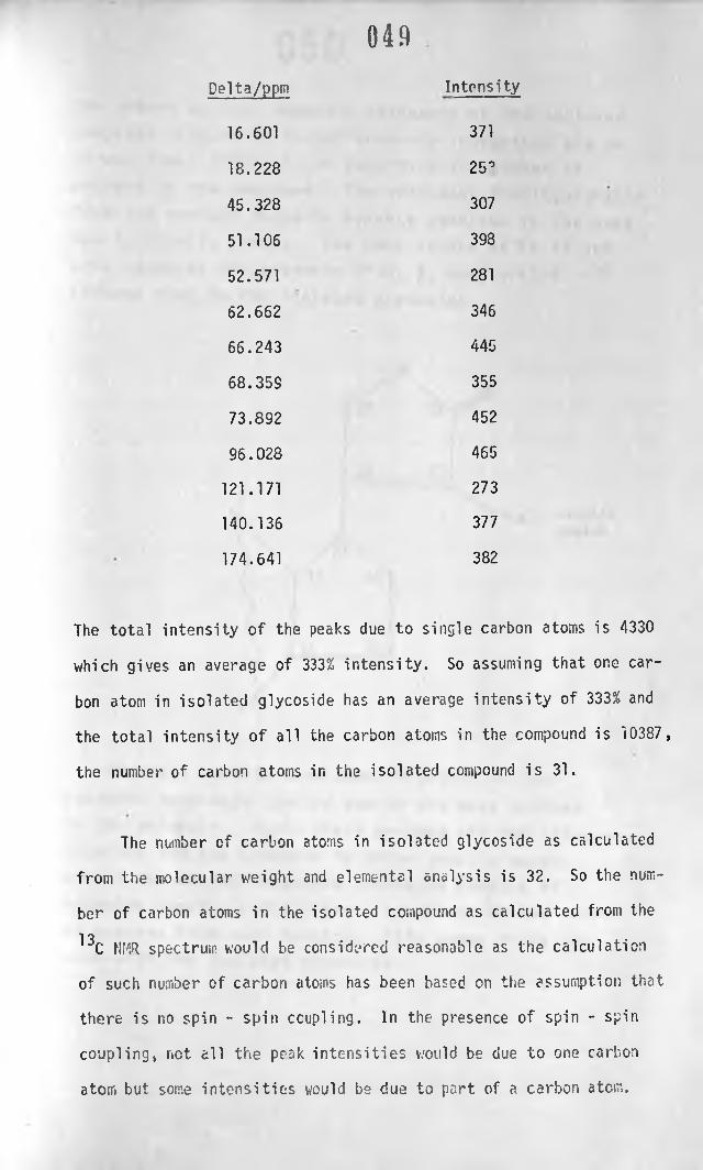
0 4 9
Delta/ppm Intensity
16.601 371
18.228 25°
45.328 307
51.106 398
52.571 281
62.662 346
66.243 445
68.35S 355
73.892 452
96.028 465
121.171 273
140.136 377
174.641 382
The total intensity of the peaks due to single carbon atoms is 4330
which gives an average of 333% intensity. So assuming that one car
bon atom in isolated glycoside has an average intensity of 333% and
the total intensity of all the carbon atoms in the compound is 10387,
the number of carbon atoms in the isolated compound is 31.
The number of carbon atoms in isolated glycoside as calculated
from the molecular weight and elemental analysis is 32. So the num
ber of carbon atoms in the isolated compound as calculated from the
13C NMR spectrum would be considered reasonable as the calculation
of such number of carbon atoms has been based on the assumption that
there is no spin - spin coupling. In the presence of spin - spin
coupling, net. all the peak intensities would be due to one carbon
atom but some intensities would be due to part of a carbon atom.

050The proton nuclear magnetic resonance of the isolated
compound (Figure 8 ) is not properly integrated and so
it was found difficult to determine the number of
protons in the compound. The only peak distinguishable
from the nuclei-r magnetic resonce spectrum is the peak
due to vinylic proton. The peak occurs a t ^ 5 . 9 2 and
this supports the presence of c<, p, unsaturated
lactone ring in the isolated glycoside.
Steroids usually have complex proton nuclear
magnetic resonance spectra due to too many protons
in the molecule. Again these protons are too close
together and the transfer fo these usually occur
making the nuclear magnetic resonance spectra of
steroids complex and difficult to determine the number
of protons from such spectra. This seems to be the
case with the isolated glycoside.

0 5 1
The inass spectrum of the isolated compound (Fig. 9) showed
features very characteristic of cardiac glycosides and helped to
confirm the chemical tests on the isolated compound. For comparison
purposes the m/e values of the fragment ions of the isolated compound,
were compared with the m/e values of somalin and digitoxigenin (Tab
le VII ). The m/e values of the fragment ions of somalin and digito
xigenin were obtained from spectra of Ardene, 1964. The parent peak
of the isolated compound was lacking from the mass spectrum and the
disappearance of the molecular ion could be due to various reasons.
In mass spectrometry, the molecular ion formed upon electron impact
(due to 70 ev energy) bears a localised charge or radical. The mo
lecular ion is more stable if there is a centre in the molecule with
comparatively little energy which can lose an electron readily upon
impact, for example, aromatic groups or some heteroatoms like nitro
gen. In such cases, the fragments resulting from the molecular ion
are few and their formation is usually predictable. In steroids,
however, a centre of comparatively weak energy for molecular ion for
mation is lacking. The tertiary and quaternary carbon atoms and
oxygen containing groups all ionize readily upon electron impact.
This recults in a large number of degradation processes with con-
committant rearrangements. As a result the parent peak may not
appear or could be very weak. In other cases, especially with poly
hydroxy steroids, they lose a molecule of water (presumably the 14
« - hydroxy) and this peak is recorded instead of the molecular peak.
Use of a large sample of the isolated glycoside might have produced
the parent peak or preparation of a suitable derivative of the com
pound.
To identify the isolated compound, its mass spectrum was com
pared with a large number of reference spectra currently available

at United Kingdon Chemical Information Service at Nottingham with
no success. The mass spectrum (Fig. 9) shows that the spectrum of
the isolated compound is complex. However, by comparing the m/e
values of the fragr ant ions of the compound wiu. those of somalin
and digitoxigenin (see Table VII ) and the fragmentation pattern of
digitoxigenin, a few deductions could be made:-
\ 652
The fragment ion with m/e 111 has been known to be formed by
McLafferty rearrangement and cleavage of the 15, 16 - bond.
The presence of this fragment ion in the mass spectrum of the iso
lated compound indicates the presence of 3 - unsaturated <f- lact
one ring and hence possibly lactone r-'ng in cardiac glycosides or
their aglycones. The same m/e value was observed to occur in sc-
malin (a cardiac glycoside and digitoxigenin (an aglycone). Ste
roids also undergo a mass fragmentation to give the fragment ion
6 and this is indicated by the fragment ion with m/e 203 andI b L o
represents parts of the rings A, B and C (Spiteller, 1963).

0 5 3
The presence of this fragment ion indicates the presence of the
steroidal skeleton. The isolated compound had a fragment ion with
m/e 205 probably due to cleavage of the ring system requiring break
ing at least two bonds giving a fragment ion This process
is frequently accompanied by the rearrangement of a hydrogen atom.
Usually loss of two methyl groups from steroids is usually
indicated by a fragment ion (m/e 404) and the removal of the side
chain at one of the methyl groups by the fragment ion (m/e 203).
Rearrangement of hydrogen atoms in the isolated compound may have
accounted for the fragment ions (m/e 405 and 205 respectively). The
mass fragment ion (m/e 393 may represent the removal of CH^ = CH0 or
CH^ = 0 + or from the molecular ion (Silvestein and Bassler,
1967). The fragment ion (m/e 364) may also be due to loss of CH^CHO
or CH-jCO from the fragment ion m/e 405.
The fragment ion with m/e 405, 393, and 364 point strongly to
the presence of oxygenated functions in the isolated compound, at
least two hydroxyl (OH) groups and some carbonyl (aldehyde or ketone
groups). The fragment ions (m/e 71 and 43) may indicate the los^ of
C2 Hj..C0 -CH3 which further suggests some oxygenated functions in the
compound.
The hydroxyl (OH) functional groups in the compound are con
firmed by the infra-red spectrum of the isolated compound (Fig. 5
broad peak at 3450 cm )̂. The presence of ketone group in the «,
g- unsaturated lactone ring has been shown by chemical tests (Ray
mond and Kedde tests were positive) and by infra-red spectrum (a
strong absorption at 1750 cm )̂ and by the mass spectrum (fragment,
ion m/e 111). The presence of such other groups as ketone (C=0) or

0 5 4
aldehyde (CHO) in the isolated compound arc yet to be determined
although there are indications that such groups may be present (frag
ment ion m/e 71, 51 and 43).
When the mass spectrum of the isolated compound was compared
with those of steroids available in literature, especially with
those of somalin and digitoxigenin, it was found that a lot of the
major peaks present in the isolated compound are also present in
these steroids. This further suggests that the isolated compound
is a steroid.
Prof. Speiser of Pharmaceutical Institute, Abterlung, Switzer
land analysed the compound for elements and for protons (NMR) and
helped to confirm the heart glycoside structure of the isolated
compound. He assigned the following structure to the compound.
From the chemical and spectroscopical analysis of the isolated
compound the following structure can be arrived at so far:-

0 5 5
The Keller - Killiani reagent indicated that the isolated compound
is a cardiac glycoside with a desoxy sugar. Therefore R̂ is a sugar
residue. An attempt to hydrolyse the glycoside and identify the
sugar did not succeed. Whether some other hydroxyl (OH) groups,
y Haldehyde (Cv ) or ketone (C=0) groups occur in the isolated compoundU ? 3 4 R 6 7
at any other position (R , R , R , R , R and R ) has as yet to be
determined. Methyl groups were shown to be present in the compound
(removal of fragment ion m/e 405 and 205). Such methyl groups could
occur at positions 18 and 1 9 .
The graph of absorbance against concentration of the isolated
compound using the data for the colour reaction with Kedde reagent
showed that Beer - Lambert law is obeyed and so the graph could be
used for quantation of the percentage yield of glycoside from the
leaves of Flaeodendron buchananii. A low yield of the glycoside was
obtained and this was about 0.29%. This low yield would be probably
due to significant losses of material during the lengthy extraction
and purification process.
/

058PHARMACOLOGICAL EFFECTS OF THE ISOLATED COMPOUND
The isolated compound was found to have a sustained increase
in blood pressure of anaesthetised rat. Similar effects were
observed with digoxin on the same preparation. With isolated per
fused rabbit heart, an increase in the force of contraction and a
slow heart rate were observed.
Cardiac glycosides have been shown to have a pressor effect
on the blood vessles (Kumar et a 1 , 1972, Vatner et a^, 1971). The
pressor effect is due to a direct vasoconstrictor action of the
cardiac glycosides on blood vessles. Also cardiac glycosides have
been shown to increase the peripheral resistance by other workers
(Higgins et_ a^, 1972, Goodman and Gilman, 1971). The increase in
force of contraction of the heart is due to a direct stimulation
by the isolated compound on the myocardium. These pharmacological
effects of the isolated compound are typical of cardiac glycosides
and these effects compare very well with those of digoxin used as
a reference.
*The pharmacological investigation carried out on the isolated
compound are only preliminary but support the chemical findings
that the isolated compound is a cardiac glycoside.

0 5 7
CHAPTER 4
CONCLUSION
The aim of the present work was to investigate the nature of
the active (poisonous) principles in F.laeodendron buchananii (Loes.)
Loes., a plant very well known because of its poisonous nature espe
cially to livestock. Investigation of the nature of the poisonous
principles led to the extraction, purification, characterisation and
partial identification of the isolated active principles. Prelimina
ry pharmacological effects of the isolated compound were carried out
to find out the pharmacological properties of the compound.
The physical and chemical properties of the compound indicated
that the compound was a cardiac glycoside. The thin-layer chroma
tographic analysis supported by other analytical techniques showed
that the isolated compound was one glycoside. The fact that the
isolated compound and the crude extract of the plant gave one spot
with same Rf value with same solvent systems and adsorbent suggest
that the isolated glycoside was the only one present in the plant.
This glycoside is probably a new or.e as indicated from literature
13survey. The infra-red, C NMR, mass and proton NMR spectra of the
isolated compound are not identical with any of the known cardiac
glycosides and the conclusion that can be reached is that this is
probably a new cardiac glycoside. All these spectra are extremely
complex which suggest that the structure of the glycoside is of
considerable complexity and to assign a molecular structure could
possibly be a major project as regards time and money.

0 5 8
The isolated glycoside was found tc have an absorption maximum
at 203 nm and had a narrow melting range 110 -111 °C.
Elaeodendron buchananii (Loes.) Loes needs further investigation
and such investigation could include:-
i) Complete structural elucidation of the cardiac glycoside'.- hyd
rolysis of the glycoside to its respective aglycone and glycone
and analysing them individually,
ii) Acetylation of the glycoside would also help in structural
elucidation.
iii) Preparation of suitable derivatives of the compound and study
of their pharmacological properties,
iv) The pharmacology of the cardiac glycoside should also be inves
tigated to establish the toxicity, absorption, distribution
and metabolism and suitable level doses.
Elaeodendron buchananii (Loes.) Loes. grows wildly in many
parts of Kenya. So the variation of glycosidal content with dif
ferent regions and seasons could also be investigated. The plant
would be easy to cultivate. So if further research on the plant
showed that the glycoside and its derivatives could be put into
medicinal use e.g. treatment of congestive heart failure, the plant
could be exploited.

059» f
T A B L E S

080
TABLE II
Investigation of the best solvent for extraction of the active prin
ciples from the leaves of Elaeodendron buchananii (Loes.) Loes.
Extracting solvent Absorbance of the coloured
complex (residue + Kedde
reagent)
1. Chloroform 0.29
2. Methanol 0.13
3. 95% alcohol 0.26
4. 70% alcohol 0 . 2 0
5. 50% alcohol + chloroform
i) alcoholic extract -
ii) chloroform extract 0.30
6 . Ethanolic/chloroform 0.29
7. Ether 0 . 1 1

001
TABLE III
Results of purification of the glycoside of Elaeodendron from chlo
roform extract using an alumina column
Eluant Fraction Volume
(ml)
Kedde reaction Comment on eluant
Alcohol 1 1 0 0 Positive yellow coloured
(95%) 2 1 0 0 +ve yellow coloured
Ethyl
acetate 3 1 0 0 +ve yellow coloured
4 1 0 0 +ve yellow coloured
Chloro- 5 1 0 0 +ve Slightly yellow
form 6 50 +ve coloured
7 50 +ve II
8 50 +ve II
9 50 +ve II
1 0 50 -ve clear eluate
11 50 -ve clear eluate

0 62
Solubility of the isolated compound in different solvents
TABLE IV
Solvent Comments on solubility
Ether Fairly soluble
Methanol Fairly soluble
Acetone Fairly soluble
Ethyl acetate Fairly soluble
Absolute alcohol Very soluble
Chloroform Extremely soluble

TABLE V
1LC analysis of the isolated glycosidc(s) of Elaeodendron compared
with digoxin and ouabain
Solvent
system
Sample No. of
spots
Distance
moved
Solvent
front
Rf Colour
after
spray
Chloroform Isol ated one 10.9 13.0 0.84
methanol glycoside
formamide
80:19:1
Ouabain one - . 13.0 pink
• chloro-
form
crude
extract one 10.85 13.0 0.84 pink
Digoxin one 1 0 . 0 13.0 0.76 pink
Ethyl Isolated
acetate- glycoside
methanol one 8.7 1 0 . 8 0.80 pink
(90:10) Digoxin one 7.7 1 0 . 8 0.71 pink
Chloro-^
form
crude
extract one 8.7 1 0 . 8 0.80 pink |
Ouabain one - 1 0 . 8 - pink'
Isolated 1

•
glycoside
from
Aj-iga
remota
|--- ' 1
one 8.9 1 0 . 8 0.82 pink
Chloroform Isolated one 6.9 11 0.63 pink
Ethanol
(7:3)
glycoside
Chloroform
crude ext
ract one 6.9 1 1 0.63 pink
Digoxin one 5 1 1 0.45 pink
Ouabain one - 1 1 - pink
Chloroform Isolated one 10.5 13.0 0.80 pink
methanol glycoside
Chloroform
crude ext- one 1 0 . 6 13.0 0.81 pink
ract
Digoxin one 9.8 13.0 0.75 pink

TABLE VI
065
Comparison of infra-red spectra of isolated glycoside with that of
digitoxigenin
Isolated compound
Note worthy peaks (cm-1)
Digitoxigenin
Note worthy peaks (cm-1)
3520 (Broad) 3440 (Broad)
2930 2940
1790 1780
1735 1760
1630 1645
1450 1465
1380 1390
1260 1260
1 1 0 0
.
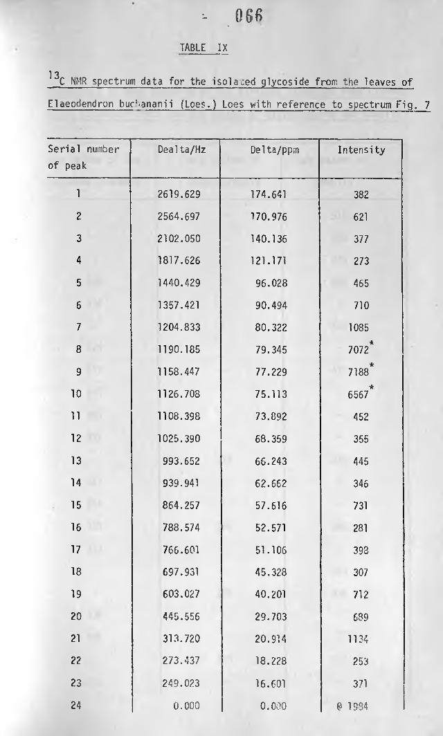
TABLE IX
13C NMR spectrum data for the isolated glycoside from the leaves of
Elaeodendron bur‘.ananii (Loes.) Loes with reference to spectrum Fig. 7
- 0 6 6
Serial number
of peak
Dealta/Hz Delta/ppm Intensity
1 2619.629 174.641 382
2 2564.697 170.976 621
3 2102.050 140.136 377
4 1817.626 121.171 273
5 1440.429 96.028 465
6 1357.421 90.494 710
7 1204.833 80.322 1085
8 1190.185 79.345 7072*
9 1158.447 77.229*
7188
1 0 1126.708 75.113 6567*
11 1108.398 73.892 452
1 2 1025.390 68.359 355
13 993.652 66.243 445
14 939.941 62.662 346
15 864.257 57.616 731
1 6 788.574 52.571 281
17 766.601 51.106 393
18 697.931 45.328 307
19 603.027 40.201 712
2 0 445.556 29.703 689
2 1 313.720 20.914 1134
2 2 273.437 18.228 253
23 249.023 16.601 371
24 0 . 0 0 0 0 . 0 0 0 0 1934

067TABLE X
Comparison of mass spectrum of the isolated glycoside from Elaeode-
ndron buchananii v.’ th the m/e values of digitoxigenin and somalin
m/e values of the
isolated compound
m/e values of
digitoxigenin
m/e values of
somalin
- - 519
- - 500
- - 474
455 - 456
436 - -
- - 430
426 - -
417 - 417
405 - -
393 - -
381i
- -
- 374 374
367 - -
364 - -
355 356 356
343 - -
- - 339
- 338 338
336 - -
331 - -
- 323 323
319 -

0 8 8
305 - -
- - 297
293 - -
69 67 -
- - 59
- 55 -
51 - -
43 41 41
- - 31
- 29 29
18
15
281 284 284
- - 275
269 - -
255 - 257
243 246 246
231 228 231
- 213 213
219 - -
218 - -
205 203 203
193 195 195
189 - -
181 - 181
- 174 175
169 - -
162 162 163
151 - -

147 145
143 - 145
131 133 -
125 124 -
119 - 119
113 - 113
111 Ill 111
100 - -
97 - 95
93 93 95
85 - 84
83 - 84
- 79 -
- - 74
71 — 71

TABLE XI
Data for the calibration curve of absorbance of the glycoside of
Elaeodendron reacted with Kedde reagent against concentration of
the glycoside
Concentration % Absorbance
0.004 0.05
0 . 0 1 2 0.15
0 . 0 2 0 0.23
0.032 0.35
0.040 0.45
0.048 0.55
0.06 0 . 6 6
T-| (unknown) 0.33
(unknown) 0.29

TABLE XII
Effects of the isolated compound, adrenalin, and digoxin on the
blood pressure of anaesthetised rat
Injection Observation Remarks
Control - 1 ml of A transient fall in blood
1 0 % w/v solution pressure. Blood pressure This fall could
of propylene gly- rose from 1 0 . 6 mm Hg to be due to vaso-
col in normal 10.3 mm Hg. Returned to dilation
saline 10.6 mm Hg after about
2 minutes.
10 mg/Adrenalin An immediate rise in blood This rise is
pressure. The blood pre- probably due to
ssure rose from 106 mm Hg the vasoconst-
to 133 mm Hg. rictor effect of
fadrenalin
1 mg of the A very slight and immedi-
isolated compound ate fall in blood pressure The fall could be
and then a gradual rise due to vesodi-
above the mean blood pre- lation
ssure. The blood pres-
sure fell from 106 to 104
then rose to 109.

G U R E S

073
FIG. 1: ELAEODENDRON BUCHANANII (LOES.) LOES
a
b
c
A branchlet (x 0.5)
Fruiting branchlet (x 0.5)
Flower (x 5)

074
A. Leaves of Elaeodendron buchananii (Loes) Loes.Natural size.
B. Your.g leaves of Elaeodendron buchananii (Loes) Loes.'

F IG . 2: ULTRAVIOLET ABSORPTION SPECTRUM OF THE ISOLATED GLYCOSIDE IN ALCOHOL- * " * — miMW if '̂ i
'vitt aV j -?.(? >;nn
450
40 ~ ,ryAl60 |
275
0 8
0-6<ucrtJQ 0 4 wo</>
-Q« 0-2
O O200 225 250 525 350
f t W o $>\*\ <aA.fc4»V
** *VN4 S.
400

FIG 3: ULTRAVIOLET ABSORPTION SPECTRUM OF THE ISOLATED GLYCOSIDE FOR THE COLOUR REACTION WITH 3:5 DINITROBENZOIC ACID

FIG. 4: ULTRAVIOLET ABSORPTION SPECTRUM OF THE ISOLATED GLYCOSIDE FOR THE COLOUR REACTION WITH 1,3 DINITROBENZENE(m - D NB )
S I "
z ^ a o s !
Concentration
Reference
Path Length
n Witt»ind«W'x5rt'.fr T T T T ,M*L:T BRtBG^ENGLAND**
Scan Speed Iasi I I s!o.\ 1 1
Absorbance Range 0 - 0 2A I lO - I O A l 1
Operator Date
----- SElSBSSSGaESBraSL-• « ! . » « E.

M ( I I / m i i l , ! I MFIG. 5: INFRA-RED SPECTRUM (KBr DISC) OF THE ISOLATED GLYCOSIDE
' » ’ * • 1 '. ,«m
4 5 5 9 10 II 12 13 14 IS I*| £> vm-l1i
lv>* -ia
!
I 11 •*- *
| k*:«*ii ic
i| 0[> '<ivf 1 ____
S )
U.i;o o o j

TCA?
•SjS
-A.'i
Xf J
FIG. 6: INFRA-RED SPECTRUM (KBr DISC) OF DIGITOXIGENIN


FIG. 8: PROTON NMR OF THE ISOLATED GLYCOSIDE
U 100 INCTOVM NO 930 .00tC*VC IOCI•h IOC 1 MHI X fH IUUhi X•V «« 1 MM| □ HOMO L )•V 40% MH| □ a“t 17 l MH| n"c 7 U1| • □ Ml ■... □ IrtDtAMfU // fitjv-fkr
t lu l
C «% 1 *— * * p/*"^
h * t y 'O 1^ l r « l̂jL, ^ '
101 VINT X tM V iru»> . XTU9C 0 0 . T ___ «AT| VO •»»
O lilMSNCC7 aH»Tm Hiam* w hm Jr ?IV hi
nui . . . .. MCI<w
row ... himet* AUK !®
w r«io _ cmno «* scm
tOCK » -UCHAI **<4.__ •» I'll 0 4*
i K N H C J O K f «
n o cu u* . __ . . cimocooc C l M ! !OffSCT HI W *>*K. jl*N C S t H A V N U D lH . .. J . Hi
«*oo* Mt(i> morn _cnwct0«J»T S lUJU. UhviMmi

FIG. 9: MASS SPECTRUM OF THE ISOLATED GLYCOSIDE
>7**•2.
*»7 t»>r-
............ till . . llln. 111.. .
Oo Or o

Absorbance
.:
-IG. 10: A GRAPH OF ABSORBANCE AGAINST CONCENTRATION OF THE ISOLATED
GLYCOSIDE

G. TI : A CONDON PRESSURE MANOMETER FOR THE STUDY OF THE EFFECTS OF THE ISOLATED GLYCOSIDE ON THE BLOOD PRESSURE OF ANAESTHETISED RAT

FIG. 12: EFFECTS OF CONTROL, ADRENALIN, TEST MIXTURE AND DIGOXIN ON ANAESTHETISED RAT

FIG. 13: EFFECTS OF ADRENALIN, ISOLATED GLYCOSIDE AND DIGOXIN ON ISOLATED PERFUSED RABBIT HEART ON A KYMOGRAPH
d & J 1* * *A
',*'«Wru|
OOcr>

0335U4
FIG. 14: EFFECTS OF CONTROL, ADRENALIN, ISOLATED GLYCOSIDE ON ISOLATED PERFUSED RABBIT HEART USING
A HARVARD SMOOTH MUSCLE/HEART TRANSDUCER
* °>
T......................■ ■ « i ' » r , » T H m i n t u „ T i i i i i | I I H I I ||M | M „ M I , 1 1 | t ) | [ ||,||n
c*
* • *4• *• • -s

R E F E R E N C E S

REFERENCES
08.0
1. Von M. V. Ardenne, R. Tummler, E. K. Weiss and T. Reichstein
Helv. Chim. Acta 47 : 1032 (1964).
2. Bailey, D.
Pharm. J. 216: 175 (1976).
3. Bellan\y, L. S.
The Infra-red Spectra of Complex Molecules
Methuem (1954) pp. 114 and 146.
4. Bowman, W. C., Rand, M. J. and West, G. B.
Textbook of Pharmacology (1970) p. 249.
5. Burns, J. S., Smith W. J., Moline, W. F.
Crop Sci. 10 : 578 - 581 (1970).
6. Canback, T.
J. Pharm. Pharmacol. 1 : 201 (1949).
7. Chen and Anderson
J. Am. Pharm. Ass. : 353 (1946).
8. Clarke, E. G.
Isolation and Identification of Drugs in Pharmaceuticals,
Body Fluids and Postmortem Materials, 2nd Ed. (1949) p. 64.
9. Clover, P. E., Stewart, J. and Cwynne, M. D.
East Afric. Agri. Forestry 0., 32 : 200 - 207 (1966).
10. Cox, E., Wright, S. E. and Roxburgh, G.
0. Pharm. Pharmacol. 11 : 535 (1959).
11.. Dyer, 0. R.
"Application of Absorption Spectroscopy of Organic Compounds"
Prentice - Hall, Inc. Englewood Cliffs, N. J. (165) p. 30
12. Feigl, F.
Tests on Organic Analysis, 6th Ed., Elsevier Publishing Co.,
New York, U.S.A. (i960) pp. 5S4.
13. Fieser, L. F. and Fieser, M.
"Steroids", Reinhold, New York and London (1959) pp. 727 - 73
09U14. Fulton, C. C., Am. J. Pharm. 104, 244 (1932).
15. Goodman, L. S. and Gilman, A.
Pharmacological Basis of Therapeutics Int. 4th Ed., The
Macmillan Company, London (1971) pp. C88.
16. Gunn, J. A.
J. Physiol., 46 : 506 (1913).
17. Higgins, C. B. et al
Circulation Research. 28 No. 2 109 - 118.
18. Higuchi, T. and Einar Brochman - Hansen
Pharmaceutical Analysis, Interscience Publishers, New York
- London (1961) p. 49.
19. Hollstein, U. Breitmaier, E. and Jung, G.
J. Am. Chem. Soc. 96 : 8036 - 8040 (1974).
20. Jacobs, Hoffman and Gustus
J. Biol. Chem. 107: 143 (1926).
21. Johnston, E. J. and Jacobs, A. L.
J. Pharm. Sci., 55̂ : 531 - 532 (1966).
22. Kedde
Pharm. Weekbland 82 : 741 (1947).
23. Kraus, L.
Chem. Zentr. J29 : 1924 (1958).
24. Laidlaw, P. P.
J. Physiol. 39 : 354 (1909).
25. Langendoff
Pflungers Arch. Ges. Physiol., 190 : 280 (1895).
26. Legal
Chem. Zentr. 54 : 652 (1883).
27. Leiberitz, D. and Roberts, J. D.
J. Am. Chem. Soc. 95 : 4996 (1972).
28. Mines, G. R.
J. Physiol. 39 : 37 - 49 (1908).

03129. Mugera, G. M. Bull. Epizoot. Dist. Afr. (1970), 18, pp. 377 - 381.
30. Poech, K. and Tracey, M. V.
Modern Methods of Plant Analysis (1956).
31. Rangaswani, S. and Subramanian, S. S.
J. Sci. Industr. Res., 148 : 78 (1955).
32. Raymond, W. D.
Analyst 61 : 400 (1938).
33. Raymond, W. D.
Analyst 63 : 480 (1938).
34. Sehandri, T. R. and Subramanian, S. S.
J. Sci. Industr. Res. 93 : 114 (1950).
35. Shellard, E. J.
Exercises in the Evaluation of Drugs and Surgical Dressings
(1958) p. 14.
36. Shoppee, E. W.
"Chemistry of Steroid" 2nd Ed, Butterworth, London (1964)
pp. 326 - 397.
37. Silverstein, R. M. and Bassler, G. C.
Spectrophotometric Identification of Organic Compounds,
2nd Ed. Wiley International (1967) pp. 61 - 63.
38. Sim, S. K.
Medicinal Plant Glycosides" 2nd Ed. University of Toronto
Press (1967) p. 30.
39. Stahl, E.
Thin Layer Chromatography, 2nd Ed. (1969) pp. 60, 135 and 873.
40. Stahl, E. and Kultenbach, U.
J. Chromatog. 5> : 458 - 460 (1961).
41. Steinegger, E. and Von der Walt, J. H.
Pharm. Acta Helv., 36 : 599 - 601 (1961).

09242. Stewart, C. P. and Stolman, A.
"Toxicology", 2 : 742 (1961).
43. Stoll, A. and Kreis, W.
Helv. Chim. Acta., 34 : 1431 - 1459 (1951).
44. Tattje, D. H. E.
J. Pharm. Pharmacol., £ : 29 (1957).
45. Thorp, R. H. and Cobbin, L. B.
"Cardiac Stimulant Substances", (Medicinal Chemistry Series),
6 : 1 (1967).
46. Trease, G. E. and Evans, E. C.
Pharmacognosy, 10th Ed. Williams and Wilkins Co., Baltimore
(1970) pp. 4 and 139.
47. Verdcourt, B. and Trump, E. C.
Common Poisonous Plants of East Africa, Collins, St. James
Place, London (1959) pp. 100.
48. Watt, J. M. and Breyer - Brandwijk, M. G.
Medicinal and Poisonous Plants of Southern and Eastern
Africa, 2nd Ed. F. S. Livingston Ltd., Ediburgh and London,
(1962).pp. 183.
49. Zoller, P. and Tamm, C.
Helv. Chim. Acta 36 : 1744- 1756 (1953).