
Burns
41
Nicole Baier, MD
-
Upload
malik-harper -
Category
Documents
-
view
58 -
download
0
description
Burns. Nicole Baier, MD. Statistics. In US: 1.2 million burns each year 60,000 hospitalizations 6000 deaths 2 nd leading cause of unintentional death in children (after MVA) Pediatric incidence by type of burn: Scald burns: 85% Flame burns: 13% - PowerPoint PPT Presentation
Transcript of Burns

Nicole Baier, MD
StatisticsIn US:
1.2 million burns each year60,000 hospitalizations6000 deaths
2nd leading cause of unintentional death in children (after MVA)
Pediatric incidence by type of burn:Scald burns: 85%Flame burns: 13%Remaining 2%: electrical and chemical burns

The SkinBarrier to:
Fluid lossEntry of infectionHeat loss
epidermis
dermis
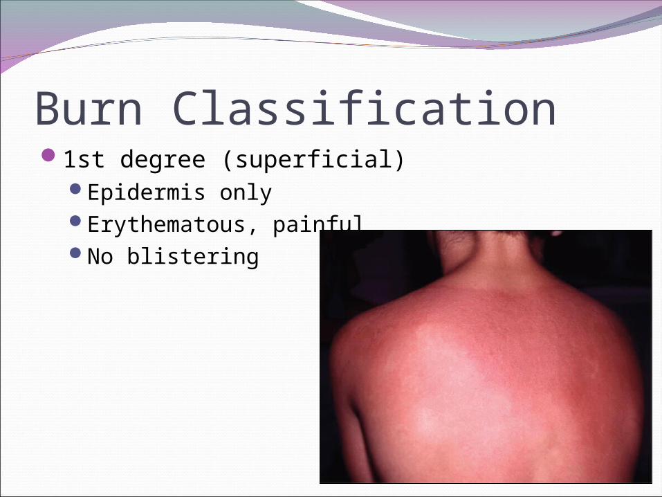
Burn Classification1st degree (superficial)
Epidermis onlyErythematous, painfulNo blistering
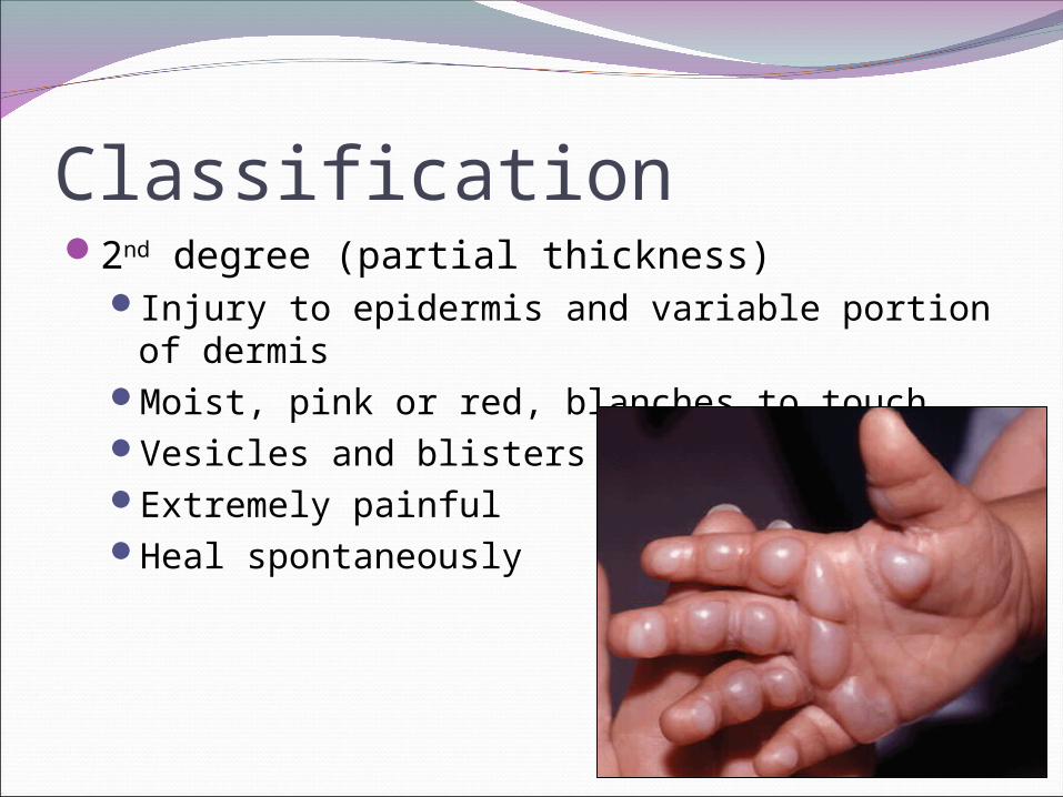
Classification2nd degree (partial thickness)
Injury to epidermis and variable portion of dermis
Moist, pink or red, blanches to touchVesicles and blistersExtremely painfulHeal spontaneously
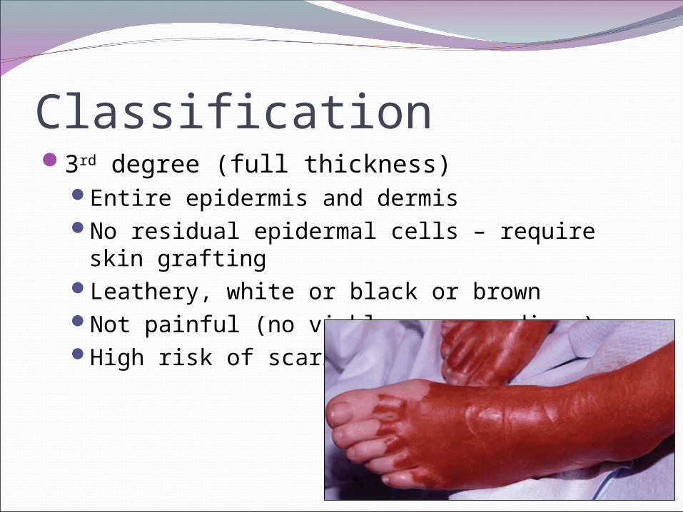
Classification3rd degree (full thickness)
Entire epidermis and dermisNo residual epidermal cells – require skin
graftingLeathery, white or black or brownNot painful (no viable nerve endings)High risk of scarring

Classification4th degree
Involve underlying structures (tendons, nerves, muscles, bone, fascia)
Reconstructive surgery often necessary
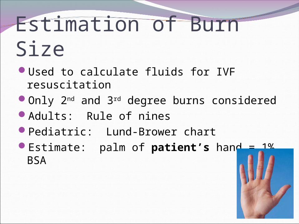
Estimation of Burn SizeUsed to calculate fluids for IVF resuscitationOnly 2nd and 3rd degree burns consideredAdults: Rule of ninesPediatric: Lund-Brower chartEstimate: palm of patient’s hand = 1% BSA

Burn Diagrams

Acute AssessmentAIRWAY
Airway edema caused by inhalational injury Direct thermal injury – supraglottic
Suspicion increased if: Facial/ oral burns Soot in mouth/nose Singed nasal hairs Wheezing, stridor, or hoarseness noted
Intubation should be performed quickly as edema can progress rapidly (over initial 24-36 hours)
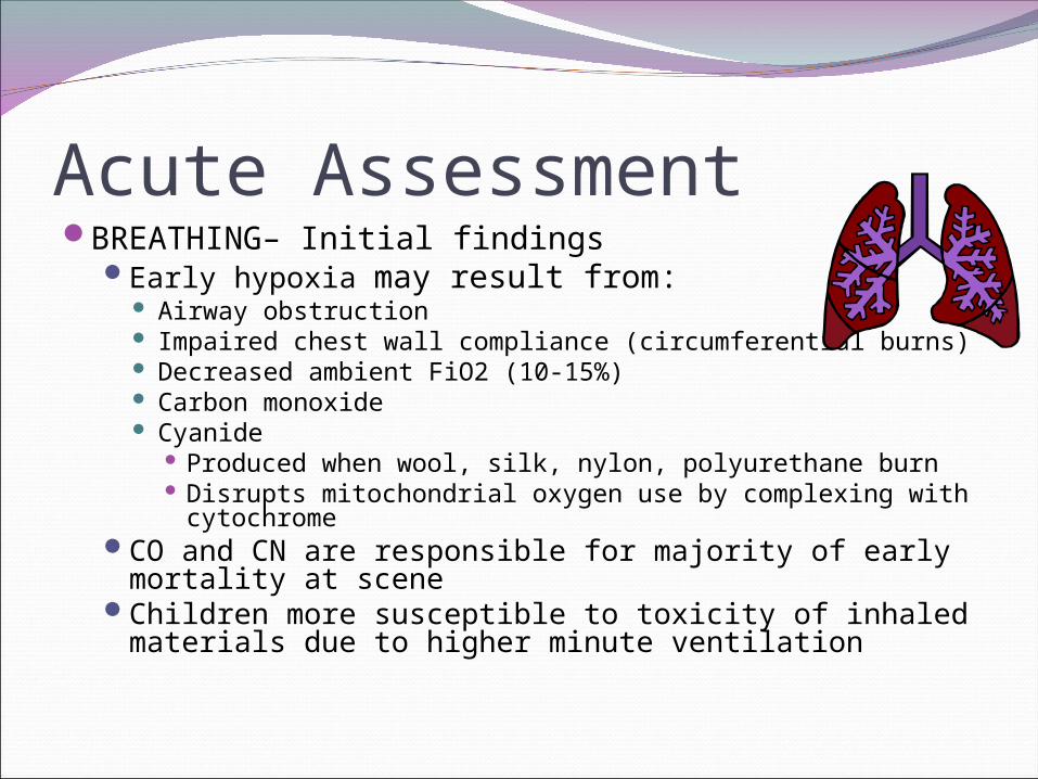
Acute AssessmentBREATHING– Initial findings
Early hypoxia may result from: Airway obstruction Impaired chest wall compliance (circumferential burns) Decreased ambient FiO2 (10-15%) Carbon monoxide Cyanide
Produced when wool, silk, nylon, polyurethane burn Disrupts mitochondrial oxygen use by complexing with
cytochromeCO and CN are responsible for majority of early
mortality at sceneChildren more susceptible to toxicity of inhaled
materials due to higher minute ventilation
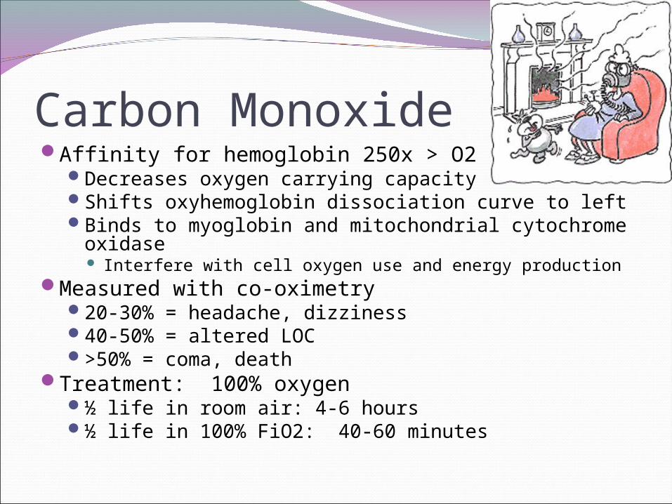
Carbon MonoxideAffinity for hemoglobin 250x > O2
Decreases oxygen carrying capacityShifts oxyhemoglobin dissociation curve to leftBinds to myoglobin and mitochondrial cytochrome
oxidase Interfere with cell oxygen use and energy production
Measured with co-oximetry20-30% = headache, dizziness40-50% = altered LOC>50% = coma, death
Treatment: 100% oxygen½ life in room air: 4-6 hours½ life in 100% FiO2: 40-60 minutes

Acute AssessmentBREATHING – Later findings
Chemical irritants injure tracheobronchial tree and lung parenchyma Lower airway edema Respiratory epithelium sloughs - cast formation causes
airway obstruction Manifests as: bronchospasm, post-obstructive atelectsis
Patients also at risk for: Surfactant deficiency due to damage to type II
pneumocytes ARDS
After 72 hours: nosocomial pneumonia may developRestrictive lung disease may develop in survivors
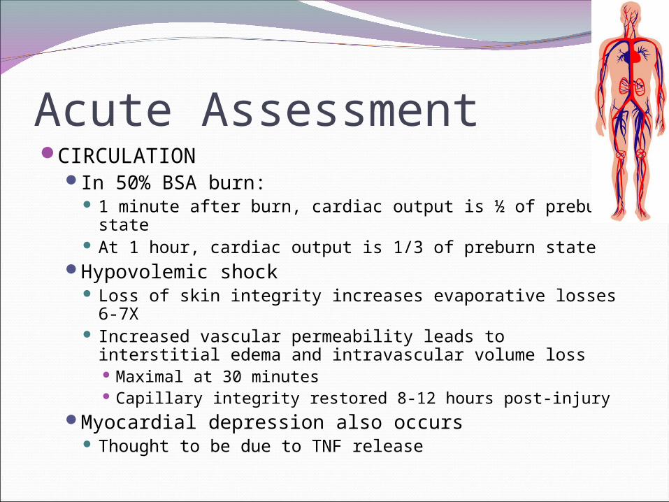
Acute AssessmentCIRCULATION
In 50% BSA burn: 1 minute after burn, cardiac output is ½ of preburn
state At 1 hour, cardiac output is 1/3 of preburn state
Hypovolemic shock Loss of skin integrity increases evaporative losses 6-7X Increased vascular permeability leads to interstitial
edema and intravascular volume loss Maximal at 30 minutes Capillary integrity restored 8-12 hours post-injury
Myocardial depression also occurs Thought to be due to TNF release
Acute ManagementCIRCULATION
Burns >15% BSA require IV fluid resuscitation to maintain perfusion
Time to IV access is a major predictor of mortality in pediatric patients who have burns greater than 80% TBSA
IV preferably placed in nonburned tissues
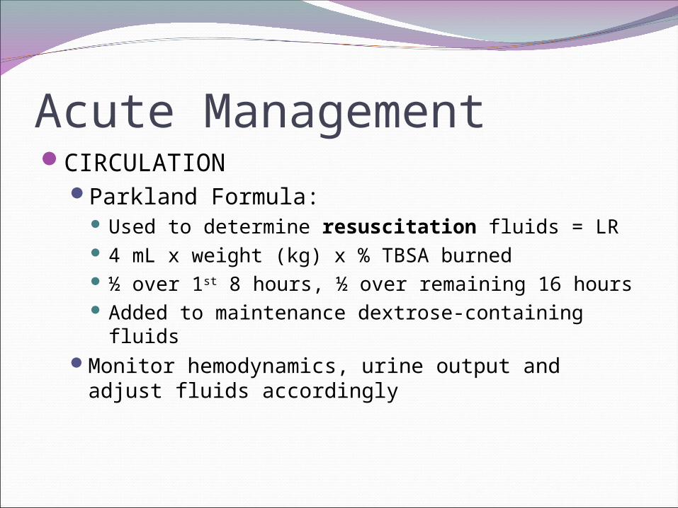
Acute ManagementCIRCULATION
Parkland Formula: Used to determine resuscitation fluids = LR 4 mL x weight (kg) x % TBSA burned ½ over 1st 8 hours, ½ over remaining 16 hours Added to maintenance dextrose-containing fluids
Monitor hemodynamics, urine output and adjust fluids accordingly
QuestionYou have a 14 month old, 11 kg infant who was
involved in a house fire and has second degree burns to both of her hands, feet, her right lower arm and both lower legs. What IV fluids should she receive over the 1st 24 hours?
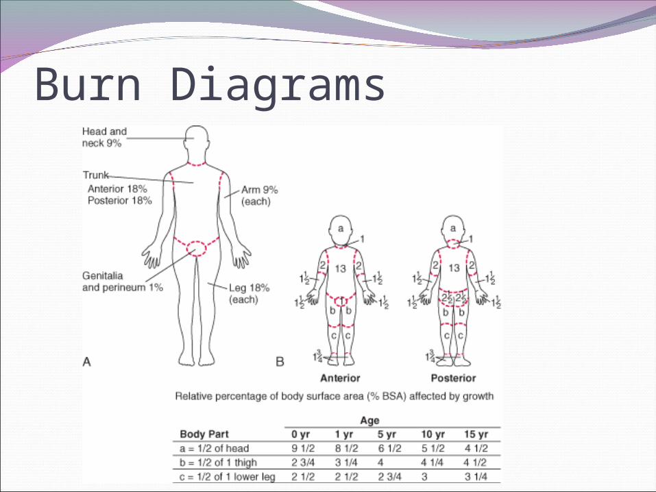
Burn Diagrams
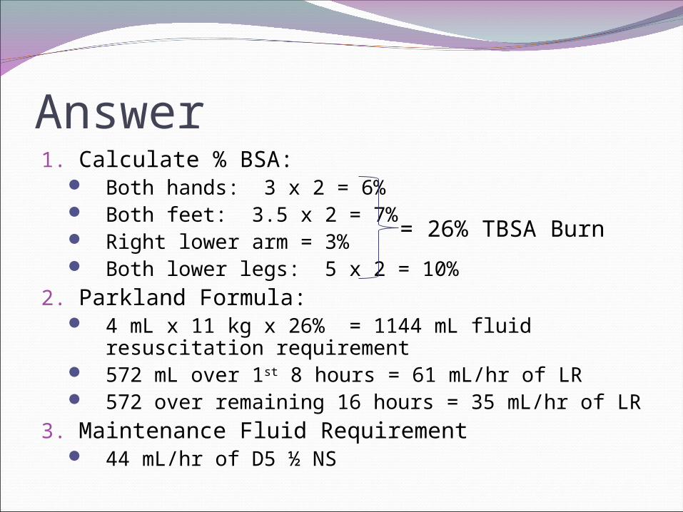
Answer1. Calculate % BSA:
Both hands: 3 x 2 = 6% Both feet: 3.5 x 2 = 7% Right lower arm = 3% Both lower legs: 5 x 2 = 10%
2. Parkland Formula: 4 mL x 11 kg x 26% = 1144 mL fluid
resuscitation requirement 572 mL over 1st 8 hours = 61 mL/hr of LR 572 over remaining 16 hours = 35 mL/hr of LR
3. Maintenance Fluid Requirement 44 mL/hr of D5 ½ NS
= 26% TBSA Burn
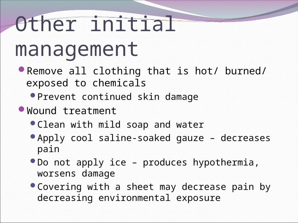
Other initial managementRemove all clothing that is hot/ burned/
exposed to chemicalsPrevent continued skin damage
Wound treatmentClean with mild soap and waterApply cool saline-soaked gauze – decreases painDo not apply ice – produces hypothermia,
worsens damageCovering with a sheet may decrease pain by
decreasing environmental exposure
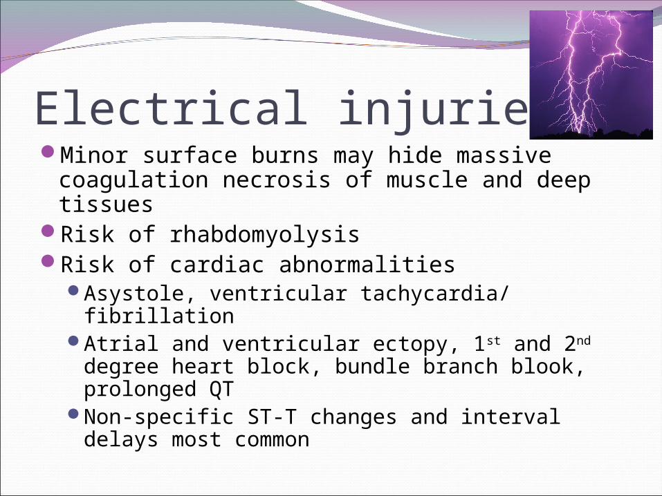
Electrical injuriesMinor surface burns may hide massive
coagulation necrosis of muscle and deep tissues
Risk of rhabdomyolysisRisk of cardiac abnormalities
Asystole, ventricular tachycardia/ fibrillationAtrial and ventricular ectopy, 1st and 2nd degree
heart block, bundle branch blook, prolonged QT
Non-specific ST-T changes and interval delays most common
Electrical InjuriesTissue injury is directly proportional to
resistanceNerves, muscles, blood vessels have lowest
resistance Electricity preferentially flows through these structures More severe damage
Increased resistance: Skin Tendons Bone Fat
Water decreases resistance, therefore moist areas (eg, axillae) tend to sustain more damage
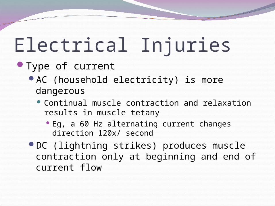
Electrical InjuriesType of current
AC (household electricity) is more dangerous Continual muscle contraction and relaxation results
in muscle tetany Eg, a 60 Hz alternating current changes direction
120x/ second
DC (lightning strikes) produces muscle contraction only at beginning and end of current flow

Electrical InjuriesCurrent Pathway
Current may flow in 1 of 3 pathways: Hand to hand
60% mortality rate due to: Spinal cord transection at C4-C8 Suffocation due to chest wall muscle tetany Myocardial muscle damage
Hand to foot 20% mortality rate due to cardiac arrhythmias
Foot to foot 5% mortality rate
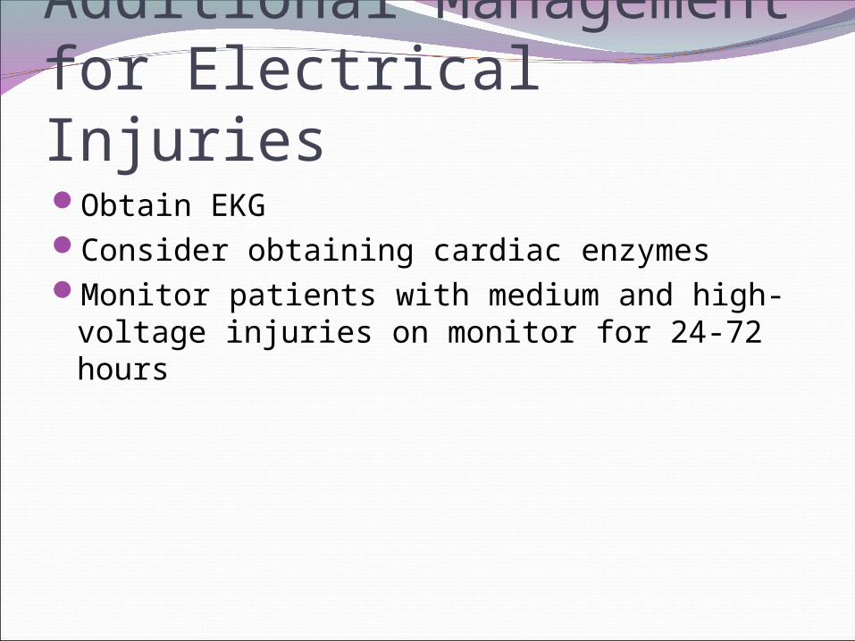
Additional Management for Electrical InjuriesObtain EKGConsider obtaining cardiac enzymesMonitor patients with medium and high-
voltage injuries on monitor for 24-72 hours

Compartment SyndromeMost common early cause of diminished pulses
is inadequate resuscitationHigh index of suspicion for elevated
compartmental pressures in circumferential burn
Emergent escharotomy or fasciotomy is indicated for limb salvage in pulseless extremity
Thoracic escharotomies are occasionally required to improve chest-wall compliance and facilitate ventilation
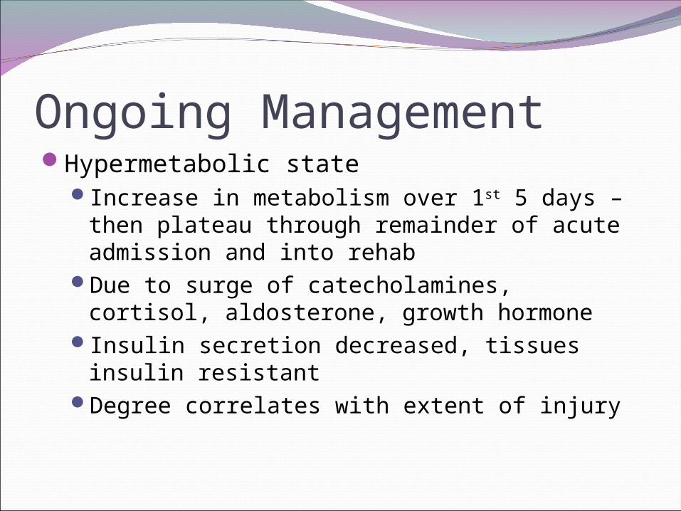
Ongoing ManagementHypermetabolic state
Increase in metabolism over 1st 5 days – then plateau through remainder of acute admission and into rehab
Due to surge of catecholamines, cortisol, aldosterone, growth hormone
Insulin secretion decreased, tissues insulin resistant
Degree correlates with extent of injury

Hypermetabolic StateManifestations
Tachycardia, increased cardiac outputHyperthermia
Baseline temp reset to 38.5 C⁰Increased gluconeogenesis, protein catabolism,
lipolysisResting energy expenditure 2-3 x normalMay be associated with:
Impaired wound healing Sepsis Loss of lean body and muscle mass
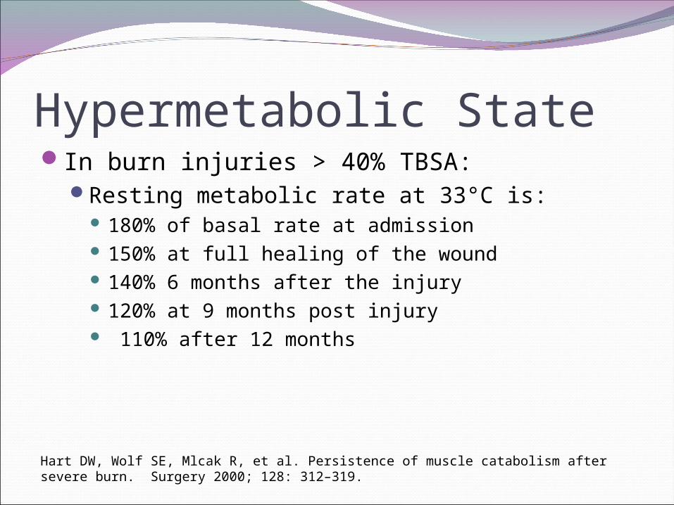
Hypermetabolic StateIn burn injuries > 40% TBSA:
Resting metabolic rate at 33°C is: 180% of basal rate at admission 150% at full healing of the wound 140% 6 months after the injury 120% at 9 months post injury 110% after 12 months
Hart DW, Wolf SE, Mlcak R, et al. Persistence of muscle catabolism after severe burn. Surgery 2000; 128: 312–319.

Hypermetabolic stateLong-term consequences
Profound muscle wastingDecreased bone mineral densityRetarded linear growth in children
In 80 patients with > 40% TBSA burn: Profound growth arrest noted during postburn year
1 Growth improved to normal by postburn year 3
Rutan FL, Herndon DN. Growth delay in postburn pediatric patients. Arch Surg 1990; 125: 392-395.

Ongoing ManagementFeeds started EARLY
Within 6 hours of admissionRequire up to 50% more calories than at
baseline Hypermetabolic state Pain and anxiety increase physiologic demands Greater heat loss occurs in young infants with
larger surface area-to-mass ratiosReduces bacterial translocation and sepsisTPN avoided due to infectious complicationsGoal: full feeds by 24-48 hours
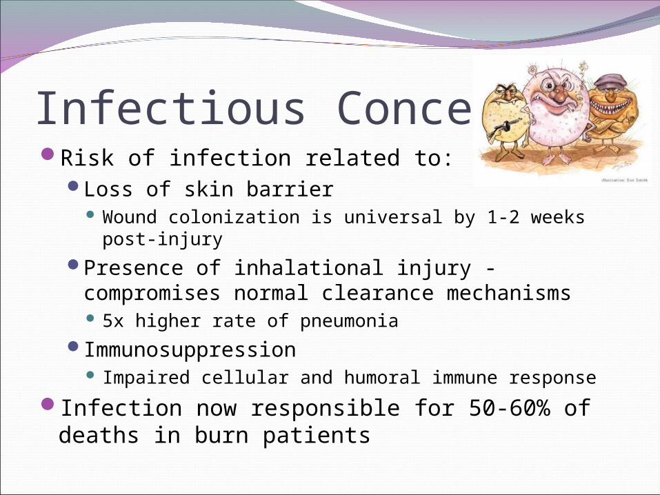
Infectious ConcernsRisk of infection related to:
Loss of skin barrier Wound colonization is universal by 1-2 weeks post-
injuryPresence of inhalational injury - compromises
normal clearance mechanisms 5x higher rate of pneumonia
Immunosuppression Impaired cellular and humoral immune response
Infection now responsible for 50-60% of deaths in burn patients

Topical TherapiesBactroban
Used for superficial burns, primarily on faceSilvadene (silver sulfadiazene)
BacteriocidalCannot be used in those with sulfa allergiesCauses neutropenia and thrombocytopenia

Topical TherapiesSulfamylon (mafenide acetate)
Better penetration of deep burns, eschars, and cartilage
BacteriostaticBetter gram negative coverage (pseudomonas)Causes fungal overgrowthPainfulCarbonic anhydrase inhibitor – causes
metabolic acidosis

Surgical Wound ManagementEarly excision and closure of full thickness
burn woundIf wound >50% TBSA is totally excised and
covered with autograft within 2–3 days:Metabolic rate 40% less compared with wound
coverage 1 week post injury
Hart DW, Wolf SE and Chinkes D, et al. Determinants of skeletal muscle catabolism. Ann Surg 2000; 233: 455–465.
Surgical Wound ManagementOther benefits of early wound excision
Decreases painProvides barrier to fluid and heat loss,
bacterial invasionDecreases length of stayAccelerates recoveryFewer septic complicationsDecreased morbidity and death
Surgical Wound ManagementSerial wound excision and grafting is the
standard of care for full-thickness burnsWhen the burned area exceeds donor site
supply (burns >30% BSA), homografts from donors or skin substitutes are usedTaken back to OR weekly to replace
homografts with autografts as donor sites heal

Criteria for Admission>15% BSA3rd degree burnsElectrical burnsInhalational injuryBurns to hands, feet, face, genitalia, joint
surfacesSuspected abuse or neglectInadequate home situation
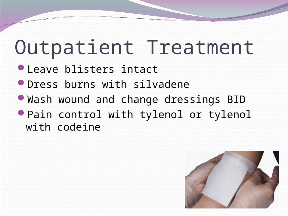
Outpatient TreatmentLeave blisters intactDress burns with silvadeneWash wound and change dressings BIDPain control with tylenol or tylenol with
codeine
Identifying abusive burns15-20% of burn injuries are the result of
abuseSuspicious patterns:
Glove or stocking burns of hands and feetDeep burns on trunk or backSmall-area full-thickness burns (cigarette)Circumferential burnsBurns localized to the perineum or buttocksSymmetric burns

Burn PreventionPreset water heaters to max of 120 F⁰
Duration of exposure required to produce full-thickness burn: 120 F: 10 minutes⁰ 130 F: 30 seconds⁰ 140 F: 5 seconds⁰ 150 F: 2 seconds⁰ 158 F: 1 second⁰
Federal Flammable Fabric ActRequires sleepwear to be flame retardant
Use of smoke detectors