Burning Issues in the Esophagus · Chronic GERD Advancing age (>50 y) Male gender Tobacco use...
45
1 Burning Issues in the Esophagus Elizabeth Montgomery, MD Johns Hopkins Medical Institutions Dr. Montgomery reports no relevant financial relationships with commercial interests. Squamous Epithelium Muscularis Mucosae Submucosa Muscularis Propria Mucosa Layers of The Esophagus [What a biopsy gets] Duct Lamina propria (note vessels) Muscularis mucosae Submucosal gland
Transcript of Burning Issues in the Esophagus · Chronic GERD Advancing age (>50 y) Male gender Tobacco use...

1
Burning Issues in the
Esophagus
Elizabeth Montgomery, MD
Johns Hopkins Medical Institutions
Dr. Montgomery reports no relevant financial relationships with
commercial interests.
Squamous
Epithelium
Muscularis
Mucosae
Submucosa
Muscularis
Propria
Mucosa
Layers
of The
Esophagus
[What a biopsy
gets]
Duct
Lamina propria (note vessels)
Muscularis mucosae
Submucosal gland

2
Barrett Esophagus -
Epidemiology
5th and 6th decade
M:F - 2:1 -4:1
White: African-American - 10:1 -
20:1
Risk of cancer – about 0.5%/year
– pooled data mostly US
Study from Denmark
Hvid-Jensen F, Pedersen L, Drewes AM, Sørensen HT, Funch-Jensen P. Incidence of Adenocarcinoma among Patients with Barrett's Esophagus. N Engl J Med. 2011 Oct 13;365(15):1375-83.
Annual risk of progression to adenocarcinoma – 0.12%
Therefore surveillance pointless (at least in Denmark)
Denmark United States
Life expectancy
(WHO)
M 77y/ F 81y
Obese population
estimate:
7%
Life expectancy
M 76y/ F 81y
Obese population
estimate:
33%

3
Barrett’s Esophagus
~ Overall incidence of progression in BE patients is 0.1 to 0.3 %/year in first five years but 9-9.5% at 20 years
Kroep S, Lansdorp-Vogelaar I, Rubenstein JH, de Koning HJ, Meester R, Inadomi JM, van Ballegooijen M. An Accurate Cancer Incidence in Barrett's Esophagus: A Best Estimate Using Published Data and Modeling. Gastroenterology. 2015 Sep;149(3):577-585
Prevalence of BE – Swedish
Study
Columnar lined esophagus in about 10.3%
BE found in [1.6%] [with goblet cells]
Alcohol, smoking were risk factors
[Gastroenterology 2005; 129: 1825]
American College of
Gastroenterology (ACG) Criteria for
Barrett’s Esophagus 2008
Barrett’s mucosa is a change of the
esophageal epithelium of any length
that
1) can be recognized at endoscopy and
2) is confirmed to have intestinal
metaplasia on biopsy

4
(ACG) Criteria for Barrett’s Esophagus 2016
“Barrett’s esophagus should be diagnosed
when there is extension of salmon-colored
mucosa into the tubular esophagus
extending > 1 cm proximal to the
gastroesophageal junction with biopsy
confirmation of intestinal metaplasia”
The authors further suggested that
endoscopic biopsy should not be performed
in the presence of a normal Z line or a Z line
with <1 cm of variability
Oh dear – Length Requirement???
How the heck do we know how much they
saw?
The ACG suggested the term “specialized
IM of the esophagogastric junction” for
things <1cm
Daily life – Biopsy labeled esophagus
Note: The above diagnosis of Barrett esophagus is made due to
presence of goblet cells (intestinal metaplasia) with the assumption that
the biopsies were obtained from columnar mucosa in the distal
esophagus located at least 1 cm proximal to the top of the gastric
folds as per 2016 American College of Gastroenterology (ACG)
guidelines.
Reference: Shaheen NJ, Falk GW, Iyer PG, Gerson LB; American
College of Gastroenterology. ACG Clinical Guideline: Diagnosis and
Management of Barrett's Esophagus. Am J Gastroenterol. 2016
Jan;111(1):30-50
5
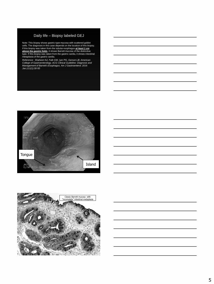
Daily life – Biopsy labeled GEJ
Note: This biopsy shows gastric-type mucosa with scattered goblet
cells. The diagnosis in this case depends on the location of this biopsy.
If this biopsy was taken from the tubular esophagus at least 1 cm
above the gastric folds, it shows Barrett mucosa of the distinctive
type. If this biopsy was taken from the gastric cardia, it shows intestinal
metaplasia of the gastric cardia.
Reference: Shaheen NJ, Falk GW, Iyer PG, Gerson LB; American
College of Gastroenterology. ACG Clinical Guideline: Diagnosis and
Management of Barrett's Esophagus. Am J Gastroenterol. 2016
Jan;111(1):30-50
Tongue
Island
Classic Barrett mucosa with “incomplete” intestinal metaplasia

6
Normal small intestinal mucosa
Normal small intestinal mucosa, PAS/AB stain
?US Will Stop Requiring Goblet
Cells?Example: Takubo et al Hum Pathol 2009;40:65-74 used
German cases and found that intestinal metaplasia
accompanied only 43% of early esophageal
adenocarcinomas and believed that most cases arose in
association with cardiac type mucosa.
Stay tuned!
ACG did not remove the requirement for goblet cells in
2008 OR 2016; AGA did not in position paper – 2010.

7
2012 and 2016 Studies from USC
ONLY FOUND DYSPLASIA OR CARCINOMA IN PATIENTS WITH INTESTINAL METAPLASIA AND EARLY CANCERS WERE ACCOMPANIED BY INTESTINAL METAPLASIA
BUT these patients were all biopsied using systematic protocols (“perfect world”) by highly experienced colleaguesChandrasoma P, Wijetunge S, DeMeester S, Ma Y, Hagen J, Zamis L, DeMeester T. Columnar-lined esophagus without intestinal metaplasia has no proven risk of adenocarcinoma. Am J Surg Pathol. 2012 Jan;36(1):1-7.
Smith J, Garcia A, Zhang R, DeMeester S, Vallone J, Chandrasoma P. Intestinal
Metaplasia is Present in Most if Not All Patients Who Have Undergone endoscopic Mucosal Resection for Esophageal Adenocarcinoma. Am J Surg Pathol. 2016 Apr;40(4):537-43. PubMed PMID: 26813746.
Cases with staging data – Johns
Hopkins
In our material at Johns Hopkins, we found
that >90% of patients with treatment naïve
esophageal adenocarcinomas had
background intestinal metaplasia
Salimian KJ, Waters KM, Eze O, Pezhouh MK, Tarabishy Y, Shin EJ, Canto MI,
Voltaggio L, Montgomery EA. Definition of Barrett Esophagus in the United States:
Support for Retention of a Requirement for Goblet Cells. Am J Surg Pathol. 2018
Feb;42(2):264-268. PubMed PMID: 29016405.
However
That’s OUR population
It may be reasonable in other populations

8
Dysplasia
Neoplastic Epithelium
Confined within the basement
membrane of the gland within
which it arose.
Diagnostic Categories
Negative for dysplasia
Indefinite for dysplasia
Dysplasia, low-grade
Dysplasia, high-grade
Intra-mucosal carcinoma
Grading Dysplasia in Barrett’s -
Algorithm
SURFACE MATURATION
[COMPARED TO UNDERLYING
GLANDS]
ARCHITECTURE
CYTOLOGIC FEATURES
INFLAMMATION
9
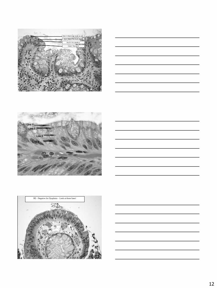
BE - Negative for Dysplasia
Surface - More mature than glands
Architecture - Abundant lamina propria
Cytology - Normal with mitoses confined to deeper glands. Nuclei with smooth nuclear membranes. Normal nuclear polarity
Inflammation - Variable
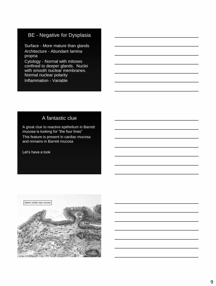
A fantastic clue
A great clue to reactive epithelium in Barrett
mucosa is looking for “the four lines”
This feature is present in cardiac mucosa
and remains in Barrett mucosa
Let’s have a look
Gastric cardiac type mucosa

10
Gastric cardiac mucosa

11
Barrett mucosa – no dysplasia
Barrett mucosa –no dysplasia
12
BE - Negative for Dysplasia – Look at those lines!

13
LGD – no lines, abrupt transition see
at arrow at left
LGD –no lines

14
Indefinite for dysplasia
Originally defined in IBD and diagnosed by
answering the questions…
a) Is this epithelium unequivocally benign or
reactive?
b) Is this epithelium unequivocally
neoplastic/adenomatous
The answer “NO” to both questions = IFD
But…..Montgomery 2001 – study on dysplasia in BE
Deliberately defined any epithelium that looked dysplastic in the bases of the pits but had surface maturation as IFD
i.e. impossible to have dysplasia with maturation
Rationale – maturation is a major feature of regenerating mucosa, so will exclude all reactive changes.

15
BE, Indefinite for Dysplasia
Surface – often more mature than glands
Architecture - slight glandular crowding
Cytology - hyperchromasia, nuclear membrane irregularities, increased mitoses in deep glands. Maintained nuclear polarity
Inflammation - Frequently a factor
Nice to see an abrupt transition to be sure something is dysplastic – and thus clonal
You might call indefinite for stratification. NeutrophilsNo abrupt transition
But using the lines, it’s reactive
Kevin Waters

16
Changes in Proportion of NFD and IFD
0
10
20
30
40
50
60
70
80
90
100
2007 2008 2009 2010 2011 2012 2013 2014 2015 2016
NFD
IFD

17

18
Low Grade Dysplasia
Clearly neoplastic
Minimal loss of
nuclear polarity
Surface involved
Only mild
architectural
crowding.

19
LGD, adenoma-like
Where is the “Minimum
Abrupt transition – appears
divergent and clonal compared to
adjacent epithelium
But without inflammation in the
specific “abnormal” focus
Our “lines” obscured
20
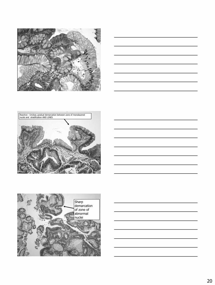
Reactive - Unclear, gradual demarcation between zone of monolayered nuclei and stratification AND LINES
Sharp demarcationof zone of abnormalnuclei

21
Does Dysplasia Start Here??”Basal crypt dysplasia”
Lomo et al (R. Odze senior au). Crypt
dysplasia with surface maturation: a
clinical, pathologic, and molecular study of
a Barrett's esophagus cohort.
Am J Surg Pathol. 2006 Apr;30(4):423-35.

22
Basal crypt dysplasia
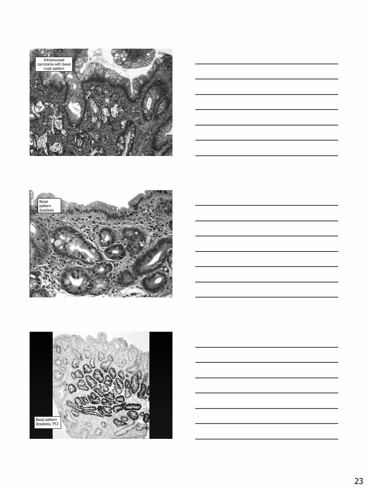
23
Intramucosal carcinoma with basal
crypt pattern
Basal pattern dysplasia
Basal pattern dysplasia, P53

24
High-Grade Dysplasia
Surface - No maturation
Architecture - Crowded
glands overrunning
lamina propria
Cytology - Nuclear
membrane irregularities,
extending to surface and
loss of nuclear polarity
Inflammation - Typically
not abundant

25
High-
Grade
Dysplasia
HGD –
Inflammation

26
HGD - InflammationInflamed HGD
Inflamed HGD
Inflamed HGD, p53

27
Barrett, intramucosal carcinoma, P53 null pattern
Barrett, intramucosal carcinoma, P53 null pattern
Barrett mucosa, Reactive changes

28
Barrett – reactive changes, wilt type pattern p53
Dysplastic pattern TP53
Wild type pattern
HGD, “small cell pattern”“non-adenomatous dysplasia”

29
HGD, “small Cell pattern”“non-adenomatousdysplasia”
HGD – small cell pattern/ “nonadenomatous
dysplasia”
“Lateral spread”

30
Intramucosal CarcinomaSurface - No maturation
Architecture - Effacement of lamina propria and syncytial growth pattern of glands. Back-to-back microglands, “dirty necrosis” in glands, DESMOPLASIA not yet developed
Cytology - as in HG –but often with nucleoli
Inflammation - variable
Lamina Propria Invasion, Esophageal Adenocarcinoma
Early intramucosal invasion

31
Intramucosal carcinoma, lateral spread of atypical glands
Intramucosal carcinoma, Budding and lateral growth
Well developed desmoplasia –Invasion into at least submucosa

32
Finding Pagetoid extension of single cells ALWAYS means there is a cancer
underneath
Additionally
There are variant forms of dysplasia – initial
studies were all using criterial for intestinal
type dysplasia but variant patterns are less
well recognized and less well understood
Intestinal type low-grade dysplasia

33
Look ! Kulchitsky cells! HGD
Foveolar Type LGD
Foveolar type HGD
34
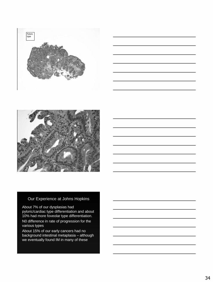
Pyloric type
Our Experience at Johns Hopkins
About 7% of our dysplasias had
pyloric/cardiac type differentiation and about
10% had more foveolar type differentiation.
N0 difference in rate of progression for the
various types
About 15% of our early cancers had no
background intestinal metaplasia – although
we eventually found IM in many of these

35
Changing the game
New endoscopic treatments and ability to
visualize the mucosa (even with molecular
markers) might slowly reduce the mortalility
of esophagus cancer
….assuming we can figure out how to get
the right patients screened in a better way
Known Risk Factors for Presence of BE
Chronic GERD
Advancing age (>50 y)
Male gender
Tobacco use
Central obesity (waist >40 inches)
Caucasian race
Known Risk Factors for Developing
Neoplasia in BE
Advanced age
Increasing length of BE
Central obesity
Tobacco use
Lack of NSAID use
Lack of PPI use
Lack of statin use

36
Estimates for Progression Risk
No dysplasia – 0.2-0.5%/year
LGD – 0.7%/yr
HGD – 7%/year
Most patients with BE WITHOUT
DYSPLASIA die of something else (>90%)
Endoscopic Resections
For Barrett, endoscopic mucosal resections
can be more practical than submucosal
dissections since they are fast and several
can be performed to achieve coverage
Subclassification of Depth of Invasion by Superficial Carcinoma Proposed by the Japan Esophageal Society

37
Relationships among Depths of Invasion and Vessel Invasion and Lymph
Node Metastasis in Superficial SQUAMOUS Carcinoma
Depth % Lymph node metastases on resection
m1 0%
m2 0%
m3 8%
sm1 17%
sm2 28%
sm3 49%
Makuuchi H et al. Rinsho-Shokakinaika (Clin Gastroenterol) 12:1749-1756, 1997;
Endo M et al. Endoscopic treatment for early carcinoma of the esophagus.
Shokaki-Shinyo Practice. Bunkodo, Tokyo,1998

38
Endoscopic Mucosal Resection [EMR] showing duplicated muscularis mucosae [Ref; Abraham et al. Am J Surg Pathol 2007; 31:1718.]
Original muscularis mucosae
Duplicated Muscularismucosae

39
Endoscopic mucosal resection – note damaged surface
from plastic cap and suction

40
Original muscularis mucosae
Endoscopic mucosal
resection -layers

41
Duplicated muscularis mucosae

42
Early submucosal invasion – measure the depth from the bottom of the muscularis mucosae to the deepest part of the cancer
43
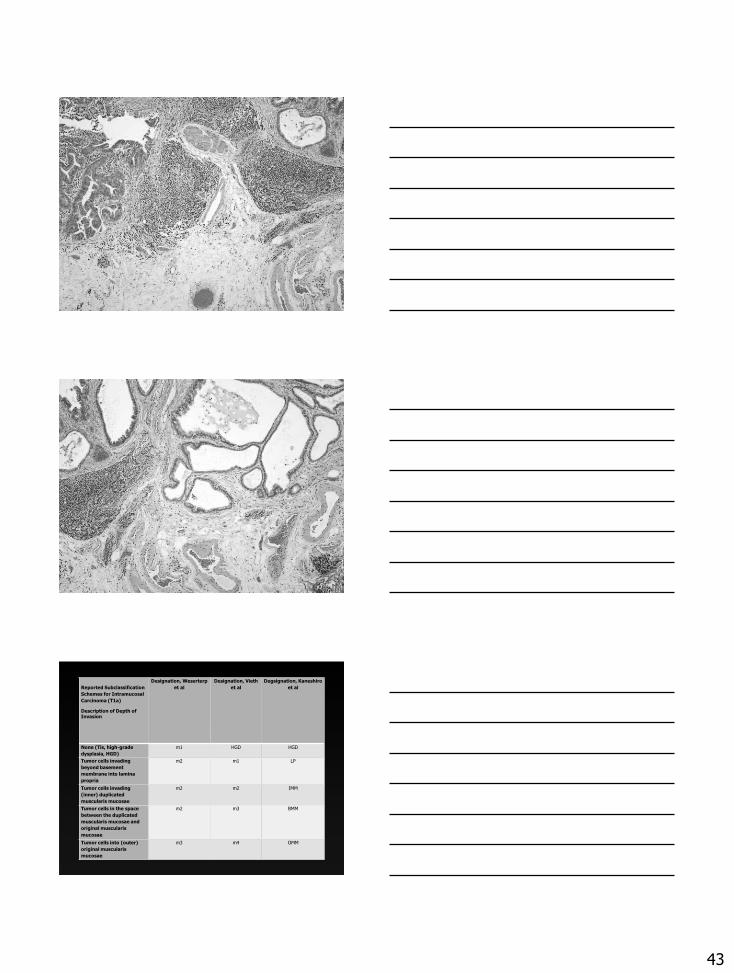
Reported Subclassification
Schemes for Intramucosal
Carcinoma (T1a)
Description of Depth of Invasion
Designation, Weserterp
et al
Designation, Vieth
et al
Degsignation, Kaneshiro
et al
None (Tis, high-grade
dysplasia, HGD)
m1 HGD HGD
Tumor cells invading
beyond basement
membrane into lamina
propria
m2 m1 LP
Tumor cells invading
(inner) duplicated
muscularis mucosae
m2 m2 IMM
Tumor cells in the space
between the duplicated
muscularis mucosae and
original muscularis
mucosae
m2 m3 BMM
Tumor cells into (outer)
original muscularis
mucosae
m3 m4 OMM

44
Studies – Early
Esophagus
Adenocarcinomas
Depth Outcome
Liu. Am J Surg Pathol
2005; 29: 1079
Lamina propria No mets
Liu. Am J Surg Pathol
2005; 1079
MM or superficial
SM
22% mets
Westertwerp. Virchow’s
Arch 2005;446:497
M1-M3, SM1 1/79 LN mets,
83% 5 yr surv
Westertwerp. Virchow’s
Arch 2005;446:497
SM2, SM3 44% LN mets;
42% 5 yr surv
Kaneshiro Am J Surg
Pathol 2011; 35:697
M1-M3
SM1
0.7% LN mets
8.6% LN mets
Guidelines ACG 2016
Surveillance NFD – every 3-5 years
IFD – repeat after optimization of acid
suppression for 3-6 mos; if repeat IFD
rebiopsy in 1 year
LGD – confirmation followed by
consideration of ablation or surveillance
every year
HGD – confirmation followed by endosopic
therapy
Follow-up After Ablation
If for HGD or IMC – every 3 months for a
year then every 6 months for a year then
annually
If for LGD – every 6 months for first year,
then annually

45
Thank you!
Mr.
Barrett says
“thank you”