BTS GUIDELINES Guidelines for radiologically guided lung...
17
BTS GUIDELINES Guidelines for radiologically guided lung biopsy A Manhire, Chairman, M Charig, C Clelland, F Gleeson, R Miller, H Moss, K Pointon, C Richardson, E Sawicka ............................................................................................................................... Thorax 2003;58:920–936 See end of article for authors’ affiliations ....................... Correspondence to: Dr A R Manhire, Department of Radiology, Nottingham City Hospital, Nottingham NG5 1PB, UK; [email protected] ....................... T hese guidelines have been developed at the request of the Standards of Care Committee of the British Thoracic Society (BTS) and with the agreement of the Royal College of Radiologists and the British Society of Interventional Radiology, and approval of the Royal College of Pathologists in respect of the pathology recommendations and the Society of Cardiothoracic Surgeons of Great Britain and Ireland. Lung biopsy is a relatively frequently per- formed procedure with considerable benefit for patient management but it may, on rare occa- sions, result in the death of the patient. It is a multidisciplinary procedure involving respiratory physicians, surgeons, and radiologists with an interest in chest diseases. The aim of the group was to produce formal evidence based guidelines for subsequent use by those referring patients for the procedure and for those performing it. The areas covered by these guidelines are as follows: N Indications N Complications, contraindications and precau- tions N Consent N Technique N Staffing issues N Patient information The following areas are not covered by these guidelines: N Lesions of the chest wall, pleura and medias- tinum N Bronchoscopic and open lung biopsy FORMULATION OF GUIDELINES Validity and grading of recommendations The criteria for assessing the levels of evidence and grading of recommendations were based on those recommended in the Scottish Inter- collegiate Guidelines Network in 1995 1 using the Agency of Health Care Policy and Research model used in some other BTS guidelines (tables 1 and 2). It should be noted that there are very few randomised trials comparing the various aspects of lung biopsy and, for that reason, more detailed systems of categorisation such as that of the Scottish Intercollegiate Guidelines Network pub- lished in 2001 were not used. 2 The papers selected by searching PubMed and Medline were assessed by the members of the working group and decisions on levels of evidence for each paper were made by two or more members. The guidelines were sent for comment to the Royal College of Radiologists, the British Thoracic Society, the British Society of Interventional Radiology, the Royal College of Pathologists, and the Society of Cardiothoracic Surgeons. Scheduled review of guidelines As methods of diagnosis and tissue sampling change and new evidence comes to light, the content and evidence base for these guidelines will be reviewed. TYPES OF LUNG BIOPSY Lung biopsies may be classified according to the method of access (percutaneously, bronchosco- pically, open operation) or by the reason for biopsy (sampling of diffuse lung disease or obtaining tissue from a mass when malignancy is suspected). Sometimes percutaneous biopsy is also defined by the tissue type obtained (cytolo- gical or histological). The indications for each will be discussed later. Fine needle aspiration biopsy (FNA, FNAB) gives cytological specimens and, although these needles tend to be of narrow bore, cutting needles (CNB) that produce histological speci- mens can also be of similar gauge. For that reason, lung biopsy in general is referred to as percutaneous transthoracic lung biopsy (PTLB) in these guidelines. Percutaneous transthoracic lung biopsy PTLB is performed with imaging guidance and most frequently by a radiologist. Usually the aim is to diagnose a defined mass. Imaging mod- alities are fluoroscopy, computed tomography (CT), and ultrasound. Ultrasound is useful only where the tissue mass is in contact with the chest wall since the ultrasound beam does not pass through air and, hence, the aerated lung. Magnetic resonance imaging (MRI) currently has a limited use because of expense, difficulty accessing the patient within the magnet, the relatively poor visualisation of lung lesions, and difficulties with ferromagnetic instruments within the magnetic field. ................................................... Abbreviations: CNB, core needle biopsy; CT, computed tomography; FNA, FNAB, fine needle aspiration biopsy; MRI, magnetic resonance imaging; NSCLC, non-small cell lung cancer; PET, positron emission tomography; PTLB, percutaneous transthoracic lung biopsy; SCLC, small cell lung cancer 920 on 18 June 2018 by guest. Protected by copyright. http://thorax.bmj.com/ Thorax: first published as 10.1136/thorax.58.11.920 on 29 October 2003. Downloaded from
Transcript of BTS GUIDELINES Guidelines for radiologically guided lung...

BTS GUIDELINES
Guidelines for radiologically guided lung biopsyA Manhire, Chairman, M Charig, C Clelland, F Gleeson, R Miller, H Moss, K Pointon, C Richardson,E Sawicka. . . . . . . . . . . . . . . . . . . . . . . . . . . . . . . . . . . . . . . . . . . . . . . . . . . . . . . . . . . . . . . . . . . . . . . . . . . . . . . . . . . . . . . . . . . . . . . . . . . . . . . . . . . . . . . . . . . . . . . . . . . . . . .
Thorax 2003;58:920–936
See end of article forauthors’ affiliations. . . . . . . . . . . . . . . . . . . . . . .
Correspondence to:Dr A R Manhire,Department of Radiology,Nottingham City Hospital,Nottingham NG5 1PB, UK;[email protected]. . . . . . . . . . . . . . . . . . . . . . .
These guidelines have been developed at therequest of the Standards of Care Committeeof the British Thoracic Society (BTS) and
with the agreement of the Royal College ofRadiologists and the British Society ofInterventional Radiology, and approval of theRoyal College of Pathologists in respect of thepathology recommendations and the Society ofCardiothoracic Surgeons of Great Britain andIreland.
Lung biopsy is a relatively frequently per-formed procedure with considerable benefit forpatient management but it may, on rare occa-sions, result in the death of the patient. It is amultidisciplinary procedure involving respiratoryphysicians, surgeons, and radiologists with aninterest in chest diseases.
The aim of the group was to produce formalevidence based guidelines for subsequent use bythose referring patients for the procedure and forthose performing it.
The areas covered by these guidelines are asfollows:
N Indications
N Complications, contraindications and precau-tions
N Consent
N Technique
N Staffing issues
N Patient information
The following areas are not covered by theseguidelines:
N Lesions of the chest wall, pleura and medias-tinum
N Bronchoscopic and open lung biopsy
FORMULATION OF GUIDELINESValidity and grading of recommendationsThe criteria for assessing the levels of evidenceand grading of recommendations were based onthose recommended in the Scottish Inter-collegiate Guidelines Network in 19951 usingthe Agency of Health Care Policy and Researchmodel used in some other BTS guidelines (tables1 and 2).
It should be noted that there are very fewrandomised trials comparing the various aspectsof lung biopsy and, for that reason, more detailedsystems of categorisation such as that of theScottish Intercollegiate Guidelines Network pub-lished in 2001 were not used.2
The papers selected by searching PubMed andMedline were assessed by the members of the
working group and decisions on levels ofevidence for each paper were made by two ormore members. The guidelines were sent forcomment to the Royal College of Radiologists,the British Thoracic Society, the British Society ofInterventional Radiology, the Royal College ofPathologists, and the Society of CardiothoracicSurgeons.
Scheduled review of guidelinesAs methods of diagnosis and tissue samplingchange and new evidence comes to light, thecontent and evidence base for these guidelineswill be reviewed.
TYPES OF LUNG BIOPSYLung biopsies may be classified according to themethod of access (percutaneously, bronchosco-pically, open operation) or by the reason forbiopsy (sampling of diffuse lung disease orobtaining tissue from a mass when malignancyis suspected). Sometimes percutaneous biopsy isalso defined by the tissue type obtained (cytolo-gical or histological). The indications for eachwill be discussed later.
Fine needle aspiration biopsy (FNA, FNAB)gives cytological specimens and, although theseneedles tend to be of narrow bore, cuttingneedles (CNB) that produce histological speci-mens can also be of similar gauge. For thatreason, lung biopsy in general is referred to aspercutaneous transthoracic lung biopsy (PTLB)in these guidelines.
Percutaneous transthoracic lung biopsyPTLB is performed with imaging guidance andmost frequently by a radiologist. Usually the aimis to diagnose a defined mass. Imaging mod-alities are fluoroscopy, computed tomography(CT), and ultrasound. Ultrasound is useful onlywhere the tissue mass is in contact with the chestwall since the ultrasound beam does not passthrough air and, hence, the aerated lung.Magnetic resonance imaging (MRI) currentlyhas a limited use because of expense, difficultyaccessing the patient within the magnet, therelatively poor visualisation of lung lesions, anddifficulties with ferromagnetic instrumentswithin the magnetic field.
. . . . . . . . . . . . . . . . . . . . . . . . . . . . . . . . . . . . . . . . . . . . . . . . . . .
Abbreviations: CNB, core needle biopsy; CT, computedtomography; FNA, FNAB, fine needle aspiration biopsy;MRI, magnetic resonance imaging; NSCLC, non-small celllung cancer; PET, positron emission tomography; PTLB,percutaneous transthoracic lung biopsy; SCLC, small celllung cancer
920
on 18 June 2018 by guest. Protected by copyright.
http://thorax.bmj.com
/T
horax: first published as 10.1136/thorax.58.11.920 on 29 October 2003. D
ownloaded from

Bronchoscopic lung biopsyBiopsy via a bronchoscope is useful for proximal endobron-chial lesions but is unable to access more peripheral lesions.Transbronchial biopsy of diffuse lung disease may be assistedby some imaging guidance. It is most commonly performedby a respiratory physician. Because it does not cross thepleura, pneumothorax is much less common than inpercutaneous biopsy.
Open lung biopsy and video assisted thoracoscopicsurgery (VATS)Although these surgical procedures are able to provide largersamples of tissue with improved accuracy and specificity, themorbidity and length of stay are greater than with the othertwo methods of biopsy.
BACKGROUNDThe indications and methods for lung biopsy have changedover the years with increased access to CT and moretherapeutic options. The total number of lung biopsiesperformed has also increased. All invasive procedures havea morbidity and mortality rate associated with them andthese are important in considering whether to subject thepatient to a procedure.
A multidisciplinary meeting will ensure the most appro-priate approach to biopsy and should include at least arespiratory physician and radiologist with an interest in chestdisease. Depending on the local circumstances, referral forbiopsy by another specialist clinician such as an oncologistmay be acceptable, but proper assessment of lung functionbefore the procedure is essential (see later).
MORTALITY AND MORBIDITYMortality rateThe mortality rate of percutaneous lung biopsy is poorlydocumented. The literature contains mainly anecdotal
reports. Sinner3 in 1975 reported no deaths in his series of5300 biopsies but, from his knowledge, he estimated amortality rate of 0.07%. A further series by Berquist et alreported two deaths in 430 procedures, a rate of 0.47%.4
Richardson et al5 performed a postal survey of the UK practiceof lung biopsies which achieved a 61% response rate. Basedon 5444 biopsies, the mortality rate was estimated at 0.15%.There is probably a tendency to under-report patient death.
Causes of mortalityMortality from PTLB is generally an early event. The causes ofmortality include acute massive haemoptysis or pulmonaryhaemorrhage, pulmonary venous air embolism leading to airwithin the intracerebral or coronary circulation, and largehaemothorax.4 6–8
MorbidityPneumothoraxThe most common complication is pneumothorax whichoccurs in 0–61% of lung biopsies. Between 3.3% and 15% ofall patients will require a chest drain.9–15 This large range forpneumothorax reflects both altered risk from the location ofthe lesion and the increased sensitivity of CT, and potentiallyultrasound, to detect very small pneumothoraces which maybe overlooked on the chest radiograph.
The risk of pneumothorax is related to the needle passingthrough aerated lung and increases significantly if the lesionis not abutting the pleura.14 In one series using CT guidedcoaxial cutting needle biopsy, the highest number ofpneumothoraces occurred when the lesions were subpleural,and were 2 cm or less in depth from the chest wall.16 Otherwork has shown that perihilar biopsy is also more likely tocause pneumothoraces because of the distance of lungcrossed.4
Post biopsy positioning has not been found to decrease therate of pneumothorax.17 18 Injection of autologous bloodthrough a coaxial needle is not commonly practised and isof uncertain benefit.19
Bilateral pneumothoraces have been reported in occasionalpatients with either unexpected lung herniation across themidline or incomplete fusion of the pleura,20 as well asfollowing heart lung transplantion where there is a singlepleural cavity.
Pulmonary haemorrhageIntrapulmonary haemorrhage may occur with or withouthaemoptysis. Intrapulmonary haemorrhage is recorded in 5–16.9% of patients and haemoptysis in 1.25–5%.3 5 Lesiondepth has been identified as the most important risk factorfor haemorrhage, with an increased risk of bleeding in lesionsdeeper than 2 cm.16
HaemothoraxThe haemothorax rate is around 1.5%. Significant haemor-rhage is rare. Haemorrhage may occur from intercostal orinternal mammary arteries or veins.8
Other complicationsCase reports are noted of tumour seeding along the needletract, cardiac tamponade, and of chest infection (pneumonia)being converted to an empyema.3 21–23
Patients are potentially at risk from drugs if they areadministered during the procedure.
Co-existing relevant pathology should also be taken intoconsideration. In line with BTS guidelines on performingbronchoscopy, lung biopsy should not be performed within6 weeks of a myocardial infarction.24 Chronic renal or hepaticinsufficiency may increase the risk of bleeding and impairdrug handling.
Table 2 Grading of recommendations
Grade Type of recommendations
A(levels Ia, Ib)
Requires at least one randomised controlled trial aspart of a body of literature of overall good qualityand consistency addressing the specificrecommendation
B(levels IIa, IIb, III)
Requires availability of well conducted clinical studiesbut no randomised clinical trials on the topic ofrecommendation
C (level IV) Requires evidence from expert committee reports oropinions and/or clinical experience of respectedauthorities. Indicates absence of directly applicablestudies of good quality
Table 1 Categories of evidence185
Level Type of evidence
Ia Evidence obtained from meta-analysis of randomisedcontrolled trials
Ib Evidence obtained from at least one randomised controlledtrial
IIa Evidence obtained from at least one well designed controlledstudy without randomisation
IIb Evidence obtained from at least one other type of welldesigned quasi-experimental study
III Evidence obtained from well designed non-experimentaldescriptive studies such as comparative studies, correlationstudies, and case controlled studies
IV Evidence obtained from expert committee reports and/orclinical experiences of respected authorities
Guidelines for radiologically guided lung biopsy 921
on 18 June 2018 by guest. Protected by copyright.
http://thorax.bmj.com
/T
horax: first published as 10.1136/thorax.58.11.920 on 29 October 2003. D
ownloaded from
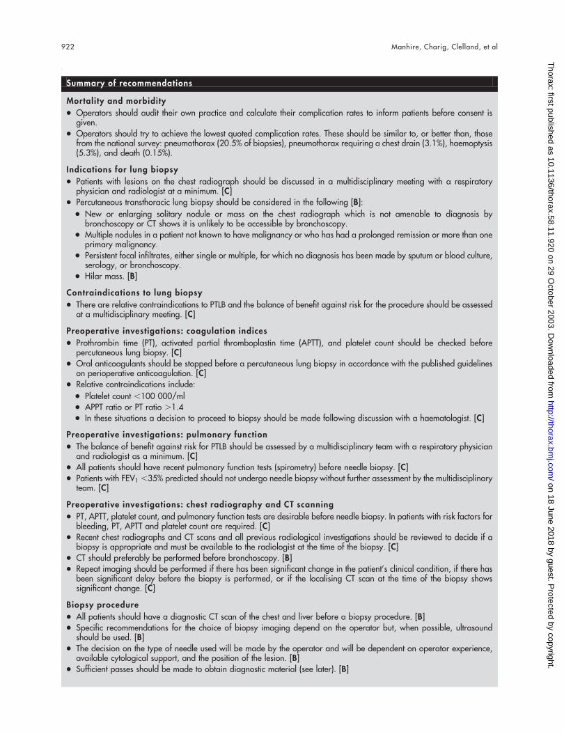
Summary of recommendations
Mortality and morbidity
N Operators should audit their own practice and calculate their complication rates to inform patients before consent isgiven.
N Operators should try to achieve the lowest quoted complication rates. These should be similar to, or better than, thosefrom the national survey: pneumothorax (20.5% of biopsies), pneumothorax requiring a chest drain (3.1%), haemoptysis(5.3%), and death (0.15%).
Indications for lung biopsy
N Patients with lesions on the chest radiograph should be discussed in a multidisciplinary meeting with a respiratoryphysician and radiologist at a minimum. [C]
N Percutaneous transthoracic lung biopsy should be considered in the following [B]:
N New or enlarging solitary nodule or mass on the chest radiograph which is not amenable to diagnosis bybronchoscopy or CT shows it is unlikely to be accessible by bronchoscopy.
N Multiple nodules in a patient not known to have malignancy or who has had a prolonged remission or more than oneprimary malignancy.
N Persistent focal infiltrates, either single or multiple, for which no diagnosis has been made by sputum or blood culture,serology, or bronchoscopy.
N Hilar mass. [B]
Contraindications to lung biopsy
N There are relative contraindications to PTLB and the balance of benefit against risk for the procedure should be assessedat a multidisciplinary meeting. [C]
Preoperative investigations: coagulation indices
N Prothrombin time (PT), activated partial thromboplastin time (APTT), and platelet count should be checked beforepercutaneous lung biopsy. [C]
N Oral anticoagulants should be stopped before a percutaneous lung biopsy in accordance with the published guidelineson perioperative anticoagulation. [C]
N Relative contraindications include:
N Platelet count ,100 000/ml
N APPT ratio or PT ratio .1.4
N In these situations a decision to proceed to biopsy should be made following discussion with a haematologist. [C]
Preoperative investigations: pulmonary function
N The balance of benefit against risk for PTLB should be assessed by a multidisciplinary team with a respiratory physicianand radiologist as a minimum. [C]
N All patients should have recent pulmonary function tests (spirometry) before needle biopsy. [C]
N Patients with FEV1 ,35% predicted should not undergo needle biopsy without further assessment by the multidisciplinaryteam. [C]
Preoperative investigations: chest radiography and CT scanning
N PT, APTT, platelet count, and pulmonary function tests are desirable before needle biopsy. In patients with risk factors forbleeding, PT, APTT and platelet count are required. [C]
N Recent chest radiographs and CT scans and all previous radiological investigations should be reviewed to decide if abiopsy is appropriate and must be available to the radiologist at the time of the biopsy. [C]
N CT should preferably be performed before bronchoscopy. [B]
N Repeat imaging should be performed if there has been significant change in the patient’s clinical condition, if there hasbeen significant delay before the biopsy is performed, or if the localising CT scan at the time of the biopsy showssignificant change. [C]
Biopsy procedure
N All patients should have a diagnostic CT scan of the chest and liver before a biopsy procedure. [B]
N Specific recommendations for the choice of biopsy imaging depend on the operator but, when possible, ultrasoundshould be used. [B]
N The decision on the type of needle used will be made by the operator and will be dependent on operator experience,available cytological support, and the position of the lesion. [B]
N Sufficient passes should be made to obtain diagnostic material (see later). [B]
922 Manhire, Charig, Clelland, et al
on 18 June 2018 by guest. Protected by copyright.
http://thorax.bmj.com
/T
horax: first published as 10.1136/thorax.58.11.920 on 29 October 2003. D
ownloaded from
Recommendations
N Operators should audit their own practice andcalculate their complication rates to inform patientsbefore consent is given.
N Operators should try to achieve the lowest quotedcomplication rates. These should be similar to, orbetter than, those from the national survey: pneu-mothorax (20.5% of biopsies), pneumothorax requir-ing a chest drain (3.1%), haemoptysis (5.3%), anddeath (0.15%).5
INDICATIONS FOR LUNG BIOPSYThe indications for PTLB have altered substantially since thetechnique was developed, reflecting changes in many areasincluding needle technology, imaging techniques, andimmunohistochemistry and cytochemistry.25 26 Furtheradvances, particularly in positron emission tomography
(PET), may alter the indications for needle biopsy and, inparticular, the management of the solitary nodule.
PTLB can be used to investigate any solid or cystic lesionbetween the chest wall and the mediastinum which is notvisible at bronchoscopy, provided it is accessible to theneedle.27–29 FNAB, providing samples for cytology, canaccurately diagnose malignancy, while the more recentdevelopment of cutting needles (CNB), providing histologicalmaterial, has enabled a firm diagnosis of benign lesions to bemade, thus improving overall diagnostic accuracy.30–32
Although PTLB can be used to investigate interstitial lungdisease (particularly in patients with focal areas of con-solidation such as cryptogenic organising pneumonia),transbronchial and thoracoscopic or open lung biopsy arepreferred to minimise the risk of pneumothorax and to obtainlarger and more representative diagnostic samples, particu-larly by open lung biopsy.33
Patients with lesions on the chest radiograph whichrequire a diagnosis should be discussed with a respiratory
Summary of recommendations (continued)
Sedation
N Biopsies should be performed without sedation whenever possible. [C]
Informed consent
N Written information should be given to all patients before the procedure. [C]
N Informed consent should be obtained in a written form from all patients. [C]
Staffing issues
N Staffing should be adequate to enable the patient to be monitored for signs of distress during and after the procedure. [C]
Expected accuracy of sampling
N False positives should be less than 1%. [C]
N Adequacy of sample should be over 90%. [C]
N Sensitivity for malignancy should be within the range of 85–90% in lesions over 2 cm. [C]
N Standards should be set and outcomes audited.
Post biopsy observation
N An erect chest radiograph should be performed 1 hour after the biopsy and is sufficient to detect the majority of postbiopsy pneumothoraces. [B]
N Patients should be informed of the risks of delayed pneumothoraces. [B]
N No specific observations are necessary after the biopsy procedure, but patients should remain in a place where staff canbe alerted if new symptoms develop in the first hour. [C]
N The chest radiograph should be reviewed by a suitably qualified member of staff. [B]
N If a pneumothorax has developed, the clinical condition of the patient and their home circumstances should be consideredbefore deciding on further management. [B]
Management of acute complications
N The operator should be able to identify and appropriately manage the complications of lung biopsy procedures.Resuscitation facilities and chest drain equipment should be immediately available. [B]
N When a complication has occurred, the pulse, blood pressure and oxygen saturations should be monitored and recordedin a severely unwell patient. [C]
Outpatient and day case biopsies
N Percutaneous lung biopsies can be performed safely on an outpatient basis. [B]
N ‘‘High risk’’ patients should not have a biopsy performed as a day case procedure. [C]
N A post biopsy erect chest radiograph should be performed at least 1 hour after the procedure and a decision should bemade at that time regarding further management if a pneumothorax is present. [B]
N Patients should be warned of delayed complications and given verbal and written instructions to return if symptomatic.[C]
N When biopsies are performed on an outpatient basis, patients should live within 30 minutes of a hospital, have adequatehome support, and have access to a telephone. [C]
Guidelines for radiologically guided lung biopsy 923
on 18 June 2018 by guest. Protected by copyright.
http://thorax.bmj.com
/T
horax: first published as 10.1136/thorax.58.11.920 on 29 October 2003. D
ownloaded from

physician and radiologist as a minimum, preferably in amultidisciplinary meeting.34 35 Clinical and radiographicinformation can be reviewed and the likely diagnosisconsidered along with the best approach to making adiagnosis. The risks and benefits of the procedure andknowledge of the wishes of the patient will enable themanagement decision to be tailored to the needs of theindividual.
The indications for PTLB include:
N A new or enlarging solitary nodule or mass on the chestradiograph which is not amenable to diagnosis bybronchoscopy, or CT shows it is unlikely to be accessibleby bronchoscopy, when a decision has been made by themultidisciplinary team that a tissue diagnosis should beobtained.
N Multiple nodules in a patient not known to havemalignancy or who has had a prolonged remission.
N Persistent infiltrates, either single or multiple, for whichno diagnosis has been made by sputum or blood culture,serology or bronchoscopy.
N Hilar mass following negative bronchoscopy.
New or enlarging solitary nodule or mass on chestradiographyThe most common indication for PTLB is to investigate thesolitary parenchymal nodule or mass on the chest radiograph.The next radiological investigation is CT to characterise thelesion and show associated hilar or mediastinal lymphadeno-pathy or evidence of other abnormalities suitable for biopsy.This enables the radiologist and respiratory physician,preferably with the thoracic surgeon, to decide the mostlikely diagnosis and the best management thereof, avoidingbronchoscopy in those patients where CT suggests that atissue diagnosis is unlikely to be obtained by this method.36–40
The likelihood of malignancy increases with the size of thelesion, patient age, a smoking history, and a history ofhaemoptysis.41–43 If the initial probability of malignancy ishigh, many surgeons feel that the correct approach inpatients with isolated small nodules who are otherwise fitand agreeable to surgery is to carry out a diagnosticresection.44 45 Distinguishing between small cell lung cancer(SCLC) and non-small cell lung cancer (NSCLC) is not anissue with these small early lesions as there is evidence ofcure of SCLC in these circumstances.45 46 Thoracic surgery hasa significant mortality (2–3% for lobectomy) and morbiditydue to cardiovascular causes and loss of lung function.45 47–50
Post-thoracotomy pain is a significant problem in approxi-mately 10% of patients in all age groups.51 Accurate diagnosisof benign lesions using CNB has reduced the need fordiagnostic surgery by up to 50%.29 31 44 The advent of PETscanning may also be helpful in determining the need forsurgery, as a lesion which strongly takes up 18F-fluorodeoxy-glucose is more likely to be malignant than benign.52–54
Patients who decline surgery or who are inoperable may beoffered radiotherapy, chemotherapy, or combination treat-ment, but this still requires the diagnosis to be establishedwherever possible.
Biopsy samples can be safely taken from masses abuttingthe pleura under ultrasound guidance using a cutting needlewhich ensures an accurate diagnosis, even in patients withlimited lung function, as the risk of pneumothorax isnegligible when aerated lung tissue is not traversed duringthe procedure.55 56
Cavitating lesions are usually caused by tumours orabscesses. The clinical picture will often distinguish betweenthese two diagnoses, but needle aspiration is helpful in
providing material for bacteriology and to guide treatment inthe latter.35 57
Multiple nodules in a patient not known to havemalignancy or who has had a prolonged remissionSlowly enlarging or new multiple nodules on the chestradiograph may occur in a number of benign conditionsincluding rheumatoid nodules, granulomatous diseases,Wegener’s granulomatosis, or infection (particularly fungal)in the immunocompromised patient and can be diagnosed bycore biopsy.58–65 Multiple lesions of varying size are mostlikely to be malignant and, if the patient is known to have aprimary tumour already, biopsy is unlikely to alter thepresumed diagnosis of metastases.66 If there has been aprolonged remission of a tumour following initial treatmentor the patient has a history of more than one primarymalignancy, oncologists may want confirmation of recur-rence to plan further treatment.
Persistent focal infil tratesPTLB may be used to obtain samples of lung tissue wheninfiltrates persist on the chest radiograph and a diagnosis hasnot been made on cultures of sputum, blood, or lung lavageor other diagnostic techniques. Tissue should be culturedbecause, although the yield is small, the investigation isinexpensive and may increase diagnostic accuracy or guidetreatment,67 particularly in patients who are immunosup-pressed.68 69 A lesion of this type which is not resolving maybe a bronchoalveolar cell carcinoma.
Hilar mass following negative bronchoscopyHilar masses can be accurately diagnosed by needle aspira-tion under CT guidance, depending on the experience of theoperator for smaller lesions.51–53 70–72 Earlier work usingfluoroscopic guidance for needle aspiration showed thatlesions at the hilum could be diagnosed with similar accuracyto peripheral lesions and that the success of the procedurewas related to the size of the lesion.
Recommendations
N Patients with lesions on the chest radiographshould be discussed in a multidisciplinary meetingwith a respiratory physician and radiologist at aminimum. [C]
N Percutaneous transthoracic lung biopsy should beconsidered in the following [B]:
N New or enlarging solitary nodule or mass on thechest radiograph which is not amenable todiagnosis by bronchoscopy or CT shows it isunlikely to be accessible by bronchoscopy.
N Multiple nodules in a patient not known to havemalignancy or who has had a prolonged remissionor more than one primary malignancy.
N Persistent focal infiltrates, either single or multi-ple, for which no diagnosis has been made bysputum or blood culture, serology, or broncho-scopy.
N Hilar mass. [B]
CONTRAINDICATIONS TO LUNG BIOPSYThere are several relative contraindications to PTLB. Patientsshould not undergo the procedure without adequate pre-biopsy assessment or if they plan to fly within 6 weeks of theprocedure. The risk is increased by abnormalities of lungfunction, respiratory failure (including mechanical ventila-tion), arterial and venous pulmonary hypertension, andcoagulation abnormalities (see preoperative investigations).35
The balance of benefit against risk for the procedure should
924 Manhire, Charig, Clelland, et al
on 18 June 2018 by guest. Protected by copyright.
http://thorax.bmj.com
/T
horax: first published as 10.1136/thorax.58.11.920 on 29 October 2003. D
ownloaded from

be assessed at a multidisciplinary meeting. The role of needlebiopsy is to establish a diagnosis to enable appropriatetreatment to be given. Failure to obtain informed consentfrom a patient is a contraindication, and management shouldbe reconsidered in these circumstances.
Previous pneumonectomy is an exclusion criterion forneedle biopsy in many series. However, if the lesion in theremaining lung is pleurally based and is accessible withouttraversing any lung tissue, it may not be considered anabsolute contraindication as the risk of pneumothorax islow.14
Mechanical ventilation will make the process of biopsymore difficult but, if the lesion is visualised by ultrasound, itmay be undertaken. Biopsy samples of intrapulmonarylesions can be taken by experienced operators under CTguidance while ventilation is controlled during the procedure,but this is difficult because of the limited space as well asaccess of medical and nursing staff during radiationexposure.
Vascular lesions, either aneurysms or arteriovenous mal-formations, should have been identified by CT and should notbe subjected to biopsy. This diagnosis should be consideredbefore the biopsy procedure at a multidisciplinary meeting.Biopsy of an unsuspected vascular lesion may lead to anincreased risk of haemorrhage.
Pulmonary arterial and venous hypertension may increasethe risk of haemorrhage but there are no data to support this.If the hypertension is significant, this would be a contra-indication to surgery; the risk of the diagnostic procedureneeds to considered against the benefit of having an answeron patient management.
The uncooperative patientIt is essential that the patient is cooperative duringpercutaneous lung biopsy. A sudden or unexpected move-ment while the biopsy needle is in the lung parenchyma maylead to a tear and subsequent intrapulmonary bleeding and/or pneumothorax. If the patient is frightened despite carefulexplanation and reassurance, an anxiolytic drug may behelpful. If the patient remains uncooperative after thesemeasures, the management should be reconsidered.
Recommendations
N There are relative contraindications to PTLB and thebalance of benefit against risk for the procedureshould be assessed at a multidisciplinary meeting.[C]
Abnormal coagulation indices and lung functionThese are discussed in more detail in the next section.
PREOPERATIVE INVESTIGATIONSCoagulation indicesFortunately, significant bleeding rarely complicates percuta-neous lung biopsies. Quoted complication rates for localpulmonary haemorrhage range between 5% and 16.9% andhaemoptysis between 1.25% and 5%.3 14 Deaths from bleedingfollowing percutaneous lung biopsy are reported althoughfewer than 10 cases have been described.8 73–77
Certain patient groups are known to be at increased risk ofbleeding. These include those who have uraemia, pulmonaryhypertension, liver disease, coagulation disorders or throm-bocytopenia.78 79 Patients with uraemia should be givenDDAVP (desmopressin acetate).
Although complication rates for pneumothorax are similarfor FNAB and automated biopsy devices, a slightly higher,but not significant, incidence of pulmonary bleeding withhaemoptysis is reported in series using automated biopsydevices.9 80
There is no specific guidance in the literature regarding thevalue of routine clotting studies before performing percuta-neous lung biopsy. Many studies have found that preopera-tive screening for coagulopathies not suspected on the basisof detailed clinical examinations is unnecessary.79 81 82 Inthese situations, however, operations and biopsies areperformed under direct vision. This is not the case inpercutaneous lung biopsy. In the absence of specific evidence,routine clotting studies are justifiable and should beperformed in order to minimise the risk of the procedure.In accordance with the British Thoracic Society guidelines ondiagnostic flexible bronchoscopy it is therefore recommendedthat the platelet count, prothrombin time (PT), and activatedpartial thromboplastin time (APTT) should be checked beforeperforming percutaneous lung biopsies.24
There is no information as to what constitutes a ‘‘safe’’level for clotting before the biopsy procedure. In transbron-chial biopsies platelet counts below 50 000/ml have beenshown to be associated with a significant risk of bleeding.83
Opinion in the world literature suggests that a PT orinternational normalised ratio (INR) or APTT ratio of morethan 1.4 and a platelet count below 100 000/ml should berelative contraindications to percutaneous lung biopsy.78
Haematological advice in these circumstances should besought before the biopsy is performed. In patients with ahaemoglobin level of less than 10 g/dl the procedure shouldbe carefully considered, although there is no evidence tosupport a particular figure and there are reports of successfulprocedures in anaemic patients.
If the patient is taking oral anticoagulants, publishedguidelines for the management of anticoagulation in theperioperative period are relevant.84 These state that oralanticoagulation should be stopped before surgery and,depending on the thrombotic risk of the condition for whichthe patient is receiving anticoagulant therapy, the INR can bemeasured and, if necessary, heparin instituted once the INRis below the therapeutic range. It is worth noting that, afterwarfarin is stopped, it takes 4 days typically for the INR toreach 1.5 and therefore oral anticoagulants should be stoppedat least 4 days before a percutaneous lung biopsy isperformed.85 There is no evidence to support stoppingantiplatelet drugs before the procedure.
Recommendations
N Prothrombin time (PT), activated partial thrombo-plastin time (APTT), and platelet count should bechecked before percutaneous lung biopsy. [C]
N Oral anticoagulants should be stopped before apercutaneous lung biopsy in accordance with thepublished guidelines on perioperative anticoagula-tion. [C]
N Relative contraindications include:
N Platelet count ,100 000/ml
N APPT ratio or PT ratio .1.4
N In these situations a decision to proceed to biopsyshould be made following discussion with ahaematologist. [C]
Pulmonary functionThe most common complication of lung biopsy is apneumothorax, and it is essential to assess whether thepatient can safely withstand the procedure, particularly asmany patients being investigated in this way will havesmoking related lung disease in addition to any changes inlung function caused by the lesion under investigation.Measurement of lung function is used to assess this and alsosuitability for surgery if appropriate. Data from several
Guidelines for radiologically guided lung biopsy 925
on 18 June 2018 by guest. Protected by copyright.
http://thorax.bmj.com
/T
horax: first published as 10.1136/thorax.58.11.920 on 29 October 2003. D
ownloaded from
retrospective studies using spirometry are conflicting. Severalstudies found no relation between forced expiratory volumein 1 second (FEV1) and the incidence of pneumothorax,14 86–89
while both Poe et al90 and Vitulo et al91 suggested that this wasrelated to the presence of hyperinflation. However, Andersonet al86 and Fish et al88 found that patients with airflowobstruction were significantly more likely to require tubedrainage if a pneumothorax occurred.
Garcia-Rio et al92 carried out a prospective study of 51patients and were able to show that the mean FEV1 %predicted, FVC (forced vital capacity) % predicted, and theFEV1/FVC ratio were significantly lower in patients whodeveloped pneumothoraces, the closest correlation being withFEV1 % predicted. They estimated that the risk of apneumothorax was 35.6% in patients with an FEV1 reducedto 70% predicted. No figures are suggested for the minimumFEV1 at which the procedure is contraindicated, althoughmost practising physicians and radiologists use a cut off of1 litre. However, this could overestimate the risk of severerespiratory embarrassment with a large pneumothorax in asmall patient while underestimating the risk in a very tallman. It is therefore suggested that the percentage predictedshould be used. An absolute figure of an FEV1 of 1 litre isapproximately 35% predicted for a white man of 70 years and1.73 metres height. This approach is in line with that adoptedin the recently published guidelines on the selection ofpatients with lung cancer for surgery,45 where an absolutefigure of a minimum FEV1 of 1.5 litres is suggested forlobectomy but a figure including percentage predicted is usedfor borderline cases. In cases of poorer lung function wherethe risk of pneumothorax is less—for instance, where thelesion abuts the pleura—biopsy may be possible.
Patients admitted to hospital as emergencies who arefound to have an incidental lesion on the chest radiographshould have their underlying condition stabilised beforereferral to a multidisciplinary team consisting of a respiratoryphysician and a radiologist as a minimum. Patients withrespiratory failure, in particular, should not undergo a biopsyprocedure without careful assessment.
Recommendations
N The balance of benefit against risk for PTLB shouldbe assessed by a multidisciplinary team with arespiratory physician and radiologist as a minimum.[C]
N All patients should have recent pulmonary functiontests (spirometry) before needle biopsy. [C]
N Patients with FEV1 ,35% predicted should notundergo needle biopsy without further assessmentby the multidisciplinary team. [C]
Chest radiographyA chest radiograph will usually have been performed at theinitial assessment of the patient. Alternatively, the lesion mayhave been found incidentally on a chest radiograph or CTscan performed for some other reason. All imaging should bereviewed at a multidisciplinary meeting to decide if a biopsyis appropriate. If there is anything to suggest that thepatient’s condition has changed at the immediate pre-biopsyclinical assessment, the patient should have a repeat chestradiograph. If there has been a substantial change such asnew collapse or consolidation, it may be necessary toreconsider if the biopsy should be performed.
With the current emphasis on speedy investigation,93
the chest radiograph is unlikely to be significantly out ofdate and the lesion will be further assessed at the time ofbiopsy.
Computed tomography (CT)Most patients thought likely to have a lung carcinomawill require imaging with CT to determine the stage oftheir disease and to help determine any appropriatetreatment such as resection, radiotherapy, or palliativeinterventions.93
There are advantages in performing a CT scan before anyfurther intervention in patients thought likely to have a lungcarcinoma. A CT scan performed before fibreoptic broncho-scopy (FOB) has been shown to increase the diagnosticyield of FOB by directing the bronchoscopist to the site mostlikely to give a diagnosis. Bronchoscopy and biopsy itself mayalter the appearance of the CT scan as a result of localhaemorrhage. It can also show carcinomas not visible onbronchoscopy, and may direct the clinician to a moreappropriate method to obtain histological specimenssuch as mediastinoscopy or biopsy of a lesion outside thechest.36–39 94–99 There is no need to perform routine FOB inpatients thought to have resectable disease in whomtissue has been obtained by CT biopsy with no preoperativeFOB.36–39 94–102
A staging CT scan may be performed before biopsy on aseparate occasion. Alternatively, the patient may attend for astaging CT scan and the decision to proceed directly to biopsymay be made while the patient remains in the CT departmentor even on the CT table. The second approach may have someadvantages in terms of patient convenience as only one visitto the department is required, but special care with thisapproach is necessary, particularly with patient consent. Inaddition, there are organisational problems, particularly inunits where care after the biopsy must be organised inadvance.
Delay since investigationAt the time of biopsy a CT chest scan is performed to assessthe best site to biopsy, the most appropriate route, andtherefore the most appropriate patient position. This may be alimited unenhanced study to localise the mass. If there hasbeen a significant delay between the original staging CT scanand the biopsy procedure, there is a risk that the tumour mayhave grown and increased in stage.103 It is imperative that theradiologist performing the procedure is fully aware of allprevious investigations and so can compare the imaging atthe time of the biopsy with previous imaging and consider if arepeat staging CT scan may be necessary. In addition, thereason for the biopsy and its relevance to the patient’smanagement must be clear so that any change in thepatient’s condition or imaging can be taken into account todecide if the biopsy is still appropriate.
Recommendations
N PT, APTT, platelet count, and pulmonary functiontests are desirable before needle biopsy. In patientswith risk factors for bleeding, PT, APTT and plateletcount are required. [C]
N Recent chest radiographs and CT scans and allprevious radiological investigations should bereviewed to decide if a biopsy is appropriate andmust be available to the radiologist at the time ofthe biopsy. [C]
N CT should preferably be performed before broncho-scopy. [B]
N Repeat imaging should be performed if there hasbeen significant change in the patient’s clinicalcondition, if there has been significant delay beforethe biopsy is performed, or if the localising CT scanat the time of the biopsy shows significant change.[C]
926 Manhire, Charig, Clelland, et al
on 18 June 2018 by guest. Protected by copyright.
http://thorax.bmj.com
/T
horax: first published as 10.1136/thorax.58.11.920 on 29 October 2003. D
ownloaded from

THE BIOPSY PROCEDUREProtective clothingThe procedure should be performed using standard universalprecautions. Protective gloves should be worn. If possible,non-powdered latex gloves or non-latex gloves should beused.104
Patient posit ioning and instructionThe patient should be positioned prone or supine dependenton the skin entry site chosen. Biopsy specimens should not betaken with the patient in a seated position because of thepotential small risk of air embolus105 or fainting during theprocedure. It is difficult for patients to maintain a consistentdecubitus position and this should be avoided if possible. Thebreathing technique required during the procedure should beexplained to the patient and practised beforehand. Deepbreaths and coughing should be avoided during the biopsyprocedure.
Imaging techniquesThe decision on the most appropriate imaging modality usedfor biopsy is made on reviewing the pre-biopsy CT scan.Fluoroscopy, CT, and ultrasound may all be used for imagingguidance, and familiarity of the operator with all threemodalities is helpful in choosing the most appropriatetechnique.35 78
The imaging technique chosen is dependent on thesize and position of the lesion, its visibility on plainradiographs, its relation to other structures such as fissures,vessels and bullae, equipment availability, and operatorpreference.35 78 106 107
Whenever possible, PTLB should be performed underultrasound guidance as this is the safest, quickest, and leastexpensive method.108 For lesions not suitable for ultrasoundguided biopsy, CT is now the preferred imaging modal-ity.26 31 35 78 108 Fluoroscopic guidance may also be used forlarger lesions visualised on a posteroanterior and lateral chestradiograph.109 110
If the biopsy is to be performed using fluoroscopy, the bestresults are usually obtained with C-arm screening (or, ifavailable, bi-plane) with vertical or horizontal needle inser-tion.106 109 110 The correct depth of needle insertion may beestimated from the pre-biopsy CT scan. If using ultrasound,needle entry into the lesion should be directly visualised andbiopsy sites away from identified areas of cavitation ornecrosis chosen.107 111–113
If using CT, a needle entry site which avoids crossingfissures, bullae and large vessels should be chosen if possibleto reduce the incidence of pneumothorax and haemorrhage.
Biopsy techniqueThe skin entry site should be sterilised with standardisedantiseptic solution and the cutaneous and subcutaneoustissue infiltrated with lidocaine (lignocaine) up to amaximum dose of 20 ml of a 2% solution. The pleura shouldnot be anaesthetised directly as this increases the risk ofpneumothorax before the biopsy itself.
When the biopsy needle is being advanced or withdrawnthe patient should suspend respiration. Most patients find itmore comfortable to hold their breath after a submaximalinspiration. For lesions at the lung bases a breath held ongentle expiration may make the biopsy procedure easier.Wherever possible a needle entry site immediately cephaladto a rib should be chosen to avoid intercostal vesselpuncture.114 Care should be taken to avoid the internalmammary vessels if the biopsy is performed adjacent to thecostal cartilages and sternum.115
In all instances the biopsy needle should be advanced orwithdrawn only during suspended respiration. The patientmay breathe gently with the needle in place. If an aspiration
biopsy is performed, the central stylet is removed and a 10 mlsyringe attached. Suction should then be applied whilerotating and moving the needle to and fro during suspendedrespiration.116 117
Instillation of autologous blood along the needle track hasbeen reported to be of benefit in some reports but is notcommonly used and its routine use is probably notwarranted.118–120
A coaxial technique may be used to allow multiple passesand to reduce the number of pleural punctures.26 78
If a CNB is performed, it is important to confirm before theprocedure that either the tip of the needle remains within thelesion once fired or stops in a safe place.
Type of needle and number of passesThe number of passes needed per procedure has not beendefined. Most operators perform at least two. Variables toconsider are: the difficulty of the procedure, complicationsarising from each biopsy, the quality of the specimenobtained, the characteristics of the lesion biopsied, and theneed for specimens for cytological, histological and micro-biological examination. The presence of an on-site cytologistor technician may reduce the number of passes required.121–125
When deciding whether to use FNAB or a cutting needlebiopsy (CNB), it is important to be aware of the accuracy ofthe technique as well as its complications.
Ideally, the technique must not only be able to diagnosemalignancy but also to make a definite diagnosis if the lesionis benign. Different populations have different ratios ofbenign to malignant disease and this can affect reportedpositive and negative predicted values.126
FNAB has an accuracy of up to 95% for malignant lesions127
but the yield for benign lesions is lower (10–50%).32 128–131
Cytology is reported to be less reliable than histology indetermining the cell type in malignant lesions,9 38 129 132
although Stewart and Stewart133 were able to diagnosecorrectly small and large cell carcinomas in their series.
There is wide variation in reported diagnostic accuracies ofFNAB between different institutions, ranging from 64% to97%.10 134 A high diagnostic accuracy is best achieved withlarge nodules134–136 and a cytopathologist present to evaluatethe adequacy of the specimens.121 133 A cytopathologist is notavailable in many centres and this factor may persuade theoperator to use a cutting needle, particularly for smallnodules.11 80 The high diagnostic figures reported from someAmerican studies may be achieved after repeated biopsiesover a short period (2 or 3 hours) rather than a singleepisode.
Several recent studies have advocated the use of 18 and 20gauge cutting needles as well as coaxial techniques toimprove the diagnostic yield and have achieved diagnosticaccuracies for malignancy of 74–95%.9 137 138 Others139 140 havefound the diagnostic yield for malignancy with CNB to belower than with FNAB. Charig and Phillips11 found thediagnostic accuracy of CNB to be similar to FNAB with an on-site cytopathologist. Their accuracy was in line with thosereported from other core biopsy studies.
Using a 21 gauge CNB needle and frozen section, Stewartand Stewart133 achieved a specific diagnosis in 77% of benignlesions; in other studies CNB improved the diagnostic yieldcompared with cytology from 10% to 40%128 and from 16.7%to 81.7%.32 The reported specific diagnosis in cases of benigndisease varies from 78.3%11 32 to 91%,9 although this mayreflect the local populations.
The false negative results for malignancy may be due to avariety of factors including the patient’s inability to coop-erate, overlying bone which may contribute to missing thelesion completely, obtaining only necrotic tissue, or samplingpneumonitis distal to an obstructing lesion.90 136 These factors
Guidelines for radiologically guided lung biopsy 927
on 18 June 2018 by guest. Protected by copyright.
http://thorax.bmj.com
/T
horax: first published as 10.1136/thorax.58.11.920 on 29 October 2003. D
ownloaded from

are the same irrespective of the choice of needle, but the falsenegative rate has been shown to be significantly lower withcutting needles.140
False positive rates of 0.8% have been reported withFNAB110 but no false positive cases have been reported usingCNB.
ComplicationsHistorically, cutting needles have been associated with arelatively high incidence of complications but many of thesedata are based on large calibre, non-automated needles usingfluoroscopic guidance. Recent studies using small gaugecutting needles have shown complication rates comparableto, or only slightly higher than, those using FNAB.9 80 141 Themost common complication following lung biopsy is pneu-mothorax, with reported rates of 0–61% for FNAB and 26–54% for CNB, requiring chest drain insertion in 1.6–18% and3.3–15% of cases, respectively.9–13 However, the pneu-mothorax rate is negligible if the lesion biopsied is peripheraland abuts the pleural surface.27
There is no correlation between the size or type of needleand the incidence of pneumothorax, although there is a non-significant trend towards increased haemorrhagic complica-tions with cutting needles.11 140 142–144 Reports show that thereis no correlation between the number of passes made withthe biopsy needle or chest drain placement and the rates ofpneumothorax,11 13 142 143 although this is counterintuitive tomost operators’ experience.
In conclusion, the literature indicates that cutting needleshave the same sensitivity in the diagnosis of malignancy asFNAB with an on-site cytopathologist (see later), are betterable to produce a specific benign diagnosis, have asignificantly lower false negative rate and, by providing ahistological core, should have a negligible false positive rate.The complication rate is not associated with needle type orsize.
Recommendations
N All patients should have a diagnostic CT scan of thechest and liver before a biopsy procedure. [B]
N Specific recommendations for the choice of biopsyimaging depend on the operator but, when possible,ultrasound should be used. [B]
N The decision on the type of needle used will be madeby the operator and will be dependent on operatorexperience, available cytological support, and theposition of the lesion. [B]
N Sufficient passes should be made to obtain diag-nostic material (see later). [B]
SEDATIONThe cooperation of the patient is paramount during thebiopsy procedure, particularly in suspending respiration. Assuch, virtually all biopsies should be performed withoutsedation. Adequate explanation before the procedure andadequate local anaesthesia during the procedure renderssedation unnecessary for the majority of patients. An oralanxiolytic drug can occasionally be helpful.
Recommendation
N Biopsies should be performed without sedationwhenever possible. [C]
INFORMED CONSENTInformed consent should be obtained in writing before thebiopsy procedure in accordance with individual hospitalpolicies. Verbal and understandable written patient informa-tion before diagnostic procedures improves the patient’s
tolerance of the procedure, as has been shown in broncho-scopy.145 146
Valid consent as defined by the Department of Health147
‘‘must be given voluntarily by an appropriately informedperson who has the capacity to consent to the intervention inquestion. Acquiescence where the person does not knowwhat the intervention entails is not ‘consent’’’. The capacityof the patient to give consent and the circumstances in whichit should be given are also set out in the document. Sufficientinformation should be provided as to the nature and purposeof the procedure and ‘‘any misrepresentation of theseelements will invalidate consent’’.
In considering what information to provide, the healthprofessional should try to ensure that the patient is able tomake a balanced judgement on whether to give or withholdconsent.
Specific risks to be mentionedFrom the above it can be concluded that the commoncomplications such as pneumothorax with, for example, thepossibility of chest drainage and haemoptysis should bediscussed and the local complication rates for these problemsmentioned. Death is extremely uncommon as a consequenceof lung biopsy and its inclusion in the discussion of theprocedure may be regarded as likely to cause unnecessaryanxiety to the patient, but a complete explanation of all risksis advocated by the General Medical Council.148
The NHS Litigation Authority has published some stan-dards aimed at managing clinical risk.149 These state that,before starting any treatment, doctors must ensure that theyhave established:
N what the patient wants to know;
N what the patient ought to know;
N that the patient understands the information which hasbeen given;
N that the patient consents to the treatment;
N proposals for treatment should be supported by writteninformation.
Who should obtain consent?The clinician performing the investigation is responsible forensuring that the patient has given valid consent beforetreatment begins. The task of seeking consent may bedelegated to another health professional provided thatprofessional is suitably trained and qualified.147
The physician or surgeon who is looking after the patientshould explain how the biopsy fits into the overall manage-ment and discuss the alternatives. These might range fromdoing nothing to excision biopsy. Most lung biopsies will beperformed by radiologists who will be in a less appropriateposition to do this.
The patient should receive an appropriately writtenpamphlet either at the time of discussion of options in theclinic or with the appointment. In some units it is proposedthat the patient signs each part of the information sheet toshow that they have read it.
The operator performing the biopsy will then be able todeal with any last minute questions and obtain writtenconsent. Patients should not be expected to make up theirmind about the biopsy when they arrive for the procedure asthere is nearly always an interval of several days from theinitial suggestion of the biopsy to the actual event.
The patient should understand:
N the nature of the proposed procedure;
N the reason for the procedure;
N the benefits of the procedure;
928 Manhire, Charig, Clelland, et al
on 18 June 2018 by guest. Protected by copyright.
http://thorax.bmj.com
/T
horax: first published as 10.1136/thorax.58.11.920 on 29 October 2003. D
ownloaded from
N the risks and complications;
N alternatives to the procedure (including an assessment ofthe relative risk:benefit ratios);
N the nature of the anaesthetic to be employed and theimaging modality.
Patient informationIt has been strongly recommended by several bodies that it isgood practice to provide written information to patients.148
Various pamphlets are available and these can be useddirectly or adapted to local circumstances. The Royal Collegeof Radiologists produces a series for several interventionalprocedures which were developed with the help of patientrepresentatives.150 Examples of a patient information sheetand biopsy request proforma are given in Appendices 1 and 2.
Patient understanding and retention of the risks can beincreased by encouraging them to recite the procedure risksbefore the biopsy.151
Recommendations
N Written information should be given to all patientsbefore the procedure. [C]
N Informed consent should be obtained in a writtenform from all patients. [C]
STAFFING ISSUESAll interventional procedures require the involvement of ateam to ensure patient comfort and safety and the technicalsuccess of the procedure. Lung biopsy has not been addressedspecifically, but two reviews of interventional radiologyprocedures have been published by the NationalConfidential Enquiry into Perioperative Deaths (NCEPOD)152
and the Royal College of Radiologists (RCR) and the RoyalCollege of Nursing (RCN).153
NCEPOD indicates that:
N the patient should be under the care of the appropriatespecialist;
N the radiologist should have sufficient expertise to performthe procedure safely and to deal with any complicationsthat may arise;
N there should be sufficient staff to perform the proceduresafely;
N monitoring of the patient should be performed during allinterventional radiological procedures and this should bedone by someone other than the radiologist performingthe procedure.
The RCR and RCN also regard the trained radiology nurseas part of the interventional team, both to allay patientanxiety and to ensure careful monitoring of the patientduring and immediately after the procedure during recovery.In some circumstances there is also a role for the nurse in thepre-assessment of the patient by instigating or checkinginvestigations according to agreed protocols and pathways.There should be sufficient nurses to fulfil these requirements,although the number may vary according to the activity ofthe department.
Recommendations
N Staffing should be adequate to enable the patient tobe monitored for signs of distress during and afterthe procedure. [C]
Experience of the operatorLung biopsies are almost always performed by or under theclose supervision of an experienced consultant.5 134 The
number of pneumothorax complications may decrease withoperator experience.154
SAMPLE EXAMINATION AT THE TIME OF BIOPSYMacroscopic appearanceMacroscopic examination (visual inspection) of the biopsyspecimen may also enable an estimate of the likelihood ofachieving a diagnosis per procedure,124 and most radiologistsperform this practice.
Immediate pathological examinationFor FNAB, there have been a number of reports of the valueof having a cytologist or cytotechnician present at the time ofthe biopsy procedure.121–123 125 155 It is likely that immediatemicroscopic examination reduces the number of biopsyspecimens required to achieve a diagnosis, but for mostcentres this is not a realistic option.
There are no data on the immediate assessment of coreadequacy following CNB, and visual inspection to confirm anadequate tissue sample seems an appropriate practice. It ispossible to perform touch preparation imprints on core biopsyspecimens to obtain cytology and these appear to offer asimilar accuracy to cytological examination.125
EXPECTED ACCURACY OF SAMPLINGOver a 10 year period in one US laboratory the annualnumber of lung FNA samples increased from 13 to 206.156 Asit has gained in popularity, the accuracy of lung FNA hascome under scrutiny. Audit has shown that this techniquehas a higher rate of positivity for malignancy than any formof endoscopic bronchial sampling.157 Problems, where theyexist, revolve mainly around adequacy and, to a lesser extent,accuracy of cell typing, but overall the technique has provedextremely successful and is continually being further refined.Core CNB does not necessarily confer any significantdiagnostic advantage over FNA in the diagnosis of malig-nancy,139 although some authors advocate it for benignlesions.32
Sensitivity, specificity, and adequacySeveral large studies of the accuracy of lung FNA have beenreported and sensitivity, specificity, and adequacy of over90% are achievable.26 35 106 158 159 The false positive rate isusually less than 1%,160 161 and the false negative rate isgenerally under 10%.30 32 70 162 Diagnostic accuracy is depen-dent on the size and site of the lesion, operator experience,needle type, choice of biopsy technique, and availability ofcytopathology expertise.78 106
Larger lesions are more likely to enable a positive diagnosisof malignancy,78 109 110 163–165 although some operators havereported no significant difference in lesions more or less than2 cm.31 62 The reports from lung cancer screening programmesalso support the ability to achieve an accurate diagnosis inlesions of less than 1 cm.166 Accuracy may be furtherimproved by reducing the number of inadequate samplesby using new techniques.35 167
Some authors expound on the value of on-site technicalassistance with smear preparation and immediate reportingto enhance the adequacy rate.133 168
Cell typingTo provide clinically useful cytology reports in terms ofappropriate treatment, accurate cell typing is required. FNAcan reliably distinguish small cell carcinoma from non-smallcell carcinoma.135 The highest accuracy is obtained with adiagnosis of small cell carcinoma with lower accuracy fordiagnosing squamous carcinoma and adenocarcinoma.10 11
Interobserver variation studies suggest that diagnosis bycytology is almost as reproducible as by histology.139 156 168
Areas of difficulty in cell typing include the distinction of
Guidelines for radiologically guided lung biopsy 929
on 18 June 2018 by guest. Protected by copyright.
http://thorax.bmj.com
/T
horax: first published as 10.1136/thorax.58.11.920 on 29 October 2003. D
ownloaded from

small cell carcinoma from poorly differentiated non-kerati-nising squamous cell carcinoma, small cell carcinoma frommalignant lymphoma, and combined small cell carcinomafrom non-small cell carcinoma.169 Accurate cell typing isreported in the setting of a small hospital,169 but specialistcytopathology training is recommended.170
ImmunocytochemistryAs an aid to diagnosis, immunocytochemistry may beundertaken in a minority of cases. Using a panel of antibodiescan assist in identifying metastatic lesions and confirming apulmonary origin for some adenocarcinomas.171
Immunocytochemistry of FNA samples can deliver compar-able results to those obtained from biopsy material,167
although it is rarely feasible to perform more than a smallpanel of immunostains on an FNA sample. Liquid-basedcytology has much to recommend it, even for the preparationof FNA samples.172 Instead of, or as well as, making smears,the sample is washed into a preservative solution. Goodfixation is ensured and it is simple to make multiplepreparations for immunocytochemistry. It is at least asaccurate as smear preparations and, by removal of red cells,debris and inflammatory cells, the slides are quicker andeasier to read. It does, however, involve extra technical workand cost.
‘‘Suspicious for malignancy’’ and ‘‘negative formalignancy’’ diagnosesThe diagnostic category ‘‘suspicious, but not diagnostic ofmalignancy’’ usually comprises 4–13% of results.158 160 Thiscan affect the sensitivity of the test as some calculations ofsensitivity include and others exclude these from themalignant category. Certainly, on follow up of FNA samplesclassified as suspicious, a high proportion of the lesions turnout to be malignant.160 173 Long term follow up of patientswith negative FNA samples shows a significant proportionfinally have a diagnosis of malignancy.160 173
Factors affecting diagnostic accuracyThe reported diagnostic accuracy rate is dependent on thesize of the lesion,10 26 31 35 78 136 163 164 the location of the lesion,operator experience,134 type of needle used (FNA orCNB),20 26 35 78 135 choice of biopsy technique,26 35 78 135 136 163 164
the pretest probability of malignancy,126 174 and the expertiseof the reporting pathologist.174
Larger lesions are more likely to allow a positive diagnosisof malignancy to be made,10 31 134 136 165 although someoperators have reported no significant differences in resultsbetween lesions more or less than 2 cm.134 The reports fromlung cancer screening programmes also support the ability toachieve an accurate diagnosis in lesions of less than 1 cm.166
Accuracy may be further improved by reducing the numberof inadequate samples using new techniques.174 175
Recommendations
N False positives should be less than 1%. [C]
N Adequacy of sample should be over 90%. [C]
N Sensitivity for malignancy should be within therange of 85–90% in lesions over 2 cm. [C]
N Standards should be set and outcomes audited.
POST BIOPSY OBSERVATIONMost complications occur immediately or within the firsthour of a PTLB. One hour following the biopsy procedure,most pneumothoraces are detectable on a chest radio-graph.13 87 176 177 Perlmutt et al,177 in a series of 673 biopsies,detected 98% of pneumothoraces on radiographs taken eitherimmediately after the procedure or at 1 hour. More recentliterature documents delayed pneumothoraces presenting24 hours after biopsy, despite normal 1 and 4 hour postbiopsy radiographs.178
No specific monitoring is required following an uncompli-cated biopsy procedure, but patients should remain inhospital for at least 1 hour, or longer if further radiographsare required to monitor a pneumothorax. Patients should bein a supervised area so staff can be alerted if they developshortness of breath, chest pain, or other symptoms.
Recommendations
N An erect chest radiograph should be performed1 hour after the biopsy and is sufficient to detect themajority of post biopsy pneumothoraces. [B]
N Patients should be informed of the risks of delayedpneumothoraces. [B]
N No specific observations are necessary after thebiopsy procedure, but patients should remain in aplace where staff can be alerted if new symptomsdevelop in the first hour. [C]
N The chest radiograph should be reviewed by asuitably qualified member of staff. [B]
N If a pneumothorax has developed, the clinicalcondition of the patient and their home circum-stances should be considered before deciding onfurther management. [B]
The British Thoracic Society guidelines state that a patientshould not travel by air within 6 weeks of thoracic surgery orresolution of a spontaneous pneumothorax.179
MANAGEMENT OF ACUTE COMPLICATIONSPneumothoraxA pneumothorax complicates up to 61% of all lungbiopsies.9–16 90 178 180–183 Acutely symptomatic pneumothoracesmay develop at the time of the lung biopsy procedure andrequire immediate drainage. Smaller or better toleratedpneumothoraces will be detected on post biopsy chestradiographs.
PresentationAcute presentation is usually with acute ipsilateral chest painand dyspnoea. Clinical findings may be minimal or mayinclude diminished breath sounds and mediastinal shift. In atension pneumothorax the patient may become tachycardicand hypotensive and develop cyanosis. Monitoring of oxygensaturation is advised, together with the administration ofoxygen as necessary. In an acutely unwell patient a chestradiograph or CT scan can be used to identify whethersymptoms relate to pulmonary haemorrhage or pneu-mothorax. In a supine patient a pneumothorax mayaccumulate inferiorly, producing a deep radiolucent costo-phrenic sulcus on the chest radiograph.
AUDIT POINTS
N Accuracy of sampling
N Pneumothorax rate as detected by chest radiographyand the numbers requiring intervention
N Day case complication rates
N Post biopsy haemorrhage requiring transfusion
N Adequate completion of pre-procedure tests
N Discussion at multidisciplinary meeting
N Death rate
930 Manhire, Charig, Clelland, et al
on 18 June 2018 by guest. Protected by copyright.
http://thorax.bmj.com
/T
horax: first published as 10.1136/thorax.58.11.920 on 29 October 2003. D
ownloaded from
Timing of chest radiographySeveral studies have shown that most significant pneu-mothoraces will be detected on a chest radiograph performed1 hour after the procedure, although they may not be visibleon radiographs taken immediately after the procedure.11 178 181
Ultrasound and CT guided biopsies enable detection of verysmall pneumothoraces,16 181 184 but will still require follow upchest radiography to assess progression. Occasional delayedpneumothoraces have been reported more than 24 hoursafter biopsy, despite the absence of a pneumothorax on chestradiographs taken 4 hours after biopsy.184
Management optionsWhere a pneumothorax is detected following a biopsyprocedure, the management options include observation,aspiration, or drain insertion. This decision will be affected byfactors such as the size of pneumothorax, co-existent lungpathology such as emphysema affecting respiratory reserve,and pain suffered. BTS guidelines on the management ofpneumothorax suggest initial treatment by aspiration, withsubsequent drainage if a leak and significant pneumothoraxpersist. A small gauge drain is usually adequate.179
Chest drains are required in 3.3–15% of all patientsundergoing lung biopsy.9–11 13 In the UK most cliniciansattach drains to an underwater seal, but the Heimlich oneway flutter valve is an alternative. This valve allowsprolonged drainage for a pneumothorax and outpatientmanagement. If the pneumothorax continues to enlarge orthe patient develops surgical emphysema, the flutter valvecan be replaced by a system attached to an underwater seal.182
Pulmonary haemorrhage or haemoptysisPulmonary haemorrhage may occur with or without haemo-ptysis. Haemorrhage is recorded in 5–16.9% and haemo-ptysis in 1.25–5% of patients.3 4 Lesion depth has beenidentified as the most important risk factor for haemorrhage.An increased risk of bleeding occurs in lesions deeper than2 cm.16
Pulmonary haemorrhage in the absence of haemoptysis isusually minor and often asymptomatic but, if larger, it maypresent with the patient becoming confused from hypoxia orshocked. The differential diagnosis includes pneumothorax,haemothorax, or an air embolism. Initial treatment shouldinclude oxygen and general resuscitation. A chest radiographis useful to identify a pneumothorax or pleural collection. Theclinical team should be contacted.
Haemoptyses are usually self-limiting. Patient reassuranceand being placed in a lateral position with the biopsy sidedown will often be adequate. If there is a more significanthaemorrhage, patient resuscitation and oxygen should beadministered and the clinical team contacted. In somecentres there may be an option of selective bronchialintubation or of performing a rigid bronchoscopy to protectthe opposite lung in patients with severe haemorrhage.5
Air embolismThe incidence of air embolism is unknown. There are singlecase reports of fatalities and some survivors.6 7 The complica-tion may be overlooked if there is a fatality, unless thepathologist has been alerted to the possibility.
Air embolism may lead to gas within the intracerebral orcoronary circulation. This may occur because of a broncho-venous fistula created at the time of the biopsy procedure orbecause, on removal of the needle stylet, air is inadvertentlyaspirated through the needle lumen into the pulmonary vein.Lung biopsies should be performed with the patient prone orsupine so that, in the event of an air embolism, air is lesslikely to travel to the cranial circulation.
Presentation may be with cardiac or neurological symp-toms and signs: chest pain or rapid circulatory collapse,
generalised seizures or focal neurological defects. Patientoutcome is variable but is usually fatal, although theincidence is unknown. Fatal dysrhythmias may occur froma small volume of air, or air may dissolve and symptomssubside within minutes. In some cases intravascular air hasbeen identified up to 48 hours after the biopsy procedure.
The diagnosis may be confirmed on the CT scan byidentifying gas within the intracranial or coronary circula-tion. Fundoscopy may show the presence of frothing blood inretinal vessels.
Treatment is to administer 100% oxygen and anticonvul-sants where necessary. The patient should be placed in theTrendelenburg position or in a left lateral decubitus positionin case of a residual gas collection within the left heart.Steroids and aspirin are also recommended. Hyperbaricoxygen therapy has been used with a successful outcome inone case report.182
HaemothoraxSignificant haemothorax is rare but may develop from biopsyprocedures through the intercostal or internal mammaryarteries.8 When a large haemothorax develops, the patientshould be given supportive care and the clinical teamcontacted. Signs of this are usually evident within the firsthour. Assistance from general or thoracic surgeons andinterventional vascular radiologists may be needed.
Recommendations
N The operator should be able to identify and appro-priately manage the complications of lung biopsyprocedures. Resuscitation facilities and chest drainequipment should be immediately available. [B]
N When a complication has occurred, the pulse, bloodpressure and oxygen saturations should be mon-itored and recorded in a severely unwell patient. [C]
OUTPATIENT AND DAY CASE BIOPSIESDelayed pneumothorax is a rare but recognised complicationfollowing lung biopsy.181 Dennie et et al183 studied 506 patientsundergoing PTLB. Patients were discharged after a 30 minutepost biopsy chest radiograph if there was no pneumothoraxand after a 60 minute radiograph if they had a stableasymptomatic pneumothorax. Symptomatic or enlargingpneumothoraces were treated with pigtail catheter insertionattached to a Heimlich valve and discharged. Seven (1.4%)patients developed a symptomatic pneumothorax afterdischarge and all required treatment. There were no deathsor other major complications. Catastrophic haemorrhagefollowing biopsy has been reported as a cause of deathfollowing PTLB.75 76 This has occurred swiftly in each case.There are no reports in the literature of delayed haemorrhagecausing death or serious morbidity.
There is no specific guidance in the literature on the choiceof patients for day case biopsy. High risk patients—that is,those with borderline lung function (see earlier) and thosewith significant co-morbid pathology or inadequate homesupport—should not have a biopsy performed as a day caseprocedure. In all the studies patients lived within 30 minutesof the hospital and had access to a telephone.11 176 183
Following an outpatient biopsy, patients should be givenverbal and written instructions to telephone or return to thehospital if they develop breathlessness, chest pain, orhaemoptysis.
Outpatient biopsies do not affect outcome either in termsof complication rate or diagnoses achieved. All studies reporta comparable diagnostic rate for biopsies performed oninpatients.11 87 176 183 There are economic advantages to out-patient biopsies. One study estimated a reduction in costs of
Guidelines for radiologically guided lung biopsy 931
on 18 June 2018 by guest. Protected by copyright.
http://thorax.bmj.com
/T
horax: first published as 10.1136/thorax.58.11.920 on 29 October 2003. D
ownloaded from

27% in an uncomplicated procedure.87 There is a reduction ininconvenience for the patient, and wasted resources causedby cancellations due to bed shortages may be prevented.
Recommendations
N Percutaneous lung biopsies can be performed safelyon an outpatient basis. [B]
N ‘‘High risk’’ patients should not have a biopsyperformed as a day case procedure. [C]
N A post biopsy erect chest radiograph should beperformed at least 1 hour after the procedure and adecision should be made at that time regardingfurther management if a pneumothorax is present.[B]
N Patients should be warned of delayed complicationsand given verbal and written instructions to returnif symptomatic. [C]
N When biopsies are performed on an outpatientbasis, patients should live within 30 minutes of ahospital, have adequate home support, and haveaccess to a telephone. [C]
Authors’ affiliations. . . . . . . . . . . . . . . . . . . . .
A Manhire, M Charig, F Gleeson, H Moss, K Pointon, Royal College ofRadiologistsA Manhire, F Gleeson, R Miller, H Moss, K Pointon, C Richardson,E Sawicka, British Thoracic SocietyA Manhire, M Charig, British Society of Interventional RadiologyC Clelland, Royal College of PathologistsA Manhire, M Charig, F Gleeson, H Moss, K Pointon, British Society ofChest Radiologists
REFERENCES1 Petrie J, Barnwell B, Grimshaw J, on behalf of the Scottish Intercollegiate
Guidelines Network. Clinical guidelines: criteria for guidelines for nationaluse. Edinburgh: Royal College of Physicians, 1995.
2 Harbour R, Miller J. A new system for grading recommendations in evidencebased guidelines. BMJ 2001;323:334–6.
3 Sinner W. Complications of percutaneous thoracic needle aspiration biopsy.Acta Radiol Diagn 1976;17:Fasc. 6 November. [III]
4 Berquist TH, Bailey PB, Cortese DA, et al. Transthoracic needle biopsy:accuracy and complications in relation to location and type of lesion. MayoClin Proc 1980;55:475–81.
5 Richardson CM, Pointon KS, Manhire AR, et al. Percutaneous lung biopsies:a survey of UK practice based on 5444 biopsies. Br J Radiol2002;75:731–5. [III]
6 Kodama F, Ogawa T, Hashimoto M, et al. Fatal air embolism as acomplication of CT guided needle biopsy of the lung. J Comput Assist Tomogr1999;23:949–51. [IV]
7 Tolly TL, Feldmeier JE, Czarnecki D. Air embolism complicating percutaneouslung biopsy. AJR 1988;150:555–6. [IV]
8 Glassberg RM, Sussman SK. Life threatening haemorrhage due topercutaneous thoracic intervention: importance of the internal mammaryartery. AJR 1990;154:47–9. [IV]
9 Klein JS, Salomon G, Stewart EA. Transthoracic needle biopsy with acoaxially placed 20- gauge automated cutting needle: results in 122 patients.Radiology 1996;198:715–20. [III]
10 Westcott JL. Percutaneous transthoracic needle biopsy. Radiology1988;169:593–601. [III]
11 Charig MJ, Phillips AJ. CT-guided cutting needle biopsy of lung lesions –safety and efficacy of an out-patient service. Clin Radiol 2000;55:964–9. [III]
12 Sakai T, Hayashi N, Kimoto T, et al. CT-guided biopsy of the chest: usefulnessof fine-needle core biopsy combined with frozen-section pathologicdiagnosis. Radiology 1994;190:243–6. [III]
13 Kazerooni EA, Lim FT, Mikhail A, et al. Risk of pneumothorax in CT-guidedtransthoracic needle aspiration biopsy of the lung. Radiology1996;198:371–375. [III]
14 Haramati LB, Austin JHM. Complications after CT-guided needle biopsythrough aerated versus non-aerated lung. Radiology 1991;181:778. [III]
15 Bungay HK, Berger J, Traill ZC, et al. Pneumothorax post CT guided lungbiopsy: a comparison between detection on chest radiographs and CT.Br J Radiol 1999;72:1160–3. [III]
16 Yeow KM, See LC, Lui KW, et al. Risk factors for pneumothorax and bleedingafter CT-guided percutaneous coaxial cutting needle biopsy of lung lesions.J Vasc Intervent Radiol 2001;12:1305–12. [III]
17 Collings CL, Westcott JL, Banson NL, et al. Pneumothorax and dependentversus non-dependent patient position after needle biopsy of the lung.Radiology 1999;210:59–64. [III]
18 Moore EH, Shepard JO, McLoud TC, et al. Positional precautions in needleaspiration lung biopsy. Radiology 1990;175:733–5.
19 Bourgouin PM, Shepard JO, McLoud TC, et al. Transthoracic needleaspiration biopsy: evaluation of the blood patch technique. Radiology1988;166:93–5. [IIb]
20 Gruden JF, Stern EJ. Bilateral pneumothorax after percutaneoustransthoracic needle biopsy. Evidence for incomplete pleural fusion. Chest1994;105:627–8. [IIb]
21 Voravud N, Shin DM, Dekmezian RH, et al. Implantation metastasis ofcarcinoma after percutaneous fine needle aspiration. Chest1992;102:313–5. [IV]
22 Raftopoulos Y, Furey WW, Kacey DJ, et al. Tumour implantation aftercomputer-tomography guided biopsy of lung cancer. J Thorac CardiovascSurg 2000;119:1288–9. [IV]
23 Kucharczyk W, Weisbrod GL, Cooper JD, et al. Cardiac tamponade as acomplication of thin needle aspiration lung biopsy. Chest 1982;82:120–1.[IV]
24 British Thoracic Society. British Thoracic Society guidelines on diagnosticflexible bronchoscopy. Thorax 2001;56(Suppl I):i7.
25 Shaham D. Semi-invasive and invasive procedures for the diagnosis andstaging of lung cancer. Radiol Clin North Am 2000;38:525–34. [IV]
26 Yankelevitz DF, Vazquez M, Henschke CI. Special techniques intransthoracic needle biopsy of pulmonary nodules. Radiol Clin North Am2000;38:267–79. [IV]
27 Conces DJ, Schwenk GR, et al. Thoracic needle biopsy: improved resultsutilizing a team approach. Chest 1987;91:813–6. [III]
28 Penketh AR, Robinson AA, Barker V, et al. Use of percutaneous needlebiopsy in the investigation of solitary pulmonary nodules. Thorax1987;42:967–71. [III]
29 Poe RH, Tobin RE. Sensitivity and specificity of needle biopsy in lungmalignancy. Am Rev Respir Dis 1980;122:725–9. [III]
30 Koniya T, Kusunoki Y, Kobayashi M, et al. Transcutaneous needle biopsy ofthe lung. Acta Radiol 1997;38:821–5. [IIa]
31 Westcott JL, Rao N, Colley DP. Transthoracic needle biopsy of smallpulmonary nodules. Radiology 1997;202:97–103. [III]
32 Greif J, Marmur S, Schwarz Y, et al. Percutaneous core needle biopsy vs.fine needle aspiration in diagnosing benign lung lesions. Acta Cytol1999;43:756–60. [III]
33 The Diffuse Parenchymal Lung Disease Group of British Thoracic Society.The diagnosis, assessment and treatment of diffuse parenchymal lung diseasein adults. Thorax 1999;54(Suppl 1):1–14. [IV]
34 Harrison BD, Thorpe RS, Kitchener PG, et al. Percutaneous Trucut lungbiopsy in the diagnosis of localised pulmonary lesions. Thorax1984;39:493–9. [IIb]
35 Klein JS, Zarka MA. Transthoracic needle biopsy: an overview. J ThoracImaging 1997;12:232–49. [IV]
36 Laroche C, Fairbairn I, Moss H, et al. Role of CT scanning of the thorax priorto bronchoscopy in the investigation of suspected lung cancer. Thorax2000;55:359–63. [IIb]
37 Bungay HK, Pal CR, Davies CWH, et al. An evaluation of CT as an aid todiagnosis in patients undergoing bronchoscopy for suspected bronchialcarcinoma. Clin Radiol 2000;55:554–60. [IIb]
38 Henschke CI, Davis SD, Auh Y, et al. Detection of bronchial abnormalities:comparison of CT and bronchoscopy. J Comput Assist Tomogr1987;11:432–5.
39 Naidich DP, Lee JJ, Garay SM, et al. Comparison of CT and fibre-opticbronchoscopy in the evaluation of bronchial disease. AJR 1987;11:432–5.
40 Collins CD, Breatnach E, Nath PH. Percutaneous needle biopsy of lungnodules following failed bronchoscopic biopsy. Eur J Radiol1992;15:49–53. [III]
41 Steele JD. The solitary pulmonary nodule. Report of cooperative study ofresected asymptomatic solitary pulmonary nodules in males. J ThoracCardiovasc Surg 1963;46:21–39.
42 Libby DM, Henschke CI, Yankelevitz DF. The solitary pulmonary nodule:update 1995. Am J Med 1995;99:491–6.
43 Siegelman SS, Khouri NF, Leo FP, et al. Solitary pulmonary nodules: CTassessment. Radiology 1986:307–12.
44 Charig MJ, Stutley JE Padley SPG, et al. The value of negative needle biopsyin suspected operable lung cancer. Clin Radiol 1991;44:147–9. [III]
45 British Thoracic Society, Society of Cardiothoracic Surgeons of Great Britainand Ireland Working Party. Guidelines on the selection of patients withcancer for surgery. Thorax 2001;56:89–108. [IV]
46 Sorensen HR, Lund C, Alstrup P. Survival in small cell lung carcinoma aftersurgery. Thorax 1986;41:174–80. [III]
47 Gebitekin C, Gupta NK, Martin PG, et al. Long term results in the elderlyfollowing pulmonary resection for non-small cell lung cancer.Eur J Cardiothorac Surg 1993;7:653–6. [IIb]
48 Ishida T, Yokoyama H, Kaneko S, et al. Long term results of operation fornon-small cell lung cancer in the elderly. Ann Thorac Surg 1990;50:919–22.[IIb]
49 Pagni S, Fredrico JA, Ponn RB. Pulmonary resection for lung cancer inoctogenarians. Ann Thorac Surg 1997;63:785–9.
50 Roxburgh JC, Thompson J, Goldstraw P. Hospital mortality and long-termsurvival after pulmonary resection in the elderly. Ann Thorac Surg1991;51:800–3. [IIb]
51 Dajczman E, Gordon A, Kreisman H, et al. Long-term postthoracotomy pain.Chest 1991;99:270–4.
52 Bury T, Dowlati A, Paulus P, et al. Evaluation of the solitary pulmonarynodule by positron emission tomography imaging. Eur Respir J1996;9:410–4.
932 Manhire, Charig, Clelland, et al
on 18 June 2018 by guest. Protected by copyright.
http://thorax.bmj.com
/T
horax: first published as 10.1136/thorax.58.11.920 on 29 October 2003. D
ownloaded from
53 Patz EF, Lowe VJ, Hoffman JM, et al. Focal pulmonary abnormalities:evaluation with F18 fluorodeoxyglucose PET scanning. Radiology1993;188:487–90. [IIa]
54 Goldsmith SJ, Kostakoglu L. Role of nuclear medicine in the evaluation of thesolitary nodule. Radiol Clin North Am 2000;38:511–24.
55 Arakawa A, Matsukawa M, Kiro M, et al. Value of ultra-sound guided core-needle biopsy for peripheral intra-thoracic and mediastinal lesions. ComputMed Imag Graphics 1997;21:23–8. [III]
56 Noppen MM, De Mey J, Meysman M, et al. Percutaneous needle biopsy oflocalized pulmonary, mediastinal and pleural diseased tissue with anautomatic disposable guillotine soft-tissue needle. Chest1995;107:1615–20. [III]
57 Conces DJ, Clark SA, Tarver RD, et al. Transthoracic aspiration needlebiopsy: value in the diagnosis of pulmonary infections. AJR 1989;152:31–4.[III]
58 Carruthers DM, Connor S, Howie AJ, et al. Percutaneous image-guidedbiopsy of lung nodules in the assessment of disease activity in Wegener’sgranulomatosis. Rheumatology (Oxford) 2000;39:776–82. [III]
59 Clore F, Virapongse C, Saterfiel. Low-risk large-needle biopsy of chestlesions. Chest 1989;95:538–41. [IIa]
60 Fekete PS, Campbell WG, Bernardino ME. Transthoracic needle aspirationbiopsy in Wegener’s granulomatosis. Morphologic findings in five cases.Acta Cytol 1990;34:155–60. [IIb]
61 Gruden JF, Klein SJ, Webb WR. Percutaneous transthoracic needle biopsy inAIDS: analysis in 32 patients. Radiology 1993;189:567–71.
62 Laurent F, Latrabe V, Vergier B, et al. CT-guided transthoracic needle biopsyof pulmonary nodules smaller than 20 mm: results with an automated 20-gauge coaxial cutting needle. Clin Radiol 2000;55:281–7. [IIa]
63 Mollers MJ, van Schaik JP, van der Putte SC. Pulmonary amyloidoma. Chest1992;102:1597–8. [III]
64 Sinner WN. Fine-needle biopsy of hamartomas of the lung. AJR1982;138:65–9. [III]
65 Sinner WN. Fine needle biopsy of tuberculomas. Chest 1984;85:836. [III]66 Patz EF, Fidler J, Knelson M, et al. Significance of percutaneous needle
biopsy in patients with multiple pulmonary nodules and a single knownprimary malignancy. Chest 1995;107:601–4. [III]
67 Forseth J, Rohwedder JJ, Levine BE, et al. Experience with needle biopsy forcoccidiodal lung nodules. Arch Intern Med 1986;146:319–20. [III]
68 Hoffer FA, Gow K, Flynn PM, et al. Accuracy of percutaneous lung biopsy forinvasive pulmonary aspergillosis. Pediatr Radiol 2001;31:144–52. [III]
69 Zachgo W, Mikloweit P, Ebermann F. Diagnosis of localized Pneumocystiscarinii and Aspergillus growth in two HIV-infected patients by ultrasonicallyguided percutaneous lung biopsy. Chest 1994;105:1285–6. [III]
70 Bocking A, Klose KC, Kyll HJ, et al. Cytologic versus histologic evaluation ofneedle biopsy of the lung, hilum and mediastinum. Sensitivity, specificity andtyping accuracy. Acta Cytol 1995;39:463–71. [III]
71 Protopapas Z, Westcott JL. Transthoracic needle biopsy of mediastinal lymphnodes for staging lung and other cancers. Radiology 1996;199:489–96. [III]
72 Powers CN, Silverman JF, Geisinger KR, et al. Fine-needle aspiration biopsyof the mediastinum: a multi-institutional analysis. Am J Clin Pathol1996;105:168–73. [III]
73 Meyer JE, Ferrucci JT, Janaver ML. Fatal complications of percutaneous lungbiopsy. Review of the literature and a report of a case. Radiology1970;96:47–8.
74 Adamson JS, Bates JH. Percutaneous needle biopsy of the lung. Arch InternMed 1967;119:164–9.
75 Milner LB, Ryan K, Gullo J. Fatal intrathoracic haemorrhage afterpercutaneous lung biopsy. AJR 1979;132:280–1.
76 Pearce JG, Patt NL. Fatal pulmonary haemorrhage after percutaneousaspiration lung biopsy. Am Rev Respir Disease 1974;110:346–9.
77 Norenberg R, Claxton CP, Takaro T. Percutaneous needle biopsy of the lung.Report of two fatal complications. Chest 1974;66:216–8.
78 Moore EH. Technical aspects of needle aspiration biopsy: a personalperspective. Radiology 1998;208:303–18.
79 Kozak EA, Brath LK. Do screening coagulation tests predict bleeding inpatients undergoing fibreoptic bronchoscopy with biopsy? Chest1994;106:703–5.
80 Lucidarme O, Howarth N, Finet J, et al. Intrapulmonary lesions:percutaneous automated biopsy with a detachable, 18-gauge, coaxialcutting needle. Radiology 1998;207:759–65.
81 Eisenberg JM, Clarke JR, Sussman SA. Prothrombin and partialthromboplastin time as pre-operative screening tests. Arch Surg1982;117:48–51.
82 Rohrer MJ, Michelotti MC, Nahrwold DL. A prospective evaluation of theefficacy of preoperative coagulation testing. Ann Surg 1988;208:554–7.
83 Papin TA, Lynch JP, Weg JG. Transbronchial biopsy in the thrombocytopenicpatient. Chest 1985;85:549–52.
84 British Committee for Standards in Haematology. Guidelines on oralanticoagulation: third edition. Br J Haematol 1998;101:374–87.
85 White RH, McKittrick T, Hutchinson R, et al. Temporary discontinuation ofwarfarin therapy: changes in international normalised ratio. Ann Intern Med1995;122:40–2.
86 Anderson CL, Acevedo Crespo JC, Lie TH. Risk of pneumothorax notincreased by obstructive lung disease in percutaneous needle biopsy. Chest1994;105:1705–8. [III]
87 Poe RH, Kallay MC. Transthoracic needle biopsy in non-hospitalizedpatients. Chest 1987;92:676–8. [III]
88 Fish GD, Stanley JH, Miller KS, et al. Postbiopsy pneumothorax: estimatingrisk by chest radiography and pulmonary function tests. AJR1988;150:71–4. [III]
89 Miller K, Fish GD, Stanley JH, et al. Prediction of pneumothorax rate inpercutaneous needle aspiration of lung. Chest 1988;93:742–5. [IIb]
90 Poe RH, Kallay MC, Wicks CM, et al. Risk of predicting a pneumothorax inneedle biopsy of the lung. Chest 1984;85:232–5. [IIb]
91 Vitulo P, Dore R, Cerveri I, et al. The role of functional respiratory tests inpredicting pneumothorax during lung needle biopsy. Chest 1996;109:612–5
92 Garcia-Rio F, Pino JM, Casadevall J, et al. Use of spirometry to predict risk ofpneumothorax in CT-guided needle biopsy of the lung. J Comput AssistTomogr 1996;20:20–3. [IIb]
93 Department of Health. Improving outcomes in lung cancer. London: NHSExecutive, June1998.
94 Gaeta M, Barone M, Russi EG, et al. Carcinomatous solitary pulmonarynodules: evaluation of the tumour-bronchi relationship with thin-section CT.Radiology 1993;187:535–9.
95 Naidich DP, Sussman R, Kutcher WL, et al. Solitary pulmonary nodules, CT-bronchoscopic correlation. Chest 1988;93:595–8.
96 Set PAK, Flower CDR, Smith IE, et al. Hemoptysis: comparative study of therole of CT and fibreoptic bronchoscopy. Radiology 1993;189:677–80.
97 McGuinness G, Beacher JR, Harkin TJ, et al. Haemoptysis: prospective highresolution CT / bronchoscopic correlation. Chest 1994;105:1155–62.
98 Miller AB, Boothroyd AE, Edwards D, et al. The role of CT in the investigationof unexplained haemptysis. Respir Med 1992;86:39–44.
99 Naidich DP, Funt S, Ettenger NA, et al. Haemoptysis: CT-bronchoscopiccorrelations in 58 cases. Radiology 1990;177:357–62.
100 Goldberg SK, Walkenstein MD, Steinbach A, et al. The role of stagingbronchoscopy in the preoperative assessment of a solitary pulmonarynodule. Chest 1993;104:94–7.
101 Mayr B, Ingrisch H, Haussinger K, et al. Tumors of the bronchi: role ofevaluation with CT. Radiology 1989;172:647–52.
102 Torrington KG, Kern JD. The utility of fibreoptic bronchoscopy in theevaluation of the solitary pulmonary nodule. Chest 1993;104:1021–4.
103 O’Rourke N, Edwards R. Lung cancer treatment waiting times and tumourgrowth. Clin Oncol 2000;12:141–4.
104 Medical Devices Agency. Latex medical gloves, MDA SN9825. London,1998.
105 Aberle DR, Gamsu G, Golden JA. Fatal systemic arterial air embolismfollowing lung needle aspiration. Radiology 1987;165:351–3. [III]
106 Murphy JM, Gleeson FV, Flower CDR. Percutaneous needle biopsy of thelung and its impact on patient management. World J Surg 2001;25:373–8.[IV]
107 Gleeson FV. Lung and pleural biopsy. In: Brewis RAL, Currin B, Geddes DM,Gibson GJ, eds. Respiratory medicine. London: WB Saunders, 2000. [IV]
108 Sheth S, Harper UH, Stanley DB, et al. US guidance for thoracic biopsy: avaluable alternative to CT. Radiology 1999;210:721. [III]
109 Flower CDR, Verney GI. Percutaneous needle biopsy of thoracic lesions: anevaluation of 300 biopsies. Clin Radiol 1979;30:215. [III]
110 Westcott JL. Direct percutaneous needle aspiration of localized pulmonarylesions: results in 422 patients. Radiology 1980;137:31–5. [III]
111 Pan JF, Yang PC, Chang DB, et al. Needle aspiration biopsy of malignantlung masses with necrotic centres: improved sensitivity with ultrasonicguidance. Chest 1993;103:1452–6. [III]
112 Yang PC. Ultrasound-guided transthoracic biopsy of peripheral lung,pleural, and chest wall lesions. J Thorac Imaging 1997;12:272–84. [III]
113 Yang PC, Chang DB, Yu CJ, et al. Ultrasound-guided core biopsy of thoracictumors. Am Rev Respir Dis 1992;146:763–7. [III]
114 Romanes GJ. Cunningham’s textbook of anatomy. London: OxfordUniversity Press, 1981.
115 Glassberg RM, Sussman SK, Glickstein MF. CT anatomy of the internalmammary vessels: Importance in planning percutaneous transthoracicprocedures. AJR 1990;155:397–400. [IV]
116 Kreula J. A new method for investigating the sampling technique of fineneedle aspiration biopsy. Invest Radiol 1990;25:245–9. [II]
117 Kreula J. Suction and cell yield in fine needle aspiration biopsy. J InterventRadiol 1990;5:131–5. [II]
118 Herman SJ, Weisbrod GL. Usefulness of the blood patch technique aftertransthoracic needle aspiration biopsy. Radiology 1990;176:395–7. [Ib]
119 Moore EH, Shelton DK, Wisner ER, et al. Needle aspiration lung biopsy: re-evaluation of the blood patch technique in an equine model. Radiology1995;196:183–6. [IIa]
120 Lang EK, Ghavami R, Schreiner V, et al. Autologous blood clot seal toprevent pneumothorax at CT-guided lung biopsy. Radiology2000;216:93–6. [Ia]
121 Austin JH, Cohen MB. Value of having a cytopathologist present duringpercutaneous fine-needle aspiration biopsy. AJR 1993;160:175–7. [IIb]
122 Pak HY, Yokota S, Teplitz RL, et al. Rapid staining techniques employed infine needle aspiration of the lung. Acta Cytol 1981;25:178–84. [IIb]
123 Miller DA, Carrasco CH, Katz RL, et al. Fine needle aspiration biopsy: therole of immediate cytologic assessment. AJR 1986;147:155–8. [IIb]
124 Williams SW, Gray W, Gleeson FV. Macroscopic assessment of pulmonaryfine needle aspirate biopsies: correlation with cytological diagnostic yield.Br J Radiol 2002;75:28–30. [IIb]
125 Hahn PF, Eisenberg PJ, Pitman MB, et al. Cytopathologic touch preparations(Imprints) from core needle biopsies: accuracy compared with that of fine-needle aspirates. AJR 1995;165:1277–9. [IIb]
126 Rosenquist CJ. Pitfalls in the use of diagnostic tests. Clin Radiol1989;40:448–50.
127 Tarver RD, Conces DJ. Interventional chest radiology. Radiol Clin North Am1994;32:689–709. [IV]
128 Greene R, Szyfelbein WM, Isler RJ, et al. Supplemental core-tissue histologyfrom fine-needle transthoracic aspiration biopsy. AJR 1985;144:787–92.[III]
Guidelines for radiologically guided lung biopsy 933
on 18 June 2018 by guest. Protected by copyright.
http://thorax.bmj.com
/T
horax: first published as 10.1136/thorax.58.11.920 on 29 October 2003. D
ownloaded from

129 Stanley JH, Fish GD, Andriole JG, et al. Lung lesions: cytologic diagnosis byfine needle biopsy. Radiology 1987;162:389–91. [III]
130 Khouri NF, Stitik FP, Erozan YS, et al. Transthoracic needle aspiration biopsyof benign and malignant lung lesions. AJR 1985;144:281–8. [III]
131 Fraser RS. Transthoracic needle aspiration: the benign diagnosis. ArchPathol Lab Med 1991;115:751–61. [IV]
132 Yilmaz A, Uskul TB, Bayramgurler B, et al. Cell type accuracy oftransthoracic fine needle aspiration material in primary lung cancer.Respirology 2001;6:91–4. [III]
133 Stewart CJ, Stewart IS. Immediate assessment of fine needle aspirationcytology of lung. J Clin Pathol 1996;49:839–43. [III]
134 Li H, Boiselle PM, Shepard JO, et al. Diagnostic accuracy and safety of CT-guided percutaneous needle aspiration biopsy of the lung: comparison ofsmall and large pulmonary nodules. AJR 1996;167:105–9.
135 Larscheid RC, Thorpe PE, Scott WJ. Percutaneous transthoracic needleaspiration biopsy: a comprehensive review of its current role in the diagnosisand treatment of lung tumors. Chest 1998;114:704–9. [III]
136 Miller JA, Pramanik BK, Lavenhar MA. Predicting the rates of success andcomplications of computed tomography-guided percutaneous core-needlebiopsies of the thorax from the findings of the preprocedure chest computedtomography scan. J Thorac Imaging 1998;13:7–13.
137 Boiselle PM, Shepard JAO, Mark EJ, et al. Routine addition of an automatedbiopsy device to fine-needle aspiration of the lung: a prospective assessment.AJR 1997;169:661–6. [III]
138 Moulton JS, Moore PT. Coaxial percutaneous biopsy technique withautomated biopsy devices: value in improving accuracy and negativepredictive value. Radiology 1993;186:515–22. [III]
139 Greif J, Marmur S, Schwarz Y, et al. Percutaneous core cutting needle biopsycompared with fine-needle aspiration in the diagnosis of peripheral lungmalignant lesions: results in 156 patients. Cancer 1998;84:144–7. [III]
140 Laurent F, Latrabe V, Vergier B, et al. Percutaneous CT-guided biopsy of thelung: comparison between aspiration and automated cutting needles using acoaxial technique. Cardiovasc Intervent Radiol 2000;23:266–72. [III]
141 Arakawa H, Nakajima Y, Kurihara H, et al. CT-guided transthoracic needlebiopsy: a comparison between automated biopsy gun and fine needleaspiration. Clin Radiol 1996;51:503–6. [III]
142 Swischuk JL, Castaneda F, Patel JC, et al. Percutaneous transthoracic needlebiopsy of the lung: review of 612 lesions. J Vasc Intervent Radiol1998;9:850–2. [III]
143 Cox JE, Chiles C, McManus CM, et al. Transthoracic needle aspirationbiopsy: variables that affect risk of pneumothorax. Radiology1999;212:165–8. [III]
144 Laurent F, Michel P, Latrabe V, et al. Pneumothoraces and chest tubeplacement after CT-guided transthoracic lung biopsy using a coaxialtechnique: incidence and risk factors. AJR 1999;172:1049–53. [III]
145 Aabakken L, Baasland I, Lygren I, et al. Development and evaluation ofwritten patient information in endoscopic procedures. Endoscopy1997;29:23–6.
146 Andrews LS, Gamble E. Patient education: pre-teaching the bronchoscopypatient: ingnorance isn’t bliss. Soc Gastrointest Assist J 1985;1:46–9.
147 Department of Health. Reference guide to consent for examination ortreatment. London: Department of Health, March 2001.
148 General Medical Council. Seeking patients’ consent: the ethicalconsiderations. London: General Medical Council, 1998.
149 Clinical Risk Management Standards. Clinical negligence scheme for trusts.London: NHS Litigation Authority, June 2000.
150 Royal College of Radiologists. Lung biopsy information 2000. (available atwww.rcr.ac.uk)
151 White CS, Mason AC, Feehan M, et al. Informed consent for percutaneouslung biopsy: comparison of two consent protocols on patient recall after theprocedure. AJR 1997;169:312.
152 National Confidential Enquiry into Perioperative Deaths. Interventionalvascular radiology and interventional neurovascular radiology. London:NCEPOD, 2000.
153 Royal Colleges of Radiologists and Nursing. Guidelines for nursing care ininterventional radiology. London: RCR, 2001.
154 Allison DJ, Hemingway AP. Percutaneous needle biopsy of the lung. BMJ1981;282:875–8. [IV]
155 Padhani AR, Scott WW, Cheema M, et al. Value of immediate cytologicevaluation for needle aspiration lung biopsy. Invest Radiol 1997;32:453.[IIb]
156 Steffee CH, Segletes LA, Geisinger KR. Changing cytologic and histologicutilization patterns in the diagnosis of 515 primary lung malignancies.Cancer 1997;25:81.
157 Johnson WW. Fine needle aspiration biopsy versus sputum and bronchialmaterial in the diagnosis of lung cancer. A comparative study of 168patients. Acta Cytol 1988;32:641–6.
158 Zarbo RJ, Fenoglio-Preiser CM. Interinstitutional database for comparison ofperformance in lung fine-needle aspiration cytology. A College of AmericanPathologists Q-probe study of 5264 cases with histologic correlation. ArchPathol Lab Med 1992;116:463–70.
159 Pilotti S, Rilke F, Gribaudi G, et al. Transthoracic fine needle aspirationbiopsy in pulmonary lesions. Updated results. Acta Cytol 1984;28:225–32.
160 Denley H, Singh N, Clelland CA. Transthoracic fine needle aspirationcytology of lung for suspected malignancy: an audit of cytological findingswith histopathological correlation. Cytopathology 1997;8:223–9.
161 Simpson RW, Johnson DA, Wold LE, et al. Transthoracic needle asprirationbiopsy. Review of 233 cases. Acta Cytol 1988;32:101–4.
162 DiBonito L, Coluatti I, Patriarca S, et al. Cytologic typing of primary lungcancer: study of 100 cases with autopsy confirmation. Diagn Cytopathol1991;7:7–10.
163 Protopapas Z, Westcott JL. Transthoracic hilar and mediastinal biopsy.Radiol Clin North Am 2000;38:281–91. [III]
164 Protopapas Z, Westcott JL. Transthoracic hilar and mediastinal biopsy.J Thorac Imaging 1997;12:250–8. [III]
165 Van Sonnenberg E, Casola G, Ho M, et al. Difficult thoracic lesions: CTguided biopsy experience in 150 cases. Radiology 1988;167:457–61. [III]
166 Henschke CI. Early Lung Cancer Action Project: overall design and findingsfrom baseline screening. Lancet 1999;354:99–105. [IIIa]
167 Bozzetti C, Franciosi V, Crafa P, et al. Biological variables in non-small celllung cancer: comparison between immunocytochemical determination onfine needle aspirates from surgical specimens and immunohistochemicaldetermination on tissue sections. Lung Cancer 2000;29:33–41.
168 Nguyen GK, Gray JA, Wong EY, et al. Cytodiagnosis of bronchogeniccarcinoma and neuroendocrine tumor of the lung by transthoracic fine-needle aspiration. Diagn Cytopathol 2000;23:431–4.
169 Hayes MM, Zhang DY, Brown W. Transthoracic fine-needle aspirationbiopsy cytology of pulmonary neoplasms. Diagn Cytopathol1994;10:315–9.
170 Zakowski MF. Fine-needle aspiration cytology of tumors: diagnosticaccuracy and potential pitfalls. Cancer Invest 1994;12:505–15.
171 Chhieng DC, Cangiarella JF, Zakowski MF, et al. Use of thyroid transcriptionfactor 1, PE-10 and cytokeratins 7 and 30 in discriminating between primarylung carcinomas and metastatic lesions in fine-needle aspiration biopsyspecimens. Cancer 2001;93:330–6.
172 Leung CS, Chiu B, Bell V. Comparison of ThinPrep and conventionalpreparations: nongynaecologic cytology evaluation. Diagn Cytopathol1997;16:368–71.
173 Dahlstrom JE, Langdale-Smith GM, James DT. Fine needle aspirationcytology of pulmonary lesions: a reliable diagnostic test. Pathology2001;33:13–6.
174 Larsen SS, Krasnik M, Vilmann P, et al. Endoscopic ultrasound guided biopsyof mediastinal lesions has a major impact on patient management. Thorax2002;57:98–103.
175 Fritscher-Ravens A, Soehendra N, Schirrow L, et al. Role of transesophagealendosonography-guided fine-needle aspiration in the diagnosis of lungcancer. Chest 2000;117:339–45.
176 Stevens GM, Jackman RJ. Outpatient needle biopsy of the lung: its safety andutility. Radiology 1984;151:301–4.
177 Perlmutt LM, Braun SD, Newman GE, et al. Timing of chest film follow-upafter transthoracic needle aspiration. AJR 1986;146:1049–50.
178 Brown KT, Brody LA, Getrajdman GI. Outpatient treatment of iatrogenicpneumothorax after needle biopsy. Radiology 1997;205:249–52. [IIa]
179 Henry M, Arnold T, Harvey J. BTS guidelines for the management ofspontaneous pneumothorax. In: BTS guidelines for the management ofpleural disease. Thorax 2003;58(Suppl II):ii39–52.
180 Swensen SJ, Jett JR, Payne WS, et al. An integrated approach toevaluation of the solitary pulmonary nodule. Mayo Clin Proc1990;65:173–86. [IV]
181 Traill ZC, Gleeson FV. Delayed pneumothorax after CT-guided percutaneousfine needle aspiration lung biopsy. Thorax 1997;52:581–2.
182 Shepard JO. Complications of percutaneous needle aspiration biopsy of thechest. Semin Intervent Radiol 1994;11:181–6.
183 Dennie CJ, Matzinger FR, Marriner JR, et al. Transthoracic needle biopsy ofthe lung: results of early discharge in 506 outpatients. Radiology2001;219:247–51. [IIb]
184 Koh DM, Burke S, Davies N, et al. Transthoracic US of the chest: clinical usesand applications. Radiographics 2002;22(1):E1.
185 Agency for Health Care Policy and Research. Acute pain management,operative or medical procedures and trauma. Clinical Practice Guideline 92-0032. Rockville, MD: Agency for Health Care Policy and ResearchPublications, 1992.
934 Manhire, Charig, Clelland, et al
on 18 June 2018 by guest. Protected by copyright.
http://thorax.bmj.com
/T
horax: first published as 10.1136/thorax.58.11.920 on 29 October 2003. D
ownloaded from

PATIENT INFORMATION: HAVING A LUNG BIOPSY
Your doctor has advised that you need a test called a lung biopsy. This leaflet explains a bit more about it.
What is a lung biopsy?A lung biopsy is a way of getting a sample of tissue from the lungs using a small needle. It helps us to find out whatis wrong.
Will I need a general anaesthetic?No. A lung biopsy is done under local anaesthetic � a small injection is used to numb the skin.
What happens now?You will receive a letter or phone call telling you when and where to report. The biopsy will normally be done within2 weeks.
Can I eat and drink before the test?You may have a light breakfast before the test � some tea and toast for example.
What happens next?The doctor who performs the test is an x-ray specialist, called a radiologist. An x-ray, CT, or ultrasound machine is used tolet the doctor know exactly where to take the samples from. The doctor then gives a local anaesthetic and passes a smallneedle into the relevant area in the lung and takes some tiny samples of tissue. The samples are then sent to the laboratoryand are examined under the microscope by a specialist called a pathologist .
Will there be any side effects after the biopsy?Most people have no problems. If you suddenly become short of breath or have severe chest pain, this may mean that therehas been an air leak (a pneumothorax). You should contact the assessment unit at the hospital straight away to arrange tobe seen and have another chest X-ray. (Tel: )
Can I drive after the biopsy?Someone else must drive you home after the test. You should be able to drive again the next day if you feel well.
Are there any problems flying in an aircraft after a biopsy?You should normally not fly for 6 weeks. If you wish to fly in less than 6 weeks please discuss this with your hospital doctor.
When can I go back to work?You should be able to go back to work the day after the lung biopsy unless advised otherwise.
When will I get the results?It can take up to a week for the results to come back to the doctor who asked for the test. You should have been given anappointment to see the doctor again. If you have not heard from the hospital within 10 days of the biopsy you shouldtelephone the consultant's secretary to make another appointment.
Please now ask any questions you have and then sign the form to confirm you have understood the information
Thank you.
Please sign below if you have read and understood ��Patient Information: Having a Lung Biopsy�� and have hadthe opportunity to ask further questions.
Will it hurt me?It shouldn't be painful at the time. Some people have a bit of pain afterwards once the anaesthetic has worn off.If you do have any pain you can take a painkiller like paracetamol (up to 2 tablets, 4 times a day)
How long does it take?It is usually a quick procedure, but may take up to 45 minutes.
Can I go home after the procedure?Most people are able to go home after a few hours, but there must be someone to stay with you overnight.You may be admitted to hospital the day before. Please bring an overnight bag with you.
It is quite normal to cough up some streaks of blood at the time or for a day or two after the procedure. Very rarely a moresignificant bleed can occur, in which case you should contact your GP or the assessment unit at the hospital.
What are the risks of having a lung biopsy?It is quite common for a little air to escape into the space around the lung during the biopsy. This may cause the lung topartially collapse. We call this a pneumothorax. Recent research shows this may happen in around 1 in 5 of procedures.Usually a pneumothorax is small and does not cause any problems. Rarely, (in up to 3 out of 100 procedures), a lot of air leaksout and causes a big pneumothorax. If that were to happen then we would treat it by either sucking the air out again with a needle(this is called aspiration) or by putting in a tube to let the air out (the tube is called a chest drain). If this happened you wouldprobably have to stay in hospital for a day or two.
Do I take my tablets on the day of the test?Yes, generally. The doctor you saw in the clinic will have checked your medicines with you. Please ask if you areto take them. You should not be taking warfarin or certain medications for diabetes.
Name Signed Date
APPENDIX 1: EXAMPLE OF PATIENT INFORMATION SHEET
Guidelines for radiologically guided lung biopsy 935
on 18 June 2018 by guest. Protected by copyright.
http://thorax.bmj.com
/T
horax: first published as 10.1136/thorax.58.11.920 on 29 October 2003. D
ownloaded from
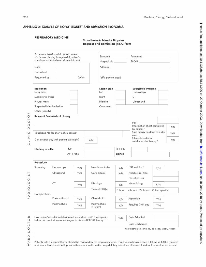
Y/N
Y/N
Y/NY/N
Y/N
Y/N
Y/N
Y/N
Y/N
Y/N
Y/N
Y/N
Y/N
Y/N
Y/N
Y/N
Y/N
Y/N
Y/N
RESPIRATORY MEDICINETransthoracic Needle BiopsiesRequest and admission (R&A) form
To be completed in clinic for all patients.No further clerking is required if patient'scondition has not altered since clinic visit
Date
Consultant
Requested by (print)
Surname Forename
Hospital No D.O.B.
Address
(affix patient label)
Indication
Clotting results:
Procedure
Screening
Complications
Has patient's condition deteriorated since clinic visit? If yes specifybelow and contact senior colleague to discuss BEFORE biopsy
Patients with a pneumothorax should be reviewed by the respiratory team. I f a pneumothorax is seen a follow up CXR is requiredin 4 hours. No patients with pneumothoraces should be discharged if they are alone at home. If in doubt request senior review.
Fluoroscopy
Ultrasound
Pneumothorax
Haemoptysis
CT
Needle aspiration
Core biopsy
Chest drain
Haemoptysis>100ml
Histology
FNA cellular?
Needle size, type
Microbiology
Aspiration
Requires O/N stay
Date Admitted
Date Discharged
No. of passes
Time of CXR(s) 1 hour 4 hours 24 hours Other (specify)
INR
APTT ratio
Platelets
Signed
Relevant Past Medical History
Lung mass
Mediastinal mass
Pleural mass
Suspected infective lesion
Lesion side
Left
Right
Bilateral
Suggested imaging
Fluoroscopy
CT
Ultrasound
Comments
Other (specify)
FEV1Information sheet completedby patient?Can biopsy be done as a daycase?Clinical conditionsatisfactory for biopsy?
Telephone No for short notice contact
Can a carer stay with patient overnight?
If not discharged same day as biopsy specify reason
WARD
DO
CTO
RRADIO
LO
GIST
CLIN
IC
DO
CTO
RAPPENDIX 2: EXAMPLE OF BIOPSY REQUEST AND ADMISSION PROFORMA
936 Manhire, Charig, Clelland, et al
on 18 June 2018 by guest. Protected by copyright.
http://thorax.bmj.com
/T
horax: first published as 10.1136/thorax.58.11.920 on 29 October 2003. D
ownloaded from