A novel restriction endonuclease GlaI for rapid and highly ...
BspRI restriction endonuclease: cloning,expression in Escherichia coli andsequential cleavage mechanismTamas Rasko1, Andras Der2, Eva Klement3, Krystyna Slaska-Kiss1, Eszter Posfai1,
Katalin F. Medzihradszky3,4, Daniel R. Marshak5,6, Richard J. Roberts5,7 and
Antal Kiss1,5,*
1Institute of Biochemistry, 2Institute of Biophysics, 3Proteomics Research Group, Biological Research Center ofthe Hungarian Academy of Sciences, Temesvari krt. 62, 6726 Szeged, Hungary, 4Department of PharmaceuticalChemistry, University of California San Francisco, San Francisco, CA 94158-2517, 5Cold Spring HarborLaboratory, Cold Spring Harbor, NY 11724, 6PerkinElmer Inc., 940 Winter Street, Waltham, MA 02451 and7New England Biolabs Inc., 240 County Road, Ipswich, MA 01938-2723, USA
Received September 16, 2009; Revised May 31, 2010; Accepted June 7, 2010
ABSTRACT
The GGCC-specific restriction endonuclease BspRIis one of the few Type IIP restriction endonucleases,which were suggested to be a monomer. Aminoacid sequence information obtained by Edmansequencing and mass spectrometry analysis wasused to clone the gene encoding BspRI. ThebspRIR gene is located adjacently to the gene ofthe cognate modification methyltransferase andencodes a 304 aa protein. Expression of thebspRIR gene in Escherichia coli was dependent onthe replacement of the native TTG initiation codonwith an ATG codon, explaining previous failures incloning the gene using functional selection. Aplasmid containing a single BspRI recognition sitewas used to analyze kinetically nicking and second-strand cleavage under steady-state conditions.Cleavage of the supercoiled plasmid went througha relaxed intermediate indicating sequential hy-drolysis of the two strands. Results of the kineticanalysis of the first- and second-strand cleavageare consistent with cutting the double-strandedsubstrate site in two independent binding events.A database search identified eight puta-tive restriction-modification systems in which thepredicted endonucleases as well as themethyltransferases share high sequence similaritywith the corresponding protein of the BspRI
system. BspRI and the related putative restrictionendonucleases belong to the PD-(D/E)XK nucleasesuperfamily.
INTRODUCTION
Type IIP restriction endonucleases (REase) arecharacterized by recognition sequences displaying dyadaxes of symmetry (palindromes), and constitute the mostabundant class of characterized restriction enzymes (1).The first Type IIP REases, which were biochemicallycharacterized, were shown to consist of two identicalsubunits: EcoRI (2), BclI (3), BstI (4) BamHI (5).Recognition of a symmetric recognition sequence by ahomodimeric protein and cutting the two strands simul-taneously using two active sites was an attractive modelalso because of the economy of the required protein syn-thesis, as first pointed out by Kelly and Smith (6). For along time, the results of crystallographic studies supportedthe generalization that Type IIP REases are homodimers,e.g. (7–11) or tetramers (12).To our knowledge, the first Type IIP REase, which was
suggested to exist as a monomer was BspRI (13). BspRI ofBacillus sphaericus recognizes the sequence GGCC andcuts after the second G to produce blunt ends (14). Theconclusion that the enzyme consists of a single subunit wasderived from a comparison of molecular massesdetermined under native (gel filtration) and denaturing(SDS–polyacryamide gel electrophoresis) conditions.Later, based mostly on similar biochemical evidence as
*To whom correspondence should be addressed. Tel: +36 62 599630; Fax: +36 62 433506; Email: [email protected] addresses:Tamas Rasko, Max-Delbruck-Zentrum fur Molekulare Medizin, Robert-Rossle-Strasse 10., D-13125 Berlin-Buch, Germany.Eszter Posfai, Friedrich Miescher Institute for Biomedical Research, Maulbeerstrasse 66, CH 4058 Basel, Switzerland.
Nucleic Acids Research, 2010, 1–12doi:10.1093/nar/gkq567
� The Author(s) 2010. Published by Oxford University Press.This is an Open Access article distributed under the terms of the Creative Commons Attribution Non-Commercial License (http://creativecommons.org/licenses/by-nc/2.5), which permits unrestricted non-commercial use, distribution, and reproduction in any medium, provided the original work is properly cited.
Nucleic Acids Research Advance Access published June 29, 2010
for BspRI, a few other Type IIP REases were alsoreported to consist of a single polypeptide chain, such asBsuRI (GG/CC) (15), BcnI (CC/SGG) (16) DpnI (GmA/TC) (17), Sau96I G/GNCC (18), BshFI (GG/CC) (19).However, because of a lack of supporting structuraldata, the notion of monomeric Type IIP REasesreceived little attention.This changed when the X-ray structure of an MspI–
DNA specific recognition complex was reported in 2004.MspI was shown to interact with its symmetric recognitionsequence (C/CGG) as a monomer (20,21). Soon otherarticles describing structures of similar asymmetriccomplexes of three other Type IIP enzymes followed:HinPI (G/CGC) (22,23), MvaI (CC/WGG) (24) andBcnI (CC/SGG) (25) establishing a new paradigm tothink about this class of REases.The gene of the BspRI methyltransferase (bspRIM), the
cognate methyltransferase (MTase) of BspRI endonucle-ase, was cloned and expressed in Escherichia coli (26), butattempts to clone the BspRI REase gene (bspRIR) werenot successful (A. Kiss, unpublished). The acceptance ofthe idea of monomeric Type IIP REases prompted us torevisit the project and to try to clone the bspRIR gene byan approach, which was not dependent on the expressionof R.BspRI in E. coli.Here we report the results of experiments, in which we
used amino acid sequence information to identify thesilent BspRI endonuclease gene on the previously clonedfragment, in the vicinity of the MTase gene. Replacing thenative TTG start codon with ATG, and cloning the genein an expression vector providing a strong promoter andShine–Dalgarno sequence resulted in high-level expressionof BspRI REase in E.coli. A database search identifiedeight putative restriction-modification systems, in whichthe predicted REases as well as the predicted MTasesshare high sequence similarity with the correspondingprotein of the BspRI system. Secondary-structure predic-tion was used to determine whether R.BspRI can beassigned to any family of characterized metal-dependentREases. The DNA cleavage mechanism of BspRI wasstudied using a plasmid containing a single BspRI recog-nition site. This substrate allowed us to analyze kineticallyboth nicking and double-strand cleavage at the uniquetarget site.
MATERIALS AND METHODS
Strains and growth conditions
Bacillus sphaericus R, originally isolated as a culturecontaminant, is the native host of the BspRIR-M system (14). Bacillus sphaericus has recentlybeen reclassified as Lysinibacillus sphaericus (27).Escherichia coli ER1821F� glnV44, e14� (McrA�)endA1 thi-1 D(mcrC-mrr)114::IS10 obtained from NewEngland Biolabs was used as cloning host.ER1821(DE3) was made by lysogenizing ER1821 with�DE3 using the �DE3 lysogenization kit of Novagen.ER1821(DE3) expresses T7 RNA polymerase upon induc-tion with isopropyl-b-D-thiogalactopyranoside (IPTG).Bacteria were grown in LB medium (28) at 30�C
(B. sphaericus) or at 37�C (E. coli). Ampicillin (Ap)and chloramphenicol (Cm) were used at 100 and25 mg/ml, respectively. For BspRI overproductionER1821(DE3+pLysS+pET3H-BspRI) was grown toOD550 � 0.5, then BspRI production was induced byadding 0.4mM IPTG to the culture and growth wascontinued for 4–5 h at 30�C.
DNA preparations
Baccillus sphaericus R genomic DNA was prepared from a50ml dense culture. Cells were sedimented by centrifuga-tion, washed with 10ml 20mM Tris–HCl pH 8.0, thenresuspended and lysed in a solution containing 50mMTris–HCl pH 7.5, 50mM EDTA, 0.2% SDS and 200 mg/ml proteinase-K. After incubation at 37�C overnight, theDNA solution was extracted three times with phenol/chloroform and precipitated with ethanol. Theprecipitated DNA was collected by a glass rod, driedand dissolved in TE buffer (10mM Tris–HCl pH 8.0,1mM EDTA).
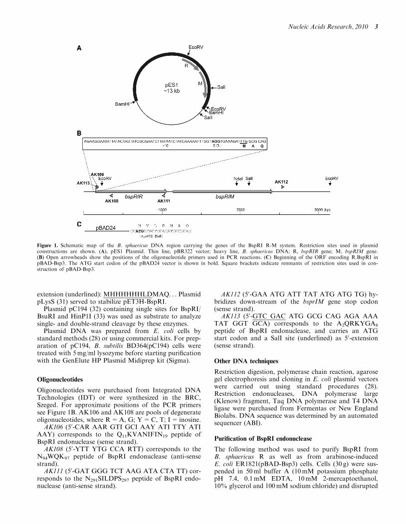
Plasmid pES1 contains the gene of the BspRI MTase ona �9kb BamHI fragment of B.sphaericus DNA cloned inpBR322 (26) (Figure 1A). pTZ-Bsp1 carries the segmentof the bspRIR gene corresponding to the Q11–K97peptide. It was constructed by PCR amplification usingB. sphaericus genomic DNA as template and AK106/AK108 as primers (Figure 1B), and subsequent cloningof the PCR product in the commercial plasmid vectorpTZ57R/T (Fermentas). pTZ-Bsp3 encodes theN-terminal M1–K97 peptide of R.BspRI. It was con-structed by PCR-synthesis using pES1 as template andAK113/AK108 as primers (Figure 1B), and cloning thePCR product in pTZ57R/T. Plasmid pTZ-Bsp5, whichcontains the complete BspRI system, was made by insert-ing the 2920 bp EcoRV fragment of pES1 carrying part ofthe bspRIR gene and the intact bspRIM gene (Figure 1A)into the unique EcoRV site of pTZ-Bsp3. The orientationof the BspRI genes in pTZ-Bsp5 is opposite to the lactranscription on the plasmid.
To construct a plasmid overexpressing R.BspRI, firstthe EcoRV fragment of pES1 (Figure 1) was cloned intothe SmaI site of pBAD24 (29) to yield pBAD-Bsp2.pBAD-Bsp2 lacks the beginning of the bspRIR gene. Toreconstruct the complete BspRI system, the SalI-NdeIfragment of pTZ-Bsp5 was inserted between the Acc65Iand NdeI sites of pBAD-Bsp2. The SalI site in pTZ-Bsp5,added by the AK113 PCR primer, immediately precedesthe ATG start codon of R.BspRI, whereas the Acc65I siteis in the pBAD24 polylinker upstream of the insertedfragment. Before ligation, the Acc65I and SalI ends werefilled-in by Klenow polymerase (Figure 1C). The resultingplasmid (pBAD-Bsp3) encodes a BspRI variant, whichcarries a four amino acid extension at the N-terminus(MVLDMAQRKY. . .). To facilitate purification ofR.BspRI, the SalI-NcoI fragment of pTZ-Bsp5 carryingthe BspRI restriction and modification genes was clonedbetween the XhoI and NcoI sites of the T7 expressionvector pET3-His (30) to yield pET3H-BspRI. TheR.BspRI variant encoded by pET3H-BspRI consists of313 amino acids and has the following N-terminal
2 Nucleic Acids Research, 2010
extension (underlined): MHHHHHHLDMAQ. . . PlasmidpLysS (31) served to stabilize pET3H-BspRI.
Plasmid pC194 (32) containing single sites for BspRI/BsuRI and HinP1I (33) was used as substrate to analyzesingle- and double-strand cleavage by these enzymes.
Plasmid DNA was prepared from E. coli cells bystandard methods (28) or using commercial kits. For prep-aration of pC194, B. subtilis BD364(pC194) cells weretreated with 5mg/ml lysozyme before starting purificationwith the GenElute HP Plasmid Midiprep kit (Sigma).
Oligonucleotides
Oligonucleotides were purchased from Integrated DNATechnologies (IDT) or were synthesized in the BRC,Szeged. For approximate positions of the PCR primerssee Figure 1B. AK106 and AK108 are pools of degenerateoligonucleotides, where R=A, G; Y=C, T; I= inosine.
AK106 (50-CAR AAR GTI GCI AAY ATI TTY ATIAAY) corresponds to the Q11KVANIFIN19 peptide ofBspRI endonuclease (sense strand).
AK108 (50-YTT YTG CCA RTT) corresponds to theN94WQK97 peptide of BspRI endonuclease (anti-sensestrand).
AK111 (50-GAT GGG TCT AAG ATA CTA TT) cor-responds to the N291SILDPS297 peptide of BspRI endo-nuclease (anti-sense strand).
AK112 (50-GAA ATG ATT TAT ATG ATG TG) hy-bridizes down-stream of the bsprIM gene stop codon(sense strand).AK113 (50-GTC GAC ATG GCG CAG AGA AAA
TAT GGT GCA) corresponds to the A2QRKYGA8
peptide of BspRI endonuclease, and carries an ATGstart codon and a SalI site (underlined) as 50-extension(sense strand).
Other DNA techniques
Restriction digestion, polymerase chain reaction, agarosegel electrophoresis and cloning in E. coli plasmid vectorswere carried out using standard procedures (28).Restriction endonucleases, DNA polymerase large(Klenow) fragment, Taq DNA polymerase and T4 DNAligase were purchased from Fermentas or New EnglandBiolabs. DNA sequence was determined by an automatedsequencer (ABI).
Purification of BspRI endonuclease
The following method was used to purify BspRI fromB. sphaericus R as well as from arabinose-inducedE. coli ER1821(pBAD-Bsp3) cells. Cells (30 g) were sus-pended in 50ml buffer A (10mM potassium phosphatepH 7.4, 0.1mM EDTA, 10mM 2-mercaptoethanol,10% glycerol and 100mM sodium chloride) and disrupted
Figure 1. Schematic map of the B. sphaericus DNA region carrying the genes of the BspRI R-M system. Restriction sites used in plasmidconstructions are shown. (A), pES1 Plasmid. Thin line, pBR322 vector; heavy line, B. sphaericus DNA; R, bspRIR gene; M, bspRIM gene.(B) Open arrowheads show the positions of the oligonucleotide primers used in PCR reactions. (C) Beginning of the ORF encoding R.BspRI inpBAD-Bsp3. The ATG start codon of the pBAD24 vector is shown in bold. Square brackets indicate remnants of restriction sites used in con-struction of pBAD-Bsp3.
Nucleic Acids Research, 2010 3
by sonication. After removing cell debris by centrifu-gation (18 000 r.p.m., 20min), the cell extract was loadedonto a 150ml phosphocellulose (Whatman P11) columnequilibrated with the same buffer. Proteins were eluted bya 0.1–1.0M NaCl gradient. Peak fractions were pooled,diluted with 10mM Tris–HCl pH 7.4, and loaded onto a30ml heparin–agarose column equilibrated with buffer A.After elution with a 0.1-1.0 M NaCl gradient peak frac-tions were pooled and loaded directly onto a 30ml hy-droxyapatite column equilibrated with buffer A. BspRIwas eluted with a 10–300mM potassium-phosphategradient. Peak fractions were pooled and dialysedagainst a buffer containing 10mM Tris–HCl pH 7.5,75mM NaCl, 5% glycerol and loaded onto a 6mlResource-S column (Pharmacia). BspRI was eluted witha gradient 0–1M NaCl in 20mM potassium-phosphate(pH 7.5) buffer.For purification of N-terminally His-tagged BspRI,
ER1821(DE3 + pLysS + pET3H-BspRI) cells obtainedfrom 1 l IPTG-induced culture were resuspended in 50mlbuffer E (50mM potassium phosphate, pH 7.4, 0.15MNaCl, 5% glycerol, 10mM 2-mercaptoethanol) containing10mM imidazole and disrupted by sonication. Cell debriswas removed by centrifugation, and the supernatantwas applied onto a 5ml Ni–agarose column (His-SelectNickel Affinity Gel, Sigma) previously equilibratedwith buffer E/10mM imidazole. Proteins were eluted witha step gradient of imidazole (50, 125, 200 and 250mM) inbuffer E. BspRI endonuclease eluted in the 200mM imid-azole step. The enzyme preparation was diluted 5-fold witha buffer containing 10mM potassium phosphate pH 6.9,100mM KCl, 10mM 2-mercaptoethanol and 5% glyceroland loaded onto a 19ml ceramic hydroxyapatite CHT(BioRad) column equilibrated with the same buffer.Proteins were eluted with a gradient containing 10–300mM potassium phosphate, 100mM KCl, 10mM2-mercaptoethanol and 5% glycerol.Both methods yielded enzyme preparations that looked
at least 99% pure by SDS–polyacrylamide gel electrophor-esis (34) after Coomassie staining. Protein concentrationwas determined by the Bradford method (35) using bovineserum albumin standard.
Edman sequencing
Purified BspRI was dialyzed against 10mM sodium–phos-phate pH 7.5, 0.05% SDS, then concentrated by evapor-ation in a SpeedVac instrument but avoiding drying of thesample. Protein samples (10–30 ml) were applied topolybrene-coated glass fiber filters and dried underargon. Filters with dried protein sample were acidifiedwith neat trifluoroacetic acid vapor and extracted withn-heptane to remove excess SDS. The filters were sub-jected to Edman degradation on an Applied Biosystems470A protein sequencer, and the resulting phenylthio-hydantoin (PTH) amino acid derivatives were analyzedby reverse phase HPLC (Applied Biosystems) accordingto the manufacturer’s specifications. PTH-amino acidswere quantitated by comparison to standards using UVabsorbance (36).
Mass spectrometry analysis
In-gel digestion. Gel pieces containing the R.BspRI bandwere cut out from SDS–polyacrylamide gels and soaked in50% acetonitrile containing 25mM NH4HCO3 to removethe Commassie stain and salts. Disulfide bridges werereduced with dithiothreitol and the free sulfhydrylgroups were alkylated with iodoacetamide. After addition-al washing steps, the protein was digested in-gel withside-chain protected porcine trypsin for 4.5 h at 37�C.The resulting peptides were extracted with 2% formicacid in 50% acetonitrile.
Peptide derivatization. A portion of the digest wasderivatized as described earlier (37). Briefly, to 5 ml ofthe digest 30 ml SPITC reagent (4-sulfophenyl isothiocyan-ate, 20 mg/ ml in 25mM NH4HCO3) was added and the pHof the reaction mixture was adjusted to pH 9.0 by theaddition of NH4OH. After 30min at 55�C, the reactionwas terminated with formic acid, then the peptides werepurified on a C18 ZipTip according to the manufacturer’sinstructions.
Matrix-Assisted Laser Desorption/Ionization Time-of-Flight (MALDI-TOF) MS. The tryptic digest wasanalyzed unfractionated prior to and after thederivatization using 2,5-dihydroxybenzoic acid as thematrix. Both mixtures were also fractionated by reversedphase HPLC (C18, 180 mm� 150mm column, flow rate1 ml/min, gradient: 5–40% B in 35min, then up to 80%B in 10min. Solvent A: 0.1 % TFA/5 % acetonitrile inwater; solvent B: 0.085% TFA in 95 % acetonitrile).Fractions were collected directly on the MALDI target.Post source decay (PSD) data were acquired in 10–12segments, lowering the reflectron voltage by 25% in eachstep, then stitching the data together.
LC-MS/MS. The underivatized digest was also subjectedto on-line LC-MS/MS analysis on an ABI QSTARESI-QqTOF mass spectrometer in information dependentacquisition (IDA) mode: 1 s MS acquisitions were followedby 5 s collision-induced dissociation (CID) analyses oncomputer-selected multiply charged ions. Nano-HPLC:C18, 75 mm� 150mm column, flow rate 300 nl/min,gradient: 5–50% B in 30min, solvent A: 0.1% formicacid in water, solvent B: 0.1% formic acid in acetonitrile.
Database searches were performed against the NCBInon-redundant protein database using the ProteinProspector software package (http://prospector.ucsf.edu).De novo sequencing was performed manually.
DNA cleavage analysis
For kinetic analysis, supercoiled pC194 plasmid DNA(2.84 nM) was incubated in 33mM Tris–acetate pH 7.9,10mM Mg–acetate, 66mM K–acetate, 0.1mg/ml BSA(Fermentas Tango buffer) with His-tagged BspRI endo-nuclease (0.0054 pM) at 37�C. Aliquots were withdrawn attimed intervals and added to excess EDTA to stop diges-tion. Digestion of pC194 with HinP1I (New EnglandBiolabs) and BsuRI (Fermentas) was tested using buffersrecommended by the manufacturers. HinP1I and BsuRI
4 Nucleic Acids Research, 2010
were used at 0.016 and 0.05 U/ml concentrations, respect-ively. BsuRI digestions were performed at room tempera-ture. Plasmid isoforms were separated by electrophoresisin 1% agarose gel at low voltage (1.25V/cm) and stainedwith ethidium bromide after the run. Amounts of DNA inthe individual bands were determined by densitometry ofthe gel photograph using the GeneTools software (version4.01, Synoptics). The kinetics of the cleavage reactionswere analyzed in the framework of the reaction schemeshown in Figure 4B using the MATLAB ProgramPackage (MathWorks Inc.,). The differential equationsystem was solved numerically, using the Newtonmethod (iteration step 0.001 s).
Bioinformatic tools
DNA and protein sequence similarity searches were per-formed using the BLAST (38,39) or the ClustalW2 (40)programs. Secondary structure predictions were per-formed with the Jpred 3 program (41). In all casesdefault settings were used.
RESULTS
Partial amino acid sequence of the BspRI REase
The gene of the BspRI MTase was originally cloned on a�9 kb BamHI fragment. E. coli cells carrying pES1(Figure 1A) expressed BspRI MTase, but did not showphage restriction and no BspRI endonuclease activitywas detectable in the cell extracts (26). Cloning of longeroverlapping fragments or screening a plasmid gene libraryfor clones restricting non-modified phage failed to yield aclone expressing BspRI endonuclease.
To use an approach that is not dependent on the ex-pression of R.BspRI in E. coli, the enzyme was purifiedfrom B. sphaericus, and a short N-terminal amino acidsequence (AQRKYGALEQKVANIFINEQVFTFKG)was determined by Edman sequencing.
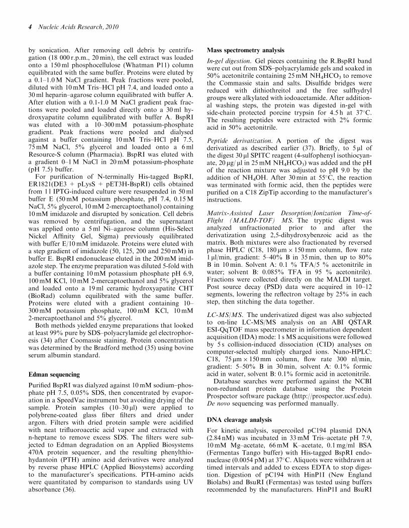
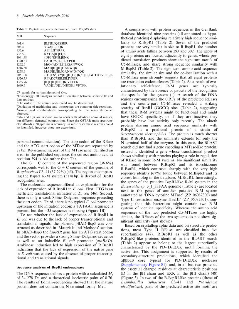
To obtain additional sequence information, purifiedBspRI was digested with trypsin and the peptides weresubjected to mass spectrometry (MS) analysis as describedin ‘Materials and Methods’ section. The tryptic digest wasextensively analyzed by MALDI and electrospray massspectrometry following off- or on-line HPLC fraction-ation. No proteins could be identified from the PSD andCID spectra by database search. To aid de novosequencing, a portion of the digest was sulfonated onthe N-termini of the peptides. Such derivatizationusually leads to almost exclusive y-ion formation in PSDanalysis. Peptide sequences determined manually from theMS/MS (PSD and/or CID) data are summarized inTable 1.
The bspRIR gene
The peptide sequences obtained by Edman degradationand MS analysis were used to design primers forPCR-amplification of a section of the bspRIR gene. Twoprimers (AK106 and AK108) were synthesized. PrimerAK106 corresponded to amino acids Q10KVANIFIN18,whereas AK108 corresponded to the tetrapeptide
NWQK (Table 1, Figure 1B). To reduce complexity ofthe AK106 pool, the neutral base inosine, which canform stable base pairs with all four bases (42), was usedat positions with greater ambiguity. PCR amplification,using B. sphaericus DNA as template, produced an�250 bp fragment, which was cloned in pTZ57R/T toyield pTZ-Bsp1. Sequencing of the insert revealed thatthe cloned fragment encodes several peptides previouslydetected by MS indicating that pTZ-Bsp1 carries aportion of the bspRIR gene. Unexpectedly, PCR synthesisusing the same primers but pES1 plasmid DNA astemplate produced a similar fragment, suggesting that atleast a part of the bspRIR gene was present on pES1.To determine the approximate distance and relative
orientation of the bspRIR and bspRIM genes, four PCRreactions were performed using B. sphaericus DNA astemplate and the following combinations of primers:AK108 + AK112, AK106 + AK112, AK106 + AK111and AK108 + AK111 (Figure 1B). AK111 and AK112were designed on basis of the previously determinedsequence flanking the bspRIM gene (43). Only theAK106 + AK111 combination yielded a PCR product(�850 bp). The same result was obtained when pES1plasmid DNA was used as template. These resultsshowed that the genes of the BspRI R-M system areclosely located in tandem arrangement with the REasegene being upstream (Figure 1).Another conclusion following from this observation
was that the entire bspRIR gene must be on the BamHIfragment cloned in pES1. This was surprising because ofthe lack of endonuclease activity in the clone. It seemedpossible that the methods used (endonuclease assay incrude extracts and phage restriction) were not sensitiveenough to detect low BspRI activity. This question wasaddressed by testing how inactivation of the MTase wouldaffect viability of the clone. To obtain an m� r+ plasmid,the small SalI fragment carrying the 30-terminal half of thebspRIM gene in pES1 (Figure 1A) was deleted.Escherichia coli cells carrying the resulting plasmid wereperfectly viable. Taking into account the large number ofBspRI sites in the E. coli genome and that restriction cutsproducing blunt ends are likely to be highly damaging dueto the absence of DNA–ligase-mediated repair (44), theviability of the m� r+ clone convincingly showed thatBspRI expression from pES1 in E. coli was undetectable.The nucleotide sequence of a 1028 bp segment preceding
and that of a 535 bp segment following the publishedsequence (43) was determined (accession number:X15758). Comparison of the deduced amino acidsequence with the peptide sequences determined byEdman sequencing and MS analysis (Figures 1 and 2) un-equivocally identified the ORF encoding R.BspRI. Agreat majority of the peptides detected in theunfractionated tryptic digest (29/34) fit to the aminoacid sequence derived from the DNA sequence(Figure 2). This ORF starts with TTG at 168 and endswith TAG at 1082 defining a 304 amino acid protein. TTGis not an unusual start codon in B. sphaericus.Approximately 8% of the genes of another B. sphaericusstrain (C3–41), whose sequence has recently been pub-lished (45), have TTG initiation codon (Xiaomin Hu,
Nucleic Acids Research, 2010 5
personal commumication). The stop codon of the REaseand the ATG start codon of the MTase are separated by77 bp. Re-sequencing part of the MTase gene identified anerror in the published sequence: the correct amino acid atposition 394 is Ala rather than Thr.The G + C content of the sequenced region (36.6%)
corresponds well to the G+C content of the genome ofB. sphaericus C3–41 (37.29%) (45). The region encompass-ing the BspRI R-M system (3178 bp) is devoid of BspRIrecognition sites.The nucleotide sequence offered an explanation for the
lack of expression of R.BspRI in E. coli. First, TTG is aninefficient translational initiatior in E. coli (46). Second,there is only a weak Shine–Dalgarno sequence precedingthe start codon. Third, there is no typical E. coli promoterupstream of the initiation codon: a TATAAT sequence ispresent, but the �35 sequence is missing (Figure 1B).To test whether the lack of expression of R.BspRI in
E. coli was due to the lack of proper transcriptional andtranslational signals, the plasmid pBAD-Bsp3 was con-structed as described in ‘Materials and Methods’ section.In pBAD-Bsp3 the bspRIR gene has an ATG start codonand the vector provides a strong Shine–Dalgarno sequenceas well as an inducible E. coli promoter (araBAD).Arabinose induction led to high expression of R.BspRIindicating that the lack of expression of the native genein E. coli was caused by the absence of proper transcrip-tional and translational signals.
Sequence analysis of BspRI endonuclease
The DNA sequence defines a protein with a calculated Mr
of 34 278 Da and a theoretical isoelectric point of 8.76.The results of Edman-sequencing showed that the matureprotein does not contain the N-terminal formyl-Met.
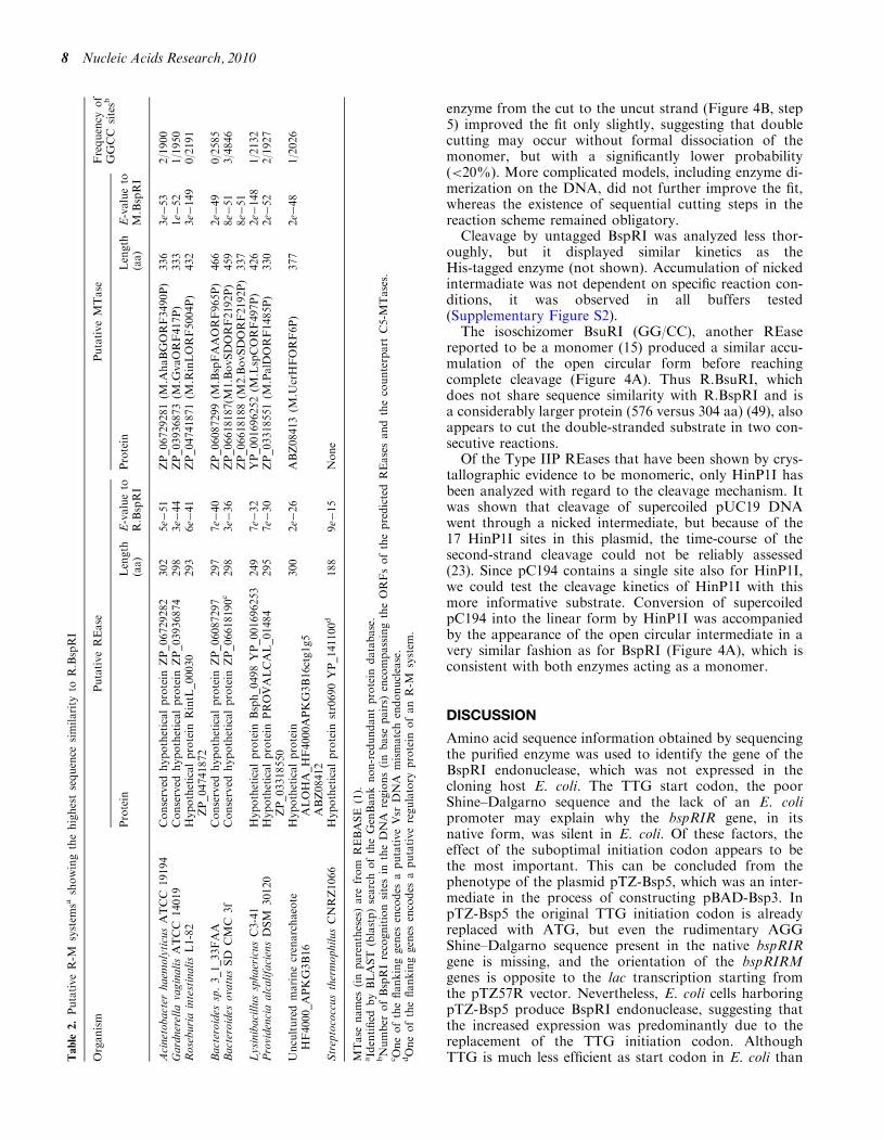
A comparison with protein sequences in the GenBankdatabase identified nine proteins (all annotated as hypo-thetical proteins) displaying relatively high sequence simi-larity to R.BspRI (Table 2). Seven of the predictedproteins are very similar in size to R.BspRI, the numberof amino acids falling between 293 and 302. The genes ofeight proteins are located adjacently to genes, whose pre-dicted translation products show the signature motifs ofC5-MTases, and share strong sequence similarity withM.BspRI (Table 2). The significant amino acid sequencesimilarity, the similar size and the co-localization with aC5-MTase gene strongly suggests that all eight proteinsare restriction endonucleases (Table 2). As a result of evo-lutionary self-defence, R-M genes are typicallycharacterized by the absence or paucity of the recognitionsite specific for the system (1). A search of the DNAregions encompassing the ORFs of the predicted REasesand the counterpart C5-MTases revealed a strikingscarcity of BspRI (GGCC) sites (Table 2), suggestingthat these R-M systems might be functional and mighthave GGCC specificity, or if they are inactive, theyprobably have lost activity only recently. The ninethprotein sharing amino acid sequence similarity withR.BspRI is a predicted protein of a strain ofStreptococcus thermophilus. The protein is much shorterthan R.BspRI, and the similarity extends for only theN-terminal half of the enzyme. In this case, the BLASTsearch did not find a gene encoding a MTase-like protein,instead it identified a gene whose translational productshows similarity with proteins playing a role in regulationof REase in some R-M systems. No significant similaritywas found between R.BspRI and its isoschizomerR.BsuRI, which contrasts sharply with the very highsequence identity (67%) found between M.BspRI and itsclosest homolog in the database, M.BsuRI. Interestingly,the genes of the putative BspRI-like R-M system in theBacteroides sp. 3_1_33FAA genome (Table 2) are locatednext to the genes of another putative R-M systemannotated as ‘DNA cytosine MTase’ (ZP_06087300) and‘type II restriction enzyme HaeIII’ (ZP_06087301), sug-gesting that this bacterium might contain two R-Msystems of identical specificity. Whereas the amino acidsequences of the two predicted C5-MTases are highlysimilar, the REases of the two systems do not show sig-nificant similarity (not shown).
Based on crystallographic data and on structure predic-tions, most Type II REases are classified into fivesuperfamilies (47). R.BspRI as well as the otherR.BspRI-like proteins identified in the BLAST search(Table 2) appear to belong to the largest superfamilycharacterized by the PD-(D/E)XK motif forming theactive site. This assignment is supported by results ofsecondary-structure predictions, which identified theabbbab core typical for PD-(D/E)XK nucleases(Supplementary Figure S1), and, in all but two proteins,the essential charged residues at characteristic positions(D in the bII chain and EXK in the bIII chain) (48)(Figure 3). In two of the R.BspRI-like proteins (those ofLysinibacillus sphaericus C3–41 and Providenciaalcalifaciens), parts of the predicted active site motif are
Table 1. Peptide sequences determined from MS/MS data
MH+ Sequence
961.4 [IL]a[IL]QESSER808.4 YGA[IL]EQK956.6 AE[IL]TNRPR936.52 KYGA[IL]EQK1041.48 {DF}bEF[IL]ENK1370.63 FADC*S[IL][IL]YPER1262.6 M(O)cAD[IL][IL]GANWQK1246.58 MAD[IL][IL]GANWQK1278.6 MAD[IL][IL]GANW(O2)
cQK2051.08 {DT/ESd}VY[IL][IL]G[QK]eE[IL]GGTDTVE[IL]K1526.75 RFADC*S[IL][IL]YPER1385.76 [IL]F[IL]NE[QK]VFTFK1669.9 VAN[IL]F[IL]NE[QK] VFTFK
C* stands for carbamidomethyl Cys.aLow-energy CID analysis cannot differentiate between isomeric Ile andLeu residues.bThe order of the amino acids could not be determined.cOxidation of methionine and tryptophan are common side-reactions.dAmino acid combinations corresponding to the mass differenceobserved.eGln and Lys are isobaric amino acids with identical nominal masses,but different elemental composition. Since the QSTAR mass spectrom-eter affords a 50 ppm mass accuracy, in most cases these residues couldbe identified, however there are exceptions.
6 Nucleic Acids Research, 2010
missing (Figure 3), suggesting that these proteins areinactive.
BspRI cleaves the two DNA strands sequentially
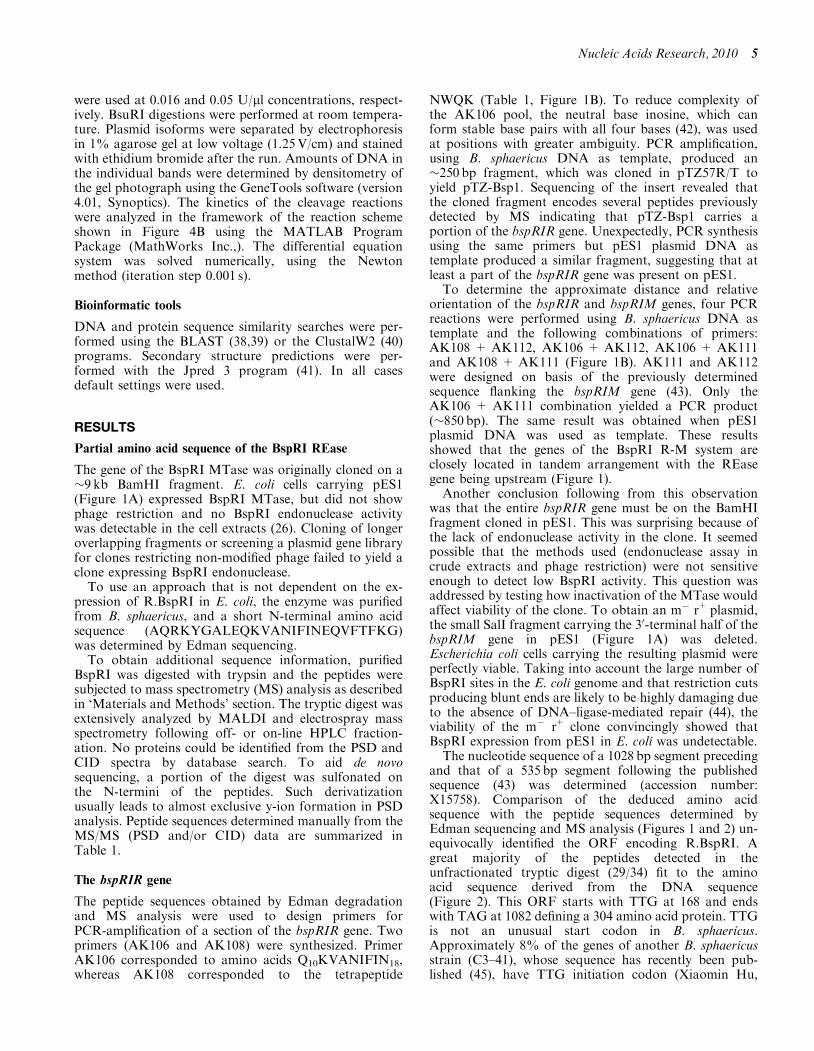
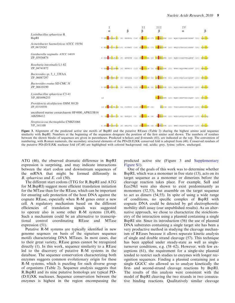
Previous results showed that R.BspRI is a monomer infree state (13). This raised questions about the mode ofsubstrate recognition and cleavage. If BspRI is a monomerwith one active site, how does it cleave the two strands ofthe symmetrical recognition sequence? The most likelymechanism appeared to be cutting the DNA in twosteps: first making a nick, then, in a second bindingevent, cleaving the other strand. To test this model, a2910 bp plasmid (pC194) having a single BspRI site wasdigested under steady state conditions with His-taggedBspRI, and conversion of the supercoiled plasmid DNAinto the nicked and linear forms was followed as describedin ‘Materials and Methods’ section. During the digestion,the nicked DNA accumulated before being converted tothe linear form (Figure 4A), showing that the enzyme firstcleaved just one strand. The amount of DNA in the
different forms was quantitated by densitometry and thekinetics of the cleavage reactions were analyzed in theframework of the proposed reaction scheme shown inFigure 4B. The fit of the derived curves to the experimen-tal data (Figure 4C) shows that the proposed reactionscheme is consistent with the results. Although the avail-able experimental data did not allow determination of thefull set of elementary rate constants shown in the reactionscheme, several of their pairwise respective ratios provedto be stable in the framework of the present model. Forexample we could establish that the rate constants of thefirst and second cleavage do not differ within the error ofthe data (�25%), suggesting independency of theseprocesses. Accumulation of a significant amount ofnicked DNA during the reaction assumes dissociation ofthe majority of the DNA�E complex before the finalcutting process takes place. This implies that BspRI cutsone strand at a time, and the second cut occurs afterbinding of the enzyme to the other strand. Introducingan alternative pathway characterized by flipping of the
1418
.80
[122
-134
]
2655
.70
[148
-170
]
1621
.89
[74-
86]
1526
.79
[110
-121
]
1664
.97
[148
-161
]
1906
.15
1856
.08
[127
-142
]
2051
.25
[49-
67]
2356
.46
[122
-142
]
2534
.45
[252
-274
]
3050
.88
[275
-301
]
1009
.63
[162
-170
]96
1.56
[98
-105
]
1086
.68
[243
-251
]
1246
.63
[87-
97]
1325
.73
[72-
81]
808.
55 [
6-12
]
918.
56 [
127-
134]
2011
.20
[13-
29]
MAQRKYGALE QKVANIFINE QVFTFKGKRY RVLKIGKPQK QGGSGEPKTD50
VYILGQELGG TDTVELKISV KKRDFEFIEN KITPIRMADI LGANWQKIIQ100
ESSERIKDRF ADCSLIYPER RGTTLKNSIT LGWKAEITNR PRTLSARMQL150
SGQQIKDLLY KGVNLPPEKR DAIVDGNIIA NSGIAEYILE TDLDDIHTTA200
DILSQMELID DANLNEAYLA FTANNYRTDT DKCDGARSLA VRVKWEIING250
KLAHHIMFGQ PLLYTGQAAV PELKQTLIQL GTIHPENMND NSILDPSIVC300
KKKRFigure 2. BspRI endonuclease sequence coverage by mass spectrometry. Upper panel, MALDI–TOF mass spectrum of BspRI unfractionated trypticdigest. Numbers in brackets indicate sequence positions of the peptides. Lower panel, amino acid sequence of BspRI restriction endonuclease.Sequences identified from MS/MS data (italic) (Table 1) or from mass only are underlined.
Nucleic Acids Research, 2010 7
enzyme from the cut to the uncut strand (Figure 4B, step5) improved the fit only slightly, suggesting that doublecutting may occur without formal dissociation of themonomer, but with a significantly lower probability(<20%). More complicated models, including enzyme di-merization on the DNA, did not further improve the fit,whereas the existence of sequential cutting steps in thereaction scheme remained obligatory.
Cleavage by untagged BspRI was analyzed less thor-oughly, but it displayed similar kinetics as theHis-tagged enzyme (not shown). Accumulation of nickedintermadiate was not dependent on specific reaction con-ditions, it was observed in all buffers tested(Supplementary Figure S2).
The isoschizomer BsuRI (GG/CC), another REasereported to be a monomer (15) produced a similar accu-mulation of the open circular form before reachingcomplete cleavage (Figure 4A). Thus R.BsuRI, whichdoes not share sequence similarity with R.BspRI and isa considerably larger protein (576 versus 304 aa) (49), alsoappears to cut the double-stranded substrate in two con-secutive reactions.
Of the Type IIP REases that have been shown by crys-tallographic evidence to be monomeric, only HinP1I hasbeen analyzed with regard to the cleavage mechanism. Itwas shown that cleavage of supercoiled pUC19 DNAwent through a nicked intermediate, but because of the17 HinP1I sites in this plasmid, the time-course of thesecond-strand cleavage could not be reliably assessed(23). Since pC194 contains a single site also for HinP1I,we could test the cleavage kinetics of HinP1I with thismore informative substrate. Conversion of supercoiledpC194 into the linear form by HinP1I was accompaniedby the appearance of the open circular intermediate in avery similar fashion as for BspRI (Figure 4A), which isconsistent with both enzymes acting as a monomer.
DISCUSSION
Amino acid sequence information obtained by sequencingthe purified enzyme was used to identify the gene of theBspRI endonuclease, which was not expressed in thecloning host E. coli. The TTG start codon, the poorShine–Dalgarno sequence and the lack of an E. colipromoter may explain why the bspRIR gene, in itsnative form, was silent in E. coli. Of these factors, theeffect of the suboptimal initiation codon appears to bethe most important. This can be concluded from thephenotype of the plasmid pTZ-Bsp5, which was an inter-mediate in the process of constructing pBAD-Bsp3. InpTZ-Bsp5 the original TTG initiation codon is alreadyreplaced with ATG, but even the rudimentary AGGShine–Dalgarno sequence present in the native bspRIRgene is missing, and the orientation of the bspRIRMgenes is opposite to the lac transcription starting fromthe pTZ57R vector. Nevertheless, E. coli cells harboringpTZ-Bsp5 produce BspRI endonuclease, suggesting thatthe increased expression was predominantly due to thereplacement of the TTG initiation codon. AlthoughTTG is much less efficient as start codon in E. coli thanT
able
2.PutativeR-M
system
sashowingthehighestsequence
similarity
toR.BspRI
Organism
PutativeREase
PutativeMTase
Frequency
of
GGCC
sitesb
Protein
Length
(aa)
E-valueto
R.BspRI
Protein
Length
(aa)
E-valueto
M.BspRI
Acinetobacter
haem
olyticusATCC
19194
Conserved
hypotheticalprotein
ZP_06729282
302
5e�
51
ZP_06729281(M
.AhaBGORF3490P)
336
3e�
53
2/1900
Gardnerella
vaginalisATCC
14019
Conserved
hypotheticalprotein
ZP_03936874
298
3e�
44
ZP_03936873(M
.GvaORF417P)
333
1e�
52
1/1950
RoseburiaintestinalisL1-82
Hypotheticalprotein
RintL_00030
ZP_04741872
293
6e�
41
ZP_04741871(M
.RinLORF5004P)
432
3e�
149
0/2191
Bacteroides
sp.3_1_33FAA
Conserved
hypotheticalprotein
ZP_06087297
297
7e�
40
ZP_06087299(M
.BspFAAORF965P)
466
2e�
49
0/2585
Bacteroides
ovatusSD
CMC
3f
Conserved
hypotheticalprotein
ZP_06618190c
298
3e�
36
ZP_06618187(M
1.BovSDORF2192P)
459
8e�
51
3/4846
ZP_06618188(M
2.BovSDORF2192P)
337
8e�
51
LysinibacillussphaericusC3-41
Hypotheticalprotein
Bsph_0498YP_001696253
249
7e�
32
YP_001696252(M
.LspCORF497P)
426
2e�
148
1/2132
ProvidenciaalcalifaciensDSM
30120
Hypotheticalprotein
PROVALCAL_01484
ZP_03318550
295
7e�
30
ZP_03318551(M
.PalD
ORF1485P)
330
2e�
52
2/1927
Unculturedmarinecrenarchaeote
HF4000_APKG3B16
Hypotheticalprotein
ALOHA_HF4000APKG3B16ctg1g5
ABZ08412
300
2e�
26
ABZ08413(M
.UcrHFORF6P)
377
2e�
48
1/2026
StreptococcusthermophilusCNRZ1066
Hypotheticalprotein
str0690YP_141100d
188
9e�
15
None
MTase
names
(inparentheses)are
from
REBASE
(1).
aIdentified
byBLAST
(blastp)searchoftheGenBanknon-redundantprotein
database.
bNumber
ofBspRIrecognitionsitesin
theDNA
regions(inbase
pairs)
encompassingtheORFsofthepredictedREasesandthecounterpart
C5-M
Tases.
cOneoftheflankinggenes
encodes
aputativeVsr
DNA
mismatchendonuclease.
dOneoftheflankinggenes
encodes
aputativeregulatory
protein
ofanR-M
system
.
8 Nucleic Acids Research, 2010
ATG (46), the observed dramatic difference in BspRIexpression is surprising, and may indicate interactionsbetween the start codon and downstream sequences ofthe mRNA that might be formed differently inB. sphaericus and E. coli (50).
The different start codons (TTG for R.BspRI and ATGfor M.BspRI) suggest more efficient translation initiationfor the MTase than for the REase, which can be importantfor ensuring safe protection of the host DNA against thecognate REase, especially when R-M genes enter a newcell. A regulatory mechanism based on the differentefficiencies of translation signals was suggestedto operate also in some other R-M systems (18,49).Such a mechanism could be an alternative to transcrip-tional control coordinating REase and MTaseexpression (51).
Putative R-M systems are typically identified in newgenome seqences on basis of the signature sequencemotifs characterizing DNA MTases. In most cases, dueto their great variety, REase genes cannot be recognizeddirectly (1). In this work, sequence similarity to a REaseled to the discovery of putative R-M systems in thedatabase. The sequence conservation characterizing bothenzymes suggests common evolutionary origin for theseR-M systems, which is puzzling for such diverse groupof organisms (Table 2). Sequence analysis suggests thatR.BspRI and its nine putative homologs are typical PD-(D/E)XK nucleases. Sequence conservation between theenzymes is highest in the region encompassing the
predicted active site (Figure 3 and SupplementaryFigure S1).One of the goals of this work was to determine whether
BspRI, which was a monomer in free state (13), acts on itstarget sequence as a monomer or dimerizes before thecleavage reaction takes place. For example, SalI andEco29kI were also shown to exist predominantly asmonomers (52,53), but assemble on the target sequenceto act as dimers (54,55). In spite of using a wide rangeof conditions, no specific complex of BspRI withcognate DNA could be detected by gel electrophoreticmobility shift assay (our unpublished results). As an alter-native approach, we chose to characterize the stoichiom-etry of the interaction using a plasmid containing a singleBspRI site. Since its introduction (56) the use of plasmidDNA substrates containing a single target site has been avery productive method in studying the cleavage mechan-ism of REases because it allows separate kinetic analysisof single and double strand cleavage (57). This techniquehas been applied under steady-state as well as single-turnover conditions, e.g. (58–62). However, with few ex-ceptions (61), the requirement for a single-site plasmidtended to restrict such studies to enzymes with longer rec-ognition sequences. Finding a plasmid containing just asingle GGCC site allowed us to analyze kinetically thefirst- and second-strand cleavage reactions by BspRI.The results of this analysis were consistent with themodel of BspRI cleaving the two strands in two consecu-tive binding reactions. Qualitatively similar cleavage
I II III IV
Lysinibacillus sphaericus R BspRI 7 GALEQKVANIFI (9) KRYRVL (14) KTDVYILG (7) TVELKISVK (23) WQKIIQESSERIKDRF (23) KAEITN
Acinetobacter haemolyticus ATCC 19194ZP_06729282 6 RDLEDYIVDLFN(10) KIYNRI (14) KTDVYVLL (7) VAEIKISVK (23) WKQILTESIDLIKERF (25) KLEVTN
Gardnerella vaginalis ATCC 14019 ZP_03936874 6 GSEEHKILSLFS (9) EFFKVN (12) KTDIYVEA (6) VKEFKISFK (23) WQEIIISAVTKLQNDF (23) KFELLN
Roseburia intestinalis L1-82ZP_04741872 5 GDAERRILAFMA (9) KNYKII (12) KTDIYILA (5) KVEIKISYK (23) WVNIIEQSTMAISDRF (23) KFELLN
Bacteroides sp. 3_1_33FAA ZP_06087297 5 INTEHCVEKLFP (9) KKYQVA (12) KTDVYIKG (6) AREIKISVK (23) ASGIIKQCLLSIQDSF (23) KFELLN
Bacteroides ovatus SD CMC 3fZP_06618190 5 GKTERKIKDLFL (9) ANYSVL (12) KTDVYVLA (6) QKEFKISVK (23) AQSIIERSIAKIKKTF (23) KFEFIN
Lysinibacillus sphaericus C3-41YP_001696253 10 YLEFKISFK (23) WQNIIESFTSSIDQKF (23) RFELVN
Providencia alcalifaciens DSM 30120 ZP_03318550 10 QTEKNLINLIN (9) CIYDVI (12) KTDVFIKT (5) KISIK (23) TDSILRSSIGTIEQSF (23) KFELLN
uncultured marine crenarchaeote HF4000_APKG3B16 ABZ08412 5 GAEKYVREIID (8) EHYQVI (9) KTDFYVLA (8) REFKISYK (23) WSEILKVQIKPIQHRF (17) RYEIEQ
Streptococcus thermophilus CNRZ1066 YP_141100 6 SKNEHKIAETLS (9) EKLTVL (12) KTDIYIEA (6) KYEFKISYK (23) WMKIIKSGTTQIKKEF (20) KYELLD
a b b b a b
Figure 3. Alignment of the predicted active site motifs of BspRI and the putative REases (Table 2) sharing the highest amino acid sequencesimilarity with BspRI. Numbers at the beginning of the sequences designate the position of the first amino acid shown. The numbers of residuesbetween the shown blocks of sequences are given in parentheses. Predicted a-helices and b-strands (41), are indicated at the top. The convention ofnumbering, with Roman numerals, the secondary structural elements of the PD-(D/E)XK conserved fold is adopted from (48). Conserved residues ofthe putative PD-(D/E)XK nuclease fold (47,48) are highlighted with colored background: red, acidic; grey, lysine; yellow, uncharged.
Nucleic Acids Research, 2010 9
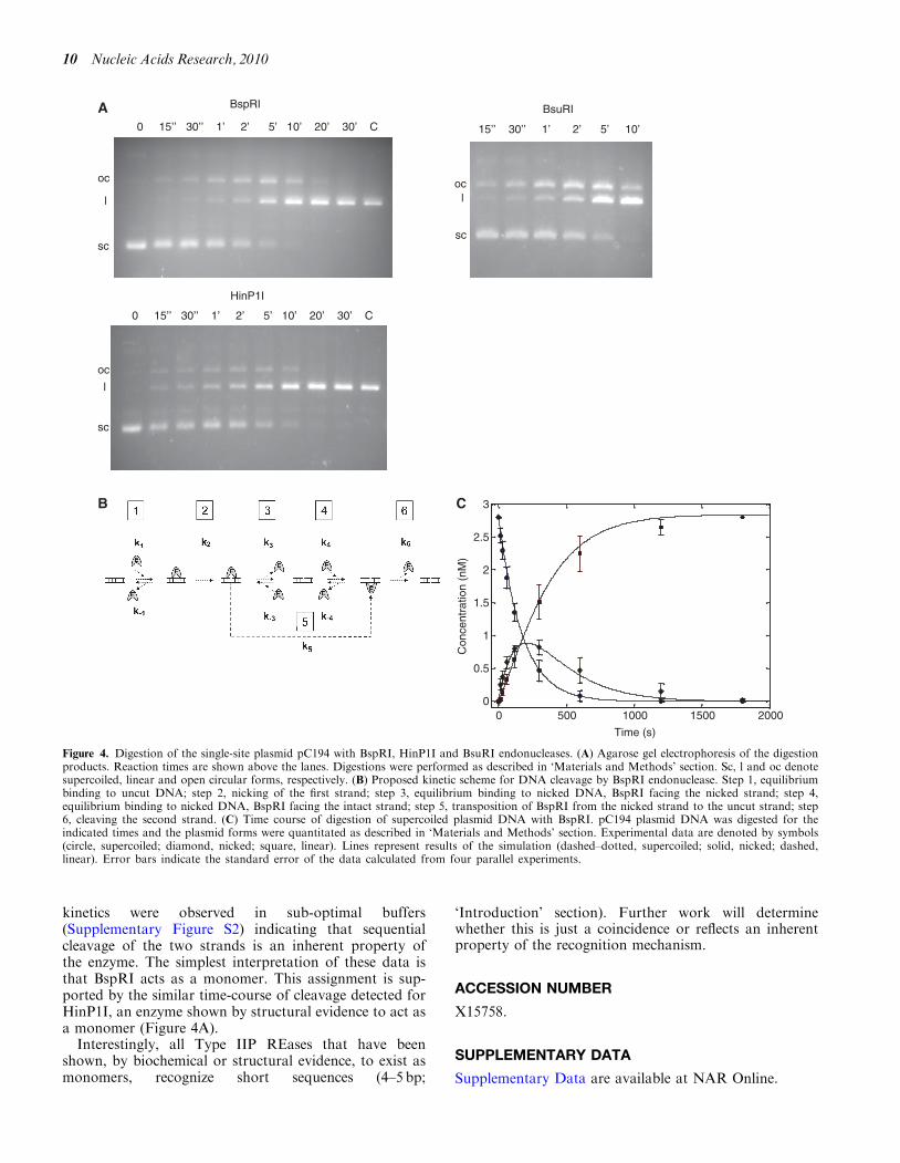
kinetics were observed in sub-optimal buffers(Supplementary Figure S2) indicating that sequentialcleavage of the two strands is an inherent property ofthe enzyme. The simplest interpretation of these data isthat BspRI acts as a monomer. This assignment is sup-ported by the similar time-course of cleavage detected forHinP1I, an enzyme shown by structural evidence to act asa monomer (Figure 4A).Interestingly, all Type IIP REases that have been
shown, by biochemical or structural evidence, to exist asmonomers, recognize short sequences (4–5 bp;
‘Introduction’ section). Further work will determinewhether this is just a coincidence or reflects an inherentproperty of the recognition mechanism.
ACCESSION NUMBER
X15758.
SUPPLEMENTARY DATA
Supplementary Data are available at NAR Online.
A15’’ 30’’ 1’ 2’ 5’ 10’
BsuRI
sc
oc
BspRI
sc
loc
l
0 15’’ 30’’ 1’ 2’ 5’ 10’ 20’ 30’ C
0 15’’ 30’’ 1’ 2’ 5’ 10’ 20’ 30’ C
HinP1I
oc
l
sc
ocl
sc
B
0 500 1000 1500 20000
0.5
1
1.5
2
2.5
3
Time (s)
Con
cent
ratio
n (n
M)
C
Figure 4. Digestion of the single-site plasmid pC194 with BspRI, HinP1I and BsuRI endonucleases. (A) Agarose gel electrophoresis of the digestionproducts. Reaction times are shown above the lanes. Digestions were performed as described in ‘Materials and Methods’ section. Sc, l and oc denotesupercoiled, linear and open circular forms, respectively. (B) Proposed kinetic scheme for DNA cleavage by BspRI endonuclease. Step 1, equilibriumbinding to uncut DNA; step 2, nicking of the first strand; step 3, equilibrium binding to nicked DNA, BspRI facing the nicked strand; step 4,equilibrium binding to nicked DNA, BspRI facing the intact strand; step 5, transposition of BspRI from the nicked strand to the uncut strand; step6, cleaving the second strand. (C) Time course of digestion of supercoiled plasmid DNA with BspRI. pC194 plasmid DNA was digested for theindicated times and the plasmid forms were quantitated as described in ‘Materials and Methods’ section. Experimental data are denoted by symbols(circle, supercoiled; diamond, nicked; square, linear). Lines represent results of the simulation (dashed–dotted, supercoiled; solid, nicked; dashed,linear). Error bars indicate the standard error of the data calculated from four parallel experiments.
10 Nucleic Acids Research, 2010
ACKNOWLEDGEMENTS
We thank David Dubnau and Lise Raleigh for the strainsBD364(pC194) and ER1821, respectively, and IbolyaAnton for the technical assistance.
FUNDING
Hungarian Scientific Research Fund (T038343 to A.K.);National Science Foundation (DMB 8217553.000 toR.J.R.); Exxon Corporation (grant to R.J.R.); NationalInstitutes of Health (NCRR RR015804 andP41RR001614 to the UCSF Mass Spectrometry Facility,Director A.L. Burlingame, to K.F.M., in part). Fundingfor open access charge: New England Biolabs.
Conflict of interest statement. None declared.
REFERENCES
1. Roberts,R.J., Vincze,T., Posfai,J. and Macelis,D. (2010)REBASE–a database for DNA restriction and modification:enzymes, genes and genomes. Nucleic Acids Res., 38, D234–236.
2. Modrich,P. and Zabel,D. (1976) EcoRI endonuclease. Physicaland catalytic properties of the homogenous enzyme. J. Biol.Chem., 251, 5866–5874.
3. Bingham,A.H., Atkinson,T., Sciaky,D. and Roberts,R.J. (1978) Aspecific endonuclease from Bacillus caldolyticus. Nucleic AcidsRes., 5, 3457–3467.
4. Clarke,C.M. and Hartley,B.S. (1979) Purification, properties andspecificity of the restriction endonuclease from Bacillusstearothermophilus. Biochem. J., 177, 49–62.
5. Smith,L.A. and Chirikjian,J.G. (1979) Purification andcharacterization of the sequence-specific endonuclease BamHI.J. Biol. Chem., 254, 1003–1006.
6. Kelly,T.J. Jr. and Smith,H.O. (1970) A restriction enzyme fromHemophilus influenzae. II. Base sequence of the recognition site.J. Mol. Biol., 51, 393–409.
7. Kim,Y.C., Grable,J.C., Love,R., Greene,P.J. and Rosenberg,J.M.(1990) Refinement of EcoRI endonuclease crystal structure: arevised protein chain tracing. Science, 249, 1307–1309.
8. Winkler,F.K., Banner,D.W., Oefner,C., Tsernoglou,D.,Brown,R.S., Heathman,S.P., Bryan,R.K., Martin,P.D., Petratos,K.and Wilson,K.S. (1993) The crystal structure of EcoRVendonuclease and of its complexes with cognate and non-cognateDNA fragments. EMBO J., 12, 1781–1795.
9. Newman,M., Strzelecka,T., Dorner,L.F., Schildkraut,I. andAggarwal,A.K. (1994) Structure of restriction endonucleaseBamHI and its relationship to EcoRI. Nature, 368, 660–664.
10. Athanasiadis,A., Vlassi,M., Kotsifaki,D., Tucker,P.A.,Wilson,K.S. and Kokkinidis,M. (1994) Crystal structure of PvuIIendonuclease reveals extensive structural homologies to EcoRV.Nat. Struct. Biol., 1, 469–475.
11. Cheng,X., Balendiran,K., Schildkraut,I. and Anderson,J.E. (1994)Structure of PvuII endonuclease with cognate DNA. EMBO J.,13, 3927–3935.
12. Deibert,M., Grazulis,S., Sasnauskas,G., Siksnys,V. and Huber,R.(2000) Structure of the tetrameric restriction endonucleaseNgoMIV in complex with cleaved DNA. Nat. Struct. Biol., 7,792–799.
13. Koncz,C., Kiss,A. and Venetianer,P. (1978) Biochemicalcharacterization of the restriction-modification system of Bacillussphaericus. Eur. J. Biochem., 89, 523–529.
14. Kiss,A., Sain,B., Csordas-Toth,E. and Venetianer,P. (1977) A newsequence-specific endonuclease (Bsp) from Bacillus sphaericus.Gene, 1, 323–329.
15. Bron,S. and Horz,W. (1980) Purification and properties of theBsu endonuclease. Methods Enzymol., 65, 112–132.
16. Petrusyte,M. and Janulaitis,A. (1982) Isolation and someproperties of the restriction endonuclease BcnI from Bacilluscentrosporus. Eur. J. Biochem., 121, 377–381.
17. de la Campa,A.G., Springhorn,S.S., Kale,P. and Lacks,S.A.(1988) Proteins encoded by the DpnI restriction gene cassette.Hyperproduction and characterization of the DpnI endonuclease.J. Biol. Chem., 263, 14696–14702.
18. Szilak,L., Venetianer,P. and Kiss,A. (1990) Cloning andnucleotide sequence of the genes coding for the Sau96I restrictionand modification enzymes. Nucleic Acids Res., 18, 4659–4664.
19. Pozidis,C., Vlatakis,G. and Bouriotis,V. (1993) Sequence-specificDNA affinity chromatography: application of a group-specificadsorbent for the isolation of restriction endonucleases.J. Chromatogr., 630, 151–157.
20. Xu,Q.S., Kucera,R.B., Roberts,R.J. and Guo,H.C. (2004) Anasymmetric complex of restriction endonuclease MspI on itspalindromic DNA recognition site. Structure, 12, 1741–1747.
21. Xu,Q.S., Roberts,R.J. and Guo,H.C. (2005) Two crystal forms ofthe restriction enzyme MspI-DNA complex show the same novelstructure. Protein Sci., 14, 2590–2600.
22. Yang,Z., Horton,J.R., Maunus,R., Wilson,G.G., Roberts,R.J. andCheng,X. (2005) Structure of HinP1I endonuclease reveals astriking similarity to the monomeric restriction enzyme MspI.Nucleic Acids Res., 33, 1892–1901.
23. Horton,J.R., Zhang,X., Maunus,R., Yang,Z., Wilson,G.G.,Roberts,R.J. and Cheng,X. (2006) DNA nicking by HinP1Iendonuclease: bending, base flipping and minor groove expansion.Nucleic Acids Res., 34, 939–948.
24. Kaus-Drobek,M., Czapinska,H., Sokolowska,M., Tamulaitis,G.,Szczepanowski,R.H., Urbanke,C., Siksnys,V. and Bochtler,M.(2007) Restriction endonuclease MvaI is a monomer thatrecognizes its target sequence asymmetrically. Nucleic Acids Res.,35, 2035–2046.
25. Sokolowska,M., Kaus-Drobek,M., Czapinska,H., Tamulaitis,G.,Szczepanowski,R.H., Urbanke,C., Siksnys,V. and Bochtler,M.(2007) Monomeric restriction endonuclease BcnI in the apo formand in an asymmetric complex with target DNA. J. Mol. Biol.,369, 722–734.
26. Szomolanyi,E., Kiss,A. and Venetianer,P. (1980) Cloning themodification methylase gene of Bacillus sphaericus R inEscherichia coli. Gene, 10, 219–225.
27. Ahmed,I., Yokota,A., Yamazoe,A. and Fujiwara,T. (2007)Proposal of Lysinibacillus boronitolerans gen. nov. sp. nov., andtransfer of Bacillus fusiformis to Lysinibacillus fusiformis comb.nov. and Bacillus sphaericus to Lysinibacillus sphaericus comb.nov.Int. J. Syst. Evol. Microbiol., 57, 1117–1125.
28. Sambrook,J., Fritsch,E.F. and Maniatis,T. (1989) MolecularCloning: A Laboratory Manual, 2nd edn. Cold Spring HarborLaboratory Press, Cold Spring Harbor, New York.
29. Guzman,L.M., Belin,D., Carson,M.J. and Beckwith,J. (1995)Tight regulation, modulation, and high-level expression by vectorscontaining the arabinose PBAD promoter. J. Bacteriol., 177,4121–4130.
30. Chen,B.P. and Hai,T. (1994) Expression vectors for affinitypurification and radiolabeling of proteins using Escherichia coli ashost. Gene, 139, 73–75.
31. Studier,F.W. (1991) Use of bacteriophage T7 lysozyme toimprove an inducible T7 expression system. J. Mol. Biol., 219,37–44.
32. Iordanescu,S., Surdeanu,M., Della Latta,P. and Novick,R. (1978)Incompatibility and molecular relationships between smallStaphylococcal plasmids carrying the same resistance marker.Plasmid, 1, 468–479.
33. Horinouchi,S. and Weisblum,B. (1982) Nucleotide sequence andfunctional map of pC194, a plasmid that specifies induciblechloramphenicol resistance. J. Bacteriol., 150, 815–825.
34. Laemmli,U.K. (1970) Cleavage of structural proteins during theassembly of the head of bacteriophage T4. Nature, 227, 680–685.
35. Bradford,M.M. (1976) A rapid and sensitive method for thequantitation of microgram quantities of protein utilizing theprinciple of protein-dye binding. Anal. Biochem., 72, 248–254.
36. Hunkapiller,M.W., Hewick,R.M., Dreyer,W.J. and Hood,L.E.(1983) High-sensitivity sequencing with a gas-phase sequenator.Methods Enzymol., 91, 399–413.
Nucleic Acids Research, 2010 11
37. Wang,D., Kalb,S.R. and Cotter,R.J. (2004) Improved proceduresfor N-terminal sulfonation of peptides for matrix-assisted laserdesorption/ionization post-source decay peptide sequencing.Rapid Commun. Mass Spectrom., 18, 96–102.
38. Altschul,S.F., Madden,T.L., Schaffer,A.A., Zhang,J., Zhang,Z.,Miller,W. and Lipman,D.J. (1997) Gapped BLAST andPSI-BLAST: a new generation of protein database searchprograms. Nucleic Acids Res., 25, 3389–3402.
39. Altschul,S.F., Wootton,J.C., Gertz,E.M., Agarwala,R.,Morgulis,A., Schaffer,A.A. and Yu,Y.K. (2005) Protein databasesearches using compositionally adjusted substitution matrices.FEBS J., 272, 5101–5109.
40. Larkin,M.A., Blackshields,G., Brown,N.P., Chenna,R.,McGettigan,P.A., McWilliam,H., Valentin,F., Wallace,I.M.,Wilm,A., Lopez,R. et al. (2007) Clustal W and Clustal X version2.0. Nucleic Acids Res., 23, 2947–2948.
41. Cole,C., Barber,J.D. and Barton,G.J. (2008) The Jpred 3secondary structure prediction server. Nucleic Acids Res., 36,W197–W201.
42. Martin,F.H., Castro,M.M., Aboul-ela,F. and Tinoco,I. Jr. (1985)Base pairing involving deoxyinosine: implications for probedesign. Nucleic Acids Res., 13, 8927–8938.
43. Posfai,G., Kiss,A., Erdei,S., Posfai,J. and Venetianer,P. (1983)Structure of the Bacillus sphaericus R modification methylasegene. J. Mol. Biol., 170, 597–610.
44. Tımar,E., Venetianer,P. and Kiss,A. (2008) In vivo DNAprotection by relaxed-specificity SinI DNA methyltransferasevariants. J. Bacteriol., 190, 8003–8008.
45. Hu,X., Fan,W., Han,B., Liu,H., Zheng,D., Li,Q., Dong,W.,Yan,J., Gao,M., Berry,C. et al. (2008) Complete genome sequenceof the mosquitocidal bacterium Bacillus sphaericus C3-41 andcomparison with those of closely related Bacillus species.J. Bacteriol., 190, 2892–2902.
46. Ringquist,S., Shinedling,S., Barrick,D., Green,L., Binkley,J.,Stormo,G.D. and Gold,L. (1992) Translation initiation inEscherichia coli: sequences within the ribosome-binding site.Mol. Microbiol., 6, 1219–1229.
47. Orlowski,J. and Bujnicki,J.M. (2008) Structural and evolutionaryclassification of Type II restriction enzymes based on theoreticaland experimental analyses. Nucleic Acids Res., 36, 3552–3569.
48. Knizewski,L., Kinch,L., Grishin,N., Rychlewski,L. andGinalski,K. (2007) Realm of PD-(D/E)XK nuclease superfamilyrevisited: detection of novel families with modified transitive metaprofile searches. BMC Struct. Biol., 7, 40.
49. Kiss,A., Posfai,G., Keller,C.C., Venetianer,P. and Roberts,R.J.(1985) Nucleotide sequence of the BsuRI restriction-modificationsystem. Nucleic Acids Res., 13, 6403–6421.
50. Stenstrom,C.M., Holmgren,E. and Isaksson,L.A. (2001)Cooperative effects by the initiation codon and its flankingregions on translation initiation. Gene, 273, 259–265.
51. Mruk,I. and Blumenthal,R.M. (2008) Real-time kinetics ofrestriction-modification gene expression after entry into a newhost cell. Nucleic Acids Res., 36, 2581–2593.
52. Maxwell,A. and Halford,S.E. (1982) The SalGI restrictionendonuclease. Purification and properties. Biochem. J., 203, 77–84.
53. Pertzev,A.V., Kravetz,A.N., Mayorov,S.G., Zakharova,M.V. andSolonin,A.S. (1997) Isolation of a strain overproducingendonuclease Eco29kI: purification and characterization of thehomogeneous enzyme. Biokhimiia, 62, 732–741.
54. Maxwell,A. and Halford,S.E. (1982) The SalGI restrictionendonuclease. Mechanism of DNA cleavage. Biochem. J., 203,85–92.
55. Ibryashkina,E.M., Sasnauskas,G., Solonin,A.S., Zakharova,M.V.and Siksnys,V. (2009) Oligomeric structure diversity within theGIY-YIG nuclease family. J. Mol. Biol., 387, 10–16.
56. Greene,P.J., Poonian,M.S., Nussbaum,A.L., Tobias,L.,Garfin,D.E., Boyer,H.W. and Goodman,H.M. (1975) Restrictionand modification of a self-complementary octanucleotidecontaining the EcoRI substrate. J. Mol. Biol., 99, 237–261.
57. Bennett,S.P. and Halford,S.E. (1989) Recognition of DNA byType II restriction enzymes. Curr. Top. Cell. Regul., 30, 57–104.
58. Rubin,R.A. and Modrich,P. (1978) Substrate dependence of themechanism of EcoRI endonuclease. Nucleic Acids Res., 5,2991–2997.
59. Halford,S.E., Johnson,N.P. and Grinsted,J. (1979) The reactionsof the EcoRI and other restriction endonucleases. Biochem. J.,179, 353–365.
60. Halford,S.E. and Johnson,N.P. (1983) Single turnovers of theEcoRI restriction endonuclease. Biochem. J., 211, 405–415.
61. Zebala,J.A., Choi,J. and Barany,F. (1992) Characterization ofsteady state, single-turnover, and binding kinetics of the TaqIrestriction endonuclease. J. Biol. Chem., 267, 8097–8105.
62. Sasnauskas,G., Jeltsch,A., Pingoud,A. and Siksnys,V. (1999)Plasmid DNA cleavage by MunI restriction enzyme:single-turnover and steady-state kinetic analysis. Biochemistry, 38,4028–4036.
12 Nucleic Acids Research, 2010