BSC 2017 Pharmacology of...
46
BSC 2017 Opioids - with special emphasis on its pharmacology - Paulo Sá Rodrigues Consultant Anaesthesiologist, Lisbon, Portugal ESA OLA Subcommittee member, EDAIC examiner and translator [email protected]
Transcript of BSC 2017 Pharmacology of...

BSC 2017
Opioids- with special emphasis on its pharmacology -
Paulo Sá RodriguesConsultant Anaesthesiologist, Lisbon, Portugal
ESA OLA Subcommittee member, EDAIC examiner and [email protected]
Presenter
Presentation Notes
Twitter: paulosarod; Google+: paulosarod; Skype: paulosarod; Facebook: [email protected]; whatsapp: +3519197960963

www.esahq.orgBSC 2017 – Pharmacology of Opioids – Paulo Sá Rodrigues
Conflict of interest declarationWHAT DECLARATION
Grants/research support/P.I. No conflict
Employee No conflict
Consultation fees No conflict
Honoraria No conflict
Speakers bureau No conflict
Company sponsored No conflict
Stock shareholder No conflict
Spouse/partner No conflict
Scientific Advisory Board No conflict
Other No conflict
www.esahq.orgBSC 2017 – Pharmacology of Opioids – Paulo Sá Rodrigues
Outline
• Introduction• Structure, receptors, mechanisms of action• Classification, potency, affinity, Emax curves• Pharmacokinetics – global aspects• Pharmacodynamics – CNS effects• Pharmacodynamics – peripheral effects• Clinical Pharmacology of some selected drugs• References
Presenter
Presentation Notes
Not all slides presented at the handout will be projected at the session. Some of the text will be suppressed. Key Points Opioids suppress pain by their action in the brain, spinal cord, and peripheral nervous system. They affect many systems, with emphasis on the respiratory and cardiovascular systems, and can cause a variety of adverse effects.. The pharmacokinetic and pharmacodynamic properties of opioids are affected by a variety of factors, such as age, body weight, organ failure, and shock. To appropriately use opioids, these factors should be taken into consideration. New pharmacokinetic principles have allowed more intelligent use of opioids along with more predictable durations of action Opioids can pharmacokinetically or pharmacodynamically interact with drugs used perioperatively. Drug interaction should be understood for proper patient management .

www.esahq.orgBSC 2017 – Pharmacology of Opioids – Paulo Sá Rodrigues
Short history and etymology
• OPIUM: • Word derived from opos,
the Greek word for juice,• The drug is derived from
the juice of the opium poppy Papaver somniferum.
• OPIATES: • are drugs derived from
opium • include the natural
products morphine, codeine, and thebaine,
• Include many semisynthetic drugs derived from them.
Presenter
Presentation Notes
The term opioid refers broadly to all compounds related to opium. The word “opium” is derived from opos, the Greek word for juice, inasmuch as the drug is derived from the juice of the opium poppy Papaver somniferum. Opiates are drugs derived from opium and include the natural products morphine, codeine, and thebaine, as well as many semisynthetic congeners derived from them. Fossilized opium poppies have been found in Neanderthal excavation sites dating back to 30,000 bc. Many old civilizations, including the Sumerians, Egyptians, Greeks, Romans, and Chinese, used opium for nutritional, medicinal, euphoric, spiritual, and religious purposes. The first written reference to the medicinal use of the opium poppy is described in a Sumerian text dated near 4,000 bc. The first undisputed reference to opium is found in the writings of Theophrastus in the third century bc. During the Middle Ages, many of the uses of opium were appreciated. Opium contains more than 20 distinct alkaloids. In 1806, Sertürner reported the isolation of a pure substance in opium that he named morphine after Morpheus, the Greek god of dreams. By the middle of the 19th century, the use of pure alkaloids rather than crude opium preparations began to spread throughout the medical world.

www.esahq.orgBSC 2017 – Pharmacology of Opioids – Paulo Sá Rodrigues
Chemical structures of common opioids
Presenter
Presentation Notes
The synthesis of heroin in 1874 was based on the empirical finding that boiling morphine with specific acids caused the replacement of the two morphine –OH groups by –OCOH3 producing diamorphine or heroin. After the structure of morphine was determined in the 1920s, the synthesis of new morphine-like opioid compounds was based on chemical principles rather than empirical discoveries. In 1937, meperidine (or pethidine) became the first synthetic opioid synthesized on the basis of the central structure of morphine. Since then many synthetic and semisynthetic opioids have been produced, including the clinically important opioid antagonists naloxone and naltrexone, by replacing the N-methyl substituent in morphine with allyl and cyclopropylmethyl groups, respectively. Semisynthetic opioids result from relatively simple modification of the morphine molecule. For example, substitution of a methyl group for the hydroxyl group on carbon 3 results in methylmorphine (codeine). Substitution of acetyl groups on carbons 3 and 6 results in diacetylmorphine (heroin). For clinical use during anesthesia the most important opioids are the piperidines fentanyl, sufentanil, alfentanil, and remifentanil. There are important pharmacokinetic and pharmacodynamic differences between these opioids. The major pharmacodynamic differences between these drugs are potency and rate of equilibration between the plasma and the site of drug effect (biophase).

www.esahq.orgBSC 2017 – Pharmacology of Opioids – Paulo Sá Rodrigues
OPIOIDS act on RECEPTORS
• Distributed throughout• the brain & spinal cord; • and also outside the CNS – vascular tissues, cardia, airway/lung,
gut and cells of the immune system.
• Rhodopsin family of GPCRs• μ-opioid receptor (MOR), κ-opioid receptor (KOR), δ-opioid
receptor (DOR), and orphanin FQ/nociception (NOP) receptor
Presenter
Presentation Notes
Opioids act on opioid receptors. The endogenous opioid system is composed of a family of structurally related endogenous peptides that act at a four-member opioid receptor family consisting of the μ-opioid receptor (MOR), κ-opioid receptor (KOR), δ-opioid receptor (DOR), and orphanin FQ/nociception (NOP) receptor. Opioid receptors are expressed in various areas in the central nervous system (CNS), including the amygdala, mesencephalic reticular formation, periaqueductal gray, and rostral ventromedial medulla. Various subtypes of opioid receptors have also been identified, with different pharmacologic functions. For example, at least three MOR subtypes have been described: μ1 is predominantly involved in opioid analgesia, μ2 is involved in opioid-induced respiratory depression, μ3 is involved in opioid-induced immune suppression.

www.esahq.orgBSC 2017 – Pharmacology of Opioids – Paulo Sá Rodrigues
OPIOID RECEPTORS (contd.)• Upon activation of the
receptors , Gi/Gscoupling causing:• Inhibition of
adenylyl cyclase activity
• Reduced opening of voltage-gated Ca2+
channels• Stimulation of K+
current through GIRKs (G protein-activated inwardly rectifying K+ channels)
• Activation of PKC (phosphokinase C) & PLCβ(phospholipase C-β)
From Millers-anesthesia-7th/chapter-27/figure-27-3
Presenter
Presentation Notes
Binding of opioid agonists with the opioid receptors leads to activation of the G protein. The activity of adenylate cyclase and the voltage-dependent Ca2+ channels is suppressed. On the other hand, the inwardly rectifying K+ channels and mitogen-activated protein kinase (MAPK) cascade are activated. AMP, adenosine monophosphate; ATP, adenosine triphosphate. Interactions between opioid ligands and selective receptors have a number of important clinical effects. Morphine-induced analgesia and respiratory depression are both induced through activation of the MOR and subsequent activation of the adenylate cyclase/cyclic AMP pathway. Recently, opioid-induced activation of alternate cell signaling pathways through interactions with many (opioid and non-opioid) receptor types have been shown to affect the growth and distribution of cancer cells.

www.esahq.orgBSC 2017 – Pharmacology of Opioids – Paulo Sá Rodrigues
Classification of Opioid Receptors
Adapted from Atcheson R, Lambert DG. Update on opioid receptors. Br J Anaesth 1994;73:132–134.
Presenter
Presentation Notes
An ideal opioid agonist would have a high specificity for receptors, producing desirable responses (analgesia) and little or no specificity for receptors associated with side effects (hypoventilation, nausea, physical dependence).

www.esahq.orgBSC 2017 – Pharmacology of Opioids – Paulo Sá Rodrigues
Opioids: mechanisms of analgesia
• Directly inhibit ascending transmission of nociceptive information from the spinal cord dorsal horn
• Activate pain control circuits that descend from the midbrain, via the rostral ventromedial medulla (RVM), to the spinal cord dorsal horn.
• The actions of opioids in the bulbospinal pathways are critical to their analgesic efficacy.
From: Molecular Mechanisms of Opioid Receptor-dependent Signaling and BehaviorAnesthes. 2011;115(6):1363-1381. doi:10.1097/ALN.0b013e318238bba6
Presenter
Presentation Notes
The analgesic effects of opioids arise from their ability to directly inhibit ascending transmission of nociceptive information from the spinal cord dorsal horn and to activate pain control circuits that descend from the midbrain, via the rostral ventromedial medulla (RVM), to the spinal cord dorsal horn. So, opioids produce analgesia not only by direct actions on the spinal cord but also by neurally mediated action in regions separate from the site of opioid administration. Local spinal mechanisms, in addition to descending inhibition, underlie the analgesic action of opioids. In the spinal cord, opioids act at synapses either presynaptically or postsynaptically. Opioid receptors are abundantly expressed in the substantia gelatinosa, where release of substance P from the primary sensory neuron is inhibited by opioids. The actions of opioids in the bulbospinal pathways are critical to their analgesic efficacy. Opioid actions in the forebrain clearly contribute to analgesia because decerebration prevents analgesia. Figure: Sites of action of opioid analgesics. The gray pathway shows the sites of action on the pain transmission pathway from periphery to central nervous system. The red pathway shows the actions on pain-modulating neurons in the midbrain and medulla. GABA = γ-aminobutyric acid; MOR = μ opioid receptor. From: Molecular Mechanisms of Opioid Receptor-dependent Signaling and Behavior; Anesthes. 2011;115(6):1363-1381. doi:10.1097/ALN.0b013e318238bba6
www.esahq.orgBSC 2017 – Pharmacology of Opioids – Paulo Sá Rodrigues
Opioids: mechanisms of analgesia
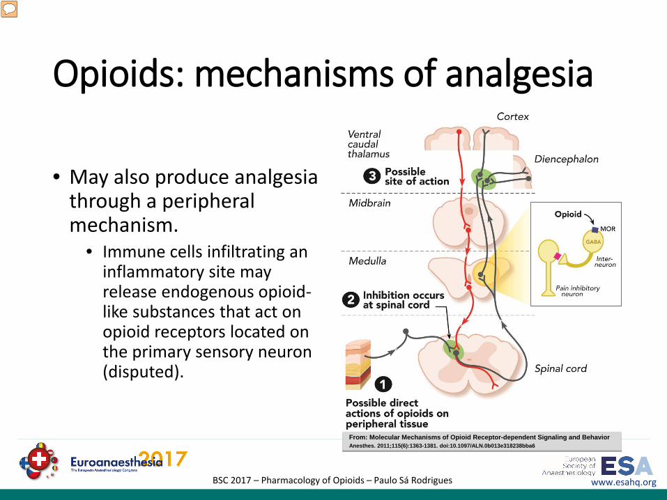
• May also produce analgesia through a peripheral mechanism.
• Immune cells infiltrating an inflammatory site may release endogenous opioid-like substances that act on opioid receptors located on the primary sensory neuron (disputed).
From: Molecular Mechanisms of Opioid Receptor-dependent Signaling and BehaviorAnesthes. 2011;115(6):1363-1381. doi:10.1097/ALN.0b013e318238bba6
Presenter
Presentation Notes
Opioids may also produce analgesia through a peripheral mechanism. Immune cells infiltrating an inflammatory site may release endogenous opioid-like substances that act on opioid receptors located on the primary sensory neuron. However, others do not support this conclusion.

www.esahq.orgBSC 2017 – Pharmacology of Opioids – Paulo Sá Rodrigues
Endogenous Opioids
• Agents found throughout the body that act on an opioid receptor
• All are peptides derived from distinct large precursor proteins -POMC, preproenkephalin, preprodynorphin
• Common amino terminal sequence: TYR-GLY-GLY-PHE-(MET OR LEU)
• Principally three classes –enkephalins, endorphins, dynorphins
Presenter
Presentation Notes
The endogenous opioid peptides include endorphins, enkephalins, and dynorphins, each of which display different affinities for the μ-, κ-, and δ-opioid receptors. β-endorphins have a high affinity for the MOR, met- and leu-enkephalins for the KOR, and Dynorphin A for the DOR. The recently discovered nociceptin has been identified as a selective endogenous ligand of the NOP receptor, while endogenous morphine acts via the μ3-receptor located on immune cells, such as human monocytes. This peptide orphanin FQ or nociceptin was so called because it lowered the pain threshold under certain conditions, in contrast to the other endogenous opioid peptides. Pharmacologic and physiologic studies have demonstrated that orphanin FQ/nociceptin has behavioral and pain modulatory properties distinct from those of the three classic opioid peptides. This opioid system is involved in a variety of regulatory functions including important roles in nociceptive, stress, emotional, and hedonic responses and modulation of thermoregulation, breathing, neuroendocrine function, GI motility, and immune responses. Opioids act not only through central and peripheral neuronal pathways, but also via non-neuronal mechanisms, such as actions on the immune system. Morphine and morphine-6-glucuronide (M6G), endogenously formed and stored in adrenal chromaffin cells and leukocytes, play an import role as modulators of non-neuronal responses in the immune system. Our considerations of endogenous opioid systems, therefore, have expanded from an opioid peptide system present in the central nervous system (CNS) and peripheral nervous system to include various peptide and non-peptide ligands in neuronal and non-neuronal cells throughout the body.

www.esahq.orgBSC 2017 – Pharmacology of Opioids – Paulo Sá Rodrigues
Exogenous Opioids
• Classified according to • Synthesis• Chemical structure• Potency• Receptor binding• Effect at opioid receptors
Presenter
Presentation Notes
Opioids may be classified on the basis of their synthesis, chemical structure, potency, receptor binding, and effect at the opioid receptors. According to their synthesis opioids are subdivided into natural (morphine), semisynthetic, and synthetic opioids. Semisynthetic opioids are derived from the morphine molecule and include buprenorphine, codeine, etorphine, heroin, hydromorphone, oxycodone, and oxymorphone. The synthetic opioids comprise the piperidines (e.g., loperamide, meperidine, alfentanil, fentanyl, sufentanil, remifentanil) and the methadones (e.g., methadone, dextro-propoxyphene). Opioid potency ranges from weak opioids such as codeine, dextro-propoxyphene, tramadol, and hydrocodone to strong opioids, which include etorphine, fentanyl, sufentanil, alfentanil, and remifentanil. Medium potency opioids include morphine, methadone, oxycodone, hydromorphone, and buprenorphine. A more practical classification of opioids is their subdivision into agents with a rapid onset and offset of action (e.g., remifentanil and alfentanil) and agents with a slow onset/offset of action (e.g., morphine and buprenorphine).

www.esahq.orgBSC 2017 – Pharmacology of Opioids – Paulo Sá Rodrigues
Classificationby
receptoraffinity
A. Opioid Agonists: Morphine, Codeine, Meperidine, Fentanyl, Sufentanil, Remifentanil, Methadone, Tramadol
B. Opioid Agonist/Antagonist & Partial Agonist: Pentazocine, Nalbuphine, Butorphanol, Buprenorphine
C. Opioid Antagonists: Nalorphine, Naloxone, Naltrexone, Naltrindole, Nalmefene
Presenter
Presentation Notes
The actions of μ-receptor agonists are invariably analgesic, whereas those of κ-receptor agonists can be either analgesic or antianalgesic. The pain-modulating effects of κ-receptor agonists in the brainstem appear to oppose those of μ-receptor agonists. The opioid antagonists naloxone and naltrexone are also partial inverse agonists at mu opioid receptors. Although naloxone is generally considered to be a pure opioid receptor antagonist, it delays gastric emptying of saline or milk, as does morphine in the rat. Furthermore, high-dose naloxone possesses partial agonistic activity on μ- and κ-opioid receptors in cultured cells. Clinically, opioid antagonists are used to restore spontaneous ventilation in patients who breathe inadequately after opioid overdose or opioid anesthesia. In addition, opioid antagonists can reduce or reverse opioid-induced nausea and vomiting, pruritus, urinary retention, rigidity, and biliary spasm associated with numerous therapies using opioids, such as neuraxial analgesic techniques.

www.esahq.orgBSC 2017 – Pharmacology of Opioids – Paulo Sá Rodrigues
Classification of Exogenous
Opioids- Strength -
• According to the strength or potency based on the plasma concentrations at which they exert their effects (C50 or the plasma concentration causing a 50% effect)
• Strong opioids include fentanyl, sufentanil, and remifentanil.
• Weak opioids include codeine and tramadol.
• An intermediate group includes morphine, methadone, oxycodone, and buprenorphine.
Presenter
Presentation Notes
Opioid potency ranges from weak opioids such as codeine, dextro-propoxyphene, tramadol, and hydrocodone to strong opioids, which include etorphine, fentanyl, sufentanil, alfentanil, and remifentanil. Medium potency opioids include morphine, methadone, oxycodone, hydromorphone, and buprenorphine. A more practical classification of opioids is their subdivision into agents with a rapid onset and offset of action (e.g., remifentanil and alfentanil) and agents with a slow onset/offset of action (e.g., morphine and buprenorphine).

www.esahq.orgBSC 2017 – Pharmacology of Opioids – Paulo Sá Rodrigues
Opioid potency comparison

www.esahq.orgBSC 2017 – Pharmacology of Opioids – Paulo Sá Rodrigues
Opioids sigmoid Emax relationships
From clinical-anesthesia-paul-barash-7th/chapter-19/figure-19-4:
Presenter
Presentation Notes
Sigmoid Emax relationships for opioids full opioid agonists with C50 = 1 and steepness parameter γ = 1 (blue line) and γ = 2 (red line), (B) full opioid agonist (blue line with Emax = 1) and a partial agonist (green line with Emax = 0.75), the rightward shift (gray line) observed by adding a competitive antagonist (such as naloxone) on top of a full agonist (blue line) causing an increase in C50 and the downward shift of adding a noncompetitive antagonist (purple line) to the full agonist causing an effect similar to that observed in (B) (i.e., a partial agonistic effect). From clinical-anesthesia-paul-barash-7th/chapter-19/figure-19-4:

Opioid Pharmacokinetics (PK)ESA Basic Sciences Course resume
www.esahq.orgBSC 2017 – Pharmacology of Opioids – Paulo Sá Rodrigues
Opioids: Physicochemical Properties
• Ionized fraction• Less lipid soluble• Attached to plasma proteins and not available to
diffuse to the action site (the receptor)• However - it is the “effective” form of the
molecule – the receptor recognizes the ionized form
• Base (free) fraction• More lipid soluble – free to diffuse to the action site
Presenter
Presentation Notes
Opioids are weak bases. When dissolved in solution, they are dissociated into protonated and free-base fractions, with the relative proportions depending on the pH and pKa. The free-base fraction is more lipid soluble than the protonated fraction. High lipid solubility facilitates transport of opioid into the biophase or site of action. Therefore, highly lipid-soluble opioids have a more rapid onset of action. However, because the opioid receptor “recognizes” an opioid molecule in the protonated form, the intensity of opioid effects is closely related to the ionized concentration of drug in the biophase. All opioids are to some extent bound to plasma proteins, including albumin and α1-acid glycoprotein. It is only the un-ionized, unbound fraction that constitutes the diffusible fraction and provides the concentration gradient that promotes diffusion of opioid from blood to the tissue of interest. Thus, the speed of onset of opioid effect is affected by both lipid solubility and protein binding.

www.esahq.orgBSC 2017 – Pharmacology of Opioids – Paulo Sá Rodrigues
Physicochemical and Pharmacokinetic Data for Commonly Used Opioid Agonists
Table from From Bailey PL, Egan TD, Stanley TH: Intravenous opioid anesthetics. In Miller RD (ed): Anesthesia, 5th ed. New York, Churchill Livingstone, 2000, p 312. t1/2a,b,d are half-lives of a 3 compartment model; Vdc volume of distribution on the central compartment; Vdss volume of distribution at steady state
Presenter
Presentation Notes
When an opioid is injected into the venous system, there is an initial rapid peak in plasma concentration. Next, the drug rapidly enters multiple organ systems with high blood flow (such as the brain, liver, kidney) from which the plasma drug concentration rapidly drops followed by a slower drop due to redistribution to organs (such as the muscles and later tissues with high fat content) that are less well perfused. These concentration changes over time can be described by non-compartmental and compartmental PK models. Non-compartmental analysis describes the drug’s PK behavior in terms of a) volume of distribution (VD = drug dose/steady-state plasma drug concentration), b) rapid and c) slow distribution half-lives, and d) elimination half-life (t½elim). A high VD is observed for lipophilic opioids with low protein-binding affinity such as fentanyl (VD = 300 L), but a low VD is observed for remifentanil and alfentanil, due to a high clearance (remifentanil) and/or high protein binding. When VD is small, clearance is responsible for the drop in plasma concentration and consequently the loss of analgesia, whereas redistribution accounts for loss of analgesic effect in drugs with a high VD. The time course of a specific effect is difficult to predict for individual patients. For some side effects such as opioid-induced respiratory depression, the prediction of onset or offset of effect is even more complicated due to counteracting forces, such as the respiratory stimulant effects of increased arterial CO2 and the presence of pain.
www.esahq.orgBSC 2017 – Pharmacology of Opioids – Paulo Sá Rodrigues
PK: Absorption
• Modestly absorbed through GI tract -oral, rectal,
• Depends on lipophilicity• High first pass metabolism (FPM)• Morphine - ~25% bioavailability by
oral route• Codeine & oxycodone – low FPM
• Well absorbed through SC & IM routes
• Nasal route – rapid absorption

www.esahq.orgBSC 2017 – Pharmacology of Opioids – Paulo Sá Rodrigues
PK:Distribution
• High concentrations in highly perfused tissues –brain, liver, kidneys & spleen
• In chronic administration –accumulation can take place & opioids are found in the plasma long after their dosage has been stopped
www.esahq.orgBSC 2017 – Pharmacology of Opioids – Paulo Sá Rodrigues
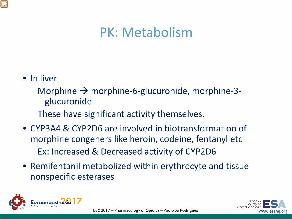
PK: Metabolism
• In liverMorphine morphine-6-glucuronide, morphine-3-
glucuronideThese have significant activity themselves.
• CYP3A4 & CYP2D6 are involved in biotransformation of morphine congeners like heroin, codeine, fentanyl etc
Ex: Increased & Decreased activity of CYP2D6 • Remifentanil metabolized within erythrocyte and tissue
nonspecific esterases
Presenter
Presentation Notes
Overview of opioid metabolism Most opioids are metabolized in the liver through either phase I (oxidative and reductive reactions catalyzed by the cytochrome P450 enzyme system) or phase II reactions (conjugation to a specific substrate). Metabolism may occur at other sites as well, such as in the enterocytes of the gastric tract, the kidney, or the brain. Three aspects of opioid metabolism have clinical importance: 1- Medications that inhibit or induce the CYP450 system may increase or decrease the clinical effect of opioids by interfering with their metabolism; 2- Opioid metabolites may either be active or inactive, which applies not only to their analgesic effect but also to their unwanted side effects. 3- Genetic variability in the CYP system has clinical implications. Cyp2d6 is involved in the conversion of codeine to morphine. So if there is a polymorphism that reduces the activity, less of codeine is converted loss of efficacy. Conversely if there is an activating polymorphism, toxicity can occur. Morphine Morphine undergoes rapid metabolism (phase II reaction) in the liver and within minutes after its administration the two most important hydrophilic metabolites appear in plasma: Morphine-3-glucuronide (M3G) and M6G. M3G is the major metabolite and about 60% of morphine is converted into M3G, while just 5% to 10% is converted to M6G. In humans M3G is without any analgesic or anti-analgesic action. M6G is a full MOR agonist but at the concentrations observed following morphine administration in a patient with normal renal function its contribution to the overall analgesic effect is minimal. Due to its low lipophilicity, passage of M6G across the blood–brain barrier is slow and consequently limited. In the hepatocytes both M3G and M6G are transported back into the bloodstream while a small part is transported into the bile ducts. In the gut both glucuronides are deglucuronidated and the resultant morphine molecule is partly absorbed by the enterocytes. Enterocytes are able to metabolize morphine and transport the resultant M3G and M6G to the bloodstream (the enterohepatic cycle). Since the morphine-glucuronides are excreted via the kidney, patients with renal failure are at risk for M6G-related side effects. Since M6G is a full MOR agonist these side effects are typical of opioids and, most importantly, include sedation and respiratory depression. In patients with compromised renal function morphine treatment causes M6G to accumulate in high concentrations that may cause loss of consciousness and severe respiratory depression. Piperidines Fentanyl, alfentanil, sufentanil, and remifentanil are lipophilic opioids that rapidly cross the blood–brain barrier. Fentanyl, alfentanil, and sufentanil are metabolized by the liver, catalyzed by the cytochrome P450 enzyme system. Fentanyl has a high hepatic extraction ratio with clearance approaching liver blood flow (1.5 L/min). The major metabolite of fentanyl is the inactive compound norfentanyl. Sufentanil also has a high hepatic extraction ratio with a clearance of 0.9 L/min. Alfentanil is metabolized by CYP3A4 and 3A5 forming the inactive compounds noralfentanil and N-phenylpropionamide. The polymorphic expression of the CYP3A5 gene accounts for the great variability in alfentanil metabolism and clearance. Remifentanil contrasts with the other piperidines in that it is not metabolized in the liver. Remifentanil contains a methyl ester side chain that is metabolized by blood (within the erythrocyte) and tissue nonspecific esterases. This causes a rapid clearance of the drug (context sensitive half-life of 2 minutes) making it the most rapidly acting opioid currently available. Clearance of remifentanil is 3 to 5 L/min, which exceeds liver blood flow affirming its extrahepatic clearance. Remifentanil is usually administered as a continuous infusion since its plasma level decreases by 50% in as little as 40 seconds. Methadone Methadone is extensively metabolized to an inactive form by CYP2B6, which is also affected by pharmacogenetic variability. Methadone has a 60% to 95% bioavailability, high potency, and a long duration of action. Furthermore, there is considerable variation among recipients in the response to the drug. While methadone has properties which make it attractive for use intravenously as a perioperative analgesic, in a controlled and well-monitored environment, these same properties may prove hazardous when methadone is administered orally for treatment of patients with chronic pain. Large numbers of patient deaths have been attributed to the long, and often unpredictable, duration of action of methadone when administered orally. Naloxone Naloxone is the most valuable and popular nonspecific MOR antagonist. Since it has a low and unpredictable bioavailability after oral intake due to an extensive (>95%) first-pass effect, naloxone is best given via the intravenous route. The most important metabolic pathway of naloxone is glucuronidation into the inactive naloxone-3-glucuronide. Its duration of effect is short, ranging from 15 to 45 minutes, which requires it to be redosed or administered as a continuous infusion when antagonism is required for long-acting opioids or for patients experiencing an opioid overdose.

www.esahq.orgBSC 2017 – Pharmacology of Opioids – Paulo Sá Rodrigues
PK:Excretion
• In kidneys, M6G & M3G are excreted by glomerular filtration.
• Chronic renal failure can cause elevated levels of these metabolites & lead to adverse effects
• Seizures• CNS depression
Presenter
Presentation Notes
Excretion of the parent drug and/or metabolites occurs via the kidney and/or via the biliary tract into the gut where some opioids (morphine, buprenorphine) may undergo reuptake of the compound into the blood stream.

www.esahq.orgBSC 2017 – Pharmacology of Opioids – Paulo Sá Rodrigues
Context-sensitive half-times for fentanyl, alfentanil, sufentanil and remifentanil
Presenter
Presentation Notes
During a drug infusion, the time needed for the drug’s plasma concentration to decrease by 50%, from a steady-state plasma concentration after the drug infusion has stopped, is called the context-sensitive half-time (CSt½) . The CSt½ depends on the duration of the infusion (the context). Remifentanil has a context-sensitive half-time of 3 to 4 minutes, regardless of the duration of infusion, whereas continuous infusion of the other opioids results in accumulation and considerable prolongation of effect, making these opioids intermediate-acting or long-acting agents, depending on the duration of infusion. Figure adapted from Egan TD, Lemmens HJ, Fiset P, Hermann DJ, Muir KT, Stanski DR, Shafer SL: The pharmacokinetics of the new short-acting opioid remifentanil (GI87084B) in healthy adult male volunteers. Anesthesiology 1993, 79:881–892.
www.esahq.orgBSC 2017 – Pharmacology of Opioids – Paulo Sá Rodrigues
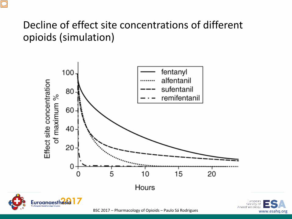
Decline of effect site concentrations of different opioids (simulation)
Presenter
Presentation Notes
Decline of effect site concentrations of different opioids after 24 hours of infusion. Because of its unique pharmacokinetic profile, remifentanil is characterized by a rapid and uniform clearance and a highly predictable onset and offset of effect. Remifentanil has a terminal half-life of approximately 10 to 20 minutes, and its context-sensitive half-time is 3 to 4 minutes, regardless of the duration of infusion. In contrast, continuous infusions of the other 4-anilidopiperidine opioids result in accumulation and considerable prolongation of effect with increased duration of infusion, making these opioids intermediate-acting or long-acting agents during prolonged infusion. The findings are expressed as percentage of the individual maximum effect site concentration. This figure was calculated using Stanpump simulation software (by Shafer S, University of Stanford, CA, USA) for a female individual (age 80 years, height 170 cm, body weight 80 kg) and the following infusion rates: remifentanil 0.15 μg/kg per minute, sufentanil 1 μg/kg per hour, alfentanil 1.5 mg/hour, and fentanyl 0.2 mg/hour.
Pharmacodynamics (PD)Central Nervous System effects

www.esahq.orgBSC 2017 – Pharmacology of Opioids – Paulo Sá Rodrigues
PD: Variability in opioid effects
• Related to • variability in PK-related parameters
• Age, weight, body fat, muscle content, renal/liver function, cardiac output, genetic polymorphism, co-medication
• Variability in PD parameters• Different opioid sensitivity• Different pain perception

www.esahq.orgBSC 2017 – Pharmacology of Opioids – Paulo Sá Rodrigues
PD: Analgesia
• When given to patients in pain• less intense, tolerable, feel more comfortable with relief of distress• Neuropathic pain responds poorly to opioids• Remifentanil may have a sedative and hypnotic effect*
• When given to normal patients• Analgesia• Drowsiness• Changes in mood• Mental clouding
Presenter
Presentation Notes
The analgesia produced by the peripheral actions of opioids is still a source of controversy. Recent reviews have concluded from meta-analysis that intra-articularly administered morphine has a definite, but mild analgesic effect. It may be dose dependent, and a systemic effect cannot be completely excluded. The addition of opioids to brachial plexus blocks has been reported to improve the success rate and postoperative analgesia. In contrast, the addition of sufentanil did not prolong the duration of brachial plexus blockade. Because of its local anesthetic effect on peripheral nerves, meperidine was tested for intravenous regional anesthesia. However, doses of meperidine large enough to produce effective postoperative analgesia cause a significant incidence of side effects. *Koitabashi et al, Anesth Analg 94:1530-1533, 2002
www.esahq.orgBSC 2017 – Pharmacology of Opioids – Paulo Sá Rodrigues
PD: Opioids as anesthetics
• EEG• High doses produce high-voltage slow (δ) waves that are suggestive
of a state consistent with anesthesia• Similar for fentanyl, alfentanil, sufentanil, and remifentanil• Ceiling effect not leading to burst suppression and flat EEG
• Evoked potentials• opioids do not appreciably alter sensory evoked potentials (SEPs)
elicited at the posterior tibial or median nerve, • SEPs can be used to monitor spinal cord function during anesthesia with
opioids.

www.esahq.orgBSC 2017 – Pharmacology of Opioids – Paulo Sá Rodrigues
PD: Cerebral Blood Flow and Cerebral Metabolic Rate
• modest decreases in the cerebral metabolic rate (CMR) and intracranial pressure
• Opioids also decrease cerebral blood flow (CBF) when combined with N2O
• However, opioid-induced neuroexcitation and focal seizure activity can cause regional increases in brain metabolism.
www.esahq.orgBSC 2017 – Pharmacology of Opioids – Paulo Sá Rodrigues
PD: CNS depression
- Respiration -
• Respiratory depression: primary cause of morbidity secondary to opioid therapy
1. Direct depression of rhythm generation in ventrolateral medulla
2. Desensitization of brainstem chemoreceptors which normally respond to rising PCO 2
3. Also desensitize the carotid & aortic chemo sensors which usually respond to hypoxia.
www.esahq.orgBSC 2017 – Pharmacology of Opioids – Paulo Sá Rodrigues
PD: Effect on Cough
• Direct inhibitory effect on the cough centre of medulla
• No loss protective glotticfunction
• Centrally acting antitussives:• Dextromethorphan• codeine, • pholcodeine
www.esahq.orgBSC 2017 – Pharmacology of Opioids – Paulo Sá Rodrigues
PD: Seizure &
Convulsions
Some opioids at a slightly higher doses can produce epileptogenic activity
• Meperidine (pethidine)• Frank seizures & myoclonus • Several mechanisms
• Inhibition of inhibitory interneurons
• Direct stimulatory effects• Actions mediated by non-opioid
receptors by their metabolites

Pharmacodynamics (PD)Peripheral effects
www.esahq.orgBSC 2017 – Pharmacology of Opioids – Paulo Sá Rodrigues
Other opioid-related side effects
a) Nausea and vomitingb) Smooth muscle effectsc) Skeletal muscle effectsd) Histamine releasee) Puritusf) Pupil effectsg) Diffuse CNS effectsh) Cardiovascular effects
Presenter
Presentation Notes
a) Opioids cause PONV due to their effects on the chemoreceptor trigger zone (CTZ) in the area postrema of the brainstem, as well as from direct effects on the GI tract. Also movement effects will contribute to PONV from effects via the vestibular system. Opioids cause an increased sensitivity of the vestibular system. The CTZ contains opioid, serotonin (5HT3), histamine, dopamine (D2), and muscarinic acetylcholine receptors. The CTZ, vagal nerve, and vestibular organs project to the vomiting center in the medulla. b) Opioid receptors are present in the enteric plexus within the smooth muscle layers of the GI tract. Opioids inhibit intestinal and pancreatic secretion, increase bowel tone, and decrease intestinal propulsive activity. Consequently, opioids cause delayed gastric emptying, constipation, bowel distension, and paralytic ileus. Although opioids affect GI motility from central sites as well, blockade of opioid receptors with opioid antagonists that do not cross the blood–brain barrier (such as methylnaltrexone) will have a favorable effect on GI motility. Due to spasms of the sphincter of Oddi and common bile duct, opioids may cause acute upper abdominal pain and colic-like complaints. Naloxone or glucagon can be used for treatment as both cause relaxation of the sphincter muscle. Activated opioid receptors present in the wall of the bladder and ureters can cause acute urine retention. It is most often seen after epidural or spinal opioid administration with a higher incidence in men than women. Urine retention is related to the inability of the urethral sphincter muscle to relax while the bladder tone increases. Opioid-induced bladder dysfunction can be treated with naloxone or the peripherally acting methylnaltrexone. c) Strong, high-dose opioids, especially when given rapidly, cause skeletal muscle rigidity, which includes thoracic, abdominal, and pharyngeal muscles. Weak opioids may give rise to an increase in muscle tension in neck and thorax. While the effect of weak opioids seems to wear off fairly rapidly (within 10 to 20 minutes), the effect of strong opioids may require muscle relaxation and intubation as ventilation may become compromised. d) High-dose morphine, codeine, and meperidine cause histamine release from mast cells, an effect that is non-opioid receptor related (naloxone does not prevent this effect). This effect is not seen with piperidines (fentanyl and congeners). The consequence of histamine release is itching and redness of the skin and hives along the trajectory of the venous injection. Histamine may also cause a reduction in vascular resistance and systemic and pulmonary pressures. e) Animal research has shown the existence of itch-specific pathways that involve itch-specific (i.e., histamine-sensitive) neurons in the spinal cord dorsal horn that travel to the thalamus via the spinothalamic tract. These neurons and pathways are distinct from pain-related neurons and pathways. Painful stimuli such as scratching can abolish histamine-induced itch. Opioids commonly cause an itch typically of the nose, after systemic administration. After spinal opioid injections itching is very common and occurs in 60% to 90% of the patients. Opioid-induced itch serves possibly as a warning signal for the presence of exogenous μ-opioids (which are normally not present in the body). Not all opioids cause itching. For example, κ-opioids (e.g., pentazocine) are antipruritic. Treatment of opioid-induced itch is difficult and many treatments have been tried including MOR antagonists (e.g., naloxone, naltrexone, nalbuphine), histamine H1 receptor antagonists, 5HT-antagonists (e.g., ondansetron), NSAIDs, prednisone, and a sub-hypnotic low-dose infusion of propofol. f) Opioids produce pupillary miosis. Of all opioid effects, miosis occurs most rapidly and at lower doses than analgesia or respiratory depression (the fentanyl C50 for miosis is about half of that for analgesia). The mechanism of pupil constriction is activation of MORs in the nucleus of Edinger–Westphal, which projects to the muscles of the iris via the oculomotor nerve. Since the iris contains MORs a direct effect of opioids on the pupil diameter is possible. g) There are a variety of CNS effects induced by opioids that can cause considerable discomfort to the patients, yet cannot be localized to a specific area of the CNS. These effects can occur after acute opioid treatment but are commonly seen in chronic pain patients on prolonged opioid treatment. These effects include dizziness, light headedness, sedation and drowsiness, euphoria, dysphoria, drug “high,” cognitive dysfunction (e.g., memory loss), inability to concentrate or focus attention, hallucinations, and so on. Some of these effects, such as dizziness, limit the use of opioids in the chronic pain setting, especially in elderly patients. h) Opioids affect the cardiovascular system at central and peripheral sites. Central effects include the activation of vagal nuclei and depression of vasomotor centers in the brainstem. Peripheral effects occur predominantly at high (supraclinical) doses and include direct myocardial depression and arterial and venous dilatation. Morphine may cause additional cardiovascular effects via the release of histamine (see above). The consequences are mild at clinical doses and include orthostatic hypotension, mild bradycardia, and a moderate reduction of systemic and pulmonary resistance. However, even at clinical doses, opioids can induce hemodynamic instability when combined with other drugs such as inhalation anesthetics, propofol, or benzodiazepines, and in severely ill patients (e.g., with sepsis). Treatment of hemodynamic instability includes the administration of atropine and vasopressors and moderate fluid therapy.

Clinical PharmacologyOf some selected drugs

www.esahq.orgBSC 2017 – Pharmacology of Opioids – Paulo Sá Rodrigues
Morphine
• Multiple routes ofadministration
• Slow rise to peak effect• Active metabolite
• Morphine-6-glucuronide is Will contribute to effects with chronic dosing especially in renal failure
Table from From Bailey PL, Egan TD, Stanley TH: Intravenous opioid anesthetics. In Miller RD (ed): Anesthesia, 5th ed. New York, Churchill Livingstone, 2000, p 312. t1/2a,b,d are half-lives of a 3 compartment model; Vdc volume of distribution on the central compartment; Vdss volume of distribution at steady state
Presenter
Presentation Notes
Morphine In humans, morphine produces analgesia, euphoria, sedation, and a diminished ability to concentrate. Other sensations include nausea, a feeling of body warmth, heaviness of the extremities, dryness of the mouth, and pruritus, especially in the cutaneous areas around the nose. Continuous, dull pain is relieved by morphine more effectively than is sharp, intermittent pain. In contrast to nonopioid analgesics, morphine is effective against pain arising from the viscera as well as from skeletal muscles, joints, and integumental structures In the absence of pain, morphine may produce dysphoria rather than euphoria. Pharmacokinetics Morphine is usually administered IV in the perioperative period, thus eliminating the unpredictable influence of drug absorption. The peak effect (equilibration time between the blood and brain) after IV administration of morphine is delayed compared with opioids such as fentanyl and alfentanil, requiring about 15 to 30 minutes. Morphine is well absorbed after IM administration, with onset of effect in 15 to 30 minutes and a peak effect in 45 to 90 minutes. The duration of action is about 4 hours. It can be administered orally for treatment of chronic pain recognizing that absorption from the gastrointestinal tract may be limited. Plasma morphine concentrations after rapid IV injections do not correlate closely with the opioid’s pharmacologic activity. Presumably, this discrepancy reflects a delay in penetration of morphine across the blood-brain barrier. CSF concentrations of morphine peak 15 to 30 minutes after IV injection and decay more slowly than plasma concentrations As a result, the analgesic and ventilatory depressant effects of morphine may not be evident during the initial high plasma concentrations after IV administration of the opioid. Only a small amount of administered morphine gains access to the CNS. For example, it is estimated that <0.1% of morphine that is administered IV has entered the CNS at the time of peak plasma concentrations. Reasons for poor penetration of morphine into the CNS include (a) relatively poor lipid solubility, (b) high degree of ionization at physiologic pH, (c) protein binding, and (d) rapid conjugation with glucuronic acid. Alkalinization of the blood, as produced by hyperventilation of the patient’s lungs, will increase the nonionized fraction of morphine and thus enhance its passage into the CNS. Nevertheless, respiratory acidosis, which decreases the nonionized fraction of morphine, results in higher plasma and brain concentrations of morphine than are present during normocarbia. This suggests that carbon dioxide–induced increases in cerebral blood flow and enhanced delivery of morphine to the brain are more important than the fraction of drug that exists in either the ionized or nonionized fraction. In contrast to the CNS, morphine accumulates rapidly in the kidneys, liver, and skeletal muscles. Morphine, unlike fentanyl, does not undergo significant first-pass uptake into the lungs. Elimination Half-Time After IV administration of morphine, the elimination of morphine-3-glucuronide is somewhat longer than for morphine. The decrease in the plasma concentration of morphine after initial distribution of the drug is principally due to metabolism, because only a small amount of unchanged opioid is excreted in the urine. Plasma morphine concentrations are higher in the elderly than in young adults. In the first 4 days of life, the clearance of morphine is decreased and its elimination half-time is prolonged compared with that found in older infants. This is consistent with the clinical observation that neonates are more sensitive than older children to the ventilatory depressant effects of morphine. Patients with renal failure exhibit higher plasma and CSF concentrations of morphine and morphine metabolites than do normal patients, reflecting a smaller volume of distribution (Vd). Possible accumulation of morphine-6-glucuronide suggests the need for caution when administering morphine to patients with renal dysfunction. Anesthesia alone does not alter the elimination half-time of morphine. Concentrations of morphine in colostrums of parturients receiving patient controlled analgesia with morphine are small and it is unlikely that significant amounts of drug will be transferred to the breast-fed neonate. Side Effects Side effects described for morphine are also characteristic of other opioid agonists, although the incidence and magnitude may vary. Cardiovascular System The administration of morphine, even in large doses (1 mg/kg IV), to supine and normovolemic patients is unlikely to cause direct myocardial depression or hypotension. The same patients changing from a supine to a standing position, however, may manifest orthostatic hypotension and syncope, presumably reflecting morphine-induced impairment of compensatory sympathetic nervous system responses. For example, morphine decreases sympathetic nervous system tone to peripheral veins, resulting in venous pooling and subsequent decreases in venous return, cardiac output, and blood pressure. Morphine can also evoke decreases in systemic blood pressure due to drug-induced bradycardia or histamine release. Morphine-induced bradycardia results from increased activity over the vagal nerves, which probably reflects stimulation of the vagal nuclei in the medulla. Morphine may also exert a direct depressant effect on the sinoatrial node and acts to slow conduction of cardiac impulses through the atrioventricular node. These actions, may, in part, explain decreased vulnerability to ventricular fibrillation in the presence of morphine. Administration of opioids (morphine) in the preoperative medication or before the induction of anesthesia (fentanyl) tends to slow heart rate during exposure to volatile anesthetics with or without surgical stimulation. Opioid-induced histamine release and associated hypotension are variable in both incidence and degree. The magnitude of morphine-induced histamine release and subsequent decrease in systemic blood pressure can be minimized by (a) limiting the rate of morphine infusion to 5 mg/minute IV, (b) maintaining the patient in a supine to slightly head-down position, and (c) optimizing intravascular fluid volume. Conversely, administration of morphine, 1 mg/kg IV, over a 10-minute period produces substantial increases in the plasma concentrations of histamine that are paralleled by significant decreases in systemic blood pressure and systemic vascular resistance. It is important to recognize, however, that not all patients respond to this rate of morphine infusion with the release of histamine, emphasizing the individual variability associated with the administration of this drug. In contrast to morphine, the infusion of fentanyl, 50 μg/kg IV over a 10-minute period, does not evoke release of histamine in any patient. Sufentanil, like fentanyl, does not evoke the release of histamine. Pretreatment of patients with H1 and H2 receptor antagonists does not alter release of histamine evoked by morphine but does prevent changes in systemic blood pressure and systemic vascular resistance. Morphine does not sensitize the heart to catecholamines or otherwise predispose to cardiac dysrhythmias as long as hypercarbia or arterial hypoxemia does not result from ventilatory depressant effects of the opioid. Tachycardia and hypertension that occur during anesthesia with morphine are not pharmacologic effects of the opioid but rather are responses to painful surgical stimulation that are not suppressed by morphine. Both the sympathetic nervous system and the renin-angiotensin mechanism contribute to these cardiovascular responses. Large doses of morphine or other opioid agonists may decrease the likelihood that tachycardia and hypertension will occur in response to painful stimulation, but once this response has occurred, administration of additional opioid is unlikely to be effective. During anesthesia, opioid agonists are commonly administered with inhaled anesthetics to ensure complete amnesia for the painful surgical stimulus. The combination of an opioid agonist such as morphine or fentanyl with nitrous oxide results in cardiovascular depression (decreased cardiac output and systemic blood pressure plus increased cardiac filling pressures), which does not occur when either drug is administered alone (Stoelting and Gibbs, 1973). Likewise, decreases in systemic vascular resistance and systemic blood pressure may accompany the combination of an opioid and a benzodiazepine, whereas these effects do not accompany the administration of either drug alone. All opioid agonists produce dose-dependent and gender–specific depression of ventilation, primarily through an agonist effect at mu2 receptors leading to a direct depressant effect on brainstem ventilation centers. Because analgesic and ventilatory effects of opioids occur by similar mechanisms it is assumed that equianalgesic doses of all opioids will produce some degree of ventilatory depression and reversal of ventilatory depression with an opioid antagonist always involves some reversal of analgesia. Opioid-induced depression of ventilation is characterized by decreased responsiveness of these ventilation centers to carbon dioxide as reflected by an increase in the resting Paco2 and displacement of the carbon dioxide response curve to the right. Opioid agonists also interfere with pontine and medullary ventilatory centers that regulate the rhythm of breathing, leading to prolonged pauses between breaths and periodic breathing. It is possible that opioid agonists diminish sensitivity to carbon dioxide by decreasing the release of acetylcholine from neurons in the area of the medullary ventilatory center in response to hypercarbia. In this regard, physostigmine, which increases CNS levels of acetylcholine, may antagonize depression of ventilation but not analgesia produced by morphine. Depression of ventilation produced by opioid agonists is rapid and persists for several hours, as demonstrated by decreased ventilatory responses to carbon dioxide. High doses of opioids may result in apnea, but the patient remains conscious and able to initiate a breath if asked to do so. Death from an opioid overdose is almost invariably attributable to depression of ventilation. Clinically, depression of ventilation produced by opioid agonists manifests as a decreased frequency of breathing that is often accompanied by a compensatory increase in tidal volume. The incompleteness of this compensatory increase in tidal volume is evidenced by predictable increases in the Paco2. Many factors influence the magnitude and duration of depression of ventilation produced by opioid agonists. For example, advanced age and the occurrence of natural sleep increases the ventilatory depressant effects of opioids. Conversely, pain from surgical stimulation counteracts depression of ventilation produced by opioids. Likewise, the analgesic effect of opioids slows breathing that has been rapid and shallow due to pain. Opioids produce dose-dependent depression of ciliary activity in the airways. Increases in airway resistance after administration of an opioid are probably due to a direct effect on bronchial smooth muscle and an indirect action due to release of histamine. Cough Suppression Opioids depress cough by effects on the medullary cough centers that are distinct from the effects of opioids on ventilation. The greatest cough suppression occurs with opioids that have bulky substitutions at the number 3 carbon position (codeine). Cough suppression is produced by dextrotatory isomers of opioids (dextromethorphan) that do not produce analgesia. Nervous System Opioids in the absence of hypoventilation decrease cerebral blood flow and possibly intracranial pressure (ICP). These drugs must be used with caution in patients with head injury because of their (a) associated effects on wakefulness, (b) production of miosis, and (c) depression of ventilation with associated increases in ICP if the Paco2 becomes increased. Furthermore, head injury may impair the integrity of the blood-brain barrier, with resultant increased sensitivity to opioids. The effect of morphine on the electroencephalogram (EEG) resembles changes associated with sleep. For example, there is replacement of rapid alpha waves by slower delta waves. Recording of the EEG fails to reveal any evidence of seizure activity after administration of large doses of opioids (see the section on Fentanyl). Opioids do not alter the responses to neuromuscular blocking drugs. Skeletal muscle rigidity, especially of the thoracic and abdominal muscles, is common when large doses of opioid agonists are administered rapidly and intravenously (Bowdle and Rooke, 1994). Clonic skeletal muscle activity (myoclonus) occurring during administration of opioids may resemble grand mal seizures, but the EEG does not reflect seizure activity. Skeletal muscle rigidity may be related to actions at opioid receptors and involve interactions with dopaminergic and gamma-aminobutyric acid–responsive neurons. Rapid IV administration of an opioid, as for induction of anesthesia, may be associated with thoracic and abdominal skeletal muscle rigidity sufficient to interfere with adequate ventilation of the lungs. Attempts to manually inflate the lungs in the presence of this skeletal muscle rigidity may result in airway pressures that interfere with venous return. Conversely, there is evidence that the major cause of difficult ventilation after induction of anesthesia with sufentanil (and presumably other opioids) is closure of the vocal cords. The incidence of opioid-induced skeletal muscle rigidity (generalized hypertonus of skeletal muscles) depends on the opioid (most common with fentanyl) and dose used and the rate of administration. Inhibition of striatal release of gamma-aminobutyric acid and increased dopamine production are the likely explanations for opioid-induced increased skeletal muscle tone. The reported incidence of difficult ventilation after a moderate dose of sufentanil ranges from 84% to 100%. Miosis is due to an excitatory action of opioids on the autonomic nervous system component of the Edinger-Westphal nucleus of the oculomotor nerve. Tolerance to the miotic effect of morphine is not prominent. Miosis can be antagonized by atropine, and profound arterial hypoxemia in the presence of morphine can still result in mydriasis. Sedation Postoperative titration of morphine frequently induces sedation that precedes the onset of analgesia. The usual recommendation for morphine titration includes a short interval between boluses (5 to 7 minutes) to allow evaluation of its clinical effect. Sedation occurs in up to 60% of patients during morphine titration and represents a common reason to discontinue morphine titration for postoperative analgesia. The assumption that sleep occurs when pain is relieved is not necessarily accurate and morphine-induced sedation should not be considered as an indicator of appropriate analgesia during intravenous morphine titration. Biliary Tract Opioids can cause spasm of biliary smooth muscle, resulting in increases in intrabiliary pressure that may be associated with epigastric distress or biliary colic. This pain may be confused with angina pectoris. Naloxone will relieve pain caused by biliary spasm but not myocardial ischemia. Conversely, nitroglycerin will relieve pain due to biliary spasm or myocardial ischemia. Equal analgesic doses of fentanyl, morphine, meperidine, and pentazocine increase common bile duct pressure 99%, 53%, 61%, and 15% above predrug levels, respectively. During surgery, opioid-induced spasm of the sphincter of Oddi may appear radiologically as a sharp constriction at the distal end of the common bile duct and be misinterpreted as a common bile duct stone. It may be necessary to reverse opioid-induced biliary smooth muscle spasm with naloxone so as to correctly interpret the cholangiogram. Glucagon, 2 mg IV, also reverses opioid-induced biliary smooth muscle spasm and, unlike naloxone, does not antagonize the analgesic effects of the opioid. Nevertheless, biliary muscle spasm does not occur in most patients who receive opioids. Indeed, the incidence of spasm of the sphincter of Oddi is about 3% in patients receiving fentanyl as a supplement to inhaled anesthetics. Contraction of the smooth muscles of the pancreatic ducts is probably responsible for increases in plasma amylase and lipase concentrations that may be present after the administration of morphine. Such increases may confuse the diagnosis when acute pancreatitis is a possibility.

www.esahq.orgBSC 2017 – Pharmacology of Opioids – Paulo Sá Rodrigues
Remifentanil
• Potency 50 to 100X morphine
• Main features• Rapid onset and offset• Brevity of action (3’ to 5’)• Total metabolism• Rapid recovery
• Metabolism by tissue andplasma esterases to inactivemetabolites.
Table from From Bailey PL, Egan TD, Stanley TH: Intravenous opioid anesthetics. In Miller RD (ed): Anesthesia, 5th ed. New York, Churchill Livingstone, 2000, p 312. t1/2a,b,d are half-lives of a 3 compartment model; Vdc volume of distribution on the central compartment; Vdss volume of distribution at steady state
Presenter
Presentation Notes
Remifentanil is a selective mu opioid agonist with an analgesic potency similar to that of fentanyl and a blood-brain equilibration (effect-site equilibration) time similar to that of alfentanil. Remifentanil is structurally unique because of its ester linkage that makes it susceptible to hydrolysis by nonspecific plasma and tissue esterases to inactive metabolites. This unique pathway of metabolism gives remifentanil (a) brevity of action, (b rapid onset (similar to that of alfentanil) and offset, ) precise and rapidly titratable effect (c) noncumulative effects, and (d) rapid recovery after discontinuation of its administration. The pharmacokinetics of remifentanil are characterized by small Vd, rapid clearance, and low interindividual variability compared to other IV anesthetic drugs. The rapid metabolism of remifentanil and its small Vd mean that remifentanil will accumulate less than other opioids. Because of its rapid systemic clearance, remifentanil provides pharmacokinetic advantages in clinical situations requiring predictable termination of drug effect. Remifentanil’s pharmacokinetics are similar in obese and lean patients. The most salient pharmacokinetic feature of remifentanil is the extraordinary clearance of nearly 3 liters/minute, which is about eight times more rapid than that of alfentanil. Remifentanil has a smaller Vd than alfentanil. The rate of decline (context-sensitive half-time) of the remifentanil plasma concentration will be nearly independent of the infusion. The rapid effect-site equilibration means that a remifentanil infusion rate will promptly approach steady-state in the plasma and its effect site. Based on analysis of the EEG response, it is concluded that remifentanil is about 19 times more potent than alfentanil (EC50 for EEG depression 20 ng/mL versus 376 ng/mL). The effect-site equilibration time, however, is similar for both opioids, suggesting that remifentanil will have an alfentanil-like onset. For example, after a rapid IV injection, the peak effect-site concentration of remifentanil will be present within 1.1 minutes, compared with 1.4 minutes for alfentanil. The effect, however, will be more transient after administration of remifentanil than alfentanil. Remifentanil is unique among the opioids in undergoing metabolism by nonspecific plasma and tissue esterases to inactive metabolites that undergo renal excretion. N-dealkylation of remifentanil is a minor metabolic pathway in humans. Remifentanil does not appear to be a substrate for butyrylcholinesterases (pseudocholinesterase), and thus its clearance should not be affected by cholinesterase deficiency or anticholinergics. Additionally, it is likely that remifentanil’s pharmacokinetics will be unchanged by renal or hepatic failure because esterase metabolism is usually preserved in these states. In this regard, the clearance of remifentanil is not altered during the anhepatic phase of liver transplantation. Hypothermic cardiopulmonary bypass decreases clearance of remifentanil by an average of 20%, presumably reflecting the effect of temperature on blood and tissue esterase activity. Esterase metabolism appears to be a very well-preserved metabolic system with little variability between individuals, which contributes to the predictability of drug effect associated with the infusion of remifentanil.

www.esahq.orgBSC 2017 – Pharmacology of Opioids – Paulo Sá Rodrigues
Remifentanil
• Pharmacokinetics• small Vd• rapid clearance
(3l/min), and • low interindividual
variability• will accumulate less than
other opioids.
• context-sensitive half-time) nearly independent of the infusion duration Table from From Bailey PL, Egan TD, Stanley TH: Intravenous opioid anesthetics. In
Miller RD (ed): Anesthesia, 5th ed. New York, Churchill Livingstone, 2000, p 312. t1/2a,b,d are half-lives of a 3 compartment model; Vdc volume of distribution on the central compartment; Vdss volume of distribution at steady state
Presenter
Presentation Notes
Elimination Half-Time An estimated 99.8% of remifentanil is eliminated during the distribution (0.9 minute) and elimination (6.3 minutes) half-time. Clinically, remifentanil behaves like a drug with an elimination half-time of 6 minutes or less. Context-Sensitive Half-Time Context-sensitive half-time for remifentanil is independent of the duration of infusion and is estimated to be about 4 minutes. This drug’s rapid clearance is responsible for the lack of accumulation even during prolonged periods of infusion. In contrast, the context-sensitive half-time for sufentanil, alfentanil, and fentanyl is longer and dependent on the duration of the infusion. Clinical Uses Anesthesia can be induced with remifentanil, 1 μg/kg IV administered over 60 to 90 seconds, or with a gradual initiation of the infusion at 0.5 to 1.0 μg/kg IV for about 10 minutes, before administration of a standard hypnotic prior to tracheal intubation (Hogue et al., 1996). The dose of hypnotic drug may need to be decreased to compensate for the synergistic effect with remifentanil. Remifentanil can be used as the analgesic component of a general anesthetic (0.25 to 1.00 μg/kg IV or 0.05 to 2.00 μg/kg/minute IV) or sedation techniques with the ability to rapidly recover from undesirable effects such as opioid-induced depression of ventilation or excessive sedation. Remifentanil, 0.05 to 0.10 μg/kg/minute in combination with midazolam, 2 mg IV, provides effective sedation and analgesia during monitored anesthesia care in otherwise healthy adult patients. Midazolam also produces a dose-dependent potentiation of remifentanil’s depressant effect on breathing rate. Changes in remifentanil drug effect predictably follow changes in the infusion rate, making it possible to more precisely titrate to the desired response than with other opioids. Before cessation of the remifentanil infusion, a longer-acting opioid may be administered to ensure analgesia when the patient awakens. The spinal or epidural administration of remifentanil is not recommended, as the safety of the vehicle (glycine, which acts as an inhibitory neurotransmitter) or opioid have not been determined. Remifentanil, 100 μg IV, attenuates the acute hemodynamic responses to electroconvulsive therapy and does not alter the duration of electroconvulsive-induced seizure activity. Side Effects The advantage of remifentanil possessing a short recovery period may be considered a disadvantage if the infusion is stopped suddenly, whether it be deliberate or accidental. It is important to administer a longer-acting opioid for postoperative analgesia when remifentanil has been administered for this purpose intraoperatively. All fentanyl analogs, including remifentanil, have been reported to induce “seizure-like” activity. Nausea and vomiting, depression of ventilation and mild decreases in systemic blood pressure and heart rate may accompany the administration of remifentanil. Depression of ventilation produced by remifentanil is not altered by renal or liver dysfunction. Histamine release does not accompany the administration of remifentanil. ICP and intraocular pressure are not changed by remifentanil). High-dose remifentanil decreases cerebral blood flow and cerebral metabolic oxygen requirements without impairing cerebrovascular carbon dioxide reactivity. Remifentanil delays drainage of dye from the gallbladder into the duodenum but the delay is shorter than with other opioids (Fragen et al., 1999). Placental passage of remifentanil is prompt but neonatal effects do not seem to occur.

www.esahq.orgBSC 2017 – Pharmacology of Opioids – Paulo Sá Rodrigues
Alfentanil
• More rapid onset (effect-site equilibration of 1.4 min) – low pKa
• Short elim halt-time but long context sensitive half-time
• High interindividual variability in metabolism
• Elimination prolonged by liver cirrhosis
Table from From Bailey PL, Egan TD, Stanley TH: Intravenous opioid anesthetics. In Miller RD (ed): Anesthesia, 5th ed. New York, Churchill Livingstone, 2000, p 312. t1/2a,b,d are half-lives of a 3 compartment model; Vdc volume of distribution on the central compartment; Vdss volume of distribution at steady state
Presenter
Presentation Notes
Alfentanil is an analogue of fentanyl that is 10x more potent than morphine. A unique advantage of alfentanil compared with fentanyl and sufentanil is the more rapid onset of action (rapid effect-site equilibration) after the IV administration of alfentanil. For example, the effect-site equilibration time for alfentanil is 1.4 minutes compared with 6.8 and 6.2 minutes for fentanyl and sufentanil, respectively. Pharmacokinetics Alfentanil has a short elimination half-time compared with fentanyl and sufentanil. Cirrhosis of the liver, but not cholestatic disease, prolongs the elimination half-time of alfentanil. Renal failure does not alter the clearance or elimination half-time of alfentanil. The elimination half-time of alfentanil is shorter in children (4 to 8 years old) than adults, reflecting a smaller Vd in these younger patients. The rapid effect-site equilibration characteristic of alfentanil is a result of the low pK of this opioid such that nearly 90% of the drug exists in the nonionized form at physiologic pH. It is the nonionized fraction that readily crosses the blood-brain barrier. The Vd of alfentanil is four to six times smaller than that of fentanyl. This smaller Vd compared with that of fentanyl reflects lower lipid solubility and higher protein binding. Despite this lesser lipid solubility, penetration of the blood-brain barrier by alfentanil is rapid because of its high degree of nonionization at physiologic pH. Alfentanil is principally bound to alpha1-acid glycoprotein, a protein whose plasma concentration is not altered by liver disease. Because protein binding is similar, it is likely that a decreased percentage of adipose tissue in children is responsible for the short elimination half-time. Metabolism Alfentanil is metabolized predominantly by two independent pathways, piperidine N-dealkylation to noralfentanil and amide N-dealkylation to N-phenylpropionamide. Noralfentanil is the major metabolite recovered in urine, with <0.5% of an administered dose of alfentanil being excreted unchanged. The efficiency of hepatic metabolism is emphasized by clearance of about 96% of alfentanil from the plasma within 60 minutes of its administration. There is wide interindividual variability in alfentanil pharmacokinetics. The most significant factor responsible for unpredictable alfentanil disposition is the tenfold interindividual variability in alfentanil systemic clearance, presumably reflecting variability in hepatic intrinsic clearance. In this regard, it is likely that population variability in P-450 3A4 (CYP3A) activity (most abundant P-450 hepatic enzyme and the major isoform of P-450 responsible for alfentanil metabolism and clearance) is the mechanistic explanation for the interindividual variability in alfentanil disposition. Alfentanil clearance is markedly influenced by CYP3A activity and alfentanil is a sensitive and validated probe for CYP3A activity. Alterations in P-450 activity may be responsible for the ability of erythromycin to inhibit the metabolism of alfentanil and a resulting prolonged opioid effect. Context-Sensitive Half-Time The context-sensitive half-time of alfentanil is actually longer than that of sufentanil for infusions up to 8 hours in duration. This phenomenon can be explained in part by the large Vd of sufentanil. After termination of a continuous infusion of sufentanil, the decrease in the plasma drug concentration is accelerated not only by metabolism but also by continued redistribution of sufentanil into peripheral compartments. Conversely, the Vd of alfentanil equilibrates rapidly; therefore, peripheral distribution of drug away from the plasma is not a significant contributor to the decrease in the plasma concentration after discontinuation of the alfentanil infusion. Thus, despite the short elimination half-time of alfentanil, it may not necessarily be a superior choice to sufentanil for ambulatory sedation techniques. Clinical Uses Alfentanil has a rapid onset and offset of intense analgesia reflecting its very prompt effect-site equilibration. This characteristic of alfentanil is used to provide analgesia when the noxious stimulation is acute but transient as associated with laryngoscopy and tracheal intubation and performance of a retrobulbar block. For example, administration of alfentanil, 15 μg/kg IV, about 90 seconds before beginning direct laryngoscopy is effective in blunting the systemic blood pressure and heart rate response to tracheal intubation. The catecholamine response to this noxious stimulation is also blunted by alfentanil, 30 μg/kg IV. Alfentanil in doses of 10 to 20 μg/kg IV blunts the circulatory but not the catecholamine release response to the sudden exposure to high inhaled concentrations of desflurane. Alfentanil, 150 to 300 μg/kg IV, administered rapidly, produces unconsciousness in about 45 seconds. After this induction, maintenance of anesthesia can be provided with a continuous infusion of alfentanil, 25 to 150 μg/kg/hour IV, combined with an inhaled drug Unlike other opioids, supplemental doses of alfentanil seem to be more likely to decrease systemic blood pressure that is increased after painful stimulation. Alfentanil increases biliary tract pressures similarly to fentanyl, but the duration of this increase is shorter than that produced by fentanyl. Alfentanil, compared with equipotent doses of fentanyl and sufentanil, is associated with a lower incidence of postoperative nausea and vomiting in outpatients.

www.esahq.orgBSC 2017 – Pharmacology of Opioids – Paulo Sá Rodrigues
Fentanil
• High potency 100x morphine – due to lipidsolubility
• Large Vdss 3-5 L• Significant first pass
pulmonar uptake 75%• Potential for
accumulation
Table from From Bailey PL, Egan TD, Stanley TH: Intravenous opioid anesthetics. In Miller RD (ed): Anesthesia, 5th ed. New York, Churchill Livingstone, 2000, p 312. t1/2a,b,d are half-lives of a 3 compartment model; Vdc volume of distribution on the central compartment; Vdss volume of distribution at steady state
Presenter
Presentation Notes
Fentanyl is a phenylpiperidine-derivative synthetic opioid agonist that is structurally related to meperidine. As an analgesic, fentanyl is around 100 times more potent than morphine. Pharmacokinetics A single dose of fentanyl administered IV has a more rapid onset and shorter duration of action than morphine. Despite the clinical impression that fentanyl produces a rapid onset, there is a distinct time lag between the peak plasma fentanyl concentration and peak slowing on the EEG. This delay reflects the effect-site equilibration time between blood and the brain for fentanyl, which is 6.4 minutes. The greater potency and more rapid onset of action reflect the greater lipid solubility of fentanyl compared with that of morphine, which facilitates its passage across the blood-brain barrier. Likewise, the short duration of action of a single dose of fentanyl reflects its rapid redistribution to inactive tissue sites such as fat and skeletal muscles, with an associated decrease in the plasma concentration of the drug. The lungs also serve as a large, inactive storage site, with an estimated 75% of the initial fentanyl dose undergoing first-pass pulmonary uptake. This nonrespiratory function of the lungs limits the initial amount of drug that reaches the systemic circulation and may play an important role in determining the pharmacokinetic profile of fentanyl. When multiple IV doses of fentanyl are administered or when there is continuous infusion of the drug, progressive saturation of these inactive tissue sites occurs. As a result, the plasma concentration of fentanyl does not decrease rapidly, and the duration of analgesia, as well as depression of ventilation, may be prolonged. Cardiopulmonary bypass causes clinically insignificant effects on the pharmacokinetics of fentanyl despite associated hemodilution, hypothermia, nonphysiologic blood flow and cardiopulmonary bypass-induced systemic inflammatory responses . Metabolism Fentanyl is extensively metabolized by N-demethylation, producing norfentanyl, hydroxyproprionyl-fentanyl, and hydroxyproprionyl-norfentanyl. Norfentanyl is structurally similar to normeperidine and is the principal metabolite of fentanyl in humans. It is excreted by the kidneys and can be detected in the urine for 72 hours after a single IV dose of fentanyl. Less than 10% of fentanyl is excreted unchanged in the urine. The pharmacologic activity of fentanyl metabolites is believed to be minimal. Fentanyl is a substrate for hepatic P-450 enzymes (CYP3A) and is susceptible to drug interactions that reflect interference with enzyme activity (less likely than with alfentanil). Elimination Half-Time Despite the clinical impression that fentanyl has a short duration of action, its elimination half-time is longer than that for morphine. This longer elimination half-time reflects a larger Vd of fentanyl because clearance of both opioids is similar. The larger Vd of fentanyl is due to its greater lipid solubility and thus more rapid passage into tissues compared with the less lipid-soluble morphine. After an IV bolus, fentanyl distributes rapidly from the plasma to highly vascular tissues (brain, lungs, heart). More than 80% of the injected dose leaves the plasma in <5 minutes. The plasma concentrations of fentanyl are maintained by slow reuptake from inactive tissue sites, which accounts for persistent drug effects that parallel the prolonged elimination half-time. A prolonged elimination half-time for fentanyl in elderly patients is due to decreased clearance of the opioid because Vd is not changed in comparison with younger adults. This change may reflect age-related decreases in hepatic blood flow, microsomal enzyme activity, or albumin production, as fentanyl is highly bound (79% to 87%) to protein. For these reasons, it is likely that a given dose of fentanyl will be effective for a longer period of time in elderly patients than in younger patients. A prolonged elimination half-time of fentanyl has also been observed in patients undergoing abdominal aortic surgery requiring infrarenal aortic cross-clamping. Context-Sensitive Half-Time As the duration of continuous infusion of fentanyl increases beyond about 2 hours, the context-sensitive half-time of this opioid becomes greater than sufentanil; This reflects saturation of inactive tissue sites with fentanyl during prolonged infusions and return of the opioid from peripheral compartments to the plasma. This tissue reservoir of fentanyl replaces fentanyl eliminated by hepatic metabolism so as to slow the rate of decrease in the plasma concentration of fentanyl when the infusion is discontinued. Clinical Uses Fentanyl is administered clinically in a wide range of doses. For example, low doses of fentanyl, 1 to 2 μg/kg IV, are injected to provide analgesia. Fentanyl, 2 to 20 μg/kg IV, may be administered as an adjuvant to inhaled anesthetics in an attempt to blunt circulatory responses to (a) direct laryngoscopy for intubation of the trachea, or (b) sudden changes in the level of surgical stimulation. Timing of the IV injection of fentanyl to prevent or treat such responses should consider the effect-site equilibration time, which for fentanyl is prolonged compared with alfentanil and remifentanil. Injection of an opioid such as fentanyl before painful surgical stimulation may decrease the subsequent amount of opioid required in the postoperative period to provide analgesia (Woolf and Wall, 1986). Administration of fentanyl 1.5 or 3 μg/kg IV 5 minutes before induction of anesthesia decreases the subsequent doses of isoflurane or desflurane with 60% nitrous oxide needed to block the sympathetic nervous system response to surgical stimulation Large doses of fentanyl, 50 to 150 μg/kg IV, have been used alone to produce surgical anesthesia. Large doses of fentanyl as the sole anesthetic have the advantage of stable hemodynamics due principally to the (a) lack of direct myocardial depressant effects, (b) absence of histamine release, and (c) suppression of the stress responses to surgery. Disadvantages of using fentanyl as the sole anesthetic include (a) failure to prevent sympathetic nervous system responses to painful surgical stimulation at any dose, especially in patients with good left ventricular function, (b) possible patient awareness, and (c) postoperative depression of ventilation. Intrathecal fentanyl (maximal benefit achieved with 25 μg) produces rapid, profound labor analgesia with minimal side effects Fentanyl may be administered as a transmucosal preparation (oral transmucosal fentanyl) in a delivery device (lozenge mounted on a handle) designed to deliver 5 to 20 μg/kg of fentanyl. The goal is to decrease preoperative anxiety and facilitate the induction of anesthesia, especially in children In children 2 to 8 years of age, the preoperative administration of oral transmucosal fentanyl, 15 to 20 μg/kg 45 minutes before the induction of anesthesia, reliably induces preoperative sedation and facilitates induction of inhalation anesthesia. These same patients, however, are likely to experience decreases in breathing frequency and arterial oxygenation and an increased incidence of postoperative nausea and vomiting that is not influenced by prophylactic administration of droperidol. In children 6 years of age and younger, the preoperative administration of oral transmucosal fentanyl, 15 μg/kg, is associated with an unacceptably high incidence of preoperative vomiting. Conversely, another report did not observe an increased incidence of vomiting or arterial oxygen desaturation after premedication with oral transmucosal fentanyl. For treatment of postoperative pain after orthopedic surgery, 1 mg of oral transmucosal fentanyl is equivalent to 5 mg of IV morphine Patients experiencing pain due to cancer may self-administer this opioid to the extent necessary to produce a desirable level of analgesia. Transdermal fentanyl preparations delivering 75 to 100 μg/hour result in peak plasma fentanyl concentrations in about 18 hours that tend to remain stable during the presence of the patch, followed by a decreasing plasma concentration for several hours after removal of the delivery system, reflecting continued absorption from the cutaneous depot. The Transdermal fentanyl systems applied before the induction of anesthesia and left in place for 24 hours decrease the amount of parenteral opioid required for postoperative analgesia). Acute toxic delirium has been observed in patients with chronic pain due to cancer being treated with transdermal fentanyl for prolonged periods of time.It is possible that renal failure and accumulation of norfentanyl contributes to the possible toxic effects of prolonged use of transdermal fentanyl. In dogs, maximal analgesic, ventilatory, and cardiovascular effects are present when the plasma concentration of fentanyl is about 30 ng/mL. This confirms that the analgesic actions of fentanyl cannot be separated from its effects on ventilation and heart rate. The fact that all receptor-mediated effects are similar at the same plasma concentration of fentanyl suggests saturation of the opioid receptors. Evidence of opioid overdose has been observed when an upper body warming blanket was placed intraoperatively and came into contact with the fentanyl patch. Side Effects The side effects of fentanyl resemble those described for morphine. Persistent or recurrent depression of ventilation due to fentanyl is a potential postoperative problem. Secondary peaks in plasma concentrations of fentanyl and morphine have been attributed to sequestration of fentanyl in acidic gastric fluid (ion trapping). Sequestered fentanyl could then be absorbed from the more alkaline small intestine back into the circulation to increase the plasma concentration of opioid and cause depression of ventilation to recur. This, however, may not be the mechanism for the secondary peak of fentanyl, because reabsorbed opioid from the gastrointestinal tract or skeletal muscles, as evoked by movement associated with transfer from the operating room, would be subject to first-pass hepatic metabolism. An alternative explanation for the secondary peak of fentanyl is washout of opioid from the lungs as ventilation to perfusion relationships are reestablished in the postoperative period. In comparison with morphine, fentanyl even in large doses (50 μg/kg IV) does not evoke the release of histamine As a result, dilatation of venous capacitance vessels leading to hypotension is unlikely. Carotid sinus baroreceptor reflex control of heart rate is markedly depressed by fentanyl, 10 μg/kg IV, administered to neonates Therefore, changes in systemic blood pressure occurring during fentanyl anesthesia have to be carefully considered because cardiac output is principally rate dependent in neonates. Bradycardia is more prominent with fentanyl than morphine and may lead to occasional decreases in blood pressure and cardiac output. Allergic reactions occur rarely in response to administration of fentanyl. In this regard, latex anaphylaxis has been erroneously attributed to fentanyl allergy Seizure activity has been described to follow rapid IV administration of fentanyl, sufentanil, and alfentanil. In the absence of EEG evidence of seizure activity, however, it is difficult to distinguish opioid-induced skeletal muscle rigidity or myoclonus from seizure activity. Indeed, recording of the EEG during periods of opioid-induced skeletal muscle rigidity fails to reveal evidence of seizure activity in the brain. Even plasma concentrations as high as 1,750 ng/mL after rapid administration of fentanyl, 150 μg/kg IV, do not produce EEG evidence of seizure activity. Conversely, opioids might produce a form of myoclonus secondary to depression of inhibitory neurons that would produce a clinical picture of seizure activity in the absence of EEG changes. Somatosensory Evoked Potentials and Electroencephalogram Fentanyl in doses exceeding 30 μg/kg IV produces changes in somatosensory evoked potentials that, although detectable, do not interfere with the use and interpretation of this monitor during anesthesia. Opioids, including fentanyl, attenuate skeletal muscle movement at doses that have little effect on the EEG. This suggests that movement in response to surgical skin incision (used to measure MAC) primarily reflects the ability of a drug to obtund noxious reflexes and may not be the most appropriate measure for assessing consciousness or loss of consciousness. This opioid effect confounds the use of bispectral analysis as a measure of anesthetic adequacy when lack of movement with surgical skin incision is used to define efficacy. Intracranial Pressure Administration of fentanyl and sufentanil to head injury patients has been associated with modest increases (6 to 9 mm Hg) in ICP despite maintenance of an unchanged Paco2. These increases in ICP are typically accompanied by decreases in mean arterial pressure and cerebral perfusion pressure. In fact, increases in ICP do not accompany the administration of sufentanil when changes in mean arterial pressure are prevented. This suggests that increases in ICP evoked by sufentanil (and presumably fentanyl) may have been due to autoregulatory decreases in cerebral vascular resistance due to decreases in systemic blood pressure resulting in vasodilation, increased blood volume and increased ICP. Nevertheless, opioid-induced increases in ICP are similar in the presence of intact or impaired autoregulation suggesting that mechanisms other than activation of the vasodilatory cascade need to be considered. Drug Interactions Analgesic concentrations of fentanyl greatly potentiate the effects of midazolam and decrease the dose requirements of propofol. The opioid-benzodiazepine combination displays marked synergism with respect to hypnosis and depression of ventilation. In clinical practice, the advantage of synergy between opioids and benzodiazepines for the maintenance of patient comfort is carefully weighed against the disadvantages of the potentially adverse depressant effects of this combination. Preinduction administration of fentanyl IV (also sufentanil and alfentanil) may be associated with reflex coughing.

www.esahq.orgBSC 2017 – Pharmacology of Opioids – Paulo Sá Rodrigues
Sufentanil
• High potency 1000x morphine
• Large Vd• Significant first pass
pulmonar uptake 60% • Potential for
accumulation in obesepatients (higher Vd)
Table from From Bailey PL, Egan TD, Stanley TH: Intravenous opioid anesthetics. In Miller RD (ed): Anesthesia, 5th ed. New York, Churchill Livingstone, 2000, p 312. t1/2a,b,d are half-lives of a 3 compartment model; Vdc volume of distribution on the central compartment; Vdss volume of distribution at steady state
Presenter
Presentation Notes
Sufentanil Sufentanil is a thienyl analogue of fentanyl. The analgesic potency of sufentanil is five to ten times that of fentanyl, which parallels the greater affinity of sufentanil for opioid receptors compared with that of fentanyl. An important distinction from fentanyl is the 1,000-fold difference between the analgesic dose of sufentanil and the dose that produces seizures in animals. This difference is 160-fold for fentanyl and may be important when large doses of opioid agonists are used to produce anesthesia. Transient skeletal muscle spasm has been described after the accidental intrathecal injection of a large dose of sufentanil (40 μg). Pharmacokinetics The elimination half-time of sufentanil is intermediate between that of fentanyl and alfentanil. A single IV dose of sufentanil has a similar elimination half-time in patients with or without cirrhosis of the liver. A prolonged elimination half-time has been observed in elderly patients receiving sufentanil for abdominal aortic surgery. The Vd and elimination half-time of sufentanil is increased in obese patients, which most likely reflects the very high lipid solubility of this opioid. A high tissue affinity is consistent with the lipophilic nature of sufentanil, which permits rapid penetration of the blood-brain barrier and onset of CNS effects (effect-site equilibration time of 6.2 minutes is similar to that of 6.8 minutes for fentanyl). A rapid redistribution to inactive tissue sites terminates the effect of small doses, but a cumulative drug effect can accompany large or repeated doses of sufentanil. Sufentanil undergoes significant first-pass pulmonary uptake (approximately 60%) after rapid IV injection. This pulmonary first-pass uptake is similar to fentanyl and greater than morphine (about 7%) and alfentanil (about 10%). The extensive protein binding of sufentanil (92.5%) compared with that of fentanyl (79% to 87%) contributes to a smaller Vd, which is characteristic of sufentanil. Binding to alpha1-acid glycoprotein constitutes a principal proportion of the total plasma protein binding of sufentanil. Levels of alpha1-acid glycoprotein vary over a threefold range in healthy volunteers and are increased after surgery, which could result in a decrease in the plasma concentration of pharmacologically active unbound sufentanil. Lower concentrations of alpha1-acid glycoprotein in neonates and infants probably account for decreases in protein binding of sufentanil in these age groups compared with that in older children and adults. The resulting increased free fraction of sufentanil in the neonate might contribute to enhanced effects of this opioid. Indeed, fentanyl and its derivatives produce anesthesia and depression of ventilation at lower doses in neonates than in adults. Metabolism Sufentanil is rapidly metabolized by N-dealkylation at the piperidine nitrogen and by O-demethylation. The products of N-dealkylation are pharmacologically inactive, whereas desmethyl sufentanil has about 10% of the activity of sufentanil. Less than 1% of an administered dose of sufentanil appears unchanged in urine. Indeed, the high lipid solubility of sufentanil results in maximal renal tubular reabsorption of free drug as well as its enhanced access to hepatic microsomal enzymes. Extensive hepatic extraction means that clearance of sufentanil will be sensitive to changes in hepatic blood flow but not to alterations in the drug-metabolizing capacity of the liver. Sufentanil metabolites are excreted almost equally in urine and feces, with about 30% appearing as conjugates. The production of a weakly active metabolite and the substantial amount of conjugated metabolite formation imply the possible importance of normal renal function for the clearance of sufentanil. Indeed, prolonged depression of ventilation in association with an abnormally increased plasma concentration of sufentanil has been observed in a patient with chronic renal failure. Context-Sensitive Half-Time The context-sensitive half-time of sufentanil is actually less than that for alfentanil for continuous infusions of up to 8 hours in duration. This shorter context-sensitive half-time can be explained in part by the large Vd of sufentanil compared to alfentanil. After termination of a sufentanil infusion, the decrease in the plasma drug concentration is accelerated not only by metabolism but by continued redistribution of sufentanil into peripheral tissue compartments. Compared with alfentanil, sufentanil may have a more favorable recovery profile when used over a longer period of time. Conversely, alfentanil has a pharmacokinetic advantage for the treatment of discrete and transient noxious stimuli because its short effect-site equilibration time allows rapid access of the drug to the brain and facilitates titration. Clinical Uses In volunteers, a single dose of sufentanil, 0.1 to 0.4 μg/kg IV, produces a longer period of analgesia and less depression of ventilation than does a comparable dose of fentanyl (1 to 4 μg/kg IV). Compared with large doses of morphine or fentanyl, sufentanil, 18.9 μg/kg IV, results in more rapid induction of anesthesia, earlier emergence from anesthesia, and earlier tracheal extubation. As observed with other opioids, sufentanil causes a decrease in cerebral metabolic oxygen requirements and cerebral blood flow is also decreased or unchanged. Bradycardia produced by sufentanil may be sufficient to decrease cardiac output. As observed with fentanyl, delayed depression of ventilation has also been described after the administration of sufentanil. Although large doses of sufentanil (10 to 30 μg/kg IV) or fentanyl (50 to 150 μg/kg IV) produce minimal hemodynamic effects in patients with good left ventricular function, the systemic blood pressure and hormonal (catecholamine) responses to painful stimulation such as median sternotomy are not predictably prevented. It seems unlikely that any clinically useful dose of sufentanil or fentanyl will abolish such responses in all patients. Use of large doses of opioids, including sufentanil or fentanyl, to produce the IV induction of anesthesia may result in rigidity of chest and abdominal musculature. This skeletal muscle rigidity makes ventilation of the patient’s lungs with positive airway pressure difficult. Difficult ventilation during sufentanil-induced skeletal muscle rigidity may reflect obstruction at the level of the glottis or above, which can be overcome by tracheal intubation.
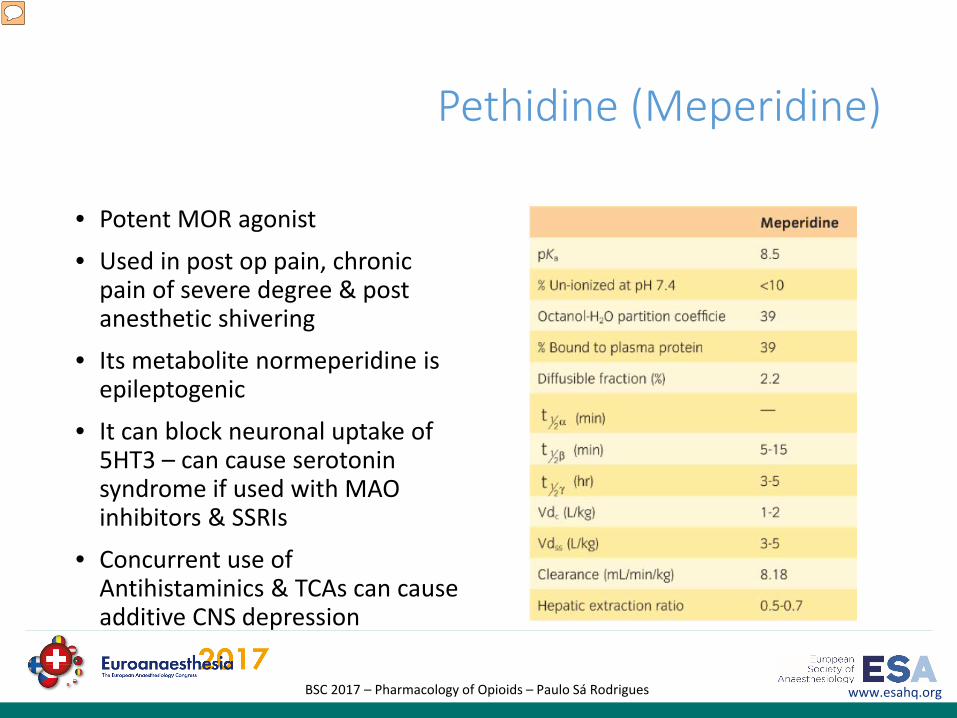
www.esahq.orgBSC 2017 – Pharmacology of Opioids – Paulo Sá Rodrigues
Pethidine (Meperidine)
• Potent MOR agonist
• Used in post op pain, chronic pain of severe degree & post anesthetic shivering
• Its metabolite normeperidine is epileptogenic
• It can block neuronal uptake of 5HT3 – can cause serotonin syndrome if used with MAO inhibitors & SSRIs
• Concurrent use of Antihistaminics & TCAs can cause additive CNS depression
Presenter
Presentation Notes
Meperidine is a synthetic opioid agonist at mu and kappa opioid receptors and is derived from phenylepiperidine. Meperidine shares several structural features that are present in local anesthetics including a tertiary amine, an ester group and a lipophilic phenyl group. Indeed, meperidine administered intrathecally blocks sodium channels to a degree comparable with lidocaine. Structurally, meperidine is similar to atropine, and it possesses a mild atropine-like antispasmodic effect. Nevertheless, the principal pharmacologic effects of meperidine resemble morphine. Pharmacokinetics Meperidine is about one-tenth as potent as morphine, with 80 to 100 mg IM being equivalent to about 10 mg IM of morphine. The duration of action of meperidine is 2 to 4 hours, making it a shorter-acting opioid agonist than morphine. In equal analgesic doses, meperidine produces as much sedation, euphoria, nausea, vomiting, and depression of ventilation as does morphine. Unlike morphine, meperidine is well absorbed from the gastrointestinal tract, but nevertheless, it is only about one-half as effective orally as when administered IM. Metabolism Hepatic metabolism of meperidine is extensive, with about 90% of the drug initially undergoing demethylation to normeperidine and hydrolysis to meperidinic acid. Normeperidine subsequently undergoes hydrolysis to normeperidinic acid. Urinary excretion is the principal elimination route and is pH dependent. For example, if the urinary pH is <5, as much as 25% of meperidine is excreted unchanged. Indeed, acidification of the urine can be considered in an attempt to speed elimination of meperidine. Decreased renal function can predispose to accumulation of normeperidine. Normeperidine has an elimination half-time of 15 hours (<35 hours in patients in renal failure) and can be detected in urine for as long as 3 days after administration of meperidine. This metabolite is about one-half as active as meperidine as an analgesic. In addition, normeperidine produces CNS stimulation. Normeperidine toxicity manifesting as myoclonus and seizures is most likely during prolonged administration of meperidine as during patient-controlled analgesia, especially in the presence of impaired renal function. Normeperidine may also be important in meperidine-induced delirium (confusion, hallucinations), which has been observed in patients receiving the drug for longer than 3 days, corresponding to accumulation of this active metabolite. Elimination Half-Time The elimination half-time of meperidine is 3 to 5 hours (see Table 3-5). Because clearance of meperidine primarily depends on hepatic metabolism, it is possible that large doses of opioid would saturate enzyme systems and result in prolonged elimination half-times. Nevertheless, elimination half-time is not altered by doses of meperidine up to 5 mg/kg IV. About 60% of meperidine is bound to plasma proteins. Elderly patients manifest decreased plasma protein binding of meperidine, resulting in increased plasma concentrations of free drug and an apparent increased sensitivity of the opioid. The increased tolerance of alcoholics to meperidine and other opioids presumably reflects an increased Vd, resulting in lower plasma concentrations of meperidine. Clinical Uses The principal use of meperidine is for analgesia during labor and delivery and after surgery. Meperidine is the only opioid considered adequate for surgery when administered intrathecally. An IM injection of meperidine for postoperative analgesia results in peak plasma concentrations that vary three- to fivefold as well as a time required to achieve peak concentrations that varies three- to sevenfold among patients. The minimum analgesic plasma concentration of meperidine is highly variable among patients; however, in the same patient, differences in concentrations as small as 0.05 μg/mL can represent a margin between no relief and complete analgesia. A plasma meperidine concentration of 0.7 μg/mL would be expected to provide postoperative analgesia in about 95% of patients. Patient-controlled analgesia delivery systems usually provide acceptable postoperative analgesia, with total doses of meperidine ranging from 12 to 36 mg/hour (White, 1985). Normeperidine toxicity has been described in patients receiving meperidine for patient-controlled analgesia. Meperidine may be effective in suppressing postoperative shivering that may result in detrimental increases in metabolic oxygen consumption. The antishivering effects of meperidine may reflect stimulation of kappa receptors (estimated to represent 10% of its activity) and a drug-induced decrease in the shivering threshold (not present with alfentanil, clonidine, propofol, or volatile anesthetics. In addition, meperidine is a potent agonist at alpha2 receptors which might contribute to antishivering effects. Indeed, clonidine is even more effective than meperidine in reducing postoperative shivering. Butorphanol (a kappa receptor agonist-antagonist) stops shivering more effectively than opioids with a predominant mu opioid receptor agonist effect. Evidence for a role of kappa receptors in the antishivering effects of meperidine and butorphanol is the failure of naloxone to completely inhibit this drug-induced effect. Oral absorption may make meperidine more useful than morphine for the treatment of many forms of pain. Unlike morphine, meperidine is not useful for the treatment of diarrhea and is not an effective antitussive. During bronchoscopy, the relative lack of antitussive activity of meperidine makes this opioid less useful. Meperidine is not used in high doses because of significant negative cardiac inotropic effects plus histamine release in a substantial number of patients Side Effects The side effects of meperidine resemble those described for morphine. In therapeutic doses, meperidine is associated with orthostatic hypotension. In fact, hypotension after meperidine injection is more frequent and more profound than after comparable doses of morphine. Orthostatic hypotension suggests that meperidine, like morphine, interferes with compensatory sympathetic nervous system reflexes. Meperidine, in contrast to morphine, rarely causes bradycardia but instead may increase heart rate, reflecting its modest atropine-like qualities. Large doses of meperidine result in decreases in myocardial contractility, which, among opioids, is unique for this drug. Delirium and seizures, when they occur, presumably reflect accumulation of normeperidine, which has stimulating effects on the CNS. Serotonin syndrome (autonomic instability with hypertension, tachycardia, diaphoresis, hyperthermia, behavioral changes including confusion and agitation, and neuromuscular changes manifesting as hyperreflexia) occurs when drugs capable of increasing serotonin administration are administered. In severe cases, coma, seizures, coagulopathy, and metabolic acidosis may develop. Administration of meperidine to patients receiving antidepressant drugs (monoamine oxidase inhibitors, fluoxetine) may elicit this syndrome. Meperidine readily impairs ventilation and may be even more of a ventilatory depressant than morphine. This opioid promptly crosses the placenta, and concentrations of meperidine in umbilical cord blood at birth may exceed maternal plasma concentrations. Meperidine may produce less constipation and urinary retention than morphine. After equal analgesic doses, biliary tract spasm is less after meperidine injection than after morphine injection but greater than that caused by codeine. Meperidine does not cause miosis but rather tends to cause mydriasis, reflecting its modest atropine-like actions. A dry mouth and an increase in heart rate are further evidence of the atropine-like effects of meperidine. Transient neurologic symptoms have been described following the administration of intrathecal meperidine for surgical anesthesia. The pattern of withdrawal symptoms after abrupt discontinuation of meperidine differs from that of morphine in that there are few autonomic nervous system effects. In addition, symptoms of withdrawal develop more rapidly and are of a shorter duration compared with those of morphine.
www.esahq.orgBSC 2017 – Pharmacology of Opioids – Paulo Sá Rodrigues
Opioid Antagonists
Effect in the presence of Agonists
• Effect on Acute opioid actions:• Increase in respiratory rate &
depth• Reversal of dysphoric &
psychotomimetic effects• Overshoot phenomenon• Rebound release of
catecholamines tachycardia, hypertension, ventricular arrhythmias
www.esahq.orgBSC 2017 – Pharmacology of Opioids – Paulo Sá Rodrigues
Opioid Antagonists
Effect in the presence of Agonists
• Effect on Opioid dependant patients:• Moderate to severe withdrawal • Depends on the dose of the
antagonist and also on the degree and duration of dependence
• Methylnaltrexone & Alvimopancan reverse the GI effects of opioid dependence without making central withdrawal syndrome.

www.esahq.orgBSC 2017 – Pharmacology of Opioids – Paulo Sá Rodrigues
References
• Opioids in Medicine– A comprehensive Review on the mode of action and the use of analgesia in different pain states; Enno Freye, Spinger, 1997
• Miller’s Anesthesia, 7th edition• Barash Clinical Anesthesia, 7th edition
• Stoelting Pharmacology and Physiology in Clinical Practice, 4th edition
• http://anesthesiology.pubs.asahq.org/article.aspx?articleid=1934470#68016492