BriefCommunications Clathrin ... · BriefCommunications...
7
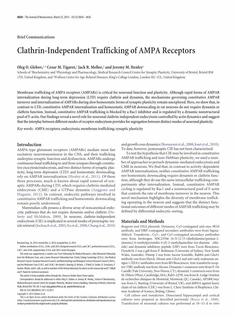
Brief Communications Clathrin-Independent Trafficking of AMPA Receptors Oleg O. Glebov, 1,3 Cezar M. Tigaret, 2 Jack R. Mellor, 2 and Jeremy M. Henley 1 Schools of 1 Biochemistry and 2 Physiology and Pharmacology, Medical Research Council Centre for Synaptic Plasticity, University of Bristol, Bristol BS8 1TD, United Kingdom, and 3 Wolfson Centre for Age-Related Diseases, King’s College London, London SE1 1UL, United Kingdom Membrane trafficking of AMPA receptors (AMPARs) is critical for neuronal function and plasticity. Although rapid forms of AMPAR internalization during long-term depression (LTD) require clathrin and dynamin, the mechanisms governing constitutive AMPAR turnover and internalization of AMPARs during slow homeostatic forms of synaptic plasticity remain unexplored. Here, we show that, in contrast to LTD, constitutive AMPAR internalization and homeostatic AMPAR downscaling in rat neurons do not require dynamin or clathrin function. Instead, constitutive AMPAR trafficking is blocked by a Rac1 inhibitor and is regulated by a dynamic nonstructural pool of F-actin. Our findings reveal a novel role for neuronal clathrin-independent endocytosis controlled by actin dynamics and suggest that the interplay between different modes of receptor endocytosis provides for segregation between distinct modes of neuronal plasticity. Key words: AMPA receptors; endocytosis; membrane trafficking; synaptic plasticity Introduction AMPA-type glutamate receptors (AMPARs) mediate most fast excitatory neurotransmission in the CNS, and their trafficking underpins synaptic function and dysfunction. AMPARs undergo continuous basal trafficking to and from synapses through constitu- tive exocytosis/endocytosis, and two distinct forms of synaptic plas- ticity, long-term depression (LTD) and homeostatic downscaling, rely on AMPAR internalization (Henley et al., 2011). Of these three processes, much is known about rapid removal of syn- aptic AMPARs during LTD, which requires clathrin-mediated endocytosis (CME) and a GTPase dynamin (Anggono and Huganir, 2012). In contrast, endocytic pathways involved in constitutive AMPAR trafficking and homeostatic downscaling remain poorly understood. Mammalian cells possess a diverse array of noncanonical endo- cytic pathways that do not require dynamin and/or clathrin (Do- herty and McMahon, 2009). In neurons, clathrin-independent endocytosis (CIE) is implicated in several modes of presynaptic ves- icle retrieval (Jockusch et al., 2005; Xu et al., 2008; Chung et al., 2010) and growth cone dynamics (Bonanomi et al., 2008; Joset et al., 2010). To date, however, postsynaptic CIE has not been characterized. To test the hypothesis that CIE may be involved in constitutive AMPAR trafficking and non-Hebbian plasticity, we used a num- ber of approaches to perturb dynamin-mediated endocytosis and CME in neurons. We find that, in contrast to activity-dependent AMPAR internalization, neither constitutive AMPAR trafficking nor homeostatic downscaling require dynamin or clathrin func- tion, although they do use the same intracellular trafficking com- partments after internalization. Instead, constitutive AMPAR cycling is regulated by Rac1 and a nonstructural pool of F-actin that controls the rate of membrane turnover in the neuron. This novel mechanism highlights the diversity of membrane traffick- ing operating in the neuron and suggests that the distinct func- tional outcomes of different modes of AMPAR trafficking may be defined by differential endocytic sorting. Materials and Methods Reagents and DNA plasmids. Dynasore, Cy3-conjugated anti-myc 9E10 antibody, and HRP-conjugated secondary antibodies were from Sigma- Aldrich. Transferrin-, Cy3-, and Cy5-conjugated secondary antibodies were from Invitrogen. NSC23766 (6-N-[2-[5-(diethylamino)pentan-2- ylamino]-6-methylpyrimidin-4-yl]-2-methylquinoline-4,6-diamine chlo- ride) and dynamin inhibitory peptide (DIP) were from Tocris Bioscience. Dynole34-2 was a gift from P. Robinson (University of Sydney, New South Wales, Australia). Pitstop 2 was from Ascent Scientific. Rabbit anti-GluA1 antibody was from Merck. Mouse anti-GluA2 and anti-early endosome an- tigen 1 (EEA1) antibodies were from BD Biosciences. Anti-transferrin recep- tor (TfR) antibody was from Abcam. Dynamin 1 constructs were from P. De Camilli (Yale University, New Haven, CT), dynamin 3 constructs were from M. Ehlers (Pfizer, Cambridge, MA), Rab5–Q79L was from R. Lodge (Institut de recherches cliniques de Montréal, Montreal, QC, Canada), AP180Cmyc was from G. Banting (University of Bristol, UK), and shRNA against heavy chain of rat clathrin (CHC) was from L. Chen (Institute of Biophysics, Chi- nese Academy of Science, Beijing, China). Cell culture and transfection. Dissociated hippocampal and cortical cultures were prepared as described previously (Rocca et al., 2008). Transfection of neuronal cultures was performed at 10 –13 d in vitro Received Aug. 26, 2014; revised Nov. 6, 2014; accepted Nov. 8, 2014. Author contributions: O.O.G., J.R.M., and J.M.H. designed research; O.O.G. and C.M.T. performed research; O.O.G., C.M.T., and J.R.M. analyzed data; O.O.G. and J.M.H. wrote the paper. This work was supported by a London Law Trust Fellowship for Medical Research, a Beit Memorial Fellowship from the Wellcome Trust, and a Junior Research Fellowship from Trinity College Cambridge (O.O.G.), the Medical Research Council, European Research Council, and Biotechnology and Biological Sciences Research Council (J.M.H.), and the Wellcome Trust (C.M.T. and J.R.M.). We thank G. Banting, B. Nichols, J. O’Neill, N. Zenkin, D. Srivastava, P. Gordon-Weeks, and G. Lalli, as well as members of the Henley laboratory for advice on the manuscript and P. Tidball and P. Rubin for technical assistance. This article is freely available online through the J Neurosci Author Open Choice option. Correspondence should be addressed to either Oleg O. Glebov or Jeremy M. Henley, School of Biochemistry, Medical Research Council Centre for Synaptic Plasticity, Medical Sciences Building, University of Bristol, University Walk, Bristol BS8 1TD, UK. E-mail: [email protected], [email protected]. DOI:10.1523/JNEUROSCI.3571-14.2015 Copyright © 2015 Glebov et al. This is an Open Access article distributed under the terms of the Creative Commons Attribution License (http://creativecommons.org/licenses/by/3.0), whichpermitsunrestricteduse,distributionandreproductioninany medium provided that the original work is properly attributed. 4830 • The Journal of Neuroscience, March 25, 2014 • 35(12):4830 – 4836
Transcript of BriefCommunications Clathrin ... · BriefCommunications...
Brief Communications
Clathrin-Independent Trafficking of AMPA Receptors
Oleg O. Glebov,1,3 Cezar M. Tigaret,2 Jack R. Mellor,2 and Jeremy M. Henley1
Schools of 1Biochemistry and 2Physiology and Pharmacology, Medical Research Council Centre for Synaptic Plasticity, University of Bristol, Bristol BS81TD, United Kingdom, and 3Wolfson Centre for Age-Related Diseases, King’s College London, London SE1 1UL, United Kingdom
Membrane trafficking of AMPA receptors (AMPARs) is critical for neuronal function and plasticity. Although rapid forms of AMPARinternalization during long-term depression (LTD) require clathrin and dynamin, the mechanisms governing constitutive AMPARturnover and internalization of AMPARs during slow homeostatic forms of synaptic plasticity remain unexplored. Here, we show that, incontrast to LTD, constitutive AMPAR internalization and homeostatic AMPAR downscaling in rat neurons do not require dynamin orclathrin function. Instead, constitutive AMPAR trafficking is blocked by a Rac1 inhibitor and is regulated by a dynamic nonstructuralpool of F-actin. Our findings reveal a novel role for neuronal clathrin-independent endocytosis controlled by actin dynamics and suggestthat the interplay between different modes of receptor endocytosis provides for segregation between distinct modes of neuronal plasticity.
Key words: AMPA receptors; endocytosis; membrane trafficking; synaptic plasticity
IntroductionAMPA-type glutamate receptors (AMPARs) mediate most fastexcitatory neurotransmission in the CNS, and their traffickingunderpins synaptic function and dysfunction. AMPARs undergocontinuous basal trafficking to and from synapses through constitu-tive exocytosis/endocytosis, and two distinct forms of synaptic plas-ticity, long-term depression (LTD) and homeostatic downscaling,rely on AMPAR internalization (Henley et al., 2011). Of thesethree processes, much is known about rapid removal of syn-aptic AMPARs during LTD, which requires clathrin-mediatedendocytosis (CME) and a GTPase dynamin (Anggono andHuganir, 2012). In contrast, endocytic pathways involved inconstitutive AMPAR trafficking and homeostatic downscalingremain poorly understood.
Mammalian cells possess a diverse array of noncanonical endo-cytic pathways that do not require dynamin and/or clathrin (Do-herty and McMahon, 2009). In neurons, clathrin-independentendocytosis (CIE) is implicated in several modes of presynaptic ves-icle retrieval (Jockusch et al., 2005; Xu et al., 2008; Chung et al., 2010)
and growth cone dynamics (Bonanomi et al., 2008; Joset et al., 2010).To date, however, postsynaptic CIE has not been characterized.
To test the hypothesis that CIE may be involved in constitutiveAMPAR trafficking and non-Hebbian plasticity, we used a num-ber of approaches to perturb dynamin-mediated endocytosis andCME in neurons. We find that, in contrast to activity-dependentAMPAR internalization, neither constitutive AMPAR traffickingnor homeostatic downscaling require dynamin or clathrin func-tion, although they do use the same intracellular trafficking com-partments after internalization. Instead, constitutive AMPARcycling is regulated by Rac1 and a nonstructural pool of F-actinthat controls the rate of membrane turnover in the neuron. Thisnovel mechanism highlights the diversity of membrane traffick-ing operating in the neuron and suggests that the distinct func-tional outcomes of different modes of AMPAR trafficking may bedefined by differential endocytic sorting.
Materials and MethodsReagents and DNA plasmids. Dynasore, Cy3-conjugated anti-myc 9E10antibody, and HRP-conjugated secondary antibodies were from Sigma-Aldrich. Transferrin-, Cy3-, and Cy5-conjugated secondary antibodieswere from Invitrogen. NSC23766 (6-N-[2-[5-(diethylamino)pentan-2-ylamino]-6-methylpyrimidin-4-yl]-2-methylquinoline-4,6-diamine chlo-ride) and dynamin inhibitory peptide (DIP) were from Tocris Bioscience.Dynole34-2 was a gift from P. Robinson (University of Sydney, New SouthWales, Australia). Pitstop 2 was from Ascent Scientific. Rabbit anti-GluA1antibody was from Merck. Mouse anti-GluA2 and anti-early endosome an-tigen 1 (EEA1) antibodies were from BD Biosciences. Anti-transferrin recep-tor (TfR) antibody was from Abcam. Dynamin 1 constructs were from P. DeCamilli (Yale University, New Haven, CT), dynamin 3 constructs were fromM. Ehlers (Pfizer, Cambridge, MA), Rab5–Q79L was from R. Lodge (Institutde recherches cliniques de Montréal, Montreal, QC, Canada), AP180Cmycwas from G. Banting (University of Bristol, UK), and shRNA against heavychain of rat clathrin (CHC) was from L. Chen (Institute of Biophysics, Chi-nese Academy of Science, Beijing, China).
Cell culture and transfection. Dissociated hippocampal and corticalcultures were prepared as described previously (Rocca et al., 2008).Transfection of neuronal cultures was performed at 10 –13 d in vitro
Received Aug. 26, 2014; revised Nov. 6, 2014; accepted Nov. 8, 2014.Author contributions: O.O.G., J.R.M., and J.M.H. designed research; O.O.G. and C.M.T. performed research; O.O.G.,
C.M.T., and J.R.M. analyzed data; O.O.G. and J.M.H. wrote the paper.This work was supported by a London Law Trust Fellowship for Medical Research, a Beit Memorial Fellowship
from the Wellcome Trust, and a Junior Research Fellowship from Trinity College Cambridge (O.O.G.), the MedicalResearch Council, European Research Council, and Biotechnology and Biological Sciences Research Council (J.M.H.),and the Wellcome Trust (C.M.T. and J.R.M.). We thank G. Banting, B. Nichols, J. O’Neill, N. Zenkin, D. Srivastava, P.Gordon-Weeks, and G. Lalli, as well as members of the Henley laboratory for advice on the manuscript and P. Tidballand P. Rubin for technical assistance.
This article is freely available online through the J Neurosci Author Open Choice option.Correspondence should be addressed to either Oleg O. Glebov or Jeremy M. Henley, School of Biochemistry,
Medical Research Council Centre for Synaptic Plasticity, Medical Sciences Building, University of Bristol, UniversityWalk, Bristol BS8 1TD, UK. E-mail: [email protected], [email protected].
DOI:10.1523/JNEUROSCI.3571-14.2015Copyright © 2015 Glebov et al.
This is an Open Access article distributed under the terms of the Creative Commons Attribution License(http://creativecommons.org/licenses/by/3.0), which permits unrestricted use, distribution and reproduction in anymedium provided that the original work is properly attributed.
4830 • The Journal of Neuroscience, March 25, 2014 • 35(12):4830 – 4836
using Lipofectamine2000 (Invitrogen) accord-ing to the instructions of the manufacturerwith minor modifications.
Biotinylation of cell surface protein. Live cor-tical neurons (15–21 d in vitro) were surfacebiotinylated on ice using the membrane-impermeant Sulfo-NHS-SS-Biotin accordingto the previously published protocol (Martinand Henley, 2004). Bands were quantified us-ing NIH ImageJ software and normalized tothe total receptor band intensity and internal-ization in the control sample.
Confocal imaging. Internalization assays fortransferrin and anti-AMPAR subunit antibod-ies were performed as described previously(Rocca et al., 2008). Each experiment was per-formed using the same batch of coverslips andneurons cultured under identical conditions.Briefly, hippocampal neurons were live labeledfor 20 min at 12–15°C, washed, and allowed tointernalize for 0 –30 min before fixation. Sur-face label was revealed by overnight stainingwith an appropriate Cy3-conjugated second-ary antibody, whereas internalized label wasstained by an appropriate Cy5-conjugated sec-ondary antibody after permeabilization. Im-ages were acquired on LSM510 and LSM710confocal microscopes (Zeiss) and analyzed us-ing NIH ImageJ software. To minimize bias,quantification of internalization was per-formed by collecting random fields from vari-ous regions of the coverslip. The followingformula for quantification of internalizationwas used:
I � (ICy5 � k � ICy3)/(ICy5 � (1 � k) � ICy3),
where ICy5 and ICy3 refer to intensities in Cy5and Cy3 channels, respectively. Correction co-efficient k was calculated as follows:
k � (ICy5t0/ICy3t0),
where ICy5t0 and ICy3t0 refer to intensities inCy5 and Cy3 at the 0 time point. To correct forthe inherent variability in intensity of surfacelabeling between different cultures, resultingintensities were normalized so that the averageintensity of the control sample was set to 1.
The fraction of internalized AMPARs(iAMPARs) was quantified as loss of surfacestaining (sAMPARs) relative to the 0 time con-trol. For colocalization analysis, surface re-maining AMPARs were blocked with the
4
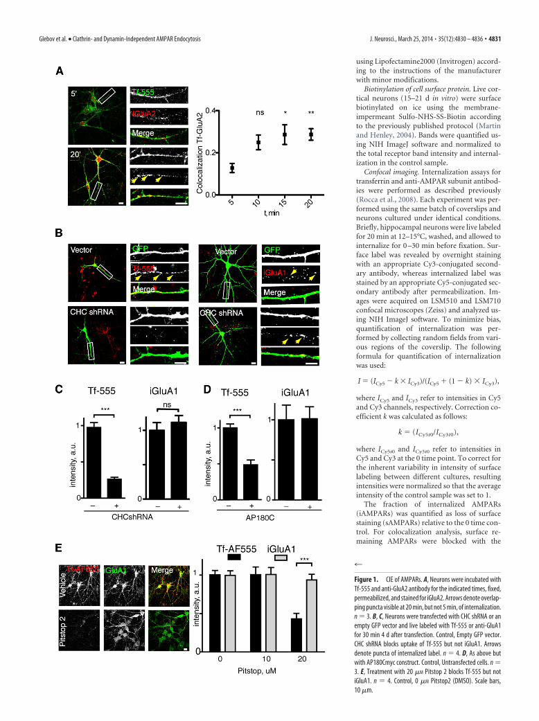
Figure 1. CIE of AMPARs. A, Neurons were incubated withTf-555 and anti-GluA2 antibody for the indicated times, fixed,permeabilized, and stained for iGluA2. Arrows denote overlap-ping puncta visible at 20 min, but not 5 min, of internalization.n � 3. B, C, Neurons were transfected with CHC shRNA or anempty GFP vector and live labeled with Tf-555 or anti-GluA1for 30 min 4 d after transfection. Control, Empty GFP vector.CHC shRNA blocks uptake of Tf-555 but not iGluA1. Arrowsdenote puncta of internalized label. n � 4. D, As above butwith AP180Cmyc construct. Control, Untransfected cells. n �3. E, Treatment with 20 �M Pitstop 2 blocks Tf-555 but notiGluA1. n � 4. Control, 0 �M Pitstop2 (DMSO). Scale bars,10 �m.
Glebov et al. • Clathrin- and Dynamin-Independent AMPAR Endocytosis J. Neurosci., March 25, 2014 • 35(12):4830 – 4836 • 4831
excess of unconjugated secondary antibody overnight. The amount ofcolocalization was quantified using an ImageJ/Fiji Colocalization plugin(http://fiji.sc/Coloc_2).
Slice preparation. Acute hippocampal slices were prepared from anes-thetized P13–P15 male Wistar rats. Brains were dissected in ice-coldartificial CSF (aCSF) equilibrated with 95% CO2 and 5% O2. Horizontalhippocampal slices (400 –500 �m thick) were cut with a VT1200 vi-bratome (Leica) and incubated in aCSF at 36°C for 30 min and then atroom temperature before use. The connections between CA3 and CA1were cut before transfer to the submerged recording chamber. The pro-cedures were performed in accordance with the United Kingdom HomeOffice and University of Bristol guidelines.
Electrophysiology. Whole-cell recordings were made from CA1 pyra-midal cells visualized with infrared differential interference contrast op-tics (Olympus BX-51 microscope) in a recording chamber perfused withaCSF (35°C) containing 50 �M picrotoxin. Patch electrodes (4 –5 M�)were filled with intracellular solution (in mM: 117 CsMeSO3, 8 NaCl, 10HEPES, 5 QX-314, 4 MgATP, 0.3 NaGTP, 0.5 EGTA, and 0.1 spermine),set to pH 7.4, 280 –285 mOsm. Cells were voltage clamped at �70 mV,and the recorded currents were filtered at 4 kHz and digitized at 10 kHzusing a CED Micro 1401 mk II board and Signal 2 software (CambridgeElectronics Design). Cells with series resistance �30 M� or showing�20% change were discarded from subsequent analysis. Baseline synap-tic responses were evoked at 0.2 Hz with a tungsten bipolar electrode (100k�, 119 �m tip spacing; FHC) placed in the stratum radiatum. Consec-utive EPSCs were averaged online every minute, and their amplitudeswere normalized offline to the first minute of recording.
For synaptic plasticity experiments, EPSCs were recorded from twoindependent pathways. LTD was induced at 15–20 min after break-in bya pairing protocol (1 Hz � 300 stimuli at �40 mV) delivered to the test
pathway, whereas the control pathway was paused. One-minute averageEPSC amplitudes (see above) were normalized to the average value over5 min before LTD induction.
Statistical analysis. Every experiment was performed at least in tripli-cate (see figure legends). Data were normalized to the control sample,and statistical analysis was performed using GraphPad Prism software(GraphPad Software). The D’Agostino–Pearson omnibus test was usedto test for normality of distribution, and, depending on results, a two-tailed t test or Mann–Whitney nonparametric test was used. ForANOVA, one-way ANOVA with Dunn’s post hoc test was used. Levels ofstatistical significance were as follows: *p � 0.05, **p � 0.01, ***p �0.001. Error bars represent � SEM.
ResultsEndogenous surface AMPARs in live cultured rat hippocampalneurons were labeled with antibodies recognizing extracellularepitopes of GluA1 and GluA2 subunits (Fig. 1A,B), and theirinternalization was monitored by immunofluorescence. Inagreement with previous reports (Lin et al., 2000; Lu et al., 2007),we observed constitutive internalization of both GluA1 andGluA2, with 31.6 � 5.5 and 36.5 � 4.9% of surface receptors,respectively, internalized after 20 min. Although little overlap wasobserved between iAMPARs and fluorescent CME cargo trans-ferrin Tf-Alexa Fluor 555 (Tf-555) at 5 min, by 15 min, there wassignificant colocalization between the two proteins (Fig. 1A).This observation raised the possibility that AMPARs and trans-ferrin may internalize via distinct endocytic mechanisms but laterconverge within the same endosomal compartments.
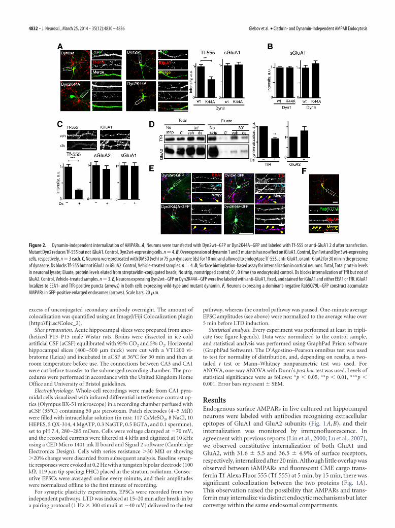
Figure 2. Dynamin-independent internalization of AMPARs. A, Neurons were transfected with Dyn2wt–GFP or Dyn2K44A–GFP and labeled with Tf-555 or anti-GluA1 2 d after transfection.Mutant Dyn2 reduces Tf-555 but not iGluA1. Control, Dyn2wt-expressing cells. n�4. B, Overexpression of dynamin 1 and 3 mutants has no effect on iGluA1. Control, Dyn1wt and Dyn3wt-expressingcells, respectively. n � 3 each. C, Neurons were pretreated with DMSO (veh) or 75 �M dynasore (ds) for 10 min and allowed to endocytose Tf-555, anti-GluA1, or anti-GluA2 for 30 min in the presenceof dynasore. Ds blocks Tf-555 but not iGluA1 or iGluA2. Control, Vehicle-treated samples. n � 4. D, Surface biotinylation-based assay for internalization in cortical neurons. Total, Total protein levelsin neuronal lysate; Eluate, protein levels eluted from streptavidin-conjugated beads; No strip, nonstripped control; 0, 0 time (no endocytosis) control. Ds blocks internalization of TfR but not ofGluA2. Control, Vehicle-treated samples. n � 3. E, Neurons expressing Dyn2wt–GFP or Dyn2K44A–GFP were live labeled with anti-GluA1, fixed, and stained for iGluA1 and either EEA1 or TfR. iGluA1localizes to EEA1- and TfR-positive puncta (arrows) in both cells expressing wild-type and mutant dynamin. F, Neurons expressing a dominant-negative Rab5Q79L–GFP construct accumulateAMPARs in GFP-positive enlarged endosomes (arrows). Scale bars, 20 �m.
4832 • J. Neurosci., March 25, 2014 • 35(12):4830 – 4836 Glebov et al. • Clathrin- and Dynamin-Independent AMPAR Endocytosis

To determine whether AMPAR internalization requires clath-rin function, we knocked down the heavy chain of rat clathrin(CHC shRNA; He et al., 2008). Expression of CHC shRNAblocked internalization of transferrin but not AMPARs (Fig.1B,C). Similar results were obtained with overexpression of adominant-negative variant of the clathrin adaptor protein AP180(AP180C; Ford et al., 2001; Fig. 1D) and Pitstop 2 (20 �M), a smallmolecule inhibitor of CME (von Kleist et al., 2011; Fig. 1E). To-gether, these data indicate that constitutive internalization ofAMPARs does not require CME.
CME and several CIE pathways require the activity of the largeGTPase dynamin to mediate scission of the budding endocyticvesicle from the plasma membrane (Doherty and McMahon,2009). Therefore, we next tested whether dynamin activity wasrequired for constitutive internalization of AMPARs. Althoughtransferrin internalization was markedly reduced by a dominant-negative K44A dynamin 2 mutant (Dyn2–K44A; Damke et al.,1994), iAMPARs (Fig. 2A) and sAMPARs (Fig. 3D) were unaf-fected. Similar results were obtained for neuron-specific isoformsdynamin 1 and dynamin 3 (Fig. 2B). Consistent with these re-sults, the dynamin GTPase inhibitor dynasore (Macia et al., 2006)blocked transferrin uptake but had no effect on iAMPARs (Fig.
2C), as did an alternative inhibitor of dynamin function,Dynole34-2 (Hill et al., 2009; data not shown). We also moni-tored the dynamin dependence of internalization using surfacebiotinylation in cultured cortical neurons. As expected, internal-ization of the TfR was inhibited by dynasore, whereas GluA2 wasunaffected (Fig. 2D), confirming that dynamin is not required forconstitutive AMPAR trafficking.
Depending on the context, cargoes endocytosed through CIEand CME may either follow separate intracellular trajectoriesor converge in endosomal compartments such as early andrecycling endosomes (Hansen and Nichols, 2009). Given thetime-dependent overlap between internalized transferrin andAMPARs (Fig. 1A), we investigated the post-internalization itin-erary of AMPARs endocytosed via CIE. AMPARs internalizedthrough CME during LTD are rapidly enriched in EEA1-positiveearly and TfR-positive recycling endosomes (Ehlers, 2000), andthis was also the case for constitutively iAMPARs in both wild-type dynamin 2 (Dyn2wt) and Dyn2–K44A-expressing cells (Fig.2E). Additionally, iAMPARs colocalized with a GTPase-defectivemutant of a small GTPase Rab5 (Fig. 2F), suggesting that consti-tutively iAMPARs traffic through Rab5-positive endosomes.Thus, AMPARs internalized via both CME and CIE traffic along
Figure 3. CME is required for NMDAR–LTD but not for basal synaptic transmission or synaptic downscaling. A, Treatment with dynasore does not affect basal excitatory synaptic transmission.Time course of EPSC amplitude evoked at 0.2 Hz at Schaffer collateral–CA1 synapses after infusion of control (n � 6), 100 �M dynasore (Ds; n � 8), vehicle (Veh, 0.1% DMSO; n � 6), or 50 �M
QVPSRPNRAP (DIP; n � 6) by inclusion in the intracellular solution. B, The pairing protocol effectively induces LTD with control intracellular solution (Control; n � 12) but intracellular infusion ofdynasore (n � 9) inhibits LTD in the test pathway. Right, Representative current traces from time points shown (1, black: 1 min before pairing; 2, gray: 25 min after pairing). C, Application of 50 �M
myristoylated DIP (myrDIP) blocks AMPAR internalization in the live-labeling assay. Control, Vehicle-treated samples. n � 3. D, Overexpression of the Dyn2–K44A construct does not affecthomeostatic downscaling. Hippocampal neurons (12–14 d in vitro) transfected with Dyn2wt–GFP or Dyn2K44A–GFP were treated with 15 mM KCl and 40 �M bicuculline for 48 h (Scaled) and labeledto quantify sGluA1 and sGluA2. Control, Dyn2wt-expressing cells under control conditions. n � 3. Scale bar, 20 �m.
Glebov et al. • Clathrin- and Dynamin-Independent AMPAR Endocytosis J. Neurosci., March 25, 2014 • 35(12):4830 – 4836 • 4833

the classical endocytic route, suggesting that convergence of thesepathways at the level of early/recycling endosomes.
To assess the functional role of CIE in basal trafficking ofsynaptic AMPARs, we measured excitatory synaptic transmissionin acute hippocampal slices. As a positive control, we used theDIP that contains the proline-rich domain from dynamin 1,which rapidly increases AMPAR currents (Luscher et al., 1999)through blockade of AMPAR internalization (Fig. 3C). AlthoughDIP effectively increased EPSC amplitude, dynasore did not alterEPSC amplitude (Fig. 3A). The discrepancy between the effects ofDIP and dynasore suggests that the effect of DIP on basal AMPARlevels does not reflect the outcome of the functional inhibition ofdynamin GTPase activity, given that the inhibitory effect of DIP isrealized through its perturbation of the Src homology 3-domaininteractions of an endocytic adaptor protein amphiphysin (Owenet al., 1998) rather than abrogation of dynamin GTPase activity,consistent with dynamin not being required for constitutive traf-ficking of AMPARs. In agreement with previous studies, dyna-sore abolished NMDAR–LTD (Man et al., 2000; Collingridge etal., 2010; Fig. 3B). In contrast, chronic enhancement of excitatorynetwork activity using increased K, and the GABAA receptorblocker bicuculline reduced sAMPARs in cells with or withoutdynamin (Fig. 3D). Together, these data indicate that inhibitionof the GTPase dynamin function abolishes LTD but does not
affect basal levels of synaptic AMPARs or their homeostaticdownscaling.
To elucidate the molecular mechanism of CIE–AMPAR, wefocused as a candidate on a small GTPase Rac1 that clustersAMPARs in developing dendrites (Wiens et al., 2005) and regu-lates dynamin-independent macropinocytosis at the growth cone(Joset et al., 2010). Because neurons overexpressing Rac1 mu-tants exhibited profound morphological aberrations, we insteadchose to pharmacologically inhibit Rac1 activation by the drugNSC23766 (Gao et al., 2004), which did not noticeably affect thecell morphology. NSC23766 increased sAMPARs and decreasediAMPARs (Fig. 4A), indicating that Rac1 activation was requiredfor constitutive AMPAR trafficking.
Given that Rac1 regulates membrane trafficking processesthrough control of actin polymerization, we tested the role ofactin in AMPAR internalization. As reported previously (Zhou etal., 2001), treatment with 5 �M Latrunculin A (LatA) decreasedF-actin and sAMPARs but increased iAMPARs (Fig. 4B). Stabili-zation of F-actin by Jasplakinolide had the opposite effect (Fig.4C), suggesting a bidirectional regulation of constitutive AMPARtrafficking by actin dynamics.
We next asked whether the effect of actin is mediated directly onAMPAR trafficking or indirectly via its structural role at the synapse.The reported existence of distinct dendritic F-actin pools with dif-
Figure 4. Nonstructural F-actin regulates AMPAR trafficking. A, Pharmacological inhibition of Rac1 inhibits AMPAR trafficking. Neurons were treated overnight with vehicle (DMSO) or 100 �M
NSC23766, and sGluA2 and iGluA2 levels were quantified. Control, DMSO. n � 4. B, Treatment with 0.5 �M LatA does not affect F-actin content but upregulates AMPAR trafficking. Neurons weretreated overnight with vehicle (DMSO) or 5 or 0.5 �M LatA, and sGluA2, iGluA2, and F-actin levels were quantified. Phalloidin–Alexa Fluor 568 was used to label F-actin. Control, DMSO. n�6 (sGluA2and iGluA2), n � 3 (F-actin). C, Stabilization of F-actin by Jasplakinolide downregulates AMPAR trafficking. Neurons were treated overnight with vehicle (DMSO) or 1 �M Jasplakinolide (Jas) andiGluA2 levels were quantified. Control, DMSO. n � 4. D, Treatment with 0.5 �M LatA increases bulk membrane uptake but not CME. Neurons (14 –21 d in vitro) were treated as in B and thenincubated with either FM1-43fx or Tf-555 for 30 min. Control, DMSO. n � 3. E, LatA at 0.5 �M disrupts synaptic downscaling of GluA2. Hippocampal neurons were treated with 15 mM KCl and 40 �M
bicuculline for 48 h (Scaled) in the presence of 0.5 �M LatA, and sGluA2 levels were quantified. Control, DMSO-treated cells under control conditions. n � 3. Scale bars, 20 �m.
4834 • J. Neurosci., March 25, 2014 • 35(12):4830 – 4836 Glebov et al. • Clathrin- and Dynamin-Independent AMPAR Endocytosis

ferent dynamics and sensitivities to LatA (Gu et al., 2010) promptedus to attempt to pharmacologically uncouple the structural and non-structural functions of actin. LatA at 0.5 �M had no effect on theoverall levels of dendritic F-actin (Fig. 4B), suggesting that the struc-tural pool of F-actin remained intact, whereas lack of effect onsynaptic AMPARs (data not shown) indicated that limited depoly-merization of F-actin did not affect AMPAR distribution, ruling outthe homeostatic effect of enhanced neurotransmitter release. Strik-ingly, 0.5 �M LatA increased iGluA2 to the same extent as 5 �M (Fig.4B) and also increased the uptake of a membrane-binding dye FM1-43fx [fixable N-(3-triethylammoniumpropyl)-4-(4-(dibutylamino)styryl) pyridinium dibromide] but not of transferrin (Fig. 4D), sug-gesting that the observed increase of AMPAR cycling is likely attrib-utable to the increase in the overall dendritic CIE. Furthermore, 0.5�M LatA abolished the reduction of sAMPARs induced by bicucul-line and KCl (Fig. 4E), indicating that this nonstructural pool of actinwas required for homeostatic downscaling.
DiscussionTo date, the majority of AMPAR endocytosis studies have beenfocused on LTD (Anggono and Huganir, 2012), and therefore, ithas been commonly assumed that AMPARs traffic exclusively viaCME. Here, we use a wide variety of acute and chronic loss-of-function approaches to show that basal internalization and ho-meostatic downscaling of AMPARs proceed normally in theabsence of clathrin or dynamin function. Although individualpharmacological and genetic tools may in some cases exhibit off-target effects (Dutta et al., 2012; Park et al., 2013; Willox et al.,2014) and compensatory upregulation (Damke et al., 1995), CIEof AMPARs is observed regardless of the methodology used. Al-though the full molecular details of the endocytic pathway re-mains to be established, our data reveal a surprising fundamentaldifference between the fast (LTD) and slow (constitutive cycling,homeostatic downscaling) modalities of neuronal receptor endo-cytosis. Therefore, it is tempting to speculate that differentialcommitment of the same cargo to distinct endocytic itineraries(Di Guglielmo et al., 2003) may provide a parsimonious mecha-nistic framework for explaining the subsequent fates of AMPARmolecules internalized during constitutive cycling, homeostaticdownscaling, and LTD (Henley et al., 2011).
Loss of synaptic AMPARs and decreased synaptic transmis-sion after actin depolymerization has been generally interpretedas an indirect consequence of the structural disruption of thespine and synapse (Zhou et al., 2001; Hotulainen and Hoogen-raad, 2010). We show that a dynamic pool of F-actin limits en-docytic AMPAR trafficking and total membrane uptake but hasno obvious role in the maintenance of the structural integrity ofthe synapse. This trafficking pool of F-actin has distinct proper-ties from the main dendritic F-actin, allowing for functional un-coupling between structural plasticity and membrane trafficking.These findings indicate that the structural role of actin at thesynapse is realized independently from its role in the organizationof local membrane trafficking and are in line with recent reportsof multiple pools of actin coexisting at the synapse (Gu et al.,2010). It remains to be determined which of the actin regulatoryproteins resident at the synapse may define the distinct identitiesof these pools.
Dynamin-independent internalization of both major AMPARsubunits and the distinct cytoskeletal regulation of bulk mem-brane uptake from CME suggest that this pathway may poten-tially function as a nonselective recycling route for any cargo thatfails to be sequestered into a adaptor-driven endocytic pathway,such as CME. Indeed, although our study has predominantly
focused on AMPAR trafficking, evidence is accumulating thatCIE may carry other cargo receptors in other synaptic contexts(Cinar and Barnes, 2001; Kumari et al., 2008; Borroni and Bar-rantes, 2011). This emerging diversity of synaptic receptor traf-ficking challenges the extant perception of CME as thepredominant endocytic mechanism in neurons and raises newquestions about the mechanisms and regulatory pathways thatallow neurons to regulate their function.
ReferencesAnggono V, Huganir RL (2012) Regulation of AMPA receptor trafficking
and synaptic plasticity. Curr Opin Neurobiol 22:461– 469. CrossRefMedline
Bonanomi D, Fornasiero EF, Valdez G, Halegoua S, Benfenati F, Menegon A,Valtorta F (2008) Identification of a developmentally regulated pathwayof membrane retrieval in neuronal growth cones. J Cell Sci 121:3757–3769. CrossRef Medline
Borroni V, Barrantes FJ (2011) Cholesterol modulates the rate and mecha-nism of acetylcholine receptor internalization. J Biol Chem 286:17122–17132. CrossRef Medline
Chung C, Barylko B, Leitz J, Liu X, Kavalali ET (2010) Acute dynamin inhi-bition dissects synaptic vesicle recycling pathways that drive spontaneousand evoked neurotransmission. J Neurosci 30:1363–1376. CrossRefMedline
Cinar H, Barnes EM Jr (2001) Clathrin-independent endocytosis of GABAA receptors in HEK 293 cells. Biochemistry 40:14030 –14036. CrossRefMedline
Collingridge GL, Peineau S, Howland JG, Wang YT (2010) Long-term de-pression in the CNS. Nat Rev Neurosci 11:459 – 473. CrossRef Medline
Damke H, Baba T, Warnock DE, Schmid SL (1994) Induction of mutantdynamin specifically blocks endocytic coated vesicle formation. J Cell Biol127:915–934. CrossRef Medline
Damke H, Baba T, van der Bliek AM, Schmid SL (1995) Clathrin-independent pinocytosis is induced in cells overexpressing a temperature-sensitive mutant of dynamin. J Cell Biol 131:69 – 80. CrossRef Medline
Di Guglielmo GM, Le Roy C, Goodfellow AF, Wrana JL (2003) Distinctendocytic pathways regulate TGF-beta receptor signalling and turnover.Nat Cell Biol 5:410 – 421. CrossRef Medline
Doherty GJ, McMahon HT (2009) Mechanisms of endocytosis. Annu RevBiochem 78:857–902. CrossRef Medline
Dutta D, Williamson CD, Cole NB, Donaldson JG (2012) Pitstop 2 is apotent inhibitor of clathrin-independent endocytosis. PLoS One7:e45799. CrossRef Medline
Ehlers MD (2000) Reinsertion or degradation of AMPA receptors deter-mined by activity-dependent endocytic sorting. Neuron 28:511–525.CrossRef Medline
Ford MG, Pearse BM, Higgins MK, Vallis Y, Owen DJ, Gibson A, Hopkins CR,Evans PR, McMahon HT (2001) Simultaneous binding of PtdIns(4,5)P2and clathrin by AP180 in the nucleation of clathrin lattices on membranes.Science 291:1051–1055. CrossRef Medline
Gao Y, Dickerson JB, Guo F, Zheng J, Zheng Y (2004) Rational design andcharacterization of a Rac GTPase-specific small molecule inhibitor. ProcNatl Acad Sci U S A 101:7618 –7623. CrossRef Medline
Gu J, Lee CW, Fan Y, Komlos D, Tang X, Sun C, Yu K, Hartzell HC, Chen G,Bamburg JR, Zheng JQ (2010) ADF/cofilin-mediated actin dynamicsregulate AMPA receptor trafficking during synaptic plasticity. Nat Neu-rosci 13:1208 –1215. CrossRef Medline
Hansen CG, Nichols BJ (2009) Molecular mechanisms of clathrin-independent endocytosis. J Cell Sci 122:1713–1721. CrossRef Medline
He Z, Fan J, Kang L, Lu J, Xue Y, Xu P, Xu T, Chen L (2008) Ca2 triggersa novel clathrin-independent but actin-dependent fast endocytosis inpancreatic beta cells. Traffic 9:910 –923. CrossRef Medline
Henley JM, Barker EA, Glebov OO (2011) Routes, destinations and delays:recent advances in AMPA receptor trafficking. Trends Neurosci 34:258 –268. CrossRef Medline
Hill TA, Gordon CP, McGeachie AB, Venn-Brown B, Odell LR, Chau N,Quan A, Mariana A, Sakoff JA, Chircop M, Robinson PJ, McCluskey A(2009) Inhibition of dynamin mediated endocytosis by the dynoles—synthesis and functional activity of a family of indoles. J Med Chem 52:3762–3773. CrossRef Medline
Glebov et al. • Clathrin- and Dynamin-Independent AMPAR Endocytosis J. Neurosci., March 25, 2014 • 35(12):4830 – 4836 • 4835

Hotulainen P, Hoogenraad CC (2010) Actin in dendritic spines: connectingdynamics to function. J Cell Biol 189:619 – 629. CrossRef Medline
Jockusch WJ, Praefcke GJ, McMahon HT, Lagnado L (2005) Clathrin-dependent and clathrin-independent retrieval of synaptic vesicles in ret-inal bipolar cells. Neuron 46:869 – 878. CrossRef Medline
Joset A, Dodd DA, Halegoua S, Schwab ME (2010) Pincher-generatedNogo-A endosomes mediate growth cone collapse and retrograde signal-ing. J Cell Biol 188:271–285. CrossRef Medline
Kumari S, Borroni V, Chaudhry A, Chanda B, Massol R, Mayor S, BarrantesFJ (2008) Nicotinic acetylcholine receptor is internalized via a Rac-dependent, dynamin-independent endocytic pathway. J Cell Biol 181:1179 –1193. CrossRef Medline
Lin JW, Ju W, Foster K, Lee SH, Ahmadian G, Wyszynski M, Wang YT, ShengM (2000) Distinct molecular mechanisms and divergent endocytoticpathways of AMPA receptor internalization. Nat Neurosci 3:1282–1290.CrossRef Medline
Lu J, Helton TD, Blanpied TA, Racz B, Newpher TM, Weinberg RJ, EhlersMD (2007) Postsynaptic positioning of endocytic zones and AMPA re-ceptor cycling by physical coupling of dynamin-3 to Homer. Neuron55:874 – 889. CrossRef Medline
Luscher C, Xia H, Beattie EC, Carroll RC, von Zastrow M, Malenka RC, NicollRA (1999) Role of AMPA receptor cycling in synaptic transmission andplasticity. Neuron 24:649 – 658. CrossRef Medline
Macia E, Ehrlich M, Massol R, Boucrot E, Brunner C, Kirchhausen T (2006)Dynasore, a cell-permeable inhibitor of dynamin. Dev Cell 10:839 – 850.CrossRef Medline
Man HY, Lin JW, Ju WH, Ahmadian G, Liu L, Becker LE, Sheng M, Wang YT(2000) Regulation of AMPA receptor-mediated synaptic transmission byclathrin-dependent receptor internalization. Neuron 25:649–662. CrossRefMedline
Martin S, Henley JM (2004) Activity-dependent endocytic sorting of kai-
nate receptors to recycling or degradation pathways. EMBO J 23:4749 –4759. CrossRef Medline
Owen DJ, Wigge P, Vallis Y, Moore JD, Evans PR, McMahon HT (1998)Crystal structure of the amphiphysin-2 SH3 domain and its role in theprevention of dynamin ring formation. EMBO J 17:5273–5285. CrossRefMedline
Park RJ, Shen H, Liu L, Liu X, Ferguson SM, De Camilli P (2013) Dynamintriple knockout cells reveal off target effects of commonly used dynamininhibitors. J Cell Sci 126:5305–5312. CrossRef Medline
Rocca DL, Martin S, Jenkins EL, Hanley JG (2008) Inhibition of Arp2/3-mediated actin polymerization by PICK1 regulates neuronal morphologyand AMPA receptor endocytosis. Nat Cell Biol 10:259 –271. CrossRefMedline
von Kleist L, Stahlschmidt W, Bulut H, Gromova K, Puchkov D, RobertsonMJ, MacGregor KA, Tomilin N, Pechstein A, Chau N, Chircop M, SakoffJ, von Kries JP, Saenger W, Krausslich HG, Shupliakov O, Robinson PJ,McCluskey A, Haucke V (2011) Role of the clathrin terminal domain inregulating coated pit dynamics revealed by small molecule inhibition. Cell146:471– 484. CrossRef Medline
Wiens KM, Lin H, Liao D (2005) Rac1 induces the clustering of AMPAreceptors during spinogenesis. J Neurosci 25:10627–10636. CrossRefMedline
Willox AK, Sahraoui YME, Royle SJ (2014) Non-specificity of Pitstop 2 inclathrin-mediated endocytosis. Biol Open 3:326 –331. CrossRef Medline
Xu J, McNeil B, Wu W, Nees D, Bai L, Wu LG (2008) GTP-independentrapid and slow endocytosis at a central synapse. Nat Neurosci 11:45–53.CrossRef Medline
Zhou Q, Xiao M, Nicoll RA (2001) Contribution of cytoskeleton to theinternalization of AMPA receptors. Proc Natl Acad Sci U S A 98:1261–1266. CrossRef Medline
4836 • J. Neurosci., March 25, 2014 • 35(12):4830 – 4836 Glebov et al. • Clathrin- and Dynamin-Independent AMPAR Endocytosis