Brian R. Mulligan Positional Fault Fact or...
11
1 Positional Fault Fact or Fiction? Dr W.A. Hing Horizon@AUT Musculoskeletal Diagnostic Imaging Unit Brian R. Mulligan Brian R. Mulligan Evolution of Evolution of Orthopaedic Manual Therapy Manual Therapy Evolution of Evolution of Orthopaedic Orthopaedic Manual Therapy Manual Therapy MWMs … MWMs …
Transcript of Brian R. Mulligan Positional Fault Fact or...
1
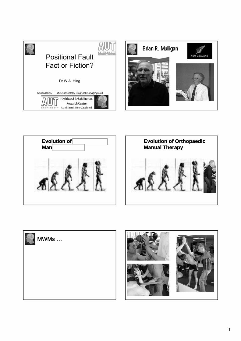
Positional FaultFact or Fiction?
Dr W.A. Hing
Horizon@AUT Musculoskeletal Diagnostic Imaging Unit
Brian R. Mulligan Brian R. Mulligan
Evolution of Evolution of OrthopaedicOrthopaedicManual TherapyManual Therapy
Evolution of Evolution of OrthopaedicOrthopaedicManual TherapyManual Therapy
MWMs …MWMs …
2
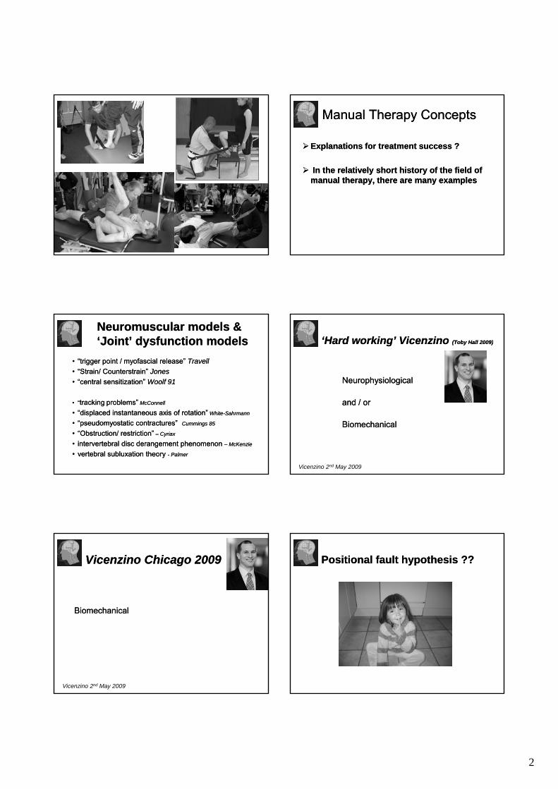
Manual Therapy ConceptsManual Therapy Concepts
Explanations for treatment success ?Explanations for treatment success ?
In the relatively short history of the field of In the relatively short history of the field of manual therapy, there are many examples manual therapy, there are many examples
Neuromuscular models & Neuromuscular models & ‘Joint’ dysfunction models‘Joint’ dysfunction models
•• “trigger point / “trigger point / myofascialmyofascial release”release” TravellTravell•• “Strain/ “Strain/ CounterstrainCounterstrain” ” JonesJones•• “central sensitization”“central sensitization” Woolf 91Woolf 91
•• ““trackingtracking problems”problems” McConnellMcConnell
•• “displaced instantaneous axis of rotation”“displaced instantaneous axis of rotation” WhiteWhite--SahrmannSahrmann
•• ““pseudomyostaticpseudomyostatic contractures”contractures” Cummings 85Cummings 85
•• “Obstruction/ restriction”“Obstruction/ restriction” –– CyriaxCyriax
•• intervertebralintervertebral disc derangement phenomenon disc derangement phenomenon –– McKenzieMcKenzie
•• vertebral vertebral subluxationsubluxation theory theory -- PalmerPalmer
‘Hard working’ ‘Hard working’ VicenzinoVicenzino (Toby Hall 2009)(Toby Hall 2009)
NeurophysiologicalNeurophysiological
and / or and / or
BiomechanicalBiomechanical
Vicenzino 2nd May 2009
VicenzinoVicenzino Chicago 2009Chicago 2009
BiomechanicalBiomechanicalBiomechanicalBiomechanical
Vicenzino 2nd May 2009
Positional fault hypothesis ??Positional fault hypothesis ??

3
Positional fault hypothesisPositional fault hypothesis
Matty & Pippi 2006
Mulligan’s TheoryMulligan’s Theory
•• Positional fault theory Positional fault theory Mulligan 95Mulligan 95
–– Joint alignment alteration due to injury or chronic/poor Joint alignment alteration due to injury or chronic/poor arthokinematicsarthokinematics
–– Inconsistent bony congruencies that occur after strain or Inconsistent bony congruencies that occur after strain or injuryinjury
–– Minor / subtle: Neither palpable nor evident on imaging?Minor / subtle: Neither palpable nor evident on imaging?–– Movement restrictions Movement restrictions -- pain resultspain results–– Responsible for movement restricted and painful jointsResponsible for movement restricted and painful joints
Positional fault hypothesisPositional fault hypothesis
MWM relocates joint in correct alignmentMWM relocates joint in correct alignment
Therefore immediate improvements in painTherefore immediate improvements in painTherefore immediate improvements in pain Therefore immediate improvements in pain and ROMand ROM
Mulligan’s explanation … is it that simple?Mulligan’s explanation … is it that simple?
Positional fault hypothesisPositional fault hypothesisPFH PFH -- EvidenceEvidence
Clinical success warrants its use Clinical success warrants its use (Mulligan)(Mulligan)
Mulligan encouraged others in the field toMulligan encouraged others in the field toMulligan encouraged others in the field to Mulligan encouraged others in the field to investigate its meritinvestigate its merit
Adequate in substantiating the PFH?Adequate in substantiating the PFH?
Evidence Based Medicine ?Evidence Based Medicine ?Positional fault hypothesisPositional fault hypothesisPFH PFH -- EvidenceEvidence
Clinical efficacy of MWMs underpinned by reasonable level of evidence (as Bill mentions!)
Less evidence to support the presence of positional faults
Is there evidence that practitioners are able to detect positional faults in their clinics with current level of technology?
4
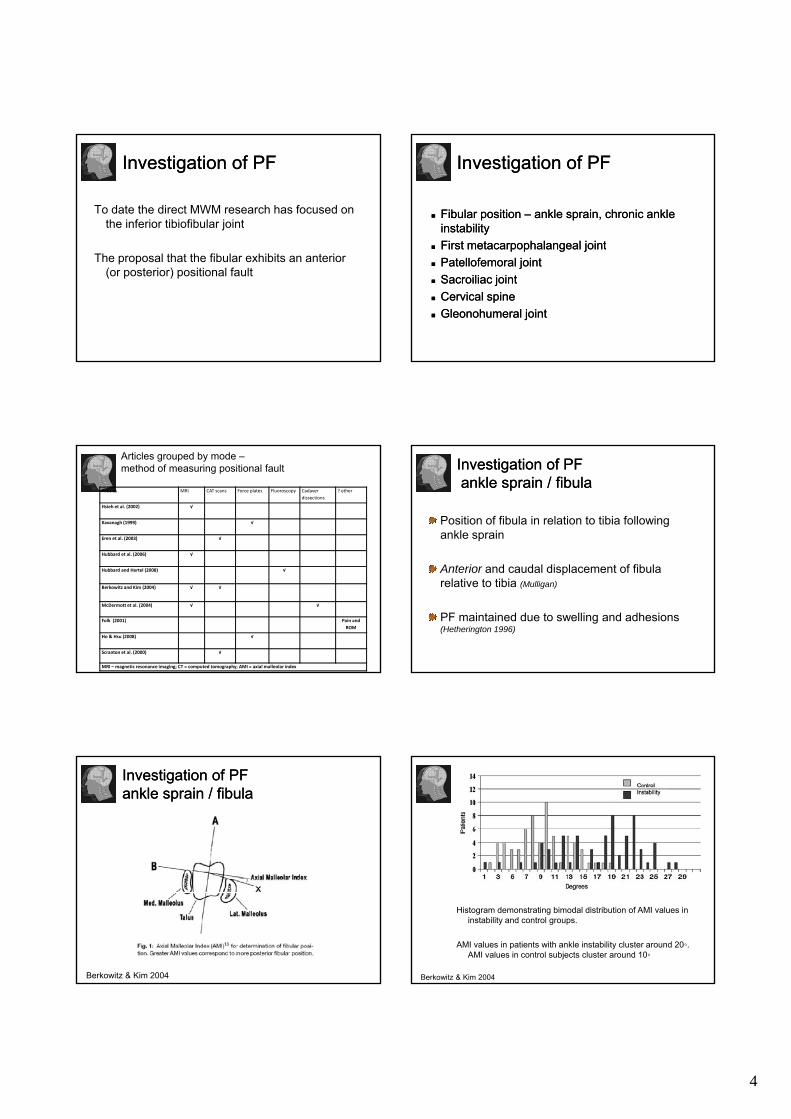
Investigation of PFInvestigation of PF
To date the direct MWM research has focused on the inferior tibiofibular joint
The proposal that the fibular exhibits an anterior (or posterior) positional fault
Investigation of PFInvestigation of PF
Fibular position Fibular position –– ankle sprain, chronic ankle ankle sprain, chronic ankle instabilityinstabilityFirstFirst metacarpophalangealmetacarpophalangeal jointjointFirst First metacarpophalangealmetacarpophalangeal jointjointPatellofemoralPatellofemoral jointjointSacroiliac jointSacroiliac jointCervical spineCervical spineGleonohumeralGleonohumeral jointjoint
Articles MRI CAT scans Force plates Fluoroscopy Cadaver dissections
? other
Hsieh et al. (2002) √
Kavanagh (1999) √
Eren et al. (2003) √
Hubbard et al. (2006) √
Articles grouped by mode –method of measuring positional fault
Hubbard and Hertel (2008) √
Berkowitz and Kim (2004) √ √
McDermott et al. (2004) √ √
Folk (2001) Pain and ROM
Ho & Hsu (2008) √
Scranton et al. (2000) √
MRI – magnetic resonance imaging; CT = computed tomography; AMI = axial malleolar index
Investigation of PFInvestigation of PFankle sprain / fibulaankle sprain / fibula
Position of fibula in relation to tibia following ankle sprain
Anterior and caudal displacement of fibula relative to tibia (Mulligan)
PF maintained due to swelling and adhesions (Hetherington 1996)
Investigation of PFInvestigation of PFankle sprain / fibulaankle sprain / fibula
Berkowitz & Kim 2004
Histogram demonstrating bimodal distribution of AMI values in instability and control groups.
AMI values in patients with ankle instability cluster around 20◦. AMI values in control subjects cluster around 10◦
Berkowitz & Kim 2004
5
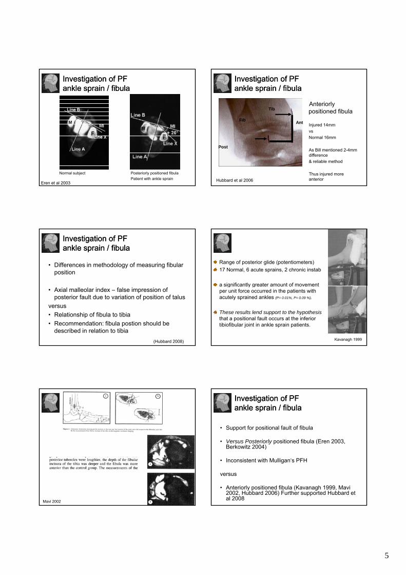
Investigation of PFInvestigation of PFankle sprain / fibulaankle sprain / fibula
Posteriorly positioned fibulaPatient with ankle sprain
Normal subject
Eren et al 2003
Investigation of PFInvestigation of PFankle sprain / fibulaankle sprain / fibula
Anteriorly positioned fibula
Ant Injured 14mmFib
Tib
Hubbard et al 2006
Post
vsNormal 16mm
As Bill mentioned 2-4mm difference & reliable method
Thus injured more anterior
Investigation of PFInvestigation of PFankle sprain / fibulaankle sprain / fibula
• Differences in methodology of measuring fibular position
• Axial malleolar index – false impression of posterior fault due to variation of position of talus
versus• Relationship of fibula to tibia • Recommendation: fibula postion should be
described in relation to tibia(Hubbard 2008)
Range of posterior glide (potentiometers)17 Normal, 6 acute sprains, 2 chronic instab
a significantly greater amount of movement g y gper unit force occurred in the patients with acutely sprained ankles (P= 0.01%, P= 0.09 %).
These results lend support to the hypothesis that a positional fault occurs at the inferior tibiofibular joint in ankle sprain patients.
Kavanagh 1999
Mavi 2002
Investigation of PFInvestigation of PFankle sprain / fibulaankle sprain / fibula
• Support for positional fault of fibula
• Versus Posteriorly positioned fibula (Eren 2003, Berkowitz 2004)Berkowitz 2004)
• Inconsistent with Mulligan‘s PFH
versus
• Anteriorly positioned fibula (Kavanagh 1999, Mavi 2002, Hubbard 2006) Further supported Hubbard et al 2008
6
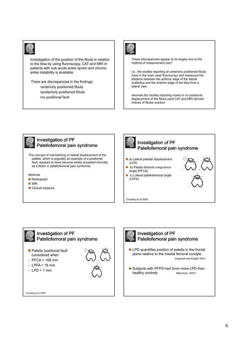
Investigation of the position of the fibula in relation to the tibia by using fluoroscopy, CAT and MRI in patients with sub-acute ankle sprain and chronic ankle instability is available
There are discrepancies in the findings: •anteriorly positioned fibula•posteriorly positioned fibula •no positional fault
These discrepancies appear to be largely due to the method of measurement use?
i.e., the studies reporting an anteriorly positioned fibula h i th i d fl d d thhave in the main used fluoroscopy and measured the distance between the anterior edge of the lateral malleolus and the anterior edge of the tibia from a lateral view
whereas the studies reporting mixed or no positional displacement of the fibula used CAT and MRI derived indices of fibular position
Investigation of PFInvestigation of PFPatellofemoral pain syndromePatellofemoral pain syndrome
The concept of mal-tracking or lateral displacement of the patella, which is arguably an example of a positional fault, appears to have become widely accepted clinically as a factor in patellofemoral pain syndrome
MethodsRadiographMRIClinical measure
Investigation of PFInvestigation of PFPatellofemoral pain syndromePatellofemoral pain syndrome
a) Lateral patellar displacement (LPD)b) Patello-femoral congruence ) gangle (PFCA)c) Lateral patellofemoral angle (LPFA)
Crossley et al 2000
Investigation of PFInvestigation of PFPatellofemoral pain syndromePatellofemoral pain syndrome
Patella ‘positional fault’ considered when
- PFCA > +58 mm- LPFA = 18 mm- LPD = 1 mm
Crossley et al 2000
Investigation of PFInvestigation of PFPatellofemoral pain syndromePatellofemoral pain syndrome
LPD quantifies position of patella in the frontal plane relative to the medial femoral condyle
(Ingersoll and Knight 1991)
Subjects with PFPS had 2mm more LPD than healthy controls (Macintyre, 2007)

7
Investigation of PFInvestigation of PFPatellofemoral pain syndromePatellofemoral pain syndrome
Clinical measureClinical measure
Herrington 2008
Investigation of PFInvestigation of PFPatellofemoral pain syndromePatellofemoral pain syndrome
Clinical measure
Herrington 2008
Investigation of PFInvestigation of PFPatellofemoralPatellofemoral pain syndromepain syndrome
• Influence of:- Patellar width?- Tibiofemoral rotation angle?
… on contact area between patella and femur
accounts 46% of variance on contact area
• Patella contact area appears to be an interesting area for future research, particularly with respect to the Mulligan concept, as the Mulligan technique used to manage PFPS attempts to change tibiofemoral rotation rather patella alignment directly.
(Salsich et al 2007)
Investigation of PFInvestigation of PF1st MCP joint1st MCP joint
Positional fault of 4 deg pronation 1st MCP
Hsieh et al 2002
Investigation of PFInvestigation of PFsacroiliac joint dysfunctionsacroiliac joint dysfunction
Hungerford et al 2004
The ShoulderThe Shoulder
Measurement Measurement with ultrasoundwith ultrasound
Posterior view Posterior view Anterior viewAnterior viewSuperior viewSuperior view
8
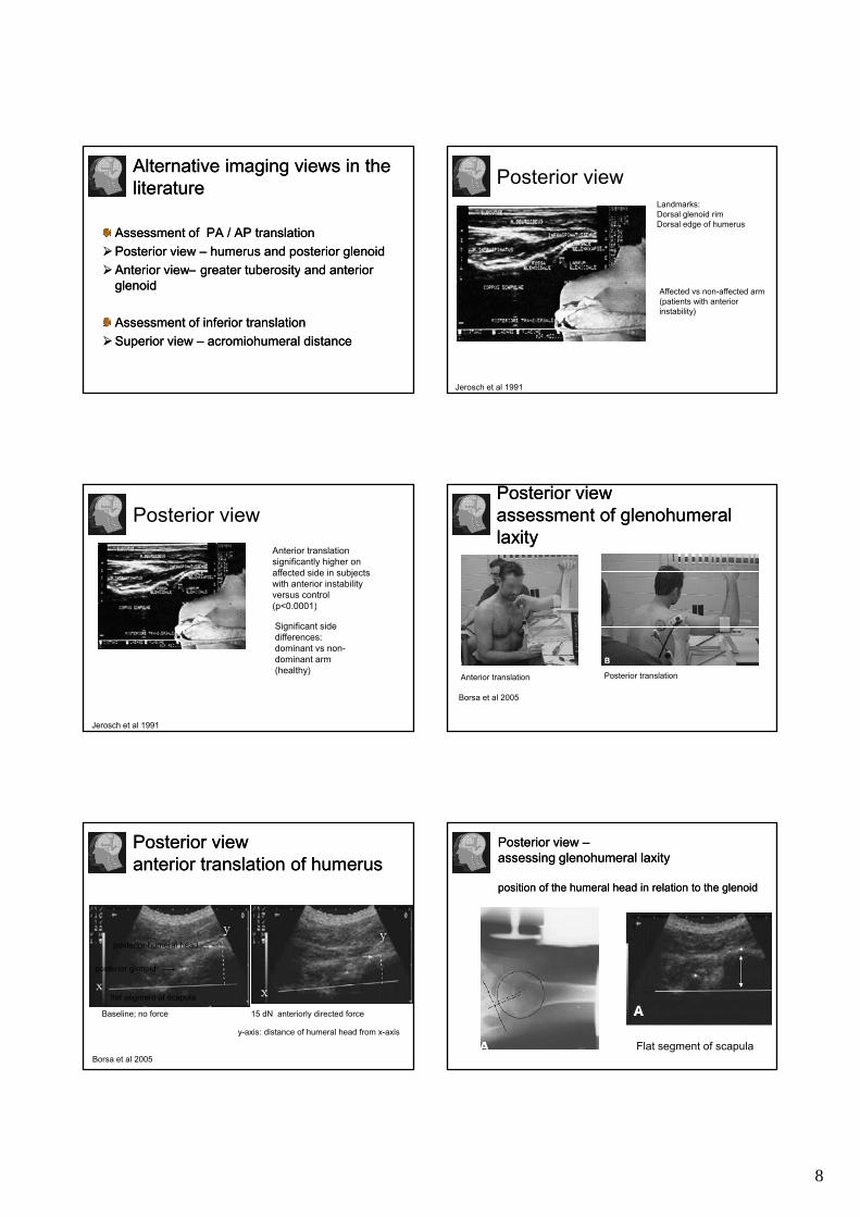
Alternative imaging views in the Alternative imaging views in the literatureliterature
Assessment of PA / AP translation Assessment of PA / AP translation Posterior view Posterior view –– humerushumerus and posterior and posterior glenoidglenoidAnterior viewAnterior view greatergreater tuberositytuberosity and anteriorand anteriorAnterior viewAnterior view–– greater greater tuberositytuberosity and anterior and anterior glenoidglenoid
Assessment of inferior translationAssessment of inferior translationSuperior view Superior view –– acromiohumeralacromiohumeral distancedistance
Posterior viewLandmarks:Dorsal glenoid rimDorsal edge of humerus
Jerosch et al 1991
Affected vs non-affected arm (patients with anterior instability)
Posterior viewAnterior translation significantly higher on affected side in subjects with anterior instability versus control (p<0 0001)
Jerosch et al 1991
Significant side differences: dominant vs non-dominant arm (healthy)
(p<0.0001)
Posterior viewPosterior viewassessment of glenohumeral assessment of glenohumeral laxitylaxity
Borsa et al 2005
Anterior translation Posterior translation
Posterior viewPosterior viewanterior translation of humerusanterior translation of humerus
posterior humeral head
Borsa et al 2005
Baseline; no force 15 dN anteriorly directed force
flat segment of scapula
posterior humeral head
posterior glenoid
y-axis: distance of humeral head from x-axis
Posterior view Posterior view ––assessing assessing glenohumeralglenohumeral laxitylaxity
position of the humeral head in relation to the position of the humeral head in relation to the glenoidglenoid
Flat segment of scapula
9
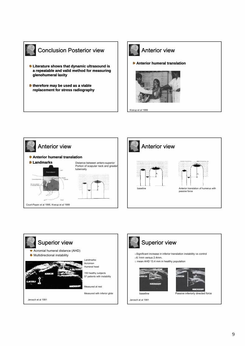
Conclusion Posterior viewConclusion Posterior view
Literature shows that dynamic ultrasound is Literature shows that dynamic ultrasound is a repeatable and valid method for measuring a repeatable and valid method for measuring glenohumeralglenohumeral laxitylaxitygg yy
therefore may be used as a viable therefore may be used as a viable replacement for stress radiographyreplacement for stress radiography
Anterior viewAnterior view
Anterior humeral translationAnterior humeral translation
Krarup et al 1999
Anterior viewAnterior view
Anterior humeral translationAnterior humeral translationLandmarksLandmarks Distance between antero-superior
Portion of scapular neck and greater tuberosity
Court-Payen et al 1995, Krarup et al 1999
Anterior viewAnterior view
baseline Anterior translation of humerus withpassive force
Superior viewSuperior viewAcromial humeral distance (AHD) Multidirectional instability
Landmarks:AcromionHumeral head
Jerosch et al 1991
150 healthy subjects57 patients with instability
Measured at rest
Measured with inferior glide
Superior viewSuperior view
oSignificant increase in inferior translation instability vs controlo6.1mm versus 2.4mm.o mean AHD 13.4 mm in healthy population
Jerosch et al 1991
baseline Passive inferiorly directed force
10
• It has been shown that the AHD ↓ during shoulder abduction using MRI
Acromiohumeral Distance AHD
•• AHD decreased with AHD decreased with shoulder impingement shoulder impingement syndrome (SIS)syndrome (SIS)
Hebert et al 2003
Acromio-Humeral Distance= AHD
Tangential distance between the humeral head surface and the lateral tip of the acromion
7 patients with SIS13 healthy subjects
Measurements at rest, 45° and 60 ° abduction
Desmeules et al (2004)
Definitive pattern of narrowing of the AHD with arm Abd
AHD values at rest tended to be higher in SIS group than in healthy group whilegroup, while
More pronounced narrowing occurred between 0º - 45º
Excessive superior translation of humeral head 0º - 45º
Desmeules et al (2004)
Sonography of Ssp tendon
young overhead athletes (basketball) in correlation with the main pathologic model of secondary SIS
Girometti et al (2006)
Measurement of SAD
Dynamic anterior impingement test (passive)
10 professional basketball players with sh pathology
10 non-athlete controls
l t l i i h l h d
Girometti et al (2006)
lateral view acromion - humeral headarm behind back position
Cutoff point < 7 mm: defined as decreased SAD (based on normative data derived from US and MRI)
Significant difference between groups only in SAD
57 patients with symptoms of unilateral SIS.
72 healthy control subjects.
Bil t l i ti i ll bj t
Cholewinsky et al (2008)
Bilateral examination in all subjects
Arm in neutral rotation
11
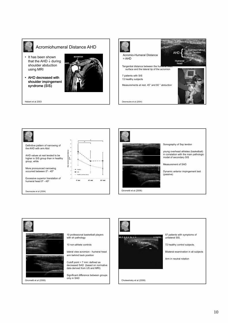
AHD and RC thickness smaller in SIS versus non affected shoulder (p<,0.001)
AHD significantly smaller in SIS vs control group (p<0 001)
Cholewinsky et al (2008)
control group (p<0.001)
AHD of more than 2.1mm in comparison to the unaffected joint may point to the dysfunction of RC muscles
AHD enables measurements of superior translation of the humeral head
ConclusionConclusion
There is both direct research into the PFH as it relates to the MWM concept
Predominantl research has not foc sed on thePredominantly research has not focused on the MWM concept but do describe investigations into minor positional ‘incongruencies’ that highlights key aspects of the PFH