Breast Cancer: Classification Based on Molecular Etiology ...cdn.intechweb.org/pdfs/24877.pdf · on...
17
4 Breast Cancer: Classification Based on Molecular Etiology Influencing Prognosis and Prediction Siddik Sarkar 1 and Mahitosh Mandal 1 1 School of Medical Science and Technology, Indian Institute of Technology Kharagpur Kharagpur, West Bengal India 1. Introduction Cancer is a group of diseases that leads to uncontrolled cell division and eventually forms a lump or mass called a tumor. They are classified and named after the part of the body where the tumor originates. Breast cancer begins in breast tissue, which is made up of glands for milk production, called lobules, and the ducts that connect lobules to the nipple. The remainder of the breast is made up of fatty, connective, and lymphatic tissue. On the basis of origin, it is of two types (i) ductal and (ii) lobular. Ductal carcinoma constitutes 80-90% and lobular carcinoma constitutes 10-20% breast cancer cases. Breast cancer is one of the most frequently diagnosed cancers in women worldwide, comprising 16% of all female cancers cases. It is estimated that this disease will affect one in eight females in America during their lifetime. It is estimated that occurrence of female breast cancer is 28% of cancers from all sites in U.S.A, and the relative risk of ever developing breast cancer is 0.125 (1 in 8) (American Cancer Society, 2009). Although breast cancer is thought to be a disease of the developed world, a majority (69%) of all breast cancer deaths occurs in developing countries (WHO Global Burden of Disease, 2004) and relative survival is poor in underdeveloped and developing countries (Coleman et al., 2008). The relative risk of developing breast cancer in the lifetime of women in the developed and developing countries is 0.048 (1 in 21) and 0.018 (1 in 56) respectively. In India, breast cancer is the leading cancer among women (Fig. 1) and the relative risk is 0.033 (1 in 30) (NCRP, 2008). 2. Risk factors of breast cancer Every woman is at risk for developing breast cancer. Several relatively strong risk factors for breast cancer that affect large proportion of the general population have been known for some time. However, the vast majority of breast cancer cases occur in women who have no identifiable risk factors other than their gender and age (Kelsey & Gammon, 1990). The other established risk factors are previous family history, age at first full-term pregnancy, early menarche, late menopause, genetic and breast tissue density. These factors are not easily modifiable and classified under unmodified factors. However, other factors associated with www.intechopen.com
Transcript of Breast Cancer: Classification Based on Molecular Etiology ...cdn.intechweb.org/pdfs/24877.pdf · on...

4
Breast Cancer: Classification Based on Molecular Etiology Influencing
Prognosis and Prediction
Siddik Sarkar1 and Mahitosh Mandal1 1School of Medical Science and Technology, Indian Institute of Technology Kharagpur
Kharagpur, West Bengal India
1. Introduction
Cancer is a group of diseases that leads to uncontrolled cell division and eventually forms a lump or mass called a tumor. They are classified and named after the part of the body where the tumor originates. Breast cancer begins in breast tissue, which is made up of glands for milk production, called lobules, and the ducts that connect lobules to the nipple. The remainder of the breast is made up of fatty, connective, and lymphatic tissue. On the basis of origin, it is of two types (i) ductal and (ii) lobular. Ductal carcinoma constitutes 80-90% and lobular carcinoma constitutes 10-20% breast cancer cases. Breast cancer is one of the most frequently diagnosed cancers in women worldwide,
comprising 16% of all female cancers cases. It is estimated that this disease will affect one in
eight females in America during their lifetime. It is estimated that occurrence of female
breast cancer is 28% of cancers from all sites in U.S.A, and the relative risk of ever
developing breast cancer is 0.125 (1 in 8) (American Cancer Society, 2009). Although breast
cancer is thought to be a disease of the developed world, a majority (69%) of all breast
cancer deaths occurs in developing countries (WHO Global Burden of Disease, 2004) and
relative survival is poor in underdeveloped and developing countries (Coleman et al., 2008).
The relative risk of developing breast cancer in the lifetime of women in the developed and
developing countries is 0.048 (1 in 21) and 0.018 (1 in 56) respectively. In India, breast cancer
is the leading cancer among women (Fig. 1) and the relative risk is 0.033 (1 in 30) (NCRP,
2008).
2. Risk factors of breast cancer
Every woman is at risk for developing breast cancer. Several relatively strong risk factors for breast cancer that affect large proportion of the general population have been known for some time. However, the vast majority of breast cancer cases occur in women who have no identifiable risk factors other than their gender and age (Kelsey & Gammon, 1990). The other established risk factors are previous family history, age at first full-term pregnancy, early menarche, late menopause, genetic and breast tissue density. These factors are not easily modifiable and classified under unmodified factors. However, other factors associated with
www.intechopen.com

Breast Cancer – Focusing Tumor Microenvironment, Stem Cells and Metastasis
70
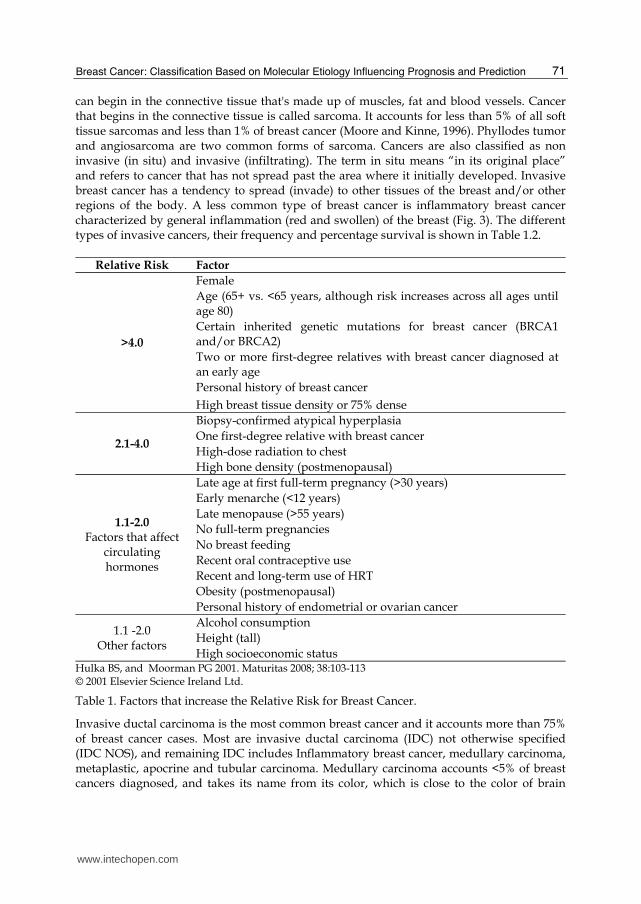
increased breast cancer risk are postmenopausal obesity, hormone replacement therapy (HRT), alcohol consumption, and physical inactivity, no breast feeding are modifiable and classified under modified factors. The relative risk of various factors responsible for breast cancer are shown in Table 1 (Hulka & Moorman, 2001).
Fig. 1. Demographic profiles of cancer cases in Indian females. Based on 2004-2005 data for Bangalore, Barshi, Bhopal, Chennai, Delhi, Mumbai, Ahmedabad and 2005 data for Kolkata.
3. Classification of breast cancer
3.1.1 Histopathological classification
Each breast has 15 to 25 sections called lobes, formed by groups of lobules, the milk glands.
Each lobule is composed of grape-like clusters of acini (also called alveoli), the hollow sacs
that make and hold breast milk. The lobes and lobules are connected by thin tubes, called
ducts that deliver milk to nipple (Fig. 2). The pink or the brown pigmented region
surrounding the nipple is called areola. Connective and fatty tissue fills the remaining space
in between the lobes and ducts. The most common type of breast cancer is ductal cancer. It is
found in the cells of the ducts. Cancer that starts in lobes or lobules is called lobular cancer.
It is more often found in both breasts than other types of breast cancer. Rarely breast cancer
Fig. 2. Anatomy of female breast.
www.intechopen.com
Breast Cancer: Classification Based on Molecular Etiology Influencing Prognosis and Prediction
71
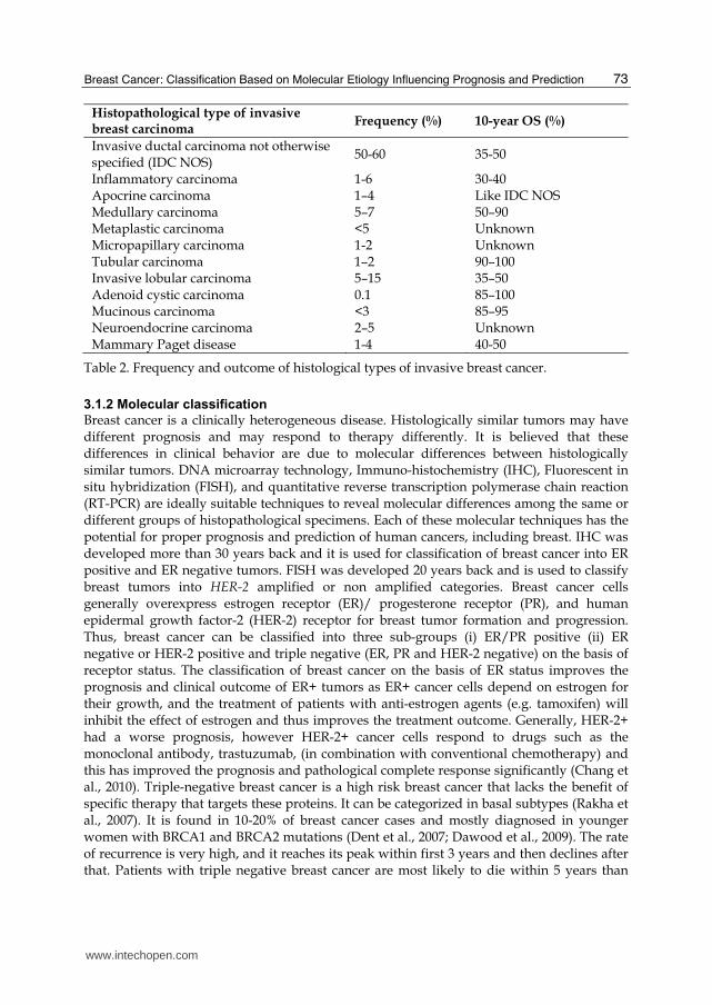
can begin in the connective tissue that's made up of muscles, fat and blood vessels. Cancer that begins in the connective tissue is called sarcoma. It accounts for less than 5% of all soft tissue sarcomas and less than 1% of breast cancer (Moore and Kinne, 1996). Phyllodes tumor and angiosarcoma are two common forms of sarcoma. Cancers are also classified as non invasive (in situ) and invasive (infiltrating). The term in situ means “in its original place” and refers to cancer that has not spread past the area where it initially developed. Invasive breast cancer has a tendency to spread (invade) to other tissues of the breast and/or other regions of the body. A less common type of breast cancer is inflammatory breast cancer characterized by general inflammation (red and swollen) of the breast (Fig. 3). The different types of invasive cancers, their frequency and percentage survival is shown in Table 1.2.
Relative Risk Factor
>4.0
Female
Age (65+ vs. <65 years, although risk increases across all ages until age 80)
Certain inherited genetic mutations for breast cancer (BRCA1 and/or BRCA2)
Two or more first-degree relatives with breast cancer diagnosed at an early age
Personal history of breast cancer
High breast tissue density or 75% dense
2.1-4.0
Biopsy-confirmed atypical hyperplasia
One first-degree relative with breast cancer
High-dose radiation to chest
High bone density (postmenopausal)
1.1-2.0 Factors that affect
circulating hormones
Late age at first full-term pregnancy (>30 years)
Early menarche (<12 years)
Late menopause (>55 years)
No full-term pregnancies
No breast feeding
Recent oral contraceptive use
Recent and long-term use of HRT
Obesity (postmenopausal)
Personal history of endometrial or ovarian cancer
1.1 -2.0 Other factors
Alcohol consumption
Height (tall)
High socioeconomic status Hulka BS, and Moorman PG 2001. Maturitas 2008; 38:103-113 © 2001 Elsevier Science Ireland Ltd.
Table 1. Factors that increase the Relative Risk for Breast Cancer.
Invasive ductal carcinoma is the most common breast cancer and it accounts more than 75% of breast cancer cases. Most are invasive ductal carcinoma (IDC) not otherwise specified (IDC NOS), and remaining IDC includes Inflammatory breast cancer, medullary carcinoma, metaplastic, apocrine and tubular carcinoma. Medullary carcinoma accounts <5% of breast cancers diagnosed, and takes its name from its color, which is close to the color of brain
www.intechopen.com

Breast Cancer – Focusing Tumor Microenvironment, Stem Cells and Metastasis
72
tissue, or medulla. It is an invasive breast cancer that forms a distinct boundary between tumor tissue and normal tissue. Metaplastic breast cancer is a form of invasive ductal cancer, meaning that it forms in the milk ducts and then moves into other tissues of the breast. Metaplastic breast carcinomas constitute a heterogeneous group of neoplasms, accounting for less than 1% of all invasive mammary carcinomas (Reis-Filho et al., 2005), such as squamous (skin) or osseous (bone) cells. The other groups of invasive breast cancers are invasive lobular carcinoma, adenoid cystic carcinoma, micropapillary carcinoma, mucinous carcinoma (formed by the mucus-producing cancer cells), etc as shown in Fig. 3.
Fig. 3. Histology of breast carcinoma. Breast carcinoma is classified into Ductal (A), Lobular carcinoma (B) and Inflammatory carcinoma. (C). It can be further classified into non-invasive (A-B) and invasive carcinoma (C-L). Invasive cancer includes Inflammatory (C), Invasive lobular (D), tubular (E) apocrine (F), medullary, (G) metaplastic (H), micropapillary, (I) adenoid cystic (J), mucunous carcinoma (K), and paget disease (L).
www.intechopen.com
Breast Cancer: Classification Based on Molecular Etiology Influencing Prognosis and Prediction
73
Histopathological type of invasive breast carcinoma
Frequency (%) 10-year OS (%)
Invasive ductal carcinoma not otherwise specified (IDC NOS)
50-60 35-50
Inflammatory carcinoma 1-6 30-40 Apocrine carcinoma 1–4 Like IDC NOS Medullary carcinoma 5–7 50–90 Metaplastic carcinoma <5 Unknown Micropapillary carcinoma 1-2 Unknown Tubular carcinoma 1–2 90–100 Invasive lobular carcinoma 5–15 35–50 Adenoid cystic carcinoma 0.1 85–100 Mucinous carcinoma <3 85–95 Neuroendocrine carcinoma 2–5 Unknown Mammary Paget disease 1-4 40-50
Table 2. Frequency and outcome of histological types of invasive breast cancer.
3.1.2 Molecular classification
Breast cancer is a clinically heterogeneous disease. Histologically similar tumors may have different prognosis and may respond to therapy differently. It is believed that these differences in clinical behavior are due to molecular differences between histologically similar tumors. DNA microarray technology, Immuno-histochemistry (IHC), Fluorescent in situ hybridization (FISH), and quantitative reverse transcription polymerase chain reaction (RT-PCR) are ideally suitable techniques to reveal molecular differences among the same or different groups of histopathological specimens. Each of these molecular techniques has the potential for proper prognosis and prediction of human cancers, including breast. IHC was developed more than 30 years back and it is used for classification of breast cancer into ER positive and ER negative tumors. FISH was developed 20 years back and is used to classify breast tumors into HER-2 amplified or non amplified categories. Breast cancer cells generally overexpress estrogen receptor (ER)/ progesterone receptor (PR), and human epidermal growth factor-2 (HER-2) receptor for breast tumor formation and progression. Thus, breast cancer can be classified into three sub-groups (i) ER/PR positive (ii) ER negative or HER-2 positive and triple negative (ER, PR and HER-2 negative) on the basis of receptor status. The classification of breast cancer on the basis of ER status improves the prognosis and clinical outcome of ER+ tumors as ER+ cancer cells depend on estrogen for their growth, and the treatment of patients with anti-estrogen agents (e.g. tamoxifen) will inhibit the effect of estrogen and thus improves the treatment outcome. Generally, HER-2+ had a worse prognosis, however HER-2+ cancer cells respond to drugs such as the monoclonal antibody, trastuzumab, (in combination with conventional chemotherapy) and this has improved the prognosis and pathological complete response significantly (Chang et al., 2010). Triple-negative breast cancer is a high risk breast cancer that lacks the benefit of specific therapy that targets these proteins. It can be categorized in basal subtypes (Rakha et al., 2007). It is found in 10-20% of breast cancer cases and mostly diagnosed in younger women with BRCA1 and BRCA2 mutations (Dent et al., 2007; Dawood et al., 2009). The rate of recurrence is very high, and it reaches its peak within first 3 years and then declines after that. Patients with triple negative breast cancer are most likely to die within 5 years than
www.intechopen.com

Breast Cancer – Focusing Tumor Microenvironment, Stem Cells and Metastasis
74
patients with other breast cancers. All deaths due to breast cancer in patients’ with triple-negative cancer occurred within 10 years of diagnosis. A novel molecular classification of breast cancer based on gene expression profiles
segregates breast cancer into four types (i) luminal, (ii) basal, (iii) HER-2 and (iv) normal
type (Perou et al., 2000; Sotiriou et al., 2003; Tamimi et al., 2008) (Fig. 4).
Fig. 4. Dendrogram of breast cancer. The tumors were separated into two main groups mainly associated with ER status as analyzed by hierarchical cluster analysis generated by using gene profile data. The dendrogram is further branched into smaller subgroups within the ER+ and ER- classes based on their basal and luminal characteristics: HER-2 subgroup, dark red; basal-like 1 subgroup, pink; luminal-like A subgroup, green; luminal-like B subgroup, yellow; and normal-like breast subgroup, blue.
Luminal express keratin 8/18, ER, GATA binding protein, X-box binding protein 1, annexin
XXXI, cytochrome P450 and basal type express keratin 5, keratin 17, integrin ┚4, matrix
metalloprotease 14, laminin ┙3, basonuclin and mutated TP53 gene. Luminal type is further
classified into luminal A and luminal B. Luminal B expresses HER-2 along with ER where as
luminal A doesn’t express HER-2. HER-2 subtype express ERB-2/HER-2, growth factor
receptor bound protein 7, TNF receptor-associated factor IV, GRB 7. Normal–breast-like
group showed the highest expression of many genes known to be expressed by adipose
tissue and other non-epithelial cell types. These tumors also showed strong expression of
basal epithelial genes and low expression of luminal epithelial genes. It expresses CD36
antigen collagen type I, glycerol 3 phosphate dehydrogenase I, lipoprotein lipase A, alcohol
dehydrogenase 2 (Sorlie et al., 2001). The molecular subclasses show difference in clinical
outcome as per as overall survival (OS) and relapse free survival (RFS) is concerned as
shown in Table 1.3. There was a significant difference in overall survival between the
subtypes with basal and HER-2 is as associated with worse outcome and shortest survival
time.
www.intechopen.com

Breast Cancer: Classification Based on Molecular Etiology Influencing Prognosis and Prediction
75
Molecular types of breast carcinoma
Frequency (%)
5-year OS+
(%) 5-year RFS* (%)
10-year OS (%)
10-year RFS (%)
Luminal A 50-60 85-95 80-90 75-85 75-85
Luminal B 5-10 70-80 65-75 55-65 54-64
Basal 10-20 63-73 60-70 57-67 45-55
ERB-2 10-20 55-65 15-20 45-55 15-30
Normal-like 10-15 84-94 80-90 75-85 72-82
Table 3. Breast cancer outcomes in molecular types of breast cancer.RFS: The percentage of people without any further symptoms of breast cancer during the interval elapsed between the date of breast surgery and the date of diagnosed further episode of breast cancer, whether the breast cancer was classified as a recurrence or second primary, and whatever the histology. OS: The percentage of people survived during the interval elapsed between the date of breast surgery and the date of breast cancer-related or un-related death (documented from hospital records).
4. Clinical outcomes of breast cancer in association with clinical, histopathological and molecular classification
Breast cancers can be classified by different schemata. Classification aspects include clinical (age, tumor, node), histopathological (grade, ER and HER-2 status, ductal, lobular, invasive) and molecular (normal-like, luminal, basal, HER-2) values. Every aspect influences treatment response and prognosis as shown in Table 2 and Table 3. The true prognostic or predictive value of the various molecular classes is unknown because there is a strong correlation between molecular class and conventional histopathologic variables (ER status, grade). For example, in one study, all luminal-type cancers were ER-positive and 63% of these were also low or intermediate grade, in contrast to 95% of basal-like cancers that were ER-negative, 91% of which were high grade (Pusztai et al., 2003). These associations partly explain the different clinical outcome observed in different molecular classes. Rouzier et al. studied the pathological outcomes of different molecular subclasses of breast cancer patients. They obtained tumor tissue biopsies from 82 patients with newly diagnosed breast cancer before they were given a commonly used chemotherapy (Taxol/5-fluorouracil, doxorubicin, and cyclophosphamide). Patients with basal-like and erbb-2+ subgroups were found to have the highest rates (45% each) of a pathological complete response (CR), while only 6% of luminal tumors had a complete response. Among the normal-like cancers, no response was seen (Rouzier et al., 2005). None of the 61genes associated with pathologic CR in the basal-like group were associated with pathologic CR in the HER-2+ group, which suggest that the mechanisms of chemotherapy sensitivity may vary across the subtypes. As molecular classification was not independently associated with pathologic CR, the predictive accuracy of the logistic regression models including (a) clinical + pathologic variables, (b) clinical variables + molecular classification, and (c) clinical + pathologic variables + molecular class (Fig. 5) was measured by constructing Receiver Operating Characteristics curve.
www.intechopen.com

Breast Cancer – Focusing Tumor Microenvironment, Stem Cells and Metastasis
76
Source of the curve AUC 95% CI p
Clinical and pathological 0.84 0.73-0.95 <0.001
Clinical variables and 0.82 0.72-0.92 <0.001molecular classification
Clinical, pathological and 0.89 0.81-0.97 <0.001
molecular classification
Reference line
Rouzier R et al. Clin Cancer Res 2005;11: 5678-5685
© by 2005 American Association for Cancer Research
Fig. 5. Receiver Operating Characteristic curves for logistic regression models. Three different prediction models were compared including clinical plus histopathologic variables (model 1), clinical variables plus molecular classification (model 2), and clinical plus histopathologic plus molecularclassification (model 3). All three models were similarly done.
The three models yielded similar area under curve (AUC). This indicates that the molecular
class alone can replace histopathological characteristics (estrogen receptor, HER-2 status, or
grade) for prediction of pathologic CR but provides little additional information when these
characteristics are included. The basal-like and HER-2 tumors were predominantly high
nuclear grade and the basal-like tumors were almost all estrogen receptor negative and 80%
of HER-2 molecular class expresses HER-2. These characteristics are known to be associated
with higher likelihood of pathologic CR to preoperative chemotherapy (Rouzier et al., 2002;
Abrial et al., 2005; Gennari et al., 2008). Because of this association, incorporation of
molecular class into a logistic regression–based predictor of response didn’t improve the
prediction accuracy compared with using routine clinical and pathologic variables only.
Therefore, it is likely that more focused gene signature–based predictors will need to be
developed through supervised outcome prediction methods that are differentially expressed
between cases of pathologic CR and residual disease.
5. Screening and detection of breast cancer
Screening uses test/techniques to check people who might have that disease (breast cancer)
and to allow it to be treated at an early stage when a cure is more likely. Breast cancer
screening is done by mammography (low dose x-ray technique to visualize the internal
structure of the breast). On average, mammography will detect about 80-90% of the breast
cancers in women without symptoms. Testing is somewhat more accurate in
postmenopausal than in premenopausal women (Michaelson et al., 2002). It can reduce
breast cancer mortality by 20-30% in women over 50 yrs old in high-income countries when
the screening coverage is over 70% (IARC, 2008). MRI, or magnetic resonance imaging, is a
technology that uses magnets and radio waves to produce detailed cross-sectional images of
the inside of the body. MRI does not use x-rays, so it does not involve any radiation
www.intechopen.com

Breast Cancer: Classification Based on Molecular Etiology Influencing Prognosis and Prediction
77
exposure. Breast MRI is not recommended as a routine screening tool for all women as MRI
screening results in more false positives results. However, it is recommended for screening
women who are at high risk for breast cancer, usually due to a strong family history and/or
a mutation in genes such as BRCA1 or BRCA2. It is also used for gathering more
information about the suspicious area found on mammogram and ultrasound and also used
for monitoring recurrence after treatment. Positron emission tomography (PET) scan creates
computerized images of chemical changes that take place in the tissue. PET scans may play
a role in determining whether a breast mass is cancerous. However, PET scans are more
accurate in detecting larger and more aggressive tumors than they are in locating tumors
that are smaller than 8 mm and/or less aggressive. They may also detect cancer when other
imaging techniques show normal results. PET scans may be helpful in evaluating and
staging recurrent disease. Clinical breast examination (CBE) is recommended for average
risk asymptomatic in the age group of 20-30 to observe any changes in shape, texture, and
location of lumps (situated in skin or deeper tissues). The breasts should also be inspected
for skin changes (e.g., dimpling, redness) and asymmetry. The area under both arms will
also be examined. CBE is also an opportunity for a woman and her health care provider to
discuss changes in her breasts, early detection testing, and factors in the woman’s history
that might make her more likely to develop. All women should become familiar with both
the appearance and feel of their breasts to detect any changes and report them promptly to
their physician. A woman who chooses to perform breast self-exams (BSE) should receive
instructions and have her technique reviewed by a health care professional who performs
clinical examinations. Finding and reporting breast changes early offers women the best
opportunity for improving breast cancer treatment and reducing breast cancer deaths.
Mammotome® is a vacuum assisted breast biopsy that uses image guidance such as
stereotactic x-ray, ultrasound, MRI and/or molecular imaging to perform breast biopsies.
Mammotome offers a full array of tissue markers to mark the biopsy site for follow-up
observations. There have been no reports of serious complications resulting from the
Mammotome breast biopsy system. Ductal lavage is another screening and investigational
technique for collecting samples of cells from breast ducts for analysis under a microscope.
A saline (salt water) solution is introduced into a milk duct through a catheter (a thin,
flexible tube) that is inserted into the opening of the duct on the surface of the nipple. Fluid,
which contains cells from the duct, is withdrawn through the catheter. The cells are checked
under a microscope to identify changes that may indicate cancer or changes that may
increase the risk for breast cancer. The procedure is used to identify precancerous cells,
called atypical cells. Ductal lavage is currently performed only on women who have
multiple breast cancer risk factors to detect breast cancer before it starts. Ductal lavage
appears to have low sensitivity and high specificity for breast cancer detection, possibly
because cancer-containing ducts fail to yield fluid or have benign or mildly atypical
cytology (Khan et al., 2004).
6. Breast cancer treatment
Breast cancer treatment depends on stage, age, hormonal and receptor status. Most women with breast cancer will undergo some type of surgery. Surgery is often combined with other treatments such as radiation therapy, chemotherapy, hormone therapy, and targeted therapy.
www.intechopen.com

Breast Cancer – Focusing Tumor Microenvironment, Stem Cells and Metastasis
78
6.1 Surgery
Most patients with breast cancer have surgery to remove the tumor mass from the breast.
The types of breast cancer surgery differ in the amount of tissue that is removed with the
tumor, depending on the tumor's characteristics, whether it has spread (metastasized), and
patient’s personal feelings. Some of the lymph nodes under the arm are usually taken out
and looked under a microscope to see if they contain cancer cells. Breast-conserving surgery
or lumpectomy is done to remove the cancer cells but not the breast itself. Lumpectomy is
almost always followed by about 5 to 7 weeks of radiation therapy. A woman who chooses
lumpectomy and radiation will have the same expected long-term survival as if she had
chosen mastectomy (Fisher et al., 2002). Simple or total mastectomy includes removal of the
entire breast. Modified radical mastectomy includes removal of the entire breast and lymph
nodes under the arm, but does not include removal of the underlying chest wall muscle, as
with a radical mastectomy. Both lumpectomy and mastectomy are often accompanied by
removal of regional lymph nodes from the axilla, or armpit, to determine the involvement of
lymph nodes and spreading of the disease. Axillary lymph node metastasis is the most
important prognostic factor for the disease-free and overall survival. Patients with multiple
unfavorable risk factors such as positive axillary lymph nodes, high nuclear grade, young
age and large tumor showed poorer local control and disease-free survival than patients
without any risk factors, and so more aggressive treatment is required for these patients.
Adjuvant radio-, chemo-, or targeted therapy has improved the prognosis of patients with
higher risk factors (Lee & Chan, 1984; Kim et al., 2005).
6.2 Radiation therapy
Radiation therapy is a cancer treatment that uses high-energy x-rays or other types of radiation to destroy cancer cells remaining in the breast, chest wall, or underarm area after surgery, or to reduce the size of a tumor before surgery (Early Breast Cancer Trialists' Collaborative Group, 2000). There are two types of radiation therapy. External radiation therapy uses a machine outside the body to send radiation toward the cancer. Internal radiation therapy uses a radioactive substance sealed in needles, seeds, wires, or catheters that are placed directly into or near the cancer. The way the radiation therapy is given depends on the type and stage of the cancer being treated. Using traditional clinical and pathological factors, patients can be classified into subgroups by the risk of loco-regional recurrence. In the high-risk groups the absolute benefit of irradiation is larger. However, the patients are over-treated in every subgroup. Substantial proportion of the patients remains free of loco-regional recurrence even in the absence of irradiation, and some patients develop loco-regional recurrence despite postoperative irradiation. Molecular subtypes on the basis of receptors may provide sufficient information to allow accurate individual risk assessment to identify patients who might benefit from receiving post mastectomy radiotherapy (PMRT). A significantly improved overall survival after PMRT was seen only among patients of luminal subtypes. No significant overall survival improvement after PMRT was found among patients with basal and ERB2 subtypes (Fig. 6). There was also smaller improvements in loco-regional recurrence of breast cancer in basal and ERB2 subtypes as compared to luminal A and luminal B (Kyndi et al., 2008). Hence, the improvement in survival resulting from the use of irradiation is more related to the prevention of local recurrences. Post-irradiation local recurrence increases the risk of mortality, but with good prognostic factors (<4 positive nodes, tumor size <2 cm, Grade 1
www.intechopen.com

Breast Cancer: Classification Based on Molecular Etiology Influencing Prognosis and Prediction
79
malignancy, ER- and PR-positive, HER-2-negative) the 10-year survival is 80-90% (Fodor, 2009).
Fig. 6. Overall survival (OS)% of different molecular subtypes of breast cancer patients after receiving post mastectomy radiation therapy (RT). P values and 95% CI of Hazard (H) ratios are shown.
6.3 Chemotherapy and molecular targeted-therapy
Chemotherapeutic drugs are applied in neoadjuvant settingsto shrink the size of tumor that has metastasized and also in adjuvant settings to delay the further growth and spread of the tumor. It is found that combinations of drugs are more effective than just one drug alone for breast cancer treatment. The most common drugs recommended to be used in combination in early breast cancer are cyclophosphamide, methotrexate, 5-fluorouracil (CMF
www.intechopen.com
Breast Cancer – Focusing Tumor Microenvironment, Stem Cells and Metastasis
80
combinations), doxorubicin (Adriamycin), epirubicin, paclitaxel (Taxol), and docetaxol (Taxotere). Although the benefit and clinical outcome of chemotherapy is dependent on clinical and histopathological parameters, but there are a percentage of cases that behave in an unexpected manner, even if the clinical and pathological parameters indicate the opposite (Gonzalez-Angulo et al., 2007). The introduction of hormonal receptor status to the classical clinical parameters improved the clinical outcome (Goldhirsch et al., 2003). The chemotherapeutic drugs are designed to target the specific molecular markers (molecular targeted therapy) overexpressed in cancer tissues. The presence of ER is correlated with a better prognosis, predicting response to hormonal therapies such as tamoxifen and aromatase inhibitors. But still 15-20% of breast cancer patients with ER+ have recurrent disease. It’s the luminal B subgroup of previously classified ER+ tumor that is irresponsive to tamoxifen treatment as they co-express EGFRs and shows poor relapse-free survival (RFS) and over-all survival (OS). Thus over-simplified classification based on ER status required additional molecular makers for sub-classification for optimal treatment. The molecular portraits based on gene profiling divides breast carcinomas into luminal (A and B), basal, HER-2 and normal like. Basal and HER-2 types normally overexpress EGFR and HER-2 respectively. EGFR and HER-2 is overexpressed in 17-30% and 20-30% respectively in breast cancer. Both EGFR and HER-2 is associated with poor prognosis and worse clinical outcome. Basal like subtypes are more aggressive and less responsive to conventional chemotherapy and expected to benefit from EGFR-targeted therapies. Tyrosine kinase inhibitors (TKI) (ZD1839, ZD6474) in combined with anthracyclines (doxorubicin, epirubicin) or taxanes based regimens will improve the clinical outcome of the basal subtypes. HER-2 might serve as a marker for tissue HER-2 status, especially for the prediction of benefit from trastuzumab and/or chemotherapy regimens (anthracyclines) (Sandri et al., 2004). Although the molecular profile of the tumor is a major determinant of disease progression and response to treatment, other factors including chemo- sensitvity or resistivity may be of considerable importance. It is found that for 100 node-negative, premenopausal women receiving chemotherapy according to standard criteria, at 5 years 3 are cured by chemotherapy, 83.50 would have been alive without chemotherapy and 13.50 die despite chemotherapy. With application of molecular profiling to predict the outcome (for the same 100 people), the number treated would be reduced to 39.05 (allowing for a false-positive rate equivalent to that seen in the van ‘t Veer study (van 't Veer et al., 2002), resulting in an increase in the proportion cured (from 3 out of 100 to 3 out of 39 or 8%). If it were possible to predict chemo-responsiveness, it is possible that the number receiving chemotherapy would reduce further from 39.05 to 29.20 (allowing for a false-positive rate equivalent to that seen in the van‘t Veer study). In this scenario, the proportion cured by chemotherapy would be 3 out of 29.20 (10.16%) (>3-fold increase in survival rate using chemotherapy), and the number of women treated has been reduced by 70.80%. Thus it is found that molecular profiling will enhance the survival benefit of chemotherapeutic regimens, which will be further improved applying the knowledge of chemo-responsiveness as shown in Fig. 7. If accurate determination of chemo-sensitivity were achieved by observing the set of genes responsible for treatment response, the overall number receiving cytotoxic treatment unnecessarily would decrease, and the overall survival benefit derived, per person treated, increase accordingly, as shown in Fig. 7. However, the absolute survival benefit of patients diagnosed with breast cancer would be unaffected and would be improved with more molecular subtypes along with the development of specific agents targeting particular biomarkers (molecular targeted therapy).
www.intechopen.com

Breast Cancer: Classification Based on Molecular Etiology Influencing Prognosis and Prediction
81
Fig. 7. Model for the effect of molecular profiling on breast cancer. The data shows numbers
of premenopausal women with node negative breast cancer receiving chemotherapy (CT),
and associated benefit at 5 years. 100 node-negative, premenopausal women receiving
chemotherapy according to standard criteria, at 5 years showed survival benefit, no benefit
and breast cancer specific death. The two bar graph represents absolute survival benefit and
% survival benefit of breast cancer patients receiving chemotherapy. Note that in neither
figure has consideration been given to the false-negative rate inherent in molecular
profiling. It has been assumed that all deaths occurring were breast cancer related.
7. Conclusion
Adjuvant chemo- and radiotherapy improves survival of patients but it is being increasingly
recognized that the benefit is not equal for all patients of breast cancer. Molecular
characteristics of the cancer affect sensitivity to chemo- and radiotherapy. In general, ER-
(Basal and HER-2) is more sensitive to chemotherapy than ER+ (Luminal A and Luminal B)
breast cancer where as ER+ is more sensitive to radiotherapy than ER- breast cancer. The
prognostic predictions made by traditional histopathological based models and molecular
based models are discordant in about 30% of the cases (van de Vijver et al., 2002), suggesting
that one of these methods may be superior to the other or at least that the information they
capture is complementary. Corollary to this, it is found that when both the type of
classifications are combined (histopathological and molecular), it yield better prognostic
values as observed in Fig. 6. It is currently unknown whether genomic tests based on
molecular signatures yield a more accurate risk prediction than conventional models. A
better prognostic test based on molecular classification with the knowledge of chemo-
responsiveness could lead to a reduction in overtreatment of low-risk individuals who are
falsely assigned to high-risk category by clinical variables. Such a test could also lead to
better overall survival by correctly identifying high-risk individuals who might currently
miss out on systemic therapy. Even if molecular classification do not prove to be better than
clinical models in prognosis and prediction outcome of breast cancer, inclusion of their
results, as additional variables, in current models could improve prognostic predictions.
www.intechopen.com

Breast Cancer – Focusing Tumor Microenvironment, Stem Cells and Metastasis
82
8. References
Abrial, C., Van Praagh, I., Delva, R., Leduc, B., Fleury, J., Gamelin, E., Sillet-Bach, I., Penault-Llorca, F., Amat, S., & Chollet, P. (2005). Pathological and clinical response of a primary chemotherapy regimen combining vinorelbine, epirubicin, and paclitaxel as neoadjuvant treatment in patients with operable breast cancer. Oncologist, Vol.10, pp.242-49.
American Cancer Society. (2010). Breast Cancer Facts and Figures 2009-10: American Cancer Society, Inc., Atlanta
Chang, H. R., Glaspy, J., Allison M. A., Kass, F. C., Elashoff, R., Chung, D. U., & Gornbein, J. (2010). Differential response of triple-negative breast cancer to a docetaxel and carboplatin-based neoadjuvant treatment. Cancer, Vol.116, pp.4227-32.
Coleman, M. P., Quaresma, M., Berrino, F., Lutz, J. M., De Angelis, R., Capocaccia, R., Baili, P., Rachet, B., Gatta, G., Hakulinen, T., Micheli, A., Sant, M., Weir, H. K., Elwood, J. M., Tsukuma, H., Koifman, S., E Silva, G. A, Francisci, S., Santaquilani, M., Verdecchia, A., Storm, H. H., & Young, J. L. (2008). Cancer survival in five continents: a worldwide population-based study (CONCORD). Lancet Oncol, Vol.9, pp.730-56.
Dawood, S., Broglio, K., Kau, S. W., Green, M. C., Giordano, S. H., Meric-Bernstam, F., Buchholz, T. A., Albarracin, C., Yang, W. T., Hennessy, B. T., Hortobagyi, G. N., & Gonzalez-Angulo, A. M. (2009). Triple receptor-negative breast cancer: the effect of race on response to primary systemic treatment and survival outcomes. J Clin Oncol, Vol.27, pp.220-26.
Dent, R., Trudeau, M., Pritchard, K. I., Hanna, W. M., Kahn, H. K., Sawka, C. A., Lickley, L. A., Rawlinson, E., Sun, P., & Narod, S. A. (2007). Triple-negative breast cancer: clinical features and patterns of recurrence. Clin Cancer Res, Vol.13, pp.4429-34.
Early Breast Cancer Trialists' Collaborative Group. (2000). Favourable and unfavourable effects on long-term survival of radiotherapy for early breast cancer: an overview of the randomised trials. Early Breast Cancer Trialists' Collaborative Group. Lancet, Vol.355, pp.1757-70.
Fisher, B., Anderson, S., Bryant, J., Margolese, R. G., Deutsch, M., Fisher, E. R., Jeong, J. H., & Wolmark, N. (2002). Twenty-year follow-up of a randomized trial comparing total mastectomy, lumpectomy, and lumpectomy plus irradiation for the treatment of invasive breast cancer. N Engl J Med, Vol.347, pp.1233-41.
Fodor, J. (2009). [Evidence-based radiotherapy in the treatment of early-stage invasive breast cancer: traditional clinical features and biomarkers]. Magy Onkol, Vol.53, pp.7-14.
Gennari. A., Sormani, M. P., Pronzato, P., Puntoni, M., Colozza, M., Pfeffer, U., & Bruzzi, P. (2008). HER2 status and efficacy of adjuvant anthracyclines in early breast cancer: a pooled analysis of randomized trials. J Natl Cancer Inst, Vol.100, pp.14-20.
Goldhirsch, A., Wood, W. C., Gelber, R. D., Coates, A. S., Thurlimann, B., & Senn, H. J. (2003). Meeting highlights: updated international expert consensus on the primary therapy of early breast cancer. J Clin Oncol, Vol.21, pp.3357-65.
Gonzalez-Angulo, A. M., Morales-Vasquez, F., & Hortobagyi, G. N. (2007). Overview of resistance to systemic therapy in patients with breast cancer. Adv Exp Med Biol, Vol.608, pp.1-22.
Hulka, B. S., & Moorman, P. G. (2001). Breast cancer: hormones and other risk factors. Maturitas, 38:103-113; discussion 103-6.
www.intechopen.com

Breast Cancer: Classification Based on Molecular Etiology Influencing Prognosis and Prediction
83
Kelsey, J. L., & Gammon, M. D. (1990). Epidemiology of breast cancer. Epidemiol Rev, Vol. 12, pp.228-40.
Khan, S. A., Wiley, E. L., Rodriguez, N., Baird, C., Ramakrishnan, R., Nayar, R., Bryk, M., Bethke, K. B., Staradub, V. L., Wolfman, J., Rademaker, A., Ljung, B. M., & Morrow, M. (2004). Ductal lavage findings in women with known breast cancer undergoing mastectomy. J Natl Cancer Inst, Vol. 96, pp.1510-17.
Kim, K. J., Huh, S. J., Yang, J. H., Park, W., Nam, S. J., Kim, J. H., Lee, J. H., Kang, S. S., Lee, J. E., Kang, M. K., Park, Y. J., & Nam, H. R. (2005). Treatment results and prognostic factors of early breast cancer treated with a breast conserving operation and radiotherapy. Jpn J Clin Oncol, Vol. 35, pp.126-33.
Kyndi, M., Sorensen, F. B., Knudsen, H., Overgaard, M., Nielsen, H. M., & Overgaard, J. (2008). Estrogen receptor, progesterone receptor, HER-2, and response to postmastectomy radiotherapy in high-risk breast cancer: the Danish Breast Cancer Cooperative Group. J Clin Oncol, Vol.26, pp.1419-26.
Lee, Y. T., & Chan, L. S. (1984). Surgical treatment of carcinoma of the breast: IV. Prognosis according to extent of involvement of the axillary lymph nodes. J Surg Oncol, Vol. 27, pp.35-41.
Michaelson, J., Satija, S., Moore, R., Weber, G., Halpern, E., Garland, A., Puri, D., & Kopans, D. B. (2002). The pattern of breast cancer screening utilization and its consequences. Cancer, Vol. 94, pp.37-43.
Moore, M. P., & Kinne, D. W. (1996). Breast sarcoma. Surg Clin North Am, Vol.76, pp.383-92. Perou, C. M., Sorlie, T., Eisen, M. B., van de Rijn, M., Jeffrey, S. S., Rees, C. A., Pollack, J. R.,
Ross, D. T., Johnsen, H., Akslen, L. A., Fluge, O., Pergamenschikov, A., Williams, C., Zhu, S. X., Lonning, P. E., Borresen-Dale, A. L., Brown, P. O., & Botstein, D. (2000). Molecular portraits of human breast tumours. Nature, Vol.406, pp.747-52.
Pusztai, L., Ayers, M., Stec, J., Clark, E., Hess, K., Stivers, D., Damokosh, A., Sneige, N., Buchholz, T. A., Esteva, F. J., Arun, B., Cristofanilli, M., Booser, D., Rosales, M., Valero, V., Adams, C., Hortobagyi, G. N., & Symmans, W. F. (2003). Gene expression profiles obtained from fine-needle aspirations of breast cancer reliably identify routine prognostic markers and reveal large-scale molecular differences between estrogen-negative and estrogen-positive tumors. Clin Cancer Res, Vol. 9, pp.2406-15.
Rakha, E. A., El-Sayed, M. E., Green, A. R., Lee, A. H., Robertson, J. F., & Ellis, I. O. (2007). Prognostic markers in triple-negative breast cancer. Cancer, Vol. 109, pp.25-32.
Reis-Filho, J. S., Milanezi, F., Carvalho, S., Simpson, P. T., Steele, D., Savage, K., Lambros, M. B., Pereira, E. M., Nesland, J. M., Lakhani, S. R., & Schmitt, F. C. (2005). Metaplastic breast carcinomas exhibit EGFR, but not HER2, gene amplification and overexpression: immunohistochemical and chromogenic in situ hybridization analysis. Breast Cancer Res, Vol. 7, pp.R1028-35.
Rouzier, R., Extra, J. M., Klijanienko, J., Falcou, M. C., Asselain, B., Vincent-Salomon, A., Vielh, P., & Bourstyn, E. (2002). Incidence and prognostic significance of complete axillary downstaging after primary chemotherapy in breast cancer patients with T1 to T3 tumors and cytologically proven axillary metastatic lymph nodes. J Clin Oncol, Vol. 20, pp.1304-10.
Rouzier, R., Perou, C. M., Symmans, W. F., Ibrahim, N., Cristofanilli, M., Anderson, K., Hess, K. R., Stec, J., Ayers, M., Wagner, P., Morandi, P., Fan, C., Rabiul, I., Ross, J.
www.intechopen.com

Breast Cancer – Focusing Tumor Microenvironment, Stem Cells and Metastasis
84
S., Hortobagyi, G. N., & Pusztai, L. (2005). Breast cancer molecular subtypes respond differently to preoperative chemotherapy. Clin Cancer Res, Vol. 11:5678-85.
Sandri, M. T., Johansson, H., Colleoni, M., Zorzino, L., Passerini, R., Orlando, L., & Viale, G. (2004). Serum levels of HER2 ECD can determine the response rate to low dose oral cyclophosphamide and methotrexate in patients with advanced stage breast carcinoma. Anticancer Res, Vol. 24, pp.1261-66.
Sorlie, T., Perou, C. M., Tibshirani, R., Aas, T., Geisler, S., Johnsen, H., Hastie, T., Eisen, M. B., van de Rijn, M., Jeffrey, S. S., Thorsen, T., Quist, H., Matese, J. C., Brown, P. O., Botstein, D., Eystein Lonning, P., & Borresen-Dale, A. L. (2001). Gene expression patterns of breast carcinomas distinguish tumor subclasses with clinical implications. Proc Natl Acad Sci U S A, Vol. 98, pp.10869-74.
Sotiriou, C., Neo, S. Y., McShane, L. M., Korn, E. L., Long, P. M., Jazaeri, A., Martiat, P., Fox, S. B., Harris, A. L., & Liu, E. T. (2003). Breast cancer classification and prognosis based on gene expression profiles from a population-based study. Proc Natl Acad Sci U S A, Vol. 100, pp.10393-98.
Tamimi, R. M., Baer, H. J., Marotti, J., Galan, M., Galaburda, L., Fu, Y., Deitz, A. C., Connolly, J. L., Schnitt, S. J., Colditz, G. A., & Collins, L. C. (2008). Comparison of molecular phenotypes of ductal carcinoma in situ and invasive breast cancer. Breast Cancer Res, Vol. 10, pp.R67.
van 't Veer, L. J., Dai, H., van de Vijver, M. J., He, Y. D., Hart, A. A., Mao, M., Peterse, H. L., van der Kooy, K., Marton, M. J., Witteveen, A. T., Schreiber, G. J., Kerkhoven, R. M., Roberts, C., Linsley, P. S., Bernards, R., & Friend, S. H. (2002). Gene expression profiling predicts clinical outcome of breast cancer. Nature, Vol. 415, pp.530-36.
van de Vijver, M. J., He, Y. D., van't Veer, L. J., Dai, H., Hart, A. A., Voskuil, D. W., Schreiber, G. J., Peterse, J. L., Roberts, C., Marton, M. J., Parrish, M., Atsma, D., Witteveen, A., Glas, A., Delahaye, L., van der Velde, T., Bartelink, H., Rodenhuis, S., Rutgers, E. T., Friend, S. H., & Bernards, R. (2002). A gene-expression signature as a predictor of survival in breast cancer. N Engl J Med, Vol. 347, pp.1999-2009.
www.intechopen.com

Breast Cancer - Focusing Tumor Microenvironment, Stem cells andMetastasisEdited by Prof. Mehmet Gunduz
ISBN 978-953-307-766-6Hard cover, 584 pagesPublisher InTechPublished online 14, December, 2011Published in print edition December, 2011
InTech EuropeUniversity Campus STeP Ri Slavka Krautzeka 83/A 51000 Rijeka, Croatia Phone: +385 (51) 770 447 Fax: +385 (51) 686 166www.intechopen.com
InTech ChinaUnit 405, Office Block, Hotel Equatorial Shanghai No.65, Yan An Road (West), Shanghai, 200040, China
Phone: +86-21-62489820 Fax: +86-21-62489821
Cancer is the leading cause of death in most countries and its consequences result in huge economic, socialand psychological burden. Breast cancer is the most frequently diagnosed cancer type and the leading causeof cancer death among females. In this book, we discussed characteristics of breast cancer cell, role ofmicroenvironment, stem cells and metastasis for this deadly cancer. We hope that this book will contribute tothe development of novel diagnostic as well as therapeutic approaches.
How to referenceIn order to correctly reference this scholarly work, feel free to copy and paste the following:
Siddik Sarkar and Mahitosh Mandal (2011). Breast Cancer: Classification Based on Molecular EtiologyInfluencing Prognosis and Prediction, Breast Cancer - Focusing Tumor Microenvironment, Stem cells andMetastasis, Prof. Mehmet Gunduz (Ed.), ISBN: 978-953-307-766-6, InTech, Available from:http://www.intechopen.com/books/breast-cancer-focusing-tumor-microenvironment-stem-cells-and-metastasis/breast-cancer-classification-based-on-molecular-etiology-influencing-prognosis-and-prediction