
Brain Tumors at the Department of Neurology in …cdn.intechweb.org/pdfs/20857.pdfThe inability of...
25
19 Brain Tumors at the Department of Neurology in Sarajevo: Retrospecitve Analysis A. Alajbegović 1 and A. Gutošić 2 1 Clinical Center University of Sarajevo, Department of Neurology 2 Regional Medical Centre Dr. Safet Mujic, Mostar Bosnia and Herzegovina 1. Introduction Brain tumors are unique and heterogeneous group of tumors which face a variety of specialty, most oncologists, neurologists, and neurosurgeons. Due to its specific location, all brain tumors are malignant, regardless of their malignant potential, because any expansion process inside the skull, increasing intracranial pressure and the destruction of surrounding structures, which may cause neurological injuries, quantitative disturbances of consciousness or death. 1.1 The incidence of brain tumors In the United States are diagnosed annually 17 000 new cases of brain tumors. In Croatia, their average frequency is 18 per 100 000 inhabitants. The incidence of brain tumors in B&H is not known. Brain tumors are more common in older people. The most common are at the age from 65 to 74 years. The incidence of brain tumors in persons older than 70 years is 70 per 100 000 inhabitants. Some authors suggest that brain tumors are more common in men, while others argue that there is no difference in incidence according to sex, except for neuromas, which occur twice as frequently in women (1). 1.2 Etiopathogenesis Genetic determination of neoplastic growth exists also in intracranial tumors and is related to a disorder of neuroectoderm and mesoderm development. Here belongs certain number of hereditary diseases which are due to the association with intracranial tumors and changes in the skin called facomatoses (neurofibromatosis, tuberous sclerosis). Tumors arise from congenital malformations where the abnormal development creates conditions for the development of neoplastic tissue (angioblasts, ganglioneuroma). It has been shown that exposure to radiation may be the cause of brain tumors while the role of trauma and infection was not significant. Under experimental conditions it was shown that injection of oncogenic virus can cause brain tumors. 1.3 Pathophysiology The inability of the skull of adults to spread if the internal pressure increases causes disorders that bear a common name as the symptoms of increased intracranial pressure. The increase in intracranial pressure is due to increase in tumors mass, due to edema that www.intechopen.com
Transcript of Brain Tumors at the Department of Neurology in …cdn.intechweb.org/pdfs/20857.pdfThe inability of...
19
Brain Tumors at the Department of Neurology in Sarajevo: Retrospecitve Analysis
A. Alajbegović1 and A. Gutošić2 1Clinical Center University of Sarajevo, Department of Neurology
2Regional Medical Centre Dr. Safet Mujic, Mostar Bosnia and Herzegovina
1. Introduction
Brain tumors are unique and heterogeneous group of tumors which face a variety of specialty, most oncologists, neurologists, and neurosurgeons. Due to its specific location, all brain tumors are malignant, regardless of their malignant potential, because any expansion process inside the skull, increasing intracranial pressure and the destruction of surrounding structures, which may cause neurological injuries, quantitative disturbances of consciousness or death.
1.1 The incidence of brain tumors In the United States are diagnosed annually 17 000 new cases of brain tumors. In Croatia, their average frequency is 18 per 100 000 inhabitants. The incidence of brain tumors in B&H is not known. Brain tumors are more common in older people. The most common are at the age from 65 to 74 years. The incidence of brain tumors in persons older than 70 years is 70 per 100 000 inhabitants. Some authors suggest that brain tumors are more common in men, while others argue that there is no difference in incidence according to sex, except for neuromas, which occur twice as frequently in women (1).
1.2 Etiopathogenesis Genetic determination of neoplastic growth exists also in intracranial tumors and is related to a disorder of neuroectoderm and mesoderm development. Here belongs certain number of hereditary diseases which are due to the association with intracranial tumors and changes in the skin called facomatoses (neurofibromatosis, tuberous sclerosis). Tumors arise from congenital malformations where the abnormal development creates conditions for the development of neoplastic tissue (angioblasts, ganglioneuroma). It has been shown that exposure to radiation may be the cause of brain tumors while the role of trauma and infection was not significant. Under experimental conditions it was shown that injection of oncogenic virus can cause brain tumors.
1.3 Pathophysiology The inability of the skull of adults to spread if the internal pressure increases causes disorders that bear a common name as the symptoms of increased intracranial pressure. The increase in intracranial pressure is due to increase in tumors mass, due to edema that
www.intechopen.com
Management of CNS Tumors
426
accompanies the tumor, and because of obstacles in the flow of cerebrospinal fluid. Since the brain is virtually incompressible, the tumor will grow with no signs of increased intracranial pressure only until it reaches a size of about 150 cc, for as long as the brain volume is smaller than the volume of the skull. As a rule increasing intracranial pressure is proportional to the growth speed of expansive processes. The brain can adapt to large tumor if it grows slowly, whereas smaller tumors that grow rapidly may give severe symptoms. One of the consequences of the expansive process of the brain is edema. Edema principally affects the white mass of the hemispheres (centrum semiovale), is less pronounced in the internal capsule, corpus callosum and optic radiation, it almost does not use at all the large brain. Even edema acts as a mass that increases the volume of the brain, causing the above flattened gyrus, a brain ventricle can be suppressed. It is believed that the edema is caused by abnormal capillary permeability, which is a consequence of venous stasis and anoxia. Since the cranial cavity from the outside is limited by bony shell and internal partitioned into three sections (falx into two supratentorial and by tentorium into the third, infratentorially cavity), the tumor will cause the clamp of the parts of the brain as follows: a. Subfalx b. Tentorially (up and down) c. Occipitaly trough foramen magnum protrudes the cerebellar tonsils. This increase in volume of the frontal parts of one hemisphere leads to pressure on the corpus callosum and gyrus subcalloosusa prolapse below the falx free edge. Because of this and move to the side chambers to the opposite side. Similarly, the increase in the volume of the brain above tentorium causes a protrusion of part or all gyrus hippocampus through tentorium incision. If this protrusion is one-sided, opposite pedunculus is being pressed to the tentorium edge. The midbrain also protrudes. Due to pressure on aqueducts hydrocephalus occurs.
1.4 Symptoms of tumors Symptoms of brain tumors can be divided into two groups: general and focal or focal. Common symptoms in most cases are preceded by focal symptoms and occur in 60% of patients with brain tumor. General symptoms are:
1.4.1 Symptoms of increased intracranial pressure (headache, vomiting, trail papilla optic nerve) Headaches are initially reported in seizures. By further tumor growth they become longer and remain constant. They are more frequent in the morning and during the night. Localization importance of headache is a small place and the pain rarely coincides with the localization of tumors. Sometimes a headache may be on the opposite side of tumors but are usually in the frontal and occipital lobe. They occur in 49% of cases. Vomiting usually occurs suddenly and without any connection with food intake. More often in the evening and morning. Route of optic nerve papilla is most often seen in tumors of the cerebellum and temporal lobes, rarely in tumors of the brain hemispheres and the pons.
1.4.2 Pulse and respiration Because of the pressure on the brain stem and nucleus vagus pulse is first slowed to below 60 per minute. Later, when intracranial pressure increases, the pulse becomes rapid and is more severe the sign than bradycardia.
www.intechopen.com
Brain Tumors at the Department of Neurology in Sarajevo: Retrospecitve Analysis
427
1.4.3 Epileptic seizures Focal epilepsy may be one of the signs of a brain tumor. Generalized seizures occur in many cases of brain tumors. It was observed that the same tumor size and localization in one patient caused epileptic seizure, while in the other does not. The most common is epilepsy in case of astrocytoma, meningioma and glioblastoma of the brain. More often is the localization of tumors in the anterior parts of the brain than in posterior. They occur in 20-50% of cases, depending on the author.
1.4.4 Mental disorders Psychological symptoms may be the first signs of a brain tumor, sometimes for months and years preceding the neurologic signs. The first symptoms of brain tumors occur in approximately 15-20% of cases. Psychic symptoms in brain tumors are classified into three groups: general symptoms (mild personality changes, anxiety, confusion, and neurasthenic catatonic states), specific symptoms (changes in affect, memory and attention disorders), and associated psychological changes (hysterical state). Focal neurological deficit is manifested by sensibility problems, dysphasia, visual field problems, hemiparesis, etc., and are depend on the tumor location (13).
1.5 Classification of brain tumors The basic division of a brain tumor is on the primary and secondary. Primary tumors are those arising from the brain parenchyma in the skull while secondary tumors of the brain are metastases, which occur by joining of malignant tumor cells of distant organs at their arrival into the brain. Primary tumors are most commonly seen in children (first decade of life). In adults, incidence increases with age and for persons over 65 years of age is 18 per 100 000. Primary tumors can be divided into malignant and benign, although essentially all brain tumors are malignant due to their specific location. The difference between benign and malignant brain tumors is in their growth rate, partly in the way of growth and the percentage of recovery. Benign brain tumors grow slowly, putting pressure on the surrounding parts of the brain, while malignant tumors grow rapidly and permeate (infiltrate) the brain. From the histological aspect and by the World Health Organization (WHO) brain tumors are: a. NEUROEPITHELIAL 1. Astrocytoma 2. Oligodendroglioma 3. Ependymoma 4. Pinealoma 5. Gangliocitoma 6. Medulloblastoma 7. Glioblastoma b. TUMORS OF THE NERVE SHEATHS c. TUMORS MENING d. PITUITARY TUMORS e. VASCULAR TUMOR f. CONGENITAL TUMORS: 1. Craniofaringioma
www.intechopen.com

Management of CNS Tumors
428
2. Dermoids 3. Lipomas g. METASTATIC TUMORS There are over 80 types of tumors that can grow inside the skull and cause brain damage. However, in adults 5 types of tumors is encountered in over 90% of cases. These are: 1. Gliomas - this includes astrocytoma, oligodendroglioma, anaplastic astrocytoma and
glioblastoma 2. Meningioma 3. Neuroma 4. Pituitary adenoma 5. Brain metastases from distant malignant tumors.
1.6 Specificity of tumor types 1.6.1 Gliomas These are the most common brain tumors. They make up 50% of the total number of brain tumors. Annually in the United States they affect 7 people per 100 000. More frequent in men than women. Most often when grow permeate the surrounding brain structures. They have great potential to become malignant. According to the degree of malignancy have three or four stages. In the first stage healing is likely. Already in the second stage the survival for more than 5 years is 50-75%. In 3rd and 4th stage survival is even shorter. After the surgery is always indicated radiation and is often used is chemotherapy.
1.6.2 Meningiomas Meningiomas are mostly benign tumors. 1-2% is malignant ones. Make up 15% to 20% of all tumors and 25% of brain tumors. Annually in the United States are affected 2 per 100 000 people. They are more often in women than men. Increase with the depletion of brain pressing the surrounding parts of the brain, nerves and blood vessels.
Fig. 1. Meningioma
The treatment is usually radical, surgical excision.
www.intechopen.com
Brain Tumors at the Department of Neurology in Sarajevo: Retrospecitve Analysis
429
1.6.3 Neuroma Arise from cells that are found in nerves. Constitute 5-10% of all brain tumors. Every year in United States 3000 new cases is diagnosed. The incidence is 1 in 100 000 people. The most common sites are the nerves of hearing and balance (neurinoma, schwanoma vestibullaris nerves, tumor of angulo pontocerebellaris). Because of that usually start with hearing impairment and dizziness. This symptom may remain for years. Later it can cause numbness and facial pain (symptomatic trigeminal neuralgia), bending of the face due to facial nerve damage, weakness of limbs due to pressure on the brain stem, hydrocephalus, etc. It is usually possible to remove completely these tumors. Alternative therapy, particularly in case of small neuromas, is the stereotaxic radiation.
1.6.4 Pituitary adenoma These are tumors that arise from the pituitary gland, glands of internal secretion. Make 14% of all brain tumors. Can secrete excessive hormones and then are usually diagnosed earlier, while still small. Depending on the hormone secreted by the patient will develop hyperprolactinemia, Cushing's disease, etc. If they secrete hormones in the gland tumor can rather grow until the first symptoms. Large adenomas are first manifested by visual disturbances. Later it can occur: reduced secretion of pituitary hormones, strabismus, double vision, frequent urination, difficulty in walking and mental symptoms and signs of hydrocephalus.
1.6.5 Brain metastases Brain metastases are the complications of systemic dissemination of primary tumors. Malignant primary tumors of the blood and lymphoma reach distant organs (usually the lungs, liver and brain), multiply and produce a new tumor. Brain metastases are the most common intracranial neoplasm in adults. Constitute about 12-27% of total CNS neoplasms. They occur in about 20-40% of cancer patients. The incidence of brain metastases is increasing. Every year in the U.S. are diagnosed between 98 000 and 170 000 new cases of brain metastases. In brain usually metastasize the tumors of the lungs (50%), breast cancer (15%), and malignant melanoma (10%). Eighty percent of brain metastases were located in the cerebral hemispheres, 15% in the cerebellum, and 5% in the brain stem. Survival of patients with brain metastases are treated only symptomatic therapy for 1-2 months, whole brain irradiation 3-6 months, a surgical procedure, and whole brain irradiation for 8-9 months.
1.7 Diagnosis of brain tumors 1. Physical examination: reveals the excesses of motion and sensitivity, impaired vision
and hearing, and the spade in the field of cranial nerves, edema of the pupil and paralysis of n. abducens with elevated intracranial pressure, cognitive problems, etc.
2. CT of the brain: on computer assisted tomography images brain tumors are showed as isodens, hypo or hiper dens lesions, and sometimes can be seen areas of hemorrhage, necrotic foci and areas of calcification.
3. MRI is much more sensitive method and is able to clearly distinguish tumor from perifocal edema
www.intechopen.com
Management of CNS Tumors
430
4. X-ray of the skull 5. EEG 6. Lumbar puncture 7. 7th Angiography 8. PET (positron emission tomography) is based on isotopes that emit positrons, which in
a collision with free electrons lead to a photon that was detected on PET. Used to differentiate tumor from scar tissue formed after the therapy.
9. Stereotaxic biopsy
1.8 Therapy of brain tumors Therapy of brain tumors may be: 1. Symptomatic 2. Surgical 3. Radiation and chemotherapy. Symptomatic treatment involves the inclusion of corticosteroids (dexason, dexamethasone) if there is brain edema. Administered are also anticonvulsants, and hypermolar analgesics solutions as needed. Brain tumor surgery involves craniotomy (opening of the skull), reaching the tumors and its complete (ablation) or partially (reduction) removal. The tumor must be separated from the surrounding structures. This is impossible if the tumor grows by infiltration (malignant tumors). In as much as a benign tumor growth in a long time and become big, it really press, and want to stick to surrounding structures. Then is often impossible to separate them and preserve function. This is the main reason why it is important to diagnose the tumor as early as possible, while they have smaller sizes. Mortality related to surgery is generally less than 1% and is usually caused by worsening of other diseases. Neurological damage associated with surgery depends on the size and tumor location. In most cases is less than 1%. In difficult localizations and large tumors occurs in 20% of patients. Accurate radiation can be achieved by using radio surgical methods, Gamma Knife and Linac. Thus precise radiation can stop tumor growth, if not already 2.5 cm in diameter and are very close to the structure of the brain that cannot tolerate radiation. Also they can stop the growth of small parts of tumors that were left behind after surgery. This method has not yet begun to be applied in our country. In developed countries it is for a long time the standard treatment. Chemotherapy usually today involves use of Tamodal. According to recent studies conducted in Australia, chemotherapy has helped in only 2% of cases suffering from cancer.
2. Problem formulation
This raises the question of the incidence of brain tumors at the Neurology Clinic Sarajevo from January 1st 2006 – December 31st 2009.
3. Problem definition
Brain tumors are neoplasms arising from nerve cells, supporting cells of the nervous tissues and cells of meninges and blood vessels of the brain. Like other tumors are
www.intechopen.com

Brain Tumors at the Department of Neurology in Sarajevo: Retrospecitve Analysis
431
divided into malignant and benign although because the specifics of their location it is difficult to talk about benign brain tumors. Its growth and pressure on surrounding structures causing increased intracranial pressure, headaches, vomiting, seizures and neurological disturbances. Brain tumors constitute 9-10% of all tumors. They are more common in men than in women. Are quite common in the child's age, where have second place in relation to other tumors. It is usually diagnosed at age of 5-10 years, mostly astrocytoma (40%) and medulloblastoma (20%). In older populations, the incidence of tumors increases with age and is highest after age of 60. The most common tumors are gliomas and make up 50% of brain tumors, and meningiomas, which represent 25% of brain tumors.
4. Goals
4.1 Primary goals 1. Prove that the incidence of brain tumors at the Neurology Clinic Sarajevo from January
1st 2006 – December 31st 2009 is less than 1%. 2. Prove that there is no statistically significant difference in the incidence of brain tumors
at the Neurology Clinic Sarajevo by years of research.
4.2 Secondary goals 1. Conduct analysis of age and sex structure of patients with brain tumors. 2. To determine the incidence of tumors in relation to the type (histological structure). 3. To determine the incidence of tumors in relation to the localization. 4. To determine the prevalence of general and focal symptoms of brain tumors. 5. To determine the incidence of brain metastases. 6. To determine the incidence of brain tumors in relation to the total number of
hospitalized patients.
5. Hypothesis
1st Working hypothesis: The incidence of brain tumors at the Clinic of Neurology CCUS from January 1st 2006 – December 31st 2009 is less than 1%. Null Hypothesis: The incidence of brain tumors at the Clinic of Neurology CCUS from January 1st 2006 – December 31st 2009 is greater than 1%. 2nd Working hypothesis: There is no difference in the incidence of brain tumors at the Clinic of Neurology CCUS by years of research. Null hypothesis: There is a difference in the incidence of brain tumors at the Clinic of Neurology CCUS by years of research.
6. Material and methods
6.1 Study design The study was conducted at Clinic of Neurology, Clinical Centre of Sarajevo University. Performed is a retrospective study that included 33 patients with brain cancer, aged between 28 and 79 years, who were treated at the Neurology Clinic Sarajevo from January 1st 2006 – December 31st 2009.
www.intechopen.com

Management of CNS Tumors
432
6.2 Methods of data collection Was reviewed medical records and history of illness of patients treated at the aforementioned Clinic. All patients were processed in an identical manner using the following questionnaire: - General information about the patient: 1. Name and surname 2. Age 3. Gender 4. Admission date 5. Discharge date - General information about the disease: 1. Symptoms (headache, vomiting, seizures, hemiparesis, disorders of the cranial nerves,
speech disorders) 2. Type of tumors (primary tumor or brain metastases; histological structure) 3. CT and MRI (localization) 4. Therapy
6.3 Statistical analysis Data collection was done by examining the medical records, and then performed a data entry in MS Excel 2003. Data after sorting, control and grouping were exported into the statistical software package SPSS 16.0, where the definition of variables made statistical analysis of data using chi-square test and Student t test. The results are presented in the appropriate number of charts and tables, statistical analysis and descriptive statistics using the software package SPSS 16.0 and MS Excel 2003.
7. Results
7.1 Demographic analysis by gender Of the total number of respondents (N = 33), 12 (36.4%) were male and 21 (63.6%) were female. It was found that there was a statistically significant difference in the higher risk of brain tumors for the female population.
Gender
N %
Male 12 36.4
Female 21 63.6
Total 33 100.0
Table 1. Presentation of gender structure of respondents with a brain tumor, CCUS, January 1st 2006 – December 31st 2009.
www.intechopen.com

Brain Tumors at the Department of Neurology in Sarajevo: Retrospecitve Analysis
433
Fig. 2. Presentation of gender structure of respondents with a brain tumor, CCUS, January 1st 2006 – December 31st 2009.
Year of the hospitalization
Total 2006 2007 2008 2009
Male N 3 0 3 6 12
% 37.5 .0 50.0 66.7 36,4
Female N 5 10 3 3 21
% 62.5 100.0 50.0 33.3 63,6
Total N 8 10 6 9 33
% 24,2 30.3 18.2 27.3 100.0
χ2=9.772, p=0.021
Table 2. Presentation of sex structure of respondents in CCUS by years of research.
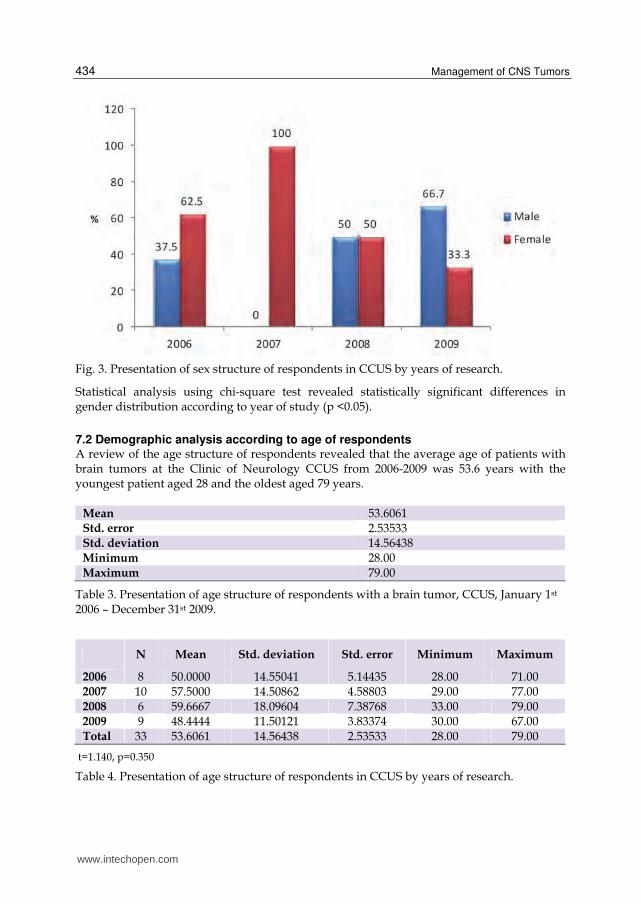
Of the total number of respondents in 2006 (N=8), three of them (37.5%) were male and 5 (62.5%) female. Total number of respondents was in 2007 - 10 (N=10) and all were female. Of the total number of patients with brain tumors in 2008 (N=6), 3 (50%) were male and the remaining 3 (50%) female. In 2009 the total number of respondents was 9, of which 6 (66.7%) males and 3 (33.3%) female.
www.intechopen.com
Management of CNS Tumors
434
Fig. 3. Presentation of sex structure of respondents in CCUS by years of research.
Statistical analysis using chi-square test revealed statistically significant differences in gender distribution according to year of study (p <0.05).
7.2 Demographic analysis according to age of respondents A review of the age structure of respondents revealed that the average age of patients with brain tumors at the Clinic of Neurology CCUS from 2006-2009 was 53.6 years with the youngest patient aged 28 and the oldest aged 79 years.
Mean 53.6061 Std. error 2.53533 Std. deviation 14.56438 Minimum 28.00 Maximum 79.00
Table 3. Presentation of age structure of respondents with a brain tumor, CCUS, January 1st 2006 – December 31st 2009.
N Mean Std. deviation Std. error Minimum Maximum
2006 8 50.0000 14.55041 5.14435 28.00 71.00 2007 10 57.5000 14.50862 4.58803 29.00 77.00 2008 6 59.6667 18.09604 7.38768 33.00 79.00 2009 9 48.4444 11.50121 3.83374 30.00 67.00 Total 33 53.6061 14.56438 2.53533 28.00 79.00
t=1.140, p=0.350
Table 4. Presentation of age structure of respondents in CCUS by years of research.
www.intechopen.com
Brain Tumors at the Department of Neurology in Sarajevo: Retrospecitve Analysis
435
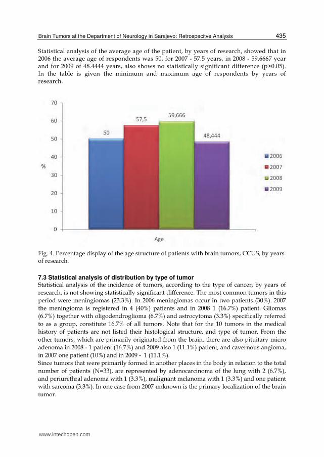
Statistical analysis of the average age of the patient, by years of research, showed that in 2006 the average age of respondents was 50, for 2007 - 57.5 years, in 2008 - 59.6667 year and for 2009 of 48.4444 years, also shows no statistically significant difference (p>0.05). In the table is given the minimum and maximum age of respondents by years of research.
Fig. 4. Percentage display of the age structure of patients with brain tumors, CCUS, by years of research.
7.3 Statistical analysis of distribution by type of tumor Statistical analysis of the incidence of tumors, according to the type of cancer, by years of research, is not showing statistically significant difference. The most common tumors in this period were meningiomas (23.3%). In 2006 meningiomas occur in two patients (30%). 2007 the meningioma is registered in 4 (40%) patients and in 2008 1 (16.7%) patient. Gliomas (6.7%) together with oligodendroglioma (6.7%) and astrocytoma (3.3%) specifically referred to as a group, constitute 16.7% of all tumors. Note that for the 10 tumors in the medical history of patients are not listed their histological structure, and type of tumor. From the other tumors, which are primarily originated from the brain, there are also pituitary micro adenoma in 2008 - 1 patient (16.7%) and 2009 also 1 (11.1%) patient, and cavernous angioma, in 2007 one patient (10%) and in 2009 - 1 (11.1%). Since tumors that were primarily formed in another places in the body in relation to the total number of patients (N=33), are represented by adenocarcinoma of the lung with 2 (6.7%), and periurethral adenoma with 1 (3.3%), malignant melanoma with 1 (3.3%) and one patient with sarcoma (3.3%). In one case from 2007 unknown is the primary localization of the brain tumor.
www.intechopen.com
Management of CNS Tumors
436
Year of the hospitalization Total
2006 2007 2008 2009
Tumor type
Unknown N 0 2 1 4 7 % .0 20.0 16.7 44.4 23.3
Pituitary micro adenoma N 0 0 1 1 2 % .0 .0 16.7 11.1 6.6
Angioma cavernosum N 0 1 0 1 2 % .0 10.0 .0 11.1 6.7
Astrocytoma N 0 0 0 1 1 % .0 .0 .0 11.1 3.3
Glioma N 1 0 1 0 2 % 10.0 .0 16.7 .0 6,7
Meningeoma N 2 4 1 0 7 % 30.0 40.0 16.7 .0 23,3
Oligodendroglioma N 1 0 1 0 2 % 10.0 .0 16.7 .0 6,7
Primary tumor -ca. mammae N 0 1 0 0 1 % .0 10.0 .0 .0 3,3
Primary tumor - unknown N 3 1 0 0 4 % 40.0 10.0 .0 .0 13,2
Primary tumor -adenocarcinoma of the lungs
N 1 0 0 1 2 % 10.0 .0 .0 11.1 6,7
Primary tumor -adenoma periurthraleN 0 0 1 0 1 % .0 .0 16.7 .0 3,3
Primary tumor-melanoma malignum N 0 0 0 1 1 % .0 .0 .0 11.1 3,3
Primary tumor -SARCOMA sterni N 0 1 0 0 1 % .0 10.0 .0 .0 3,3
Total N 8 10 6 9 33 % 16,7 33.3 20.0 30.0 100.0
χ2=44.722 , p=0.358
Table 5. Type of tumor
Year of the hospitalizationTotal
2006 2007 2008 2009
Metastases No
N 6 7 5 5 23 % 75.0 70.0 83.3 55.6 69,7
Meta cerebri N 2 3 1 4 10 % 25.0 30.0 16.7 44.4 30,3
Total N 8 10 6 9 33 % 24,2 30.3 18.2 27.3 100.0
χ2=1.487, p=0.685
Table 6. Brain metastases
www.intechopen.com
Brain Tumors at the Department of Neurology in Sarajevo: Retrospecitve Analysis
437
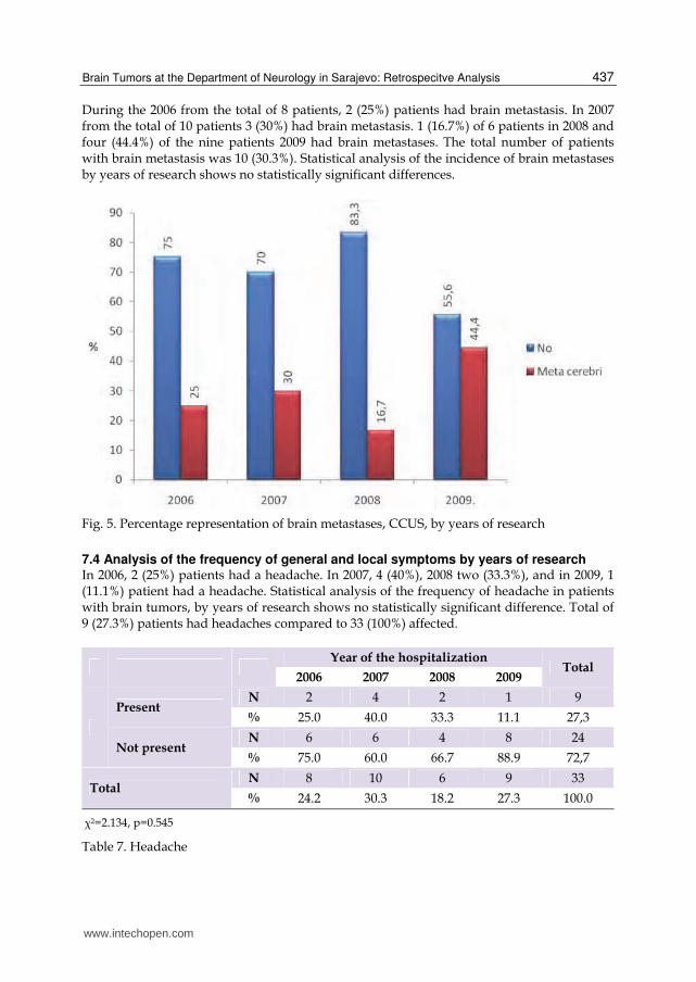
During the 2006 from the total of 8 patients, 2 (25%) patients had brain metastasis. In 2007 from the total of 10 patients 3 (30%) had brain metastasis. 1 (16.7%) of 6 patients in 2008 and four (44.4%) of the nine patients 2009 had brain metastases. The total number of patients with brain metastasis was 10 (30.3%). Statistical analysis of the incidence of brain metastases by years of research shows no statistically significant differences.
Fig. 5. Percentage representation of brain metastases, CCUS, by years of research
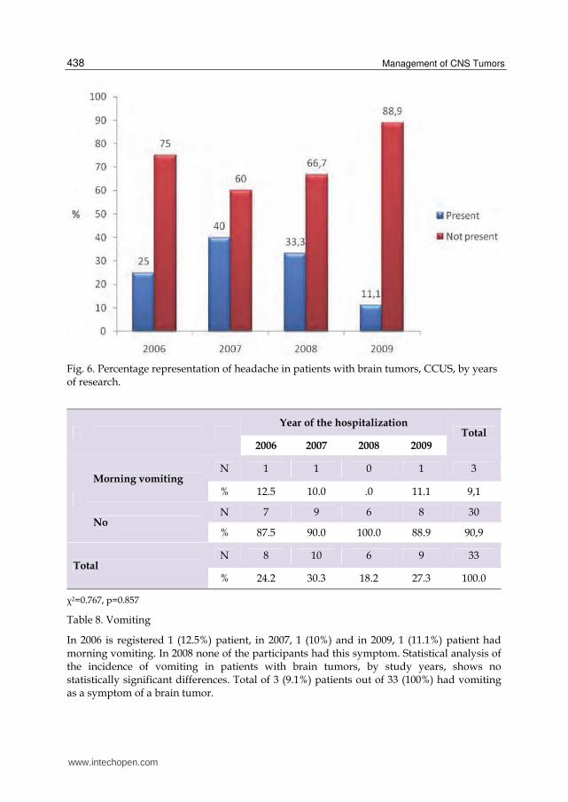
7.4 Analysis of the frequency of general and local symptoms by years of research In 2006, 2 (25%) patients had a headache. In 2007, 4 (40%), 2008 two (33.3%), and in 2009, 1 (11.1%) patient had a headache. Statistical analysis of the frequency of headache in patients with brain tumors, by years of research shows no statistically significant difference. Total of 9 (27.3%) patients had headaches compared to 33 (100%) affected.
Year of the hospitalization
Total 2006 2007 2008 2009
Present N 2 4 2 1 9
% 25.0 40.0 33.3 11.1 27,3
Not present N 6 6 4 8 24
% 75.0 60.0 66.7 88.9 72,7
Total N 8 10 6 9 33
% 24.2 30.3 18.2 27.3 100.0
χ2=2.134, p=0.545
Table 7. Headache
www.intechopen.com
Management of CNS Tumors
438
Fig. 6. Percentage representation of headache in patients with brain tumors, CCUS, by years of research.
Year of the hospitalization
Total 2006 2007 2008 2009
Morning vomiting N 1 1 0 1 3
% 12.5 10.0 .0 11.1 9,1
No N 7 9 6 8 30
% 87.5 90.0 100.0 88.9 90,9
Total N 8 10 6 9 33
% 24.2 30.3 18.2 27.3 100.0
χ2=0.767, p=0.857
Table 8. Vomiting
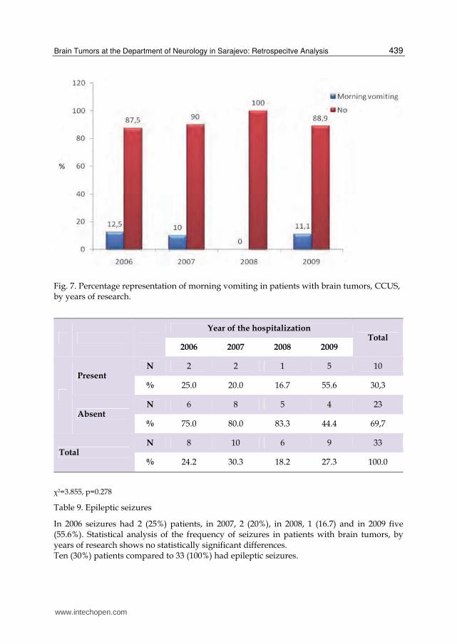
In 2006 is registered 1 (12.5%) patient, in 2007, 1 (10%) and in 2009, 1 (11.1%) patient had morning vomiting. In 2008 none of the participants had this symptom. Statistical analysis of the incidence of vomiting in patients with brain tumors, by study years, shows no statistically significant differences. Total of 3 (9.1%) patients out of 33 (100%) had vomiting as a symptom of a brain tumor.
www.intechopen.com
Brain Tumors at the Department of Neurology in Sarajevo: Retrospecitve Analysis
439
Fig. 7. Percentage representation of morning vomiting in patients with brain tumors, CCUS, by years of research.
Year of the hospitalization
Total 2006 2007 2008 2009
Present N 2 2 1 5 10
% 25.0 20.0 16.7 55.6 30,3
Absent N 6 8 5 4 23
% 75.0 80.0 83.3 44.4 69,7
Total N 8 10 6 9 33
% 24.2 30.3 18.2 27.3 100.0
χ2=3.855, p=0.278
Table 9. Epileptic seizures
In 2006 seizures had 2 (25%) patients, in 2007, 2 (20%), in 2008, 1 (16.7) and in 2009 five (55.6%). Statistical analysis of the frequency of seizures in patients with brain tumors, by years of research shows no statistically significant differences. Ten (30%) patients compared to 33 (100%) had epileptic seizures.
www.intechopen.com

Management of CNS Tumors
440
Fig. 8. Percentage representation of seizures in patients with brain tumors, CCUS, by years of research.
Year of the hospitalization
Total 2006 2007 2008 2009
No N 6 9 2 7 24
% 75.0 90.0 33.3 77.8 72,7
Hemiparesis cerebralis lateris dextri N 1 1 3 1 6
% 12.5 10.0 50.0 11.1 18,2
Hemiparesis cerebralis lateris sinistri N 1 0 1 1 3
% 12.5 .0 16.7 11.1 9,1
Total N 8 10 6 9 33
% 24.2 30.3 18.2 27.3 100.0
χ2=7.257, p=0.298
Table 10. Hemiparesis
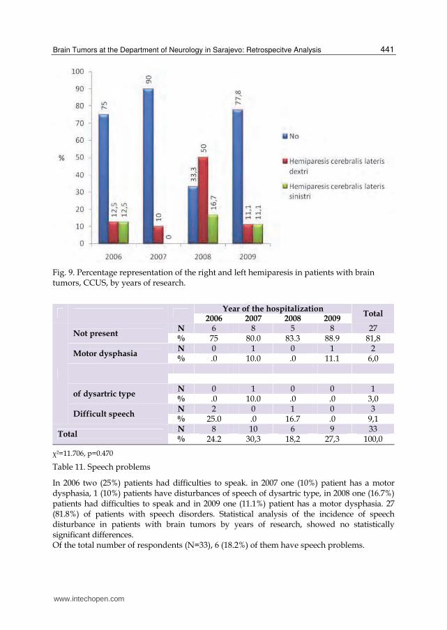
In 2006 is registered one patient (12.5%), in 2007, 1 (10%), in 2008, 3 (50%), and in 2009, 1 (11.1%) patient had a right hemiparesis. Left hemiparesis in 2006 occurs in one patient (12.5%), in 2008. at 1 (16.5%) and 2009 at 1 (11.1%) patients. In 2007 there was no patient with left hemiparesis. Statistical analysis of the frequency of hemiparesis in patients with brain tumors, CCUS, by years of research showed no statistically significant differences. Total of 9 (27.3%) patients at 33 (100%) had hemiparesis.
www.intechopen.com
Brain Tumors at the Department of Neurology in Sarajevo: Retrospecitve Analysis
441
Fig. 9. Percentage representation of the right and left hemiparesis in patients with brain tumors, CCUS, by years of research.
Year of the hospitalizationTotal
2006 2007 2008 2009
Not present N 6 8 5 8 27 % 75 80.0 83.3 88.9 81,8
Motor dysphasia N 0 1 0 1 2 % .0 10.0 .0 11.1 6,0
of dysartric type N 0 1 0 0 1 % .0 10.0 .0 .0 3,0
Difficult speech N 2 0 1 0 3 % 25.0 .0 16.7 .0 9,1
Total N 8 10 6 9 33 % 24.2 30,3 18,2 27,3 100,0
χ2=11.706, p=0.470
Table 11. Speech problems
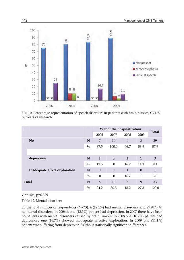
In 2006 two (25%) patients had difficulties to speak. in 2007 one (10%) patient has a motor dysphasia, 1 (10%) patients have disturbances of speech of dysartric type, in 2008 one (16.7%) patients had difficulties to speak and in 2009 one (11.1%) patient has a motor dysphasia. 27 (81.8%) of patients with speech disorders. Statistical analysis of the incidence of speech disturbance in patients with brain tumors by years of research, showed no statistically significant differences. Of the total number of respondents (N=33), 6 (18.2%) of them have speech problems.
www.intechopen.com
Management of CNS Tumors
442
Fig. 10. Percentage representation of speech disorders in patients with brain tumors, CCUS, by years of research.
Year of the hospitalization Total
2006 2007 2008 2009
No N 7 10 4 8 29
% 87.5 100.0 66.7 88.9 87,9
depression N 1 0 1 1 3
% 12.5 .0 16.7 11.1 9,1
Inadequate affect exploration N 0 0 1 0 1
% .0 .0 16.7 .0 3,0
Total N 8 10 6 9 33
% 24.2 30.3 18.2 27.3 100.0
χ2=6.406, p=0.379
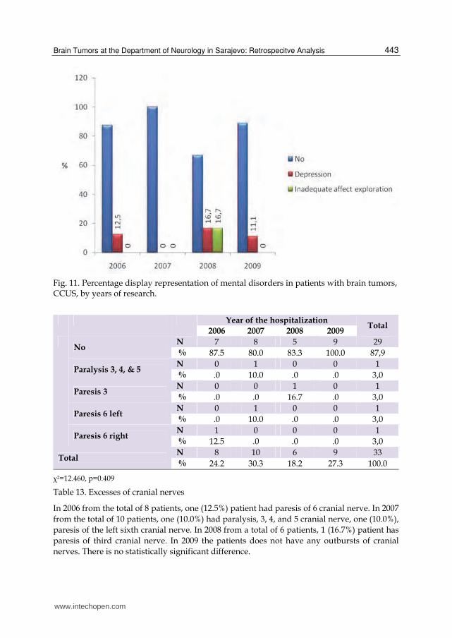
Table 12. Mental disorders
Of the total number of respondents (N=33), 4 (12.1%) had mental disorders, and 29 (87.9%) no mental disorders. In 2006th one (12.5%) patient had depression. In 2007 there have been no patients with mental disorders caused by brain tumors. In 2008 one (16.7%) patient had depression, one (16.7%) showed inadequate affective exploration. In 2009 one (11.1%) patient was suffering from depression. Without statistically significant differences.
www.intechopen.com
Brain Tumors at the Department of Neurology in Sarajevo: Retrospecitve Analysis
443
Fig. 11. Percentage display representation of mental disorders in patients with brain tumors, CCUS, by years of research.
Year of the hospitalization
Total 2006 2007 2008 2009
No
N 7 8 5 9 29 % 87.5 80.0 83.3 100.0 87,9
Paralysis 3, 4, & 5 N 0 1 0 0 1 % .0 10.0 .0 .0 3,0
Paresis 3 N 0 0 1 0 1 % .0 .0 16.7 .0 3,0
Paresis 6 left N 0 1 0 0 1 % .0 10.0 .0 .0 3,0
Paresis 6 right N 1 0 0 0 1 % 12.5 .0 .0 .0 3,0
Total N 8 10 6 9 33 % 24.2 30.3 18.2 27.3 100.0
χ2=12.460, p=0.409
Table 13. Excesses of cranial nerves
In 2006 from the total of 8 patients, one (12.5%) patient had paresis of 6 cranial nerve. In 2007 from the total of 10 patients, one (10.0%) had paralysis, 3, 4, and 5 cranial nerve, one (10.0%), paresis of the left sixth cranial nerve. In 2008 from a total of 6 patients, 1 (16.7%) patient has paresis of third cranial nerve. In 2009 the patients does not have any outbursts of cranial nerves. There is no statistically significant difference.
www.intechopen.com

Management of CNS Tumors
444
Fig. 12. Percentage representation of failure of the cranial nerves, CCUS, after years of research.
7.5 Analysis of the localization of brain tumors From 33 analyzed patients showed that the brain tumors are located mostly in the cerebellum (21%), then frontoparietal (18.1%) in the brain stem (15.1%), parietal (12%). In sphenoid sinus are located 9.1% of tumors. The frontal, occipital, and parietoccipital in the cavernous sinus are located 6.1% of the tumors. Statistical analysis of the incidence of tumors in relation to their location, by years of research, showed no statistically significant difference.
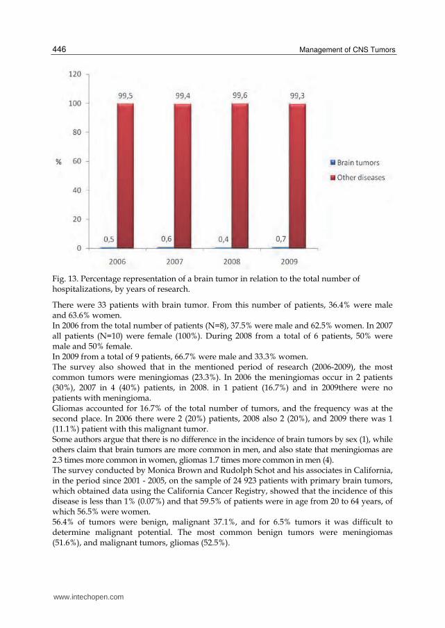
7.6 Analysis of the incidence of tumors in relation to the total number of hospitalized patients A review of years of hospitalization, showed that most patients with brain tumors in relation to the total number of patients were hospitalized during 2009 (9 or 0.7%) where the total number of patients was 1731. Followed 2007 when from the 1699 hospitalized patients 10 or 0.6% was with brain tumors. In 2008 hospitalized was 1700 patients, of which 6 or 0.4 with brain tumors, and in 2006 from the 1731 patients hospitalized, of which 8 or 0.5% with brain tumors. The frequency of brain tumors at Neurology Clinic CCUS, from January 1st 2006 – December 31st 2009 is 33 of 6358 patients, or 0.5%.
www.intechopen.com
Brain Tumors at the Department of Neurology in Sarajevo: Retrospecitve Analysis
445
Year of the hospitalization Total
2006 2007 2008 2009
Frontal
N 1 .0 .0 1 2 % 12.5 .0 .0 11.1 6.1
Parietal N 2 0 0 2 4 % 25.0 .0 .0 22.2 12.1
Occipital N 1 0 1 0 2 % 12.5 .0 16.7 .0 6,1
Frontoparietal N 1 0 3 2 6 % 12.5 .0 50.0 22.2 18.1
Parietoccipital N 0 2 0 0 2 % .0 20.0 .0 .0 6,1
Brain stem N 0 5 0 0 5 % .0 50.0 .0 .0 15,1
Sphenoid sinus N 0 1 1 1 3 % .0 10.0 16.7 11.1 9,1
Cerebellum N 2 1 1 3 7 % 25.0 10.0 16.7 33.3 21,1
Cavernous sinus N 1 1 0 0 2 % 12.5 10.0 .0 .0 6,1
Total N 8 10 6 9 33 % 24.2 30.3 18.2 27.3 100.0
χ2=65.519, p=0.389
Table 14. Tumor location
Year of the hospitalization
Total 2006 2007 2008 2009
Brain tumor N 8 10 6 9 33 % 0.5 0.6 0.4 0.7 0,5
Other diseases N 1723 1689 1694 1219 6325 % 99.5 99.4 99.6 99.3 99,5
Total N 1731 1699 1700 1228 6358 % 27.2 26.7 26.7 19.3 100.0
Table 15. Tumors frequency
8. Discussion
In a study conducted at the Neurology Clinic Sarajevo from January 1st 2006 – December 31st 2009, it was found that the incidence of brain tumors is less than 1%, and there is no significant statistical difference in the incidence of tumors by years of research, except when it comes to the gender distribution of patients.
www.intechopen.com
Management of CNS Tumors
446
Fig. 13. Percentage representation of a brain tumor in relation to the total number of hospitalizations, by years of research.
There were 33 patients with brain tumor. From this number of patients, 36.4% were male and 63.6% women. In 2006 from the total number of patients (N=8), 37.5% were male and 62.5% women. In 2007 all patients (N=10) were female (100%). During 2008 from a total of 6 patients, 50% were male and 50% female. In 2009 from a total of 9 patients, 66.7% were male and 33.3% women. The survey also showed that in the mentioned period of research (2006-2009), the most common tumors were meningiomas (23.3%). In 2006 the meningiomas occur in 2 patients (30%), 2007 in 4 (40%) patients, in 2008. in 1 patient (16.7%) and in 2009there were no patients with meningioma. Gliomas accounted for 16.7% of the total number of tumors, and the frequency was at the second place. In 2006 there were 2 (20%) patients, 2008 also 2 (20%), and 2009 there was 1 (11.1%) patient with this malignant tumor. Some authors argue that there is no difference in the incidence of brain tumors by sex (1), while others claim that brain tumors are more common in men, and also state that meningiomas are 2.3 times more common in women, gliomas 1.7 times more common in men (4). The survey conducted by Monica Brown and Rudolph Schot and his associates in California, in the period since 2001 - 2005, on the sample of 24 923 patients with primary brain tumors, which obtained data using the California Cancer Registry, showed that the incidence of this disease is less than 1% (0.07%) and that 59.5% of patients were in age from 20 to 64 years, of which 56.5% were women. 56.4% of tumors were benign, malignant 37.1%, and for 6.5% tumors it was difficult to determine malignant potential. The most common benign tumors were meningiomas (51.6%), and malignant tumors, gliomas (52.5%).
www.intechopen.com
Brain Tumors at the Department of Neurology in Sarajevo: Retrospecitve Analysis
447
Retrospective survey conducted by Srdjana and Sinisa Telarovic, in Istria (Croatia), in the period from January 1st 1986 to December 31st 2000 at the Neurological Department of the General Hospital in Pula, found the incidence of tumors less than 1%. Of the 364 patients, 213 (57.69%) were male and 151 patients (42.31%) female. The most common tumors were gliomas 47.7%. The survey by Peter D. and associates in Cyprus in the period since 1998 - 2001, 56% of the total 150 patients were male and 44% were women. The most common cancers in the population aged 50-69 years were gliomas. It should be noted that at the 2007 at the Clinic of Neurology was only female population, and that same year recorded four meningiomas from a total of 7 cancers. Previous two studies and research at the Clinic of Neurology, which are included in this survey, confirming the dependence of the frequency of brain tumors by sex on the incidence of brain tumors in relation to the type of tumors. The research at Clinic of Neurology, which is included in this study, has registered 10 (30%) of 33 patients with brain metastases. In four brain metastases was not found listed species or location of primary tumors in the patient’s medical history. Primary brain tumors of the other 6 metastases were adenocarcinoma of the lung metastases (N=2), malignant melanoma (N=1), periurethral adenoma (N=1), sarcoma (N=1), and breast cancer (N=1). According to world literature metastases make 12-25% of brain tumors (1). The survey conducted by Teletovic and associates, in Istria (Croatia), at the Neurological Department of the General Hospital in Pula, in the period from 1986 to 2000, it was found that 56.87% were primary, mostly malignant tumors, 43.13% brain metastases. Thus a large number of brain metastases in Istria are explained by stress caused by war during nineties in Croatian territory, and weakening of immunity, and increased use of cigarettes and alcohol. According to a retrospective study, the incidence of brain tumors at the Department of Neurology CCUS, for the period from January 1st 1990 to December 31st 1999, which conducted Alajbegovic and Hrnjica with associates, 36% of patients (N=105) had brain metastasis. Research at the Clinic of Neurology, which is included in this study showed that the average age of patients with brain tumor is 53.6 years with the youngest patient aged 28 and the oldest aged 79 years. According to a study conducted by Srdjan and Sinisa Telarovic with associates, found the illness mostly in the population aged 50-59 years. A study conducted by Peter D. and associates in Cyprus, confirmed that the brain tumors usually occur in the population of at age 50-69 years and in 40% of cases. In 33 analyzed patients showed that the brain tumors are located in the cerebellum (21%), then frontal parietal (18.1%) in the brain stem (15.1%) and parietal (12%). In sfenoidal sinus was located 9.1%tumors. The frontal, occipital, and partial occipital in the cavernous sinus was located 6.1% tumors. In other words, most of the tumors was located in the cerebral hemispheres, and in the cerebellum, and then in the brain stem. This coincides with a study by Peter D. in Cyprus by which 34.8% tumors are located in the cerebral hemispheres, 4.7% in the cerebellum, 2% in the brain stem and 2% in the spinal cord. For the other 48% tumors location was not specified. According to a study by Sinisa and Srdjan Teletovic, 43% tumors were localized in the cerebral hemispheres, 15.2% in the cerebellum and the rest of the tumors were located in the brain stem and spinal cord. The research at Department of Neurology is covered a general and focal symptomatology. Most were represented seizures (30.3%), followed by headache (27.3%), hemiparesis 27.3% (18.2% right, left 9.1%), speech disturbances 18.2%, mental disorders 12.0%, morning vomiting 9.1%, and the problems of the cranial nerves with 9.0%. These data correspond to the data
www.intechopen.com
Management of CNS Tumors
448
given in world literature (1). According to a retrospective study conducted by Alajbegovic, Loga and associates at the Clinic of Neurology CCUS, from January 1st 2001 – December 31st 2005, 15-20% of patients had psychological disturbances. According to the already mentioned study, conducted by Alajbegovic and Hrnjica about 35% of patients had seizures.
9. Conclusions
1. The incidence of brain tumors at the Neurology Clinic Sarajevo from January 1st 2006 – December 31st 2009 was 0.5% (less than 1%).
2. There was no statistically significant difference in the incidence of tumors by years of research, except in the frequency of sex.
3. Of 33 patients, 36.4% were male and 63.6% women. 4. The mean age of patients with brain tumors was 53.6 years, with the youngest patient
aged 29 and the oldest aged 79 years. 5. The most common tumors were meningiomas (23.3%), and gliomas (16.7%). 6. Brain metastases were confirmed in 10 (30%) patients. 7. The most common tumor site was the cerebral hemispheres, and in the cerebellum, and
then in the brain stem. 8. The most common symptoms of brain tumors were seizures (30.3%) followed by
headache and hemiparesis with 27.3%.
10. References
[1] Alajbegović Azra, Hrnjica Muhamed, Dimitrijević Jovan, Zukić Tarik, Subašić Nihada. Central nervous system neoplasms in clinical data from the Neurology CU Sarajevo, 1990-1999.
[2] Alajbegović Azra, Logo Nataša, Alajbegović Salem, Resić Halima. Psihičke promjene pacijenata sa moždanim tumorom, 2008.
[3] Aleksandar kostić, Milorad Radić, Goran Ignjatović, Miloš Janićijević. Odnos preoperativne veličine tumora I preživljavanja kod obolelih od astrocitoma mozga,2009.
[4] Chan TA, Weingart JD, ParisiM, et al. Treatment of recurrent glioblastoma multiforme with GliaSite brachytherapy. Int J Radiat Oncol Biol Phys 2005.
[5] Dr.sc. Lijerka Makulin- Grgić. Radioterapija i kemoterapija tumora živčanog sistema. [6] Glantz MJ, Cole BF, Forsyth PA, et al. Parctice parameter : Anticonvulsant prophylaxis in
patients with newly diagnosed brain tumors. Neurology, 2000; 54:1886 - 1893. [7] Kondziolk D, Martin J J. Indication for resection and radiosurgery for brain metastases.
Current Opinion in Oncology 2005. [8] Monica Brown, Rudolf Schrot, Katrina Bauer, Deanna Le Tendre. Incidence of First
Primary Central Nervous System Tumors in California, 2005. [9] Nacional Cancer Institute of USA. Brain and Central Nervous systems cancer, 2005. [10] Peter D., Elaine Row. Brain and Other Central Nervous System Cancer, 2001. [11] Šamija M, Vrdoljak E, Krajina Z. Klinička onkologija, Medicinska naklada Zagreb 2006. [12] Srđana Telarović, Siniša Telarović, Maja Relja. Impact of War on Central Nervous
Sistem, 2000. [13] Tosoni A, Ermani M, Brandes A A. the pathogenesis and treatment of brain metastases:
a komprehesivereview, critical Reviews in Oncology/Hematology 2004.
www.intechopen.com

Management of CNS TumorsEdited by Dr. Miklos Garami
ISBN 978-953-307-646-1Hard cover, 464 pagesPublisher InTechPublished online 22, September, 2011Published in print edition September, 2011
InTech EuropeUniversity Campus STeP Ri Slavka Krautzeka 83/A 51000 Rijeka, Croatia Phone: +385 (51) 770 447 Fax: +385 (51) 686 166www.intechopen.com
InTech ChinaUnit 405, Office Block, Hotel Equatorial Shanghai No.65, Yan An Road (West), Shanghai, 200040, China
Phone: +86-21-62489820 Fax: +86-21-62489821
Management of CNS Tumors is a selected review of Central Nervous System (CNS) tumors with particularemphasis on pathological classification and complex treatment algorithms for each common tumor type.Additional detailed information is provided on selected CNS tumor associated disorders.
How to referenceIn order to correctly reference this scholarly work, feel free to copy and paste the following:
A. Alajbegovic and A. Gutosić (2011). Brain Tumors at the Department of Neurology in Sarajevo: RetrospecitveAnalysis, Management of CNS Tumors, Dr. Miklos Garami (Ed.), ISBN: 978-953-307-646-1, InTech, Availablefrom: http://www.intechopen.com/books/management-of-cns-tumors/brain-tumors-at-the-department-of-neurology-in-sarajevo-retrospecitve-analysis