
BRAIN ReynoldsUnwrapped.comReynoldsUnwrapped.com offers FANTASTIC, inexpensive daily email...
215
BRAI N ReynoldsUnwrapped.com offers FANTASTIC, inexpensive daily email subscriptions, where you can receive a HILARIOUS new cartoon every day, and it is a MARVELOUS idea for a UNIQUE gift for your family and friends as well. That is how I learned about this...one of my fellow teachers gave me a subscription as a birthday present. He also has FUNNY greeting cards and BEAUTIFUL paintings for sale as well. You can also get reprints suitable for framing, or originals. Here is more info about his work and a YOUTUBE 1
-
Upload
adam-hancock -
Category
Documents
-
view
215 -
download
0
Transcript of BRAIN ReynoldsUnwrapped.comReynoldsUnwrapped.com offers FANTASTIC, inexpensive daily email...

BRAIN
ReynoldsUnwrapped.com offers FANTASTIC, inexpensive daily email subscriptions, where you can receive a HILARIOUS new cartoon every day, and it is a MARVELOUS idea for a UNIQUE gift for your family and friends as well. That is how I learned about this...one of my fellow teachers gave me a subscription as a birthday present.
He also has FUNNY greeting cards and BEAUTIFUL paintings for sale as well.You can also get reprints suitable for framing, or originals. Here is more info about his work and a YOUTUBE video.https://nccnews.expressions.syr.edu/?p=11515
1

MENTAL HOSPITAL PHONE MENU• Hello and thank you for calling The State Mental Hospital.
Please select from the following options menu: • If you are obsessive-compulsive, press 1 repeatedly.
If you are co-dependent, please ask someone to press 2 for you.
If you have multiple personalities, press 3, 4, 5 and 6.
If you are paranoid, we know who you are and what you want, stay on the line so we can trace your call.
If you are delusional, press 7 and your call will be forwarded to the Mother Ship.
If you are schizophrenic, listen carefully and a little voice will tell You which number to press.
If you are manic-depressive, hang up. It doesn't matter which number you press, nothing will make you happy anyway.
If you are dyslexic, press 9-6-9-6.
If you are bipolar, please leave a message after the beep or before the beep or after the beep. • But Please wait for the beep.
If you have short-term memory loss, press 9. If you have short-term memory loss, press 9. If you have short-term memory loss, press 9.
If you have low self-esteem, please hang up. Our operators are too busy to talk with you.
If you are menopausal, put the gun down, hang up, turn on the fan, lie down and cry. You won't be crazy forever.
2

THE BRAIN
ANATOMICAL REGIONS A. Cerebrum B. Diencephalon
Thalamus Hypothalamus
C. Brain Stem Midbrain Pons Medulla oblongata
D. Cerebellum
3

THE BRAIN
FUNCTIONAL REGIONS A. MOTOR AREAS B. SENSORY AREAS C. HIGHER FUNCTIONS
4

• MAJOR ANATOMICAL REGIONS OF THE BRAIN – Cerebrum– Diencephalon– Brain Stem– Cerebellum
5
The Brain
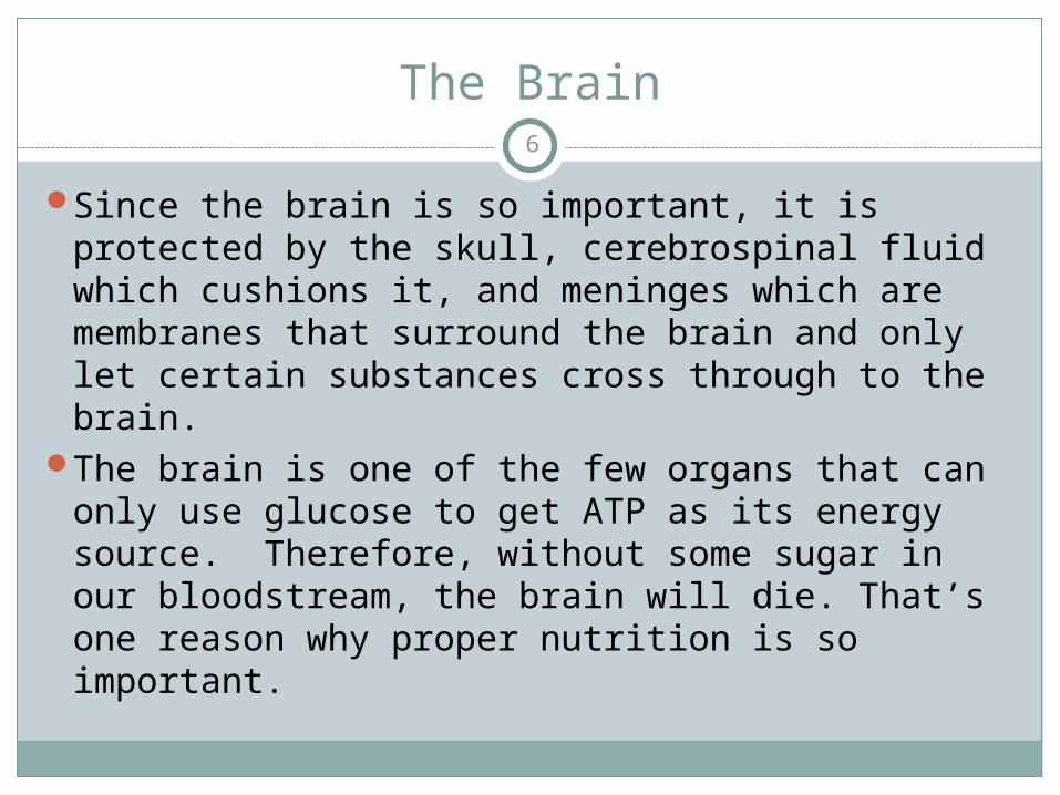
Since the brain is so important, it is protected by the skull, cerebrospinal fluid which cushions it, and meninges which are membranes that surround the brain and only let certain substances cross through to the brain.
The brain is one of the few organs that can only use glucose to get ATP as its energy source. Therefore, without some sugar in our bloodstream, the brain will die. That’s one reason why proper nutrition is so important.
6

The Brain
By the way, geniuses have the same size brain as everyone else; they are just more efficient at forming synapses. They also have more synapses because they have more dendrites. You can develop more dendrites and synapses by keeping your brain active by learning and reading new things.
We don’t use 90% of our brains, we use 100%.
Fun Fact: -Scientists say the higher your I.Q. The more you dream.
7

CEREBRUM
The brain is divided into parts, and is bilaterally symmetrical.
In general, the left side controls the right half of the body, and the right side of the brain controls the left half of the body.
The largest portion is the CEREBRUM, which makes up 80% of the brain.
The cerebrum controls logical thought and conscious awareness of the environment.
It is also the area responsible for the highest sensory and motor activity.
The cerebrum is made up mostly of grey matter (cell bodies, dendrites, and unmyelinated axons).
8

GYRUS AND SULCUS
The superficial region of the brain (and all other organs) is called the cortex.
The surface is not smooth, it’s convoluted. Each bump on the surface of the cerebrum is called a GYRUS, and each shallow groove on the surface of the cerebrum is called a SULCUS.
This formation increases the surface area, and the surface (cortex) is where the information processing is.
9

Figure 13.7a10

11

CEREBRUM
The cerebrum is divided into 2 halves called CEREBRAL HEMISPHERES, which are separated by the longitudinal fissure.
The right cerebral hemisphere controls the activity of, and receives sensory input from the left side of the body.
The left cerebral hemisphere controls the activity of, and receives sensory input from the right side of the body.
Each hemisphere is divided into lobes, named for the bones on top of them.
12

The Cerebral Hemispheres and lobes
Figure 13.7b, c
13

The Cerebral Hemispheres and lobes
The FRONTAL LOBE and PARIETAL LOBE are separated by the CENTRAL SULCUS.
The TEMPORAL LOBE is between the parietal and frontal lobe, separated by the LATERAL SULCUS.
The OCCIPITAL LOBE does not have a real border; it’s just a region.
These are the anatomical areas, but the functional areas are more important.
Central sulcus
Lateral sulcus
14

CORPUS CALLOSUM
If you slice the brain down the center in a mid-sagittal section, you will slice through a white colored tissue called the CORPUS CALLOSUM, which is the area that connects the right and left halves of the brain.
15

CORPUS CALLOSUM
The CORPUS CALLOSUM is the area that connects the right and left halves of the brain.
16

Corpus callosumSheep brain
17

CORPUS CALLOSUM
This is the area that is responsible for the right half of the brain communicating with the left half of the brain.
If the corpus callosum was cut, there would be no communication between the right and left halves of the brain.
Autism is a neurological disease that includes problems with communication between the right and left cerebral hemispheres.
Music therapy for autism: http://ezinearticles.com/?The-Benefits-of-Music-Therapy-for-Autism&id=432566
18

Phineas Gage
Phineas was a railroad construction foreman who survived an accident in which a large iron rod was driven completely through his head, severing connections in his left frontal lobe.
It changed his personality; he became emotional and had frequent outbursts.
This was the first case suggesting that damage to specific regions of the brain might affect personality and behavior.
19

Phineas Gage
The left side of the brain is responsible for critical thinking, and the right side is responsible for emotion.
Since his left frontal lobe was damaged, his emotions went unchecked.
20

CORPUS CALLOSUM: Fun Fact
Women have a wider corpus callosum than men.They tend to use both sides of their brain more than
men do.That’s why they like to talk more.Give a little girl a doll, and she will hold it like a baby.Give a little boy a doll, and he will take the head off to
see what it looks like inside.This is a difference between using both sides of the
brain vs. just one side.There is a new book out in paperback called “How to
Understand Women”
21
How To Understand Women22

WHY WOMEN CAN'T SLEEP23
In a woman’s brain, every one of those little balls is a thought about something thatneeds to be done, a decision that has to be made, or a problem that needs to be solved.
A man has only 2 balls. They consume all his thoughts, and he sleeps like a baby.

Diencephalon
Consists of two parts:Thalamus
The superior portion of the diencephalon Processes sensory information according to
importance Major relay station for sensory impulses to the
cerebrumHypothalamus
The inferior portion of the diencephalon Makes hormones which maintains the
homeostasis of the body
24

Figure 13.15
25

THALAMUS
The THALAMUS functions to sort out all the sensory information.
It compares the input and determines what information is worth sending to the cortex.
Your body ignores most sensory information.Up until now, have you noticed the sound of
the air conditioner? It’s not important, so it goes unnoticed.
This area also compares information from the right and left eyes for stereoscopic vision, and the right and left ear to determine direction of sound.
26

Thalamus
Hypothalamus
Pituitary gland
27

Pituitary gland
Hypothalamus
Thalamus
28

HYPOTHALAMUS
This small area exerts more control over autonomic functioning than any other part.
Makes hormones which provide homeostatic control over the body
It maintains homeostasis by controlling the autonomic nervous reflexes, glucose and hormone levels.
It is also the main visceral control center, so it controls body temperature, hunger and thirst, and blood pressure.
The hypothalamus is part of the limbic system (which is involved in memories), so that’s why a painful memory can increase blood pressure.
29

Figure 13.1530

HYPOTHALAMUS
The hypothalamus synthesizes and secretes hormones, and these in turn stimulate or inhibit the secretion of pituitary hormones.
By secreting hormones, the hypothalamus controls blood pressure, body temperature, hunger, thirst, fatigue, sleep, autonomic nervous reflexes, and circadian cycles.
31

• BRAIN STEM– MIDBRAIN– PONS– MEDULLA OBLONGATA
32

Midbrain
The top of the brain stem is the MIDBRAIN. It controls automatic behaviors (fight or flight) The midbrain also contains a pigmented area called
the substantia nigra.The Substantia nigra is involved in addictions and
in initiating body movement. The substantia nigra secretes the neurotransmitter
dopamine.When the neurons in the substantia nigra become
damaged, dopamine levels decrease, causing Parkinson's Disease.
Treatment is to replace the dopamine
33

Dopamine
Remember that acetylcholine is the neurotransmitter that functions to contract skeletal muscles?
There are many other types of neurotransmitters as well. One is called dopamine.
Dopamine is the neurotransmitter that controls the flow of information between various areas of the brain.
Dopamine is lacking in Parkinson's Disease, in which the person has muscular rigidity and tremors, so they lose the ability to start movements. They need a service dog to help them get out of a chair or to take a first step. They also have a pill-rolling tremor at rest.
34

VIDEOS
Parkinson’s gaitParkinson’s patient
35

Dopamine
Dopamine plays a major role in the brain system that is responsible for reward-driven learning. Every type of reward that has been studied increases the level of dopamine transmission in the brain, and a variety of highly addictive drugs, including stimulants such as cocaine and methamphetamine, act directly on the dopamine system.
There is evidence that people with extraverted (reward-seeking) personality types tend to show higher levels of dopamine activity than people with introverted personalities.
36

Dopamine
Several important diseases of the nervous system are associated with dysfunctions of the dopamine system. Parkinson's disease, an age-related degenerative condition causing tremor and motor impairment, is caused by loss of dopamine-secreting neurons in the substantia nigra.
Schizophrenia has been shown to involve elevated levels of dopamine activity in regions of the brain and decreased levels of dopamine in other regions.
Attention deficit hyperactivity disorder (ADHD) is also believed to be associated with decreased dopamine activity.
37

Dopamine
Because dopamine cannot cross the blood–brain barrier, patients with diseases such as Parkinson's disease are given L-DOPA (the precursor of dopamine) because it crosses the blood-brain barrier relatively easily.
It is then converted by the body to dopamine.
38

VIDEOS
Huntington’s chorea gaitHuntington’s chorea patient
39

Endorphins
From the Greek: word endo meaning "within" and morphine, from Morpheus, the god of sleep.
Endorphins are neurotransmitters made within our body that are produced by the pituitary gland during exercise, excitement, pain, acupuncture, consumption of spicy food, love and orgasm, and they resemble opiates in their abilities to produce analgesia (pain suppression) and a feeling of well-being.
They cause more dopamine to be released. How drugs cause dopamine release: Mouse Party http://learn.genetics.utah.edu/content/addiction/mouse/
40

Corpora Quadrigemina “Quadruplet bodies”
They control visual and audio (hearing) reflexes.
Throw something at your face, you blink = visual reflex. Loud noise (BANG!) causing a startle, is the audio reflex.
The two superior bodies are for eye blinking and fast eye movements.
The two inferior bodies are for sound reflexesThe corpora quadrigemina are linked to the
midbrain.
41

Midbrain
Corpora quadrigemina
42

Pons
Farther down the brainstem is the PONS, which relays sensory information between the cerebellum and cerebrum.
43

MidbrainMedulla Oblongata Pons
Spinal cord
44

Pons
Midbrain
Medulla Oblongata
45

Medulla Oblongata
At the base of the brainstem is the MEDULLA OBLONGATA, which contains the cardiac, respiratory, vomiting and vasomotor (blood vessel constriction) centers.
It also effects heart rate, blood pressure, and breathing.
Damage here causes coma. Swelling from an injury causes pressure, which can damage this area, which can cause a coma.
Concussions cause nausea and a decrease in blood pressure; patients with these symptoms need an MRI to see if this is early signs of damage to medulla oblongata
Boxers who are knocked out can recover, but repeated knock-outs can cause permanent brain damage.
46

What’s the difference in function between the medulla oblongata and the hypothalamus?
The medulla oblongata controls blood pressure directly (using nerves), and the hypothalamus controls it indirectly (using hormones).
47

48

Reticular Formation
The reticular formation is a group of cells scattered throughout the brainstem.
They play a role in rousing and maintaining consciousness.
49

Melatonin in animals
Hormone found in animals, plants, and microbes.
In animals, circulating levels of melatonin vary in a daily or seasonal cycles, thereby allowing the circadian rhythms of several biological functions.
It allows reptiles to change the color of their skin
The change in duration of secretion also serves as a biological signal for seasonal reproduction, behavior, coat growth, and camouflage coloring in animals.
50

Melatonin
Another timekeeping function of melatonin is its role in orchestrating seasonal changes. For example, it plays a major part in signaling the body of animals to hibernate. In this capacity, it is believed that melatonin is instrumental in causing tissues to shift into a state of metabolic inactivity during hibernation. This capability has led some researchers to propose that melatonin might be able to induce a hibernation-like state and donor organs prior to transplantation in order to prolong their shelf life.
51

Melatonin
In seasonally reproducing animals, melatonin plays a part in directing the reproductive system to become inactive. Because of this ability to regulate seasonal activities, there has been interest in the role that melatonin might play in seasonal affective disorder (SAD), which makes some people depressed in the winter. The present evidence indicates that the short days of winter cause excess melatonin to be produced in individuals susceptible to this disorder. It appears that in people with SAD, the melatonin system does not respond to artificial lighting, while in unaffected people the system ceases production of melatonin in response to artificial lighting. Interestingly, in many people with SAD, special bright lights that mimic sunlight are effective in shutting down the melatonin and alleviating depression.
52

Melatonin in humans
In humans, melatonin helps us sleep. Infants' melatonin levels become regular in about the third month after birth, so they sleep through the night better.
Production of melatonin by the pineal gland is inhibited by light and permitted by darkness.
Secretion peaks in the middle of the night, with normal variations in timing according to an individual's chronotype.
A chronotype is an attribute reflecting at what time of the day their physical functions (hormone level, body temperature, cognitive faculties, eating and sleeping) are active, change, or reach a certain level.
Are you a morning person or a night owl?
53

Melatonin
One of the clearest effects of melatonin is the ability to reset the body's internal clock for blind people or those suffering from jet lag or in night shift workers. Melatonin administered in the afternoon shifts the sleep cycle so that people wake up and go to sleep earlier. Melatonin given in the morning causes people to wake up and go to sleep later. This capability of melatonin to shift the body's internal clock will likely find increasing use in the future as travel for business and pleasure become more international. It should also be especially useful in sports where teams playing in international competitions now have to arrive a week before an event in order for there internal rhythms to shift naturally so they can play at their best.
54

Melatonin
Although melatonin is widely advertised as a sleep aid, there is controversy among scientists about what role it plays in sleep it does not appear to work like a sleeping pill that simply induces sleep, rather it seems to produce a physiological bias toward sleep. As people get older, the amount of melatonin they produce at night decreases, while insomnia and other sleep problems increase. Alzheimer's patients have less melatonin than normal. Fortunately, a few studies have already shown that melatonin treatment can cause significant improvements in the sleep quality of both elderly insomniacs and Alzheimer's patients. If melatonin can be used to reestablish more normal sleep patterns in Alzheimer's patients, it should help delay their institutionalization and reduce the psychological, physical, and monetary burden of this devastating disease.
55

Other effects of melatonin
Melatonin stimulates the immune system There is new research being done on giving melatonin to
people with autoimmune diseases, such as ulcerative colitis.
It is an antioxidant, protecting mitochondrial DNA
It increases REM sleep time (dreaming)It causes the onset of puberty
Melatonin is mainly secreted by the pineal body.
56

PINEAL BODY (Pineal gland)
The PINEAL BODY secretes melatonin.How much it secretes depends on the sensory
information it receives from the eyes about how many hours of daylight are present.
The amount of melatonin secreted and circulating in the blood then determines the circadian rhythm, or the 24-hour biological clock (cycles influenced by light).
Therefore, the pineal body detects the number of hours of light and dark, and sets the body’s 24-hour clock.
57

Thalamus
Pineal body58

Pineal body
59

JET LAG
When you get jet lag, it’s because the information the pineal gland has been getting doesn’t match with where you are now.
You can help yourself get over jet lag by being outdoors in the daylight and being indoors in a dark room at night, and the pineal body with reset the clock.
60

Chronic Insomnia
First eliminate caffeine, then modify the diet (no sugars) and increase exercise.
If that does not work, try Benadryl. It is an antihistamine, but its side-effect is drowsiness. It is good because it does not interfere with REM sleep and it is not addictive. Some try melatonin as well.
Prescription sleep meds can be addictive and they interfere with REM sleep, so you don’t feel rested.
Ambien (a prescription sleep med) can cause people to sleep walk, and even drive in their sleep!
61

CEREBELLUM
The cerebellum is the second largest portion of the brain, is responsible for balance and muscle coordination, and is a comparator.
62

CEREBELLUM
The cerebellum functions as a comparator. Action potentials from the cerebral motor cortex descend into the spinal cord to move the muscles.
There are branches that are sent to the cerebellum to give it information on the intended movement.
At the same time, the cerebellum receives information from proprioreception neurons (sensory, tell what position each body part is in).
The cerebellum compares all this information to allow smooth movements. That is why it is called a comparator.
63

64

Cerebellar Function Evaluation
Finger to noseHeel to shinAtaxic gait of alcoholismTandem gait (walk straight line)
65

FUNCTIONAL REGIONS
A. SENSORY AREASB. MOTOR AREASC. HIGHER FUNCTIONS
66

CORTEX AND ASSOCIATION AREAS
Each area of the brain has a region where the sensory information comes in, and another area where the information is understood.
The area where the information comes in is a cortex, and the area where it is understood is the association area.
Therefore, there will be a visual cortex and association area, an auditory cortex and association area, and a somatic (sense of touch) cortex and association area. There is also a motor cortex and association area.
67

Functional and Structural Areas of the Cerebral Cortex
Figure 13.11a68

Dyslexia
Dyslexia is a very broad term defining a learning disability that impairs a person's fluency or comprehension accuracy in being able to read, and which can manifest itself as a difficulty with phonological awareness (speaking properly), phonological decoding (understanding speech), orthographic coding (writing properly and reading properly), auditory short-term memory, or rapid naming.
Dyslexia is distinct from reading difficulties resulting from other causes, such as a non-neurological deficiency with vision or hearing, or from poor or inadequate reading instruction. Dyslexia affects 5-10 % of the population.
69

Dyslexia
There are three proposed cognitive subtypes of dyslexia: auditory, visual and attentional.
Dyslexia is the most common learning disability. Researchers at MIT found that people with dyslexia exhibited impaired voice-recognition abilities.
Adult dyslexics can read with good comprehension, but they tend to read more slowly than non-dyslexics and perform more poorly at spelling and nonsense word reading, a measure of phonological awareness. Dyslexia and IQ are not interrelated.
70

71

FUNCTIONAL REGIONS
A. SENSORY AREASB. MOTOR AREASC. HIGHER FUNCTIONS
72

SENSORY AREAS
PRIMARY SOMATOSENSORY CORTEX Somatic = touch SOMATOSENSORY ASSOCIATION AREA PRIMARY VISUAL CORTEX VISUAL ASSOCIATION AREA
PRIMARY AUDITORY CORTEX AUDITORY ASSOCIATION AREA
PRIMARY GUSTATORY CORTEX (sense of taste) GUSTATORY ASSOCIATION AREA
73

SOMATOSENSORY AREAS
1. Primary somatosensory cortex2. Somatosensory association areaThe primary somatosensory cortex receives
signals for touch and pressure.
The somatosensory association area interprets the sensation. When I put my hand in my pocket, I know (gnosis) that is my keys I am feeling.VIDEO
12
74

VISUAL AREAS
1. Primary visual cortex2. Visual association areaThe primary visual cortex receives signals from
the optic nerves.
The visual association area interprets the signals. When I look at my keys, I can identify them as keys. 1
2
75

VISUAL ASSOCIATION AREA
Within the visual association area is a region called Brodmann areas 18 +19.
Damage to this area results in an inability to recognize what one sees.
The person can see a chair in their way, move around it, but they can’t identify the object as a chair.
Some people with this damage can’t distinguish one person from another because they can’t recognize their faces.
For more information on these types of brain damages, there’s a book called The Man Who Mistook his Wife for a Hat.
76

HEARING AREAS
1. Primary auditory cortex2. Auditory association areaThe primary auditory cortex receives signals
from the ear.
The auditory association area interprets the signals. When I hear a sound, I can tell you what it is that I am hearing.
1 2
77

AUDITORY AREA
The auditory area is where language is formed.
Language is natural to humans. A group of deaf children in South America
were found to have created their own language, using nouns, verbs, pronouns, and adjectives, even though no one there knew any sign language to teach them.
There are certain strokes that cause injury to this area, and the person can’t use adjectives, but everything else is normal!
78

Auditory Association Area• The auditory association area contains two special regions• BROCA'S AREA is a region of the brain that allows for speech.
– Injury (stroke) in this location causes impairment of speaking certain words. They know what they want to say, they just cannot get the words out. Not being able to speak at all is called aphasia.
• WERNICKE’S AREA is the region of the brain that allows understanding of words.
• It does not affect a person’s speech.• They can say what they want to, but they cannot comprehend someone else’s speech.
79

Fun Fact
Deaf people are not using their auditory cortex and association area, but that region of the brain is not left inactive. Signals from the optic nerve branch out and synapse there, and they use that area of the brain to develop better peripheral vision.
Blind people are not using their visual cortex and association area, so that region of the brain is used to develop more fine connections for their sense of touch. As they learn to read Braille, they can discern the small bumps of print with their fingers better than a sighted person.
80

Gustatory Cortex and Association Area
The gustatory cortex and association area are located on the insula (“insulated”).
Neurons there respond to sweetness, saltiness, bitterness, and sourness, and they code the intensity of the taste stimulus.
81

82

FUNCTIONAL REGIONS
A. SENSORY AREASB. MOTOR AREASC. HIGHER FUNCTIONS
83

MOTOR AREAS
PRIMARY MOTOR CORTEX PRIMARY MOTOR ASSOCIATION AREA
84

MOTOR AREAS
1. PRIMARY MOTOR CORTEX 2. PRIMARY MOTOR ASSOCIATION
AREA 1
2
85

PRIMARY MOTOR CORTEX
Contains UPPER MOTOR NEURONS, which extend down the spinal cord and synapse on LOWER MOTOR NEURONS which then leave the spinal cord to innervate every skeletal muscle.
A motor unit is a neuron and all of the muscle fibers it connects to.
Some muscles have more motor units than others (hands, eyes, etc).
86
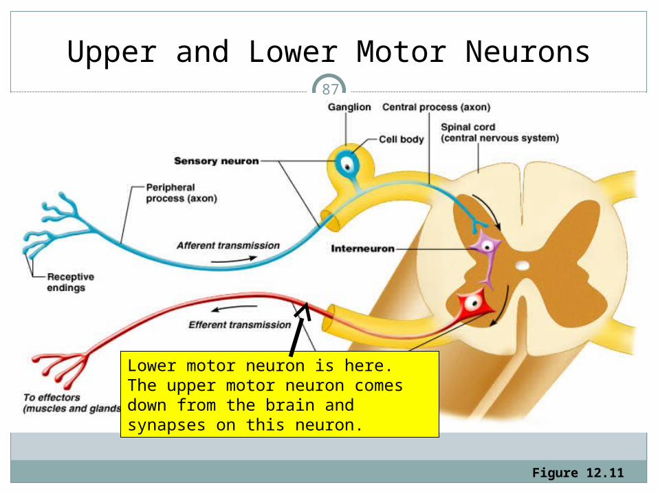
Upper and Lower Motor Neurons
Figure 12.11
Lower motor neuron is here. The upper motor neuron comes down from the brain and synapses on this neuron.
87

PRIMARY MOTOR ASSOCIATION AREA
A. Planning movement: This is when you plan to reach for a new item.
You have not rehearsed it, but you know to extend your forearms, lift, etc.
A signal is sent to the primary motor cortex to turn on specific motor units to do that.
Damage from a stroke= loose function to that area, but you can compensate by using other muscles, and re-learn that movement.
88

PRIMARY MOTOR ASSOCIATION AREA
B. Learned motor skills: these are preprogrammed skills, like when you know how to type or swing a golf club. You practiced it so often, it’s now automatic.
When someone asks you how to spell a word, but you can’t do it until you write it out, it’s because that memory is now a motor skill.
The same happens when you know how to tie your own shoelace or necktie, but can’t easily teach someone else how to do it. It initially is learned by repetition.
Then, to do it later triggers a series of information which turns on those muscles in the right order.
89

Fun Fact
Use your right foot to draw a clockwise circle in the air.
While keeping your foot going, draw the number “6” in the air with your hand.
Did your foot start moving counter-clockwise?
90

Pre-Central Gyrus91

Pre-Central Gyrus
Within the primary motor area of the brain, there is a structure called the pre-central gyrus which contains a precise map of the different body parts.
This map is called a motor homunculus (Latin: little man) All the neurons that innervate the lips would have their cell
bodies in one particular region in this area. All the neurons that innervate the hands have their cell bodies in this area. All those that innervate the back have their cell bodies here.
However, we don’t have as many neurons innervating the back as we do for the lips and hands.
The homunculus is drawn to represent how many neuron cell bodies we have that innervate each region of our body.
92

93

Pre-Central Gyrus
Not all body parts are equally represented by cell density in the motor area in proportion to their size in the body.
Lips, parts of the face and hands are drawn large because there are many cells in the motor area that innervate those regions of the body.
The face region of the homunculus is large representing many neurons, so we can have many facial expressions. The hands and tongue are large, indicating that we have many fine motor skills in those areas as well.
There is also a somatosensory homunculus.
94

Depiction of the motor homunculus
95

Depiction of the sensory homunculus
96

Partial Brain Removal for Boy Suffering Seizures• http://fxn.ws/ZG8SZw• Medication controls epileptic seizures in about
45 percent of patients.
97

• But Spike continued to have 70 seizures a day. He then tried alternative treatments – including the ketogenic diet, which is a low-carb, high-fat diet – so precise, every ingredient has to be measured to a tenth of a gram.
• At just 4 years old, a typical meal for Spike included heavy cream, heavy butter, a tiny piece of meat and a couple of blueberries.
• Spike then had his right frontal lobe removed – the part of the brain responsible for attention span and executive function.
• “We do this type of procedure on 50 to 60 patients a year,” Dr. Lara Jehi, lead author of the study and director of research at the Cleveland Clinic Epilepsy Center.
• Her study looked at 158 patients over a 15 year period, and found that epileptic patients who opt to have frontal lobe surgery have an 80 percent chance of living seizure-free for the rest of their lives.
98

• The part of Spike’s brain causing the seizures was considered dead, and not doing him any good. There was a chance he could have temporary paralysis and/or attention deficit issues, or he might be slower to learn things – but in the end, he would eventually recover all normal function.
• Spike, who is now 6. He can play football or kickbox. These days, he’s a regular little boy who enjoys hiking.
99

FUNCTIONAL REGIONS
A. SENSORY AREASB. MOTOR AREASC. HIGHER FUNCTIONS
For a great video of a neurologist describing what it felt like when she had a stroke:
How it feels to have a stroke: http://www.youtube.com/watch?v=UyyjU8fzEYU
100

HIGHER FUNCTIONS
1. PLANNING AND JUDGMENT2. MEMORY3. EMOTIONS
101

102

If you can read this you have a strong mind:
7H15 M3554G3 53RV35 7O PR0V3 H0W 0UR M1ND5 C4N D0 4M4Z1NG 7H1NG5! 1MPR3551V3 7H1NG5! 1N 7H3 B3G1NN1NG 17 WA5 H4RD BU7 N0W, 0N 7H15 LIN3 Y0UR M1ND 1S R34D1NG 17 4U70M471C4LLY W17H 0U7 3V3N 7H1NK1NG 4B0U7 17, B3 PROUD! 0NLY C3R741N P30PL3 C4N R3AD 7H15.
103

PLANNING AND JUDGMENT
This is coordinated by the frontal lobe: How much time do you need to be ready for the test? This is calculated by the frontal lobe.
Damage to the frontal lobe causes people to become docile and do what they are told.
1930’s when people were overly aggressive, they did a frontal lobotomy by going up the eyelid, crack through the skull, and stirring up the brain. The problem is that it permanently altered their personalities.
Stopped in 1960’s; we do it with drugs now (Ritalin).
104

105

PLANNING AND JUDGMENT
There was a 16 year old rebel who shot himself in the head, but the bullet went too far forward, and he gave himself a lobotomy and his personality improved! Don’t try this at home.
Ritalin suppresses CNS in children, stimulates it in adults, so it is a high theft item.
In a criminal psych ward, an inmate with a lobotomy got his hand caught in the electric door, and while his hand was dangling half off, a nurse asked him if it hurt, and he just calmly said, “Yes, quite a lot.” No emotion.
Remember, when you kill a neuron, it does not regenerate; it’s gone forever.
106

MEMORY: HIPPOCAMPUS
We talked about motor memory. You can also have memory of events.
This is controlled by the HIPPOCAMPUS (“sea horse”; that’s its shape). The hippocampus plays a major role in storing and retrieving memories.
But memories are not stored there or in any other single site in the brain. They are stored throughout the brain, especially in the cerebral cortex.
107

Memory: Hippocampus
Hippocampus
108
Amygdala

109

Amygdala
The amygdala functions to take new memories and store them, making them long-term memories.
One man, who had severe epilepsy, had his amygdala removed. His seizures stopped, but he developed anterograde amnesia and could no longer remember anything new.
http://en.wikipedia.org/wiki/Amygdala
110

Amygdala
The amygdala functions in emotional and social responses.
If all your friends remember an event differently than you do (who held the umbrella that day? They say it was you, but you remember it was Susan), you will tend to change what you believe is right (I guess it must have been me holding the umbrella, since everyone else says it was me).
That is generating a false memory due to social pressure.
111

Amygdala
The amygdala is like a bouncer at a nightclub. It determines what information is important
enough to go into long term storage by allowing it to go through the hippocampus.
The memories that carry emotional weight are allowed in.
These are the memories shape our identity. Social pressure will cause us to change our
memories to conform, so social pressure shapes who we are.
112

Amygdala
When you first experience a traumatic event, if you try to minimize the strong emotions you feel, it will not go into long term memory to continue to haunt you.
For example, if someone does something that bothers you, try to forget it and do not dwell on it, otherwise it will go into long term memory.
If you rehearse the event over and over, you will even start dreaming about it.
The average person spends about 6 years dreaming.
113

Amygdala
Making a new memory is like writing a word on paper with an ink pen. At first, the ink is wet (the memory is fresh) and you can smear it with your thumb so you can’t read the word (don’t dwell on the memory and it will not become a firm memory).
But once the ink is dry (the memory is stored), the memory can be retrieved.
Every time you recall a memory, it is like creating a brand new memory. It is like tracing over the old word in fresh ink. It becomes bolder, stronger, easier to read.
114

Amygdala
When you want to remember something, go over and over it throughout the day, every day.
If it is a painful memory, write it down and read it out loud every day while in a relaxed environment, followed by a logical discussion of the event.
Try to eliminate the emotional distress of the memory during this time.
That will retrain your brain so it does not recall the event as being so emotionally disturbing.
Therefore, it will not cause as many bad dreams.
115

What happens in my brain when I can't recall something I know?
A Neural Network (computer software) is just a simple model of the brain - NN is composed of interconnected neurons with synapses (software model artifacts.)
Each neuron is an adder with a threshold, and each synapse has a weight. Both the threshold and the weight holds a small unit of information (could be digital oranalog.) The entire NN has a certain information capacity, and used wisely (as in VOT (voice to text) or OCR (optical character recognition)) they do quite a job!
However, NN theory (and practice) shows that when this capacity has been not used and retrieved more than 11% of the time while learning, the network starts ' forgetting!'
What you memorize today goes into short term memory and may not be in your memory tomorrow. To get short term memory to convert into long term memory, you need to let your brain know that information is important by retrieving the information frequently. In other words, you need to go over and over the same information several times a day, every day, even after you know it. If you wait a week before you go over it again, some of it will be lost.
Short term memory also flies out of your head under stress, such as during an exam. It is better to study daily rather that do a marathon study session on the weekends.
116

Word of the Day
Hypermnesia: abnormally vivid or complete memory or recall of the past.
Perhaps the most famous individual to exhibit hypermnesia was a Russian man known as "S," whose amazing photographic memory was studied for 30 years by a psychologist in the early part of the 20th century.
"Hypermnesia" sometimes refers to cases like that of "S," but it can also refer to specific instances of heightened memory (such as those brought on by trauma or hypnosis) experienced by people whose memory abilities are unremarkable under ordinary circumstances.
117

Word of the Day
The word "hypermnesia," which has been with us since at least 1882, was created in New Latin as the combination of "hyper-" (meaning "beyond" or "super") and "-mnesia" (to remember).
It ultimately derives from the Greek word "mnasthai," meaning "to remember."
118

Memory
Memory consists of four processes Encoding: during exposure to new thing Consolidation: short-term memory forms; retained for a
few seconds to a few days. The average person can only remember about 7 new things at a time in a few minutes. When new information is presented, old information in short term memory is eliminated. If temporal lobe is damaged, consolidation may not occur and the person only remembers things learned in the last few minutes plus things stored in long-term memory, before the injury.
Storage: long-term memory forms for a few minutes to permanently, depending on how often it is retrieved and used.
Retrieval: using the stored information
119

Memory
To convert short-term memory into long-term memory, you should learn things in a variety of ways: Prepare Listen Write notes Review daily Watch videos Do labs
This allows easy access to that information again by going through the hippocampus.
120

MEMORY
The reason we sleep is to allow our brains to form memories. Anything with a strong emotional attachment will form a
stronger memory during the sleep process. Whatever you are afraid of during the day, you will dream
about more, and remember more. You will have more nightmares if you watch a disturbing TV
show before going to bed. If you have nightmares about your personal life, stop dwelling on those things during the day! Resolve your conflicts while you are awake, and you will sleep better.
The best way to remember what you study is to go over it before going to bed. Study with fear and you will remember it more!
121

122

Mammillary Bodies
A pair of small round bodies at the anterior end of the fornix
Part of the diencephalon; they form part of the limbic system.
They relay information (recognition memory) from the hippocampus. They also add the element of smell to memories.
Damage to the mammillary bodies due to thiamine deficiency (vit B1) or alcohol causes Wernicke-Korsakoff syndrome (anterograde amnesia)
123

Memory: Hippocampus
Hippocampus
124
Amygdala

Fornix
Sheep brain
mammillary body
125

Fornix
mammillary body
126

127

Korsakoff syndrome
Sergei Korsakoff (1854–1900), a Russian neuropsychiatrist ‘Korsakoff syndrome’ refers to a group of symptoms—known as
amnesic syndrome—which includes inattentiveness, memory defect for recent events, retrograde amnesia and other disorders of recall and recognition, and disorientation in time, place, and situation.
The Korsakoff syndrome develops most often in chronic alcoholics who fail to take an adequate diet. This may cause an acute deficiency of thiamine (vitamin B1). When or if the patient recovers he will probably be left with the typical features of the Korsakoff syndrome.
The memory defect is revealed in the difficulty the patient shows in finding his way about, his forgetfulness in simple matters, and especially his failure to retain information.
Also, presented with an object he has been shown a few minutes before, he tends to respond to it as not identical or as in some manner changed.
128

Fornix
Carries signals from the mammillary bodies to the rest of the hippocampus.
129

130

ANTEROGRADE AMNESIA
Damage to the mammillary bodies or hippocampus; they remember things before the injury occurred, but all new information is lost within minutes.
Nemo’s fish friend, Dori, has this type of amnesia.You can get around it by motor memory. Give an
amnesiac a new puzzle; they’ll do it in 30 mins. The next day, they don’t recognize the puzzle, but they do it in 20 mins, the next day in 10. Therefore, they are learning by motor memory. They can learn their route from home to the market by repetition. But they can’t make a detour, and if anything bumps them off track, they’ll be lost.
131

132

RETROGRADE AMNESIA
Retrograde amnesia is a form of amnesia where someone is unable to recall events that occurred before the development of the amnesia.
Retrograde amnesia is caused by trauma that results in brain injury.
Retrograde amnesia is often temporally graded, meaning that remote memories are more easily accessible than events occurring just prior to the trauma.
Events nearest in time to the event that caused memory loss may never be recovered.
They can remember new things.
133

134

FUN FACTS • Bees tell other members of the hive where food is by doing a
special dance that relays distance and direction. Octopus have the ability to sort out complex problems on their own
• A dolphin doesn’t need to see to catch a meal. It sends out sound waves which bounce back and create a 3D ultrasound picture of it, even in complete darkness. A dolphin’s brain in relation to its body is larger than that of a chimpanzee or great ape.
• Dolphins are one of the most intelligent mammals; they've learned to sleep using only half their brains. While one half rests, the other remains alert and continues the respiratory process, without interruptions.
• The brain of a roach is located in its body, and if by accident (natural or human) it should lose its head, it can live up to nine days completely decapitated. It dies from starvation.
135

STROKES
A hemorrhage in the brain (broken blood vessel) or a clot deprives an area of the brain of oxygen.
This is called a stroke.It is one of the most likely causes of amnesia. Amnesia that is caused by a blow to the head
is not cured by a second blow!
136

Another problem with memory:
ALZHEIMER’S DISEASE
Dementia is a symptom, not a disease. Dementia is loss of memory.
Alzheimer’s disease is the most common form of dementia.
About 10% of people over the age of 65 and 50% of people over the age 85 suffer from it.
It is irreversible, incurable, and fatal (6th leading cause of death in the USA, surpassing diabetes). The person dies because they can no longer eat, swallow, etc. There are treatments to delay symptoms.
137

Normal Brain vs. Alzheimer’s138

Alzheimer’s Disease vs. other dementia
Alzheimer's disease is typically a slowly progressive disorder that involves loss of memory for recent information (short-term memory) and one or more other abilities, such as speech and language, personality, decision-making and judgment or awareness and ability to interact with the environment.
Abilities that are typically not impaired in a patient with Alzheimer's disease include memory for information of long ago (long-term memory), vision, ability to feel things and muscle strength.
139

Memory
Even when these memory systems are working well, some memories will be stored or recalled more easily than others.
A memory with a strong emotional component, such as where you were on September 11, 2001, will likely be retained for the rest of your life.
Information is also more likely to be stored properly when it is recognized as important.
140

Memory
New information is also more likely to be retained and recalled if it is related to information that is already stored.
The links between the new and old information serve as retrieval clues.
The more numerous the links and the stronger the associations, the more accessible and clear the memory will be.
However, if the new information is too similar or two different from an existing memory, it may be discarded.
Forging new memories depends in large part on staying interested, active, and alert.
141

142

Learning new things
It’s hard to learn anything brand new; you have to either use repetition or do something to put the new information in your head by associating it with something you are already familiar with.
That’s why mnemonics are good. The word “supinate” was a brand new word, but it sounds like “soup”, and its motion looks like you’re holding a bowl of soup, so it’s easy now to remember.
143

Learning new things
If the word “cerebrum” is a brand new word, it sounds like “Sir read broom”, which are words you already know and can visualize. Think of Harry Potter asking a wizard to read the strange words on his new broom: “Sir read broom”, and the wizard scratches his brain (cerebrum) as he tries to read the words. Now it’s easy to remember because you can relate it to something you already know and can picture.
144

145

IMPLANTED MEMORY
When the Challenger shuttle exploded in the 1980’s, a freshman college professor told his students to write down where they were and what they were doing when they heard about it.
Four years later, he asked them again. 65% answered the same way, but 35% remembered it completely differently, but the students insisted they were right.
146

IMPLANTED MEMORY
Another college professor found all the freshmen students with older siblings at the college, and he told the older siblings to tell this story to their younger siblings:
“When you were 5, we went to a fancy restaurant to celebrate mom’s birthday, and you spilled something on her dress and you were really embarrassed.”
A few weeks later, the professor asked the freshmen to write down a story about anything embarrassing that happened to them when they were five, and to include all the details they remembered.
The freshmen recounted the fake story as though it was real because they thought they remembered it.
They also included details that they were not told, such as the name of the restaurant, the color of the dress, and what was spilled. The freshmen filled in the story to make a complete memory.
147

MEMORY LAPSES
More likely to occur when a person is tired, sick, distracted, or under stress.
People who are depressed are also more likely to have memory problems.
The brain contains about 100 billion neurons. Only a few neurons die over a person's lifetime, but they do shrink. This shrinkage may partially explain why mental functioning slows down in middle and older age.
148

Attention Deficit Disorder149

Memory aids
Place all commonly lost items in a designated spot. Write things down Concentrate and relax Get plenty of sleep Say words out loud: saying” I have turned off the stove”
after you have done so will give you a verbal reminder when you later charged recall whether the stove is still on. Incorporating people’s names into the conversation immediately after you have met them serves the same purpose.
Use memory aids: use a pocket notepad, personal digital assistant, wristwatch alarm, or voice recorder to help you remember what you need to do more to keep track of information.
150

Memory aids
Use visual images: when learning new information, such as a persons name, create a visual image in your mind to make the information more vivid and more memorable. If you have just been introduced to Mr. Hackman, visualize him hacking his way through a dense jungle with a machete.
Group items using mnemonics: when memorizing lists, names, addresses, and so on, alphabetize them or group them into an acronym -- a word made from the first letter of a series of words. You could also use the first letter of each word to create new words to form sentences or phrase. You can use rhymes or create a story that connects each element to be remembered. The more compact or meaningful the mnemonic, the easier it will be to remember the information.
151

EMOTIONS: LIMBIC SYSTEM
The prefrontal lobe and the hippocampus are part of a system of structures in the brain.
The LIMBIC SYSTEM also includes the olfactory nerves (sense of smell). Therefore, memory, emotion, and smell are linked.
Crayolas are created today with the same old waxy scent because it reminds people of their happy times in childhood.
152

The Limbic System(everything in orange)
Figure 13.23153

Limbic System
The limbic system includes the olfactory cortex (sense of smell), and portions of the diencephalon and cerebrum
It influences emotions, motivations, and moodIt initiates responses necessary for survival,
such as hunger and thirst.
154

MENINGES• These are tissues that cover the entire CNS.
They are three layers that serve to protect and cushion the brain.
155

Meninges
1. DURA MATER is the thickest and most superficial of the meninges.
2. ARACHNOID MATER is the middle layer and is not nearly as dense. It also does not go down into the sulci, it only covers over the top of the gyri.
3. PIA MATER is the thin, shiny layer that DOES follow the brain surface into the sulci.
SUBDURAL SPACE is between the dura mater and the arachnoid mater. It has the blue, venous blood.
The SUBARACHNOID SPACE is between the arachnoid and pia mater, and is filled with CEREBRAL SPINAL FLUID (CSF).
156

1. DURA MATER (“Tough mother”)
Dense regular connective tissue. It consists of two layers. Under the skull is the first layer of dura mater, called
the PERIOSTEAL LAYER. Just under this is the second layer, called the MENINGEAL LAYER. There are these two layers everywhere except around the spinal cord, where it’s just one layer, the meningeal layer of the dura mater; no periosteal layer.
Between the meningeal and periosteal layers of the dura mater are DURAL SINUSES, which are filled with venous blood which is drained from the brain.
157

Dural sinus and subarachnoid space
158

Clinical Significance
In the spinal cord, between L3 and L4, a doctor can inject anesthetic above the dura mater, so only the nerves there are affected. What is that called?
The dura and arachnoid mater both have lots of blood vessels, which might rupture in an injury, called a SUBDURAL or SUBARACHNOID HEMORRHAGE, which is potentially fatal. Blood accumulates and squeezes the brain. When the blood clots, it is called a hematoma.
Treatment = drill a hole, preferably before it clots.
Epidural.
159

VENTRICLES OF THE BRAIN
The brain contains hollow spaces called ventricles, which are filled with CSF. They are extensive. The names are simple.
LATERAL VENTRICLE is the largest, extends throughout the cerebrum.
THIRD VENTRICLE: in a sheep, it forms a figure “3” under the fornix and around the corpora quadrigemina. In a human model, it looks like a cavity between the fornix and a red arch.
FOURTH VENTRICLE is at the base of the cerebellum; it is continuous with the central canal of the spinal cord, and also with the subarachnoid space. CEREBRAL AQUEDUCT: connects the 3rd and 4th
ventricles. The used CSF drains out of the brain at the aqueduct and returns to the blood circulation.
The ventricles, subarachnoid space , and cerebral aqueduct are filled with CSF. The subdural space is NOT filled with CSF; it is filled with venous blood.
160

Figure 13.6a, b
VENTRICLES OF THE BRAIN (blue)
161

Fornix
mammillary body
3rd ventricle
162

Fourth ventricle
Cerebral aqueductLateral ventricle
163

Lateral ventricleThird ventricle (forms a number “3”)
Fourth ventricle
Cerebral aqueduct
Sheep brain
164

CerebroSpinal Fluid (CSF)
CSF is similar to plasma because it is derived from plasma.
CSF is made in the ventricles by a group of capillaries called the CHOROID PLEXUS.
The choroid plexus capillaries have holes that allow the blood plasma to leak into the subarachnoid space. It is now called cerebrospinal fluid (CSF).
165

Dural sinus and subarachnoid space166

CerebroSpinal Fluid (CSF)
The CSF that has been depleted of its nutrients is absorbed back into the blood through the ARACHOID GRANULATIONS.
Arachnoid granulations are small protrusions of the arachnoid mater (the thin second layer covering the brain) through the dura mater (the thick outer layer).
They protrude into the venous sinuses of the brain, and allow cerebrospinal fluid (CSF) to enter the brain, and circulate.
800ml of CSF is made per day, but there is actually only 150 ml there because the extra is continually removed from the brain and returned to the bloodstream.
167

FUNCTION OF FLUID-FILLED VENTRICLES
1. Allows the brain to float. The brain has the consistency of Jell-O, and weighs three pounds. Its weight would crush the inferior structures if it didn’t float.
2. It cushions. In sudden movement, like riding a bike into a tree, and hitting the head on the tree, the brain hits inside the skull in the front, and then in recoil it hits the back of the skull = closed head injury, not necessarily with a fracture.
3. Acts as the lymphatic system of the brain (it doesn’t have one).
168

Protection of the Brain – Cerebrospinal Fluid (CSF)
Figure 13.27b169

PROBLEMS WITH MENINGES
HYDROCEPHALY is accumulation of CSF inside the ventricles.
It is usually congenital, caused by a blockage of the cerebral aqueduct. The CSF is made but can’t leave, and the brain gets expanded.
The skull bones in a newborn can expand, so although it CAN damage the brain, it does NOT always cause mental retardation if it is treated early enough. The head becomes enlarged.
Treatment is to put in a tube to drain it.Hydrocephaly in adults can be caused by a tumor,
and since the skull no longer expands, it’s very dangerous.
170

HYDROCEPHALY171

HYDROCEPHALY172

MENINGITIS
Meningitis is inflammation of the meninges. Can be caused from bacteria (can be fatal in 24 hours) or
virus (fatal in a week or more) or fungi (fatal in a week or so). It kills 3 people in 10 who get it.
The main symptom is a headache, so when this occurs in an infant, they can’t say where they hurt.
So when an infant presents with a high fever of 104˚ with no other symptoms, they might test for meningitis, because if they miss it, it’s fatal.
The test is a SPINAL TAP, where a needle is inserted between L4 and L5 because that is below the level of the spinal cord.
They draw the CSF to look at. It it’s cloudy or bloody, it’s usually meningitis. Untreated meningitis can lead to this next one:
173

ENCEPHALITIS
This is inflammation of the brain. It can be caused by mosquito-borne protozoa,
or bacteria. Why is infection of the brain so dangerous?
The swelling crushes the brain. Any injury may lead to brain swelling.Treatment is to remove a piece of the skull
bone to allow the swelling.
174

Luke’s Storyhttp://www.caringbridge.org/visit/lukefallon
Luke was hit in a football game (freshman at high school) and complaining of a severe headache he limped off the field and as soon as they took off his helmet he began seizing, so he was rushed to Long Beach Memorial (he was playing at Los Alamitos HS) and once wheeled into the emergency room the neuro surgeon happened to be in there and one look at Luke and they prepped the OR and when they opened his head up blood shot 15 feet in the air. Come to discover he had an arachnoid cyst which had burst/torn, most likely from the impact of the hit (shows you how brutal football can be). The neuro surgeon thinks the arachnoid cyst was congenital and may have never presented in Luke’s life but for something like this happening to him. Anyways he was in ICU for about a week and then released to the rehab but had developed an infection (not MRSA but something pretty awful) and so back to the OR to remove the infected bone flap and then he was on antibiotic for about 6 weeks (he had a PICC line in so he could go home) but was supposed to wear a helmet until they replaced the bone flap with his prosthetic (but of course he didn’t) and so had his 3rd surgery Dec 30th and then made an amazing recovery, after months of speech, physical and occupational therapy he appears fully recovered.
175

176

177

Luke’s Storyhttp://www.caringbridge.org/visit/lukefallon
I actually think he was suffering from a concussion he had received in practice prior to the game, because he had been complaining of headaches a week prior so much so I took him into the pediatrician but she said to keep an eye on him. Ironically that week he had his physical that is required for playing sports at high school…so 2 doctors visits and nothing uncovered a problem, which goes to show you how much medicine is just luck! Anyway, we are just thankful that he is alive and well…a true miracle child. His blow by blow story can be viewed atwww.caringbridge.org
His name is Luke Fallon and you can see a log of events Have to say the nursing staff in the ICU blew my away…amazing people, completely different level of care than the regular rehab Millers Children’s’ Hospital offered, but that’s as it should be.
178

179

180

181

182

183

184
Meningioma

MRI of Meningioma185

186


MRI of Meningioma, post-op188

Aging
Aging affects the nervous system Decline in sensory functionsDecline in motor functionsDecline in short-term memoryInsomnia
189

Brain Cancer190

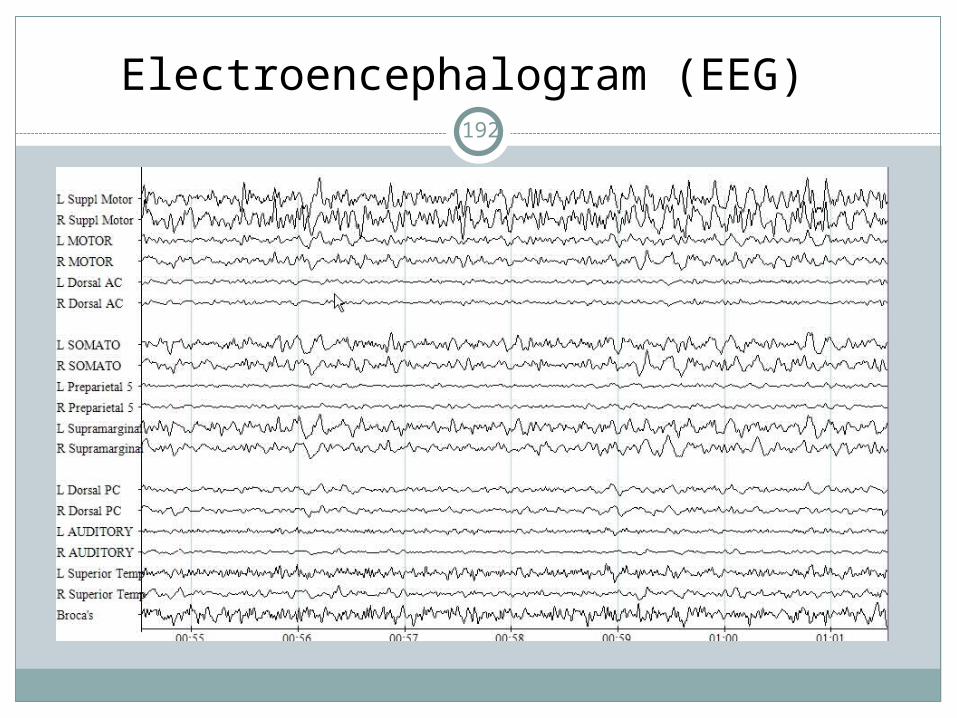
Brain Wave Activity is
recorded on an EEG
Not to be confused with an
EKG, which is for the
heart.
Electroencephalogram (EEG)191
Electroencephalogram (EEG)192

Brain Waves
Types of brain wavesAlpha (active during wakeful relaxation of
closed eyes, such as meditation, prayer). When you pray or meditate for a long time, you feel refreshed!
Beta (active when learning, thinking and concentrating)
Theta (active when just falling asleep)Delta (active during deepest stage of sleep)
193

Human Experiments
We can figure out how the brain works by examining people’s motor and sensory abilities after a head injury, and comparing them to normal.
A brain tumor can sometimes cause epilepsy. If the surgery does not show where the tumor is, the patient needs to be under mild sedation only, so they can probe the area, get feedback from the patient, and see the results. That’s how they can find the tumor.
194

Brain Tumor TherapiesGamma Knife Therapy
The gamma knife and its associated computerized treatment planning software enable physicians to locate and irradiate relatively small targets in the head (mostly inside the brain) with extremely high precision. Intense doses of radiation can be given to the targeted area(s) while largely sparing the surrounding tissues.
The gamma knife is usually unsuitable for targets larger than three or four centimeters in size.
195

Gamma Knife Therapy
The target is placed exactly in the center of approximately 200 precision-aimed, converging beams of (Cobalt-60 generated) gamma radiation.
Treatment takes anywhere from several minutes to a few hours to complete depending on the shape and size of the target and the dose required.
Patients do not feel the radiation. Following treatment the headframe is removed
and the patient may return to normal activity.
196

Gamma Knife Therapy197

Tumor-Starving Therapy: Avastin
Avastin is an innovative tumor-starving therapy designed to block the VEGF (Vascular Endothelial Growth Factor) protein that is produced by normal cells and overproduced by cancer cells, and is needed for cell growth.
Avastin is not chemotherapy and therefore works differently.
VEGF is important for the formation of blood vessels. Tumors rely on blood vessels to get the nutrients and oxygen they need to survive.
198

Paralyzed woman moves robotic arm using thought alone
Robotic Arm Woman (3 mins)
Robotic Arm Man (7mins) https://www.youtube.com/watch?v=QRt8QCx3BCo
199

200
Look at this picture.

201
Stare at this picture and you will see the man turn his face.
Your brain cannot imagine half a face so it will correct the image so his face will appear sideways!

202

How do drugs affect the brain?
AlcoholDrugsNicotine
203

DRUG ABUSE
Many drugs can alter the mood or emotional state, but they also have other side effects.
Drug abuse quickly leads to dependence, which is when a person spends much time thinking about the drug or arranging to get it and they take more of the drug than was intended because they develop tolerance to it and then need more to get the same effect.
They get withdrawal symptoms when they try to stop.
Drug use occurs when people want to avoid dealing with their personal problems and unpleasant emotions.
204

ALCOHOL
Alcohol affects the cerebellum (balance area of the brain)
You can see that area of the brain has been affected by alcohol because you cannot walk a straight line or close your eyes and touch your finger to your nose.
205

ALCOHOL
Alcohol is metabolized (broken down) in the liver, where it disrupts the normal working of the liver so that fats cannot be broken down, and they accumulate.
This fat accumulation, which is the first stage of liver deterioration, begins after only one night of heavy drinking. If the drinking continues, scar tissue appears in the second stage. If the drinking stops, the liver can still recover and become normal again. If not, the final stage, cirrhosis of the liver occurs, and the liver cells die and harden and cannot be repaired.
Alcohol crosses the placenta in pregnant women and causes fetal alcohol syndrome, which is characterized by mental retardation.
206

NICOTINE
This is from tobacco, and it quickly goes into the entire nervous system and is highly addictive. It also increases the heart rate and blood pressure. Withdrawal symptoms include headache, irritability, and insomnia.
Lung cancer has passed breast cancer as a cause of death. Nicotine also causes harm to the fetus. Interestingly, alcohol is the most toxic drug available (more toxic than illegal drugs) and tobacco is the one of the most addictive, yet these two substances are legal.
207

COCAINE
A cocaine binge can go on for days, after which the individual suffers from a crash. The cocaine high is followed by depression because it depletes dopamine. That’s why their mood does not just return to normal.
During the binge, the person has no desire for food or sleep. During the crash, the user is tired, depressed, irritable, and has memory and concentration problems. It usually winds up causing a loss of sex drive and impotence.
Too much can cause seizures and cause the heart to stop beating and the lungs to stop breathing.
Babies born to addicts suffer brain and developmental problems. If someone uses cocaine every day for 30 days, there is a 100% chance of becoming addicted.
208

What to do if someone has a seizure:
http://www.fbhc.org/Patients/Modules/ep_ifsomeone.cfm Roll the person on his or her side to prevent choking on any fluids or vomit. Cushion the person’s head. Loosen any tight clothing around the neck. Keep the person’s airway open. If necessary, grip the person’s jaw gently
and tilt his or her head back. Do NOT restrict the person from moving unless he or she is in danger. Do NOT put anything into the person’s mouth, not even medicine or liquid.
These can cause choking or damage to the person’s jaw, tongue, or teeth. Contrary to widespread belief, people cannot swallow their tongues during a seizure or any other time.
Remove any sharp or solid objects that the person might hit during the seizure.
Note how long the seizure lasts and what symptoms occurred so you can tell a doctor or emergency personnel if necessary.
Stay with the person until the seizure ends.
209

What to do if someone has a seizure:
Call 911 if:The person is pregnant or has diabetes. The seizure happened in water. The seizure lasts longer than 5 minutes. The person does not begin breathing again and return to
consciousness after the seizure stops. Another seizure starts before the person regains consciousness. The person injures himself or herself during the seizure. This is a first seizure or you think it might be. If in doubt, check to see
if the person has a medical identification card or jewelry stating that they have epilepsy or a seizure disorder.
After the seizure ends, the person will probably be groggy and tired. He or she also may have a headache and be confused or embarrassed. Be patient with the person and try to help him or her find a place to rest if he or she is tired or doesn’t feel well. If necessary, offer to call a taxi, a friend, or a relative to help the person get home safely.
210

HEROIN
Side effects include nausea, vomiting, and a decrease in breathing and circulation, which can cause death.
The user becomes so tolerant to it, they have to take more and more of it just to prevent the withdrawal symptoms. These symptoms include sweating, shakes, abdominal cramps, and an increase in heart rate.
Infants born of addicts also suffer these withdrawal symptoms.
211

MARIJUANA
This causes alteration in vision, judgment, and motor coordination.
Causes distortions of space and time. They lack motor coordination, including the
ability to speak in a way that is understandable.
Heavy use causes hallucinations, anxiety, depression, body image distortion, paranoia and loss of sense of reality.
Long term use can lead to brain impairment.
212

Ecstasy
http://www.ecstasy.ws/e-side-effects.htmWhen the drug has worn off, serotonin levels
become severely depleted. This causes the depressive episodes called
Suicide Tuesday because of the midweek depression that users often experience after having used the drug on the weekend.
213

214

For Fun
• Intelligence – Lumosity Brain training gameshttp://www.parade.com/games/lumosity/word-bubble.html
• Food Additives and Children's Hyperactivityhttp://www.time.com/time/magazine/article/0,9171,1661703,00.html
• Cure for epilepsy? New device can reduce, stop seizures
• http://www.foxnews.com/health/2012/11/13/cure-for-epilepsy-new-device-can-reduce-stop-seizures/
• Narcolepsy
215