Brain mechanisms underlying neuropsychiatric symptoms in ...
22
REVIEW Open Access Brain mechanisms underlying neuropsychiatric symptoms in Alzheimer’s disease: a systematic review of symptom- general and –specific lesion patterns Yaojing Chen 1,2† , Mingxi Dang 1,2† and Zhanjun Zhang 1,2* Abstract Neuropsychiatric symptoms (NPSs) are common in patients with Alzheimer’s disease (AD) and are associated with accelerated cognitive impairment and earlier deaths. This review aims to explore the neural pathogenesis of NPSs in AD and its association with the progression of AD. We first provide a literature overview on the onset times of NPSs. Different NPSs occur in different disease stages of AD, but most symptoms appear in the preclinical AD or mild cognitive impairment stage and develop progressively. Next, we describe symptom-general and -specific patterns of brain lesions. Generally, the anterior cingulate cortex is a commonly damaged region across all symptoms, and the prefrontal cortex, especially the orbitofrontal cortex, is also a critical region associated with most NPSs. In contrast, the anterior cingulate-subcortical circuit is specifically related to apathy in AD, the frontal-limbic circuit is related to depression, and the amygdala circuit is related to anxiety. Finally, we elucidate the associations between the NPSs and AD by combining the onset time with the neural basis of NPSs. Keywords: Neuropsychiatric symptoms, Alzheimer’s disease, Neuroimaging, Brain lesion pattern, Brain circuit Background As the worldwide population ages, over 50 million people are now living with dementia, and this number is set to increase to 152 million by 2050 [1]. Dementia has reached epidemic proportions, with major social, med- ical, and economic burdens [2]. The leading cause of de- mentia is Alzheimer’s disease (AD), whose main clinical manifestation is cognitive impairment, but 80% of AD patients also show various behavioural and psychological symptoms, collectively known as neuropsychiatric symp- toms (NPSs) [3]. These symptoms are associated with more rapid progression to severe dementia and an earlier death [4]. They also adversely reduce the quality of life of patients and caregivers [5]. Cross-sectional and longitudinal studies have indicated that different NPSs occur mostly at different stages of AD [6–8]. For example, hallucinations seem to be more common in patients with severe AD, while irritability tends to occur in the early stages of the disease [7]. Even if some NPSs seem to appear together and share some of the same pathological features (for example, both de- pression and apathy are expressed as loss of interest and motivation), they have different pathological mecha- nisms. A clear understanding of the pathological mecha- nisms of differential NPSs is crucial for the early detection and treatment of the disease and the NPSs. Many empirical studies have been conducted to under- stand the neural pathogenesis of NPSs in AD and its role in the progression of AD primarily using neuroimaging © The Author(s). 2021 Open Access This article is licensed under a Creative Commons Attribution 4.0 International License, which permits use, sharing, adaptation, distribution and reproduction in any medium or format, as long as you give appropriate credit to the original author(s) and the source, provide a link to the Creative Commons licence, and indicate if changes were made. The images or other third party material in this article are included in the article's Creative Commons licence, unless indicated otherwise in a credit line to the material. If material is not included in the article's Creative Commons licence and your intended use is not permitted by statutory regulation or exceeds the permitted use, you will need to obtain permission directly from the copyright holder. To view a copy of this licence, visit http://creativecommons.org/licenses/by/4.0/. The Creative Commons Public Domain Dedication waiver (http://creativecommons.org/publicdomain/zero/1.0/) applies to the data made available in this article, unless otherwise stated in a credit line to the data. * Correspondence: [email protected] † Yaojing Chen and Mingxi Dang contributed equally to this work. 1 State Key Laboratory of Cognitive Neuroscience and Learning, Beijing Normal University, Beijing 100875, China 2 BABRI Centre, Beijing Normal University, Beijing 100875, China Chen et al. Molecular Neurodegeneration (2021) 16:38 https://doi.org/10.1186/s13024-021-00456-1
Transcript of Brain mechanisms underlying neuropsychiatric symptoms in ...
REVIEW Open Access
Brain mechanisms underlyingneuropsychiatric symptoms in Alzheimer’sdisease: a systematic review of symptom-general and –specific lesion patternsYaojing Chen1,2†, Mingxi Dang1,2† and Zhanjun Zhang1,2*
Abstract
Neuropsychiatric symptoms (NPSs) are common in patients with Alzheimer’s disease (AD) and are associated withaccelerated cognitive impairment and earlier deaths. This review aims to explore the neural pathogenesis of NPSsin AD and its association with the progression of AD. We first provide a literature overview on the onset times ofNPSs. Different NPSs occur in different disease stages of AD, but most symptoms appear in the preclinical AD ormild cognitive impairment stage and develop progressively. Next, we describe symptom-general and -specificpatterns of brain lesions. Generally, the anterior cingulate cortex is a commonly damaged region across allsymptoms, and the prefrontal cortex, especially the orbitofrontal cortex, is also a critical region associated with mostNPSs. In contrast, the anterior cingulate-subcortical circuit is specifically related to apathy in AD, the frontal-limbiccircuit is related to depression, and the amygdala circuit is related to anxiety. Finally, we elucidate the associationsbetween the NPSs and AD by combining the onset time with the neural basis of NPSs.
Keywords: Neuropsychiatric symptoms, Alzheimer’s disease, Neuroimaging, Brain lesion pattern, Brain circuit
BackgroundAs the worldwide population ages, over 50 millionpeople are now living with dementia, and this number isset to increase to 152 million by 2050 [1]. Dementia hasreached epidemic proportions, with major social, med-ical, and economic burdens [2]. The leading cause of de-mentia is Alzheimer’s disease (AD), whose main clinicalmanifestation is cognitive impairment, but 80% of ADpatients also show various behavioural and psychologicalsymptoms, collectively known as neuropsychiatric symp-toms (NPSs) [3]. These symptoms are associated withmore rapid progression to severe dementia and an
earlier death [4]. They also adversely reduce the qualityof life of patients and caregivers [5].Cross-sectional and longitudinal studies have indicated
that different NPSs occur mostly at different stages ofAD [6–8]. For example, hallucinations seem to be morecommon in patients with severe AD, while irritabilitytends to occur in the early stages of the disease [7]. Evenif some NPSs seem to appear together and share someof the same pathological features (for example, both de-pression and apathy are expressed as loss of interest andmotivation), they have different pathological mecha-nisms. A clear understanding of the pathological mecha-nisms of differential NPSs is crucial for the earlydetection and treatment of the disease and the NPSs.Many empirical studies have been conducted to under-
stand the neural pathogenesis of NPSs in AD and its rolein the progression of AD primarily using neuroimaging
© The Author(s). 2021 Open Access This article is licensed under a Creative Commons Attribution 4.0 International License,which permits use, sharing, adaptation, distribution and reproduction in any medium or format, as long as you giveappropriate credit to the original author(s) and the source, provide a link to the Creative Commons licence, and indicate ifchanges were made. The images or other third party material in this article are included in the article's Creative Commonslicence, unless indicated otherwise in a credit line to the material. If material is not included in the article's Creative Commonslicence and your intended use is not permitted by statutory regulation or exceeds the permitted use, you will need to obtainpermission directly from the copyright holder. To view a copy of this licence, visit http://creativecommons.org/licenses/by/4.0/.The Creative Commons Public Domain Dedication waiver (http://creativecommons.org/publicdomain/zero/1.0/) applies to thedata made available in this article, unless otherwise stated in a credit line to the data.
* Correspondence: [email protected]†Yaojing Chen and Mingxi Dang contributed equally to this work.1State Key Laboratory of Cognitive Neuroscience and Learning, BeijingNormal University, Beijing 100875, China2BABRI Centre, Beijing Normal University, Beijing 100875, China
Chen et al. Molecular Neurodegeneration (2021) 16:38 https://doi.org/10.1186/s13024-021-00456-1

techniques. Yet, there has only been a small, thoughgrowing, number of reviews on this body of literature.Moreover, most of these reviews were merely of qualita-tive with regard to the brain regions associated withNPSs [9–12]. In our current efforts, we attempted toprovide a more comprehensive review focusing on thequantitative aspects of relevant reports available in theliterature. In doing so, we will manage to summarize thenumber of significant associations between NPSs andbrain regions to describe quantitatively symptom-generaland -specific patterns of brain lesions, so that we can de-termine the core damage regions of each symptom, withmore detailed pathological information of NPSs in AD.Accurate assessments of NPSs are the basis of NPSs
pathogenesis neuroimaging research. Although these as-sessments are well-established and available in the NPSsliterature with or without the use of neuroimaging, re-views of NPSs have only briefly summarized these NPSstest [13, 14]. We believe that a more detailed descriptionof these tests is needed, including the applicable popula-tion of the tests, their advantages and disadvantages,among others. More importantly to the study of AD, wecannot ignore the problem of assessing NPSs in AD pa-tients, accurate assessment of whose neurological and be-havioural symptoms is critical and yet compounded withNPSs. Finally, a number of researches have shown that theonset time and the association of NPSs with different cog-nitive domains are variable, better understanding of whichhelps to comprehend the association between NPSs andAD. To the best of our knowledge, such issues have notbeen systematically summarized in review.Overall, the current understanding of the pathological
mechanism of NPSs in patients with AD is limited, espe-cially the relationship between NPSs and AD, so a sys-tematic review is needed to clarify these problems. Wetherefore provide an extensive review to 1) summarizethe clinical assessment, onset time, and association withcognitive impairment of NPSs, and 2) quantitatively de-scribe symptom-general and -specific patterns of brainlesions and brain circuits; and 3) elucidate the associa-tions between the NPSs and AD.
Main textMethodsSearch strategy and selection criteriaWe performed a systematic literature review followingthe guidelines for Preferred Reporting Items for System-atic Reviews and Meta-Analyses (PRISMA, http://www.prisma-statement.org/). Research papers published up toMarch 2020 were identified in the databases PubMedand PsycINFO databases, using the following terms:‘Alzheimer disease’ or ‘mild cognitive impairment’ AND‘neuropsychiatric symptoms’, ‘apathy’, ‘delusions’, ‘de-pression’, ‘agitation’, ‘hallucination’, ‘anxiety’, ‘euphoria’,
‘disinhibition’, ‘irritability’, ‘aberrant motor behavior’,‘sleep disturbances’, ‘appetite disturbances’, or ‘eatingdisorder’. The selection criteria were as follows: (1) Tolimit the heterogeneity, we focused only on AD and ex-cluded studies that included non-AD dementia and non-amnesic MCI; (2) To ensure the reliability of the re-search results, articles with a sample size less than 10were excluded; (3) Articles with subjects younger than50 years of age were excluded.
Study quality assessmentAll included original manuscripts were assessed by twoindependent reviewers to avoid possible bias and report-ing quality using the Joanna Briggs Institute-QualitativeAssessment and Review instrument (JBI-QARI) [15]. Sixpapers were considered low quality and were excluded.Twenty-five papers comparing the pathological mecha-nisms of subjects with and without NPSs, withoutmatching confounding variables such as age and cogni-tive ability, but without methodological problems, wererated as of moderate quality. The rest of the papers wererated as high quality with low bias.Finally, 114 studies were included, including 66 neuro-
imaging studies to explore the pathological mechanismof NPSs and 48 non-imaging studies to summarize clin-ical assessments of NPSs (N = 25), onset times of NPSs(N = 8), and the associations of NPSs with cognitive im-pairment (N = 15). sFigure 1 summarizes the process forstudy selection and inclusion. AD is defined by thestandard diagnostic criteria, primarily NINCDS-ADRDA(approximately 78%), followed by the CDR, DSM-IV,and CERAD. Amnestic MCI (aMCI) is defined byPetersen criteria.
Neuropsychiatric symptoms in Alzheimer’s diseaseIn this section, we will first summarize the existing clin-ical assessment scales of NPSs and discuss the mainproblems these assessments have when used to measureNPSs in AD patients. Then, we will summarize the re-sults on the onset times of NPSs. Final, we willsummarize the report findings on the association be-tween NPSs and the cognitive impairment.
Clinical assessment of neuropsychiatric symptomsWe summarized the scales for measuring neuropsychi-atric and behavioural symptoms, listed the measuredsymptoms, applicable population, and described eachscale in Table 1. Multiple instruments are available forassessing NPSs in AD, but there are several problemsthat need to attention when selecting a measurementscale. First, since NPSs overlap with dementia symptoms,attempts should be made to rule out the effects of cogni-tive impairment on the measures. For example, the Be-havioral Pathology in Alzheimer’s Disease Rating scale
Chen et al. Molecular Neurodegeneration (2021) 16:38 Page 2 of 22

Table 1 Summary of clinical assessment scales for neuropsychiatric symptoms
Measured symptoms Scale Applicablepopulation
Scale description
Delusions, hallucinations, anxiety, agitation, euphoria,disinhibition, irritability, apathy, aberrant motorbehavior, sleep and eating disturbance
NeuropsychiatricInventory
Generallyapplicable
Assessment of broader psychopathology; Collectinformation that may distinguish the different causesof dementia.
Paranoid and delusional ideation, hallucinations,activity disturbances, aggressiveness, diurnal rhythmdisturbances, affective disturbances, anxieties andphobias
BehavioralPathology inAlzheimer’s diseaserating scale
AD Specifically for patients with AD, excluding the effectsof cognitive impairment on measurement.
Anxiety, depression, aberrant motor behavior,delusions and hallucinations, disturbance ofconsciousness
Behavior RatingScale for Dementia
AD Detailed content; High variability and sensitivity
Psychotic disorders, mood disorders, substance usedisorders, anxiety disorders, etc
Diagnostic andStatistical Manual ofMental Disorders
Generallyapplicable
The scale included multidimensional and single-dimensional assessments. There were three self-assessment versions: the adult, the child/adolescent,and the parent/guardian.
Mental health, walking, eating, diurnal rhythm,aggressive behavior, sexual behavior, incontinence,individual behavioral abnormalities
Present BehavioralExamination
Generallyapplicable
Interviews were conducted with primary caregivers forpatients with dementia or other neuropsychiatricdisorders. It assesses behavior over the preceding 4weeks.
Motor, intellectual and emotional functions anddifferent symptoms characteristic for dementia.
Gottfries–Brane–Steen scale
Generallyapplicable
It can measure changes in dementia symptoms over acertain amount of time and evaluate the effect oftreatment.
Apathy (Unidimension) Apathy EvaluationScale
AD/PD/stroke
Three versions of the AES (clinician, informant, and self-rated) were used to evaluate the emotional apathy ofpatients in the past 4 weeks.
Dementia ApathyInterview and Rating
AD Attempt to differentiate limited activity andengagement due to lack of interest from the inabilityor longstanding, premorbid personality traits throughquestion construction, and interview format.
Apathy Inventory AD/PD/MCI It consists of two sets of questionnaires, one forcaregivers and the other for patient-based assessments.Each problem involves frequency and severity.
Apathy (Multidimension) Dimensional ApathyScale
Generallyapplicable
A comprehensive and robust measure ofmultidimensional apathy
Lille Apathy RatingScale
PD The scale is based on a structured interview, including33 items, divided into nine domains. Responses arescored on a dichotomous scale.
Apathy MotivationIndex
Healthypeople
Identified subtypes of apathy in behavioral, social, andemotional domains.
Depression HamiltonDepression Scale
Mild AD Emphasis on patient perception and memory; onlyappropriate for evaluating patients with mild dementia
Cornell Scale forDepression inDementia
AD Accurately distinguish depressive symptoms in ADpatients from their cognitive dysfunction
Depressive SignsScale
Severedementia
Can not assess depressive symptoms in patients mildor moderate dementia.
The Center forEpidemiologicStudies DepressionScale
Generallyapplicable
The scale is a short self-report scale designed to meas-ure depressive symptomatology in the general popula-tion. More emphasis is placed on the individual’semotional physical examination, less on the somaticsymptoms of depression.
Montgomery-AsbergDepression RatingScale
People withdepression
A clinical interview with ten items, each scored on ascale from 0 to 6, particularly sensitive to treatmenteffects.
Geriatric depressionscale
Elderly withdepression
More sensitively examine somatic symptoms specific toolder depressed patients, with 30 core items.
Anxiety Hamilton AnxietyScale
Generallyapplicable
Can not distinguish depression and anxiety well; Theassessment of AD depression lacks specificity
Chen et al. Molecular Neurodegeneration (2021) 16:38 Page 3 of 22

(BEHAVE-AD) is commonly used to assess non-cognitive behavioural disorders in patients with AD [16].Secondly, some scales are only applicable to subjects with acertain level of cognition. For example, the Hamilton depres-sion scale (HAMD) is only used to assess patients with milddementia [17]; while the Depressive Signs Scale (DSS) cannotassess depressive symptoms in patients with mild or moder-ate dementia [18]. In addition, multiple NPSs tend to occursimultaneously (e.g. apathy and depression), and the func-tional relationships among these different NPSs are not clear.It is recommended to adopt a scale that can measure mul-tiple NPSs simultaneously, and all of them should be inde-pendent measurements, such as Neuropsychiatric Inventory(NPI) scale [19]. Finally, the patient and the caregiver mayexaggerate or conceal the severity of the symptoms becauseof the pathological injury and the caregiver’s emotions, re-spectively, so the instrument should be graded based on in-formation from as many sources as possible, such as theDimensional Apathy Scale [20].Therefore, although many scales have been developed to
measure neuropsychiatric and behavioural symptoms,scales that can accurately measure different NPSs in pa-tients with AD are still inadequate or lacking. In practice,we should carefully select appropriate scales according tothe population to be assessed and their needs.
The onset time of neuropsychiatric symptomsTable 2 summarizes the study findings that provideinformation on the onset time of NPSs. For example,one study suggested that the prevalence of delusion
was significantly increased in mild AD compared toaMCI [21]; and another study showed that the preva-lence of aberrant motor behaviours, delusion, halluci-nations and sleep disturbances were significantlyhigher in moderate AD than in mild AD [22]. For thepurpose of this study, the AD continuum was dividedinto four stages as aMCI (or preclinical AD), mildAD, moderate AD, and severe AD, and these findingsare summarized in sTable 1 and Fig. 1. sTable 1shows that several studies have found that the preva-lence of NPSs are significantly higher at a certainstage than at its previous one. Each rise of the curvein Fig. 1 represents at least one study suggesting asignificant increase in the prevalence of the NPSscompared to the previous stage.Most NPSs occur in the preclinical AD or aMCI
phase, including apathy, depression, anxiety, irritability,agitation, sleep disturbances, and abnormal motorbehaviour (Fig. 1, sTable 1). Delusions and eating distur-bances occur in the mild AD phase. Finally, disinhib-ition, hallucinations, and euphoria occur in themoderate AD phase. In addition to disinhibition andappetite changes, the onset of the other NPSs is progres-sive, meaning the prevalence of these symptoms con-tinues increasing as the disease progresses.
Neuropsychiatric symptoms and cognitive dysfunction inADBy collecting and sorting out the literature findings onthe association between NPSs and cognitive decline, we
Table 1 Summary of clinical assessment scales for neuropsychiatric symptoms (Continued)
Measured symptoms Scale Applicablepopulation
Scale description
Worry Scale Milddementia/non-dementiaadults
The Worry Scale is a brief, unidimensional scale withgood reliability and concurrent validity.
Rating Anxiety inDementia
Dementia The items in the scale were rated according to theperson’s symptoms and signs of anxiety over theprevious 2 weeks, including worry, sleep disturbance,irritability, and a number of somatic symptoms
Depression, Anxiety Hospital Anxiety andDepression Scale
Adults It contains two subscales of anxiety and depression,each with seven questions. The psychiatric assessmentfor each patient lasted about 20 min.
Aggressive Rating Scale ForAggressive Behaviorin the Elderly
Elderly Not only to assess patients in nursing homes orhospitals, but also in the community
Cohen-MansfieldAgitation Inventory
Generallyapplicable
The CMAI is a caregivers’ rating questionnaireconsisting of 29 agitated behaviors, each rated on a 7-point scale of frequency. A disruptiveness scale wasadded to later versions.
Sleep disturbance Women’s HealthInitiative InsomniaRating Scale
Women A brief, five-item scale evaluating the frequency and in-tensity of certain sleep difficulties in respondents andrequiring between 2 and 5 min
Abbreviations: AD Alzheimer’s disease, MCI mild cognitive impairment, MMSE Mini-mental State Examination, PD Parkinson’s disease
Chen et al. Molecular Neurodegeneration (2021) 16:38 Page 4 of 22

found that NPSs were closely related to global cognitiveimpairment [27, 28] and activities of daily living decline[29, 30]. In general, different NPSs in patients with ADwere related to specific cognitive impairment (Table 3,sTable 2). Mental symptoms and agitation seem to beassociated with more cognitive domains and more rapidcognitive decline, all accompanied by impaired languageand memory function [28, 29, 31–34]. In addition, delu-sions are associated with decreased executive function,reasoning ability, and conceptualization [31]; hallucina-tions are associated with decreased visuospatial function[33]; while agitation is associated with decreasedexecutive function, visuospatial function, andconceptualization [29, 32, 35]. Apathy is closely relatedto executive function [29, 36–38] and comportment(which stands for Social Behavior) [29, 32]. Depressionare accompanied by a decline in executive [38, 39] andmemory function [39, 40]. Abnormal motor behaviour isassociated with executive and language impairment.Disinhibition and sleep disturbances are related only toexecutive and memory functions, respectively [29, 35,41]. However, we did not find that euphoria, irritabil-ity, or eating disturbances were associated with anyspecific cognitive domains. Hence, executive functionis the most closely related to NPSs among allcognitive domains, implying the executive functiondeficits and some NPSs may stem from commonneurobiological mechanisms.
Table 2 The onset time of neuropsychiatric symptoms: study characteristics
Source Stages Contrast Findings
Craig, 2005[7]
ProbableAD
MMSE: < 10 vs 10–20 vs >20
Depression and apathy were the earliest to appear, and hallucinations, euphoria, and aberrantmotor behavior were the latest symptoms to emerge. Hallucinations were significantly morecommon in severe dementia (MMSE< 10). Irritability was most prevalent in early disease(MMSE> 20).
Cheng, 2012[22]
AD Moderate AD vs mild AD The prevalence of aberrant motor behavior, delusion, hallucination and sleep disturbance wassignificantly higher in moderate AD than in mild AD.
Burns, 1990[23]
AD Severe AD vs moderateAD
The prevalence of aberrant motor behavior and sexual disinhibition was significantly higher insevere AD than in moderate AD.
Hwang, 2004[21]
aMCI/Mild AD
aMCI vs controls; mild ADvs aMCI
There were significant differences in apathy, irritability, anxiety, agitation and abnormal motorbehavior between the aMCI and controls. Delusion was significantly increased in mild ADcompared to aMCI.
Iulio, 2010[24]
aMCI/AD aMCI vs controls; The prevalence of depression, apathy, agitation and irritability was significantly higher in aMCIthan in normal controls.
Ehrenberg,2018 [25]
AD Braak I/II, Braak III/IV, BraakV/VI vs controls
In Braak I/II, significantly increased odds were detected for agitation, anxiety, appetite changes,depression, and sleep disturbances, compared to controls. Increased odds of agitationcontinue into Braak III/IV. Braak V/VI is associated with higher odds for delusions.
Jost, 1996 [6] AD Time order Apathy, depression, sleep disturbance and anxiety appeared before the diagnosis of AD.Irritability and delusions occurred within 5 months after diagnosis; Inappropriate sexualbehavior, wandering, agitation within 5–10 months after diagnosis; Hallucination andaggression appear 10 months after diagnosis.
Linde, 2016[26]
AD Persistence Apathy and abnormal behavior showed high persistence; Irritability, agitation, depression andanxiety showed moderate persistence; Delusions, hallucination, appetite changes, and sleepdisturbance showed short persistence.
Abbreviations: AD Alzheimer’s disease, aMCI amnestic mild cognitive impairment, NPSs neuropsychiatric symptoms, MMSE Mini-mental State Examination
Fig. 1 Schematic diagram of the onset time of NPSs. Diseaseprogression is divided into five stages: healthy stage, preclinical ADor aMCI, mild AD, moderate AD, and severe AD. Each rise of thecurve represents a significant increase in the prevalence of the NPSscompared to the previous stage. Abbreviations: AD, Alzheimer’sdisease; aMCI, amnestic mild cognitive impairment
Chen et al. Molecular Neurodegeneration (2021) 16:38 Page 5 of 22

Neuroimaging findings in neuropsychiatric symptomsWe summarized the neuroimaging findings of NPSs inAD (Table 4), of which approximately two-thirds used theNPI scale to evaluate NPSs and three-quarters used theNICDS-ADRDA criteria for the diagnosis of AD. Based onthe reports we identified for this review, we defined the fre-quency of the lesion regions in the NPS-specific brain lesionpattern, and the high frequency represented that the regionwas most affected by the symptom (Figs. 2, 3, 4 and 5,sFigure 2–5). “Lesion” was defined as a pathologicallesion associated with NPSs, including gray mattervolume atrophy, cortical thinning, decreased whitematter integrity, decreased metabolism, and
increased Aβ deposition. In addition, brain circuitsfor apathy, depression, and anxiety in AD patientswere also reviewed in this section.
Apathy in ADApathy altered anterior cingulate cortex in ADWhite matter studies on apathy consistently show that pa-tients with low fractional anisotropy (FA)—a measurerepresenting white matter integrity and information trans-fer speed—of the anterior cingulate cortex (ACC) aremore likely to present with apathy symptoms [49–51]. Inaddition, FA in the right thalamus and bilateral parietallobes and white matter hyperintensities in the frontal lobe
Table 3 The relationship between NPSs and cognitive dysfunction: study characteristics
Source Participants(Number)
Findings
Senanarong,2005 [29]
AD (N = 73) Clock-drawing test correlated with agitation, apathy, and disinhibition; Verbal Fluency correlated withagitation; Activities of Daily Living and Functional Assessment Questionnaire scores correlated with agitation,apathy, and disinhibition; Comportment predicted total NPI-12 score and apathy; Memory predicted agitation/aggression.
McPHERSON,2002 [36]
AD (N = 80) AD patients with apathy performed significantly worse on tests of executive function (WAIS–R Digit Symbol,Trail-Making, Stroop Color Interference Test) than AD patients without apathy.
Grossi, 2013 [37] AD (N = 32) The apathetic AD had poorer performance than non-apathetic AD on frontal tasks (Inverse Motor Learningtest).
Jeste, 1992 [31] AD (N = 107) Patients with delusions were significantly more impaired than those without delusions on the MMSE, BlessedInformation-Memory-Concentration Test, Dementia Rating Scale (especially its conceptualization and memorysubtests), verbal fluency, modified Wisconsin Card Sorting Test, and the Similarities subtest of the WechslerAdult Intelligence Scale-revised.
Son, 2013 [40] AD (N = 49) Seventeen AD patients with depression versus 32 patients with dementia only showed decreased immediaterecall for a word list and constructional praxis scores.
Scarmeas,2005 [42]
Early AD (N = 456) Delusions and hallucinations predict cognitive (Columbia MMSE score) and functional (Blessed DementiaRating Scale score) decline.
Boyle, 2003 [43] AD (N = 45) Apathy correlated with Activities of Daily Living.
Chen, 1998 [35] AD (N = 31) Deficits in four executive skills tests were significantly associated with the Agitation/Disinhibition factor scoreand total neuropsychiatric score on the Neurobehavioral Rating Scale, as well as the Activities subscore on theBlessed Dementia Scale.
Sultzer, 2014 [34] AD (N = 88) Patients with delusions had lower Dementia Rating Scale memory subscale scores.
Westerberg,2010 [41]
aMCI (N = 10) Inadequate memory consolidation in aMCI patients is related to declines in subjective sleep indices.
Rozum, 2017[32]
Severe dementia(N = 89)
Comportment (Social Behavior) was correlated with Apathy, while conceptualization (Sorting by Color),language (Naming, Comprehension), memory (Remote Recall, Learning), and visuospatial ability (FigureTracing, Drawing) were each correlated with agitation/aggression. Comportment and memory wereassociated with total NPI-12.
Nagata,2010 [44]
AD (N = 75) Aberrant motor behaviors correlated with Frontal Assessment Battery total and the subtest scores (lexicalfluency, conflicting instructions).
Wilson, 2000 [33] AD (N = 410) Compared with AD patients without hallucination, the average annual rate of decline was increased aboutmemory, visuoconstruction, repetition, and naming in those with hallucination.
Lopez, 1991 [28] AD (N = 17) AD patients with delusions and hallucinations had a more rapid rate of decline, as measured by the MMSE, aspecific defect in receptive language, and a greater frequency of aggression and hostility.
Nakaaki, 2008[38]
AD (N = 88) Total Frontal Assessment Battery scores differed significantly between the AD patients with depression/apathyand those without depression/apathy.
Onofrio, 2012[30]
AD (N = 166) A significant association was also found between the impairment of the instrumental activities of daily livingand agitation/aggression, anxiety, aberrant motor activity, depression, apathy, irritability/lability, sleep andeating disturbances in AD.
Abbreviations: AD Alzheimer’s disease, aMCI Amnestic mild cognitive impairment, MMSE Mini-mental State Examination, N number, NPI Neuropsychiatric Inventory,Aβ Amyloid-β
Chen et al. Molecular Neurodegeneration (2021) 16:38 Page 6 of 22

Table 4 Detailed brain changes correlates of neuropsychiatric symptoms in AD
NPS items Neuroimagingmarkers
Author, year Subject(Number)
Diagnostic criteria Scale Structures associated Referencesno.
Apathy Atrophy Apostolova,2007
AD (N = 35) NINCDS-ADRDA NPI Bilateral anterior cingulateand left medial frontal cortex
[45]
Bruen, 2008 Mild AD(N = 31)
NINCDS-ADRDA NPI Anterior cingulate and frontalcortex bilaterally, the head ofthe left caudate nucleus andin bilateral putamen
[46]
Cortical thinning Donovan,2014
HC (N =229), MCI(N = 395),AD (N =188)
NINCDS-ADRDA NPI Inferior temporal region [47]
Apostolova,2007
AD (N = 35) NINCDS-ADRDA NPI Left cingulate [45]
Tunnard,2011
AD (N =111)
NINCDS-ADRDA andDSM- IV criteria
NPI Left caudal anterior cingulatecortex and left lateralorbitofrontal cortex, as well asleft superior and ventrolateralfrontal regions
[48]
FA Tighe, 2012 MCI (N =22), mildAD (N = 23)
NINCDS-ADRDA,CDR
NPI Anterior cingulum [49]
Kim, 2011 Very mildor mildprobableAD (N = 51)
NINCDS-ADRDA NPI Left anterior cingulum [50]
Ota, 2012 AD (N = 21) NINCDS-ADRDA Apathy Scale Right anterior cingulate, rightthalamus, and bilateralparietal regions
[51]
Aβ Mori, 2014 Aβ-positiveAD (N = 28)
NINCDS-ADRDA NPI Bilateral frontal and rightanterior cingulate
[52]
Hypometabolism Marshall, 2007 AD (N = 41) NINCDS-ADRDA NPI Bilateral anterior cingulateregion extending inferiorly tothe medial orbitofrontalregion and the bilateralmedial thalamus
[53]
Holthoff, 2005 AD (N = 53) NINCDS-ADRDA NPI Left orbitofrontal regions [54]
Neurofibrillarytangle
Marshall, 2006 AD (N = 29) CERAD NPI Anterior cingulate [55]
Tekin, 2001 AD (N = 31) CERAD NPI Left anterior cingulate [56]
Depression Atrophy Son, 2013 AD (N = 49) DSM IV-TR criteria GDS Left inferior temporal gyrus [40]
Zahodne,2013
MCI (N =334)
Petersen criteria NPI Anterior cingulate cortex [57]
Morra, 2009 AD (N =100), MCI(N = 200),HC (N =100)
NINCDS-ADRDA,CDR
GDS Right hippocampal [58]
Cortical thickness Lebedev,2014
Mild AD/LBD (N =71)
NINCDS-ADRDA MADRS Prefrontal and temporal areas [59]
Zahodne,2013
MCI (N =334)
Petersen criteria NPI Entorhinal cortex [57]
Lebedeva,2014
AD (N =189)
NINCDS-ADRDA,DSM-IV/ICD-10
CSDD, GDS Left parietal and temporalbrain regions, includingsupramarginal, superior andinferior temporal and fusiformgyri, right posterior cingulateand precuneus
[60]
Chen et al. Molecular Neurodegeneration (2021) 16:38 Page 7 of 22

Table 4 Detailed brain changes correlates of neuropsychiatric symptoms in AD (Continued)
NPS items Neuroimagingmarkers
Author, year Subject(Number)
Diagnostic criteria Scale Structures associated Referencesno.
Gray matterhypodensities
Brommelhoff,2011
AD (N =192)
NINCDS-ADRDA History ofdepression
Caudate nucleus andlentiform nucleus
[61]
White matteratrophy
Lee, 2012 MCI (N =243)
Petersen NPI Frontal, parietal, and temporal [62]
Aβ Chung, 2015 aMCI (N =78)
Petersen GDS/NPI bilateral frontal cortex [63]
Hypometabolism Lee, 2017 MCI (N =36)
Petersen HRSD Right superior frontal gyrus [64]
Hirono, 1998 AD (N = 53) DSM-IV, NINCDS-ADRDA
NPI Bilateral superior frontal andleft anterior cingulate cortices
[65]
Holthoff, 2005 AD (N = 53) NINCDS-ADRDA NPI Dorsolateral prefrontalregions.
[54]
Anxiety Atrophy Poulin, 2011 Very mildand mildAD (N1 =90; N2 =174)
NINCDS-ADRDA NPI Amygdala [66]
Mah, 2015 aMCI (N =376)
Petersen NPI Entorhinal cortical [67]
Tagai, 2014 Mild AD(N = 26)
NINCDS-ADRDA Behave-AD Right precuneus and inferiorparietal lobule
[68]
Nour, 2020 AD (N = 35) NINCDS-ADRDA NPI left parahippocampal,posterior cingulate gyrus, leftinsula and bilateral putamen
[69]
White matterhyperintensities
Berlow, 2010 AD (N = 37) NINCDS-ADRDA NPI – [70]
Bensamoun,2016
HC (N =230), MCI(N = 308),AD (N =119)
NINCDS-ADRDA NPI All diagnostic groups: frontal,cingulate, and global cerebral;MCI subgroup: frontal andglobal cerebral
[71]
Hyperperfusion Tagai, 2014 Mild AD(N = 26)
NINCDS-ADRDA Behave-AD Bilateral anterior cingulatecortices
[68]
Hashimoto,2006
AD (N = 41) NINCDS-ADRDA NPI Bilateral entorhinal cortex.Anterior parahippocampalgyrus, left anterior superiortemporal gyrus and insula
[72]
Delusion Atrophy Serra, 2010 AD (N = 27),aMCI (N =19), HC(N = 23)
NINCDS-ADRDA NPI Right hippocampus [73]
Geroldi, 2002 Mild AD(N = 41)
Standardizedclinical,neuropsychological,and instrumentalevaluation
NPI Left frontal and righttemporal lobe
[74]
Geroldi, 2000 AD (N = 41) Standardizedclinical,neuropsychological,and instrumentalevaluation
NPI Right medial temporal lobe [75]
Cortical thickness Whitehead,2012
AD (N =113)
NINCDS-ADRDA,DSM-IV
NPI Left medial orbitofrontal andleft superior temporal region
[76]
FA Nakaaki, 2013 AD (N = 25) NINCDS-ADRDA NPI Left parieto-occipital region,body of the corpus callosum,superior temporal gyrus
[77]
Chen et al. Molecular Neurodegeneration (2021) 16:38 Page 8 of 22
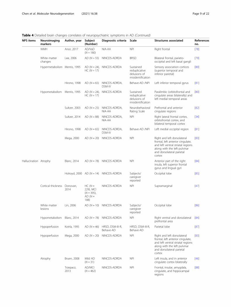
Table 4 Detailed brain changes correlates of neuropsychiatric symptoms in AD (Continued)
NPS items Neuroimagingmarkers
Author, year Subject(Number)
Diagnostic criteria Scale Structures associated Referencesno.
WMH Anor, 2017 AD/VaD(N = 180)
NIA-AA NPI Right frontal [78]
White matterchanges
Lee, 2006 AD (N = 55) NINCDS-ADRDA BRSD Bilateral frontal, parieto-occipital and left basal gangli
[79]
Hypermetabolism Mentis, 1995 AD (N = 24),HC (N = 17)
NINCDS-ADRDA Sustainedreduplicativedelusions ofmisidentification
Sensory association cortices(superior temporal andinferior parietal)
[80]
Hirono, 1998 AD (N = 65) NINCDS-ADRDA,DSM-IV
Behave-AD /NPI Left inferior temporal gyrus [81]
Hypometabolism Mentis, 1995 AD (N = 24),HC (N = 17)
NINCDS-ADRDA Sustainedreduplicativedelusions ofmisidentification
Paralimbic (orbitofrontal andcingulate areas bilaterally) andleft medial temporal areas
[80]
Sultzer, 2003 AD (N = 25) NINCDS-ADRDA,NIA-AA
NeurobehavioralRating Scale
Prefrontal and anteriorcingulate regions
[82]
Sultzer, 2014 AD (N = 88) NINCDS-ADRDA,NIA-AA
NPI Right lateral frontal cortex,orbitofrontal cortex, andbilateral temporal cortex
[34]
Hirono, 1998 AD (N = 65) NINCDS-ADRDA,DSM-IV
Behave-AD /NPI Left medial occipital region [81]
Mega, 2000 AD (N = 20) NINCDS-ADRDA NPI Right and left dorsolateralfrontal, left anterior cingulate,and left ventral striatal regionsalong with the left pulvinarand dorsolateral parietalcortex
[83]
Hallucination Atrophy Blanc, 2014 AD (N = 78) NINCDS-ADRDA NPI Anterior part of the rightinsula, left superior frontalgyrus and lingual gyri
[84]
Holroyd, 2000 AD (N = 14) NINCDS-ADRDA Subjects/caregiverreported
Occipital lobe [85]
Cortical thickness Donovan,2014
HC (N =229), MCI(N = 395),AD (N =188)
NINCDS-ADRDA NPI Supramarginal [47]
White matterlesions
Lin, 2006 AD (N = 10) NINCDS-ADRDA Subjects/caregiverreported
Occipital lobe [86]
Hypometabolism Blanc, 2014 AD (N = 78) NINCDS-ADRDA NPI Right ventral and dorsolateralprefrontal area
[84]
Hypoperfusion Kotrla, 1995 AD (N = 46) HRSD, DSM-III-R,Behave-AD
HRSD, DSM-III-R,Behave-AD
Parietal lobe [87]
Hypoperfusion Mega, 2000 AD (N = 20) NINCDS-ADRDA NPI Right and left dorsolateralfrontal, left anterior cingulate,and left ventral striatal regionsalong with the left pulvinarand dorsolateral parietalcortex
[83]
Atrophy Bruen, 2008 Mild AD(N = 31)
NINCDS-ADRDA NPI Left insula, and in anteriorcingulate cortex bilaterally
[46]
Trzepacz,2013
AD/MCI(N = 462)
NINCDS-ADRDA NPI Frontal, insular, amygdala,cingulate, and hippocampalregions
[88]
Chen et al. Molecular Neurodegeneration (2021) 16:38 Page 9 of 22

Table 4 Detailed brain changes correlates of neuropsychiatric symptoms in AD (Continued)
NPS items Neuroimagingmarkers
Author, year Subject(Number)
Diagnostic criteria Scale Structures associated Referencesno.
Hsu, 2015 AD (N =129), MCI(N = 31)
NIA-AA NPI Posterior cingulate andparieto-occipital sulcus andsulci of the parietal lobes andprecuneus
[89]
FA Tighe, 2012 MCI (N =22), mildAD (N = 23)
NINCDS-ADRDA,CDR
NPI Anterior cingulum [49]
Increasedfunctionalconnectivity
Balthazar,2014
Mild tomoderateAD (N = 20)
NINCDS-ADRDA NPI Anterior cingulate cortex andright insula areas
[90]
Neurofibrillarytangles
Tekin, 2001 AD (N = 31) CERAD NPI Left orbitofrontal cortex andleft anterior cingulate
[56]
Hypometabolism Weissberger,2017
AD (N = 88) NINCDS-ADRDA,NIA-AA
NPI Right temporal, middle, andsuperior gyri, Right calcarinecortex, Right lingual gyrus,Right fusiform gyrus, Rightcuneus, Bilateral cingulate,middle, and posterior
[91]
Irritability Atrophy Poulin, 2011 Very mildand mildAD (N1 =90; N2 =174)
NINCDS-ADRDA NPI Amygdala [66]
FA Tighe, 2012 MCI (N =22), mildAD (N = 23)
NINCDS-ADRDA,CDR
NPI Anterior cingulum [49]
Increasedfunctionalconnectivity
Balthazar,2014
Mild tomoderateAD (N = 20)
NINCDS-ADRDA NPI Anterior cingulate cortex andright insula areas
[90]
Aβ Bensamoun,2016
HC (N =230), MCI(N = 308),AD (N =119)
NINCDS-ADRDA NPI All diagnostic groups: frontal,cingulate, parietal and globalcerebral; AD:parietal
[71]
Hypometabolism Weissberger,2017
AD (N = 88) NINCDS-ADRDA,NIA-AA
NPI Right temporal, middle, andsuperior gyri, Right insula,Right precentral andpostcentral gyri, Right frontal,middle, and inferior
[91]
AberrantMotorBehavior
Atrophy Poulin, 2011 Very mildand mildAD (N1 =90; N2 =174)
NINCDS-ADRDA NPI Amygdala [66]
Increasedfunctionalconnectivity
Balthazar,2014
Mild tomoderateAD (N = 20)
NINCDS-ADRDA NPI Anterior cingulate cortex andright insula areas
[90]
Hypometabolism Meguro, 1997 Moderatelysevere AD(N = 10)
NINCDS-ADRDA Subjects/caregiverreported
Striatum and the frontal andtemporal lobes
[92]
Hypermetabolism Reilly, 2011 AD (N =135)
DSM-IV NPI Orbitofrontal cortex [93]
Neurofibrillarytangles
Tekin, 2001 AD (N = 31) CERAD NPI Left orbitofrontal cortex [56]
Euphoria Increasedfunctionalconnectivity
Balthazar,2014
Mild tomoderateAD (N = 20)
NINCDS-ADRDA NPI Anterior cingulate cortex andright insula areas
[90]
Chen et al. Molecular Neurodegeneration (2021) 16:38 Page 10 of 22

were correlated with apathy [51]. Gray matter studies onapathy showed that gray matter atrophy in the bilateralACC and left medial frontal cortex [45]. Moreover,decreased gray matter density in the bilateral ACC, frontalcortex, head of the left caudate nucleus and bilateralputamen [46], and decreased cortical thickness in theleft caudal ACC, left orbitofrontal cortex (OFC), leftsuperior, and ventrolateral frontal region [48] and in-ferior temporal region [47] were all correlated withthe severity of apathy.
Positron emission tomography (PET) studies have dem-onstrated that the AD patients with apathy had glucosehypometabolism in the ACC, OFC, ventral striatum, andmedial thalamus [53, 54], higher neurofibrillary tanglesburden in the ACC [55, 56], and higher amyloid-β (Aβ)deposition in the bilateral frontal lobe and right ACC [52].
Anterior cingulate circuit lesions cause apathy in ADApathy has been conceptualized as a motivationalbarrier or defect in goal-directed behaviour [101]. As
Table 4 Detailed brain changes correlates of neuropsychiatric symptoms in AD (Continued)
NPS items Neuroimagingmarkers
Author, year Subject(Number)
Diagnostic criteria Scale Structures associated Referencesno.
Disinhibition Atrophy Serra, 2010 AD (N = 27) NINCDS-ADRDA NPI Bilateral cingulate and rightmiddle frontal gyri
[73]
Increasedfunctionalconnectivity
Balthazar,2014
Mild tomoderateAD (N = 20)
NINCDS-ADRDA NPI Anterior cingulate cortex andright insula areas
[90]
Sleepdisturbance
FA Tighe, 2012 MCI (N =22), mildAD (N = 23)
NINCDS-ADRDA,CDR
NPI Anterior cingulum [49]
Hypometabolism Liguori, 2017 AD (N = 18) NIA-AA Polysomnography Hypothalamic [94]
Hyperperfusion Ismail, 2009 AD (N = 55) NINCDS-ADRDA NPI, CSDD Right middle frontal gyrus [95]
Appetitedisturbance
Atrophy Grundman,1996
AD (N = 58),HC (N = 16)
NINCDS-ADRDA Body mass index Mesial temporal cortex [96]
Hypometabolism Hu, 2002 AD (N = 27) NINCDS-ADRDA Body mass index Anterior cingulate cortex [97]
Hypoperfusion Ismail, 2008 AD (N = 66) NINCDS-ADRDA NPI left anterior cingulate and leftorbitofrontal cortices
[98]
Abbreviations: AD Alzheimer’s disease, aMCI amnestic mild cognitive impairment, LBD Lewy body dementia, VaD vascular dementia, HC healthy controls, NPSneuropsychiatric symptom, WMH white matter hyperintensities, GDS Geriatric Depression Scale, MADRS Montgomery-Asberg Depression Rating Scale, HRSDHamilton Rating Scale for Depression, Behave-AD Behavioral Pathology in Alzheimer’s Disease Scale, CDR Clinical Dementia Rating, DSM-IV Diagnostic andStatistical Manual of Mental Disorders, forth version, ICD-10 International Statistical Classification of Disease, tenth version, NIA-AA National Institute on Aging-Alzheimer’s Association workgroups, CERAD Consortium to Establish a Registry for AD
Fig. 2 The brain lesion pattern and anterior cingulate-subcortical circuit of apathy. a The lesion brain region with the highest frequency of apathyis the anterior cingulate cortex (especially on the left), followed by the left medial frontal, medial orbitofrontal, medial thalamus, left lateralorbitofrontal, left superior and ventrolateral frontal regions, as well as the parietal, the head of the left caudate nucleus, putamen, and otherregions of the frontal lobe. b The anterior cingulate-subcortical circuit begins in the anterior cingulate cortex and projects to the ventral striatum,which includes the nucleus accumbens, ventral putamen, ventromedial caudate, and olfactory tubercle. The ventral striatum has circuit linkagesto the ventral pallidum and rostrodorsal substantia nigra. Then the ventral pallidum provides limited input to the mediodorsal thalamus. Theanterior cingulate circuit is closed with projections from the dorsal portion of the magnocellular mediodorsal thalamus to the anterior cingulate.Abbreviations: GP: globus pallidus, SN: substantia nigra. b A visual adaptation of a figure from Nobis et al. [13], with permission
Chen et al. Molecular Neurodegeneration (2021) 16:38 Page 11 of 22

reported, the normal motivational behaviour is re-lated to the anterior cingulate-subcortical circuit[102]. The anterior cingulate circuit, linking the ven-tral striatum to the thalamus via the rostromedialventrolateral globus pallidus interna and ventralpallidum, originates in the ACC and medial OFC
[13, 103, 104]. The disruption of this circuit may becrucially involved in effort-based decision makingand executive functions [13]. In particular, lesions tothe medial OFC and ventral striatum can lead to theinability to connect emotions with ongoing or up-coming behaviour [105].
Fig. 3 The brain lesion patterns and frontal-limbic circuit of depression in AD. a The brain region with the highest frequency of depressionlesions is the superior frontal lobe, followed by the left inferior temporal and other frontal regions, as well as other temporal regions, the anteriorcingulate, entorhinal, right hippocampal, caudate nucleus, lentiform nucleus, fusiform, right posterior cingulate, precuneus, supramarginal andparietal lobe. b The frontal-limbic circuit is composed of a dorsal part dominated by the dorsolateral prefrontal cortex and ventral part dominatedby the subgenual cingulate and inferior temporal cortex. A direct projection from the subgeniual cingulate to the dorsolateral prefrontal cortexand a bidirectional indirect pathway through multiple marginal regions, including the posterior cingulate, hypothalamus, hippocampus, andinsula are delineated. Abbreviations: rACC = rostral anterior cingulate; BG = basal ganglia; Th = thalamus. b A visual adaptation of a figure fromMayberg et al. [99], with permission
Fig. 4 The brain lesion pattern and amygdala circuit of anxiety in AD. a The anterior and posterior cingulate cortex, entorhinal cortex,parahippocampal gyrus and insula cortex are the highest frequency of anxiety lesion regions, and the second is the amygdala, right precuneus,inferior parietal lobule, left anterior superior temporal, putamen, middle cingulate cortex and the frontal lobe. b The afferent arm of the anxietycircuit includes the exteroceptive sensory systems of the brain, which convey the sensory information contained in anxiety-inducing stimuli tothe dorsal thalamus. An exception is the olfactory system, which carries information through the amygdala and entorhinal cortex, not thethalamus. Visceral afferent pathways alter the function of the locus coeruleus and amygdala. The thalamus relays sensory information to theprimary sensory receptive areas of the cortex, which project to adjacent unimodal and polymodal cortical association areas. The corticalassociation areas send projections to the amygdala, entorhinal cortex, orbitofrontal cortex, and cingulate gyrus. The efferent pathways involvingthe amygdala, locus coeruleus, hypothalamus, periaqueductal gray, and striatum mediate autonomic, neuroendocrine, and skeletal-motorresponses are associated with anxiety. Abbreviations: BNST = bed nucleus of the stria terminalis. b A visual adaptation of a figure from Charneyet al. [100], with permission
Chen et al. Molecular Neurodegeneration (2021) 16:38 Page 12 of 22

SummaryApathy is the most common NPS in AD and has beenthe focus of past research on NPSs. Although the re-gions of apathy lesions are not the same in differentstudies, it is consistently shown that apathy is closely re-lated to changes in the structure and function of the
medial frontal cortex and the ACC in AD. Meanwhile,subcortical alterations in the ventral striatum, medialthalamus, and ventral pallidum are also related to ap-athy. The imaging findings in the regions of apathy le-sion supports the association of apathy with anteriorcingulate circuit lesions in AD (Fig. 2, sFigure 2).
Depression in ADCortical and subcortical limbic brain regions abnormalitiesDepression associated gray matter volume atrophy andcortical thinning mainly occur in the frontal and temporallobes, especially in the left dorsolateral prefrontal, rightmedial prefrontal, OFC, ACC, and inferior temporal gyrus[40, 57, 59, 60]. The severity of depression was also associ-ated with gray matter changes in the right hippocampal[58], entorhinal [57], left parietal [60], and striatal [61] re-gions. Similarly, depression causes white matter lesions inthe frontal, parietal, and temporal lobes [62]. Hypometa-bolism in the bilateral superior frontal, left anterior cingu-late, and dorsolateral prefrontal regions have been notedin patients with depression [54, 64, 65]. The presence ofdepression was also associated with the accumulation ofAβ in the frontal lobe in aMCI [63].
Frontal-limbic circuits abnormalities and depression in ADMost research shows that depression is associated withfrontal-striatal and subcortical limbic circuits in AD[106, 107]. Mayberg’s frontal-limbic model of depressioninvolves the dorsal, ventral, and rostral compartments[108]. The disturbances in the dorsal compartment,which includes the dorsolateral prefrontal, dorsal ACC,and posterior cingulate cortex, cause attentional andcognitive disturbances. The ventral compartment, whichconsists of the paralimbic cortical, subcortical, andbrainstem regions, is associated with the vegetative andsomatic symptoms of depression, such as insomnia andloss of appetite. The rostral ACC connects the dorsaland ventral compartments and plays an important regu-latory role in the whole network.Other researchers believe that the hippocampus is the
most common area of structural brain changes in de-pression [58, 109], and is associated with prefrontal cor-tex damage. A hippocampal–prefrontal cortex modelwas proposed, in which the hippocampus—a central partof memory function—regulates mood disorders and cog-nitive dysfunction in depressed patients [110]. Themodel also emphasizes the role of the limbic system,such as the cingulate gyrus and amygdala.
SummaryDepression may precede a cognitive decline in AD [8]and accelerate the rate of cognitive decline [111]. Bothdepression and apathy were associated with structuralbrain changes in the frontal, temporal, and occipital
Fig. 5 The brain lesion patterns of other neuropsychiatric symptomsin AD. The degree of damage to different regions varies withdifferent symptoms, while the anterior cingulate cortex (black box) isan area of common damage for all symptoms and is the mostcommon damaged area for agitation, irritability, disinhibition, andeating disturbances. In addition, delusions are closely associatedwith damage to the orbitofrontal and superior temporal lobes,followed by the occipital and other areas of the frontotemporallobes. Hallucinations are associated with damage to the left superiorfrontal lobe, followed by the occipital, parietal, and dorsolateralprefrontal lobes. Agitation is associated with damage to theposterior cingulate gyrus, followed by the middle cingulate gyrusand insula. Irritability is closely associated with damage to the rightinsula. Disinhibition is also closely associated with damage to theinsula and the middle frontal lobe, cingulate other regions. Aberrantmotor behaviour and eating disturbances mainly affect theorbitofrontal area, and sleep disturbances are also associated withthe right middle frontal gyrus and hypothalamus
Chen et al. Molecular Neurodegeneration (2021) 16:38 Page 13 of 22

lobes, but apathy was more associated with the anteriorcingulate-subcortical circuit, and depression was moreassociated with neuropathology in the frontal-subcorticallimbic circuits in AD (Fig. 3, sFigure 3). The subcorticallimbic system of depression mainly includes the hippo-campus, amygdala, locus ceruleus, substantia nigra, andhypothalamus [106].
Anxiety in ADSubcortical brain region lesions in AD anxietyAn anxiety state predicts a decreased entorhinal corticalvolume [67] and may be associated with amygdala atro-phy [66]. The severity of anxiety was also associated withhyperperfusion of the bilateral ACC, decreased gray mat-ter volume in the right precuneus, inferior parietal, leftparahippocampal, posterior cingulate gyrus, left insula,and bilateral putamen lobes [68, 69], and hypometabo-lism in the bilateral entorhinal, anterior hippocampal,left superior temporal and insula regions [72]. 18F-Flor-betapir-PET studies show the patients with anxiety hadhigher Aβ deposition in the precuneus—posterior cingu-late, frontal, parietal, anterior cingulate cortex and globalcerebral [71]. The locus coeruleus in the hypothalamusis also thought to be the centre of the anxiety-relatednetwork [25], and anxiety cells are enriched in the CA1in the ventral hippocampus [112].
Amygdala-medial prefrontal cortex mediated anxiety in ADAnxiety can be thought of as a set of expected emo-tional, cognitive, and behavioural changes to the uncer-tainty of potential future threats, accompanied by fear[113]. The amygdala plays a pivotal role in the transmis-sion and interpretation of fear and anxiety because it re-ceives extensive afferents from the thalamus andextracortical sensory systems and as a subcortical vis-ceral afferent pathway (Fig. 4) [100]. The neuronal inter-actions between the amygdala and cortical regions, suchas the OFC, provide a framework for the initiation ofcoping behaviors based upon the nature of the threatand prior experiences [114]. Grupe DW and Nitschke JB[115] developed the ‘Uncertainty and AnticipationModel of Anxiety’, which emphasizes that activity in thedorsomedial prefrontal regions and OFC reflects prob-abilistic estimates of future events and expected costs,respectively, and mainly include the amygdala, bed nu-cleus of the stria terminalis (BNST), ventromedial pre-frontal cortex, OFC, anterior mid-cingulate cortex andanterior insula.An alternative network is also proposed. Under this
network, the hippocampus receives convergent, inte-grated inputs from all sensory systems through the pro-jections of the entorhinal cortex [116], and it works withthe entorhinal cortex on situational fear conditioning.Projections from the hippocampus to the BNST and
projections from the BNST to hypothalamic and brain-stem sites may be involved in the expression of context-ual fear conditioning. Theta oscillations within thehippocampus-amygdala-medial prefrontal cortex circuitare associated with anxious behavior [117]. Both this cir-cuit and the ‘Uncertainty and Anticipation Model ofAnxiety’ suggest that the amygdala plays a pivotal role inthe assessment of, and response to, danger.
SummaryAnxiety is more common in individuals with dementiathan in those without dementia [21], and it has been de-scribed as a risk factor for AD [118]. Anxiety is primarilyassociated with damage to the subcortical regions in AD:the amygdala plays an important role in risk assessmentand response, the locus coeruleus plays an importantrole in the efferent response system, and the hypothal-amus plays an important role in the integration of auto-nomic and neuroendocrine responses to threats. Theanterior mid-cingulate cortex is closely linked to thesebrain regions and plays a central role in a series of mal-adaptive responses to uncertainty (Fig. 4, sFigure 4).
Delusions and hallucinations in ADFrontotemporal region lesions in AD delusionDelusions are characterized by asymmetrical brain struc-ture change in the frontal and temporal regions as wellas mainly atrophy in the right temporal lobe and leftfrontal lobe [74, 75, 119]. In addition, delusions are asso-ciated with gray matter change in the right hippocam-pus, left frontal lobe, right frontoparietal cortex, and leftclaustrum [46, 73], and white matter changes in the bi-lateral frontal, parieto-occipital region, left basal ganglia,the body of the corpus callosum and the superior tem-poral gyrus [77–79]. The severity of delusions is associ-ated with hypometabolism in the frontal lobe, especiallyin the right lateral frontal cortex, ACC and OFC [34, 80,82], but the association with metabolism in the temporallobe and occipital lobe is inconsistent. Some studiesshowed delusions had hypometabolism in the bilateraltemporal cortex and the left medial occipital region [34,81], while other studies showed delusions had highermetabolism in the superior temporal, left inferior tem-poral gyrus and inferior parietal lobe [80, 81] or no con-nection [82]. The discrepancies among previous studiesare likely due to sex differences. One study showed thatwomen with delusions had frontotemporal atrophy inAD, but men with delusions did not have brain atrophycompared to men without delusions [76].
Anterior-posterior neural network lesions in ADhallucinationHallucinations are associated with atrophy of the gray mat-ter in the anterior right insula, left superior frontal gyrus,
Chen et al. Molecular Neurodegeneration (2021) 16:38 Page 14 of 22

lingual gyrus and lateral occipital lobes in AD [47, 84], aswell as hypometabolism in the right ventral and dorsolat-eral prefrontal areas [84]. Patients with hallucinations alsohad hypoperfusion in the parietal lobe [87]. The damagedbrain areas associated with hallucinations mainly includethe anterior (e.g., dorsolateral prefrontal area) -posterior(e.g., occipital lobes) neural network and the anterior insula.It is worth noting that these studies do not distinguish be-tween the types of hallucinations. The most common formof hallucinations is visual in AD [120]. Visual hallucinationsare mainly caused by atrophy and white matter lesions inthe occipital lobe [85, 86]. These lesions are related to thedisturbance in the lateral frontal cortex, namely, the ventralvisual stream system [121].
SummaryBoth delusions and hallucinations occur in the middle orlate AD stage, and hallucinations may be more commonin severe dementia [122]. Compared to delusions, only alimited number of studies have looked specifically at hal-lucinations in AD, so researchers seem to focus more ondelusions (despite conflicting evidence and patientsclinging to false beliefs) [123]. The brain lesions of delu-sions mainly occur in the frontotemporal lobe, accom-panied by lateralization and sex differences, while brainlesions of hallucinations mainly occur in the anterior-posterior neural network and anterior insula region(Fig. 5, sFigure 5).
Other neuropsychiatric symptoms in ADHyperactivity syndrome in ADHyperactivity syndrome includes agitation, disinhibition,irritability, euphoria, and aberrant motor behavior [124],which is related to increased functional connectivity inthe anterior cingulate cortex and right insula areas ofthe salience network [90]. Other neuroimaging findingssupport this view, especially for the agitation. Diffusiontensor imaging studies showed that irritability and agita-tion are related to the decreased white matter integrityin the ACC, which is a core component of the saliencenetwork [125]. Magnetic resonance imaging studiesshowed that agitation is related to greater atrophy in thefrontal, cingulate, insular, amygdala, and hippocampalregions [46, 88] while aberrant motor behaviour is re-lated to greater atrophy in the amygdala [66]. These arepredominantly frontolimbic regions and compose manycomponents of the significance network. PET studiesshowed that aberrant motor behaviour is associated withhypometabolism of the striatum and frontotemporallobes and hypermetabolism of the OFC [92, 93]. Inaddition, a study found that the severity of the agitationis correlated with the atrophy score of the posterior tem-poral lobe [89].
Eating disturbance in ADAD patients sometimes suffer from eating disturbancesand weight loss, and nearly half of patients with AD ex-perience appetite changes in the mild stage [126]. A lon-gitudinal study found that patients had acceleratedweight loss as many as 6 years before the diagnosis ofAD [127]. A functional neuroimaging study found hypo-perfusion of the ACC, OFC, and left middle mesial tem-poral cortices can predict appetite disturbances [98].Other neuroimaging studies found weight loss is associ-ated with atrophy of the mesial temporal cortex [96] andhypometabolism of the ACC [97]. In addition, the amyg-dala and OFC affect the internal balance between hungerand satiety and external motivational control of appetite[128]. Therefore, eating disturbances may be related tothe network of the ventral (orbitobasal) frontal cortex,medial temporal cortex, and amygdala in AD.
Sleep disturbances in ADCommon sleep disturbances in AD include fragmenta-tion of sleep at night, decreased duration of sleep atnight, daytime sleepiness, and inversion of the sleep-wake cycle [129]. The relationship between sleep andAD is complex and bidirectional, and the underlyingmechanism is the interaction between sleep and Aβ—sleep disturbances increase the generation of Aβ and de-crease the elimination of Aβ; and once Aβ accumulates,there is increased sleep disturbance [130, 131]. Amyloiddeposition appears to be associated with decreased sleepquality, but not with changes in sleep quantity in thepreclinical stage of AD [132]. Worse sleep quality in-creases the Aβ burden in the precuneus [133], and ashorter sleep quantity at night increases the Aβ burdenin the right hippocampus and thalamus in healthy olderpeoples [134]. In addition, tau pathology as the secondhallmark of AD can also cause sleep disturbances. Sleepregulating areas mainly include the brain stem, thalamus,hypothalamus, midbrain and basal forebrain [135]. Manyof these areas show tau pathology at pretangle stages orstages by Braak staging, before any cortical tau or amyl-oid pathology development [136]. Orexin, as an import-ant sleep-wake regulatory marker, increases in thecerebrospinal fluid in patients with moderate to severeAD and is positively correlated with tau protein levels[137]. Hence, tau pathology may also play an importantrole in sleep disturbances in AD.
SummaryDysfunction of the orbitofrontal–subcortical circuit ischaracterized by personality changes including disin-hibition, agitation, and irritability; this circuit con-nects the frontal monitoring systems to the limbicsystem [104]. Few studies on the relationships be-tween other NPSs and neuroimaging have been
Chen et al. Molecular Neurodegeneration (2021) 16:38 Page 15 of 22

conducted in AD, but they all are associated with alesion in the ACC (Fig. 5, sFigure 5).
Relationship between NPSs and Alzheimer’s diseaseBehavioral and neuropsychiatric symptoms are associ-ated with abnormalities in specific brain regions, such asthe prefrontal and subcortical limbic regions, which dis-rupt the normal balance of neurotransmission. Accord-ing to neuroimmunoregulation theory, this, in turn, isassociated with inflammatory pathways that lead tomicroglial activation and aggregation and the formationof neurofibrillary tangles, ultimately triggering neuronalloss [138]. In addition, NPSs are related to the dysfunc-tion of various neurotransmitter pathways related to AD,including the dopamine system, the serotonin system,and the cholinergic system [139]. In the current section,we will further discuss the molecular and cellularchanges associated with stages of AD progression andtheir relationship to NPSs.
NPSs in preclinical Alzheimer’s diseaseNPSs are variable and sporadic throughout the course ofthe disease, but an important group appears early (Fig. 1),especially emotional symptoms (e.g., depression, anxiety,and apathy), before the clinical diagnosis of cognitive im-pairment [140, 141].The presence of microglial activation and inflamma-
tory signals in patients with AD prior to “clinical diagno-sis” may explain the occurrence of NPSs in the earlystages of the disease. Activation of microglia has beenshown to be associated with deficits in social interaction[142]. Meanwhile, apathy, anxiety, depression, and agita-tion were associated with increased pro-inflammatorycytokines (systemic tumor necrosis factor α) detected inthe serum of AD patients [143, 144]. Similarly, anotherstudy found an association between the levels of diversecytokines present in CSF of patients with dementia, dis-covering that anti-inflammatory interleukin-6 (IL-6)cytokine levels were inversely proportional to anxietyscores in AD patients [145]. Importantly, Ledo et al.found that depressive-like behavior induced by Alzhei-mer’s Aβ oligomers in mice is mediated by inflammationthrough microglial cell activation in the hippocampus,decreasing 5-HT levels in the hippocampus and pre-frontal cortex [146].Depression is also influenced by the reduction of
dopamine and serotonin in the brain, while AD has beenassociated with loss of serotonergic neurons and a de-crease in 5-hydrotryptamine (5-HT) levels in the post-mortem brains with this disease [147, 148]. In a healthybrain, dopamine is constantly released into the hippo-campus, which links emotional feelings with cognitiveprocesses [149, 150]. In AD, a decrease in dopaminelevels coupled with a decrease in serotonin triggers
depression, which is regarded as a prodromal symptomof AD. In addition, late-life depression and AD sharecommon genetic factors, including brain-derived neuro-trophic factor, apolipoprotein E, interleukin-1, andmethylenetetrahydrofolate reductase, while inflammatorypathways are activated in both disorders [151, 152]. Inthis context, the changes produced by late-life depres-sion seem to have an impact on the hippocampus, indu-cing inflammatory events that activate microglia, whichtrigger the overproduction of pro-inflammatory cyto-kines, as described in earlier time about the conceptualscheme of the neuroimmunomodulation theory [138].
NPSs in mild to severe Alzheimer’s diseaseAs AD progresses, most NPSs present in the early stagesof AD become more severe and common, and some psy-chiatric and behavioral symptoms begin to appear. Onehypothesis that has been suggested is that AD progres-sive cholinergic loss (resulting in a loss of inhibition ofthe dopamine system), in the context of a relativelyspared dopaminergic system, may increase the tendencyof AD patients to develop psychosis because of a relativestriatal hyperdopaminergia [153]. Available evidencesuggested that striatal dopamine (D2/D3) receptors areincreased in AD patients with delusional compared withAD patients without delusions [154], and that higherstriatal D2 receptors are associated with wandering be-havior [92].In addition, several studies have shown a correlation be-
tween serotonin deficiency and NPSs. In patients withAD, lower levels of serotonin1A receptors were associatedwith more severe depressive symptoms [155], and lowerconcentrations of serotonin in the temporal cortex wereassociated with hyperactivity and psychosis [148]. The de-pressed AD patients showed larger and more extensive re-ductions in serotonin transporters including the midbrain,nucleus accumbens, and thalamus [156]. Further, thestudy showed glucose metabolism in the right dorsolateralprefrontal cortex was positively correlated with 5-HTtransporter ([11C]-DASB) levels in the striatum in AD pa-tients, suggesting that subcortical serotonergic dysfunctionmay affect cortical function in regions involved in affectiveprocessing such as dorsolateral prefrontal cortex. For ex-ample, prefrontal cortex interactions with the hypothal-amus mediate reward aspects of eating such as foodcravings [157].In the final stage of AD, the pathology of all NPSs be-
comes complicated and difficult to treat. Worseningmental symptoms (delusions and hallucinations) causeconfusion between reality and morbid fantasies, and pa-tients exhibit severe abnormal motor behaviour, oftencharacterized by scratching, which can lead to recurrenthyperfascial skin infections. These destructive NPSsaccelerate the death of AD patients.
Chen et al. Molecular Neurodegeneration (2021) 16:38 Page 16 of 22

SummaryWe believe that there may be two connective mecha-nisms between NPSs and AD: (A) NPSs arise as a conse-quence of AD pathology. AD affects key brain regions ofunderlying behavior, emotion, or mental, so NPSs maybe a direct non-cognitive manifestation of AD neurode-generative disease [158]. AD-related cognitive declinemay also develop into depression, anxiety, or similarNPSs as a psychological response. Other NPSs in theAD stage are caused by AD through reverse causality orpsychological responses, and the onset of NPSs will ag-gravate the pathology of AD. (B) NPSs and AD path-ology arise as a consequence of some shared pathologicprocess. In this case, there is no causal relationship be-tween NPSs and AD pathology, but a third factor, suchas brain vascular disease or white matter change, leadsto the occurrence of AD and NPSs [159, 160].
ConclusionsNPSs almost universal existence in the AD, combinedwith their disabling effects on patients and caregivers, iscontrasted by the fact that few effective and safe treat-ments exist, which is mainly attributed to the followingthree reasons: (1) Lack of reliable and effective measure-ment of NPSs in AD; (2) Biomarkers associated withsymptom-specific in patients with AD have not yet beendeveloped; (3) The relationship between NPS and thepathological mechanism of AD remains unclear.The current review provides a good complement to
these treatment issues. Firstly, we summarized the de-tailed scale information, as well as some possible prob-lems in the NPSs measurement process, which may behelpful for accurate assessment of NPSs. Next, we de-scribed symptom-general and -specific patterns of brainlesions. The anterior cingulate cortex is a commonlydamaged region across all symptoms, and the prefrontalcortex, especially the orbitofrontal cortex, is also a crit-ical region associated with most NPSs. This conclusionwas supported by an intervention study, which foundthat greater reduction in orbitofrontal blood flow hasbeen associated with a greater behavioural response totreatment with donepezil [161].In contrast, the anterior cingulate-subcortical circuit is
specifically related to apathy in AD, the frontal-limbiccircuit to depression, and the amygdala circuit to anx-iety. Understanding symptom-specific brain lesion net-works may help track treatment response for targeteddrug therapy. For example, it is important to understandwhether observed network changes are the result offunctional remodeling of defective networks or reflectthe plasticity of compensatory circuitry complement intreatment development. Finally, we elucidated the twopossible connective mechanisms between NPSs and AD:
etiologic pathways and interactions, and summarized theonset time of NPSs. Different NPSs occur in differentdisease stages of AD, but most symptoms appear in thepreclinical AD or mild cognitive impairment stage anddevelop progressively, which suggested that the criticaltreatment window for NPSs should be advanced to theearly stage of AD.There are still some limitations in the study of the
pathological mechanism of NPSs in AD patients, andmore studies are needed to solve them in the future.Firstly, we found the differences between the subtypes ofeach symptom in exploring the NPS-specific pathologicmechanisms. For example, in addition to the weight losscaused by the loss of appetite mentioned above, AD pa-tients may also experience increased appetite, difficultyswallowing, and other symptoms of eating disturbances.The relationship between dementia stages and eatingdisorders may depend on the type of eating disorder. Forexample, people with mild AD are more likely to experi-ence anorexia, while people with moderate AD have anincreased appetite and changes in food preferences andeating habits, and people with severe AD have difficultyswallowing [126]. We suspect that in AD, the relation-ship between the dementia stage and other NPSs mayalso differ depending on the subtype of symptoms.Therefore, future studies should further explore the rela-tionship between various symptom subtypes and the se-verity of dementia in order to better understand thepathological association.In addition, we should pay attention to the patho-
logical superposition of multiple NPSs. One type ofNPSs is unlikely to appear alone and instead is mostlikely to occur with other types of symptoms in AD[162, 163]. Among patients with dementia, 55% reporttwo or more symptoms, and 44% report three or moresymptoms [3]. However, most studies, including someincluded in the current review, did not adjust the pres-ence of other NPSs when exploring the mechanism of aparticular NPS. This is a major limitation of the currentreview.Some longitudinal studies showed that individuals with
two NPSs had an additional 1.9-fold elevated risk of de-veloping dementia compared with those with zero orone NPS, while those with three or more symptoms hadan additional risk of 3 [118] and significantly higherodds of having functional limitations [164]. Anotherstudy also showed the number of comorbid NPSs, butnot symptom clusters, are associated with an increasedrisk of dementia [165]. These findings suggest that un-derstanding the comorbid pattern of NPSs will help usto further clarify the pathogenesis of NPSs in AD andcontribute to clinical evaluation. However, there are fewneuroimaging studies on comorbid NPSs and futurestudies should focus on this issue.
Chen et al. Molecular Neurodegeneration (2021) 16:38 Page 17 of 22

AbbreviationsAD: Alzheimer’s disease; NPSs: Neuropsychiatric symptoms; CSDD: CornellScale for Depression in Dementia; BEHAVE-AD: The Behavioral Pathology inAlzheimer’s disease rating scale; BRSD: Behavior Rating Scale for Dementia;HAMD: Hamilton depression scale; DSS: Depressive Signs Scale;HAMD: Hamilton Depression Scale; HAMA: Hamilton Anxiety Scale;NPI: Neuropsychiatric Inventory; FA: Fractional anisotropy; ACC: Anteriorcingulate cortex; OFC: Orbitofrontal cortex; PET: Positron emissiontomography; Aβ: Amyloid-β; BNST: Bed nucleus of the stria terminalis;MCI: Mild cognitive impairment; rACC: Rostral anterior cingulate; BG: Basalganglia; Th: Thalamus; IL-6: Interleukin-6
Supplementary InformationThe online version contains supplementary material available at https://doi.org/10.1186/s13024-021-00456-1.
Additional file 1.
AcknowledgementsWe are grateful to Kewei Chen (Banner Alzheimer’s Institute) andChuansheng Chen (University of California, Irvine) for helpful comments onearlier version of this review.
Authors’ contributionsYJC and MXD led the writing of the manuscript, devised all the figures andedited the manuscript. ZJZ supervised the writing and co-edited the manu-script. All authors read and approved the final manuscript.
FundingThis work was supported by National Key Research and Development Projectof China (grant number 2018YFC1315200), National Science Fund forDistinguished Young Scholars (grant number 81625025), Funds forInternational Cooperation and Exchange of the National Natural ScienceFoundation of China (grant number 81820108034), National Natural ScienceFoundation of China (grant number 31700997, and 82071205).
Availability of data and materialsNot applicable.
Declarations
Ethics approval and consent to participateNot applicable.
Consent for publicationNot applicable.
Competing interestsThe authors declared no competing of interests.
Received: 24 March 2021 Accepted: 11 May 2021
References1. International AsD. World Alzheimer report 2019: attitudes to dementia. In:
Book World Alzheimer Report 2019: Attitudes to dementia (Editor ed.^eds.).London: Alzheimer’s Disease Internationals; 2019.
2. Sindi S, Mangialasche F, Kivipelto M. Advances in the prevention ofAlzheimer's disease. F1000prime Rep. 2015;7:50.
3. Lyketsos CG, Lopez O, Jones B, Fitzpatrick AL, Breitner J, DeKosky SJJ.Prevalence of neuropsychiatric symptoms in dementia and mild cognitiveimpairment: results from the cardiovascular health study. Jama. 2002;288:1475–83.
4. Peters ME, Schwartz S, Han D, Rabins PV, Steinberg M, Tschanz JT, et al.Neuropsychiatric symptoms as predictors of progression to severeAlzheimer’s dementia and death: the Cache County Dementia ProgressionStudy. Am J Psychiatr. 2015;172:460–5.
5. Shin I-S, Carter M, Masterman D, Fairbanks L, Cummings JL.Neuropsychiatric symptoms and quality of life in Alzheimer disease. Am J
Geriatr Psychiatry. 2005;13(6):469–74. https://doi.org/10.1097/00019442-200506000-00005.
6. Jost BC, Grossberg GT. The evolution of psychiatric symptoms in Alzheimer’sdisease: a natural history study. J Am Geriatr Soc. 1996;44(9):1078–81.https://doi.org/10.1111/j.1532-5415.1996.tb02942.x.
7. Craig D, Mirakhur A, Hart DJ, McIlroy SP, Passmore AP. A cross-sectionalstudy of neuropsychiatric symptoms in 435 patients with Alzheimer'sdisease. Am J Geriatr Psychiatry. 2005;13(6):460–8. https://doi.org/10.1097/00019442-200506000-00004.
8. Jicha GA, Carr SA. Conceptual evolution in Alzheimer's disease: implicationsfor understanding the clinical phenotype of progressive neurodegenerativedisease. J Alzheimers Dis. 2010;19(1):253–72. https://doi.org/10.3233/JAD-2010-1237.
9. Rosenberg PB, Nowrangi MA, Lyketsos CG. Neuropsychiatric symptoms inAlzheimer's disease: what might be associated brain circuits? Mol Asp Med.2015;43:25–37.
10. Nowrangi MA, Lyketsos CG, Rosenberg PB. Principles and management ofneuropsychiatric symptoms in Alzheimer’s dementia. Alzheimer's Res Ther.2015;7:12.
11. Geda YE, Schneider LS, Gitlin LN, Miller DS, Smith GS, Bell J, et al.Neuropsychiatric symptoms in Alzheimer's disease: past progress andanticipation of the future. Alzheimers Dement. 2013;9(5):602–8. https://doi.org/10.1016/j.jalz.2012.12.001.
12. Tascone LS, Bottino CMC. Neurobiology of neuropsychiatric symptoms inAlzheimer's disease: a critical review with a focus on neuroimaging.Dementia Neuropsychologia. 2013;7:236–43.
13. Nobis L, Husain M. Apathy in Alzheimer's disease. Curr Opin Behav Sci. 2018;22:7–13. https://doi.org/10.1016/j.cobeha.2017.12.007.
14. Seignourel PJ, Kunik ME, Snow L, Wilson N, Stanley M. Anxiety in dementia:a critical review. Clin Psychol Rev. 2008;28(7):1071–82. https://doi.org/10.1016/j.cpr.2008.02.008.
15. Institute JB. Joanna Briggs institute reviewers’ manual: 2014 edition.Adelaide: The Joanna Briggs Institute; 2014.
16. Reisberg B, Auer SR, Monteiro IM. Behavioral pathology in Alzheimer'sdisease (BEHAVE-AD) rating scale. Int Psychogeriatr. 1997;8(S3):301–8.https://doi.org/10.1017/S1041610297003529.
17. Hamilton M. A rating scale for depression. J Neurol, Neurosurg PsychiatryInvestig. 1960;23:56.
18. Katona C, Aldridge C. The dexamethasone suppression test and depressivesigns in dementia. J Affect Disord. 1985;8(1):83–9. https://doi.org/10.1016/0165-0327(85)90076-X.
19. Cummings JL, Mega M, Gray K, Rosenberg-Thompson S, Carusi DA,Gornbein J. The neuropsychiatric inventory: comprehensive assessment ofpsychopathology in dementia. Neurology. 1994;44:2308.
20. Radakovic R, Abrahams S. Developing a new apathy measurement scale:dimensional apathy scale. Psychiatry Res. 2014;219:658–63.
21. Hwang TJ, Masterman DL, Ortiz F, Fairbanks LA, Cummings JL. Mildcognitive impairment is associated with characteristic neuropsychiatricsymptoms. Alzheimer Dis Assoc Disord. 2004;18(1):17–21. https://doi.org/10.1097/00002093-200401000-00004.
22. Cheng S-T, Kwok T, Lam LC. Neuropsychiatric symptom clusters ofAlzheimer's disease in Hong Kong Chinese: prevalence and confirmatoryfactor analysis of the neuropsychiatric inventory. Int Psychogeriatr. 2012;24:1465–73.
23. Burns A, Jacoby R, Levy R. Psychiatric phenomena in Alzheimer's disease. IV:disorders of behaviour. Br J Psychiatry. 1990;157(1):86–94. https://doi.org/10.1192/bjp.157.1.86.
24. Di Iulio F, Palmer K, Blundo C, Casini AR, Gianni W, Caltagirone C, et al.Occurrence of neuropsychiatric symptoms and psychiatric disorders in mildAlzheimer's disease and mild cognitive impairment subtypes. IntPsychogeriatr. 2010;22:629–40.
25. Ehrenberg AJ, Suemoto CK, Resende F, de Paula E, Petersen C, Leite REP,et al. Neuropathologic correlates of psychiatric symptoms in Alzheimer’sdisease. J Alzheimers Dis. 2018;66:115–26.
26. van der Linde RM, Dening T, Stephan BC, Prina AM, Evans E, Brayne C.Longitudinal course of behavioural and psychological symptoms ofdementia: systematic review. Br J Psychiatry. 2016;209(5):366–77. https://doi.org/10.1192/bjp.bp.114.148403.
27. Wadsworth LP, Lorius N, Donovan NJ, Locascio JJ, Rentz DM, Johnson KA,et al. Neuropsychiatric symptoms and global functional impairment along
Chen et al. Molecular Neurodegeneration (2021) 16:38 Page 18 of 22

the Alzheimer’s continuum. Dement Geriatr Cogn Disord. 2012;34(2):96–111.https://doi.org/10.1159/000342119.
28. Lopez OL, Becker JT, Brenner RP, Rosen J, Bajulaiye O, Reynolds C.Alzheimer's disease with delusions and hallucinations: neuropsychologicaland electroencephalographic correlates. Neurology. 1991;41:906.
29. Senanarong V, Poungvarin N, Jamjumras P, Sriboonroung A, Danchaivijit C,Udomphanthuruk S, et al. Neuropsychiatric symptoms, functionalimpairment and executive ability in Thai patients with Alzheimer's disease.Int Psychogeriatr. 2005;17:81–90.
30. D’Onofrio G, Sancarlo D, Panza F, Copetti M, Cascavilla L, Paris F, et al.Neuropsychiatric symptoms and functional status in Alzheimer’s disease andvascular dementia patients. Curr Alzheimer Res. 2012;9:759–71.
31. Jeste DV, Wragg RE, Salmon DP, Harris MJ, Thal LJ. Cognitive deficits ofpatients with Alzheimer's disease with and without delusions. Am JPsychiatry. 1992;149(2):184–9.
32. Rozum W, Cooley B, Richens A, Matyi J, Vernon E, Tschanz J. Specificcognitive/behavioral domains predict neuropsychiatric symptoms in severedementia; 2017.
33. Wilson RS, Gilley DW, Bennett DA, Beckett LA, Evans DA. Hallucinations,delusions, and cognitive decline in Alzheimer's disease. J Neurol,NeurosurgPsychiatry. 2000;69:172–7.
34. Sultzer DL, Leskin LP, Melrose RJ, Harwood DG, Narvaez TA, Ando TK, et al.Neurobiology of delusions, memory, and insight in Alzheimer disease. Am JGeriatr Psychiatry. 2014;22(11):1346–55. https://doi.org/10.1016/j.jagp.2013.06.005.
35. Chen ST, Sultzer DL, Hinkin CH, Mahler ME, Cummings JL. Executivedysfunction in Alzheimer's disease: association with neuropsychiatricsymptoms and functional impairment. J Neuropsychiatry Clin Neurosci.1998;10:426–32.
36. McPHERSON S, Fairbanks L, Tiken S, Cummings JL, Back-Madruga C. Apathyand executive function in Alzheimer's disease. J Int Neuropsychol Soc. 2002;8(3):373–81. https://doi.org/10.1017/S1355617702813182.
37. Grossi D, Santangelo G, Barbarulo AM, Vitale C, Castaldo G, Proto MG, et al.Apathy and related executive syndromes in dementia associated withParkinson’s disease and in Alzheimer’s disease. Behav Neurol. 2013;27:515–22.
38. Nakaaki S, Murata Y, Sato J, Shinagawa Y, Hongo J, Tatsumi H, et al.Association between apathy/depression and executive function in patientswith Alzheimer's disease. Int Psychogeriatr. 2008;20:964–75.
39. Pietrzak RH, Lim YY, Neumeister A, Ames D, Maruff P. Amyloid-β, anxiety,and cognitive decline in preclinical Alzheimer disease: a multicenter,prospective cohort study. JAMA Psychiatry. 2015;72(3):284–91. https://doi.org/10.1001/jamapsychiatry.2014.2476.
40. Son JH, Han DH, Min KJ, Kee BS. Correlation between gray matter volume inthe temporal lobe and depressive symptoms in patients with Alzheimer'sdisease. Neurosci Lett. 2013;548:15–20.
41. Westerberg CE, Lundgren EM, Florczak SM, Mesulam M-M, Weintraub S, ZeePC, et al. Sleep influences the severity of memory disruption in amnesticmild cognitive impairment: results from sleep self-assessment andcontinuous activity monitoring. Alzheimer Dis Assoc Disord. 2010;24(4):325–33. https://doi.org/10.1097/WAD.0b013e3181e30846.
42. Scarmeas N, Brandt J, Albert M, Hadjigeorgiou G, Papadimitriou A, Dubois B,Sarazin M, Devanand D, Honig L, Marder K. Delusions and hallucinations areassociated with worse outcome in Alzheimer disease. J Arch Neurol.. 2005;62:1601-8.
43. Boyle PA, Malloy PF, Salloway S, Cahn-Weiner DA, Cohen R, Cummings JL.Executive dysfunction and apathy predict functional impairment in Alzheimerdisease. Am J Geriatr Psychiatry. 2003;11:214-21.
44. Nagata T, Shinagawa S, Ochiai Y, Kada H, Kasahara H, Nukariya K, NakayamaK: Relationship of frontal lobe dysfunction and aberrant motor behaviors inpatients with Alzheimer's Int Psychogeriatr. 2010;22:463-9.
45. Apostolova LG, Akopyan GG, Partiali N, Steiner CA, Dutton RA, Hayashi KM,et al. Structural correlates of apathy in Alzheimer’s disease. Dement GeriatrCogn Disord. 2007;24:91–7.
46. Bruen PD, McGeown WJ, Shanks MF, Venneri A. Neuroanatomical correlatesof neuropsychiatric symptoms in Alzheimer's disease. Brain. 2008;131(9):2455–63. https://doi.org/10.1093/brain/awn151.
47. Donovan NJ, Wadsworth LP, Lorius N, Locascio JJ, Rentz DM, Johnson KA,et al. Regional cortical thinning predicts worsening apathy andhallucinations across the Alzheimer disease spectrum. Am J GeriatrPsychiatry. 2014;22(11):1168–79. https://doi.org/10.1016/j.jagp.2013.03.006.
48. Tunnard C, Whitehead D, Hurt C, Wahlund L, Mecocci P, Tsolaki M, et al.Apathy and cortical atrophy in Alzheimer's disease. Int J Geriatr Psychiatry.2011;26(7):741–8. https://doi.org/10.1002/gps.2603.
49. Tighe SK, Oishi K, Mori S, Smith GS, Albert M, Lyketsos CG, et al. Diffusiontensor imaging of neuropsychiatric symptoms in mild cognitive impairmentand Alzheimer’s dementia. J Neuropsychiatry Clin Neurosci. 2012;24(4):484–8.https://doi.org/10.1176/appi.neuropsych.11120375.
50. Kim JW, Lee DY, Choo IH, Seo EH, Kim SG, Park SY, et al. Microstructuralalteration of the anterior cingulum is associated with apathy in Alzheimerdisease. Am J Geriatr Psychiatry. 2011;19:644–53.
51. Ota M, Sato N, Nakata Y, Arima K, Uno M. Relationship between apathy anddiffusion tensor imaging metrics of the brain in Alzheimer's disease. Int JGeriatr Psychiatry. 2012;27(7):722–6. https://doi.org/10.1002/gps.2779.
52. Mori T, Shimada H, Shinotoh H, Hirano S, Eguchi Y, Yamada M, et al. Apathycorrelates with prefrontal amyloid β deposition in Alzheimer's disease. JNeurol Neurosurg Psychiatry. 2014;85:449–55.
53. Marshall GA, Monserratt L, Harwood D, Mandelkern M, Cummings JL, SultzerDL. Positron emission tomography metabolic correlates of apathy inAlzheimer disease. Arch Neurol. 2007;64:1015–20.
54. Holthoff VA, Beuthien-Baumann B, Kalbe E, Lüdecke S, Lenz O, Zündorf G,et al. Regional cerebral metabolism in early Alzheimer’s disease withclinically significant apathy or depression. Biol Psychiatry. 2005;57(4):412–21.https://doi.org/10.1016/j.biopsych.2004.11.035.
55. Marshall GA, Fairbanks LA, Tekin S, Vinters HV, Cummings JL.Neuropathologic correlates of apathy in Alzheimer’s disease. Dement GeriatrCogn Disord. 2006;21:144–7.
56. Tekin S, Mega MS, Masterman DM, Chow T, Garakian J, Vinters HV, et al.Orbitofrontal and anterior cingulate cortex neurofibrillary tangle burden isassociated with agitation in Alzheimer disease. Ann Neurol. 2001;49(3):355–61. https://doi.org/10.1002/ana.72.
57. Zahodne LB, Gongvatana A, Cohen RA, Ott BR, Tremont G, Initiative AsDN.Are apathy and depression independently associated with longitudinaltrajectories of cortical atrophy in mild cognitive impairment? Am J GeriatrPsychiatry. 2013;21(11):1098–106. https://doi.org/10.1016/j.jagp.2013.01.043.
58. Morra JH, Tu Z, Apostolova LG, Green AE, Avedissian C, Madsen SK, et al.Automated 3D mapping of hippocampal atrophy and its clinical correlatesin 400 subjects with Alzheimer's disease, mild cognitive impairment, andelderly controls. Hum Brain Mapp. 2009;30:2766–88.
59. Lebedev AV, Beyer MK, Fritze F, Westman E, Ballard C, Aarsland D. Corticalchanges associated with depression and antidepressant use in Alzheimerand Lewy body dementia: an MRI surface-based morphometric study. Am JGeriatr Psychiatry. 2014;22:4–13. e11.
60. Lebedeva A, Westman E, Lebedev AV, Li X, Winblad B, Simmons A, et al.Structural brain changes associated with depressive symptoms in theelderly with Alzheimer’s disease. Neurol Neurosurg Psychiatry. 2014;85(8):930–5. https://doi.org/10.1136/jnnp-2013-307110.
61. Brommelhoff JA, Spann BM, Go JL, Mack WJ, Gatz M. Striatal hypodensities,not white matter hypodensities on CT, are Associated with Late-OnsetDepression in Alzheimer's Disease. J Aging Res. 2011;2011(3):187219.
62. Lee GJ, Lu PH, Hua X, Lee S, Wu S, Nguyen K, et al. Depressive symptoms inmild cognitive impairment predict greater atrophy in Alzheimer's disease-related regions. Biol Psychiatry. 2012;71(9):814–21. https://doi.org/10.1016/j.biopsych.2011.12.024.
63. Chung JK, Plitman E, Nakajima S, Chow TW, Chakravarty MM, Caravaggio F,et al. Lifetime history of depression predicts increased amyloid-βaccumulation in patients with mild cognitive impairment. J Alzheimers Dis.2015;45(3):907–19. https://doi.org/10.3233/JAD-142931.
64. Lee HS, Choo IH, Lee DY, Kim JW, Seo EH, Kim SG, et al. Frontal dysfunctionunderlies depression in mild cognitive impairment: a FDG-PET study.Psychiatry Investig. 2010;7(3):208–14. https://doi.org/10.4306/pi.2010.7.3.208.
65. Hirono N, Mori E, Ishii K, Ikejiri Y, Imamura T, Shimomura T, et al. Frontallobe hypometabolism and depression in Alzheimer's disease. Neurology.1998;50:380–3.
66. Poulin SP, Dautoff R, Morris JC, Barrett LF, Dickerson BC, Initiative AsDN.Amygdala atrophy is prominent in early Alzheimer's disease and relates tosymptom severity. Psychiatry Res Neuroimaging. 2011;194:7–13.
67. Mah L, Binns MA, Steffens DC, Initiative AsDN. Anxiety symptoms inamnestic mild cognitive impairment are associated with medial temporalatrophy and predict conversion to Alzheimer disease. Am J GeriatrPsychiatry. 2015;23:466–76.
Chen et al. Molecular Neurodegeneration (2021) 16:38 Page 19 of 22

68. Tagai K, Nagata T, Shinagawa S, Nemoto K, Inamura K, Tsuno N, et al.Correlation between both morphologic and functional changes and anxietyin Alzheimer's disease. Dement Geriatr Cogn Disord. 2014;38(3-4):153–60.https://doi.org/10.1159/000358822.
69. Nour AEAM, Jiao Y, Teng GJ. Neuroanatomical associations of depression,anxiety and apathy neuropsychiatric symptoms in patients with Alzheimer'sdisease. Acta Neurol Belgica. 2020(5). https://doi.org/10.1007/s13760-020-01349-8.
70. Berlow YA, Wells WM, Ellison JM, Sung YH, Renshaw PF, Harper DG.Neuropsychiatric correlates of white matter hyperintensities in Alzheimer'sdisease. Int J Geriatr Psychiatry. 2010;25(8):780–8. https://doi.org/10.1002/gps.2418.
71. Bensamoun D, Guignard R, Furst AJ, Derreumaux A, Manera V, Darcourt J,et al. Associations between neuropsychiatric symptoms and cerebralamyloid deposition in cognitively impaired elderly people. J Alzheimers Dis.2016;49(2):387–98. https://doi.org/10.3233/JAD-150181.
72. Hashimoto H, Monserratt L, Nguyen P, Feil D, Harwood D, Mandelkern MA,et al. Anxiety and regional cortical glucose metabolism in patients withAlzheimer’s disease. J Neuropsychiatry Clin Neurosci. 2006;18(4):521–8.https://doi.org/10.1176/jnp.2006.18.4.521.
73. Serra L, Perri R, Cercignani M, Spanò B, Fadda L, Marra C, et al. Are thebehavioral symptoms of Alzheimer's disease directly associated withneurodegeneration? J Alzheimers Dis. 2010;21:627–39.
74. Geroldi C, Bresciani L, Zanetti O, Frisoni GB. Regional brain atrophy inpatients with mild Alzheimer's disease and delusions. Int Psychogeriatr.2002;14(4):365–78. https://doi.org/10.1017/S1041610202008566.
75. Geroldi C, Akkawi NM, Galluzzi S, Ubezio M, Binetti G, Zanetti O, et al.Temporal lobe asymmetry in patients with Alzheimer's disease withdelusions. J Neurol Neurosurg Psychiatry. 2000;69:187–91.
76. Whitehead D, Tunnard C, Hurt C, Wahlund L, Mecocci P, Tsolaki M, et al.Frontotemporal atrophy associated with paranoid delusions in women withAlzheimer's disease. Int Psychogeriatr. 2012;24(1):99–107. https://doi.org/10.1017/S1041610211000974.
77. Nakaaki S, Sato J, Torii K, Oka M, Negi A, Nakamae T, et al. Decreased whitematter integrity before the onset of delusions in patients with Alzheimer’sdisease: diffusion tensor imaging. Neuropsychiatr Dis Treat. 2013;9:25.
78. Anor CJ, O'Connor S, Saund A, Tang-Wai DF, Keren R, Tartaglia MC.Neuropsychiatric symptoms in Alzheimer disease, vascular dementia, andmixed dementia. Neurodegener Dis. 2017;17:127–34.
79. Lee DY, Choo IH, Kim KW, Jhoo JH, Youn JC, Lee UY, et al. White matterchanges associated with psychotic symptoms in Alzheimer’s diseasepatients. J Neuropsychiatry Clin Neurosci. 2006;18(2):191–8. https://doi.org/10.1176/jnp.2006.18.2.191.
80. Mentis MJ, Weinstein EA, Horwitz B, McIntosh AR, Pietrini P, Alexander GE,et al. Abnormal brain glucose metabolism in the delusionalmisidentification syndromes: a positron emission tomography study inAlzheimer disease. Biol Psychiatry. 1995;38(7):438–49. https://doi.org/10.1016/0006-3223(94)00326-X.
81. Hirono N, Mori E, Ishii K, Kitagaki H, Sasaki M, Ikejiri Y, et al. Alteration ofregional cerebral glucose utilization with delusions in Alzheimer's disease.J Neuropsychiatry Clin Neurosci. 1998;10:433–9.
82. Sultzer DL, Brown CV, Mandelkern MA, Mahler ME, Mendez MF, Chen ST,et al. Delusional thoughts and regional frontal/temporal cortex metabolismin Alzheimer’s disease. Am J Psychiatr. 2003;160(2):341–9. https://doi.org/10.1176/appi.ajp.160.2.341.
83. Mega MS, Lee L, Dinov ID, Mishkin F, Toga AW, Cummings JL. Cerebralcorrelates of psychotic symptoms in Alzheimer's disease. J Neurol NeurosurgPsychiatry. 2000;69(2):167–71. https://doi.org/10.1136/jnnp.69.2.167.
84. Blanc F, Noblet V, Philippi N, Cretin B, Foucher J, Armspach J-P, et al. Right anteriorinsula: core region of hallucinations in cognitive neurodegenerative diseases. PLoSOne. 2014;9(12):e114774. https://doi.org/10.1371/journal.pone.0114774.
85. Holroyd S, Shepherd ML, Downs JH III. Occipital atrophy is associated withvisual hallucinations in Alzheimer's disease. J Neuropsychiatry Clin Neurosci.2000;12:25–8.
86. Lin S-H, Yu C-Y, Pai M-C. The occipital white matter lesions in Alzheimer'sdisease patients with visual hallucinations. Clin Imaging. 2006;30(6):388–93.https://doi.org/10.1016/j.clinimag.2006.09.025.
87. Kotrla KJ, Chacko RC, Harper RG, Jhingran S, Doody R. SPECT findings onpsychosis in Alzheimer's disease. Am J Psychiatry. 1995;152:1470–5.
88. Trzepacz PT, Yu P, Bhamidipati PK, Willis B, Forrester T, Tabas L, et al.Frontolimbic atrophy is associated with agitation and aggression in mild
cognitive impairment and Alzheimer's disease. Alzheimer's Dement. 2013;9:S95–S104. e101.
89. Hsu J-L, Lee W-J, Liao Y-C, Lirng J-F, Wang S-J, Fuh J-L. Posterior atrophyand medial temporal atrophy scores are associated with different symptomsin patients with Alzheimer’s disease and mild cognitive impairment. PLoSOne. 2015;10(9):e0137121.
90. Balthazar ML, Pereira FR, Lopes TM, da Silva EL, Coan AC, Campos BM, et al.Neuropsychiatric symptoms in Alzheimer's disease are related to functionalconnectivity alterations in the salience network. Hum Brain Mapp. 2014;35(4):1237–46. https://doi.org/10.1002/hbm.22248.
91. Weissberger GH, Melrose RJ, Narvaez TA, Harwood D, Mandelkern MA,Sultzer DL. 18F-Fluorodeoxyglucose positron emission tomography corticalmetabolic activity associated with distinct agitation behaviors in Alzheimerdisease. Am J Geriatr Psychiatry. 2017;25(6):569–79. https://doi.org/10.1016/j.jagp.2017.01.017.
92. Meguro K, Yamaguchi S, Itoh M, Fujiwara T, Yamadori A. Striatal dopaminemetabolism correlated with frontotemporal glucose utilization inAlzheimer's disease: a double-tracer PET study. Neurology. 1997;49:941–5.
93. Reilly TJ, Staff RT, Ahearn TS, Bentham P, Wischik CM, Murray AD. Regionalcerebral blood flow and aberrant motor behaviour in Alzheimer's disease.Behav Brain Res. 2011;222(2):375–9. https://doi.org/10.1016/j.bbr.2011.04.003.
94. Ismail Z, Herrmann N, Francis P, Rothenburg L, Lobaugh N, Leibovitch F,et al. A SPECT study of sleep disturbance and Alzheimer’s disease. DementGeriatr Cogn Disord. 2009;27:254–9.
95. Liguori C, Chiaravalloti A, Nuccetelli M, Izzi F, Sancesario G, Cimini A, et al.Hypothalamic dysfunction is related to sleep impairment and CSFbiomarkers in Alzheimer’s disease. J Neurol. 2017;264(11):2215–23. https://doi.org/10.1007/s00415-017-8613-x.
96. Grundman M, Corey-Bloom J, Jernigan T, Archibald S, Thal L. Low bodyweight in Alzheimer's disease is associated with mesial temporal cortexatrophy. Neurology. 1996;46:1585–91.
97. Hu X, Okamura N, Arai H, Higuchi M, Maruyama M, Itoh M, et al.Neuroanatomical correlates of low body weight in Alzheimer's disease: aPET study. Prog Neuro-Psychopharmacol Biol Psychiatry. 2002;26:1285–9.
98. Ismail Z, Herrmann N, Rothenburg LS, Cotter A, Leibovitch FS, Rafi-Tari S,et al. A functional neuroimaging study of appetite loss in Alzheimer’sdisease. J Neurol Sci. 2008;271:97–103.
99. Mayberg HS, Liotti M, Brannan SK, McGinnis S, Mahurin RK, Jerabek PA, et al.Reciprocal limbic-cortical function and negative mood: converging PETfindings in depression and normal sadness. Am J Psychiatr. 1999;156:675–82.
100. Charney DS, Grillon C, Bremner JD. The neurobiological basis of anxiety andfear: circuits, mechanisms, and neurochemical interactions (part I).Neuroscientist. 1998;4(1):35–44. https://doi.org/10.1177/107385849800400111.
101. Levy R, Dubois B. Apathy and the functional anatomy of the prefrontalcortex–basal ganglia circuits. Cereb Cortex. 2006;16:916–28.
102. Bonelli RM, Cummings JL. Frontal-subcortical circuitry and behavior.Dialogues Clin Neurosci. 2007;9:141.
103. Le Heron C, Apps MAJ, Husain M. The anatomy of apathy: a neurocognitiveframework for amotivated behaviour. Neuropsychologia. 2017;118:54-67.
104. Tekin S, Cummings JL. Frontal–subcortical neuronal circuits and clinicalneuropsychiatry: an update. J Psychosom Res. 2002;53(2):647–54. https://doi.org/10.1016/S0022-3999(02)00428-2.
105. Levy R. Dubois BJCc: apathy and the functional anatomy of the prefrontalcortex–basal ganglia circuits. Cereb Cortex. 2006;16:916–28.
106. Tagariello P, Girardi P, Amore M. Depression and apathy in dementia: samesyndrome or different constructs? A critical review. Arch Gerontol Geriatr.2009;49:246–9.
107. Mortby ME, Maercker A, Forstmeier S. Apathy: a separate syndrome fromdepression in dementia? A critical review. Aging Clin Exp Res. 2012;24:305–16.
108. Deckersbach T, Dougherty DD, Rauch SL. Functional imaging of mood andanxiety disorders. J Neuroimaging. 2006;16(1):1–10. https://doi.org/10.1177/1051228405001474.
109. Campbell S, MacQueen G. The role of the hippocampus in thepathophysiology of major depression. J Psychiatry Neurosci. 2004;29(6):417–26.
110. Sampath D, Sathyanesan M, Newton SS. Cognitive dysfunction in majordepression and Alzheimer’s disease is associated with hippocampal–prefrontal cortex dysconnectivity. Neuropsychiatr Dis Treat. 2017;13:1509.
111. Rapp MA, Schnaider-Beeri M, Wysocki M, Guerrero-Berroa E, Grossman HT,Heinz A, et al. Cognitive decline in patients with dementia as a function ofdepression. Am J Geriatr Psychiatry. 2011;19(4):357–63. https://doi.org/10.1097/JGP.0b013e3181e898d0.
Chen et al. Molecular Neurodegeneration (2021) 16:38 Page 20 of 22

112. Jimenez JC, Su K, Goldberg AR, Luna VM, Biane JS, Ordek G, et al. Anxietycells in a hippocampal-hypothalamic circuit. Neuron. 2018;97:670–683. e676.
113. Tuma A H , Maser J . Anxiety and the anxiety disorders. Int ClinPsychopharmacol. 1990;5(1):75.
114. Rempel-Clower NL. Role of orbitofrontal cortex connections in emotion.Ann N Y Acad Sci. 2007;1121:72–86.
115. Grupe DW, Nitschke JB. Uncertainty and anticipation in anxiety: an integratedneurobiological and psychological perspective. Nat Rev Neurosci. 2013;14:488–501.
116. ten Donkelaar HJ. The Limbic System. In Clinical Neuroanatomy: BrainCircuitry and Its Disorders. Berlin: Springer Berlin Heidelberg; 2011:633-710.https://doi.org/10.1007/978-3-642-19134-3_14.
117. Jacinto LR, Cerqueira JJ, Sousa N. Patterns of theta activity in limbic anxietycircuit preceding exploratory behavior in approach-avoidance conflict. FrontBehav Neurosci. 2016;10:171.
118. Acosta I, Borges G, Aguirre-Hernandez R, Sosa AL, Prince M, Group DR.Neuropsychiatric symptoms as risk factors of dementia in a Mexicanpopulation: a 10/66 Dementia Research Group Study. Alzheimers Dement.2018;14:271–9.
119. Rossi R, Geroldi C, Bresciani L, Zanetti O, Frisoni G. Delusions in ad areassociated with less severe brain structural changes. Res Pract Alzheimer'sDis. 2003;7:168–73.
120. Jeste DV, Finkel SI. Psychosis of Alzheimer's disease and related dementias:diagnostic criteria for a distinct syndrome. Am J Geriatr Psychiatry. 2000;8:29–34.
121. Collerton D, Perry E, McKeith I. Why people see things that are not there: anovel perception and attention deficit model for recurrent complex visualhallucinations. Behav Brain Sci. 2005;28:737–57.
122. Bassiony MM, Lyketsos CG. Delusions and hallucinations in Alzheimer’sdisease: review of the brain decade. Psychosomatics. 2003;44(5):388–401.https://doi.org/10.1176/appi.psy.44.5.388.
123. Reeves SJ, Gould RL, Powell JF, Howard RJ. Origins of delusions inAlzheimer's disease. Neurosci Biobehav Rev. 2012;36:2274–87.
124. Aalten P, Verhey FR, Boziki M, Bullock R, Byrne EJ, Camus V, et al.Neuropsychiatric syndromes in dementia. Dement Geriatr Cogn Disord.2007;24(6):457–63. https://doi.org/10.1159/000110738.
125. Tighe SK, Oishi K, Mori S, Smith GS, Albert M, Lyketsos CG, et al. Diffusiontensor imaging of neuropsychiatric symptoms in mild cognitive impairmentand Alzheimer’s dementia. J Neuropsychiatry Clin Neurosci. 2012;24:484–8.
126. Kai K, Hashimoto M, Amano K, Tanaka H, Fukuhara R, Ikeda M. Relationshipbetween eating disturbance and dementia severity in patients withAlzheimer’s disease. PLoS One. 2015;10(8):e0133666.
127. Gao S, Nguyen JT, Hendrie HC, Unverzagt FW, Hake A, Smith-Gamble V,et al. Accelerated weight loss and incident dementia in an elderly African-American cohort. J Am Geriatr Soc. 2011;59(1):18–25. https://doi.org/10.1111/j.1532-5415.2010.03169.x.
128. Hinton EC, Parkinson JA, Holland AJ, Arana FS, C. Roberts A, Owen AM.Neural contributions to the motivational control of appetite in humans. EurJ Neurosci. 2004;20:1411–8.
129. Peter-Derex L, Yammine P, Bastuji H, Croisile B. Sleep and Alzheimer'sdisease. Sleep Med Rev. 2015;19:29–38.
130. Ju Y-ES, Lucey BP, Holtzman DM. Sleep and Alzheimer diseasepathology—a bidirectional relationship. Nat Rev Neurol. 2014;10:115.
131. Holth JK, Patel TK, Holtzman DM. Sleep in Alzheimer's disease–beyondamyloid. Neurobiol Sleep Circadian Rhythms. 2017;2:4–14.
132. Ju Y-ES, McLeland JS, Toedebusch CD, Xiong C, Fagan AM, Duntley SP, et al.Sleep quality and preclinical Alzheimer disease. JAMA Neurol. 2013;70(5):587–93. https://doi.org/10.1001/jamaneurol.2013.2334.
133. Spira AP, Gamaldo AA, An Y, Wu MN, Simonsick EM, Bilgel M, et al. Self-reported sleep and β-amyloid deposition in community-dwelling olderadults. JAMA Neurol. 2013;70:1537–43.
134. Shokri-Kojori E, Wang G-J, Wiers CE, Demiral SB, Guo M, Kim SW, et al. β-amyloid accumulation in the human brain after one night of sleepdeprivation. Proc Natl Acad Sci. 2018;115(17):4483–8. https://doi.org/10.1073/pnas.1721694115.
135. Saper CB, Scammell TE, Lu J. Hypothalamic regulation of sleep and circadianrhythms. Nature. 2005;437:1257–63.
136. Braak H, Thal DR, Ghebremedhin E, Del Tredici K. Stages of the pathologicprocess in Alzheimer disease: age categories from 1 to 100 years. JNeuropathol Exp Neurol. 2011;70:960–9.
137. Liguori C, Romigi A, Nuccetelli M, Zannino S, Sancesario G, Martorana A,et al. Orexinergic system dysregulation, sleep impairment, and cognitive
decline in Alzheimer disease. JAMA Neurol. 2014;71(12):1498–505. https://doi.org/10.1001/jamaneurol.2014.2510.
138. Maccioni RB, Rojo LE, Fernandez JA, Kuljis RO. The role ofneuroimmunomodulation in Alzheimer's disease. Ann N Y Acad Sci. 2009;1153(1):240–6. https://doi.org/10.1111/j.1749-6632.2008.03972.x.
139. Tonkonogy J. The American Psychiatric Publishing textbook of Alzheimerdisease and other dementias. Psychiatr Serv. 2010;61(2):206–7. https://doi.org/10.1176/ps.2010.61.2.206a.
140. Masters MC, Morris JC, Roe CM. “Noncognitive” symptoms of earlyAlzheimer disease: a longitudinal analysis. Neurology. 2015;84:617–22.
141. Ballard CG, Gauthier S, Cummings JL, Brodaty H, Grossberg GT, Robert P,et al. Management of agitation and aggression associated with Alzheimer disease.Nat Rev Neurol. 2009;5(5):245–55. https://doi.org/10.1038/nrneurol.2009.39.
142. Zhan Y, Paolicelli RC, Sforazzini F, Weinhard L, Bolasco G, Pagani F, et al.Deficient neuron-microglia signaling results in impaired functional brainconnectivity and social behavior. Nat Neurosci. 2014;17(3):400–6. https://doi.org/10.1038/nn.3641.
143. Lasselin J, Elsenbruch S, Lekander M, Axelsson J, Benson S. Mood disturbanceduring experimental endotoxemia: predictors of state anxiety as a psychologicalcomponent of sickness behavior. Brain Behav Immun. 2016;57:30–7.
144. Holmes C, Cunningham C, Zotova E, Culliford D, Perry VH. Proinflammatorycytokines, sickness behavior, and Alzheimer disease. Neurology. 2011;77(3):212–8. https://doi.org/10.1212/WNL.0b013e318225ae07.
145. Larksater M, Holmgren S, Freund-Levi Y, Aarsl D, Hjorth E. Neuropsychiatricsymptoms in dementia-a role for neuroinflammation? Brain Res Bull. 2014;108:88-93.
146. Ledo JH, Azevedo EP, Beckman D, Ribeiro FC, Ferreira ST. Cross talk betweenbrain innate immunity and serotonin signaling underlies depressive-likebehavior induced by Alzheimer's amyloid-oligomers in mice. J Neurosci. 2016;36(48):12106–16. https://doi.org/10.1523/JNEUROSCI.1269-16.2016.
147. Kovacs GG, Klöppel S, Fischer I, Dorner S, Lindeck-Pozza E, Birner P, et al.Nucleus-specific alteration of raphe neurons in human neurodegenerativedisorders. Neuroreport. 2003;14(1):73–6. https://doi.org/10.1097/00001756-200301200-00014.
148. Garcia-Alloza M, Gil-Bea FJ, Diez-Ariza M, Chen C-H, Francis PT, Lasheras B,et al. Cholinergic–serotonergic imbalance contributes to cognitive andbehavioral symptoms in Alzheimer’s disease. Neuropsychologia. 2005;43:442–9.
149. Russo SJ, Nestler E. The brain reward circuitry in mood disorders. Nat RevNeurosci. 2013;14(9):609–25. https://doi.org/10.1038/nrn3381.
150. Rocchetti J, Isingrini E, Dal Bo G, Sagheby S, Menegaux A, Tronche F, et al.Presynaptic D2 dopamine receptors control long-term depressionexpression and memory processes in the temporal hippocampus. BiolPsychiatry. 2015;77(6):513–25. https://doi.org/10.1016/j.biopsych.2014.03.013.
151. Ye Q, Bai F, Zhang Z. Shared genetic risk factors for late-life depression and Alzheimer’sdisease. J Alzheimers Dis. 2016;52(1):1–15. https://doi.org/10.3233/JAD-151129.
152. Stefano P, Concetta C, Luigi D, Marco C, Antonis P, Ioannis L, et al. Role ofneurodevelopment involved genes in psychiatric comorbidities andmodulation of inflammatory processes in Alzheimer's disease. J Neurol Sci.2016;370:162–6. https://doi.org/10.1016/j.jns.2016.09.053.
153. Hirao K, Pontone GM, Smith GS. Molecular imaging of neuropsychiatricsymptoms in Alzheimer's and Parkinson's disease. Neurosci Biobehav Rev.2015;49:157–70. https://doi.org/10.1016/j.neubiorev.2014.11.010.
154. Reeves S, Brown R, Howard R, Grasby P. Increased striatal dopamine (D2/D3)receptor availability and delusions in Alzheimer disease. Neurology. 2009;72(6):528–34. https://doi.org/10.1212/01.wnl.0000341932.21961.f3.
155. Lai MKP, Tsang SW, Esiri MM, Francis PT, Wong TH, Chen CP. Differentialinvolvement of hippocampal serotonin1A receptors and re-uptake sites in non-cognitive behaviors of Alzheimer's disease. Psychopharmacology. 2011;213:431–9.
156. Ouchi Y, Yoshikawa E, Futatsubashi M, Yagi S, Ueki T, Nakamura K. Alteredbrain serotonin transporter and associated glucose metabolism inAlzheimer disease. J Nucl Med. 2009;50(8):1260–6. https://doi.org/10.2967/jnumed.109.063008.
157. Öngür D, Price J. The organization of networks within the orbital andmedial prefrontal cortex of rats, monkeys and humans. Cereb Cortex. 2000;10(3):206–19. https://doi.org/10.1093/cercor/10.3.206.
158. Jicha GA, Carr SA, Lovell MA. Conceptual evolution in Alzheimer's disease:implications for understanding the clinical phenotype of progressiveneurodegenerative disease. J Alzheimers Dis. 2010;19:253–72.
159. Alexopoulos GS. The vascular depression hypothesis: 10 years later. BiolPsychiatry. 2006;60:1304–5.
Chen et al. Molecular Neurodegeneration (2021) 16:38 Page 21 of 22

160. Taylor WD, Aizenstein HJ, Alexopoulos GS. The vascular depressionhypothesis: mechanisms linking vascular disease with depression. MolPsychiatry. 2013;18(9):963–74. https://doi.org/10.1038/mp.2013.20.
161. Mega MS, Dinov ID, Lee L, O’Connor SM, Masterman DM, Wilen B, et al.Orbital and dorsolateral frontal perfusion defect associated with behavioralresponse to cholinesterase inhibitor therapy in Alzheimer's disease. JNeuropsychiatry Clin Neurosci. 2000;12:209.
162. Dechamps A, Jutand MA, Onifade C, Richard-Harston S, Bourdel-MarchassonI. Co-occurrence of neuropsychiatric syndromes in demented and psychoticinstitutionalized elderly. Int J Geriatr Psychiatry. 2008;23(11):1182–90. https://doi.org/10.1002/gps.2052.
163. Wetzels RB, Zuidema SU, de Jonghe JF, Verhey FR, Koopmans RT. Course ofneuropsychiatric symptoms in residents with dementia in nursing homesover 2-year period. Am J Geriatr Psychiatry. 2010;18(12):1054–65. https://doi.org/10.1097/JGP.0b013e3181f60fa1.
164. Okura T, Plassman BL, Steffens DC, Llewellyn DJ, Potter GG, Langa KM.Prevalence of neuropsychiatric symptoms and their association withfunctional limitations in older adults in the United States: the aging,demographics, and memory study. J Am Geriatr Soc. 2010;58(2):330–7.https://doi.org/10.1111/j.1532-5415.2009.02680.x.
165. Mortby ME, Burns R, Eramudugolla R, Ismail Z, Anstey KJ. Neuropsychiatricsymptoms and cognitive impairment: understanding the importance of co-morbid symptoms. J Alzheimers Dis. 2017;59(1):141–53. https://doi.org/10.3233/JAD-170050.
Publisher’s NoteSpringer Nature remains neutral with regard to jurisdictional claims inpublished maps and institutional affiliations.
Chen et al. Molecular Neurodegeneration (2021) 16:38 Page 22 of 22


![symptoms occur...Serious neuropsychiatric symptoms have been patients being reported in treated with CHANTIX [see Boxed Warning and Adverse Reactions (6.2)]. These postmarketing reports](https://static.fdocuments.in/doc/165x107/601dcf256326592b1921c2b4/symptoms-occur-serious-neuropsychiatric-symptoms-have-been-patients-being-reported.jpg)