Human brain mechanisms of pain perception and regulation ...

Brain Mechanisms of Vision t
I l I
A functional architecture that may underlie processing of sensory
information in the cortex is revealed by studies of the activity and
the organization in space of neurons in the primary visual cortex
by David H. Bubel and Torsten N. Wiesel l Viewed as a kind of invention by
evolution, the cerebral cortex must be one of the great success
stories in the history of living things. In vertebrates lower than mammals the cerebral cortex is minuscule, if it can be said to exist at all. Suddenly impressive in the lowest mammals, it begins to dominate the brain in carnivores, and it increases explosively in primates; in man it almost completely envelops the rest of the brain, tending to obscure the other parts. The degree to which an animal depends on an organ is an index of the organ's importance that is even more convincing than size, and dependence on the cortex has increased rapidly as mammals have evolved. A mouse without a cortex appears fairly normal;at least to casual inspection; a man without a cortex is almost a vegetable, speechless, sightless, senseless.
Understanding of this large and indispensable organ is still woefully deficient. This is partly because it is very complex, not only structurally but also in its functions, and partly because neurobiologists' intuitions about the functions have so often been wrong. The outlook is changing, however, as techniques improve and as investigators learn how to deal with the huge numbers of intricately connected neurons that are the basic elements of the cortex, with the impulses they carry and with the synapses that connect them. In this article we hope to sketch the present state of knowledge of one subdivision of the cortex: the primary visual cortex (also known as the striate cortex or area 17), the most elementary of the cortical regions concerned with vision. That will necessarily lead us into the related subject of visual perception, since the workings of an organ cannot easily be separated from its biological purpose.
T he cerebral cortex, a highly folded plate of neural tissue about two mil
limeters thick, is an outermost crust wrapped over the top of, and to some extent tucked under, the cerebral hemispheres. In man its total area, if it were spread out, would be about 1.5 square
150
feet. (In a 1963 article in Scientific American one of us gave the area as 20 square feet and was quickly corrected by a neuroanatomist friend in Toronto, who said he thought it was 1.5 square feet-"at least that is what Canadians have.") The folding is presumably mainly the result of such an unlikely structure's having to be packed into a box the size of the skull.
A casual glance at cortical tissue under a microscope shows vast numbers of neurons: about 105 (100,000) for each square millimeter of surface, sujl'gesting that the cortex as a whole has so:..1e 1010 (10 billion) neurons. The cell bodies are arranged in half a dozen layers that are alternately cell-sparse and cell-rich. In contrast to these marked changes in cell density in successive layers at different depths in the cortex there is marked uniformity from place to place in the plane of any given layer and in any direction within that plane. The cortex is morphologically rather uniform in two of its dimensions.
One of the first great insights about cortical organization came late in the 19th century, when it was gradually realized that this rather uniform plate of tissue is subdivided into a number of different regions that have very different functions. The evidence came from clinical, physiological and anatomical sources. It was noted that a brain injury, depending on its location, could cause paralysis or blindness or numbness or speech loss; the blindness could be total or limited to half or less of the visual world, and the numbness could involve one limb or a few fingers. The consistency of the relation between a given defect and the location of the lesion gradually led to a charting of the most obvious of these specialized regions, the visual, auditory, somatic sensory (body sensation), speech and motor regions.
In many cases a close look with a microscope at cortex stained for cell bodies showed that in spite of the relative uniformity there were structural variations, particularly in the layering pattern, that correlated well with the clinically defined subdivisions. Additional confirmation came from observations
of the location (at the surface of the\ brain) of the electr~cal brain w~ves pro. duced when an ammal was stimulated 1
by touching the body, sounding clicks! or tones in the ear or flashing light in the eye. Similarly, motor areas could,~
be mapped by stimulating the cortex~ electrically and noting what part of the animal's body moved.
T.his systematic mapping of the cortex 11
soon led to a fundamental realization: most of the sensory and motor,:areas contained systematic two-dimen-1 sional maps of the world they represented. Destroying a particular small region of cortex could lead to paralysis of one arm; a similar lesion in another small region led to numbness of one hand or~~ of the upper lip, or blindness in one small part of the visual world; if electrodes were placed on an animal's cor-~ tex, touching one limb produced a cor· r respondingly localized series of electric potentials. Clearly the body was system· atically mapped onto the somatic senso-~ ry and motor areas; the visual world was mapped onto the primary visual cortex, an area on the occipital lobe that . in man and in the macaque monkey (the I animal in which our investigations have mainly been conducted) covers about 15 square centimeters. i
In the primary visual cortex the map \ is uncomplicated by breaks and discon· j'
tinuities except for the remarkable split . of the visual world down the exact mid·f die, with the left half projected to the
1 right cerebral cortex and. the right half projected to the left cortex. The map of J:
the body is more complicated and is sti~l perhaps not completely understood. It IS 'j nonetheless systematic, and it is similar- . ly crossed, with the right side of the l body projecting to the left hemisphere ; and the left side projecting to the right hemisphere. (It is worth remarking that no one has the remotest idea why there should be this amazing tendency for ner· l vous-system pathways to cross.)
An important feature of cortical maps -~ is their distortion. The scale of the maps varies as it does in a Mercator projec- J~·~· tion, the rule for the cortex being that
OCUI terns f sual cc autora (aetna slice c rons r• region
DO IV autor Jar, to au tor layer ons I

:>rtex f the
)rtex j .liza-j 10(01",.'
nensentgion ·one mall
1~; i ~~f~[-em-11so-~ :>rid mall' that. (the ave t 15
' nap I
I on· • plit l ~~; ~~ talf . 'of : ;ti_ll 1 .tIS I
ar· I thet ~re
sht I 1at ~re ( er-
OCULAR-DOMINANCE COLUMNS, one of the two major systems that characterize the functional architecture of the primary visual cortex, are revealed as periodic bright patches in this dark-field autoradiograph of a section of macaque monkey cortex. The columns (actually curving slabs of cortex, seen here in cross section in a brain slice cut perpendicularly to the surface) are regions in which all neurons respond more actively to the right eye than to the left one; dark regions separating the bright patches are columns of left-eye prefer-
DOMINANCE PATTERN is seen face on in an axonal-transport autoradiograph of a brain section parallel, rather than perpendicular, to the surface of the primary visual cortex. As can be seen in the autoradiograph at the top of the page, the label is brightest in one layer of the folded cortex, layer IV. This is the level at which the axons bringing visual information to the cortex terminate and where
ence. The autoradiograph was made by injecting a radioactively labeled amino acid into the right eye of an anesthetized animal. The amino acid was taken up by cell bodies in the retina and transported via the lateral geniculate nucleus, a way station in the brain, to cells in the cortex. A brain slice was coated with a photographic emulsion, which was exposed for several months and then developed. Exposed silver grains overlying the regions of radioactivity form the light-scattering patches that represent ocular-dominance columns.
the label therefore accumulates. This section was cut in a plane tangential to the dome-shaped surface of the cortex and just below layer IV, which therefore appears as a ring of roughly parallel bright bands. These are the radioactively labeled ocular-dominance regions, which are now seen from above instead of edge on. The actual width of the ocular-dominance regions is typically about .4 millimeter.
151

I sis ana I inform~ l ter desc
that tak :~ tex we~ I ing und
tions h~ chitect1
I cessiblc
lw:. I
l I
' • 1
PRIMARY VISUAL CORTEX, also known as the striate cortex or the two occipital lobes. It also curves around the medial surface be-l area 17, is a region of the cerebral cortex: a layered plate of neurons tween the two cerebral hemispheres. It continues in a complex fold that envelops the primate brain. In the macaque brain, seen here underneath the convex outer surface, as is shown in a parasagittat~ from the side (left) and from above and behind (right), the primary section (see top illustration on opposite page) that was cut along the I visual cortex (colored areas) occupies most of the exposed surface of colored line and is viewed in the direction indicated by the arrows.
the regions of highest discrimination or have been revising their estimates up- The implications of this are far-reach-~ delicacy of function occupy relatively ward. The important basic notion is that ing. Whatever any given region of the more cortical area. For the body sur- information on any given modality such cortex does, it does locally. At stages_ face, a millimeter of surface on the fin- as sight or sound is transmitted first to a where there is any kind of detailed, sys:·· gers, the lips or the tongue projects to primary cortical area and from there, tematic topographical mapping the analmore cortex than a millimeter of trunk, either directly or via the thalamus, to ysis must be piecemeal. For example, buttocks or back; in vision the central successions of higher areas. A modern in the somatic sensory cortex the roespart of the retina has a representation guess as to the number of cortical areas sages concerning one finger can be com-some 35 times more detailed than the might be between 50 and 100. bined and compared with an input from far peripheral part. elsewhere on that same finger or with I
Important as the advances in mapping The second major insight into cortical input from a neighboring finger, but cortical projections were, they tended organization came from the work of they can hardly be combined with the , for some time to divert thought from the the anatomist Santiago Ramon y Cajal influence from the trunk or from a foot.~real problem of just how the brain ana- and his pupil Rafael Lorente de N6. Tl\e same applies to the visual world. lyzes information. It was as though the This was the realization that the op- Given the detailed order of the input to representation could be an end in itself erations the cortex performs on the in- the primary visual cortex, there is no . instead of serving a more subtle pur- formation it receives are local. What likelihood that the region will do any- f pose-as though what the cortex did was that means can best be understood by thing to correlate information coming 1
1.
to cater to some little green man who sat considering the wiring diagram that in from both far above and far below inside the head and surveyed images emerged from the Golgi method used by the horizon, or from both the left and~-playing across the cortex. In the course Cajal and Lorente de N6. In essence the the right part of the visual scene. It folof this article we shall show that, for wiring is simple. Sets of fibers bring in- lows that this cannot by any stretch of vision !it least, the world is represented formation to the cortex; by the time sev- the imagination be the place where ac· . in a far more distorted way; any little era! synapses have been traversed the tual perception is enshrined. Whatever I green man trying to glean information influence of the input has spread verti- these cortical areas are doing, it must be I from the cortical projection would be cally to all cell layers; finally several some kind of local analysis of the sense-puzzled indeed. other sets of fibers carry modified mes- ry world. One can only assume that as·~·-
The first major insight into cortical sages out of the area. The detailed con- the information on vision or touch or organization was nonetheless the recog- nections between inputs and outputs dif- sound is relayed from one cortical area nition of this subdivision into areas hav- fer from one area to the next, but with- to the next the map becomes progresing widely different functions, with a in a given area they seem to be rather sively more blurred and the information j tendency to ordered mapping. Just how stereotyped. What is common to all re- carried more abstract. many such areas there are has been a gions is the local nature of the wiring. Even though the Golgi-method stud- j subject of wide speculation. Anatomists' The information carried into the cortex ies of the early 1900's made it clear that~,estimates have on the whole been rather by a single fiber can in principle make the cortex must perform local analyses, high-up to several hundred areas, de- itself felt through the entire thickness in it was half a century before physiolo· 1. pending on the individual worker's sen- about three or four synapses, whereas gists had the least inkling of just what sitivity to fine differences in microscopic the lateral spread, produced by branch- the analysis was in any area of the cor· ( patterns and sometimes also on his abili- ing trees of axons and dendrites, is lim- tex. The first understanding came in the ty to fool himself. Physiologists began ited for all practical purposes to a few primary visual area, which is now the ( with lower estimates, but lately, with millimeters, a small proportion of. the best-understood of any cortical region. more powerful mapping methods, they vast extent of the cortex. and is still the only one where the analy-
152
retina · each e: about togethc are the retina. sitive e alread: apses types c at the amour inforrr
Ala bers P· cells d genicu apses. send tl visual a! syn numb• borin~ target even I late b< path i the rn corte} path. early stract
As the OJ genic are cc and <
right convc late < late a both the o
To al pa 1950 Be gil optic trod( to fir in flu retin patt( size, bad ary I
b\.lt: that the c on c

·ea )f >tag l, s :an mp
)m· bel ft a Itf tch :re ate ust sen hat tch tl a ogr nati
sis and consequent transformations of information are known in any detail. After describing the main transformations that take place in the primary visual cortex we shall go on to show how increasing understanding of these cortical functions has revealed an entire world of architectural order that is otherwise inaccessible to observation.
We can best begin by tracing the visual path in a primate from the
retina to the cortex. The output from each eye is conveyed to the brain by about a million nerve fibers bundled together in the optic nerve. These fibers are the axons of the ganglion cells of the retina. The messages from the light-sensitive elements, the rods and cones, have already traversed from two to four synapses and have involved four other types of retinal cells before they arrive at the ganglion cells, and a certain amount of sophisticated analysis of the information has already taken place.
A large fraction of the optic-nerve fibers pass uninterrupted to two nests of cells deep in the brain called the lateral geniculate nuclei, where they make synapses. The lateral geniculate cells in turn send their axons directly to the primary visual cortex. From there, after several synapses, the messages are sent to a number of further destinations: neighboring cortical areas and also several targets deep in the brain. One contingent even projects back to the lateral geniculate bodies; the function of this feedback path is not known. The main point for the moment is that the primary visual cortex is in no sense the end of the visual path. It is just one stage, probably an early one in terms of the degree of abstraction of the information it handles.
As a result of the partial crossing of the optic nerves in the optic chiasm, the geniculate and the cortex on the left side are connected to the two left half retinas and are therefore concerned with the right half of the visual scene, and the converse is the case for the right geniculate and the right cortex. Each geniculate and each cortex receives input from both eyes, and each is concerned with the opposite half of the visual world.
To examine the workings of this visual pathway our strategy since the late 1950's has been (in ,principle) simple. Beginning, say, with the fibers of the optic nerve, we record with microelectrodes from a single nerve fiber and try to find out how we can most effectively influence the firing by stimulating the retina with light. For this one can use patterns of light of every conceivable size, shape and color, bright on a dark background or the reverse, and stationary or moving. It may take a long time, but sooner or later we satisfy ourselves thac we have found the best stimulus for the .~ell being tested, in this case a ganglion ':ell of the retina. (Sometimes we are
SECTION OF VISUAL CORTEX along the colored line in the illustration on the opposite page was stained by the Nissl method, which makes cell bodies but not fibers visible. The visual cortex is seen to be a continuous layered sheet of neurons about two millimeters thick. The black rectangle outlines a section like the one that is further enlarged in the illustration below.
tl
Ill
IVa
IVb
IVc
v
VI
CROSS SECTION OF PRIMARY VISUAL CORTEX in the macaque, stained here by the Nissl method and enlarged about 35 diameters, shows the layered structure and gives the conventional designations of the six layers (left). The white gaps are sectioned blood vessels.
153
!jj :l,
" ~~ i: iq
!I I! :; i>
i! !''

wrong!) We note the results and then go on to another fiber. After studying a few hundred cells we may find that new types become rare. Satisfied that we know roughly how the neurons at this stage work, we proceed to the next stage (in this case the geniculate) and repeat the process. Comparison of the two sets of results can tell us something about what the geniculate does. We then go on to the next stage, the primary cortex, and repeat the procedure.
Working in this way, one finds that both a retinal ganglion cell and a geniculate cell respond best to a roughly circular spot of light of a particular size in a particular part of the visual field. The size is critical because each cell's receptive field (the patch of retinal receptor cells supplying the cell) is divided, with an excitatory center and an inhibitory surround (an "on center" cell) or exactly the reverse configuration (an "off center" cell). This is the center-surround configuration first described by Stephen W. Kuffier at the Johns Hopkins Univer>ity School of Medicine in 1953. A spot exactly filling the center of an on-center cell is therefore a more effective stimulus than a larger spot that invades the inhibitory area, or than diffuse light. A line stimulus (a bar of light) is effective if it covers a large part of the center region and only a small part of the surround. Because these cells have circular
RIGHT EYE
symmetry they respond well to such a line stimulus whatever its orientation. To sum up, the retinal ganglion cells and the cells of the lateral geniculate-the cells supplying the input to the visual cortex-are cells with concentric, center-surround receptive fields. They are primarily concerned not with assessing levels of illumination but rather with making a comparison between the light level in one small area of the visual scene and the average illumination of the immediate surround.
T he first of the two major transformations accomplished by the visual
cortex is the rearrangement of incoming information so that most of its cells respond not to spots of light but to specifically oriented line segments. There is a wide variety of cell types in the cortex, some simpler and some more complex in their response properties, and one soon gains an impression of a kind of hierarchy, with simpler cells feeding more complex ones. In the monkey there is first of all a large group of cells that behave (as far as is known) just like geniculate cells: they have circularly symmetrical fields. These cells are all in the lower part of one layer, called layer IV, which is precisely the layer that receives the lion's share of the geniculate input. It makes sense that these least sophisticated cortical cells should be
VISUAL PATHWAY is traced schematically in the human brain, seen here from below. The output from the retina is conveyed, by ganglion-cell axons bundled in the optic nerves, to the lateral geniculate nuclei; about half of the axons cross over to the opposite side of the brain, so that a representation of each half of the visual scene is projected on the geniculate of the opposite hemisphere. Neurons in the geniculates send their axons to the primary visual cortex.
154
the ones most immediately connected to the input.
Cells outside layer IV all respond best to specifically oriented line segments. A typical cell responds only when light falls in a particular part of the visual world, but illuminating that area diffusely has little effect or none, and small spots of light are not much better. The best response is obtained when a line that has just the right tilt is flashed in the region or, in some cells, is swept across the region. The most effective orientation varies from cell to cell and is usually defined sharply enough so that a change of 10 or 20 degrees clockwise or counterclockwise reduces the response markedly or abolishes it. (It is hard to convey the precision of this discrimination. If 10 to 20 degrees sounds like a wide range, one should remember that the angle between 12 o'clock and one o'clock is 30 degrees.) A line at 90 degrees to the best orientation almost never evokes any response.
Depending on the particular cell, the stimulus may be a bright line on a dark background or the reverse, or it may be a boundary between light and dark regions. If it is a line, the thickness is likely to be important; increasing it beyond some optimal width reduces the response, just as increasing the diameter of a spot does in the case of ganglion and geniculate cells. Indeed, for a particular part of the visual field the geniculate receptive-field centers and the optimal cortical line widths are comparable.
N eurons with orientation specificity vary in their complexity. The
simplest, which we call "simple" cells, behave as though they received their input directly from several cells with center-surround, circularly symmetrical fields-the type of cells found in layer IV. The response properties of these simple cells, which respond to an optimally oriented line in a narrowly defined location, can most easily be accounted for by requiring that the centers of the incoming center-surround fields all be excitatory or all be inhibitory, and that they lie along a straight line. At present we have no direct evidence for this scheme, but it is attractive because of its simplicity and because certain kinds of indirect evidence support it. According to the work of Jennifer S. Lund of the University of Washington School of Medicine, who in the past few years has done more than anyone else to advance the Golgi-stain anatomy of this cortical area, the cells in layer IV project to the layers just above, which is roughly where the simple cells are found.
The second major group of orientation-specific neurons are the far more numerous "complex" cells. They come in a number of subcategories, but their main feature is that they are less particular about the exact position of a line.
RIGHT EYE
)LATERAL
\ is a layered! bottom tot
. and those i1 the same si
, Complex I received t simple eel field orie1 I in the e~
This sche1 steady firi
1 ~~tJs:~: cells pref• to the op] ble circui
11 plain this
nism is st A !thou
1 that orier I thing to< certainly sent som<
l
I a
r J I
I \
'-...
l J
J I I RECEP1 ,., c:ompare< l lak nuch
' an rnhibi1 fanlng or ba> of lig tb< cente

LEFT EYE RIGHT EYE
LATERAL GENICULATE NUCLEUS of a normal monkey (left) is a layered structure in which cells in layers 1, 4 and 6 (numbered from bottom to top) receive their input from the eye on the opposite side and those in layers 2, 3 and 5 receive information from the eye on the same side. The maps are in register, so that the neurons along
any radius (black line) receive signals from the same part of the visual scene. The layered nature of the input is demonstrated in the two geniculates of an animal that had vision in the left eye only (two micrographs at right): in each geniculate cells in the three layers with input from right eye have atrophied. Geniculates are enlarged 10 diameters.
Complex cells behave as though they ··• received their input from a number of -simple cells, all with the same receptive
field orientation but differing slightly in the exact location of their fields. This scheme readily explains the strong steady firing evoked in a complex cell as a line is kept in the optimal orientation
·-·.· .• , .•.• -.-. and is swept across the receptive field. With the line optimally oriented many cells prefer one direction of movement
.. - to the opposite direction. Several possible circuits have been proposed to explain this behavior, but the exact mecha-
·1. nism is still not known.
Although there is no direct evidence that orientation-sensitive cells have any-
·.• .. 1~ thing to do with visual perception, it is . certainly tempting to think they repre-
sent some early stage in the brain's anal··~ l a
l I I j
/
I \ '-
" ' I
ysis of visual forms. It is worth asking which cells at this early stage would be expected to be turned on by some very simple visual form, say a dark blob on a light background. Any cell whose receptive field is entirely inside or outside the boundaries of such an image will be completely unaffected by the figure's presence because cortical cells effectively ignore diffuse changes in the illumination of their entire receptive fields.
The only cells to. be affected will be those whose field is cut by the borders. For the circularly symmetrical cells the ones most strongly influenced will be those whose center is grazed by a bound- . ary (because for them the excitatory and inhibitory subdivisions are most unequally illuminated). For the orientation-specific cells the only ones to be ac-
tivated will be those whose optimal orientation happens to coincide with the prevailing direction of the border. And among these the simple cells will be much more exacting than the complex ones, responding optimally only when the border falls along a line separating an excitatory and an inhibitory region. It is important to realize that this part of the cortex is operating only locally, on bits of the form; how the entire form is analyzed or handled by the brain-how
· this information is worked on and synthesized at later stages, if indeed it is-is still not known.
T he second major function of the monkey visual cortex is to combine
the inputs from the two eyes. In the lateral geniculate nuclei a neuron may re-
1 t t
1 l
RECEPTIVE FIELDS of various cells in the visual pathway are tompared. Retinal ganglion cells and neurons in the lateral geniculatt' nucleus have drcular fields with either an excitatory center and an inhibitory surround (a) or the opposite arrangement. A spot of light falling on the tenter stimulates a response from such a cell; so does a bat of light falling on the field in any orientation, provided it falls on the center. In the visual cortex there is a hierarchy of neurons with in-
creasingly complex response properties. The cortical cells that receive signals directly from the geniculate have circularly symmetrical fields. Cortical cells farther along the pathway, however, respond only to a line stimulus in a particular orientation. A "simple" cell (b) responds to such a line stimulus only in a particular part of its field. A "complex" cell (c) responds to a predsely oriented line regardless of where it is in its field and also to one moving in a particular direction (arrow).
155
li !

spond to stimulation of the left eye or of the right one, but no cell responds to
· stimulation of both eyes. This may seem surprising, since each geniculate receives inputs from both eyes, but the fact is that the geniculates are constructed in a way that keeps inputs from the two eyes segregated. Each geniculate body is divided into six layers, three lefteye layers interdigitated with three right-eyt< ones. The opposite-side half of the visual world is mapped onto each layer (with the six maps in precise register, so that in a radial pathway traversing the six layers the receptive fields of all the cells encountered have virtually identical positions in the visual field). Since any one layer has input from only one eye, the individual cells of that layer must be monocular.
Even in the visual cortex the neurons to which the geniculate cells project directly, the circularly symmetrical cells in layer IV, are all (as far as we can tell) strictly monocular; so are all the simple cells. Only at the level of the complex cells do t}};.;;~~~ .. ftoi~Lthe two .eyes converge, ~a~.iLey.¢n there the blendmg of informatioil is incomplete and takes a special form. About half of the complex cells are monocular, in the sense that any one ceU can be activated only by stimulating one eye. The rest of the cells can be influenced independently by both eyes.
2
3
4
5
6
7
8
9
If one maps the right-eye and left-eye receptive fields of a binocular cell (by stimulating first through one eye and then through the other) and compares the two fields, the fields turn out to have identical positions, levels of complexity, orientation and directional preference; everything one learns about the cell by stimulating one eye is confirmed through the other eye. There is only one
should be present in two duplicate copies. It is perhaps even more surprising that all of this can be observed in a newborn animal. The wiring is mostly innate, and it presumably is genetically determined. (In one particular respect, however, some maturation of binocular wiring does take place mostly after birth.)
exception: if first on~ eye ang. then the we now turn to a consideration of other are tested with identical stimuli, the way these cells are grouped in the two responses are usually not quan- the cortex. Ar:e cells with similar chartitatively identical; in many cases one acteristics-complexity, receptive-field eye is dominant, consistently producing position, orientation and ocular domi- · a higher frequency of firing than the nance-grouped together or scattered at other eye. random? From the description so far it
From cell to cell all degrees of ocu- will be obvious that cells of like comlar dominance can be found, from com- plexity tend to be grouped in layers, with plete monopoly by one eye through the circularly symmetrical cells low in equality to exclusive control by the oth- layer IV, the simple cells just above er eye. In the monkey the cells with a them and the complex cells in layers II, marked eye preference are somewhat III, V and VI. Complex cells can be furcommoner than the cells in which the ther subcategorized, and the ones found two eyes make about equal contribu- in each layer are in a number of ways tions. Apparently a binocular cell in the very different. primary visual cortex · •. ,:i~l~~~~-~ff.~r~ces f~;.o~ lay_er to}ayer to the two eyes that are . ·~~l(~f··on ,aifcled mterest m v1ew of the tually identical, but the important discovery, confirmed by sev-two sets of connections is not necessar- eral physiologists and anatomists during ily the same. the past few decades, that fibers project-
It is remarkable enough that the elab- ing from particular layers of the cortex orate sets of wiring that produce speci- have particular destinations. For examficity of orientation and of direction of pie, in the visual cortex the deepest laymovement and other special properties er, layer VI, projects mainly (perhaps
only) back to the lateral geniculate body; layer V projects to the superior colliculus, a visual station in the midbrain; layers II and HI send their projections to other parts of the cortex. This relation between layer and projection site probably deserves to be ranked as a third major insight into cortical organization.
The next stimulus variable to be considered is the position of the receptive field in the visual field. In describing the lateral geniculate nucleus we pointed out that in each layer the opposite-half visual field forms an ordered topographical map. In the projection from lateral geniculate to primary visual cortex this order is preserved, producing a cortical map of the visual field. Given this ordered map it is no surprise that neighboring cells in this part of the cortex always have receptive fields that are close together; usually, in fact, they overlap. If one plunges a microelectrode into the cortex at a right angle to the surface and records from cell after cell (as many as 100 or 200 of them) in successively deeper layers, again the receptive fields mostly overlap, with each new field heaped on all the others. The extent of the entire pile of fields is usually
POSITIONS OF RECEPTIVE FIELDS (numbered from I to 9) of cortical neurons mapped by an electrode penetrating at roughly a right angle to the surface are essentially the same (left), although the fields are different sizes and there is some scatter. In an oblique penetration (right) from two to four cells were recorded, at .!-millimeter intervals, at each of four sites (numbered from I to 4) one millimeter apart. Each group includes various sizes and some scatter, but now there is also a systematic drift: fields of each successive group of cells are somewhat displaced.
several times the size of any one typical field.
There is some variation in the size of these receptive fields. Some of the variation is tied to the layering: the largest fields in any penetration tend to be in
156
ORIEN' micro ell most eff wisedir• in this c tation. 1
ORIEJ'Ii tures ·ir Michae into a 1
early rn
ORIEr f•lex. 1 the SOl

l ) 150 SURFACE OF CORTEX
l
l l
I l I 1 l I I I I I t ~
f I
120
90 (j)
60 w w a:
30 (C) w
~ :· \
' ' g_ 0 ~~~
z 'If; 0-30 • t=
' ~ -60 • z •• ~ -90 0
-120
-150
- -- '\ ,,
~
LAYER IV g " " "
4 5 -180~--~----~--~~---7--~ 0 ~ 1~ 2 2.5
"
TRACK DISTANCE (MILLIMETERS) \
ORIENTATION PREFERENCES of 23 neurons encountered as a microelectrode penetrated the cortex obliquely are charted (left); the most effective tilt of the stimulus changed steadily in a counterclockwise direction. The results of a similar experiment are plotted (center); in this case, however, there were several reversals in dire.ction of rotation. The results of a large number of such experiments, together
ORIENTATION COLUMNS are visualized as anatomical structures ·in a deoxyglucose autoradiograph made by the authors and Michael P. Stryker. Radioactively labeled deoxyglucose was injected into a monkey; it was taken up primarily by active neurons, and an early metabolite accumulated in the cells, Immediately after the in-
ORIENTATION PATTERN, seen face on, is unexpectedly complc;x, This deoxyglucose autoradiograph is of a section tangential to tb, somewhat curved layers of the cortex. The darker regions repre-
with the observation that a microelectrode penetrating the cortex perpendicularly encounters only cells that prefer the same orientation (apart from the circularly symmetrical cells in layer IV, which have no preferred orientation), suggested that the cortex is subdivided into roughly parallel slabs of tissue, with each slab, called an orientation column, containing neurons with like orientation specificity (right).
jection the animal was stimulated with a pattern of vertical stripes, so that cells responding to vertical lines were most active and became most radioactive. In this section perpendicular to surface active-cell regions are narrow bands about .5 millimeter apart Layer IV (wlth no orientation preference) is, as expected, uniformly radioactive.
sent continuously labeled layer IV. In the other layers the orientation regions are intricately curved bands, something like the walls of a maze seen from above, but distance from one band to next is uniform.
157
LEFT EYE RIGHT EYE
HORIZONTAL HORIZONTAL
VERTICAL VERTICAL
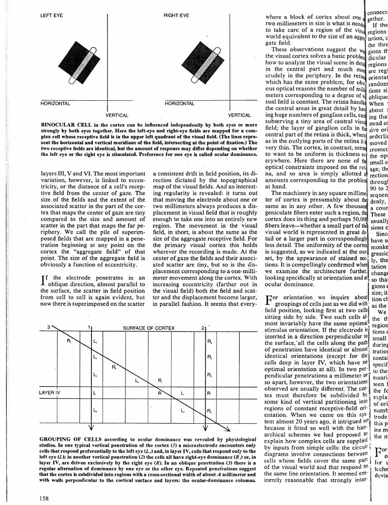
BINOCULAR CELL in the cortex can be influenced independently by both eyes or more strongly by both eyes together. Here the left-eye and right-eye fields are mapped for a complex _cell whose receptive field is in the upper left quadrant of the visual field. (The lines represent the horizontal and vertical meridians of the field, intersecting at the point of fixation.) The two receptive fields are identical, but the amount of response may differ depending on whether the left eye or the right eye is stimulated. Preference for one eye is called ocular dominance.
layers III, V and VI. The most important variation, however, is linked to eccentricity, or the distance of a cell's receptive field from the center of gaze. The size of the fields and the extent of the associated scatter in the part of the cortex that maps the center of gaze are tiny compared to the size and amount of scatter in the part that maps the far periphery. We call the pile of superimposed fields that are mapped in a penetration beginning at any point on the cortex the "aggregate field" of that poipt. The size of the aggregate field is obviously a function of eccentricity.
I f the electrode penetrates in an oblique direction, almost parallel to
the surface, the scatter in field position from cell to cell is again evident, but now there is superimposed on the scatter
a consistent drift in field position, its direction dictated by the topographical map of the visual fields. And an interesting regularity is revealed: it turns out that moving the electrode about one or two millimeters always produces a displacement in visual field that is roughly enough to take one into an entirely new region. The movement in the visual field, in short, is about the same as the size of the aggregate receptive field. For the primary visual cortex this holds wherever the recording is made. At the center of gaze the fields and their associated scatter are tiny, but so is the displacement corresponding to a one-millimeter movement along the cortex. With increasing eccentricity (farther out in the visual field) both the field and scatter and the displacement become larger, in parallel fashion. It seems that every-
3' 1 SURFACE OF CORTEX 2
~
""" L, R,
~ ~
R, '
~ L, R,
r--..... R,
LAYER IV L ~ L R
L, 0 ""'
R,
~ \!/ \J ~
GROUPING OF CELLS according to ocular dominance was revealed by physiological studies. In one typical vertical penetration of the cortex (J) a microelectrode encounters only cells that respond preferentially to the left eye (L r> and, in layer IV, cells that respond only to the left eye (L); in another vertical penetration (2) the cells all have right-eye dominance (Rz) or, in layer IV, are driven exclusively by the right eye (R). In an oblique penetration (3) there is a regular alternation of dominance by one eye or the other eye. Repeated penetrations suggest that the cortex is subdivided into regions with a cross-sectional width of about .4 millimeter and with walls perpendicular to the cortical surface and layers: the ocular-dominance columns.
158
·_connect' where a block of cortex about one gether. two millimeters in size is what is neede If the to take care of a region of the visu regions, world C?quivalent to the size of an aggr tation, c gate field. the thre
These observations suggest the wa gions tt the visual cortex solves a basic proble dicular how to analyze the visual scene in det regions. in the central part and much m are regi crudely in the periphery. In the reti oriental which has the same problem, for obvi random ous optical reasons the number of mill' meters corresponding to a degree of · sual field is constant. The retina handle the central areas in great detail by hav ing huge numbers of ganglion cells, eac subserving a tiny area of central visu field; the layer of ganglion cells in th sive ori central part of the retina is thick, where· orderlil as in the outlying parts of the retina it· moved very thin. The cortex, in contrast, see cromet to want to be uniform in thickness ev the op1 erywhere. Here there are none of small s1 optical constraints imposed on the reti age; the na, and so area is simply allotted i rection amounts corresponding to the proble througl at hand. 90 to 2
The machinery in any square millime sequen ter of cortex is presumably about denly, same as in any other. A few thousan a coun geniculate fibers enter such a region, th These cortex does its thing and perhaps 50,000 · usuall) fibers leave-whether a small part of th~_ sions c visual world is represented in great de·· Sinc1
tail or a larger part in correspondingly have sc less detail. The uniformity of the cortex _ monke is suggested, as we indicated at the out gressic set, by the appearance of stained sec· · ly the tions. It is compellingly confirmed when ·_ tation we examine the architecture further, · change looking specifically at orienta.tion and a • 80 thai ocular dominance. gions<
size; it For orientation_ we inquire about tion ct
groupings of cells just as we did witht1 as the field position, looking first at two cells We sitting side by side. Two such cells a!· the tt m_ost invari~bly ~ave the same optim~l . region
. stunulus onentatlon. If the electrode lSI tions < inserted in a direction perpendicular to small the surface,' all the cells along the path durinl of penetration have id-entical or almost! tratioJ identical orientations (except for the contai cells deep in layer IV, which have no specif optimal orientation at all). In two per·. to the pendicular penetrations a millimeter or'" essari so apart, however, the two orientations seen J observed are usually different. The cor· the fc tex must therefore be subdivided by expla' some kind of vertical partitioning int~ of ori regions of constant receptive-field on· j numb entation. When we came on this sys·1 trode tern almost 20 years ago, it intrigued u1 this p because it fitted so well with the hier· • ine m archical schemes we had proposed to i the rr. explain how complex cells are supplied : by inputs from simple cells: the circuit i diagrams involve connections between j'
cells whose fields cover the same part of the visual world and that respond to.: the same line orientation. It seemed em· inently reasonable that strongly inter·
po~ for 1
kchn de vis

l connected cells should be grouped to>ne 0! gether. Ceded .····• If the cortex is diced up into small visua .·regions of constant receptive-field orien-3.ggre" tation, can one say anything more about
r the three-dimensional shape of the re~ wa1 . gions than that their walls are perpen•blelll: dicular to the surface? Are neighboring detail'\ regions related in any systematic way or more are regions subserving all the possible etin~ .· orientations scattered over the cortex at obvj.~ random? We began to study these quesmi!lj. tions simply by penetrating the cortex of vj. obliquely or parallel to the surface . . ndles When we first did this experiment in ·hav.· about 1961, the result was so surpris. ~ac~ ing that we could hardly believe it. InllSUa] stead of a random assortment of succesn th sive orientations there was an amazing here.l orderliness. Each time the electrode a it~ moved forward as little as 25 or 50 mieerns crometers (thousandths of a millimeter) •s ev.j the optimal orientation changed by a f ~e 1 small step, about 10 degrees on the aver-re~FI age; the steps continued in the same di
!d Ill.,, rection, clockwise or counterclockwise, bleml through a total angle of anywhere from . ·. 90 to 270 degrees. Occasionally such a
lime. sequence would reverse direction sud-
.t thel·· denly, from a clockwise progression to sand a counterclockwise one or vice versa. 1• the These reversals were unpredictable, lf,O~~ usually coming after steady progres. the sions of from 90 to 270 degrees. t de· Since making this first observation we ngly have seen similar order in almost every >rtex monkey. Either there is a steady proout· gression in orientation or, less frequentsec-~! Iy, there are stretches in which orien
vhen tation stays constant. The successive ther, changes in orientation are small enough ld at. so that it is hard to be sure that the re-I gions of constant orientation are finite in
size; it could be that the optimal orientaJOUt tion changes in some sense continuously with •
( as the electrode moves along the cortex. ;ells We became increasingly interested in ; al· j the three-dimensional shape of these
l.~~!.l·.· regional subdivisions. From considerations of geometry alone the existence of
r to >ath small or zero changes in every direction during a horizontal or tangential pene-
1ost thel. !ration points to parallel slabs of tissue
0 . containing cells with like orientation n j specificity, with each slab perpendicular
Jer· · to the surface. The slabs would not nee~~~~-· essarily be planar, like slices of bread;
seen from above they might well have ;~; · the form of swirls, which could easily
l explain the reversals in the direction nto
of orientation changes. Recording large Jfi· \ :ys· 1 numbers of cells in several parallel elec-
t trode penetrations seemed to confirm u!"l· thi, prediction, but it was hard to exam-
ietr0· · me more than a tiny region of brain with ied the microelectrode.
uit ('
::r i to
m· er·
portunately an ideal anatomical meth-')d was invented at just the right time
for us. This was the 2-deoxyglucose tec<•nique for assessing brain activity, de, :sed by Louis Sokoloff and his group
at the National Institute of Mental Health and described elsewhere in this issue [see "The Chemistry of the Brain," by Leslie L. Iversen, page 134]. The method capitalizes on the fact that brain cells depend mainly on glucose as a source of metabolic energy and that the closely similar compound 2-deoxyglucose can to some extent masquerade as glucose. If deoxyglucose is injected into an animal, it is taken up actively by neurons as though it were glucose; the more
active the neuron, the greater the uptake~ The compound begins to be metabolized, but for reasons best known to biochemists the sequence stops with a metabolite that cannot cross the cell wall and therefore accumulates within the cell.
The Sokoloff procedure is to inject an animal with deoxyglucose that has been labeled with the radioactive isotope carbon 14, stimulate the animal in a way calculated to activate certain neurons
ANATOMICAL CONFIRMATION of ocular-dominance columns came from various staining methods and from axonal-trausport autoradiograpbs such as those shown in color on page 151. This composite autoradiograph visualizing the pattern over an area some 10 millimeters wide was made by cutting out and pasting together the regions representing layer IV in a number of parallel sections: the one in bottom illustration on page 151 and others at different depths.
RECONSTRUCTION of the ocular-dominance pattern over the entire exposed part of the right primary visual cortex was made by the authors and Simon LeVay from a series of sections stained by a reduced-silver method be developed. The left-band margin is at the medial edge of occipital lobe, where cortex folds downward; pattern is enlarged about six diameters.
159

and then immediately examine the brain for radioactivity, which reveals active areas where cells will have taken up more deoxyglucose than those in quiescent areas. The usual way of examining the brain for this purpose is to cut very thin slices of it (as one would for microscopic examination) and press them against a photographic plate sensitive to the radioactive particles. When the film is developed, any areas that were in contact with radioactive material are seen as dark masses of developed silver grains. Together with Michael P. Stryker we adapted the Sokoloff method to our problem, injecting an anesthetized animal with deoxyglucose and then moving a pattern of black and white vertical stripes back and forth 1.5 meters in front of the animal for 45 minutes. We then cut the brain into slices, either perpendicular to the surface of the cortex or parallel to it.
The autoradiographs quickly confirmed the physiological results. Sections .cut perpendicular to the surface showed narrow bands . of radioactivity about every 570 micrometers (roughly half a millimeter), extending through the full thickness of the cortex. Evidently these were the regions containing cells responsive to vertical lines. The deep
part of layer IV was uniformly radioactive, as was expected from the fact that the cells in the layer have circularly symmetrical receptive fields and show no orientation selectivity.
Sections cut parallel to the surface showed an unexpectedly complex set of periodically spaced bands, often swirling, frequently branching and rejoining, only here and there forming regular parallel slabs. What was particularly striking was the uniformity of the distance from one band to the next over the entire cortex. This fitted perfectly with the idea of a uniform cortex. Moreover, the distance between stripes fitted well with the idea that the cortical machinery must repeat itself at least every millimeter. If the distance were, for example, 10 millimeters from vertical through 180 degrees and back to vertical, sizable parts of the visual field would lack cells sensitive to any given orientation, making for a sketchy and extremely. bizarre representation of the visual scene.
T he final variable whose associated architecture needs to be considered
is eye preference. In microelectrode studies neighboring cells proved almost invariably to prefer the same eye. If in vertical penetrations the first cell we en-
BLOCK OF CORTEX about a millimeter square and two millimeters deep (light color) can be considered an elementary unit of the primary visual cortex. It contains one set of orientation slabs subserving all orientations and one set of ocular-dominance slabs subserving both eyes. The pattern is reiterated throughout the primary visual area. The placing of the boundaries (at the right or the left eye, at a vertical, horizontal or oblique orientation) is arbitrary; representation of the slabs as flat planes intersecting at right angles is an oversimplification.
countered preferred the right eye, then • the three c so did all the cells, right down to the 1eniculate bottom of layer VI; if the first cell pre. · This met ferred the left eye, so did all the rest, path fron Any penetration favored one eye or the ~· ·ynapse to· other with equal probability. (Since the ~als, howe' cells of layer IV are monocular, there it ~e path a was a matter not of eye preference but . !971 Bern: of eye monopoly.) If the penetration Was !University oblique. or horizontal, there was an a}. ·that after a ternation of left and right preferences, eye of a m with a rather abrupt switchover about 1111aterial e! every half millimeter. The cortex thus . terminals 2
proved to be diced up into a second set in the geni• of regions separated by vertical walls ;their axon: that extend through the full cortical !thought th thickness. The ocular-dominance sys. in a monk· tern was apparently quite independent . ography, rr of the orientation system, because in llate termin oblique or tangential penetrations the of the visu two sequences had 'no apparent relation to each other. ·our first
The basis of these ocular-dominance I negat columns, as they have come to be called, hints of a f seems to be quite simple. The terminals er IV. It v of geniculate fibers, some subserving ~
1.1hat we n
the left eye and others the right, group dark-field 1
themselves as they enter the cortex so vantage oJ that in layer IV there is no mixing. This ·lies of silv produces left-eye and right-eye patches I' sensitivity at roughly half-millimeter intervals. A a dark-fie] neuron above or below layer IV receives looked at connections from that layer from.up to ·l"croscope, 1 about a millimeter away in every direc- were the pc tion. Probably the strongest connections er IV [see are from the region of layer IV closest . The nex to the neuron, so that it is presumably pattern fa< dominated by whichever eye feeds that ! parallel to region. · tex is dome
Again we were most curious to learn aile! to th( what these left-eye and right-eye regions IV shows might look like in three dimensions; any oval, whil of several geometries could lead to the shows it as cross-sectional appearance the physi- of such o· ology had suggested. The answer first sections 01
came from studies with the silver-degen- I over a wic .eration method for mapping connec- I From tl tions, devised by Walle J. H. Nauta of I mediately the Massachusetts Institute of Techno!- ~··pattern is ogy. Since then we have found three oth- senting te er independent anatomical methods for jected eye demonstrating these columns. , ing the otl
A particularly effective method (be- j not regula cause it enables one to observe in a sin- 1. ourselves gle animal the arrangement of columns all, biolo.l over the entire primary visual cortex) is ~~- represent! based on the phenomenon of axonal stnpes, or transport. The procedure is to inject a where a radioactively labeled amino acid into an 1 branches. area of nervous tissue. A cell body takes 1. monest n up the amino acid, presumably incorpo· ~ along the rates it into a protein and then transports J The stript it along the axon to its terminals. When ,..1; dicular ~o we injected the material into one eye . mary v1s1 of a monkey, the retinal ganglion cells , area 18, a. took it up and transported it along their ! est such i axons, the optic-nerve fibers. We could fall macaq then examine the destinations of these . of the pat fibers in the lateral geniculate nuclei by t to the ne coating tissue slices with a silver ernul- . sphere to sion and developing the emulsion; the l The wi· radioactive label showed up clearly in constant,

' te . ,eniculate on each stde. :n i'~e three compleme~tary layers of the
~- · · This method does not ordinarily trace t. ·.,path from one axon terminal across a te ,,ynapse to the next neuron and its termite nals, however, and we wanted to follow it the path all the way to the cortex. In 11 !971 Bernice Grafstein of the Cornell ts !University Medical College discovered 1- that after a large enough injection in the s, eye of a mouse some of the radioactive t! !'material escaped from the optic-nerve s terminals and was taken up by the cells :t in the geniculate and transported along s , their axons to the cortex. We had the :1 ~·thought that a similarly large injection .- in a monkey, combined with autoradi-t 0graphy, might demonstrate the genicu~ !'late terminals from one eye in layer IV
of the visual cortex.
'our first attempt yielded dismayingly I negative results, with only faint
I hints of a few silver grains visible in layer IV. It was only after several weeks
~,that we realized that by resorting to dark-field microscopy we could take advantage of the light-scattering proper-
1 ties of silver grains and so increase the I sensitivity of the method. We borrowed a dark-field condenser, and when we looked at our first slide under the mi-
' 1-croscope, there shining in all their glory were the periodic patches of label in layer IV [see top illustration on page 151].
The next step was to try to see the pattern face on by sectioning the cortex
' parallel to its surface. The monkey cor-! texis dome-shaped, arld so a section parallel to the surface and tangent to layer IV sho:-vs that layer as a circle or an
I. oval, while a section below layer IV shows it as a ring. By assembling a series of such ovals and rings from a set of
. sections one can reconstruct the pattern
! over a wide expanse of cortex. From the reconstructions it was im
mediately obvious that the main overall '. pattern is one of parallel stripes repre-senting terminals belonging to the injected eye, separated by gaps representing the other eye. The striping pattern is
I not regular like wallpaper. (We remind ourselves occasionally that this is, after all, biology!) Here and there a stripe
• representing one eye branches into two stripes, or else it ends blindly at a point where a stripe from the other eye
, branches. The irregularities are com-
1 monest near the center of gaze and
I along the line that maps the horizon. The stripes always seem to be perpen
.,. dicular to the border between the pri-
1 mary visual cortex and its neighbor, arec., 18, and here the regularity is greatest. Such general rules seem to apply to
i an macaque brains, although the details
li of the pattern vary from one individual to lhe next and even from one hemiSph"re to the other in the same monkey.
The width of a set of two stripes is con:;tant, about .8 millimeter, over the
VERTICAL
\ L R L R L R
R L L
HORIZONTAL
HYPOTHETICAL PATTERN OF CORTICAL ACTIVITY that might result from stimulation of the left eye with a single short horizontal line, placed in the upper left quadrant of the visual field, is shown by the colored patches on a diagram of an area of the right cortex, seen face on, The area receiving input from the object in the visual field is indicated by the broken black line. If ocular-dominance and orientation columns are arrayed as shown, activated cells will be those that respond optimally to approximately horizontal stimuli from the left eye.
entire primary visual cortex, once more emphasizing the uniformity of the cortex. Again the widths fit perfectly with the idea that all of the apparatus needed to look after an area the size of an aggregate field must be contained within any square millimeter of cortex. The two techniques, deoxyglucose labeling and amino acid transport, have the great advantage of being mutually compatible, so that we have been able to apply both together, one to mark orientation lines and the other to see the ocular-dominance columns. The number of brains examined so far is too small to justify any final conclusions, but the two systems appear to be quite independent, neither parallel nor at right angles but intersecting at random.
The function served by ocular-dominance columns is still a mystery. We know there are neurons with all grades of eye preference throughout the entire binocular part of the visual fields, and it may be that a regular, patterned system of converging inputs guarantees that the distribution will be uniform, with neither eye favored by accident in any one place. Why there should be all these grades of eye preference everywhere is itself not clear, but our guess is that it has something to do with stereoscopic depth perception.
Given what has been learned about the primary visual cortex, it is clear
that one can consider an elementary piece of cortex to be a block about a millimeter square and two millimeters
deep. To know the organization of this chunk of tissue is to know the organization for all of area 17; the whole must be mainly an iterated version of this elementary unit. Of course the elementary unit ~hould not be thought of as a discrete, separable block. Whether the set of orientation slabs begins with a slab representing a vertical orientation, an oblique one or a horizontal one is completely arbitrary; so too is whether an ocular-dominance sequence begins with a left-plus-right pair of dominance slabs or a right-plus-left pair. The same thing is true for a unit crystal of sodium chloride or for any complex repetitive pattern such as is found in wallpaper.
What, then, does the visual scene really look like as it is projected onto the visual cortex? Suppose an animal fixes its gaze on some point and the only object in the visual field is a straight line above and a bit to the left of the point where the gaze is riveted. If each active cell were to light up, and if one could stand above the cortex and look down at it, what would the pattern be? To make the problem more interesting, suppose the pattern is seen by one eye only. In view of the architecture just described the pattern turns out to be not a line but merely a set of regularly spaced patches [see illustration above]. The reasoning can be checked directly by exposing a monkey with one eye closed to a set of vertical stripes and making a deoxyglucose autoradiograph. The resulting pattern should not be a great surprise: it is a set of regularly spaced patches, which sim-
161

ply represents the intersection of the two sets of column systems. Imagine the surprise and bewilderment of a little green man looking at such a version of the outside world!
Why evolution has gone to the trouble of designing such an elaborate architecture is a question that continues to fascinate us. Perhaps the most plausible notion is that the column systems are a solution to the problem of portraying more than two dimensions on a two-dimensional surface. The cortex is dealing with at least four sets of values: two for the x andy position variables in the visual field, one for orientation and one for the different degrees of eye preference. The two surface coordinates are used up in designating field position; the other two variables are accommodated by dicing up the cortex with subdivisions so fine that one can run through a complete set of orientations or eye preferences and meanwhile have a shift in visualfield position that is small with respect to the resolution in that part of the visual world.
The strategy of subdividing the cortex with small vertical partitions is certainly not limited to the primary visual area. Such subdivisions were first seen in the somatic sensory area by Vernon B. Mountcastle of the Johns Hopkins University School of Medicine about 10 years before our work in the visual area. In the somatic sensory area, as we point-
ed out above, the basic topography is a map of the opposite half of the body, but superimposed on that there is a twofold system of subdivisions, with some areas where neurons respond to the movement of the joints or pressure on the skin and other areas where they respond to touch or the bending of hairs. As in the case of the visual columns, a complete set here (one area for each kind of neuron) occupies a distance of about a millimeter. These subdivisions are analogous to ocular-dominance columns in that they are determined in the first instance by inputs to the cortex (from either the left or the right eye and from either deep receptors or receptors in the upper skin layers) rather than by connections within the cortex, such as those that determine orientation selectivity and the associated system of orientation regions.
The columnar subdivisions associated with the visual and somatic sensory systems are the best-understood ones, but there are indications of similar vertical subdivisions in some other areas: several higher visual areas, sensory parietal regions recently studied by Mountcastle and the auditory region, where Thomas J. Imig, H. 0. Adrian and John F. Brugge of the University of Wisconsin Medical School and their colleagues have found subdivisions in which the two ears seem alternately to add their information or to compete.
ACTUAL PATTERN of cortical activity was elicited by exposing only the left eye to a set of vertical stripes. The deoxyglucose autoradiograph is of a tangential section in the outer layers of the cortex. The pattern of regularly spaced dark patches of radioactivity represents intersection of ocular-dominance and orientation systems. Magnification is about eight diameters.
162
For most of these physiologically de.l fined systems (except the visual ones) there are so far no anatomical corre. lates. On the other hand, in the past few years several anatomists, notably Ed.~··. ward G. Jones of the Washington Uni. versity School of Medicine and Nauta and Patricia Goldman at M.I.T., have shown that connections from one region I of the cortex to another (for example from the somatic sensory area on one side to the corresponding area on the · other side) terminate in patches that have a regular periodicity of about a millimeter. Here the columns are evi. dent morphologically, but one has no idea of the physiological interpretation. It is clear, however, that fine periodic . subdivisions are a very general feature • of the cerebral cortex. Indeed, Mount- [ castle's original observation of that feature may be said to supply a fourth profound insight into cortical organization. I. •
I t would surely be wrong to assume that this account of the visual cortex .
1 in any way exhausts the subject. Color, 1 movement and stereoscopic depth are probably all dealt with in the cortex, but . to what extent or how is still not clear.[J U 1 There are indications from work we and others have done on depth and from work on color by Semir Zeki of Uni- .
1." If you'
versity College London that higher cor- . life with tical visual areas to which the primary . 'n the eyt area projects directly or indirectly may ·.· ~ou shm be specialized to handle these variables, · magazir but we are a long way from knowing never 81 what the handling involves. [sphere~
What happens beyond the primary vi- . OMY m sual area, and how is the information on excitem orientation exploited at later stages? Is !"light se£ one to imagine ultimately finding a cell · standint that responds specifically to some very to ASTF particular item? (Usually one's grand- , Mont mother is selected as the particular item, I' tory-que for reasons that escape us.) Our answer paintinc is that we doubt there is such a cell, but ASTRO we have no goo. d alternative to offer. To ~·;of spac1
speculate broadly on how the brain may What v-. work is fortunately not the only course and m open to investigators. To explore the formed brain is more fun and seems to be more planatic profitable. Toda
There was a time, not so long ago, enterec when one looked at the millions of neu- "OMY. t rons in the various layers of the cortex omy m; and wondered if anyone would ever article~ have any idea of their function. Did they all work in parallel, like the cells of the liver or the kidney, achieving their objectives by pure bulk, or were they each doing something special? For the visual cortex the answer seems now to be known in broad outline: Particular stimuli turn neurons on or off; groups of neu- i rons do indeed perform particular trans- ' , formations. It seems reasonable to think that if the secrets of a few regions such as this one can be unlocked, other re-gions will also in time give up their se-crets.