BRAIN MARKERS of · Brain markers of psychosis and autism . Academisch Proefschrift . Ter...
187
UvA-DARE is a service provided by the library of the University of Amsterdam (http://dare.uva.nl) UvA-DARE (Digital Academic Repository) Brain markers of psychosis and autism: the brain in concert Bloemen, O.J.N. Link to publication Citation for published version (APA): Bloemen, O. J. N. (2011). Brain markers of psychosis and autism: the brain in concert. General rights It is not permitted to download or to forward/distribute the text or part of it without the consent of the author(s) and/or copyright holder(s), other than for strictly personal, individual use, unless the work is under an open content license (like Creative Commons). Disclaimer/Complaints regulations If you believe that digital publication of certain material infringes any of your rights or (privacy) interests, please let the Library know, stating your reasons. In case of a legitimate complaint, the Library will make the material inaccessible and/or remove it from the website. Please Ask the Library: https://uba.uva.nl/en/contact, or a letter to: Library of the University of Amsterdam, Secretariat, Singel 425, 1012 WP Amsterdam, The Netherlands. You will be contacted as soon as possible. Download date: 11 Mar 2020
Transcript of BRAIN MARKERS of · Brain markers of psychosis and autism . Academisch Proefschrift . Ter...

UvA-DARE is a service provided by the library of the University of Amsterdam (http://dare.uva.nl)
UvA-DARE (Digital Academic Repository)
Brain markers of psychosis and autism: the brain in concert
Bloemen, O.J.N.
Link to publication
Citation for published version (APA):Bloemen, O. J. N. (2011). Brain markers of psychosis and autism: the brain in concert.
General rightsIt is not permitted to download or to forward/distribute the text or part of it without the consent of the author(s) and/or copyright holder(s),other than for strictly personal, individual use, unless the work is under an open content license (like Creative Commons).
Disclaimer/Complaints regulationsIf you believe that digital publication of certain material infringes any of your rights or (privacy) interests, please let the Library know, statingyour reasons. In case of a legitimate complaint, the Library will make the material inaccessible and/or remove it from the website. Please Askthe Library: https://uba.uva.nl/en/contact, or a letter to: Library of the University of Amsterdam, Secretariat, Singel 425, 1012 WP Amsterdam,The Netherlands. You will be contacted as soon as possible.
Download date: 11 Mar 2020

Brain markers of psychosis and autism� e brain in concert
Brain markers of psychosis and autism O
swald J.N
. Bloemen Oswald J.N. Bloemen
∆BPND
ND
∆BPND
ND
∆BPND
BOLD
BOLD
BOLDBOLD
BOLD BO
LD
GLU
GABA
FA
FA
FA
GLU
GLU
GABA
GABA GABA
GABA
DA
DA

Brain
markers
of
psychosis
and
autism
Brain markers of psychosis and autism Copyright © 2011 Oswald J.N. Bloemen, Amsterdam, the Netherlands Printing of this thesis was financially supported by: the University of Amsterdam, the Graduate School Neurosciences Amsterdam Rotterdam (ONWAR), Janssen Cilag, AstraZeneca, Eli Lilly and GE Healthcare. Lay-out: O.J.N. Bloemen Cover: Image adapted with permission from a painting of Dancing to the Spirit of the
Wood (an artisan gallery located in Hendersonville, Tennessee which donates part of its profits to autism related causes). Cover created with kind help from Nadine van Asbeck.
Printing Off Page ISBN: 978-90-9025933-8

Brain markers of psychosis and autism
Academisch Proefschrift
Ter verkrijging van de graad van doctor aan de Universiteit van Amsterdam op gezag van de Rector Magnificus
Prof. dr. D.C. van den Boom ten overstaan van een door het college van promoties
ingestelde commissie, in het openbaar te verdedigen in de Aula der Universiteit
op vrijdag 11 februari 2011, te 13:00 uur
door
Oswald Jan Nicolaas Bloemen
Geboren te Amsterdam

P r o m o t i e c o m m i s s i e
Promotores: Prof. dr. D.H. Linszen Prof. dr. J. Booij Co-promotor: Dr. T.A.M.J. van Amelsvoort Overige Leden: Prof. dr. D.A.J.P. Denys
Prof. dr. R.J. van der Gaag Prof. dr. B.L.F. van Eck Prof. dr. W. van den Brink Dr. M.A. Mehta
Faculteit der Geneeskunde

C o n t e n t s
Chapter 1. General introduction. 7
Chapter 2. Early intervention in patients at ultra high risk of psychosis: 21 benefits and risks.
Chapter 3. White matter markers for psychosis in a prospective ultra high 53 risk cohort.
Chapter 4. Challenge and therapeutic studies using alpha-methyl-para- 69 tyrosine (AMPT) in neuropsychiatric disorders: a review.
Chapter 5. Striatal dopamine D2/3 receptor binding following dopamine 91 depletion in subjects at Ultra High Risk for psychosis.
Chapter 6. Hippocampal glutamate levels and striatal dopamine D2/3 111 receptor occupancy in subjects at Ultra High Risk of Psychosis.
Chapter 7. Psychosis and autism. An in vivo magnetic resonance imaging 117 study of brain anatomy.
Chapter 8. White matter integrity in Asperger syndrome: a preliminary 139 diffusion tensor imaging study.
Chapter 9. Summary and general discussion. 161
Chapter 10. Summary in Dutch. 173
Chapter 11. Acknowledgements. 183 List of Publications. 189 Curriculum Vitae. 191


C h a p t e r 1
G e n e r a l i n t r o d u c t i o n

S c h i z o p h r e n i a
Schizophrenia is a severe mental disorder which has a chronic course in the majority of patients. The disorder is regarded as the most disabling health condition in the Netherlands, and as the third most disabling condition worldwide (Ustun et al 1999). The disorder is characterized by disturbances in perception, thought, volition, cognition and affect. Most symptoms and signs are commonly categorized as positive and negative symptoms. Positive or psychotic symptoms are experiences that are present in patients with schizophrenia, while usually absent in healthy individuals, such as delusions, hallucinations, disorganized behavior or speech formal disorders of thought (incoherence) and ‘catatonic’ movement and behavioral disorders (American Psychiatric Association 1987). Negative symptoms can be described as absence of functions that are present in healthy individuals, such as flattening of affect, anhedonia, avolition, and social withdrawal. Cognitive symptoms such as attention and memory problems and problems with planning are often also present. Lifetime prevalence of schizophrenia is about 0.8-1% and incidence is 0.2-0.4 per 1000 (Mueser and McGurk 2004). Symptoms typically appear during adolescence and in young adulthood. Prevalence is roughly equal in both sexes, although women are often older when they are diagnosed with schizophrenia and clinical presentation is different between men and women (Mueser and McGurk 2004).
SCHI ZOPHREN IA CON CEPT The concept of schizophrenia is only just over 100 years old, although it is very likely that the disorder has been present in man for a much longer period of time. The term “démence precoce” was first used by Bénédict Augustin Morel (1809-1873) to describe a mental disorder which initially struck young males, and eventually led to deterioration of mental functioning and disability. The term dementia praecox was used by Emil Kraepelin (1856-1926) in the late 19th century to unify three psychiatric disorders at that time (paranoia, hebephrenia, catatonia), in one syndrome that shared the characteristics of those described by Morel. Eugen Bleuler (1857-1939) changed the name into schizophrenia in 1908 with some original components of dementia praecox described as subtypes (paranoid, disorganized (hebephrenic), catatonic, undifferentiated and residual subtype) which have remained largely constant since then. Nevertheless, after about 100 years, these categories may disappear in the next edition of the Diagnostic and Statistical Manual of Mental Disorders (DSM) and exchanged for symptom dimensions (DSM-5 is currently scheduled for 2013).
8

AE TIOL OGY OF S CHIZOPH REN IA The aetiology of schizophrenia is unknown, but the prevailing view today is that genetic and environmental factors interact to the development of the disorder. Two major theories of schizophrenia are the neurodevelopmental and the dopamine hypotheses. A third model which is gaining support is the dimensional model. The neurodevelopmental theory directs our attention to early in life, and states that early aberrations in development, probably in concert with other risk factors, cause the emergence of schizophrenia at the end of adolescence. The most important risk factors are genetic. Overall heritability of schizophrenia is estimated at 83% (Cardno and Gottesman 2000), and having an affected monozygotic twin sibling (or two affected parents) increases the chance of developing the disorder to about 50% (Cardno and Gottesman 2000) while dizygotic twins only have a concordance rate of 28%. Children with one affected parent have about 10% chance of developing the disorder. On a molecular level, schizophrenia has been associated with several types of susceptibility genes, such as neuroregulin, dysbindin, and DISC1 that are coding for proteins that play a role in the development of the brain.. A major functional susceptibility gene involved in dopamine regulation is the gene encoding for COMT (Rapoport et al 2005). Heritability does not appear to follow simple Mendelian single-gene inheritance patterns, it is more likely that susceptibility genes, each with small effects, act in concert with epigenetic and environmental factors (Mueser and McGurk 2004). Known environmental risk factors early in life include pre- and peri-natal events (such as viral infections and obstetric complications), and cannabis use, urbanicity and social isolation during adolescence although effect sizes are small (Mueser and McGurk 2004;Murray et al 2008). In conclusion, symptoms of schizophrenia are familial and heritable, that may interact with environmental factors and there is clear evidence for a developmental pathogenesis.
The dimensional model focuses our attention to the general population. Interestingly, mild subclinical psychotic experiences are common in the general population and are thought to signal the mild end of a continuum for which psychosis or schizophrenia is the extreme end (van Os et al 2000). Subclinical symptoms may also be present in children (Poulton et al 2000), and share the risk factors of psychotic symptoms in schizophrenia, such as for instance being familial and heritable, associated with early impairments in cognitive functioning, linked to premorbid behavioral, emotional, and educational problems, urbanicity, low birth weight and perinatal complications (Polanczyk et al 2010). Furthermore these ‘quasi-psychotic’ phenomena are associated with an increased risk of developing schizophrenia in these children (Cannon et al 2002). Thus there is evidence for the dimensional model as subdiagnostic symptoms can be seen in the general adult population and in children, which share characteristics of psychotic symptoms in
9

schizophrenia like heritability and familiarity and may progress to psychotic severity later in life.
The dopamine hypothesis finally directs our attention to current symptomatology in psychosis and its treatment. The classical dopamine theory was postulated around 40 years ago and states that increased dopaminergic function in the mesolimbic pathway may be associated with the positive symptoms of schizophrenia, whereas decreased dopaminergic function in the mesocortical pathway may be responsible for the negative symptoms. . This insight was fueled by the discovery of chlorpromazine in 1950 (Delay et al 1952) and by reports that stimulating dopamine release with amphetamines could cause psychosis (Connell 1958). Later studies found multiple genes and neuronal pathways leading to psychosis with dopamine as the last step in a complex developmental cascade towards schizophrenia (Murray et al 2008). The theory was recently amended by Kapur, who suggested that excess stimulus-independent striatal dopamine is responsible for inappropriate assigning of salience to normal external and internal stimuli (Kapur 2003). Delusions may be a top-down cognitive explanation for these experiences of increased salience.
More recently, excess striatal dopaminergic activity in schizophrenia has been hypothesized to occur secondary to dysfunctional glutamatergic transmission (Carlsson et al 2001;Lisman et al 2008). The hippocampus has extensive glutamatergic projections to the striatum. When these output neurons are experimentally stimulated, both the number of spontaneously active dopaminergic neurons and the amount of dopamine released in the striatum are increased (Lodge and Grace 2006). Disinhibition of hippocampal glutamate may in turn be caused by loss of hippocampal gamma-aminobutyric acid (GABA) tone due to blockade of glutamatergic N-methyl-D-aspartate (NMDA) receptors on GABAergic interneurons. Glutamate itself (measured by Proton Magnetic Resonance Spectroscopy (1H-MRS)) may also be related to symptomatology in psychosis, independently from dopaminergic abnormalities. As medication that modulates glutamatergic function has more effect on negative symptoms, and formal thought disorder, and dopamine affecting drugs have more effect on positive symptoms, it is thought that these systems are responsible for different features of schizophrenia (Stone et al 2007).
MORBID ITY Schizophrenia is a severe mental disorder often accompanied with social decline and recurrent hospitalizations. Patients are about evenly distributed throughout all social and cultural subcultures, and approximately 5% of patients manage to stay employed
10

(Honkonen et al 2007). Outcome is poor in less than 50% of patients and, similarly, outcome is also good in less than 50% of patients (van Os and Kapur 2009). As Bleuler already had observed in his patients, the course and outcome of schizophrenia is characterized by mainly unexplained heterogeneity rather than uniform poor outcome. Of schizophrenia patients, approximately 5% dies of suicide, but up to 40% may attempt a tentamen suicidii (Palmer et al 2005). Life expectancy is approximately 22½ years shorter and patients with schizophrenia have increased health problems as for instance cardiovascular events, obesity, metabolic aberrations, smoking and alcohol abuse (von Hausswolff-Juhlin et al 2009) compared to the general population. These problems are related to the treatment with antipsychotics, the disorder itself and/or lifestyle.
THE “UL T RA HIGH RIS K” SYNDROME Schizophrenia is usually preceded by a prodromal period before the first psychotic episode, starting with non specific symptoms (anxiety, depression), with negative symptoms and ending with emerging mild psychotic symptoms, and a decline in psychosocial functioning. Researchers in Australia, the USA and Germany developed instruments for assessment of symptoms and signs that predicted transition to psychosis in a help-seeking young population prospectively (Klosterkotter et al 2001;Miller et al 2003;Yung et al 2003). Recently, these findings were replicated with transition rates varying from 10-40% after 1 to 2 years follow-up (Cannon et al 2008;Ruhrmann et al 2010;Yung et al 2008). Patients in these studies were described to have an At Risk Mental State (ARMS) or to have an Ultra High Risk (UHR) to develop a first psychotic episode. These studies have defined UHR subjects as help-seeking patients with either 1) attenuated psychotic symptoms, 2) a genetic risk for schizophrenia plus a recent decrease in functioning, or 3) brief limited psychotic symptoms that spontaneously disappear within a week (Miller et al 2003;Yung et al 2003). Until now the main problem of the attenuated psychotic symptoms syndrome remains the problem of the ‘false positives’; the majority of help seeking patients in the UHR state do not develop a first psychotic episode. Currently there is no replicated therapy to prevent transition to psychosis.
A u t i s m s p e c t r u m d i s o r d e r
Autism Spectrum Disorder (ASD) includes autism, Asperger syndrome and atypical autism. ASD is an increasingly diagnosed neurodevelopmental disorder affecting up to 1:100 children (Baird et al 2006). It is thought to be highly genetic (Bailey et al 1995;Frith 2001) and is characterized by significant difficulties in social interaction, communication, and unusual or stereotyped routines and behavior (International Statistical Classification of Diseases and Health Related Problems—10th revision). People with Asperger syndrome
11

typically do not have delay in the acquisition of language but they still show the other characteristic ‘autistic’ impairments (i.e., difficulties in social interaction and unusual or stereotyped routines and behavior).
It has been suggested that people with ASD are also at increased risk of developing psychosis (Petty et al 1984). Autism was initially thought to be an early manifestation of schizophrenia, and was often referred to as “schizophrenic syndrome of childhood” or “childhood psychosis”. In fact, it was only in 1971 that ASD was finally distinguished from schizophrenia (Kolvin 1971). This late division can be understood as symptoms of the two disorders overlap phenotypically, such as deficits in social behavior, oddness of speech, unusual responsiveness to the sensory environment, isolated skill areas, and inappropriate affect. Bleuler named “autism” as one of the four core symptoms of schizophrenia (the others being flattened affect, loosening of association and ambivalence (Stotz-Ingenlath 2000)).
The risk for both disorders is influenced by pre- and peri-natal factors as for instance low birth weight, maternal infection and fetal stress. On an intermediate phenotype level dopamine antagonists help to alleviate certain symptoms in both disorders, and neurophysiologically both share markers such as impaired stimulus filtering as measured by prepulse inhibition (PPI) (Kumari et al 2008;McAlonan et al 2002;Perry et al 2007). Furthermore, structural neuroimaging studies suggest that lower grey matter volumes within limbic-striato-thalamic circuitry are common to both ASD and schizophrenia (Cheung et al 2010).
On a genetic level there are also similarities between ASD and schizophrenia. Both have heritability estimates of around at least 80%, and inheritance is complex with multiple genetic and environmental factors influencing the chance of developing the disorders. Single Nucleotide Polymorphisms (SNP’s), copy number variations (CNV’s) e.g. NRXN1 and specific rare loci and alleles are associated with increased risks for ASD and schizophrenia (Carroll and Owen 2009).
Nevertheless there are apparent differences between ASD and schizophrenia. ASD is usually diagnosed in childhood and is clearly a developmental disorder with symptoms typically coming to attention of parents and doctors from two years of age. Schizophrenia may have some symptoms and signs in the premorbid phase as stated above, but is usually diagnosed in the late teens or early twenties, although prodromal symptoms often start two to five years earlier (Hafner et al 1992). People with ASD usually have intellectual disabilities, whereas this is not the case with schizophrenia. Also there are numerous genetic risk loci that differ between them and specific differences in imaging studies.
12

As symptoms overlap, it can be difficult to differentiate between ASD and psychosis. Previous diagnostic classifications also specifically excluded the presence of both disorders. Nevertheless there is growing consensus that diagnosis of psychosis in ASD is warranted and 14 – 34% of ASD patients have been reported to have a co-morbid psychotic disorder (Mouridsen et al 2008;Stahlberg et al 2004;Tsakanikos et al 2007). Certain specific subtypes of ASD (although not incorporated in DSM-IV) like multiple complex developmental disorder (McDD) are also associated with an increased risk of developing psychosis, with risk between 22% and 64% (Sprong et al 2008;van Engeland and van der Gaag 1994). One could therefore tentatively state that ASD patients, as UHR patients, are at clinically or even at ultra high risk to develop psychosis.
I m a g i n g
DT-MRI Diffusion Tensor Magnetic Resonance Imaging (DT-MRI) is a brain imaging technique, which has been widely used to study white matter (WM) in chronic and in first episode schizophrenia (Basser et al 1994). DT-MRI can be used to investigate orientation and integrity of WM tracts. This is achieved by measuring the amount and direction of water diffusion, which can be isotropic (the same amount in every direction) or anisotropic. Diffusion of water molecules in WM tends to be greater along the direction of WM tracts and thus predominantly anisotropic, with the degree of anisotropy in a particular tissues often being quantified through its fractional anisotropy (FA) value. The degree of anisotropy depends on a number of factors, for instance, myelination, fibre diameter and density. It is thought that a lower FA is indicative of lower connectivity or integrity of WM tracts (Basser 1995;Beaulieu 2002;Pierpaoli and Basser 1996).
Studies have reported significantly lower FA of widespread brain regions in patients with chronic schizophrenia (Konrad and Winterer 2008). Furthermore, first episode patients were also reported to have significant localized FA reductions, but to a lesser extent than chronic patients (Friedman et al 2008). Recently there have been reports of WM abnormalities in genetic and clinical high risk samples although structural changes have not been consistent across studies. It may thus be that WM abnormalities are present before the onset of frank psychosis.
White matter has also been researched in ASD. It has been proposed that ASD is a ‘connectivity disorder’ (Frith 2004) and that people with ASD have local overconnectivity but reduced long range (global) connectivity (Just et al 2004). Previous studies have also shown that children with autism have specific reductions of FA (Alexander et al
13

2007;Barnea-Goraly et al 2004;Keller et al 2007;Lee et al 2007). In addition, it has been suggested that brain abnormalities may increase with age, or are ‘exponentially distributed”. This would infer that abnormal brain development may persist into adulthood. It would thus be interesting to study adult patients with ASD. Of the ASD subtypes, most research has been done on normal or high functioning autism, while Asperger syndrome has been much less studied.
NU CL EAR IM AGIN G Nuclear imaging (scintigraphic) techniques such as Positron Emission Tomography (PET) and Single Photon Emission Computed Tomography (SPECT) are used to provide information about the central dopaminergic system. Using radiotracers that label dopaminergic receptors, dopamine transporters or enzymes involved in dopaminergic metabolism, these imaging techniques enable direct measurement of the dopaminergic system. These techniques have shown direct evidence of disruption of dopaminergic neurotransmission in the striatum of patients with schizophrenia. For instance [18F]-DOPA PET studies showed increased presynaptic striatal DOPA uptake in schizophrenia (Howes et al 2007). Also increased striatal [18F]-DOPA uptake has recently been demonstrated in UHR patients (Howes et al 2009) and in first degree relatives of patients with schizophrenia (Huttunen et al 2008). This suggests that dopaminergic abnormalities are present in subjects that have not (yet) developed psychosis but are at increased (genetic) risk to do so.
[123I]-IBZM SPECT is used to measure the amount of dopamine D2/3 receptors. As receptors can be occupied by endogenous dopamine in normal circumstances, only a portion of D2/3 receptors can be measured by a baseline SPECT (i.e., the free receptors available to bind to the radiopharmaceutical IBZM). Removing endogenous dopamine from the receptors, and performing a second (depletion) IBZM SPECT scan, provides information on the “total” amount of D2/3 receptors. Furthermore subtracting the baseline from the depletion SPECT scan provides information on the amount of occupancy of the D2/3 receptors by endogenous dopamine in baseline condition. Dopamine can be depleted with alpha-methyl-para-tyrosine (AMPT), which is a competitive and reversible inhibitor of tyrosine hydroxylase, the rate-limiting enzyme of catecholamine synthesis.
Dopamine depletion studies showed increased occupancy of postsynaptic striatal dopamine D2/3 receptors by endogenous dopamine in patients with schizophrenia compared to controls (Abi-Dargham et al 2000;Abi-Dargham et al 2009;Kegeles et al 2010). Interestingly, studies using a dopamine stimulation challenge instead of dopamine depletion show increased dopamine release after amphetamine-induced dopamine
14

stimulation (Abi-Dargham et al 2009;Laruelle et al 1996). This increased release is correlated to the increased occupancy by endogenous dopamine in the same patients (Abi-Dargham et al 2009). In conclusion, challenge and non-challenge studies show evidence for dopaminergic abnormalities in schizophrenia and there is preliminary evidence from non-challenge studies that UHR patients already have dopaminergic abnormalities.
A i m a n d o u t l i n e o f t h e t h e s i s
The overall aim of the studies described in this thesis was to increase our knowledge on the brain structure and function that may be underlying early schizophrenia and ASD. We focused on these groups of patients as they are thought to be at increased or at ultra high risk for developing psychosis and expected to find brain changes preceding the development of psychosis. We used various imaging techniques such as IBZM SPECT, structural MRI, DT-MRI and 1H-MRS, and thus looked at dopaminergic and glutamatergic function, brain morphometry and connectivity. We also related clinical symptoms to imaging findings where possible.
In Chapter 2 we discuss the UHR concept and review therapeutic strategies in patients with an ultra high risk to develop psychosis. In Chapter 3 we perform a DT-MRI study in UHR patients and describe WM abnormalities in UHR patients who later develop psychosis, compared to UHR patients who do not develop psychosis and also include age, gender and IQ matched healthy controls. Chapter 4 is a review of challenge studies in neuropsychiatric disorders using AMPT in vivo. All reported clinical and therapeutic effects as well as side effects of AMPT are discussed. Chapter 5 reports on striatal dopamine D2/3 receptor binding following dopamine depletion with AMPT in subjects at ultra high risk for developing psychosis compared to age, gender and IQ matched healthy controls. In Chapter 6 we discuss the relationship between hippocampal glutamate levels and striatal dopamine D2/3 receptor availability and occupancy by endogenous dopamine in UHR subjects and controls in response to a recently published report of a presynaptic relationship between dopamine and glutamate. In Chapter 7 we compare people with autism and psychosis to people with autism without psychosis and to healthy controls and report on structural brain differences between these groups. In Chapter 8 we perform a DT-MRI study and compare white matter integrity of adults with Asperger syndrome to age, gender and IQ matched healthy controls. In Chapter 9 we summarize the findings of the studies of this thesis. Implications and limitations are discussed and we look at future directions in research.
15

R e f e r e n c e s Abi-Dargham A, Rodenhiser J, Printz D, Zea-Ponce Y, Gil R, Kegeles LS et al (2000): Increased baseline occupancy of D2 receptors by dopamine in schizophrenia. Proc Natl Acad Sci U S A 97: 8104-8109.
Abi-Dargham A, van de Giessen E, Slifstein M, Kegeles LS, Laruelle M (2009): Baseline and amphetamine-stimulated dopamine activity are related in drug-naive schizophrenic subjects. Biol Psychiatry 65: 1091-1093.
Alexander AL, Lee JE, Lazar M, Boudos R, Dubray MB, Oakes TR et al (2007): Diffusion tensor imaging of the corpus callosum in Autism. Neuroimage 34: 61-73.
American Psychiatric Association (1987): Diagnostic and Statistical Manual of Mental Disorders (3rd edition, revised) (DSM-II-R) : APA.
Bailey A, Le Couteur A, Gottesman I, Bolton P, Simonoff E, Yuzda E et al (1995): Autism as a strongly genetic disorder: evidence from a British twin study. Psychol Med 25: 63-77.
Baird G, Simonoff E, Pickles A, Chandler S, Loucas T, Meldrum D et al (2006): Prevalence of disorders of the autism spectrum in a population cohort of children in South Thames: the Special Needs and Autism Project (SNAP). Lancet 368: 210-215.
Barnea-Goraly N, Kwon H, Menon V, Eliez S, Lotspeich L, Reiss AL (2004): White matter structure in autism: preliminary evidence from diffusion tensor imaging. Biol Psychiatry 55: 323-326.
Basser PJ (1995): Inferring microstructural features and the physiological state of tissues from diffusion-weighted images. NMR Biomed 8: 333-344.
Basser PJ, Mattiello J, LeBihan D (1994): MR diffusion tensor spectroscopy and imaging. Biophys J 66: 259-267.
Beaulieu C (2002): The basis of anisotropic water diffusion in the nervous system - a technical review. NMR Biomed 15: 435-455.
Cannon M, Caspi A, Moffitt TE, Harrington H, Taylor A, Murray RM et al (2002): Evidence for early-childhood, pan-developmental impairment specific to schizophreniform disorder: results from a longitudinal birth cohort. Arch Gen Psychiatry 59: 449-456.
Cannon TD, Cadenhead K, Cornblatt B, Woods SW, Addington J, Walker E et al (2008): Prediction of psychosis in youth at high clinical risk: a multisite longitudinal study in North America. Arch Gen Psychiatry 65: 28-37.
Cardno AG, Gottesman II (2000): Twin studies of schizophrenia: from bow-and-arrow concordances to star wars Mx and functional genomics. Am J Med Genet 97: 12-17.
Carlsson A, Waters N, Holm-Waters S, Tedroff J, Nilsson M, Carlsson ML (2001): Interactions between monoamines, glutamate, and GABA in schizophrenia: new evidence. Annu Rev Pharmacol Toxicol 41: 237-260.
Carroll LS, Owen MJ (2009): Genetic overlap between autism, schizophrenia and bipolar disorder. Genome Med 1: 102.
Cheung C, Yu K, Fung G, Leung M, Wong C, Li Q et al (2010): Autistic disorders and schizophrenia: related or remote? An anatomical likelihood estimation. PLoS One 5: e12233.
Connell,PH (1958): Amphetamine psychosis. Maudsley Monographs, No. 5 : Oxford University Press, London.
Delay J, Deniker P, Harl J, Grasset J (1952): [N-dimethylamino-prophylchlorophenothiazine (4560 RP) therapy of confusional states.]. Ann Med Psychol (Paris) 110: 398-403.
Friedman JI, Tang C, Carpenter D, Buchsbaum M, Schmeidler J, Flanagan L et al (2008): Diffusion tensor imaging findings in first-episode and chronic schizophrenia patients. Am J Psychiatry 165: 1024-1032.
16
Frith C (2004): Is autism a disconnection disorder? Lancet Neurol 3: 577.
Frith U (2001): Mind blindness and the brain in autism. Neuron 32: 969-979.
Hafner H, Riecher-Rossler A, Hambrecht M, Maurer K, Meissner S, Schmidtke A et al (1992): IRAOS: an instrument for the assessment of onset and early course of schizophrenia. Schizophr Res 6: 209-223.
Honkonen T, Stengard E, Virtanen M, Salokangas RK (2007): Employment predictors for discharged schizophrenia patients. Soc Psychiatry Psychiatr Epidemiol 42: 372-380.
Howes OD, Montgomery AJ, Asselin MC, Murray RM, Grasby PM, McGuire PK (2007): Molecular imaging studies of the striatal dopaminergic system in psychosis and predictions for the prodromal phase of psychosis. Br J Psychiatry Suppl 51: s13-s18.
Howes OD, Montgomery AJ, Asselin MC, Murray RM, Valli I, Tabraham P et al (2009): Elevated striatal dopamine function linked to prodromal signs of schizophrenia. Arch Gen Psychiatry 66: 13-20.
Huttunen J, Heinimaa M, Svirskis T, Nyman M, Kajander J, Forsback S et al (2008): Striatal dopamine synthesis in first-degree relatives of patients with schizophrenia. Biol Psychiatry 63: 114-117.
Just MA, Cherkassky VL, Keller TA, Minshew NJ (2004): Cortical activation and synchronization during sentence comprehension in high-functioning autism: evidence of underconnectivity. Brain 127: 1811-1821.
Kapur S (2003): Psychosis as a state of aberrant salience: a framework linking biology, phenomenology, and pharmacology in schizophrenia. Am J Psychiatry 160: 13-23.
Kegeles LS, Abi-Dargham A, Frankle WG, Gil R, Cooper TB, Slifstein M et al (2010): Increased synaptic dopamine function in associative regions of the striatum in schizophrenia. Arch Gen Psychiatry 67: 231-239.
Keller TA, Kana RK, Just MA (2007): A developmental study of the structural integrity of white matter in autism. Neuroreport 18: 23-27.
Klosterkotter J, Hellmich M, Steinmeyer EM, Schultze-Lutter F (2001): Diagnosing schizophrenia in the initial prodromal phase. Arch Gen Psychiatry 58: 158-164.
Kolvin I (1971): Studies in the childhood psychoses. I. Diagnostic criteria and classification. Br J Psychiatry 118: 381-384.
Konrad A, Winterer G (2008): Disturbed structural connectivity in schizophrenia primary factor in pathology or epiphenomenon? Schizophr Bull 34: 72-92.
Kumari V, Fannon D, Geyer MA, Premkumar P, Antonova E, Simmons A et al (2008): Cortical grey matter volume and sensorimotor gating in schizophrenia. Cortex 44: 1206-1214.
Laruelle M, Abi-Dargham A, van Dyck CH, Gil R, D'Souza CD, Erdos J et al (1996): Single photon emission computerized tomography imaging of amphetamine-induced dopamine release in drug-free schizophrenic subjects. Proc Natl Acad Sci U S A 93: 9235-9240.
Lee JE, Bigler ED, Alexander AL, Lazar M, Dubray MB, Chung MK et al (2007): Diffusion tensor imaging of white matter in the superior temporal gyrus and temporal stem in autism. Neurosci Lett 424: 127-132.
Lisman JE, Coyle JT, Green RW, Javitt DC, Benes FM, Heckers S et al (2008): Circuit-based framework for understanding neurotransmitter and risk gene interactions in schizophrenia. Trends Neurosci 31: 234-242.
Lodge DJ, Grace AA (2006): The hippocampus modulates dopamine neuron responsivity by regulating the intensity of phasic neuron activation. Neuropsychopharmacology 31: 1356-1361.
McAlonan GM, Daly E, Kumari V, Critchley HD, van Amelsvoort T, Suckling J et al (2002): Brain anatomy and sensorimotor gating in Asperger's syndrome. Brain 125: 1594-1606.
17
Miller TJ, McGlashan TH, Rosen JL, Cadenhead K, Cannon T, Ventura J et al (2003): Prodromal assessment with the structured interview for prodromal syndromes and the scale of prodromal symptoms: predictive validity, interrater reliability, and training to reliability. Schizophr Bull 29: 703-715.
Mouridsen SE, Rich B, Isager T (2008): Psychiatric disorders in adults diagnosed as children with atypical autism. A case control study. J Neural Transm 115: 135-138.
Mueser KT, McGurk SR (2004): Schizophrenia. Lancet 363: 2063-2072.
Murray RM, Lappin J, Di FM (2008): Schizophrenia: from developmental deviance to dopamine dysregulation. Eur Neuropsychopharmacol 18 Suppl 3: S129-S134.
Palmer BA, Pankratz VS, Bostwick JM (2005): The lifetime risk of suicide in schizophrenia: a reexamination. Arch Gen Psychiatry 62: 247-253.
Perry W, Minassian A, Lopez B, Maron L, Lincoln A (2007): Sensorimotor gating deficits in adults with autism. Biol Psychiatry 61: 482-486.
Petty LK, Ornitz EM, Michelman JD, Zimmerman EG (1984): Autistic children who become schizophrenic. Arch Gen Psychiatry 41: 129-135.
Pierpaoli C, Basser PJ (1996): Toward a quantitative assessment of diffusion anisotropy. Magn Reson Med 36: 893-906.
Polanczyk G, Moffitt TE, Arseneault L, Cannon M, Ambler A, Keefe RS et al (2010): Etiological and clinical features of childhood psychotic symptoms: results from a birth cohort. Arch Gen Psychiatry 67: 328-338.
Poulton R, Caspi A, Moffitt TE, Cannon M, Murray R, Harrington H (2000): Children's self-reported psychotic symptoms and adult schizophreniform disorder: a 15-year longitudinal study. Arch Gen Psychiatry 57: 1053-1058.
Rapoport JL, Addington AM, Frangou S, Psych MR (2005): The neurodevelopmental model of schizophrenia: update 2005. Mol Psychiatry 10: 434-449.
Ruhrmann S, Schultze-Lutter F, Salokangas RK, Heinimaa M, Linszen D, Dingemans P et al (2010): Prediction of psychosis in adolescents and young adults at high risk: results from the prospective European prediction of psychosis study. Arch Gen Psychiatry 67: 241-251.
Sprong M, Becker HE, Schothorst PF, Swaab H, Ziermans TB, Dingemans PM et al (2008): Pathways to psychosis: a comparison of the pervasive developmental disorder subtype Multiple Complex Developmental Disorder and the "At Risk Mental State". Schizophr Res 99: 38-47.
Stahlberg O, Soderstrom H, Rastam M, Gillberg C (2004): Bipolar disorder, schizophrenia, and other psychotic disorders in adults with childhood onset AD/HD and/or autism spectrum disorders. J Neural Transm 111: 891-902.
Stone JM, Morrison PD, Pilowsky LS (2007): Glutamate and dopamine dysregulation in schizophrenia--a synthesis and selective review. J Psychopharmacol 21: 440-452.
Stotz-Ingenlath G (2000): Epistemological aspects of Eugen Bleuler's conception of schizophrenia in 1911. Med Health Care Philos 3: 153-159.
Tsakanikos E, Sturmey P, Costello H, Holt G, Bouras N (2007): Referral trends in mental health services for adults with intellectual disability and autism spectrum disorders. Autism 11: 9-17.
Ustun TB, Rehm J, Chatterji S, Saxena S, Trotter R, Room R et al (1999): Multiple-informant ranking of the disabling effects of different health conditions in 14 countries. WHO/NIH Joint Project CAR Study Group. Lancet 354: 111-115.
van Engeland H, van der Gaag RJ (1994): MCDD in childhood: a precursor of schizophrenic spectrum disorders. Schizophr Res 11: 197.
18

van Os J, Hanssen M, Bijl RV, Ravelli A (2000): Strauss (1969) revisited: a psychosis continuum in the general population? Schizophr Res 45: 11-20.
van Os J, Kapur S (2009): Schizophrenia. Lancet 374: 635-645.
von Hausswolff-Juhlin Y, Bjartveit M, Lindstrom E, Jones P (2009): Schizophrenia and physical health problems. Acta Psychiatr Scand Suppl : 15-21.
Yung AR, Nelson B, Stanford C, Simmons MB, Cosgrave EM, Killackey E et al (2008): Validation of "prodromal" criteria to detect individuals at ultra high risk of psychosis: 2 year follow-up. Schizophr Res 105: 10-17.
Yung AR, Phillips LJ, Yuen HP, Francey SM, McFarlane CA, Hallgren M et al (2003): Psychosis prediction: 12-month follow up of a high-risk ("prodromal") group. Schizophr Res 60: 21-32.
19

C h a p t e r 2
Early intervention in patients at ultra high risk of psychosis: benefits and risks.
M.B. de Koning, O.J.N. Bloemen, T.A.M.J. van Amelsvoor, H.E. Becker, D.H. Nieman, M. van der Gaag, D.H. Linszen: Acta Psychiatrica Scandinavica 2009: 119: 426–442.
A b s t r a c t OBJECTIVE: Prediction of transition to psychosis in the prodromal phase of schizophrenia has raised interest in intervention prior to the onset of frank psychosis. The aim of this review was to examine whether interventions in the prodromal phase have a favorable benefit/risk ratio.
METHOD: A literature search in PubMed, EMBASE and PsycINFO was performed.
RES UL TS: Three randomized clinical trials with antipsychotic medication and/or cognitive behavioral therapy as clinical intervention suggested a positive effect at the end of treatment, but no significant differences were found at the end of follow-up periods from 1 to 4 years. Naturalistic studies present a hypothesis about a possible preventive effect of antidepressive medication. The results of eight other studies are more difficult to interpret. Side-effects of antipsychotic medication and non-adherence with medication are essential problems.
CON CL US ION: At the present time, the data concerning the benefits and risks do not justify prodromal intervention as standard clinical practice.
22
C l i n i c a l r e c o m m e n d a t i o n s • Several treatments have been proposed for patients who are at ultra high risk
(UHR) of developing a psychosis: different types of medication (antipsychotics: olanzapine, risperidone, aripiprazol, amisulpride, haloperidol; Selective Serotonin Reuptake Inhibitors; omega-3 fatty acids; glycine); CBT; skills training; psychoeducation and family interventions. Treatments are aimed at reducing the risk of transition to psychosis and ⁄ or treatment of the actual symptoms.
• A definitive conclusion about the efficacy and safety of all these interventions cannot be drawn at this moment.
• UHR patients should be monitored regularly and actively and defined comorbid syndromes such as depression and substance-use disorders should be dealt with adequately.
• A possible strategy when treating an UHR patient is providing extensive information about the possible benefit and risks of the different interventions and providing treatment based on the preferences of each individual patient.
A d d i t i o n a l c o m m e n t s • Only a few randomized trials have been published, but pharmacological
interventions and CBT have shown encouraging results that justify further research.
• Prediction algorithms might be improved, thus lowering the number of false positives. With improved prediction algorithms, the validity of efficacious and effective interventions might increase, and analysis of benefits and risks might be more favorable.
• Even with better prediction algorithms, the emphasis of the UHR criteria will be on attenuated psychotic symptoms and brief limited psychosis. Neurocognitive ‘dysfunctioning and negative symptoms are core features of schizophrenia and of the UHR state which are until now far more difficult to influence.
23

I n t r o d u c t i o n Schizophrenia is a serious illness that usually manifests itself in adolescence or early adulthood. It has a potentially chronic course and the outcome is often poor. Over the last two decades, interest has grown in the potential benefits of intervention before the onset of psychosis. In this paper, the literature on interventions in the phase preceding the first psychosis will be reviewed, but first relevant concepts and preceding issues will be shortly addressed.
PREVEN TIVE IN TERVEN TION S: D EFIN ITION S Throughout medicine, the last decades have shown a movement towards prevention. In 1994, Mrazek and Haggerty (Mrazek and Haggerty 1994) described the difficulties with the traditional public health classification system of primary, secondary and tertiary prevention, which makes the classification system less useful for prevention of mental health disorders. They questioned the use of the term ‘prevention’ for situations in which a disorder is already present, as in ‘tertiary prevention’. Furthermore, they pointed at the unclear definition of ‘secondary prevention’. The term ‘secondary prevention’ is used in two different ways: i) early detection of a disease and preventing progression of the disease and ii) detection of prodromal symptoms of a disease and preventing full manifestation of the disease.
To overcome these difficulties, they described a mental health intervention spectrum, in which the term ‘prevention’ is reserved for interventions that take place when there is (still) no clinically diagnosable disorder. Then, the aim of prevention was to reduce the occurrence of new cases of a clinically diagnosable disorder (Munoz et al 1996). ‘Prevention’ is divided into three subcategories: universal prevention, selective prevention and indicated prevention (Mrazek and Haggerty 1994). Universal prevention is prevention in the whole population, selective prevention is prevention in a subgroup with risk factors but without any symptoms and indicated prevention is prevention in a group of persons with minimal but detectable symptoms but no clinically diagnosable disorder.
As the risk of developing a disorder increases along these three subgroups, the criteria for economically and ethically justified interventions get less strict. Universal prevention is acceptable when costs are low, the intervention is effective and the risk of adverse effects is low. Indicated prevention may be reasonable even at high costs and when there is some risk of adverse effects, especially if a serious disorder is implied and if the incidence of the disorder in the targeted subgroup is high (Mrazek and Haggerty 1994;Munoz et al 1996).
Universal and selective prevention are only possible when the aetiology of a disease is known, and when aetiological risk factors can be eliminated. In the case of psychosis, many risk factors are known, but each makes a small contribution to the total risk, and they are hard to influence. Effective universal and selective prevention strategies are, until now, not
24

available (Faraone et al 2002;Hafner et al 2004). In this paper, studies on indicated preventive interventions will be reviewed.
THE PUTATIVEL Y PROD ROM AL S TATE: OPERATION AL IS ATION CRITERIA In the phase preceding a first psychotic episode, many symptoms may be present: depressed mood, anxiety, irritability, changes in volition, cognitive changes (e.g. thought blocking), physical symptoms (e.g. sleep disturbances), behavioral changes (e.g. social withdrawal), impaired tolerance to normal stress and attenuated psychotic symptoms (Klosterkotter et al 2001;Yung and McGorry 1996;Yung and McGorry 2007). Many of these symptoms are not specific for the psychotic prodrome and might also be the first manifestation of another disorder, for example a major depression (Hafner et al 2005). Attenuated psychotic symptoms occur late in the disease process (Yung and McGorry 2007).
As ‘prodromal’ is a retrospective concept (Yung and McGorry 1996), a patient with ‘prodromal symptoms’ actually has ‘putatively prodromal symptoms’ (McGlashan et al 2006). Criteria had to be developed to operationalize this putatively prodromal state. Different research groups have developed different ‘early detection instruments’ and operationalization criteria, which will be discussed below. All different names used for the putatively prodromal state [e.g. ‘ultra high risk’ state (UHR), ‘at-risk mental state’ (ARMS), ‘early initial prodromal state’ (EIPS) and ‘clinical high risk state’ (CHR)] have their own definition, which are sometimes much alike, but almost never identical. The most widely used name is ‘UHR’: UHR patients and UHR state. Although this name originally referred to a specific set of operationalization criteria, nowadays it is often used to refer to the whole group of patients with putatively prodromal symptoms. Therefore, in this review, the name ‘UHR’ has been used, when referring to putatively prodromal symptoms or patients with these symptoms. For each described study, the specific operationalization criteria of the UHR state in that study have been mentioned.
The UHR approach In 1994, the Personal Assessment and Crisis Evaluation (PACE) Clinic in Melbourne (Australia) started to develop the UHR approach. The researchers described a set of criteria, combining risk factors (age, family history) with clinical symptoms. The symptom scores are based on subjective information from the patient and on observation by the rater. Individuals meeting a defined combination of risk factors are at UHR of developing a psychosis (Yung and McGorry 1996), indicating an increased risk for developing a first psychotic episode within a year.
Yung and McGorry (Yung and McGorry 2007) described the current UHR criteria as follows [for more details see Yung et al. (Yung et al 2003;Yung et al 2004)]: ‘The current UHR criteria require that a help seeking young person be aged between 14 and 29 years, is referred to a clinical service and meets criteria for one or more of the following groups:
25

i. Attenuated psychotic symptoms group (APS): have experienced sub-threshold, attenuated psychotic symptoms during the past year;
ii. Brief limited intermittent psychotic symptoms group (BLIPS): have experienced episodes of frank psychotic symptoms that have not lasted longer than a week and have been spontaneously abated;
iii. State and trait risk factor group: have schizotypal personality disorder or a first-degree relative with a psychotic disorder, and have experienced a significant decrease in functioning during the previous year’.
The PACE research team developed the first early assessment instrument ‘Comprehensive Assessment of At-Risk Mental States (CAARMS)’ in 1996 and refined it in the next 9 years (Yung et al 2005). The CAARMS is a structured diagnostic interview on positive and negative symptoms, and other symptoms (e.g. cognitive changes, behavioural changes, emotional disturbances) that can occur in the prodromal state. The criteria for being at UHR were precisely defined, and consisted of thresholds for intensity, frequency and duration of the positive symptoms (for APS and BLIPS) and a precise definition of ‘significant decrease in functioning’ (for the state and trait risk factor group). Criteria for experiencing a psychotic episode (at present or in the past) were precisely defined as well and are based on intensity, frequency and duration of positive symptoms. Patients fulfilling the prodromal criteria in the CAARMS have been named ‘UHR patients’, but also ‘ARMS patients’ or ‘patients fulfilling PACE criteria’.
Shortly after the development of the first version of the CAARMS in 1996, the Prevention through Risk Identification, Management, and Education (PRIME) prodromal research team at Yale University (USA) developed the Structured Interview for Prodromal Syndromes (SIPS), including the scoring list Scale of Prodromal Symptoms (SOPS) (Miller et al 1999). The instrument was further developed in the next years (Miller et al 2003). The SIPS and SOPS were based on the UHR approach developed in the PACE Clinic by Yung et al. (Yung and McGorry 1996). As in the CAARMS, the SIPS includes precisely defined criteria for the UHR state (the Criteria of Prodromal Syndromes, COPS) and for the psychosis-threshold (Presence of Psychotic Syndrome, POPS) (Miller et al 2003). Patients fulfilling the prodromal criteria in the COPS have been named ‘UHR patients’, The structured interviews of the SIPS and the CAARMS have converged over the times, as have the UHR and psychosis criteria. Subtle differences are mainly found in frequency and duration criteria.
The UHR criteria (COPS or CAARMS) have been used by several research clinics besides the PACE clinic and the PRIME clinic (e.g. the Early Identification and Intervention Evaluation Clinic in Manchester, UK), sometimes with slight modifications. The 12-month transition rate to psychosis varies between 9 and 54% [for a summary see Haroun et al. (Haroun et al 2006)].
26

The basic symptom approach Another early detection instrument was developed in Germany, and it originated from a different approach. Departing from the fundamental symptoms of schizophrenia as hypothesized by the Swiss psychiatrist Bleuler, the concept of basic symptoms was developed by the German psychiatrists Gross and Huber. The basic symptoms are subjectively experienced well-defined symptoms, amongst others cognitive symptoms, that were thought to be core signs of schizophrenia. They were translated to the Bonn Scale for the assessment of Basic Symptoms (BSABS) (Gross et al 1987).
Klosterkotter et al. (Klosterkotter et al 2001) studied the predictive capacity of basic symptoms with a shortened 66-item version of the BSABS in a group of 160 patients who were not psychotic but were referred by their clinician as being at risk for developing schizophrenia. During a follow-up period of 9.6 years, 79 of the 160 patients developed schizophrenia. The presence of at least one basic symptom at baseline had a positive predictive value of 0.70, and the absence of basic symptoms had a negative predictive value of 0.96. From the 66 BSABS items, ten items fulfilled the criteria of sensitivity >0.25 and a positive predictive value of >0.70, that were defined on forehand (e.g. thought interference, thought blockages, and acoustic perception disturbances).
Based on this study, the authors developed a new instrument, the BSABS – Prediction List (BSABS-P) (Schultze-Lutter and Klosterkotter 2002), a list of nine symptoms, with a cut-off score for being at high risk of developing schizophrenia when two or more of these symptoms are present. The predictive validity of this instrument is currently being examined in the European Prediction of Psychosis Study (EPOS) (Klosterkotter et al 2005). Because the basic symptoms refer to subtle subjectively experienced abnormalities, they may refer to an earlier phase in the disease process than the UHR criteria in the CAARMS and the COPS.
POTEN TIAL BEN EFITS OF PREPS YCHOTIC IN TERVEN TION There are, at least, three possible mechanisms for improving the course of the disease by intervention before onset of psychosis. Firstly, it might be possible to prevent psychosis by intervening in a crucial phase of beginning symptoms.
Secondly, it might be possible to improve the course of the disease by improving the mental state in the prodromal phase, or by postponing the first psychotic episode. It has been hypothesized that much harm is done already in the prodromal phase. This hypothesis refers to possible loss of grey matter at the time of transition to psychosis (Pantelis et al 2003), and to functional decline (Hafner et al 1995). The major source of disability in schizophrenia is decline in social and work skills. By improving these skills in the prodromal phase, and/or by giving a patient more healthy years in the crucial adolescent phase of building relationships and studying, functional decline might be limited (Lieberman et al 2001).
27

Finally, the first psychotic episode might have a more favorable course after intervention in the prodromal phase, because the patient is already enrolled in a mental health treatment program: a psychosis will be discovered soon after onset, and the patient might be more willing to accept treatment, thus shortening the duration of untreated psychosis (DUP) (McGorry et al 2003;McGorry et al 2002).
POTEN TIAL RIS KS OF PREPS YCHOTIC IN TERVEN TION Risks of intervention primarily concern two issues: drug side effects and stigma or anxiety because of the word “psychosis” being used. Both risks are especially important because of the false positive UHR subjects who will not develop a psychosis. In intervention studies, it is impossible to know the number of false positive subjects, because there may be “false false positives”: subjects that do not make the transition to psychosis, but who would have if they had not been treated (Yung et al 2003). False positives and “false false positives” cannot be distinguished at follow-up, so it is impossible to know how many subjects have been treated unnecessarily.
The potential risk of stigmatising has often been discussed. One should keep in mind, however, that UHR subjects are subjects who have psychological or psychiatric problems. They are help-seeking, often low functioning individuals. In practice, researchers have found that good education about psychosis and about the risk and uncertainty of transition, is often accepted and does not seem to be stigmatizing (McGlashan 2005).
EARL IER REVIEWS ON IN TERVEN TION S TUD IES IN UHR S UBJECTS The most recent reviews on this topic that were found, date from 2006: a Cochrane review (database search until March 2006) (Marshall and Rathbone 2006) and a comprehensive review article on prospective investigations of the prodromal state of schizophrenia by Olsen and Rosenbaum (database search until August 2005) (Olsen and Rosenbaum 2006).
Since then, many new results have been published, which justifies a new review. Furthermore, in the Cochrane review only three randomized controlled trials were included. The results of the Cochrane review were inconclusive. Since in clinical practice the need for knowledge about interventions in UHR subjects is high, in this review all available information will be included in this review.
Olsen and Rosenbaum gave a complete overview of prospective studies until August 2005, focusing on operationalisation criteria of the putatively prodromal state, and prediction of transition to psychosis in naturalistic studies, as well as on effects of interventions. They paid much attention to conceptual and methodological issues in the field. In contrast, the present review aims to examine whether interventions in UHR subjects have a favorable benefit/risk ratio, by giving a clinical overview that focuses on effects of interventions only, and especially on the meaning of research results for clinical practice.
28

AIM S OF THE S TUD Y The aim of this paper is to review all interventions in UHR subjects through a literature search. The main question is whether benefits of intervention during this phase outweigh the risks associated with intervention.
29
M a t e r i a l s a n d M e t h o d s We searched PubMed, EMBASE and PsycINFO databases from January 1980 until June 2008, using the keywords (early intervention OR prevent*) AND (psychosis) AND (ultra high risk OR prodrom*). Only publications in English, or with at least an abstract available in English, were included. The reference lists of retrieved articles were searched for additional articles. If a retrieved article was part of a special theme issue on the subject, the whole issue was screened. Abstract books of conferences on the subject were screened from January 2005 until June 2008.
In line with the aim of this paper, we searched for studies that gave information about the efficacy of interventions in UHR subjects. We did not include: studies assessing the feasibility or tolerability of interventions or the feasibility of running a clinical service for UHR subjects, naturalistic studies describing the percentage of transition to psychosis and the prediction of psychosis, descriptions of UHR subjects at the point of inclusion in a study, and validation studies on operationalisation criteria or early detection instruments. Neither did we include unpublished studies in progress.
IN CL US ION OF S TUD IES : D ES IGN AN D QUAL ITY Because of the limited number of randomized clinical trials, studies with other methodological designs will also be reviewed. We included intervention studies without a control group, because these studies are often the first indication of a possible positive effect of an intervention, and can prompt further research. We also included naturalistic studies in which the effects of different non-experimental interventions are compared retrospectively. Although this kind of design leads to a bias, it may generate interesting hypotheses. Finally we included results presented in abstracts, which can provide important additional information, when there are so few trials with results published in full articles.
IN CL US ION OF S TUD IES : IN TERVEN TION Different research groups have used – or evaluated, in the case of naturalistic, retrospective studies - several different interventions, sometimes in combination: different types of medication (antipsychotics: olanzapine, risperidon, aripiprazol, amisulpride, haloperidol; Selective Serotonin Reuptake Inhibitors (SSRI’s); omega-3 fatty acids; glycine); different types of CBT; skills training; psychoeducation and family interventions). Therefore, it is difficult to compare the different studies. Because of the novelty of the field, and the impossibility to decide on forehand which interventions are the most promising, every study will be reviewed, irrespective of the type of intervention used.
30

IN CL US ION OF S TUD IES : OUTCOM E M EAS URES Two main outcome measures can be discerned: the percentage of transition to psychosis versus acute treatment effects in the present phase. They will both be addressed in this review, because in clinical practice both outcome measures are important.
31
R e s u l t s With the search in PubMed, EMBASE and PsycINFO, 56 articles could be retrieved that considered the subject. Of these 56 articles, 12 articles described the effect of an intervention. The other 44 articles were descriptions of study designs, descriptions of interventions and clinical settings, reviews, commentaries and case reports. By searching the reference lists of retrieved articles, special theme issues and abstract books, eight additional articles/abstracts could be retrieved that described the effect of an intervention. Three articles/abstracts described the same results; the most recent and complete description was selected. Thus, a total number of 18 articles/abstracts was found.
Studies focusing on the prevention of transition to psychosis are reviewed in section 1 and are listed in table 1. Randomized controlled trials, trials without control intervention, and naturalistic studies with retrospective comparisons of interventions are reviewed separately. Studies focusing on acute treatment effects are reviewed in section 2, and are listed in table 2.
PREVEN TION OF TRAN S ITION TO PS YCHOS IS The first study, to our knowledge, that focused on detection of possible prodromal symptoms was conducted by Falloon et al. (Falloon 1992). In an area of 35,000 inhabitants they offered education, stress management techniques, and low-dose antipsychotic medication to patients with possible prodromal symptoms (defined by DSM-III-prodromal symptoms and ten additional symptoms). The authors found a tenfold reduction of the incidence of schizophrenia within the area, compared to a previous period. The study has multiple methodological limitations, of which the most important is the uncertain comparability with the historical control group (Larsen et al 2001). However, the results of this innovative and provocative study have inspired many researchers in the field of prepsychotic intervention.
Randomized controlled trials Until June 2008, five research groups have reported results from randomized controlled trials evaluating the efficacy of different treatment approaches in reducing the transition rate from UHR symptoms to psychosis. Definitive results have been published for three of the five trials (McGlashan et al 2006;McGorry et al 2002;Morrison et al 2004); for two of these, long term follow-up results have also been published (Morrison et al 2007;Phillips et al 2007). Results of the fourth and fifth trial have only been presented orally and in abstracts (Amminger et al 2007;Amminger et al 2008;Bechdolf et al 2008;Bechdolf et al 2006).
A sixth randomized controlled trial evaluated the efficacy of an intervention in reducing the transition rate from schizotypal disorder to psychosis (Nordentoft et al 2006). This is a
32

different patient group, but we included the study because of the partial overlap between UHR criteria and criteria for schizotypal disorder.
The PACE clinic study (Personal Assessment and Crisis Evaluation) (Australia) The first randomized trial compared the outcome in two groups of UHR participants, selected with the PACE criteria, which were later elaborated in the CAARMS: a group who received a combination of 1-2mg risperidone plus CBT (Specific Intervention: SI, n=31) and a group who received supportive psychotherapy only (Needs Based Intervention: NBI, n=28) (McGorry et al 2002). The main outcome measure was transition to psychosis, operationally defined using threshold scores on the Brief Psychiatric Rating Scale (BPRS) (Overall and Gorham 1962) and the Comprehensive Assessment of Symptoms and History (CASH) (Andreasen et al 1992). Study participants and clinicians were not blind to treatment; research interviewers intended to be blind, but this proved to be difficult. Treatment was provided for six months with follow-up six months later. Follow-up in these twelve months was 100%.
When analyzed by intention-to-treat, significantly more people in the NBI group had developed a psychotic episode by the end of the treatment phase than in the SI group (10/28 = 36% versus 3/31 = 10%). This difference was no longer significant at the follow-up at twelve months due to the progression to psychosis of three more SI participants between month 6 and month 12. Levels of all symptoms improved in both groups. There was no difference in symptom improvement between the NBI and the SI group. Levels of functioning remained stable in both groups. Neuroleptic adverse effects were present in four patients, and were relieved by dose reduction.
Follow-up interviews took place three to four years after study entry (Phillips et al 2007). Follow-up was 24/31 (77%) in the SI group and 17/28 (61%) in the NBI group. Between the 12-month and the 3-4 year follow-ups, two people from the NBI group and four people from the SI group developed psychosis. There was no significant difference in the probability of developing psychosis between the NBI and the SI group over the entire duration of the study. Neither were there any differences in symptomatology or functioning between the groups.
The PRIME study (Prevention through Risk Identification, Management and Education) (USA) The PRIME study is a randomized double-blind placebo-controlled trial which compared the outcome of UHR participants who received olanzapine 5-15mg (n=31) with participants who received placebo (n=29) (McGlashan 2005). The UHR state was defined by COPS criteria. The main outcome measure was transition to psychosis, operationally defined by the POPS (McGlashan et al 2003). Treatment was provided for one year, with a further 1-year follow-up.
33
Participation at follow-up was low: 14/31 (45%) in the olanzapine group and 19/29 (66%) in the placebo group; the difference in drop-out rate was not significant. All drop-out took place in the first year. In the olanzapine group 5/31 participants became psychotic during the intervention (16%) and three more during the follow-up year (total 8/31 = 26%). In the placebo group 11/29 participants became psychotic during the treatment year (38%) and two more during the follow-up year (total 13/29 = 45%). Although the rate of conversion to psychosis seemed higher in the placebo group during the first year, this difference was not significant. This may be due to a lack of power. Neither were there any significant differences between the two groups in changes in symptom and functioning scores, although after the treatment year the olanzapine group showed a greater improvement in positive symptoms than the placebo group, tending to significance. When analyzed with a mixed-effects model repeated-measures analysis, significant between-treatment differences were observed between week 8 and week 28, when the reductions in positive symptom scores were significantly greater for the olanzapine patients. At follow-up, positive symptom scores worsened significantly in the former olanzapine group. During the treatment year, fatigue was reported by 29% of patients in the olanzapine group and 3% of patients in the placebo group; this difference was significant. The difference in weight gain was also significant: 8,8 kg in the olanzapine group versus 0,3 kg in the placebo group.
The Early Detection and Intervention Evaluation study (EDIE) (UK) This randomized trial compared the outcome in two groups of UHR participants. UHR was operationally defined using an adaptation of the PACE criteria, based on the Positive and Negative Syndrome Scale (PANSS) (Kay et al 1987). The treatment group received cognitive therapy (CT) (n=37) and the control group received only monitoring (n=23) (Morrison et al 2004). The main outcome measure was transition to psychosis, operationally defined based on the PACE criteria and the PANSS, and/or prescription of antipsychotic medication, and/or a DSM-IV diagnosis.
Participants and clinicians could not be blinded due to the nature of the intervention. Research interviewers intended to be blind, but this proved to be impossible. Treatment was provided for six months with follow-up six months later. Two of the 37 participants in the CT group were excluded from analysis because at the first post-randomisation assessment they both reported having had concealed psychotic symptoms at baseline.
In the CT group 26/35 (74%) completed CT and follow-up. In the monitoring group 16/23 (70%) completed follow-up. The authors reported a significant difference between the CT and monitoring group in percentage of transition to psychosis at follow-up at 12 months (2/35 (6%) and 5/23 (22%) respectively), suggesting that CT significantly reduced the likelihood of developing psychosis. However, the omission of two participants from analysis because of having been psychotic at the time of randomisation, is questionable, and might not be compatible with true intention to treat analysis. When included in the
34

analysis and counted as transitions to psychosis, the transition rate in the CT group is 4/37 (11%), and the difference is no longer significant (Marshall and Rathbone 2006).
Positive symptoms diminished in both groups during the 12-month trial, significantly more in the CT group than in the monitoring group. There were no differences in levels of functioning between the groups.
Follow-up interviews took place 3 years after study entry (Phillips et al 2007). Follow-up rates were quite low: 17/35 (49%) in the CBT group and 10/23 (43%) in the monitoring group. Between the 12-month and the 3 year follow-up period, there were five more transitions to psychosis in the CT group and none in the monitoring group, when transition was defined by PACE/PANSS and DSM-IV criteria. There was no significant difference anymore between the two groups in transition rate. However, when defining transition by being prescribed antipsychotic medication, there was a significant difference with a lower percentage of transitions in the CT group. These findings are difficult to interpret because of the different methods used for defining transition. Prescription of antipsychotic medication might be considered to be the most objective measure of transition because it does not rely upon self-report data from interviews. On the other hand, antipsychotic medication is increasingly used in clinical practice for other disorders or UHR symptoms (Cannon et al 2008). At the 3 year follow-up, no results were published about symptom or functioning levels.
The German Research Network on Schizophrenia (GRNS): Early intervention in the Initial Prodromal State (EIPS) (Germany) The GRNS study is different from the other three studies, because it combines the “ultra high-risk approach” with the “basic-symptom approach”, thus distinguishing two different putatively prodromal stage phases:
i. Early Initial Prodromal State (EIPS): presence of at least one basic symptom out of ten basic symptoms that were found to have a high sensitivity and positive predictive value for developing schizophrenia in a 9,6-year follow-up period (Klosterkotter et al 2001) and/or a “state and trait risk factor”. The “state and trait risk factor” in this study is a combination of a reduction in GAF score of at least 30 points within the last year and one of the following risk-factors: first-degree relative with diagnosis of schizophrenia, a schizophrenia spectrum disorder in the help-seeking person or pre- or perinatal complications.
ii. Late Initial Prodromal State (LIPS): attenuated positive symptoms (APS) and/or brief limited intermittent psychotic symptoms (BLIPS)
The EIPS patients (n=128) were randomized to receive either a comprehensive CBT or supportive counseling (SC) for 12 months. The CBT was a multimodal treatment program comprising individual cognitive therapy, group intervention, cognitive remediation and
35

psychoeducational family intervention. The LIPS patients (n=124) were randomly assigned to a needs-focused-intervention (NFI) or to NFI plus amisulpride.
For the LIPS group, only results on short-term acute treatment effects have been published, which will be summarized below. For the EIPS group, preliminary results concerning prevention of psychosis have been published without statistical analysis (Hafner et al 2004;Ruhrmann et al 2003). Definitive results have only been described in abstracts and presented at conferences (Bechdolf et al 2008;Bechdolf et al 2006):
The risk of transition from EIPS to LIPS was lower in the CBT group than in the SC group at month 12 (3,2% versus 16,9%, p=0,008) and at month 24 (6,3% versus 20,0%, p=0,019). The risk of transition from EIPS to psychosis was also lower in the CBT group than in the SC group at month 12 (1,6% versus 13,8%, p=0,020). At month 24 no results of transition from EIPS to psychosis have been published.
Medical University of Vienna: Omega-3 fatty acids (Austria) A research group of the Child & Adolescent Psychiatry Unit of the Medical University of Vienna, in cooperation with The Schlössli Clinic, Ötwil am See (Switzerland) and ORYGEN’s Research Centre, Parkville (Australia), has chosen a different intervention to evaluate. They hypothesized that omega-3 fatty acids might be effective in preventing psychosis in UHR individuals, based on earlier studies which implicate that fatty acid deficiencies may contribute to neurodevelopmental disorders (Berger et al 2007).
They conducted a randomized, double-blind, placebo-controlled trial testing the effects of 1,5g/day omega-3 fatty acids in 81 adolescents (mean age 16,4; range 13-24 year) with UHR symptoms for developing psychosis. UHR state was defined by CAARMS criteria.
Supplementation was administered for 12 weeks; follow-up was 12 months. The primary outcome measure was transition to psychosis, operationally defined using PANSS criteria, and criteria for frequency and duration of symptoms. Secondary outcome measures were PANSS scores and GAF scores.
Preliminary results have been described in two abstracts and presented orally (Amminger et al 2007;Amminger et al 2008):
i. Results at 12-week follow-up have been described for 76 of the 81 adolescents. In the omega-3 fatty acids group 1 of 38 adolescents (2,6%) made the transition to psychosis in 12 weeks, compared to 8 of 38 adolescents (21,2%) in the placebo group. This difference was significant (p=0,028). There were also significant differences at week 12 in changes from baseline on PANSS scores (positive symptoms and global symptoms) and GAF score, in favour of the treatment group. No side effects were observed.
36

ii. Results at 1-year follow-up were described for the whole group. In the omega-3 fatty acids group 2 of 41 adolescents (4,9%) had made the transition to psychosis, compared to 11 of 40 (27,5%) adolescents in the placebo group. This difference was significant (p=0,006).
OPUS trial, Copenhagen (Denmark) The OPUS trial compared integrated treatment versus standard treatment in 547 patients who recently got a ICD-10 diagnosis in the schizophrenia spectrum (schizophrenia, acute or transient psychotic disorder, schizoaffective disorder, other delusional disorders, and schizotypal disorder) (Petersen et al 2005). The majority of these patients had already experienced a first psychotic episode, a diagnosis beyond the scope of this review. Seventy-nine patients, however, received a diagnosis of schizotypal disorder, and never had a psychotic episode. This group has been analysed separately (Nordentoft et al 2006). The criteria for schizotypal disorder in ICD-10 have some overlap with UHR criteria (e.g. odd beliefs or magical thinking, suspiciousness or paranoid ideas and unusual perceptions are among the criteria for schizotypal disorder and are items in the SOPS and the CAARMS). Patients with a schizotypal disorder are at a higher risk for developing a psychotic episode, which makes this group interesting to study for indicated prevention.
The integrated treatment in this trial comsisted of Assertive Community Treatment (ACT) with programmes for family involvement and social skills training, and lasted two years. Medication prescription was based on the decision of the psychiatrist of the individual patient in each treatment condition.
The primary outcome measure was transition to psychotic disorder, defined as fulfilling the criteria of an ICD-10 diagnosis of a psychotic disorder. Secondary outcome measures were positive, negative and disorganized symptoms on the PANSS.
In the integrated treatment group 37/42 patients (88%) completed one-year follow-up, and 36/42 (86%) patients two-year follow-up. In the standard treatment group 30/37 patients (81%) completed one-year follow-up and 29/37 (78%) patients the two-year follow-up. Patients lost to follow-up were excluded from analysis.
After one year, 3/37 (8,1%) patients were diagnosed with a psychotic disorderin the integrated treatment group, compared to 10/30 (25,0%) in the standard treatment group. After two years, the number of patients diagnosed with a psychotic disorder was 9/36 (25,0%) in the integrated treatment group and 14/29 (48,3%) in the standard treatment group. In a multivariate analysis, integrated treatment significantly reduced the risk of being diagnosed with a psychotic disorder after two years (relative risk = 0,36, p = 0,02).
The level of positive and disorganized symptoms was not different between the two treatment groups. The level of negative symptoms was significantly lower in the integrated treatment group after one year, but not after two years.
37

Antipsychotic medication was prescribed to many patients: 68% and 61% of the patients in one-year and two-year follow-up. As far as we know, no data have been published about the proportion of patients using antipsychotic medication in the group that made transition to psychosis and in the group that did not make transition to psychosis.
Trials without control intervention Szeged, Hungary: haloperidol and risperidone This Hungarian study has been published in a Hungarian journal (Keri et al 2006). Only the English translation of the abstract was included in the present review.
A group of 52 UHR subjects (defined with PACE criteria) was treated for six months with low-dose haloperidol or risperidone (0,5-2mg/day), together with psychoeducation and supportive psychotherapy, with a follow-up period of six months. There was no control group. Transition to psychosis was the primary outcome measure. The operational definition of transition is not mentioned in the abstract.
After one year, 42/52 (81%) patients completed the study. Of these 42 patients, three patients developed schizophrenia during the study (7,1%), all three during the first six months. Side effects were mild and transient.
The authors conclude that low-dose haloperidol or risperidone seems to be effective in preventing or postponing transition to psychosis, because the transition rate is lower than the transition rate in UHR subjects who do not get any treatment, which is, according to the authors, 30-60%.
Naturalistic studies Cornblatt and colleagues present a different vision on the subject (Cornblatt 2002;Cornblatt et al 2001;Cornblatt et al 2003;Cornblatt et al 2007). In 2001 they pointed out that information was lacking on several important topics in prodromal intervention research. For example, in their opinion knowledge was insufficient to make a rational choice which intervention to study, and to know how to measure the effectiveness.
They suggested that naturalistic studies were necessary to provide this information In 2007 they conclude in a review that the situation is still very much the same. They point out the disadvantages of randomized controlled clinical trials for prodromal intervention research:
i. Lack of generalizability
ii. Medication non-adherence is a major problem which often does not get enough attention in clinical trials. It is a potential confounder.
iii. In a clinical trial one treatment option has to be chosen. Naturalistic studies are essential because the range of potential treatments is not limited. Results might
38
show treatment options with promising results, that can be evaluated in a randomized clinical trial afterwards.
The Hillside Recognition and Prevention (RAP) program in New York is a clinical program with a naturalistic treatment strategy, for adolescents (12-22 year) with UHR symptoms. A diagnostic algorithm is used that makes use of the SOPS, but with different operationalisation criteria of the putatively prodromal state. The authors use the term “clinical high-risk” (CHR) for patients fulfilling their inclusion criteria. They discerned two CHR subgroups (Cornblatt et al 2003):
i. CHR– : patients that exhibited only attenuated negative symptoms (this group is not included in UHR criteria in CAARMS and COPS, and is considered by the authors to represent the earliest putatively prodromal stage)
ii. CHR+: patients that exhibited attenuated positive symptoms (APS, defined as in COPS)
The UHR categories “BLIPS” and “state and trait risk factor” are no inclusion criteria in the RAP program. From 1998-2005 152 adolescents were enrolled in the program, of which 30 already had a psychosis, but did not meet criteria for schizophrenia. Of the remaining 122, 44 only had attenuated negative symptoms (CHR–). Seventy-eight adolescents had APS (CHR+). From these 78 adolescents, 48 were selected for analysis, with the following criteria: (1) received pharmacological treatment for at least eight weeks, and (2) was followed up for at least six months (mean 30 months, maximum 60 months) (Cornblatt et al 2007). Of these 48 adolescents, 20 never received antipsychotics (APs), but received antidepressants (ADs) alone or in combination with other medication (mainly mood stabilizers). Twenty-eight adolescents received a second-generation AP, often with an AD as co-medication.
During the follow-up of maximum five years, 12 of the 28 adolescents who used an AP converted to psychosis (43%), while none of the 20 adolescents who used an AD without an AP converted. This difference was statistically significant. A logical explanation of the high transition rate in the AP group would be that the AP group was a more severely ill group at baseline, being in a later phase of the prodrome, which could have been the exact reason they got prescribed an AP. However, the baseline symptom profiles of the two groups did not differ except for disorganized thinking which was more severe in the AP subgroup.
The high transition rate in the AP group could also be due to a high percentage of non-adherence. Seventeen of the 28 adolescents who used an AP were non-adherent. Eleven of the 12 converters to psychosis were non-adherent, thus only one adherent AP-using adolescent converted to psychosis. Non-adherence to APs was much higher than non-adherence to ADs (61% versus 20%, p=0,005). The authors conclude that ADs might be an effective treatment for UHR subjects, possibly because of the higher adherence to ADs
39

than to APs. They emphasize that the naturalistic character of the study limits the comparisons that can be made between the AD and AP subgroup.
The results of Cornblatt et al. (Cornblatt et al 2007) prompted the research group of “Outreach And Support In South London” (OASIS), a clinical service for people with UHR symptoms (defined by the CAARMS) in London, to do the same analysis in their group (Fusar-Poli et al 2007). At the OASIS clinic, patients are invited to make their own choice out of several interventions, amongst others symptom monitoring, CBT, antipsychotic medication and antidepressant medication, after being informed about the possible benefits and risks (Broome et al 2005). They found similar results: during a follow-up of two years, 10 of 35 adolescents who used an AP converted to psychosis (29%), while only one of the 13 adolescents who used an AD converted (8%) (Fusar-Poli et al 2007). No statistical analysis has been published. As in Cornblatt’s group, the question is whether the AP group was more severely ill at baseline. Furthermore, intrinsic patient characteristics may have influenced the outcome, because the choice for an AP or an AD was based on the patient’s preferences.
ACUTE TREATM EN T EFFECTS IN UHR S UBJECTS The UHR state is characterized not only by symptoms and signs, but also by a decrease in functioning in a vast majority of patients (Bechdolf et al 2005). Therefore, some researchers focus on the syndrome itself and the acute treatment effects on symptoms and functioning, independently from a possible conversion to psychosis in the future. Furthermore, in studies with short follow-up periods (< 6 months) this focus on symptom levels and functioning is necessary, because of the often very small number of transitions to psychosis after such a short follow-up period. For example, in the four studies described below the follow-up period is twelve weeks or less.
Olanzapine versus Placebo (PRIME study, USA) The above described double-blind, randomized, placebo-controlled trial of olanzapine in UHR subjects from McGlashan et al. (McGlashan et al 2006) also generated acute treatment results. Results of the first eight weeks were analyzed separately (Woods et al 2003). Drop-out was high in the first eight weeks (11/31 = 35% in the olanzapine subgroup, 8/29 = 28% in the placebo subgroup). The olanzapine group improved significantly more than the placebo group on different symptom scales. However, this was only the case when a mixed effects, repeated-measures analysis was used, combined with post hoc analyses. In “last observation carried forward” analyses, which had been planned on forehand, the scores at end point were not significantly different, although there was a trend of more improvement in the olanzapine group. In the olanzapine subgroup weight gain was significantly higher than in the placebo subgroup.
40

Amisulpride versus a needs-focused intervention (GRNS: LIPS trial, Germany) The GRNS study has been described above. The patients in the Late Initial Prodromal State (LIPS, n=124) were randomly assigned to a needs-focused-intervention (NFI, n=59) or to NFI plus amisulpride, in a dose range from 50 to 800mg (n=65) (Ruhrmann et al 2007). The study was open-label. Of the 124 patients, 102 were considered for analysis. In the amisulpride group seven patients were excluded from analysis: three because treatment had already started before baseline assessment (protocol violation), and four because they did not return after randomisation.
In the NFI alone group fifteen patients were excluded from analysis: ten because they did not return after randomisation, one because of a serious somatic problem, and four because at the baseline assessment they proved to be psychotic and/or used antipsychotic medication. It is questionable if all these exclusions do not violate the intention to treat principle. In the remaining sample, drop-out was 15/58 (26%) in the amisulpride group and 15/44 (34%) in the NFI alone group. At week 12 scores on different symptom and functioning scales were reported, as well as side-effects. Symptoms and functioning ameliorated in both groups during these 12 weeks. The NFI plus amisulpride group ameliorated significantly more than the NFI alone group on all measures. The body mass index (BMI) increased significantly in the amisulpride group, and not in the NFI alone group. Other side-effects were mainly associated with increased prolactin levels, like diminished sexual desire.
Aripiprazole: an open-label pilot study Woods et al. (Woods et al 2007) included 15 UHR participants with a mean age of 17.1 years. UHR state was operationalised by COPS criteria. The participants were enrolled in an open-label trial with aripiprazole treatment for eight weeks, without control intervention. Aripiprazole dosing varied between 5-30 mg/day. During these eight weeks, two participants dropped out (13%). The principal outcome measure was the severity of prodromal symptoms (SOPS total score). Improvement from baseline was statistically significant, with mixed-effects repeated-measures analysis. No participant converted to psychosis during these eight weeks. Mean weight gain was 1.2 kg. Acathisia emerged in eight participants. The authors conclude that aripiprazole is possibly effective and relatively safe for UHR subjects, but they emphasize that placebo-controlled studies are needed.
Glycine: an open-label pilot study Woods et al. (Woods et al 2006) included ten UHR patients with a mean age of 17.3 years. The UHR state was defined by COPS criteria. The participants were enrolled in an open-label trial with glycine 0,4 g/kg for eight weeks, with follow-up of sixteen more weeks, without control intervention. Glycine is an amino acid neurotransmitter that acts as a coagonist with glutamate at NMDA receptors. It is no standard treatment for psychosis. It is hypothesized that NMDA hypofunction is associated with developing schizophrenia, which makes glycine an interesting potential treatment (xii). Outcome measures were
41

changes on SOPS total scores, and positive, negative, disorganization and general symptom subscales. To our knowledge, the results were only described in an abstract. During the eight weeks of treatment, 3/10 patients dropped out (one because of lack of efficacy and two because of transportation or family difficulties). Patients improved significantly from baseline on the SOPS total score (p<0,001) and on all subscales except the negative symptom subscale. The authors conclude that the NMDA-agonist glycine might be effective in prodromal patients and that placebo-controlled trials are necessary.
42

D i s c u s s i o n The main conclusion of this review is, that we are unable to draw a final conclusion about the efficacy and safety of interventions for UHR subjects.
Prevention of transition to psychosis In the PACE study, CBT + risperidone significantly lowered the transition rate to psychosis compared to the NBI group at the end of the 6-month treatment phase, but during the follow-period of 1-4 years this effect was no longer present (McGorry et al 2002;Phillips et al 2007). This might suggest that the intervention is effective in delaying psychosis for months; that might be an indication for continuation of the intervention. The separate contribution of risperidone and CBT is not known. Neuroleptic adverse effects were present in four patients.
In the PRIME study, no significant effects were found of olanzapine compared to placebo on the transition rate to psychosis (McGlashan et al 2006). Conversely, significantly higher adverse effects were reported in the olanzapine group (weight gain and fatigue mainly). Drop-out was high, especially in the olanzapine group. The study may have been underpowered, because the transition rate seemed much lower in the olanzapine group, especially at the end of the treatment year.
In the EDIE study, six months of CT significantly lowered the transition rate to psychosis compared to the monitoring group after one year (Morrison et al 2004). However, a methodological discussion about the exclusion of two patients complicates this finding. At 3 year follow-up, the transition rate in the CT group was still significantly lower, but only when transition was defined as being prescribed antipsychotic medication, which was only one of the three operationalisations of the primary outcome measure (Morrison et al 2007).
Results of the GRNS trial have only been described in abstracts (Bechdolf et al 2008;Bechdolf et al 2006). The absolute numbers and information about drop-out and other methodological issues have not yet been published, so no definitive conclusions can be drawn. Data thus far suggest that 12 months of this specially developed comprehensive CBT are effective in lowering transition from EIPS to LIPS and from EIPS to psychosis.
One study on the possible therapeutic effects of 12 weeks of omega-3 fatty acids was described in two abstracts (Amminger et al 2007;Amminger et al 2008). Supplementation of omega-3 fatty acids for twelve weeks significantly lowered the transition to psychosis at week 12 and at 1-year follow-up. No side effects were observed. These findings suggest that omega-3 fatty acids could be effective and safe in preventing psychosis in UHR subjects, possibly with a lasting effect after supplementation of only twelve weeks. Since
43
results have only been presented in abstracts so far, methodological issues will have to be evaluated after publication of a full article.
One study on the efficacy of integrated treatment (ACT with family intervention and social skills training) (Nordentoft et al 2006) compared to standard treatment in patients with schizotypal disorder showed a significantly lower relative risk of transition to psychosis in the integrated treatment group. An important limitation of the study is the exclusion of dropped-out patients from statistical analysis. It is not clear if the significant effect would have been found with an intention to treat analysis. Another limitation is the prescription of antipsychotic medication to many patients, which can be a confouding factor. However, results of the intervention are promising. Although the patient group is different from patients fulfilling UHR criteria, the intervention could be of great importance for UHR patients also. Due to the study design, it is not possible to conclude what elements from the integrated treatment were the effective ones.
The last study on transition to psychosis found a transition of 7,1% in UHR subjects treated with low-dose haloperidol or risperidone for 6 months (Keri et al 2006). There was no control group. Although this transition rate is lower than in many studies without a specific treatment, the variation in transition rates in naturalistic studies is high (Haroun et al 2006), and one cannot be sure that the studied subjects are comparable to subjects in earlier research groups with no treatment. A control group is necessary to be able to interpret the results.
Two naturalistic studies show a possible effect of ADs in patients with UHR symptoms, lowering the transition rate to psychosis (Cornblatt et al 2007;Fusar-Poli et al 2007). It is possible that a low mood plays a causal role in the development of a psychosis because it leads to a more paranoid interpretation of beginning anomalous experiences (Yung et al 2007). The major question is the possibility that the patients who were prescribed ADs were a subgroup with a better prognosis than the patients who were prescribed APs. In Fusar-Poli’s group the outcome may also have been influenced by intrinsic patient characteristics, because the choice for an AP or an AD was based on the patient’s preferences (Broome et al 2005). Randomized trials are necessary to test the possible therapeutic effect of ADs and to evaluate drug safety and tolerability (Fusar-Poli et al 2007).
Acute treatment effects Summarizing four studies on acute treatment effects, we conclude that a significant symptomatic improvement was found for olanzapine and for amisulpride compared with a control intervention (Ruhrmann et al 2007;Woods et al 2003). However, methodological issues complicate the findings: in the first study different statistical methods are used, and in the second study 22 patients are excluded from analysis on forehand for different reasons. The third and the fourth study are studies without a control intervention, which makes conclusions about the effect of the medication (aripiprazol and the NMDA-agonist
44

glycine respectively) impossible (Woods et al 2007;Woods et al 2006). In these four studies drop-out varied from 13 to 35% in 8-12 weeks. Weight gain and acathisia were the main side effects of the antipsychotics.
In conclusion, all treatment studies with antipsychotic medication and/or CT/CBT and/or family intervention and/or social skills training, suggest a positive effect of the intervention at the end of treatment. Omega-3 fatty acids give promising results, but publication of a full article has to be awaited to evaluate methodological issues. For ADs and glycine no conclusions can be drawn. The positive effect of antipsychotic medication and CT/CBT seems to have disappeared at follow-up. A methodological problem when comparing the different studies, is the operational definition of the UHR state and of transition to psychosis. Different instruments have been used for this purpose.
POTEN TIAL BEN EFITS OF AN TIPS YCHOTIC M ED ICATION AN D CT/CBT AS PREPS YCHOTIC IN TERVEN TION S The results suggest that, if there is a therapeutic effect, it seems to be delay of onset of psychosis, and not prevention, because the possible therapeutic effects disappear after the end of the intervention period. Hypotheses about improving clinical outcome after the first psychosis by delaying its onset have not been proven .
Furthermore, in all follow-up studies the functioning of the UHR subjects remained poor, even if they did not convert to psychosis. UHR symptoms seem to indicate a serious mental health problem. This might mean that treatment (e.g. antipsychotic medication or CBT) is necessary and should not be stopped after a certain period. However, until now, no studies with treatment periods longer than 1 year have been published.
POTEN TIAL RIS KS OF AN TIPS YCHOTIC M ED ICATION AN D CT/CBT AS PREPS YCHOTIC IN TERVEN TION S The known side effects of second-generation antipsychotic medication are clearly present in UHR subjects, as are adherence problems (Cornblatt et al 2007). Extrapyramidal symptoms, weight gain, and metabolic complications are major issues nowadays in the treatment of psychotic patients. When used as prepsychotic intervention, with no proven long-term effects, the benefit/risk ratio is even more unfavourable. Furthermore, antipsychotic medication may lead to upregulation of D2 receptors, which might raise the chance of transition to psychosis after stopping the medication.
These risks do not hold for CT/CBT, but the potential risk of stigmatising or anxiety induction when using the word “psychosis” does. However, in all studies the education about psychosis and about the uncertainty of transition, seems to be very well accepted. Furthermore, Fusar-Poli et al. (Fusar-Poli et al 2007) found in their service that CBT is more easily accepted by their UHR subjects than any type of medication.
45

Finally, when intervening in the UHR state, the question when the intervention should stop, is very complex, and has not yet been answered. “False false positives” cannot be discerned from “false positives”, so when a subject with UHR symptoms does not progress to psychosis, one does not know if that was due to the intervention or not. But even if it would be sure that a subject has not progressed to psychosis due to the intervention, it is not known for how long the intervention has to be continued. Prescribing an intervention for many years without knowing the answers to these questions is not ethical, especially when side effects can be serious, as is the case for antipsychotic medication.
FUTURE D EVEL OPM EN TS: BETTER PRED ICTION? The transition of UHR symptoms and signs to a psychotic disorder is obviously uncertain, so the problem of false positives remains. Cannon and colleagues (Cannon et al 2008) have demonstrated that prediction algorithms can be improved, resulting in a positive predictive power (PPP) of 68-80%, which is much higher than the PPP of the criteria used until now. This improvement of PPP resulted from adding certain features to the prediction model, e.g. “greater social impairment”, “history of substance abuse”, “higher levels of suspicion/paranoia”). If this finding of an increased PPP is replicated, the effect of interventions might increase as well, and the benefits and risks ratio might be more favorable. However, Yung (Yung 2008) comments on Cannon’s results, noting that the sensitivity may be reduced when the PPP is increased by adding certain selection criteria. It depends on the aim of the study if sensitivity should be sacrificed to minimize false positives.
Even with better prediction algorithms, the emphasis of the selection criteria will be on attenuated positive symptoms and brief limited psychosis, simply because they are the most specific putatively prodromal symptoms for psychosis. In the case of schizophrenia, these symptoms are probably not core risk factors, but the more dramatic manifestations of the disease. The core features of schizophrenia are probably neurocognitive dysfunctions and negative symptoms, which are far more difficult to influence (Cornblatt 2002). Therefore, delaying or even preventing transition to psychosis can be important, but may not be correlated with a long-term better clinical outcome. An interesting study would be the comparison between the outcomes of treated prodromal subjects who converted to psychosis and patients with a first psychotic episode who never received treatment during the prodromal phase, as suggested by Phillips et al. (Phillips et al 2007).
Furthermore it is important to realize that, even with highly available early intervention services, there will always be patients who present with a first psychotic episode without having sought help during the prodromal phase.
IM PL ICATION S FOR CL IN ICAL PRACTICE Antipsychotic medication as prepsychotic intervention might delay the onset of psychosis, and might have therapeutic effects on the prepsychotic symptoms, but this has not yet been
46
proven. On the other hand, serious side effects and adherence problems are present, as is the risk that antipsychotic medication leads to upregulation of D2 receptors and raises the chance of transition to psychosis after stopping the medication. We conclude that the results are promising enough to justify further research, but that antipsychotic medication should be no standard clinical practice in the UHR state.
The therapeutic effect of CT/CBT and family intervention have not been proven either, but we think these are more suitable options, because side effects have not been demonstrated. Antidepressive medication and omega-3 fatty acids might be promising alternatives, but further research is necessary.
As there is no conclusive evidence for one type of intervention, a possible strategy when treating an UHR subject is providing extensive information about the possible benefit and risks of social support, symptom monitoring, CBT, antidepressant and antipsychotic medication, family intervention, psychoeducation, and social skills training, and providing treatment based on the patient’s preferences. The patient might choose only to receive social support and monitoring, but might also ask for CBT or even antipsychotic medication. For example, the OASIS clinic in London is following this strategy (Broome et al 2005).
International clinical practice guidelines for early psychosis have been published in 2005 (59). An update will probably be published in 2009. Given the available research at present, these guidelines still seem to be valid. The most important advices in the guidelines for UHR subjects are (modified from (International Early Psychosis Association Writing Group 2005)):
i. regular monitoring of mental state and offer support
ii. specific treatment for syndromes, such as depression, anxiety or substance misuse, and assistance with problem areas such as interpersonal, vocational and family stress if present
iii. psychoeducation
iv. family education and support
v. provide information in a flexible, careful and clear way about risks for mental disorders as well as about existing syndromes
vi. antipsychotic medication is not usually indicated. Exceptions should be considered when rapid deterioration is occurring.
vii. the evidence of effectiveness of treatments aimed specifically at reducing the risk of transition psychosis (e.g. cognitive and family therapy, antipsychotic medication or experimental neuroprotective drug strategies) remains preliminary. More data are required and the risk/benefit ratio of various interventions needs to be determined.
47

R e f e r e n c e s Amminger,GP, Schaefer,MR, Papageorgiou,K, Becker,J, McGorry,PD, and Berger,G. Omega-3 fatty acids reduce the risk of early transition to psychosis in ultra-high risk individuals: a double blind randomized placebo controlled treatment study. Schizophr.Bull. 33[Sup], 417-419. 2007.
Amminger,GP, Schaefer,MR, Papageorgiou,K, McGorry,PD, and Berger,G. Indicated prevention of psychotic disorders with long-chain omega-3 fatty acids: a randomized, placebo-controlled trial. Schizophr.Res. 102[S252]. 2008.
Andreasen NC, Flaum M, Arndt S (1992): The Comprehensive Assessment of Symptoms and History (CASH). An instrument for assessing diagnosis and psychopathology. Arch Gen Psychiatry 49: 615-623.
Bechdolf A, Pukrop R, Kohn D, Tschinkel S, Veith V, Schultze-Lutter F et al (2005): Subjective quality of life in subjects at risk for a first episode of psychosis: a comparison with first episode schizophrenia patients and healthy controls. Schizophr Res 79: 137-143.
Bechdolf,A, Wagner,M, and Ruhrmann,S. CBT in the early initial prodromal state: 24-months results. Schizophr.Res. 86[Sup], S8. 2008.
Bechdolf,A, Wagner,M, Veith,V, Ruhrmann,S, Janssen,B, Bottlender,R, Moeller,HJ, Gaebel,W, Maier,W, and Klosterkotter,J. A randomised controlled multicentre trial oof cognitive behaviour therapy in the early initial prodromal state of psychosis. Schizophr.Res. 86[Sup 1], S8. 2006.
Berger G, Dell'Olio M, Amminger GP, Cornblatt B, Phillips L, Yung A et al (2007): Neuroprotection in emerging psychotic disorders. Early Interv in Psych 1: 114-127.
Broome MR, Woolley JB, Johns LC, Valmaggia LR, Tabraham P, Gafoor R et al (2005): Outreach and support in south London (OASIS): implementation of a clinical service for prodromal psychosis and the at risk mental state. Eur Psychiatry 20: 372-378.
Cannon TD, Cadenhead K, Cornblatt B, Woods SW, Addington J, Walker E et al (2008): Prediction of psychosis in youth at high clinical risk: a multisite longitudinal study in North America. Arch Gen Psychiatry 65: 28-37.
Cornblatt BA (2002): The New York high risk project to the Hillside recognition and prevention (RAP) program. Am J Med Genet 114: 956-966.
Cornblatt BA, Lencz T, Kane JM (2001): Treatment of the schizophrenia prodrome: is it presently ethical? Schizophr Res 51: 31-38.
Cornblatt BA, Lencz T, Smith CW, Correll CU, Auther AM, Nakayama E (2003): The schizophrenia prodrome revisited: a neurodevelopmental perspective. Schizophr Bull 29: 633-651.
Cornblatt BA, Lencz T, Smith CW, Olsen R, Auther AM, Nakayama E et al (2007): Can antidepressants be used to treat the schizophrenia prodrome? Results of a prospective, naturalistic treatment study of adolescents. J Clin Psychiatry 68: 546-557.
Falloon IR (1992): Early intervention for first episodes of schizophrenia: a preliminary exploration. Psychiatry 55: 4-15.
Faraone SV, Brown CH, Glatt SJ, Tsuang MT (2002): Preventing schizophrenia and psychotic behaviour: definitions and methodological issues. Can J Psychiatry 47: 527-537.
Fusar-Poli P, Valmaggia L, McGuire P (2007): Can antidepressants prevent psychosis? Lancet 370: 1746-1748.
Gross,G, Huber,G, Klosterkotter,J, Linz,M (1987): Bonner Skala für die Beurteilung von Basissymptomen . Berlin, Heidelber, New York: Springer-Verlag.
48

Hafner H, Maurer K, Ruhrmann S, Bechdolf A, Klosterkotter J, Wagner M et al (2004): Early detection and secondary prevention of psychosis: facts and visions. Eur Arch Psychiatry Clin Neurosci 254: 117-128.
Hafner H, Maurer K, Trendler G, an der HW, Schmidt M, Konnecke R (2005): Schizophrenia and depression: challenging the paradigm of two separate diseases--a controlled study of schizophrenia, depression and healthy controls. Schizophr Res 77: 11-24.
Hafner H, Nowotny B, Loffler W, an der HW, Maurer K (1995): When and how does schizophrenia produce social deficits? Eur Arch Psychiatry Clin Neurosci 246: 17-28.
Haroun N, Dunn L, Haroun A, Cadenhead KS (2006): Risk and protection in prodromal schizophrenia: ethical implications for clinical practice and future research. Schizophr Bull 32: 166-178.
International Early Psychosis Association Writing Group. International clinical practice guidelines for early psychosis. Br.J.Psychiatry 187[Sup 48], s120-4. 2005.
Kay SR, Fiszbein A, Opler LA (1987): The positive and negative syndrome scale (PANSS) for schizophrenia. Schizophrenia Bulletin 13: 261-276.
Keri S, Kelemen O, Janka Z (2006): [Therapy of mental states at high risk for psychosis: preliminary results from Hungary]. Orv Hetil 147: 201-204.
Klosterkotter J, Hellmich M, Steinmeyer EM, Schultze-Lutter F (2001): Diagnosing schizophrenia in the initial prodromal phase. Arch Gen Psychiatry 58: 158-164.
Klosterkotter J, Ruhrmann S, Schultze-Lutter F, Salokangas RK, Linszen D, Birchwood M et al (2005): The European Prediction of Psychosis Study (EPOS): integrating early recognition and intervention in Europe. World Psychiatry 4: 161-167.
Larsen TK, Friis S, Haahr U, Joa I, Johannessen JO, Melle I et al (2001): Early detection and intervention in first-episode schizophrenia: a critical review. Acta Psychiatr Scand 103: 323-334.
Lieberman JA, Perkins D, Belger A, Chakos M, Jarskog F, Boteva K et al (2001): The early stages of schizophrenia: speculations on pathogenesis, pathophysiology, and therapeutic approaches. Biol Psychiatry 50: 884-897.
Marshall M, Rathbone J (2006): Early intervention for psychosis. Cochrane Database Syst Rev : CD004718.
McGlashan TH (2005): Early detection and intervention in psychosis: an ethical paradigm shift. Br J Psychiatry Suppl 48: s113-s115.
McGlashan TH, Zipursky RB, Perkins D, Addington J, Miller T, Woods SW et al (2006): Randomized, double-blind trial of olanzapine versus placebo in patients prodromally symptomatic for psychosis. Am J Psychiatry 163: 790-799.
McGlashan TH, Zipursky RB, Perkins D, Addington J, Miller TJ, Woods SW et al (2003): The PRIME North America randomized double-blind clinical trial of olanzapine versus placebo in patients at risk of being prodromally symptomatic for psychosis. I. Study rationale and design. Schizophr Res 61: 7-18.
McGorry PD, Yung AR, Phillips LJ (2003): The "close-in" or ultra high-risk model: a safe and effective strategy for research and clinical intervention in prepsychotic mental disorder. Schizophr Bull 29: 771-790.
McGorry PD, Yung AR, Phillips LJ, Yuen HP, Francey S, Cosgrave EM et al (2002): Randomized controlled trial of interventions designed to reduce the risk of progression to first-episode psychosis in a clinical sample with subthreshold symptoms. Arch Gen Psychiatry 59: 921-928.
Miller TJ, McGlashan TH, Rosen JL, Cadenhead K, Cannon T, Ventura J et al (2003): Prodromal assessment with the structured interview for prodromal syndromes and the scale of prodromal symptoms: predictive validity, interrater reliability, and training to reliability. Schizophr Bull 29: 703-715.
49

Miller TJ, McGlashan TH, Woods SW, Stein K, Driesen N, Corcoran CM et al (1999): Symptom assessment in schizophrenic prodromal states. Psychiatr Q 70: 273-287.
Morrison AP, French P, Parker S, Roberts M, Stevens H, Bentall RP et al (2007): Three-year follow-up of a randomized controlled trial of cognitive therapy for the prevention of psychosis in people at ultrahigh risk. Schizophr Bull 33: 682-687.
Morrison AP, French P, Walford L, Lewis SW, Kilcommons A, Green J et al (2004): Cognitive therapy for the prevention of psychosis in people at ultra-high risk: randomised controlled trial. Br J Psychiatry 185: 291-297.
Mrazek,P, Haggerty,R (1994): Reducing risks for mental disorders: Frontiers for preventive intervention research . Washington, DC.
Munoz RF, Mrazek PJ, Haggerty RJ (1996): Institute of Medicine report on prevention of mental disorders. Summary and commentary. Am Psychol 51: 1116-1122.
Nordentoft M, Thorup A, Petersen L, Ohlenschlaeger J, Melau M, Christensen TO et al (2006): Transition rates from schizotypal disorder to psychotic disorder for first-contact patients included in the OPUS trial. A randomized clinical trial of integrated treatment and standard treatment. Schizophr Res 83: 29-40.
Olsen KA, Rosenbaum B (2006): Prospective investigations of the prodromal state of schizophrenia: review of studies. Acta Psychiatrica Scandinavica 113: 247-272.
Overall J, Gorham DR (1962): The Brief Psychiatric Rating Scale. Psychol Rep 10: 799-812.
Pantelis C, Velakoulis D, McGorry PD, Wood SJ, Suckling J, Phillips LJ et al (2003): Neuroanatomical abnormalities before and after onset of psychosis: a cross-sectional and longitudinal MRI comparison. Lancet 361: 281-288.
Petersen L, Jeppesen P, Thorup A, Abel MB, Ohlenschlaeger J, Christensen TO et al (2005): A randomised multicentre trial of integrated versus standard treatment for patients with a first episode of psychotic illness. BMJ 331: 602.
Phillips LJ, McGorry PD, Yuen HP, Ward J, Donovan K, Kelly D et al (2007): Medium term follow-up of a randomized controlled trial of interventions for young people at ultra high risk of psychosis. Schizophrenia Research 96: 25-33.
Ruhrmann S, Bechdolf A, Kuhn KU, Wagner M, Schultze-Lutter F, Janssen B et al (2007): Acute effects of treatment for prodromal symptoms for people putatively in a late initial prodromal state of psychosis. Br J Psychiatry Suppl 51: s88-s95.
Ruhrmann S, Schultze-Lutter F, Klosterkotter J (2003): Early detection and intervention in the initial prodromal phase of schizophrenia. Pharmacopsychiatry 36 Suppl 3: S162-S167.
Schultze-Lutter,F, Klosterkotter,J (2002): Bonn Scale for the assessment of Basic Symptoms – Prediction List, BSABS-P . Cologne: University of Cologne.
Woods SW, Breier A, Zipursky RB, Perkins DO, Addington J, Miller TJ et al (2003): Randomized trial of olanzapine versus placebo in the symptomatic acute treatment of the schizophrenic prodrome. Biol Psychiatry 54: 453-464.
Woods SW, Tully EM, Walsh BC, Hawkins KA, Callahan JL, Cohen SJ et al (2007): Aripiprazole in the treatment of the psychosis prodrome: an open-label pilot study. Br J Psychiatry Suppl 51: s96-101.
Woods,SW, Walsh,BC, Pearlson,GD, and McGlashan,TH. Glycine treatment of prodromal symptoms. Schizophr.Res. 2006[86], S7. 2006.
Yung A (2008): Prognostic factors for progression to psychosis in high risk youth. Evid Based Ment Health 11: 72.
50
Yung AR, Buckby JA, Cosgrave EM, Killackey EJ, Baker K, Cotton SM et al (2007): Association between psychotic experiences and depression in a clinical sample over 6 months. Schizophr Res 91: 246-253.
Yung AR, McGorry PD (1996): The prodromal phase of first-episode psychosis: past and current conceptualizations. Schizophr Bull 22: 353-370.
Yung AR, McGorry PD (2007): Prediction of psychosis: setting the stage. Br J Psychiatry Suppl 51: s1-s8.
Yung AR, Phillips LJ, Yuen HP, Francey SM, McFarlane CA, Hallgren M et al (2003): Psychosis prediction: 12-month follow up of a high-risk ("prodromal") group. Schizophr Res 60: 21-32.
Yung AR, Phillips LJ, Yuen HP, McGorry PD (2004): Risk factors for psychosis in an ultra high-risk group: psychopathology and clinical features. Schizophrenia Research 67: 131-142.
Yung AR, Yuen HP, McGorry PD, Phillips LJ, Kelly D, Dell'Olio M et al (2005): Mapping the onset of psychosis: the Comprehensive Assessment of At-Risk Mental States. Aust N Z J Psychiatry 39: 964-971.
51

C h a p t e r 3
White matter markers for psychosis in a prospective ultra high risk cohort. O.J.N. Bloemen, M.B. de Koning, N. Schmitz, D.H. Nieman, H.E. Becker, L. de Haan, P. Dingemans, D.H. Linszen, T.A.M.J. van Amelsvoort: Psychological Medicine (2010), 40, 1297–1304.

A b s t r a c t
BACKG ROUN D. Subjects at “Ultra High Risk” (UHR) for developing psychosis have differences in white matter (WM) compared to healthy controls. WM integrity has not yet been investigated in UHR subjects in relation to the development of subsequent psychosis. Hence we investigated a prospective cohort of UHR subjects and compared whole brain fractional anisotropy (FA) of those who would later develop psychosis (UHR-P) to those who would not (UHR-NP).
METHOD S. We recruited 37 subjects who fulfilled UHR criteria, and 10 healthy controls. 3 Tesla MRI scans and PANSS ratings were obtained at baseline. UHR subjects were assessed at 9, 18 and 24 months for development of frank psychosis. We compared baseline FA of UHR-P to controls and UHR-NP subjects. Furthermore, we related clinical data to MRI outcome in the patient population.
RES UL TS. Of the 37 UHR subjects, 10 had transition to psychosis. UHR-P subjects showed significantly lower FA values than control subjects in medial frontal lobes bilaterally. UHR-P subjects had lower FA values than UHR-NP subjects, lateral to the right putamen and in the left superior temporal lobe. UHR-P subjects showed higher FA values, compared to UHR-NP, in the left medial temporal lobe.
In UHR-P, positive PANSS negatively correlated to FA in the left middle temporal lobe. In the total UHR group positive PANSS negatively correlated to FA in the right superior temporal lobe.
CON CL US ION S. UHR subjects who later develop psychosis have differences in WM integrity, compared to UHR subjects who do not develop psychosis and to healthy controls, in brain areas associated with schizophrenia.
54
I n t r o d u c t i o n
Several connectivity models have led to an increasing interest in brain white matter (WM) structure in schizophrenia (Konrad and Winterer 2008). Studies of WM in subjects at “Ultra High Risk” (UHR) for psychosis have nevertheless been scarce. UHR subjects are help-seeking patients with either 1) attenuated positive psychotic symptoms, 2) genetic risk for schizophrenia plus a recent decrease in functioning, or 3) psychotic symptoms that spontaneously fade within a week (Miller et al 2003). At this moment there are no proven clinical interventions for this putative pre-psychotic or prodromal group of patients (de Koning et al 2009). The UHR diagnosis is associated with a 40% chance of developing psychosis within twelve months (Yung et al 2003), although recent literature suggests lower transition rates (Olsen and Rosenbaum 2006).
Diffusion Tensor Magnetic Resonance Imaging (DT-MRI) is a brain imaging technique, which has been widely used to study WM in chronic and in first episode schizophrenia (Basser et al 1994). DT-MRI can be used to investigate orientation and integrity of WM tracts. This is achieved by measuring the amount and direction of water diffusion, which can be isotropic (the same amount in every direction) or anisotropic. Diffusion of water molecules in WM tends to be greater along the direction of WM tracts and thus predominantly anisotropic, with the degree of anisotropy in a particular tissues often being quantified through its fractional anisotropy (FA) value. The degree of anisotropy depends on a number of factors, for instance, myelination, fibre diameter and density. It is thought that a lower FA is indicative of lower connectivity or integrity of WM tracts (Basser 1995;Beaulieu 2002;Pierpaoli and Basser 1996).
Studies have reported significantly lower FA of widespread brain regions in patients with chronic schizophrenia (Konrad and Winterer 2008). Furthermore, first episode patients were also reported to have significant localised FA reductions, but to a lesser extent than chronic patients (Friedman et al 2008). Recently there have been reports of WM abnormalities in genetic and clinical high risk samples although structural changes have not been consistent across studies. For instance Walterfang and colleagues reported corpus callosal shape alterations in UHR subjects and larger frontal WM volumes in UHR subjects who later developed psychosis (Walterfang et al 2008a;Walterfang et al 2008b), and our group reported preliminary evidence for frontal FA decreases (Peters et al 2009). In genetic high risk subjects a decrease in FA of the anterior limb of the internal capsule was reported (Muñoz Maniega et al 2008) and others reported reduced FA of WM in the left inferior frontal gyrus, left posterior cingulate and angular gyrus and increased FA of WM in the left subgenual anterior cingulate, bilateral pontine tegmentum and right
55

middle/superior frontal lobes (Hoptman et al 2008). Some WM abnormalities appear to progress after transition into schizophrenia (Witthaus et al 2008), and the length of the prodromal period may correlate positively with the extent of WM density differences (Lappin et al 2007).
Thus schizophrenia patients have differences in WM structure that may already be apparent in the prodromal stage, and which may increase with the progression of the disease to its chronic stage. No one has yet investigated whole brain WM integrity differences in UHR subjects who develop psychosis. Hence we investigated a prospective cohort of UHR subjects and compared FA maps between those who later developed psychosis (UHR-P), those who did not (UHR-NP) and a non psychiatric control group. We hypothesized that UHR-P subjects would show differences in FA compared to UHR-NP and control subjects, and that FA would correlate to clinical symptom severity at baseline.
M e t h o d s UHR subjects were recruited through our clinical research program in early psychosis. They were referred to this program when psychotic symptoms or an increased risk for developing psychosis were suspected. Subjects were eligible to participate if they were aged between 16 and 35 years and fulfilled UHR criteria (Structured Interview for Prodromal Syndromes (SIPS, Table 1) (Miller et al 2003)). Exclusion criteria were a previous psychotic episode for more than one week, (mild) psychotic symptoms due to an organic etiological factor and an IQ below 85. At 9, 18 and 24 months follow-up, subjects were assessed for potential transition to psychosis. Control subjects were recruited through local advertisement. Subjects were eligible to participate if they were aged between 16 and 35 years, had IQ >85 and no previous or current psychiatric diagnosis. This study was approved by local and national medical ethics committees and all participants of the study gave written informed consent.
CL IN ICAL M EAS URES All subjects were assessed for UHR status by a psychiatrist and research psychologist as described previously (Peters et al 2008). Subjects were categorized as UHR after SIPS assessment when having 1) attenuated psychotic symptoms (e.g. odd beliefs, paranoid ideation) or 2) brief psychotic moments with spontaneous remission in less than one week and/or 3) a decline in functioning in the past year (30 % reduction in Global Assessment of Functioning scale) plus a genetic risk (first degree relative with schizophrenia-like disorder or a schizo-typal personality disorder) (Miller et al 2003).
56

Positive, negative and general symptom severity was measured with the Positive and Negative Syndrome Scale (PANSS) (Kay et al 1987). Diagnosis of psychosis or other major psychiatric disorders was established with the Structured Clinical Interview for Diagnosis (SCID), sections B and C (Spitzer et al 1992).
Drug and alcohol abuse was assessed with the Comprehensive International Diagnostic Interview (CIDI), sections J and L (World Health Organization 1993). Overall intellectual ability (IQ) was estimated for all subjects with the Dutch version of the National Adult Reading Test (Schmand et al 1991).
MR ACQUIS I TION All participants were scanned with a clinical 3 Tesla Magnetic Resonance (MR) imaging system (Achieva, Philips Medical System, Best, The Netherlands). For each individual, all images were acquired in the same session. Prior to the neuroimaging investigation participants were made familiar with the scanner and the scanning procedures.
Structural 3D T1-weighted high resolution, gradient echo images were acquired (repetition time (TR)/echo time (TE) of 9.8/4.6 ms; axial orientation; 120 continuous (no inter-slice gap) slices; slice-thickness 1.2 mm; flip angle 8°; 224 mm field of view (FOV); acquisition matrix 256x256; acquisition voxel size 1.20 x 0.8 x 0.8 mm) to identify grey matter (GM), WM and Cerebral Spinal Fluid (CSF).
Diffusion Tensor Imaging data were acquired using 3D multi-slice spin echo single shot echo-planar imaging (EPI) with: TR/TE: 8872/51 diffusion sensitivities of b=0 and b=1000 s/mm2; six orthogonal diffusion gradients (YZ, XY, XZ, Y-Z, X-Y and X-Z); 48 continuous (no inter-slice gap) slices, slice-thickness 3 mm, 224 mm FOV; acquisition matrix 112x112; acquisition voxel size 2 x 2 x 3 mm. The DT-MRI data were post processed using Philips Achieva software to create FA value maps.
QUAN TITA TIVE N EU ROIM AGIN G AN AL YS IS All data were processed using SPM2 (Wellcome-Department of Cognitive Neurology, London, UK) modified for optimized voxel based morphometry (VBM) on a MATLAB platform (The MathWorks Inc., USA; version 7.4). All images were pre-processed and checked for artefacts and image corruption before entering statistical analysis.
PROCES S IN G OF T1 AN D DT-MRI D AT A We used optimized VBM implemented in SPM2, to identify regional FA differences of UHR-P compared to UHR-NP and control subjects, and to identify regional differences in WM concentration (density) of UHR-P compared to UHR-NP subjects. Optimized
57

VBM techniques including customized template creation, spatial normalization, tissue segmentation and smoothing were employed (Ashburner and Friston 2000;Ashburner and Friston 2001). A participant based B0-template was created, using all original B0 images of the complete sample. Next to the customized B0-template, prior images of GM, WM, and CSF were generated based on the existing (MNI) T1-weighted template in SPM2, and smoothed with a Gaussian kernel of 8 mm full width at half-maximum (FWHM). Thereafter, automated optimizations in SPM2 (Department of Psychiatry, University of Jena, Germany) were used to spatially normalize all B0 images, based on the customized T1-weighted template. The prior images of WM were used for segmentation and stripping. All standard presets in SPM2 were maintained. The FA images were co-registered (write normalized) to the spatially normalized B0 image of the corresponding subject. The degree of smoothing to apply is still a subject of much discussion as different smoothing levels result in varying results (Jones et al 2005) and the only rule of thumb is that the full width at half maximum (FWHM) must be 2–3 times the voxel dimension. In the absence of a specific hypothesis about the spatial extent of any abnormalities, we applied a 10 mm FWHM smoothing filter as this is commonly used and yields robust results in neuroimaging literature, to render the data more normally distributed to achieve optimal outcome in parametric statistical comparisons and further to aid between-subject anatomical matching and improve the signal-to-noise ratio.
STA TIS TI CS SPSS (SPSS 16·02 for windows, SPSS Inc) was used for statistical analysis. Group differences in age and IQ were examined using independent samples t-tests (two tailed). Group differences in sex, handedness, antipsychotic medication and cannabis use were tested with chi-square tests. Level of statistical significance was defined as p<0·05 (two tailed).
DT-MRI: Regional FA differences between UHR-P and controls and between UHR-P and UHR-NP were assessed using t-statistics implemented in the General Linear Model approach of SPM2, using contrast between 1 and -1: Analysis of variance (ANOVA) was carried out, investigating group differences on a voxel-by-voxel basis for FA value maps. Two-way ANOVA (t-tests) for group comparisons were thresholded in a successive order, (i) at p<0.001, uncorrected for multiple comparisons with height threshold (t) at Z=4.02 at voxel level, with a minimal cluster size (cluster extend threshold at p<0.001) of 50 voxels; then, (ii) individual significant clusters (p<0.05 (at cluster level, Pc)); and (iii) false discovery rate (FDR) and family wise error (FWE) corrections of multiple voxel comparisons were applied to the data, where possible. For results (since apparent only in the WM segments) co-ordinates are given as an indication of the voxel location in a
58

standardized brain. Additionally, resulting voxels and cluster maps of FA images were overlaid for visualisation. To further localize significant voxel clusters, up to date atlases were consulted (Mori et al 2005;Talairach and Tournoux 1988).
Structural data: ANOVA were carried out, investigating group differences on a voxel-by-voxel basis for GM, WM, CSF segments. Statistical comparisons were implemented in the General Linear Model approach of SPM2. We used ANOVA to calculate regional WM differences between 10 UHR-P and 27 UHR-NP subjects. To prevent type 1 and type 2 errors, we corrected our data for multiple comparisons. This procedure was done in a successive order, (i) at p<0.001, uncorrected for multiple comparisons with height threshold (t) at Z=3.10 at voxel level, with a minimal cluster size (cluster extend threshold at p<0.001) of 50 voxels; then, (ii) individual significant clusters were corrected for multiple comparisons (p<0.05 (at cluster level, Pc)); and, where possible, (iii) FDR and FWE corrections of multiple voxel comparisons were applied to the data. Data not surviving correction for multiple comparisons (at a cluster level) are to be considered as preliminary findings. Voxels and clusters were localized using the Montreal-Neurological-Institute (MNI) space and transformed into Talairach and Tournoux (Mori et al 2005;Talairach and Tournoux 1988) co-ordinates. For results in WM segments, co-ordinates are given as an indication of the voxel location in a standardized brain. To further localize significant voxel clusters and tracts, up to date atlases were consulted (Mori et al 2005;Talairach and Tournoux 1988).
We then carried out a preliminary (post hoc) analysis in which we correlated FA values of the UHR-P group and the total UHR group to clinical symptom severity at baseline as measured by the sum of the positive PANSS items. Correlation was done with SPM, using ´simple regression` algorithm with the PANSS scores as a regressor.
59

R e s u l t s
DEM OGRA PHICS We recruited 10 control subjects and 37 UHR subjects. After follow-up 10 UHR subjects developed psychosis and 27 did not. The UHR-P and UHR-NP groups did not differ significantly in age, IQ, gender, handedness, medication use or cannabis use. Two UHR-NP subjects had used amphetamines within one month prior to the study. No other illicit drugs were used within one month prior to the study. UHR-P and controls did not differ significantly in age, gender, IQ , handedness or cannabis use (Table 1). Of the UHR-NP group five subjects were lost to follow-up before 18 months but had not developed psychosis at the time of the last contact. Of the remaining UHR-NP subjects; 20 had completed the full 24 months follow-up and 2 had completed 18 months follow-up. The mean transition time to psychosis in the UHR-P group was 16 months, median was 14 months (range 6-37 months).
IM AGIN G RES UL TS/ DT-MRI VOXEL-BAS ED AN AL YS IS UHR-P subjects had significantly lower FA than control subjects of WM in left (p<0.0001 FWE and FDR-corrected at voxel-level, Z=5.81, cluster size 11065, T&T: -14, 42, 43), and right (p<0.0001 FDR-corrected at voxel-level, Z=4.43, cluster size 2022, T&T: 16, 26, 49) superior frontal lobe.
UHR-P subjects had a significantly lower FA than UHR-NP subjects of WM lateral to the right putamen (p<0.0001 corrected at cluster-level, Z=3.40, cluster size 572, T&T: 24, 12, -4) and the left superior temporal lobe (p<0.0001 corrected at cluster-level, Z=3.23, cluster size 152, T&T: -52 , -48, 12). Also, UHR-P subjects had a corresponding decrease in WM density lateral to the right putamen (p<0.001, uncorrected, Z=2.72, T&T: 24, 13, -3), although this density result did not survive correction for multiple comparisons. No other areas differed significantly in WM density.
UHR-P subjects had higher FA than UHR-NP subjects of left medial temporal lobe WM (p<0.0001 corrected at cluster-level, Z 3.78, cluster size= 370 T&T: -39, -64, 20). The corresponding brain regions in these areas are depicted in Figure 1.
Additionally in the UHR-P group we found a significant relationship between reduced FA and more severe positive symptoms (PANSS) in the left middle temporal lobe (p<0.0001 corrected at cluster-level, Z=3.72, cluster size 123, T&T: -36, 12, -36).
60
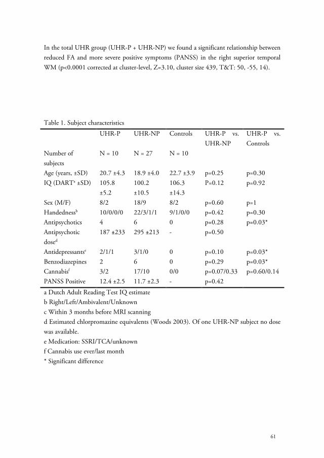
In the total UHR group (UHR-P + UHR-NP) we found a significant relationship between reduced FA and more severe positive symptoms (PANSS) in the right superior temporal WM (p<0.0001 corrected at cluster-level, Z=3.10, cluster size 439, T&T: 50, -55, 14).
Table 1. Subject characteristics UHR-P UHR-NP Controls UHR-P vs.
UHR-NP UHR-P vs. Controls
Number of subjects
N = 10 N = 27 N = 10
Age (years, ±SD) 20.7 ±4.3 18.9 ±4.0 22.7 ±3.9 p=0.25 p=0.30 IQ (DARTa ±SD) 105.8
±5.2 100.2 ±10.5
106.3 ±14.3
P=0.12 p=0.92
Sex (M/F) 8/2 18/9 8/2 p=0.60 p=1 Handednessb 10/0/0/0 22/3/1/1 9/1/0/0 p=0.42 p=0.30 Antipsychotics 4 6 0 p=0.28 p=0.03* Antipsychotic dosed
187 ±233 295 ±213 - p=0.50
Antidepressantse 2/1/1 3/1/0 0 p=0.10 p=0.03* Benzodiazepines 2 6 0 p=0.29 p=0.03* Cannabisf 3/2 17/10 0/0 p=0.07/0.33 p=0.60/0.14 PANSS Positive 12.4 ±2.5 11.7 ±2.3 - p=0.42
a Dutch Adult Reading Test IQ estimate b Right/Left/Ambivalent/Unknown c Within 3 months before MRI scanning d Estimated chlorpromazine equivalents (Woods 2003). Of one UHR-NP subject no dose was available. e Medication: SSRI/TCA/unknown f Cannabis use ever/last month * Significant difference
61

Figure 1
UHR-P vs. UHR-NP. Significant clusters; blue depicts lower FA and red depicts higher FA in UHR-P subjects. (Although clusters may appear to touch grey matter, they are in
white matter only and have been depicted on a SPM T1 template image to aid rough visual reference)
Figure 2
UHR-P vs. non-psychiatric controls. Significant clusters of lower FA in UHR-P are yellow/red.
62
D i s c u s s i o n
This study is the first comparison of whole brain WM integrity between UHR subjects who develop psychosis and those who do not. Areas where UHR-P differed from the healthy control group were located in the frontal lobe WM, particularly in the left hemisphere, containing the anterior thalamic radiation (ATR) and the inferior fronto-occipital fasciculus (IFO). Our voxel based whole brain methodology does not allow for the identification of fibre tracts, and so the implication of these tracts remains tentative. This is in line with a recent meta-analysis that reported that reduced frontal (and temporal) FA is one of the most consistent DT-MRI findings in schizophrenia (Ellison-Wright and Bullmore 2009). Areas which differed between UHR-P and UHR-NP included WM lateral of the right putamen, containing the uncinate fasciculus (UNC), the IFO and the superior longitudinal fasciculus (SLF). There is consensus that the striatum is important in schizophrenia, due to its extensive dopaminergic input, the fact that its dopamine D2 receptors are the target of most anti-psychotic drugs and the role of the striatum in cognitive, sensory, and motor processing (Shenton et al 2001). UHR subjects have been reported to have increased dopamine function in the striatum (Howes et al 2009). Furthermore, there is evidence for an association between size of the striatum and executive function in schizophrenia (Antonova et al 2004). Our findings suggest that WM abnormalities in striatal brain regions may play a role in the development of psychosis in UHR subjects.
In addition we found that UHR-P subjects had significantly lower FA in the left superior temporal lobe than UHR-NP subjects, including the SLF, IFO and inferior longitudinal fasciculus (ILF). The left superior temporal lobe WM might therefore have a role in the development of schizophrenia. Witthaus and colleagues reported decreased WM volume in the right superior temporal lobe in UHR subjects compared to healthy controls (Witthaus et al 2008) and schizophrenia has been consistently associated with left temporal GM and WM abnormalities (Ellison-Wright and Bullmore 2009;Konrad and Winterer 2008). Again, our temporal findings seem specific to the selected subcategories of UHR subjects, indicating a difference between transition and non transition. Hence, lower integrity of WM in the superior temporal lobe might increase the vulnerability for the development of frank psychosis in at risk subjects.
UHR-P subjects had higher FA than UHR-NP in the left medial temporal lobe, in WM containing the posterior thalamic radiation (PTR), IFO and ILF. A finding of higher FA in pathological studies is unusual. Reduced FA may be caused by decreases in myelination and putatively reflects a decrease in connectivity. A higher FA may be caused by for
63

instance increased neuronal density(Beaulieu 2002), and thus may reflect heightened connectivity. Our WM density analysis did not provide support for this hypothesis. Conversely, an increase in FA may also reflect tissue loss. It can arise when a region in the control group has a low FA due to crossing fibers and when loss of one of these fiber populations in the patient group causes an increase in FA. Interpretation of our result of higher FA in the UHR-P group is difficult. The result may be an artefact, but since we used a corrected conservative analysis, and the voxels are not near any ventricle we think it nonetheless reflects different microstructural integrity. It is important to realise that we compared to UHR subjects who later did not develop psychosis. Using functional MRI it was shown that activation in the medial temporal lobe is associated with positive symptoms in genetic high risk populations (Whalley et al 2007), and our study adds to evidence that the medial temporal lobe has a role in the progression to schizophrenia in high risk subjects.
We found a significant negative association between FA in the left middle temporal lobe and positive symptom severity on the PANSS (at baseline) in the UHR-P group. The middle temporal lobe has previously been associated with positive symptoms, for instance the middle (and superior) temporal lobe have been associated with formal though disorder in schizophrenia (Kircher et al 2001). Our results suggest that lower integrity of WM in the middle temporal lobe is related to increased positive symptoms, specifically in a subgroup of UHR subjects who later develop psychosis. We found a significant negative correlation in the combined UHR group between FA in the right superior temporal lobe and positive symptoms on the PANSS. Hence, superior temporal FA correlates to positive symptoms in UHR subjects, but are not specific for the development of psychosis.
Others recently reported frontal WM volume differences in UHR-P subjects compared to UHR-NP (Walterfang et al 2008a). We did not find white matter frontal differences between the UHR subgroups in our sample, but did find these when comparing to non-psychiatric controls. This could be due to the different methodology, as we used FA values and not volumetric measures, or could be because of lack of power. Further, we did not find a bilateral decrease in FA near the striatum. This may be due to lack of power or may reflect a true lateralisation, as others previously reported (Buchsbaum et al 1998) a decrease in anisotropy adjacent to the right putamen in schizophrenia, and co-registered positron emission tomography (PET) data revealed a corresponding deficit in functional connectivity between the right putamen and right frontal lobe. Also, a relative decrease in WM volume was found in the putamen and other striatal regions in patients with chronic schizophrenia (Tamagaki et al 2005).
64

This preliminary study has its limitations, particularly the relatively small sample size. Nevertheless we had sufficient power to detect significant group differences in FA. The results we report are likely to represent true differences in FA because of the use of a conservative analysis which was corrected for multiple comparisons to reduce the risk of type 1 errors.
Some subjects did not complete the full 24 months follow-up or were lost to follow-up. It is possible that more transitions to psychosis may have occurred in the UHR-NP group and this may have influenced our results. To reduce this chance, we contacted all dropouts by telephone and interviewed them about their current complaints and their current work or study. All drop-out subjects were working or in school and none reported increased symptoms. Although, in absence of a standard SCID no “objective” conclusions could be drawn, none were suspected of having developed psychosis. Also we expect that any missed transition in the UHR-NP group would attenuate the observed FA differences rather than intensify them; hence we do not think our results can be wholly explained by this.
Although the UHR-P and UHR-NP groups did not differ significantly in the number cannabis users, they may have differed in quantity of use, and we cannot exclude an effect on our results. The UHR-P group differed significantly from the healthy control group (but not from the UHR-NP group) in medication and cannabis use and thus these factors may have influenced our results.
Finally, apart from other limitations discussed in the methods section, voxel based whole brain DT-MRI methodology does not allow for the identification of fibertracts. Nevertheless this study may provide hypotheses for future fibertracking studies that could identify which tracts are implicated.
CON CL US ION We found differences in FA values and WM density between UHR-P and UHR-NP subjects in striatal and temporal regions, and between UHR-P and non-psychiatric controls in frontal lobes. These regions are known to be involved in psychosis and are associated with severity of psychotic symptoms in other samples. The putative changes in connectivity that these FA differences represent may play a role in the development of psychosis or its symptomatology, or conversely may be a result of this process. As the UHR paradigm essentially consists of attenuated psychotic symptoms, it is not possible to actually differentiate between cause and result. Still, it provides an interesting possibility to study psychosis in a “true prodromal” stage. From our data it appears that, compared to the more extensive frontal differences with non-psychiatric controls, relatively subtle
65

differences in WM integrity in temporal and striatal brain regions may influence whether a UHR subject develops psychosis..
F u n d i n g
This work was supported by the European Commission in Brussels, Belgium [QLGU-CT-2001-01081] and by Zon-MW [2630.0001]. Zon-MW is funded by the Dutch government. The funding body had no further role in study design; in the collection, analysis and interpretation of the data; in the writing of the report; and in the decision to submit the paper for publication.
A c k n o w l e d g e m e n t s
All authors contributed substantially to the conception, design, and interpretation of data. O. Bloemen took the main responsibility for drafting the article and O. Bloemen and N. Schmitz for the analysis of data. All authors contributed to revising the article for important intellectual content and gave final approval of the version to be published. No other authors have contributed substantially to this manuscript. All authors declare they have no conflict of interest.
66

R e f e r e n c e s Antonova E, Sharma T, Morris R, Kumari V (2004): The relationship between brain structure and neurocognition in schizophrenia: a selective review. Schizophrenia Research 70: 117-145.
Ashburner J, Friston KJ (2000): Voxel-based morphometry--the methods. Neuroimage 11: 805-821.
Ashburner J, Friston KJ (2001): Why voxel-based morphometry should be used. Neuroimage 14: 1238-1243.
Basser PJ (1995): Inferring microstructural features and the physiological state of tissues from diffusion-weighted images. NMR Biomed 8: 333-344.
Basser PJ, Mattiello J, LeBihan D (1994): MR diffusion tensor spectroscopy and imaging. Biophys J 66: 259-267.
Beaulieu C (2002): The basis of anisotropic water diffusion in the nervous system - a technical review. NMR Biomed 15: 435-455.
Buchsbaum MS, Tang CY, Peled S, Gudbjartsson H, Lu D, Hazlett EA et al (1998): MRI white matter diffusion anisotropy and PET metabolic rate in schizophrenia. Neuroreport 9: 425-430.
de Koning MB, Bloemen OJ, van Amelsvoort TA, Becker HE, Nieman DH, van der Gaag M et al (2009): Early intervention in patients at ultra high risk of psychosis: benefits and risks. Acta Psychiatrica Scandinavica .
Ellison-Wright I, Bullmore E (2009): Meta-analysis of diffusion tensor imaging studies in schizophrenia. Schizophrenia Research 108: 3-10.
Friedman JI, Tang C, Carpenter D, Buchsbaum M, Schmeidler J, Flanagan L et al (2008): Diffusion tensor imaging findings in first-episode and chronic schizophrenia patients. Am J Psychiatry 165: 1024-1032.
Hoptman MJ, Nierenberg J, Bertisch HC, Catalano D, Ardekani BA, Branch CA et al (2008): A DTI study of white matter microstructure in individuals at high genetic risk for schizophrenia. Schizophrenia Research 106: 115-124.
Howes OD, Montgomery AJ, Asselin MC, Murray RM, Valli I, Tabraham P et al (2009): Elevated striatal dopamine function linked to prodromal signs of schizophrenia. Arch Gen Psychiatry 66: 13-20.
Jones DK, Symms MR, Cercignani M, Howard RJ (2005): The effect of filter size on VBM analyses of DT-MRI data. Neuroimage 26: 546-554.
Kay SR, Fiszbein A, Opler LA (1987): The positive and negative syndrome scale (PANSS) for schizophrenia. Schizophrenia Bulletin 13: 261-276.
Kircher TT, Liddle PF, Brammer MJ, Williams SC, Murray RM, McGuire PK (2001): Neural correlates of formal thought disorder in schizophrenia: preliminary findings from a functional magnetic resonance imaging study. Archives of General Psychiatry 58: 769-774.
Konrad A, Winterer G (2008): Disturbed structural connectivity in schizophrenia primary factor in pathology or epiphenomenon? Schizophr Bull 34: 72-92.
Lappin JM, Dazzan P, Morgan K, Morgan C, Chitnis X, Suckling J et al (2007): Duration of prodromal phase and severity of volumetric abnormalities in first-episode psychosis. British Journal of Psychiatry - Supplement 51: s123-s127.
Miller TJ, McGlashan TH, Rosen JL, Cadenhead K, Cannon T, Ventura J et al (2003): Prodromal assessment with the structured interview for prodromal syndromes and the scale of prodromal symptoms: predictive validity, interrater reliability, and training to reliability. Schizophr Bull 29: 703-715.
Mori,S, Wakana,S, Nagae-Poetscher,L, van Zijl,P (2005): MRI Atlas of Human White Matter. Amsterdam: Elsevier.
67

Muñoz Maniega S, Lymer GK, Bastin ME, Marjoram D, Job DE, Moorhead TW et al (2008): A diffusion tensor MRI study of white matter integrity in subjects at high genetic risk of schizophrenia. Schizophrenia Research 106: 132-139.
Olsen KA, Rosenbaum B (2006): Prospective investigations of the prodromal state of schizophrenia: review of studies. Acta Psychiatrica Scandinavica 113: 247-272.
Peters BD, de Haan L., Dekker N, Blaas J, Becker HE, Dingemans PM et al (2008): White Matter Fibertracking in First-Episode Schizophrenia, Schizoaffective Patients and Subjects at Ultra-High Risk of Psychosis. Neuropsychobiology 58: 19-28.
Peters BD, Schmitz N, Dingemans PM, van Amelsvoort TA, Majoie CB, den Heeten GJ et al (2009): Preliminary evidence for reduced frontal white matter integrity in subjects at ultra-high-risk for psychosis. Schizophrenia Research : Epub 2009 Apr 15.
Pierpaoli C, Basser PJ (1996): Toward a quantitative assessment of diffusion anisotropy. Magn Reson Med 36: 893-906.
Schmand B, Bakker D, Saan R, Louman J (1991): [The Dutch Reading Test for Adults: a measure of premorbid intelligence level]. Tijdschrift voor Gerontologie en Geriatrie 22: 15-19.
Shenton ME, Dickey CC, Frumin M, McCarley RW (2001): A review of MRI findings in schizophrenia. Schizophrenia Research 49: 1-52.
Spitzer RL, Williams JB, Gibbon M, First MB (1992): The Structured Clinical Interview for DSM-III-R (SCID). I: History, rationale, and description. Archives of General Psychiatry 49: 624-629.
Talairach,J, Tournoux,P (1988): Co-planar stereotaxic atlas of the human brain . New York: Thieme.
Tamagaki C, Sedvall GC, Jonsson EG, Okugawa G, Hall H, Pauli S et al (2005): Altered white matter/gray matter proportions in the striatum of patients with schizophrenia: a volumetric MRI study. The American Journal of Psychiatry 162: 2315-2321.
Walterfang M, McGuire PK, Yung AR, Phillips LJ, Velakoulis D, Wood SJ et al (2008a): White matter volume changes in people who develop psychosis. British Journal of Psychiatry 193: 210-215.
Walterfang M, Yung A, Wood AG, Reutens DC, Phillips L, Wood SJ et al (2008b): Corpus callosum shape alterations in individuals prior to the onset of psychosis. Schizophrenia Research 103: 1-10.
Whalley HC, Gountouna VE, Hall J, McIntosh A, Whyte MC, Simonotto E et al (2007): Correlations between fMRI activation and individual psychotic symptoms in un-medicated subjects at high genetic risk of schizophrenia. BMC Psychiatry 7: 61.
Witthaus H, Brune M, Kaufmann C, Bohner G, Ozgurdal S, Gudlowski Y et al (2008): White matter abnormalities in subjects at ultra high-risk for schizophrenia and first-episode schizophrenic patients. Schizophrenia Research 102: 141-149.
Woods SW (2003): Chlorpromazine equivalent doses for the newer atypical antipsychotics. Journal of Clinical Psychiatry 64: 663-667.
World Health Organization. Composite International Diagnostic Interview - Version 1.1. World Health Organization. WHO . 1993. Geneva.
Yung AR, Phillips LJ, Yuen HP, Francey SM, McFarlane CA, Hallgren M et al (2003): Psychosis prediction: 12-month follow up of a high-risk ("prodromal") group. Schizophr Res 60: 21-32.
68

C h a p t e r 4
Challenge and therapeutic studies using alpha-methyl-para-tyrosine (AMPT) in neuropsychiatric disorders: a review. O.J.N. Bloemen, M.B. de Koning E. Boot, J. Booij, T.A.M.J. van Amelsvoort: Central Nervous System Agents in Medicinal Chemistry, 2008, 8, 249-256.

A b s t r a c t Alpha-methyl-para-tyrosine (AMPT) temporarily inhibits tyrosine hydroxylase, the rate limiting step in the dopamine biosynthesis cascade. AMPT has been approved for clinical use in phaeochromocytoma in 1979. Recently however, AMPT has been increasingly employed as a pharmacological challenge in acute dopamine depletion studies including neuroimaging studies. The use of this exciting challenge technique allows us to increase our understanding of dopaminergic neurotransmission in the brain. In addition, there have been clinical reports that AMPT may be useful to treat movement disorders like dystonia, dyskinesia and Huntington’s chorea, psychiatric disorders like mania, psychosis, obsessive compulsive disorder and substance abuse as well as behavioral problems in 22q11 deletion syndrome. In this review we will discuss the effects of AMPT in challenge studies that have been reported in humans. Furthermore we will review all studies reporting therapeutic effects of AMPT in neuropsychiatric disorders and adverse effects associated with AMPT use reported in both challenge and therapeutic research.
70

I n t r o d u c t i o n Depletion of selected monoamines, such as dopamine and serotonin, is a method that is used as a way of elucidating the pathogenesis of neuropsychiatric disorders. In these so called “challenge studies” different techniques are used to acutely and temporarily lower the levels and consequently the function of monoamines. The currently most established depletion tests for norepinephrine (NE) and dopamine (DA) are phenylalanine/tyrosine depletion (APTD) and alpha-methyl-para-tyrosine (AMPT). Depletion of serotonin (5-HT) is mainly achieved by acute tryptophan depletion (ATD) and para-chlorophenylalanine (PCPA) (Booij et al 2003).
Many neuropsychiatric diseases have underlying DA and or NE abnormalities. For example schizophrenia and psychosis are hypothesized to be related to mesolimbic and striatal hyperdopaminergic states according to the DA hypothesis (van Rossum 1966). Other disorders related to dopamine dysfunction include movement disorders such as Parkinson’s disease, dystonia, dyskinesia and Huntington’s disease, and neuropsychiatric disorders such as attention deficit hyperactivity disorder (ADHD), addiction, and obsessive compulsive disorder (OCD).
AMPT is a specific competitive inhibitor of tyrosine hydroxylase, the rate-limiting enzyme in the synthesis of DA (and NE) from tyrosine (Engelman et al 1968b). Studies of human cerebrospinal fluid after acute AMPT administration found a 50–70% decrease in the DA metabolite homovanillic acid (HVA), and no change in the 5-HT metabolite 5-hydroxyindoleacetic acid (5-HIAA) (Brodie et al 1971;Bunney, Jr. et al 1971). Urinary levels of the NE metabolite vanillylmandelic acid (VMA) and 3-methoxy-4-hydroxy-phenylglycol (MHPG) are decreased by 25-50% (Brodie et al 1971;Engelman et al 1968a). The DA depleting effect of AMPT is being used clinically in the management of phaeochromocytoma, to reduce catecholamine-induced pre- and intra-operative complications. Moreover, by using AMPT it is also possible to specifically investigate DA/NE neurotransmitter systems through challenge paradigms.
In the past decade, AMPT has been increasingly used as a pharmacological challenge in humans. In addition, observations have been made of potential beneficial effects of AMPT in the treatment of neuropsychiatric disorders. In this review we will discuss the effects of AMPT on humans in neuropsychiatric research and the clinical management of neuropsychiatric disorders.
71

M e t h o d s Medical databases (PubMed) were used to identify relevant literature (period between 1965 and May 2008). Keywords used were: “ampt”, "alpha-methyl-para-tyrosine", “demser”, “metyrosine” and "alpha-methyl-p-tyrosine". Additionally reference lists of retrieved articles were checked. We included all human challenge and treatment studies relevant to neuropsychiatric disorders found in PubMed. We excluded animal and in vitro studies from the review.
72

C l i n i c a l u s e o f A M P T Currently AMPT has only been approved for clinical use in phaeochromocytoma (since 1979), which is a catecholamine-producing tumor of the sympathetic nervous system with an incidence of 0.2-0.4% in hypertensive patients (Yeo and Roman 2005). Most of these tumors occur in the adrenal glands and secrete norepinephrine and epinephrine but they may also rarely secrete L-DOPA and/or dopamine. The typical manifestation is sustained or paroxysmal hypertension, with the triad of severe headaches, palpitations and diaphoresis resulting from hormone excess (Reisch et al 2006). Before the introduction of catecholamine blockade, surgical resection of phaeochromocytoma was associated with a mortality rate of 24 to 50% (Levine and McDonald 1984;Pullerits et al 1988). This was due to the fact that intra-operative catecholamine release, caused by anesthesia or tumor manipulation, induced severe hypertension, arrhythmia’s and stroke.
Adding AMPT to the alpha-adrenic blockade with phenoxybenzamine has been associated with the need for less intra-operative medication for the control of blood pressure, lower intra-operative fluid requirements, and lower blood loss and decreased surgical morbidity (Perry et al 1990;Steinsapir et al 1997). In recent years surgical mortality has dropped to 0% to 2,9% (Kinney et al 2002). Nevertheless, sometimes hypertensive crisis still occurs when AMPT is not combined with adequate alpha-blockade (Green et al 1982;Hauptman et al 1983;Ram et al 1985). Although AMPT has demonstrated its effectiveness in the management of patients with phaeochromocytoma in reducing symptoms (Engelman et al 1968a), it does not have a significant beneficial effect on essential hypertension. Thus the clinical use of AMPT in the treatment of hypertension seems limited to hypertension induced by catecholamine producing phaeochromocytoma, and is based on depleting the causal agent for the associated symptoms, norepinephrine.
Some reports have suggested that AMPT may have beneficial effects in the treatment of other conditions. Glaucoma is a group of diseases characterized by progressive damage of the optic nerve involving loss of retinal ganglion cells in a characteristic pattern of optic neuropathy. Intraocular pressure is a significant risk factor for developing glaucoma. It is the second most common cause of irreversible blindness in the world and affects more than 70 million people worldwide (Congdon et al 2003). Although glaucoma is frequently mentioned in literature as an indication for AMPT use, there are no studies to support this. Engelman discussed the issue briefly in his study where two patients with glaucoma were included, and found no improvement (Engelman et al 1968a). In a more recent study AMPT appeared to have a beneficial effect in vitro on hyperpigmentation (Drago et al 1999), a side effect of the anti-glaucoma drug latanoprost, but another study could not replicate this (Loeffler et al 2001). No other studies were found, and thus it seems fair to conclude that there is no evidence that AMPT has beneficial effects on patients with glaucoma.
73

A M P T a s a p h a r m a c o l o g i c a l c h a l l e n g e i n n e u r o p s y c h i a t r i c r e s e a r c h
Challenge studies using AMPT are abundant, covering a wide area of disciplines both in human and in animal populations. In the following paragraph we will discuss all human studies with relevance to neuropsychiatric disorders. Animal and in vitro studies were not included, as most researched highly specific physiological and pathological biochemical pathways which are beyond the neuropsychiatric scope of this review. Common AMPT depletion procedures administer a total amount of 4500 – 8000 mg of AMPT in a 25 to 48 hour study period (Booij et al 2003). The first challenge studies date back to the 1970’s. In that period Shopsin studied depressed patients who showed an antidepressant response to the tricyclic drug imipramine, to elucidate the biochemical pathway behind this antidepressant (Shopsin et al 1975). AMPT did not cause relapse of depression and thus Shopsin concluded that serotonergic mechanisms were most likely involved in the antidepressant effects of imipramine (Shopsin et al 1975).
HEAL THY S UBJE CTS An AMPT challenge using common doses does not usually induce depressive symptoms in healthy, but may have a slight negative effect on mood, which relatively (and absolutely) increases when combined with sleep deprivation (McCann et al 1992;McCann et al 1993). Furthermore AMPT has been reported to decrease attention, alertness, happiness and increase sleepiness, tension and anger (Fujita et al 2000;Krahn et al 1999;Laruelle et al 1997;McCann et al 1992;McCann et al 1993;McCann et al 1995;Salomon et al 1997;Verhoeff et al 2003;Verhoeff et al 2001). Most symptoms can be reversed by administering L-dopa and thus seem to be dopaminergically modulated (McCann et al 1995). Additionally, Tychsen (Tychsen and Sitaram 1989) reported an AMPT-induced increase in saccadic eye movements in a small study (N=3) when administering 3g/day for 3 consecutive days.
Acute administration of AMPT also has a negative effect on subjective wellbeing. Subjects showed declining scores on mental functioning, emotional regulation and physical functioning, as measured by the Subjective Well-being Under Neuroleptic Treatment Scale (SWN) (Boot et al 2008). Transient extrapyramidal side effects (EPS) of varying severity (hypokinesia, rigidity, tremor, salivation) have been commonly reported after acute DA depletion with AMPT (de Haan et al 2005;Engelman et al 1968a;Fujita et al 2000;Krahn et al 1999;Laruelle et al 1997;McCann et al 1990;Verhoeff et al 2001).
Furthermore increased anxiety is often reported (de Haan et al 2005;Engelman et al 1968a;Krahn et al 1999;McCann et al 1993), again varying from slight increases to recurrent panic attacks (Fujita et al 2000;McCann et al 1991) requiring subject withdrawal from the study, but symptoms never persist after discontinuing AMPT medication. Catecholamines thus appear to be involved in anxiety regulation, since administering L-dopa also reverses AMPT-induced anxiety (McCann et al 1995). However other studies
74

did not find increased anxiety after AMPT (Laruelle et al 1997;Verhoeff et al 2001), and no apparent dose-response effect can be distilled from the studies. Healthy subjects were also reported to display transient decreases in total sleep for a few days after stopping AMPT (Ahlenius et al 1973;Anand et al 1999;Charalampous and Brown 1967;Engelman et al 1968a;Sjoerdsma et al 1965;Wyatt et al 1971).
Imaging studies have shown that AMPT depletion provides a non-invasive method for estimating synaptic DA concentrations in healthy subjects using positron emission tomography (PET) (Verhoeff et al 2001) and single photon emission computed tomography (SPECT) (Fujita et al 2000;Laruelle et al 1997). The effect of DA depletion can be measured in-vivo using radiotracers for the D2 receptor. Indeed, acute dopamine depletion induced by AMPT leads to an increased D2 receptor binding, but does not significantly alter D1 receptor binding (Verhoeff et al 2002). Data suggest that endogenous DA occupies between 20% and 30% of D2 receptors in healthy subjects which is reflected in an increase in D2 binding potential following DA depletion. Importantly, it has been argued that such an increase cannot be explained by D2 receptor up-regulation, which would require longer than a week of DA depletion (Laruelle et al 1997;Narang and Wamsley 1995). Relatively low doses of AMPT can be used to obtain adequate results (Boot et al 2008), while producing acceptable levels of side effects. A recent PET study (Cropley et al 2008) reported that [18F]fallypride combined with AMPT challenge may be unreliable for estimating tonic or baseline dopamine levels in humans, as no significant change in binding potential was found in striatal and extrastriatal regions after AMPT administration. These findings were in contrast with another recent study which found that a higher dose of AMPT (66 mg/kg/24h vs. 23 mg/kg/24h; total dose was 71 mg/kg vs. 43 mg/kg) significantly increased the binding potential in the caudate nucleus, putamen, ventral striatum, and substantia nigra (Riccardi et al 2008). Although the dose was lower, Cropley et al (Cropley et al 2008) reported similar plasma levels of AMPT but absent side effects. These results are also in contrast to previous SPECT and PET studies using different radioligands, which reported increases of D2 radioligand binding in the striatum after an AMPT challenge using [123I]IBZM (Laruelle et al 1997) or [11C]raclopride (Verhoeff et al 2002;Verhoeff et al 2001), and in the temporal cortex with [123I]epidepride (Fujita et al 2000). These conflicting results may be the result of small sample size, interference with the amphetamine challenge one week before in Cropley’s study, dose related, or due to different characteristics of the radioligands used.
Prolactin (PRL) is commonly used as a marker for DA depletion, as DA inhibits the release of PRL at the level of the pituitary gland (Freeman et al 2000). Treatment with AMPT consistently produces two- to four-fold increases in PRL levels (Verhoeff et al 2002) although DA is not the only factor controlling PRL levels, and there is no direct relation between DA levels in the hypophysial stalk and serum PRL levels (Freeman et al 2000). Women display higher AMPT induced PRL secretion than men, possibly explained by the PRL synthesis promoting effect of estradiol on the pituitary gland (Zimmermann et al 1996).
75

Melatonin, and its urinary metabolite 6-hydroxymelatonin (6-MS) are regulated by noradrenergic neurons located in the sympathetic superior cervical ganglion via NE-induced stimulation of postsynaptic beta-adrenergic receptors on the pineal gland (Moore 1978). Melatonin and 6/MS were reported to be a superior marker for NE depletion by AMPT compared to MHPG as there are sex differences in MHPG secretion and only 20-65% of circulating MHPG is derived from the brain (Krahn et al 1999;Zimmermann et al 1994;Zimmermann et al 1996). The same research group investigated the possibility that leptin would be modulated by NE, but found no evidence for this when performing an AMPT challenge on ten healthy individuals (Zimmermann et al 1998).
Although short-term effects of AMPT include TSH stimulation through the decrease in DA’s inhibiting effect (Nicoletti et al 1986;Plosker et al 1995), prolonged use of AMPT was also reported to inhibit thyroid-stimulating hormone (TSH) secretion and significantly attenuate the circadian rhythm of TSH (Zimmermann et al 2001). This is probably caused by NE’s inhibitory effect on thyroid-releasing hormone (TRH); animal research has indicated that the depletion of NE occurs later in time than DA depletion by AMPT (Kitsikis and Roberge 1973).
In conclusion, AMPT challenge has little effect on mood but can cause transient sedation, EPS and anxiety in healthy subjects, particularly at higher doses.
MOOD D IS ORD ERS Some patients with a history of medically-treated depression experience a return of symptoms (Brodie et al 1971;Bunney, Jr. et al 1971;Engelman et al 1968a;Moreno et al 2006) after DA depletion with AMPT, which seems to depend on the mechanism of prior anti-depressant medication (Delgado et al 1993;Delgado et al 2002;Miller et al 1996a;Miller et al 1996b) but also on individual vulnerability to depression (Berman et al 1999). AMPT did not lead to worsening of their condition in the actively ill (Berman et al 2002;Heninger et al 1996;Miller et al 1996b). Furthermore, AMPT also seems to induce relapse in seasonal affective disorder (Lam et al 2001;Neumeister et al 1998), but did not so in eight bipolar patients who were stable on lithium medication (Anand et al 1999). A recent study (Bremner et al 2003) showed that the effect of AMPT on prefrontal, orbitofrontal and thalamic metabolism (measured with [18F]FDG PET) could differentiate between patients who would experience a return of depressive symptoms after AMPT depletion and those who would not (all participants were patients with a depression in stable remission on NE re-uptake inhibitors). The patients with a return of symptoms showed a decreased metabolism (from elevated baseline) and the patients without a return of symptoms showed an increase in metabolism (from relatively lower baseline). In conclusion, occurrence of a relapse of depressive symptoms in stable patients after AMPT seems dependent on the mechanism of prior anti-depressant medication and AMPT does not worsen depression in actively ill patients. Brain glucose metabolism in successfully treated depressed patients may predict depressive responses to AMPT treatment.
76
SCHI ZOPHREN IA Patients with schizophrenia showed a decrease in subjective wellbeing after an AMPT challenge, and those patients who had a history of persistent dysphoria in response to previous neuroleptic therapy had significantly greater dysphoric responses scored on the Drug Attitude Inventory (DAI) and the Addiction Research Center Inventory (ARCI) than patients who did not have such a history (Voruganti et al 2001). In the same study, Voruganti reported an inverse correlation between subjective responses plus EPS (increases in dysphoria scores on ARCI and DAI and increases on the Barnes Akathisia Scale and Simpson-Angus Scale) and the changes in D2 receptor binding ratios. This suggests that schizophrenic patients with relatively lower (but probably still increased compared to healthy controls) endogenous DA activity are more susceptible to dysphoric response and EPS following dopamine challenge with AMPT than schizophrenic patients with a relatively higher DA activity. Later research by the same group replicated these results (Voruganti and Awad 2006). Abi-Dargham reported an increased D2 receptor availability after AMPT in the schizophrenia group as compared to the control group, which implies that schizophrenic patients have elevated neostriatal DA levels compared to controls (Abi-Dargham et al 2000). 22q11 Deletion syndrome (22q11DS) was also reported to have disrupted dopaminergic neurotransmission, which might explain their susceptibility for psychiatric disorders like psychosis and schizophrenia (Boot et al 2007). Thus, AMPT seems useful in elucidating the role of DA in the pathogenesis and susceptibility to side effects of medication of these disorders.
SUBS TAN CE-IN D UCED D IS ORD ERS McCann reported differential responses to an AMPT challenge in cognitive measures of speed and impulsivity and of sleep architecture when comparing 25 abstinent 3, 4-methylenedioxymethamphetamine (MDMA, the main component of the recreational drug commonly known by its street name XTC) users to 23 controls (McCann et al 2007). AMPT administration led to more prominent increases in cognitive speed and impulsivity in MDMA users than in controls and MDMA users had less difficulty falling asleep and had larger increases in rapid eye movement (REM) latency following AMPT. This supports the theory that MDMA can lead to subtle functional deficits in brain functions involving reciprocal 5-HT/catecholamine interactions.
77

A M P T a s a t h e r a p e u t i c i n n e u r o p s y c h i a t r i c r e s e a r c h
DYS TON IA AN D D YS KIN ESIA There have been some reports of beneficial effects of AMPT in patients with movement disorders including dystonia, dyskinesia, and akathisia, although most are case-reports. A recent paper presented three case studies in which AMPT was used in patients suffering form dystonia and dyskinesia. In the first case, AMPT was added to antipsychotic treatment in a woman who after brain injury had hallucinations and “attacks” of painful dystonia. She had only mild dystonia once a week after adding AMPT, which deteriorated when AMPT was stopped. In the second case tardive dystonia improved after adding AMPT in a retarded woman who had used clozapine. In the third case tardive dyskinesia was successfully treated with AMPT (Ankenman and Salvatore 2007).
Treating dystonia with AMPT was also studied by Fahn (Fahn 1983b), who reported little benefit in an unspecified number of patients. Lang et al (Lang and Marsden 1982) reported that some individual dystonic patients seemed to benefit from the addition of AMPT, but that the results overall were disappointing. Interestingly, McCann reported that 5 of 24 healthy volunteers even developed dystonia when administered AMPT at a total dose between 5,25 and 6,75 grams (McCann et al 1990). The paradoxal fact that AMPT is used to treat dystonia but that AMPT can also induce it may be explained by Hornykiewicz’ view that any disturbance in the balance between striatal dopamine, norepinephrine, serotonin, and acetylcholine systems that leads to a functional overactivity of cholinergic brain mechanisms may be sufficient to cause dystonia in susceptible individuals (Hornykiewicz et al 1986).
In patients with tardive dyskinesia, Gerlach et al reported a decrement in frequency and amplitude, but an increase of duration of tongue protrusion and/or mouth opening after treatment with AMPT. In contrast, biperiden (an anticholinergic agent) had opposite effects (Gerlach 1977a;Gerlach 1977b;Gerlach and Thorsen 1976). Fahn et al published some promising results on the treatment of tardive dyskinesia with AMPT. He used AMPT on 14 patients diagnosed with (neuroleptic-induced) tardive dyskinesia and akathisia in an uncontrolled open label trial and reported improvement when using AMPT as a supplement to reserpine treatment, which is a pre-synaptically acting dopamine depletor (Fahn 1983b). He reported on this earlier (Fahn 1978), when he treated 7 patients with reserpine and AMPT, (5 responded to blindly substituting AMPT for placebo and back) and later on reported more long-term results (Fahn 1983a). The last report concluded that the addition of AMPT to the pre-synaptic dopamine depletors reserpine or tetrabenazine (TBZ) increased the potency of the treatment.
It has been proposed, as a possible biochemical pathway, that the additive effect of AMPT in dystonia and tardive dyskinesia may lie in the fact that AMPT mainly limits dopamine
78

synthesis in the cytosolic pool (or newly synthesized pool) of dopaminergic cells, and that reserpine and TBZ prevent dopamine uptake into the vesicular pool (stored pool) (Kannari et al 2000;Yavich 1996).
In conclusion, AMPT appears to have beneficial effects on patients with tardive dyskinesia, and may potentiate reserpine and TBZ treatment, nevertheless evidence is scarce and there is obvious need for protocolised double-blind, placebo-controlled trials to confirm this. AMPT appears to benefit some individual patients with dystonia, although the results overall are not promising.
HUN TIN G TON’S CHO REA Lang (Lang and Marsden 1982) reported that of 9 patients with Huntington’s chorea, 3 were unable to tolerate the addition of AMPT to their TBZ or reserpine treatment and two had no change in symptoms, but 4 experienced a marked improvement in movements scores compared to their scores on TBZ alone. Placebo substitution yielded significant increases in chorea in all 4 patients, and as an add-on, AMPT appeared to potentiate the effect of TBZ, as the mean TBZ dose could be reduced from 142mg to 58mg after addition of AMPT (Lang and Marsden 1982). Already in the 1960’s Birkmayer reported a decline in chorea in six patients within a day after daily administration of 200 mg AMPT intravenously (Birkmayer and Mentasti 1967) and total absence of chorea in one of five subjects 60 minutes after administering 500 mg AMPT intravenously (Birkmayer 1969).
WES T SYN D ROM E Twelve children with newly diagnosed untreated infantile spasms, also known as West Syndrome, were treated with AMPT. Response to therapy was determined objectively with 24-h polygraphic/video monitoring techniques and was defined as cessation of spasms and disappearance of the hypsarrhythmic EEG pattern. Two of 12 patients treated with AMPT responded to therapy and one patient had a return of symptoms after AMPT medication was stopped (Hrachovy et al 1989), which provides evidence that AMPT had a beneficial role on the symptoms in this disorder.
MAN IA In two early studies AMPT appeared to have a beneficial effect on some patients with a manic episode, although studies were very small and open-labelled (Brodie et al 1971;Bunney, Jr. et al 1971). After withdrawal of AMPT some patients experienced a transient relapse of hypomanic symptoms and less need for sleep (Anand et al 1999;Brodie et al 1971;Bunney, Jr. et al 1977). Interestingly, decreased need for sleep, decreased total sleep hours and decreased REM sleep are also reported in healthy subjects (Ahlenius et al 1973;Anand et al 1999;Charalampous and Brown 1967;Engelman et al 1968a;Sjoerdsma et al 1965;Wyatt et al 1971). Equally, depressed patients displayed significant decreased sleep and even hypomanic symptoms after stopping AMPT (Bunney, Jr. et al 1977). This effect had no correlation with the dose of AMPT (Bunney, Jr. et al 1977) and was hypothesized to be related to changes in melatonin secretion (Zimmermann et al 1994). Bunney et al reported that post-AMPT hypomanic patients had nearly equal decreases in
79

sleep as post-AMPT non-hypomanic bipolar patients, and thus argued that this decrease cannot be fully explained by an increase in mania (Bunney, Jr. et al 1977). Then again, since healthy subjects and depressed patients also showed decreases in sleep and hypomania post-AMPT, it is tempting to suggest that the “insomnia” is not an isolated symptom but rather part of a hypomanic spectrum, involving a larger portion of subjects. The hypomanic symptoms and the decrease in sleep may be explained by increased sensitivity (hyper- or supersensitivity) of DA receptors following depletion of DA, as proposed by Bunney (Anand et al 1999;Bunney, Jr. et al 1977). So although there are some early reports of beneficial effects of AMPT, Brody et al reported it to be inferior to lithium carbonate (Brodie et al 1971), and no later therapeutic studies were performed in manic subjects. There appears to be a rebound effect after stopping AMPT medication, yielding hypomanic symptoms and/or sleep decrements in manic, depressed and healthy subjects which is hypothesized to be caused by increased DA receptor sensitivity.
OCD One (double-blind, placebo-controlled) study reported no effect in 6 non-medicated patients with non-tic-related OCD (Longhurst et al 1999). To our knowledge, no other AMPT studies on OCD were done. Sweet et al administered AMPT to 6 patients with Tourette syndrome, which induced a sustained dramatic decline of profound movement and vocal tics in one patient for at least a year and improved symptoms in two more for a shorter period (Sweet et al 1974).
SUBS TAN CE A BUS E As dopamine is known to be involved in substance abuse, AMPT was also studied for its potential to suppress drug craving and drug dependence. Pozuels reported cessation of amphetamine, methadone and heroin use and in craving in a case report (Pozuels 1976) and Jönssen et al reported lowered amphetamine-induced euphoria in patients with AMPT treatment (Jonsson et al 1971). This lowered euphoria was also observed in relation to alcohol use (Ahlenius et al 1973). Regarding cocaine abuse, Stine’s study, although reporting a trend towards a diminished cocaine-induced “high”, did not provide strong support for the therapeutic potential of AMPT depletion in cocaine abuse (Stine et al 1997).
22Q11DS There is preliminary evidence that patients with 22q11DS might profit from AMPT medication. Recently, Boot et al reported beneficial subjective effects of acute AMPT medication (Boot et al 2007) and in an earlier small uncontrolled open-label trial (Graf et al 2001), three out of four patients continued AMPT after the trial due to increased subjective well-being. Furthermore a recent case report showed reduction of psychotic symptoms and mood lability in a 22q11DS patient who did not respond to regular treatment, and was treated with AMPT (Carandang and Scholten 2007). This is interesting as a lot of evidence is pointing to dopaminergic dysregulation, caused by the genetic anomalies in this disorder (Boot et al 2007).
80

SCHI ZOPHREN IA This anti-psychotic effect of AMPT has also been studied in schizophrenic patients. Carlson’s group (Carlsson et al 1972;Carlsson et al 1973;Larsson et al 1984;Walinder et al 1976) consistently showed that addition of AMPT led to reduced doses of concurrent neuroleptic treatment among stable chronic schizophrenia patients. Nevertheless, others could not reproduce these results (Charalampous and Brown 1967;Gershon et al 1967;Nasrallah et al 1977;Voruganti and Awad 2006;Wolkin et al 1994), or found the neuroleptic-potentiating effect to be too limited in relation to adverse effects (Magelund et al 1979a;Magelund et al 1979b). Abi-Dargham reported that high synaptic dopamine levels predicted improvement of positive symptoms after AMPT medication (Abi-Dargham et al 2000).
In conclusion, the current literature, although scarce, suggests that AMPT may have beneficial effects on dyskinesia, Huntington´s chorea, Tourette syndrome, mania, substance abuse, 22q11DS and psychosis. However, double-blind, placebo-controlled studies are lacking and are needed before firm conclusions can be drawn.
S a f e t y i s s u e s Depletion of catecholamines can cause adverse effects of varying severity. The most common adverse reactions to acute AMPT administration are moderate to severe sedation, anxiety and EPS, all of which were described in the AMPT challenge section above.
Diarrhea is reported in up to 10% of subjects, but is not reported in most acute challenge studies (Engelman et al 1968a;Laruelle et al 1997;McCann et al 1995;Perry et al 1990). Galactorrhea could be expected due to higher PRL levels in chronic use, but is not commonly reported. Engelman et al reported that one of 52 of chronic AMPT users patients experienced galactorrhea (Engelman et al 1968a). Acute dystonia is also reported in 0-25% patients after AMPT challenge (Abi-Dargham et al 2000;Fujita et al 2000;Laruelle et al 1997;McCann et al 1990) or after AMPT withdrawal (Krahn et al 1999), and akathisia was reported in 0-60% of patients (Fujita et al 2000;Sweet et al 1974;Voruganti and Awad 2006). Crystalluria was reported in 0-66% of patients (Abi-Dargham et al 2000;Krahn et al 1999;Steinsapir et al 1997;Sweet et al 1974). Animal studies have indicated that crystallization of AMPT in the urinary tract can be prevented by high fluid intake (Hook and Moore 1969;Moore et al 1967). Bearing this in mind most studies recommended that patients drink at least 2 liters of fluid per day throughout the study, and urinary sediments were examined for crystals.
Some individual subjects may react dramatically to AMPT depletion, and experience a wide range of symptoms including obsessive-compulsive symptoms, thought disorders, and anxiety and depressive symptoms, highlighting the importance of the role of dopamine in major psychiatric disorders (de Haan et al 2005). Steinsapir reported visual hallucinations and psychosis in one patient taking AMPT for at least 3 weeks at a dose of 1-2 grams per
81

day as pre-operative treatment for pheochromocytoma (Steinsapir et al 1997). One case report (Burke et al 1981) described a patient with Huntington’s chorea who was treated with TBZ (350 mg per day) and AMPT (250 mg per day) for 7 months, who developed hyperpyrexia, hyperthermia and dystonia, consistent with a diagnosis of neuroleptic malignant syndrome (NMS), suggesting that not only DA receptor blockade but also (low dose) DA depletion can cause this potentially deadly condition.
When using AMPT therapeutically, obviously one should analyze if the benefits of the treatment outweigh the discomfort due to possible side effects. Side effects are always reported to be temporary, and disappear after the withdrawal of the AMPT medication. Apart from sedation, which is almost unanimously seen in patients, the different transient adverse effects can be totally absent in one study and quite common in others. The reason for this is unclear. It can be a result of the applied dose of AMPT, the regimen of administration, the length or methodology of the study. Common AMPT depletion procedures administer a total amount of 4500 – 8000 mg of AMPT in a 25 to 48 hour study period (Booij et al 2003). To the best of our knowledge, the relation between dose of AMPT and the occurrence of side effects has not been studies yet. Nevertheless there is evidence that high doses of AMPT cause some of the more severe symptoms (de Haan et al 2005), and that moderate weight-adjusted doses (40 mg AMPT per kilogram body weight) yield satisfactory DA depletion results (Boot et al 2008). Therefore, limiting the dose of AMPT as much as appropriate seems recommendable.
C o n c l u s i o n AMPT is increasingly popular as a means of depleting DA in human clinical research and provides an useful paradigm for elucidating the function of catecholaminergic pathways. Nevertheless the depletion methodology is time-consuming and produces variable dose-dependent relationships. Furthermore there have been no test-retest studies to our knowledge. As a therapeutic agent it has been approved to treat the consequences of phaeochromocytoma. In addition, beneficial effects have been studied in a range of other neuropsychiatric disorders and there is promising evidence of varying quality and persuasiveness that AMPT has some beneficial effects on dyskinesia, Huntington´s chorea, Tourette syndrome, mania, substance abuse, 22q11DS and psychosis. These results might warrant further research since controlled studies are mostly lacking. This is equally true with respect to the unclear dose-response relationship for side-effects. Severe adverse effects are rare, and side effects are transient and do not commonly cause subjects to withdraw from studies. Nevertheless discomfort due to higher doses of AMPT is common and it can be recommended to limit the dose of AMPT administered to subjects as much as appropriate.
82
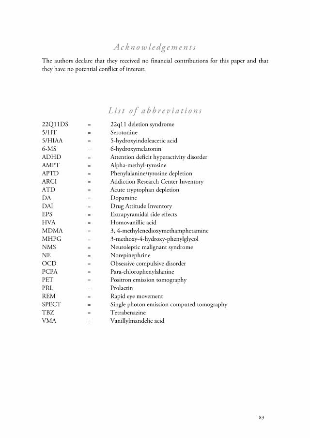
A c k n o w l e d g e m e n t s The authors declare that they received no financial contributions for this paper and that they have no potential conflict of interest.
L i s t o f a b b r e v i a t i o n s 22Q11DS = 22q11 deletion syndrome 5/HT = Serotonine 5/HIAA = 5-hydroxyindoleacetic acid 6-MS = 6-hydroxymelatonin ADHD = Attention deficit hyperactivity disorder AMPT = Alpha-methyl-tyrosine APTD = Phenylalanine/tyrosine depletion ARCI = Addiction Research Center Inventory ATD = Acute tryptophan depletion DA = Dopamine DAI = Drug Attitude Inventory EPS = Extrapyramidal side effects HVA = Homovanillic acid MDMA = 3, 4-methylenedioxymethamphetamine MHPG = 3-methoxy-4-hydroxy-phenylglycol NMS = Neuroleptic malignant syndrome NE = Norepinephrine OCD = Obsessive compulsive disorder PCPA = Para-chlorophenylalanine PET = Positron emission tomography PRL = Prolactin REM = Rapid eye movement SPECT = Single photon emission computed tomography TBZ = Tetrabenazine VMA = Vanillylmandelic acid
83

R e f e r e n c e s Abi-Dargham A, Rodenhiser J, Printz D, Zea-Ponce Y, Gil R, Kegeles LS et al (2000): Increased baseline occupancy of D2 receptors by dopamine in schizophrenia. Proc Natl Acad Sci U S A 97: 8104-8109.
Ahlenius S, Carlsson A, Engel J, Svensson T, Sodersten P (1973): Antagonism by alpha methyltyrosine of the ethanol-induced stimulation and euphoria in man. Clin Pharmacol Ther 14: 586-591.
Anand A, Darnell A, Miller HL, Berman RM, Cappiello A, Oren DA et al (1999): Effect of catecholamine depletion on lithium-induced long-term remission of bipolar disorder. Biol Psychiatry 45: 972-978.
Ankenman R, Salvatore MF (2007): Low dose alpha-methyl-para-tyrosine (AMPT) in the treatment of dystonia and dyskinesia. J Neuropsychiatry Clin Neurosci 19: 65-69.
Berman RM, Narasimhan M, Miller HL, Anand A, Cappiello A, Oren DA et al (1999): Transient depressive relapse induced by catecholamine depletion: potential phenotypic vulnerability marker? Arch Gen Psychiatry 56: 395-403.
Berman RM, Sanacora G, Anand A, Roach LM, Fasula MK, Finkelstein CO et al (2002): Monoamine depletion in unmedicated depressed subjects. Biol Psychiatry 51: 469-473.
Birkmayer W (1969): Der Alpha-Methyl-P-tyrosin-Effekt bei extrapyramidalen Erkrankungen. Wien Klin Wochenschr 81: 10-12.
Birkmayer W, Mentasti M (1967): [Further experimental studies on the catecholamine metabolism in extrapyramidal diseases (Parkinson and chorea syndromes)]. Arch Psychiatr Nervenkr 210: 29-35.
Booij L, Van der Does AJ, Riedel WJ (2003): Monoamine depletion in psychiatric and healthy populations: review. Mol Psychiatry 8: 951-973.
Boot E, Booij J, Hasler G, Zinkstok JR, de HL, Linszen DH et al (2008): AMPT-induced monoamine depletion in humans: evaluation of two alternative [(123)I]IBZM SPECT procedures. Eur J Nucl Med Mol Imaging .
Boot E, Booij J, Zinkstok J, Abeling N, de HL, Baas F et al (2007): Disrupted Dopaminergic Neurotransmission in 22q11 Deletion Syndrome. Neuropsychopharmacology .
Bremner JD, Vythilingam M, Ng CK, Vermetten E, Nazeer A, Oren DA et al (2003): Regional brain metabolic correlates of alpha-methylparatyrosine-induced depressive symptoms: implications for the neural circuitry of depression. JAMA 289: 3125-3134.
Brodie HK, Murphy DL, Goodwin FK, Bunney WE, Jr. (1971): Catecholamines and mania: the effect of alpha-methyl-para-tyrosine on manic behavior and catecholamine metabolism. Clin Pharmacol Ther 12: 218-224.
Bunney WE, Jr., Brodie HK, Murphy DL, Goodwin FK (1971): Studies of alpha-methyl-para-tyrosine, L-dopa, and L-tryptophan in depression and mania. Am J Psychiatry 127: 872-881.
Bunney WE, Jr., Kopanda RT, Murphy DL (1977): Sleep and behavioral changes possibly reflecting central receptor hypersensitivity following catecholamine synthesis inhibition in man. Acta Psychiatr Scand 56: 189-203.
Burke RE, Fahn S, Mayeux R, Weinberg H, Louis K, Willner JH (1981): Neuroleptic malignant syndrome caused by dopamine-depleting drugs in a patient with Huntington disease. Neurology 31: 1022-1025.
Carandang CG, Scholten MC (2007): Metyrosine in psychosis associated with 22q11.2 deletion syndrome: case report. J Child Adolesc Psychopharmacol 17: 115-120.
Carlsson A, Persson T, Roos BE, Walinder J (1972): Potentiation of phenothiazines by -methyltyrosine in treatment of chronic schizophrenia. J Neural Transm 33: 83-90.
84

Carlsson A, Roos BE, Walinder J, Skott A (1973): Further studies on the mechanism of antipsychotic action: potentiation by alpha-methyltyrosine of thioridazine effects in chronic schizophrenics. J Neural Transm 34: 125-132.
Charalampous KD, Brown S (1967): A clinical trial of a-methyl-para-tyrosine in mentally ill patients. Psychopharmacologia 11: 422-429.
Congdon NG, Friedman DS, Lietman T (2003): Important causes of visual impairment in the world today. JAMA 290: 2057-2060.
Cropley VL, Innis RB, Nathan PJ, Brown AK, Sangare JL, Lerner A et al (2008): Small effect of dopamine release and no effect of dopamine depletion on [(18)F]fallypride binding in healthy humans. Synapse 62: 399-408.
de Haan L, Booij J, Lavalye J, van AT, Linszen D (2005): Subjective experiences during dopamine depletion. Am J Psychiatry 162: 1755.
Delgado PL, Miller HL, Salomon RM, Licinio J, Heninger GR, Gelenberg AJ et al (1993): Monoamines and the mechanism of antidepressant action: effects of catecholamine depletion on mood of patients treated with antidepressants. Psychopharmacol Bull 29: 389-396.
Delgado PL, Moreno FA, Onate L, Gelenberg AJ (2002): Sequential catecholamine and serotonin depletion in mirtazapine-treated depressed patients. Int J Neuropsychopharmacol 5: 63-66.
Drago F, Marino A, La MC (1999): alpha-Methyl-p-tyrosine inhibits latanoprost-induced melanogenesis in vitro. Exp Eye Res 68: 85-90.
Engelman K, Horwitz D, Jequier E, Sjoerdsma A (1968a): Biochemical and pharmacologic effects of alpha-methyltyrosine in man. J Clin Invest 47: 577-594.
Engelman K, Jequier E, Udenfriend S, Sjoerdsma A (1968b): Metabolism of alpha-methyltyrosine in man: relationship to its potency as an inhibitor of catecholamine biosynthesis. J Clin Invest 47: 568-576.
Fahn S (1978): Treatment of tardive dyskinesia with combined reserpine and alpha-methyltyrosine. Trans Am Neurol Assoc 103: 100-103.
Fahn S (1983a): Long-term treatment of tardive dyskinesia with presynaptically acting dopamine-depleting agents. Adv Neurol 37: 267-276.
Fahn S (1983b): Treatment of tardive dyskinesia: use of dopamine-depleting agents. Clin Neuropharmacol 6: 151-158.
Freeman ME, Kanyicska B, Lerant A, Nagy G (2000): Prolactin: structure, function, and regulation of secretion. Physiol Rev 80: 1523-1631.
Fujita M, Verhoeff NP, Varrone A, Zoghbi SS, Baldwin RM, Jatlow PA et al (2000): Imaging extrastriatal dopamine D(2) receptor occupancy by endogenous dopamine in healthy humans. Eur J Pharmacol 387: 179-188.
Gerlach J (1977a): Relationship between tardive dyskinesia, L-Dopa-induced hyperkinesia and parkinsonism. Psychopharmacology (Berl) 51: 259-263.
Gerlach J (1977b): The relationship between parkinsonism and tardive dyskinesia. Am J Psychiatry 134: 781-784.
Gerlach J, Thorsen K (1976): The movement pattern of oral tardive dyskinesia in relation to anticholinergic and antidopaminergic treatment. Int Pharmacopsychiatry 11: 1-7.
Gershon S, Hekimian LJ, Floyd A, Jr., Hollister LE (1967): Alpha-methyl-p-tyrosine (AMT) in schizophrenia. Psychopharmacologia 11: 189-194.
Graf WD, Unis AS, Yates CM, Sulzbacher S, Dinulos MB, Jack RM et al (2001): Catecholamines in patients with 22q11.2 deletion syndrome and the low-activity COMT polymorphism. Neurology 57: 410-416.
85

Green KN, Larsson SK, Beevers DG, Bevan PG, Hayes B (1982): Alpha-methyltyrosine in the management of phaeochromocytoma. Thorax 37: 632-633.
Hauptman JB, Modlinger RS, Ertel NH (1983): Pheochromocytoma resistant to alpha-adrenergic blockade. Arch Intern Med 143: 2321-2323.
Heninger GR, Delgado PL, Charney DS (1996): The revised monoamine theory of depression: a modulatory role for monoamines, based on new findings from monoamine depletion experiments in humans. Pharmacopsychiatry 29: 2-11.
Hook JB, Moore KE (1969): The renal handling of alpha-methyltyrosine. J Pharmacol Exp Ther 168: 310-314.
Hornykiewicz O, Kish SJ, Becker LE, Farley I, Shannak K (1986): Brain neurotransmitters in dystonia musculorum deformans. N Engl J Med 315: 347-353.
Hrachovy RA, Frost JD, Jr., Glaze DG, Rose D (1989): Treatment of infantile spasms with methysergide and alpha-methylparatyrosine. Epilepsia 30: 607-610.
Jonsson LE, Anggard E, Gunne LM (1971): Blockade of intravenous amphetamine euphoria in man. Clin Pharmacol Ther 12: 889-896.
Kannari K, Tanaka H, Maeda T, Tomiyama M, Suda T, Matsunaga M (2000): Reserpine pretreatment prevents increases in extracellular striatal dopamine following L-DOPA administration in rats with nigrostriatal denervation. J Neurochem 74: 263-269.
Kinney MA, Narr BJ, Warner MA (2002): Perioperative management of pheochromocytoma. J Cardiothorac Vasc Anesth 16: 359-369.
Kitsikis A, Roberge AG (1973): Behavioral and biochemical effects of alpha-methyltyrosine in cats. Psychopharmacologia 31: 143-155.
Krahn LE, Lin SC, Klee GG, Lu PY, Ory SJ, Zimmermann RC (1999): The effect of presynaptic catecholamine depletion on 6-hydroxymelatonin sulfate: a double blind study of alpha-methyl-para-tyrosine. Eur Neuropsychopharmacol 9: 61-66.
Lam RW, Tam EM, Grewal A, Yatham LN (2001): Effects of alpha-methyl-para-tyrosine-induced catecholamine depletion in patients with seasonal affective disorder in summer remission. Neuropsychopharmacology 25: S97-101.
Lang AE, Marsden CD (1982): Alpha methylparatyrosine and tetrabenazine in movement disorders. Clin Neuropharmacol 5: 375-387.
Larsson M, Ohman R, Wallin L, Walinder J, Carlsson A (1984): Antipsychotic treatment with alpha-methyltyrosine in combination with thioridazine: prolactin response and interaction with dopaminergic precursor pools. J Neural Transm 60: 115-132.
Laruelle M, D'Souza CD, Baldwin RM, bi-Dargham A, Kanes SJ, Fingado CL et al (1997): Imaging D2 receptor occupancy by endogenous dopamine in humans. Neuropsychopharmacology 17: 162-174.
Levine SN, McDonald JC (1984): The evaluation and management of pheochromocytomas. Adv Surg 17: 281-313.
Loeffler KU, Sahm M, Spitznas M (2001): Short-time application of latanoprost does not stimulate melanogenesis in bovine ocular melanin-containing cells in vitro. Ophthalmic Res 33: 102-106.
Longhurst JG, Carpenter LL, Epperson CN, Price LH, McDougle CJ (1999): Effects of catecholamine depletion with AMPT (alpha-methyl-para-tyrosine) in obsessive-compulsive disorder. Biol Psychiatry 46: 573-576.
Magelund G, Gerlach J, Casey DE (1979a): Neuroleptic-potentiating effect of alpha-methyl-p-tyrosine compared with haloperidol and placebo in a double-blind cross-over trial. Acta Psychiatr Scand 60: 185-189.
86

Magelund G, Gerlach J, Casey DE (1979b): Neuroleptic-potentiating effect of metyrosine. Am J Psychiatry 136: 732.
McCann UD, Penetar DM, Belenky G (1990): Acute dystonic reaction in normal humans caused by catecholamine depletion. Clin Neuropharmacol 13: 565-568.
McCann UD, Penetar DM, Belenky G (1991): Panic attacks in healthy volunteers treated with a catecholamine synthesis inhibitor. Biol Psychiatry 30: 413-416.
McCann UD, Penetar DM, Shaham Y, Thorne DR, Gillin JC, Sing HC et al (1992): Sleep deprivation and impaired cognition. Possible role of brain catecholamines. Biol Psychiatry 31: 1082-1097.
McCann UD, Penetar DM, Shaham Y, Thorne DR, Sing HC, Thomas ML et al (1993): Effects of catecholamine depletion on alertness and mood in rested and sleep deprived normal volunteers. Neuropsychopharmacology 8: 345-356.
McCann UD, Peterson SC, Ricaurte GA (2007): The effect of catecholamine depletion by alpha-methyl-para-tyrosine on measures of cognitive performance and sleep in abstinent MDMA users. Neuropsychopharmacology 32: 1695-1706.
McCann UD, Thorne D, Hall M, Popp K, Avery W, Sing H et al (1995): The effects of L-dihydroxyphenylalanine on alertness and mood in alpha-methyl-para-tyrosine-treated healthy humans. Further evidence for the role of catecholamines in arousal and anxiety. Neuropsychopharmacology 13: 41-52.
Miller HL, Delgado PL, Salomon RM, Berman R, Krystal JH, Heninger GR et al (1996a): Clinical and biochemical effects of catecholamine depletion on antidepressant-induced remission of depression. Arch Gen Psychiatry 53: 117-128.
Miller HL, Delgado PL, Salomon RM, Heninger GR, Charney DS (1996b): Effects of alpha-methyl-para-tyrosine (AMPT) in drug-free depressed patients. Neuropsychopharmacology 14: 151-157.
Moore KE, Wright PF, Bert JK (1967): Toxicologic studies with alpha-methyltyrosine, an inhibitor of tyrosine hydroxylase. J Pharmacol Exp Ther 155: 506-515.
Moore RY (1978): Neural control of pineal function in mammals and birds. J Neural Transm Suppl : 47-58.
Moreno FA, McGahuey CA, Freeman MP, Delgado PL (2006): Sex differences in depressive response during monoamine depletions in remitted depressive subjects. J Clin Psychiatry 67: 1618-1623.
Narang N, Wamsley JK (1995): Time dependent changes in DA uptake sites, D1 and D2 receptor binding and mRNA after 6-OHDA lesions of the medial forebrain bundle in the rat brain. J Chem Neuroanat 9: 41-53.
Nasrallah HA, Donnelly EF, Bigelow LB, Rivera-Calimlim L, Rogol A, Potkin S et al (1977): Inhibition of dopamine synthesis in chronic schizophrenia. Clinical ineffectiveness of metyrosine. Arch Gen Psychiatry 34: 649-655.
Neumeister A, Turner EH, Matthews JR, Postolache TT, Barnett RL, Rauh M et al (1998): Effects of tryptophan depletion vs catecholamine depletion in patients with seasonal affective disorder in remission with light therapy. Arch Gen Psychiatry 55: 524-530.
Nicoletti I, Filipponi P, Fedeli L, Ambrosi F, Giammartino C, Spinozzi F et al (1986): Catecholamines and pituitary function. IV. Effects of low-dose dopamine infusion and long-term bromocriptine treatment on the abnormal thyrotroph (TSH) dynamics in patients with pathological hyperprolactinaemia. Acta Endocrinol (Copenh) 111: 154-161.
Perry RR, Keiser HR, Norton JA, Wall RT, Robertson CN, Travis W et al (1990): Surgical management of pheochromocytoma with the use of metyrosine. Ann Surg 212: 621-628.
Plosker SM, Rabinovici J, Montalvo M, Jaffe RB (1995): Endogenous catecholamines suppress thyrotropin secretion during the early follicular phase of the menstrual cycle. J Clin Endocrinol Metab 80: 2530-2533.
87

Pozuels J (1976): Suppression of craving and withdrawal in humans addicted to narcotics or amphetamines by administration of alpha-methyl-para-tyrosine (AMPT) and 5-butylpicolinic acid (fusaric acid) treatment of narcotic and amphetamine addiction in humans: preliminary report. Cleve Clin Q 43: 89-94.
Pullerits J, Ein S, Balfe JW (1988): Anaesthesia for phaeochromocytoma. Can J Anaesth 35: 526-534.
Ram CV, Meese R, Hill SC (1985): Failure of alpha-methyltyrosine to prevent hypertensive crisis in pheochromocytoma. Arch Intern Med 145: 2114-2115.
Reisch N, Peczkowska M, Januszewicz A, Neumann HP (2006): Pheochromocytoma: presentation, diagnosis and treatment. J Hypertens 24: 2331-2339.
Riccardi P, Baldwin R, Salomon R, Anderson S, Ansari MS, Li R et al (2008): Estimation of baseline dopamine D2 receptor occupancy in striatum and extrastriatal regions in humans with positron emission tomography with [18F] fallypride. Biol Psychiatry 63: 241-244.
Salomon RM, Miller HL, Krystal JH, Heninger GR, Charney DS (1997): Lack of behavioral effects of monoamine depletion in healthy subjects. Biol Psychiatry 41: 58-64.
Shopsin B, Gershon S, Goldstein M, Friedman E, Wilk S (1975): Use of synthesis inhibitors in defining a role for biogenic amines during imipramine treatment in depressed patients. Psychopharmacol Commun 1: 239-249.
Sjoerdsma A, Engelman K, Spector S, Udenfriend S (1965): Inhibition of catecholamine synthesis in man with alpha-methyl-tyrosine, an inhibitor of tyrosine hydroxylase. Lancet 2: 1092-1094.
Steinsapir J, Carr AA, Prisant LM, Bransome ED, Jr. (1997): Metyrosine and pheochromocytoma. Arch Intern Med 157: 901-906.
Stine SM, Krystal JH, Petrakis IL, Jatlow PI, Heninger GR, Kosten TR et al (1997): Effect of alpha-methyl-para-tyrosine on response to cocaine challenge. Biol Psychiatry 42: 181-190.
Sweet RD, Bruun R, Shapiro E, Shapiro AK (1974): Presynaptic catecholamine antagonists as treatment for Tourette syndrome. Effects of alpha methyl para tyrosine and tetrabenazine. Arch Gen Psychiatry 31: 857-861.
Tychsen L, Sitaram N (1989): Catecholamine depletion produces irrepressible saccadic eye movements in normal humans. Ann Neurol 25: 444-449.
van Rossum JM (1966): The significance of dopamine-receptor blockade for the mechanism of action of neuroleptic drugs. Arch Int Pharmacodyn Ther 160: 492-494.
Verhoeff NP, Christensen BK, Hussey D, Lee M, Papatheodorou G, Kopala L et al (2003): Effects of catecholamine depletion on D2 receptor binding, mood, and attentiveness in humans: a replication study. Pharmacol Biochem Behav 74: 425-432.
Verhoeff NP, Hussey D, Lee M, Tauscher J, Papatheodorou G, Wilson AA et al (2002): Dopamine depletion results in increased neostriatal D(2), but not D(1), receptor binding in humans. Mol Psychiatry 7: 233, 322-233, 328.
Verhoeff NP, Kapur S, Hussey D, Lee M, Christensen B, Psych C et al (2001): A simple method to measure baseline occupancy of neostriatal dopamine D2 receptors by dopamine in vivo in healthy subjects. Neuropsychopharmacology 25: 213-223.
Voruganti L, Slomka P, Zabel P, Costa G, So A, Mattar A et al (2001): Subjective effects of AMPT-induced dopamine depletion in schizophrenia: correlation between dysphoric responses and striatal D(2) binding ratios on SPECT imaging. Neuropsychopharmacology 25: 642-650.
Voruganti LN, Awad AG (2006): Subjective and behavioural consequences of striatal dopamine depletion in schizophrenia--findings from an in vivo SPECT study. Schizophr Res 88: 179-186.
Walinder J, Skott A, Carlsson A, Roos BE (1976): Potentiation by metyrosine of thioridazine effects in chronic schizophrenics. A long-term trial using double-blind crossover technique. Arch Gen Psychiatry 33: 501-505.
88

Wolkin A, Duncan E, Sanfilipo M, Wieland S, Cooper TB, Rotrosen J (1994): Persistent psychosis after reduction in pre- and post-synaptic dopaminergic function. J Neural Transm Gen Sect 95: 49-61.
Wyatt RJ, Chase TN, Kupfer DJ, Scott J, Snyder F (1971): Brain catecholamines and human sleep. Nature 233: 63-65.
Yavich L (1996): Two simultaneously working storage pools of dopamine in mouse caudate and nucleus accumbens. Br J Pharmacol 119: 869-876.
Yeo H, Roman S (2005): Pheochromocytoma and functional paraganglioma. Curr Opin Oncol 17: 13-18.
Zimmermann RC, Krahn L, Klee G, Delgado P, Ory SJ, Lin SC (1994): Inhibition of presynaptic catecholamine synthesis with alpha-methyl-para-tyrosine attenuates nocturnal melatonin secretion in humans. J Clin Endocrinol Metab 79: 1110-1114.
Zimmermann RC, Krahn L, Klee G, Lu PY, Ory SJ, Lin SC (1996): The impact of gender on alpha-methyl-para-tyrosine mediated changes in prolactin secretion and 6-hydroxymelatonin sulfate excretion. Psychoneuroendocrinology 21: 469-478.
Zimmermann RC, Krahn L, Rahmanie N, Sauer MV (1998): Prolonged inhibition of presynaptic catecholamine synthesis does not alter leptin secretion in normal-weight men and women. Hum Reprod 13: 822-825.
Zimmermann RC, Krahn LE, Klee GG, Ditkoff EC, Ory SJ, Sauer MV (2001): Prolonged inhibition of presynaptic catecholamine synthesis with alpha-methyl-para-tyrosine attenuates the circadian rhythm of human TSH secretion. J Soc Gynecol Investig 8: 174-178.
89


C h a p t e r 5
Striatal dopamine D2/3 receptor binding following dopamine depletion in subjects at Ultra High Risk for psychosis.
O.J.N. Bloemen, M.B. de Koning, T. Gleich, J. Meijer, L. de Haan, D.H. Linszen, J. Booij, T.A.M.J. van Amelsvoort: submitted.
A b s t r a c t OBJECTIVE. Altered striatal dopaminergic neurotransmission is thought to be fundamental to schizophrenia. Increased presynaptic dopaminergic activity ([18F]-DOPA PET) may predate the onset of psychotic symptoms and correlates to clinical symptoms in subjects at Ultra High Risk (UHR) for developing psychosis. Postsynaptic dopaminergic neurotransmission has not been investigated yet in UHR patients. We hypothesized that occupancy of striatal dopamine D2/3 receptors by endogenous dopamine would be increased in UHR patients, and that occupancy would be related to symptom severity.
METHOD. 16 UHR patients and 15 age and IQ matched controls were recruited for an [123I]-IBZM SPECT scan at baseline and again after dopamine depletion with alpha-methyl-para-tyrosine (AMPT). We measured changes in radiotracer binding potential, compared these between UHR patients and controls, and correlated these to symptom severity.
RES UL TS. A subgroup of UHR patients with more severe positive symptoms had significantly more striatal D2/3 receptors occupied by endogenous dopamine than the subgroup with milder symptoms. The UHR group as a whole did not differ significantly from controls. AMPT significantly reduced symptom severity in the UHR group. Higher occupancy predicted larger reduction of positive symptoms following depletion in the UHR group.
CON CL US ION S. These results suggest the presence of significant postsynaptic dopaminergic abnormalities in a subgroup of UHR patients, characterized clinically by more severe positive symptoms. Positive symptoms responded to dopamine depletion in UHR patients, comparable to observations in schizophrenia, suggesting a similar mechanism. More endogenous dopamine was associated with more severe positive symptoms and a greater reduction of these symptoms following depletion.
92

I n t r o d u c t i o n Schizophrenia typically emerges in late adolescence, and is characterized by disturbances in perception, thought, volition and cognition. It is usually preceded by a prodromal period, with mild positive psychotic and negative symptoms, non-specific symptoms and a decline in psychosocial functioning. Researchers in Australia, USA and Germany developed instruments for assessment of symptoms and signs that predicted transition to psychosis prospectively (Klosterkotter et al 2001;Miller et al 2003;Yung et al 2003). Recently, these findings were replicated with transition rates varying from 10-40% after a two-year follow-up (Cannon et al 2008;Ruhrmann et al 2010;Yung et al 2008). Patients in these studies were described to have an At Risk Mental State (ARMS) or have an Ultra High Risk (UHR) to develop psychosis. Currently there is no proven therapy or strategy to prevent transition to psychosis (de Koning et al 2009).
Dopamine receptors have been a focus in schizophrenia research as they are the target of anti-psychotic drugs. Imaging studies have shown direct evidence of disruption of dopaminergic neurotransmission in the striatum of patients with schizophrenia and this is thought to be fundamental to development of psychotic symptoms. More specifically, the majority of [18F]-DOPA positron emission tomography (PET) studies showed increased striatal DOPA uptake (Howes et al 2007) and dopamine depletion studies showed increased occupancy of striatal dopamine D2/3 receptors by endogenous dopamine in patients with schizophrenia compared to controls (Abi-Dargham et al 2000;Abi-Dargham et al 2009;Kegeles et al 2010).
An increased presynaptic striatal [18F]-DOPA uptake has recently been demonstrated in UHR patients (Howes et al 2009) and first degree relatives of patients with schizophrenia (Huttunen et al 2008), and has been related to abnormal frontal brain function (Fusar-Poli et al 2010;Fusar-Poli et al 2009) and hippocampal glutamate levels (Stone et al 2010). However to our knowledge imaging of postsynaptic D2/3 receptors following dopamine depletion has not yet been investigated in UHR patients.
[123I]-IBZM Single Photon Emission Computed Tomography (SPECT) imaging measures the in-vivo binding of [123I]-IBZM to striatal dopamine D2/3 receptors not occupied by endogenous dopamine (non-displaceable binding potential). Occupancy of D2/3 receptors by endogenous dopamine can be estimated by employing a well-validated challenge paradigm (Bloemen et al 2008) in combination with a second scan following dopamine depletion with alpha-methyl-para-tyrosine (AMPT). AMPT is a competitive inhibitor of tyrosine hydroxylase, the rate-limiting enzyme of catecholamine synthesis. The percentage change in non-displaceable binding potential between the baseline and depletion is a proxy of baseline receptor occupancy by endogenous dopamine.
93

We hypothesized that occupancy of striatal D2/3 receptors is increased in UHR patients, as has been demonstrated in schizophrenia. We furthermore hypothesized that receptor occupancy by endogenous dopamine is positively related to symptom severity.
94

M e t h o d s
SUBJEC TS Help seeking UHR patients were recruited through our clinical early psychosis program (Academic Medical Centre, Amsterdam). Inclusion criteria were age between 18 and 35 years and fulfillment of UHR criteria (see below). Healthy control subjects were recruited through local advertisement.
Exclusion for all participants criteria were: (1) present treatment with antipsychotic or stimulant medication or previous treatment for longer than one week, (2) present use (checked by urine drug screen) or lifetime history of substance dependence or abuse, (3) neurological disorders, (4) pregnancy (checked by urine test), (5) participation in research with radioactive load in past year prior to study. Additional exclusion criteria for healthy controls were: present or past DSM-IV diagnosis or family history of psychotic illness. The study was approved by local and national medical ethics committees and all participants of the study gave written informed consent after the full procedure had been explained to them.
CLINICAL MEASURES All subjects were assessed by a psychiatrist and a research psychologist. UHR diagnosis was assessed with the “Comprehensive Assessment of At Risk Mental State” (CAARMS) (Yung et al 2004). UHR subjects were furthermore dichotomized based on median scores on the total positive symptoms on the CAARMS, above 10 was classified as “high positive symptoms”. At the time of imaging all patients were assessed using the following instruments: the Positive and Negative Syndrome Scale (PANSS) for Schizophrenia to assess symptom severity (Kay et al 1987), Structured Clinical Interview for Diagnosis (SCID), sections B and C (Spitzer et al 1992) to asses other DSM-IV diagnoses, the Comprehensive International Diagnostic Interview (CIDI), sections J and L to assess substance abuse (World Health Organization 1993), and 7 subtests of the Wechsler Adult Intelligence Scale-Revised (WAIS-R) to assess full scale intelligence quotient (FSIQ) (Weschler 1987).
DEPL E TION PRO TOCOL Dopamine depletion was induced by oral administration of AMPT over 24 hours. The exact AMPT dose was calculated on a per weight basis (40 mg / kg body weight, with a maximum of 4 grams). This dose was selected as it induces sufficient depletion while causing minimal adverse effects (Bloemen et al 2008;Boot et al 2008;Hasler et al 2008). Three doses were given one day prior to imaging; at 10 a.m., 4 p.m. and 10 p.m. The last AMPT dose was given at 10 a.m., one hour prior to the acquisition of the second SPECT scan. To prevent the formation of AMPT crystals in the urine, subjects were instructed to drink plenty of fluids.
95

SPECT PROTO COL Participants were not allowed to consume coffee, alcohol or nicotine on scan days (Kaasinen et al 2004). All subjects took potassium iodide orally to block thyroid uptake of free radioactive iodide. Subjects underwent two SPECT scans with the selective dopamine D2/3 receptor tracer iodine-123 labeled (S)-(-)-3-iodo-2-hydroxy-6-methoxy-N-[(1-ethyl-2-pyrrolidinyl)methyl]benzamide ([123I]-IBZM), using the sustained equilibrium/constant infusion technique (Laruelle et al 1995). A total [123I]-IBZM dose (specific activity > 200MBq/nmol and radiochemical purity >95% produced according to GMP criteria at GE Healthcare, Eindhoven, The Netherlands) of approximately 80 MBq was given as a bolus, followed by continuous infusion of approximately 20 MBq per hour for the duration of the experiment. The bolus to hourly infusion ratio was approximately 4.0 (Booij et al 1997a). SPECT data were acquired for approximately 60 min, from 120 to 180 min after the initiation of [123I]-IBZM administration (19). SPECT studies were performed using a brain-dedicated scanner (Neurofocus 810, upgrade of Strichmann Medical Equipment). Axial slices were acquired in 5 mm steps as earlier described (Booij et al 1997a;Boot et al 2008). The first scan was obtained in the absence of pharmacological intervention (baseline scan). The second scan was performed identically following dopamine depletion.
PERIPHERAL MEASURES Blood samples were taken at 9 a.m. (baseline) before administering [123I]-IBZM on the baseline day and at 9 a.m. (pre-SPECT) and 12 a.m. (post-SPECT) on the depletion day for determination of plasma levels of prolactin and homovanillic acid. Urine samples were collected at the same times for determination of dopamine.
Prolactin was measured as earlier described (Boot et al 2008). The total assay variation ranged from 5.8-7.6%. Homovanillic acid levels were measured as earlier described (Boot et al 2008). Intra-and inter-assay variations, calculated on low, mid, and high levels, ranged from 1.2 to 7.8% (intra-assay) and 4.8–10.4% (inter-assay) respectively. Concentrations of dopamine in urine were determined as earlier described (Abeling et al 1984;Stroomer et al 1990). Dopamine variation ranged from 2.4 to 4.1% (intra-assay) and 2.7–6.7 % (inter-assay). Plasma AMPT levels were measured using gas chromatography/mass spectrometry. Inter- and intra-assay coefficient of variation was less than 5%.
IM AGE RECON S T RU CTION AN D AN AL YS IS SPECT data were reconstructed and analyzed by a single blinded investigator. Images were corrected for attenuation and reconstructed in three-dimensional mode, as earlier described (Booij et al 1997b). For quantification, region-of-interest (ROI) analyses were performed. Fixed ROI templates were used for left and right striatum and occipital cortex and placed on four consecutive axial slices containing the highest striatal binding. Individual variation required movement of the fixed ROIs, without changing size and shape, for optimal fitting.
96
Mean activation across all slices was calculated the striatum and for the occipital reference region. Nondisplaceable binding potential was calculated as the ratio of specific to non-specific activity (total activity in striatum minus activity in occipital cortex, divided by activity in occipital cortex (BPND (Innis et al 2007)). To assess the occupancy of dopamine receptors by endogenous dopamine (∆BPND), the difference between BPND after depletion and BPND at baseline was expressed as percentage change in nondisplaceable binding potential compared to baseline BPND (Abi-Dargham et al 2000).
STA TIS TI CAL AN AL YS ES Demographic variables were analyzed using independent t-tests or chi-square test as appropriate. All variables were tested for normality, and parametric or non-parametric tests were used as appropriate. A probability value of 0.05 (two-tailed) was selected for all tests. All statistical analyses were performed with SPSS, release 18.0.0 for Windows (SPSS Inc, Chicago, Illinois, USA, 2009). Peripheral measures were compared using repeated-measures analysis of variance (ANOVA). Between-group comparisons of nondisplaceable binding potential were done using repeated-measures ANOVA. The group differences were further explored “post hoc” using paired-sample t-tests. AMPT effects on positive PANSS scores were tested with repeated-measures ANOVA. The relation between percentage change in nondisplaceable binding potential and change in positive symptoms (total score on positive subscales of the PANSS) following AMPT was tested using a linear regression analysis. Relationships between clinical measures and dopamine receptor occupancy were analyzed using Pearson correlation coefficient.
97

R e s u l t s
SUBJEC TS AN D D EM OGRAPHICS We recruited 16 UHR patients and 15 controls. Two UHR patients did not return for the depletion scan due to anxiety during baseline imaging and for unknown reason, respectively. Demographic characteristics and PANSS scores are listed in Table 1.
Table 1 Demographic variables
Variable UHR group S.D. e controls S.D. e p-value
N 16 15 Gender (M/F) a 12/4 13/2 Age in years (average ± SD) 21.69 3.76 22.17 3.54 0.72 Weight in kg (average ± SD) 79.03 18.36 77.73 15.05 0.83
FSIQ (average ± SD) b 103.67 11.84 111.47 18.62 0.31 PANSS positive total c baseline 12.25 2.72 7.07 0.28 <0.001* PANSS positive total d depletion 10.43 3.18 7.00 0.00 0.001* PANSS total baseline 49.75 12.74 30.79 2.16 <0.001*
PANSS total depletion 47.43 14.66 31.14 2.00 0.003* a M=Male, F=Female; b Full-scale intelligence quotient measured by seven subtests of the abbreviated Wechsler Adult Intelligence Scale-III; c The total of the positive symptom sub-scores of the PANSS; d The total of all sub-scores of the PANSS; e Standard deviation * Significantly different between UHR and controls.
AD VERS E AN D CL IN ICAL EFFECTS None of the subjects had to be withdrawn due to side effects. One UHR subject experienced an anxiety attack after AMPT depletion, but completed the depletion SPECT on another day. Some UHR patients reported feeling better after taking AMPT. AMPT significantly decreased positive PANSS scores in the UHR group (mean=1.92, SD=2.56, p=0.014, eta2= 0.38).
There was no interaction between pre- and post-AMPT PANSS positive scores and high/low positive symptom subgroup (Figure 1) (PANSS x subgroup interaction F(1,12)=2.81, p=0.12). We found a significant main effect of AMPT on positive PANSS scores (F(1,12)=10.38, p=0.007, partial eta2=0.46,) and no significant main effect of subgroup on positive PANSS scores (F(1, 12)=0.001, p=0.979). PANSS reductions after depletion were significant in the high positive symptom UHR subgroup (t(5)=2.78, p=0.039, eta2=0.61), but not in the low positive symptom UHR subgroup (t(7)=1.37, p=0.21).
High striatal receptor occupancy predicted good response of positive symptoms to AMPT (r2=0.56, p=0.01).
98

Figure 1 PANSS decrease after AMPT in UHR subgroups.
D2 / 3 RECEPTOR BINDING There was no statistically significant AMPT x group interaction (F(1,27)=1.25, p=0.27) for nondisplaceable binding potential. The main effect of AMPT on nondisplaceable binding potential was significant (F(1,27)=5.09, p=0.03, partial eta2=0.16), but there was no significant effect of group on nondisplaceable binding potential (F(1, 27)=0.001, p=0.976) (Figure 2). Percentage change in nondisplaceable binding potential was 5.20% (SD=21) for UHR patients and 10.94% (SD=13) for controls, but did not differ significantly between the two groups (t(20.7)=0.87, p=0.394). There was no significant difference between groups in nondisplaceable binding potential at baseline (t(29)= -0.45; p= 0.66) or after depletion (t(27)= 0.64; p= 0.53) (Figure 2 and Table 2).
When we compared the UHR subgroups with high versus low positive symptoms there was a significant interaction with nondisplaceable binding potential (Figure 3, F(1,12)=6.65, p=0.024, partial eta2 = 0.36). Occupancy was significantly higher in the high positive symptom UHR subgroup (Figure 4, t(12)=-2.72, p=0.019, eta2=0.38), compared to the low positive symptom UHR subgroup. Difference in baseline nondisplaceable binding potential just failed to reach significance (t(14)=2.12, p= 0.052) and no difference was found after depletion (t(12)=-1.53, p=0.15) between the subgroups. When comparing the high positive symptom UHR subgroup to healthy controls, the interaction between nondisplaceable binding potential and group was significant after removing one outlier that showed a decrease of almost 20% after AMPT (t(1,18)=5.79, p=0.027, partial eta2=0.24).
99

Percentage change in nondisplaceable binding potential was significantly correlated to baseline total CAARMS positive subscale scores (r=0.78, p=0.001), baseline PANSS total (r=0.57, p=0.032), baseline PANSS positive subscales total (r=0.75, p=0.002) in the whole UHR group.
Table 2 [123I]-IBZM nondisplaceable binding potential at baseline and following AMPT administration
Group n Mean a S.D. b p-value
Baseline condition Control Group 15 0.82 0.12
0.66c UHR Group 16 0.84 0.15
Depletion condition Control Group 15 0.90 0.11
0.53c UHR Group 14 0.87 0.13
Percentage change after depletion
Control Group 15 10.94 12.60
UHR Group 14 5.20 21.47 0.39c
Percentage change after depletion
Low positive CAARMS 8 -5.89 9.16
High positive CAARMS 6 19.97 24.97 0.019d a mean displaceable binding potential; b Standard deviation, c P-value for the comparison between UHR patients and controls, d P-value for the comparison of UHR patients with high vs. low scores on CAARMS positive subscales.
Figure 2 Statistically significant increase in nondisplaceable binding potential for controls and UHR patients after depletion.
100

Figure 3 Baseline and depletion nondisplaceable binding potential between UHR subgroups with high or low total positive symptom scores on the PANSS.
Figure 4 Distribution of percentage change in nondisplaceable binding potential after dividing the UHR group in half on the basis of the median scores of the positive symptom CAARMS scores.
101

PERIPHE RAL M EAS URES There was a statistical significant decrease of dopamine and homovanillic acid and an increase of prolactin after AMPT, but no between-group differences (Table 3, Dopamine: AMPT x group interaction (F(1,24)=1.36, p=0.25), main effect of AMPT on dopamine (F(1,24=298.55, p=<0.001, eta2=0.93), main effect of group on dopamine (F(1, 24)=4.08, p=0.055). Homovanillic acid: AMPT x group interaction (F(1,25)=1.90, p=0.181), main effect of AMPT on homovanillic acid (F(1,25=80.80, p=<0.001, eta2=0.76), main effect of group on homovanillic acid (F(1, 25)=0.52, p=0.48). Prolactin: AMPT x group interaction (F(1,26)=0.36, p=0.551), main effect of AMPT on prolactin (F(1,26=50.50, p=<0.001, eta2=0.67), main effect of group on prolactin (F(1, 26)=0.94, p=0.34).
Table 3 Pharmacological measures taken from blood and urine samples of participants
Baselinee S.D. h Pre-SPECT f S.D. h Post-SPECT g S.D. h
AMPTa - - 8.34 2.01 15.37 3.45
Dopamineb 145.58 29.60 79.3 23.68 76.77 27.13
Prolactinc 14.31 5.79 24.93 6.90 38.21 17.48
Homovanillic acid d 68.62 30.39 33.54 15.02 21.25 9.99 a Alpha-methyl-para-tyrosine, measured in mg/L in plasma; b Dopamine measured in nmol/mmol kreatinine in urine c Prolactin measured in μg/L in plasma; d Homovanillic acid nmol/L in plasma, e Baseline= Day 1 at 9 a.m.; f Pre-SPECT= Day 2 at 9 a.m.; g Post-SPECT= Day 2 at 12 a.m., h Standard deviation.
102

D i s c u s s i o n This is the first study reporting on striatal dopamine neurotransmission before and after dopamine depletion in drug-naïve UHR patients. Our results demonstrate that the AMPT depletion paradigm achieved sufficient depletion; AMPT induced 1) a statistically significant increase in nondisplaceable binding potential in the combined sample and in controls, 2) significant symptom reduction on the PANSS in UHR patients, 3) a significant increase in prolactin and significant decrease in dopamine and homovanillic acid. Moreover, our results suggest that UHR patients are a heterogeneous group and that there may be a subgroup of UHR patients that has abnormalities in dopaminergic neurotransmission. This subgroup is characterized clinically by high scores on positive symptom scales. High occupancy of dopamine D2/3 receptors predicted good response of positive symptoms to AMPT in UHR patients.
Doses of AMPT used in challenge studies have been variable. Some groups used relatively high AMPT doses (Abi-Dargham et al 2009;Martinez et al 2009;Verhoeff et al 2001), which may lead to more pronounced side-effects and may cause subjects to drop out of the study (Bloemen et al 2008;de Haan L. et al 2005). We have shown previously that lower doses (40 mg/kg) of AMPT achieve a satisfactory central and peripheral dopaminergic depletion with few side effects (Boot et al 2008). In the present study depletion of dopamine with AMPT significantly decreased dopamine in urine and homovanillic acid in plasma, and significantly increased plasma prolactin. Homovanillic acid was decreased by approximately 70% in both UHR and controls, in line with previous AMPT depletion literature (Laruelle et al 1997;Martinez et al 2009;Verhoeff et al 2001). AMPT caused a statistically significant increase in nondisplaceable binding potential of 10% in controls, which is comparable to findings of previous depletion studies (Abi-Dargham et al 2000;Abi-Dargham et al 2009;Martinez et al 2009;Riccardi et al 2008). Variance in nondisplaceable binding potential was quite large in the UHR group, which underlines the heterogeneity of the group. This increased variance is also observed in schizophrenia patients (Abi-Dargham et al 2000).
There was no significant difference in percentage change in nondisplaceable binding potential between UHR patients and healthy controls. A possible explanation for this is the heterogeneity of the UHR group, as only 10-40% develop subsequent psychosis. The heterogeneity of the UHR group is inherent to its concept but decreases our power to detect differences in nondisplaceable binding potential. Another speculative explanation is that the postsynaptic dopaminergic abnormalities seen in schizophrenia (Abi-Dargham et al 2000;Abi-Dargham et al 2009) may be a later phenomenon caused by the psychosis, following presynaptic abnormalities that have been reported in UHR patients (Howes et al 2009).
We found interesting differences between UHR patients with relatively severe positive sub-psychotic symptoms and UHR patients who had milder positive symptoms. There was a significant interaction between nondisplaceable binding potential and high or low
103

symptom UHR subgroup. Further, UHR patients with relatively severe positive symptoms had a significantly higher percent change in nondisplaceable binding potential than healthy controls and UHR with milder symptoms. Effect sizes were large and these findings suggest that there is a subgroup of UHR patients that has dopaminergic abnormalities. Follow-up data of this study will have to show if this subgroup is also more likely to develop psychosis.
The dopaminergic abnormalities we observed were significantly correlated to the amount of positive symptoms they experience on the CAARMS, and to positive PANSS and to total PANSS, but not to total CAARMS. Howes and colleagues reported a correlation between presynaptic striatal dopamine levels and total CAARMS, but not to positive subscale totals (Howes et al 2009). Abi Dargham and colleagues reported no relation between PANSS scores and striatal dopamine in schizophrenia (Abi-Dargham et al 2000). This suggests that presynaptic dopamine may be more related to overall functioning whereas postsynaptic neurotransmission may be more related to positive symptoms. The reason that in schizophrenia this relationship is lost is unclear, but may be due to longer duration of symptoms, having had frank psychotic symptoms, or to possible ceiling effect of high dopamine and symptomatology.
It is also interesting that the group with milder symptoms appears to have a very low percentage change in nondisplaceable binding potential (Figure 3). Martinez and colleagues (Martinez et al 2009) recently showed low striatal receptor occupancy in abstinent cocaine users. Although the patients in our sample had no history of drug abuse, a similar effect of AMPT was seen in the striatum. This finding suggests that low striatal receptor occupancy may not be specific for addiction. In this context it is of interest that all dopaminergic depletion imaging studies showed clear variations in striatal receptor occupancy, also in controls. . Future studies should focus on etiological factors and clinical correlates of this variation.
Dopamine depletion with AMPT significantly decreased positive symptoms in UHR patients as measured by the total score on the subscales for positive symptoms on the PANSS. This novel finding adds to evidence that the positive symptoms that UHR patients experience are related to dopamine. This was strengthened by the fact that some of the UHR patients asked if they could continue AMPT medication (see also “patient perspectives”). The high positive symptom UHR subgroup appeared to benefit most from dopamine depletion, as shown by the trend level subgroup X PANSS interaction and the significant decrease in positive PANSS scores, whereas the low symptom group did not have a significant decrease in symptoms. Furthermore high striatal dopamine occupancy predicted good response of positive symptoms to dopamine depletion. This has also been reported in schizophrenia, and also predicted response to antipsychotic treatment (Abi-Dargham et al 2000). This is interesting, in particular regarding the ongoing discussion about treatment of UHR symptoms (de Koning et al 2009;McGorry et al 2009), as our results suggest there might be a subgroup of UHR patients with high striatal receptor
104

occupancy who could benefit from antipsychotics to treat their current positive symptoms. Our current data do not allow any conclusions with regard to transition to psychosis. Nevertheless at the same time there is an UHR subgroup with quite low striatal receptor occupancy that would presumably have no benefit at all and might experience more side effects (Voruganti et al 2001), and at this time there is no available clinical test to assess striatal receptor occupancy in a single patient apart from the depletion protocols used in studies. Hence at this time we are unable to determine which patients might possibly profit from medication, although our results suggest some may.
It is unlikely that the higher percentage of nondisplaceable binding potential is caused by up-regulation of D2/3 receptors after acute dopamine depletion with AMPT as previous studies have shown that the duration of treatment is too short to induce detectable up-regulation (Laruelle et al 1997). We can therefore assume that comparing baseline and depletion nondisplaceable binding potential is an indirect measure of the proportion of D2/3 receptors occupied by dopamine in the baseline state. So what would explain higher occupancy of the receptors? Increased sensitivity of the receptors for dopamine could explain this, although this is not supported by a recent [11C]-(+)-PHNO PET study. This study reported no evidence for elevated D2 receptor sensitivity in schizophrenia patients, although depletion studies are urgently needed to confirm this was not masked by endogenous dopamine. Alternatively increased occupancy could be caused by increased presynaptic dopamine levels, which is supported by recent literature in UHR patients (Howes et al 2009).
Furthermore the strong link between positive symptoms and dopamine in our study suggests that increased postsynaptic receptor occupancy may not be a risk factor for psychosis but is co-occurring with increased psychotic symptomatology. Thus, it may be more indicative of state than of trait of the UHR patients and these findings are in line with a dimensional approach of psychiatric disorders.
This study has several strengths. As the first depletion study in UHR patients studying dopaminergic neurotransmission it provides unique data about occupancy of D2/3 receptors by dopamine in the striatum, and its relation to symptoms. Furthermore all subjects were drug-naïve and comparable in age, weight, FSIQ and sex. We achieved significant depletion with a relatively low AMPT dose and subjects had very little side effects. Limitations of this study are the relatively small sample size and the failure of two subjects to complete the depletion scan.
In conclusion, the results of this first depletion study in UHR patients suggest that there may be a subgroup of UHR patients that already has abnormalities in dopaminergic neurotransmission. This subgroup is characterized clinically by high scores on positive symptom scales. Dopamine depletion leads to significant positive symptom reduction in UHR patients, and high occupancy of dopamine D2/3 receptors predicted good response of positive symptoms to AMPT in UHR patients. Our results add to evidence of dopaminergic abnormalities in a subgroup of UHR patients. Furthermore the results show
105

that positive symptoms respond to dopamine depletion in the UHR group as they do in schizophrenia. Follow-up studies are now ongoing and will provide information on transition to psychosis in the UHR group. These data may elucidate the relation between transition to psychosis and striatal receptor occupancy and may eventually answer the relevant question whether dopamine depletion studies can predict the transition to psychosis.
A c k n o w l e d g e m e n t s We thank the patients and volunteers for participating in this study and thank members of the early psychosis Amsterdam team who were involved in this study: Sara Dragt, Bouke Sterk, Eva Velthorst, Danielle van Dam and Soleil Keeman.
P a t i e n t p e r s p e c t i v e s Ms. D was a 23-year-old woman who experienced mild ideas of reference and paranoia and had perceptual disturbances like hearing her name or hearing mumbling when no one was there. She was very keen to participate in the study “to help doctors understand my symptoms better” even though she was mildly nervous about being in the scanner. She did not report any side effects of AMPT , only that she was more sleepy than usual. She enjoyed the psychological tests (“they were fun!”).
Mr. S was a 20-year-old male that had mild perceptional disturbances, had heard a voice some time ago and had transitory feelings that ideas were taken from him. He reported feeling better after taking AMPT (“could I have a prescription for these pills?”). He did not enjoy the psychological tests too much (“so many questions!”) but slept peacefully in the scanner.
Participants were interested to hear about the final results of the study when the study was finished and participation in the study was overall regarded as interesting, fun and “good for science”.
106

R e f e r e n c e s Abeling NG, van Gennip AH, Overmars H, Voute PA (1984): Simultaneous determination of catecholamines and metanephrines in urine by HPLC with fluorometric detection. Clin Chim Acta 137: 211-226.
Abi-Dargham A, Rodenhiser J, Printz D, Zea-Ponce Y, Gil R, Kegeles LS et al (2000): Increased baseline occupancy of D2 receptors by dopamine in schizophrenia. Proc Natl Acad Sci U S A 97: 8104-8109.
Abi-Dargham A, van de Giessen E, Slifstein M, Kegeles LS, Laruelle M (2009): Baseline and amphetamine-stimulated dopamine activity are related in drug-naive schizophrenic subjects. Biol Psychiatry 65: 1091-1093.
Bloemen OJ, de Koning MB, Boot E, Booij J, van Amelsvoort TA (2008): Challenge and therapeutic studies using alpha-methyl-para-tyrosine (AMPT) in neuropsychiatric disorders: a review. Central Nervous System Agents in Medicinal Chemistry 8: 249-256.
Booij J, Korn P, Linszen DH, Van Royen EA (1997a): Assessment of endogenous dopamine release by methylphenidate challenge using iodine-123 iodobenzamide single-photon emission tomography. Eur J Nucl Med 24: 674-677.
Booij J, Tissingh G, Boer GJ, Speelman JD, Stoof JC, Janssen AG et al (1997b): [123I]FP-CIT SPECT shows a pronounced decline of striatal dopamine transporter labelling in early and advanced Parkinson's disease. J Neurol Neurosurg Psychiatry 62: 133-140.
Boot E, Booij J, Hasler G, Zinkstok JR, de HL, Linszen DH et al (2008): AMPT-induced monoamine depletion in humans: evaluation of two alternative [123I]IBZM SPECT procedures. Eur J Nucl Med Mol Imaging 35: 1350-1356.
Cannon TD, Cadenhead K, Cornblatt B, Woods SW, Addington J, Walker E et al (2008): Prediction of psychosis in youth at high clinical risk: a multisite longitudinal study in North America. Arch Gen Psychiatry 65: 28-37.
de Haan L., Booij J, Lavalye J, van AT, Linszen D (2005): Subjective experiences during dopamine depletion. Am J Psychiatry 162: 1755.
de Koning MB, Bloemen OJ, van Amelsvoort TA, Becker HE, Nieman DH, van der Gaag M et al (2009): Early intervention in patients at ultra high risk of psychosis: benefits and risks. Acta Psychiatr Scand 119: 426-442.
Fusar-Poli P, Howes OD, Allen P, Broome M, Valli I, Asselin MC et al (2010): Abnormal frontostriatal interactions in people with prodromal signs of psychosis: a multimodal imaging study. Arch Gen Psychiatry 67: 683-691.
Fusar-Poli P, Howes OD, Allen P, Broome M, Valli I, Asselin MC et al (2009): Abnormal prefrontal activation directly related to pre-synaptic striatal dopamine dysfunction in people at clinical high risk for psychosis. Mol Psychiatry .
Hasler G, Fromm S, Carlson PJ, Luckenbaugh DA, Waldeck T, Geraci M et al (2008): Neural response to catecholamine depletion in unmedicated subjects with major depressive disorder in remission and healthy subjects. Arch Gen Psychiatry 65: 521-531.
Howes OD, Montgomery AJ, Asselin MC, Murray RM, Grasby PM, McGuire PK (2007): Molecular imaging studies of the striatal dopaminergic system in psychosis and predictions for the prodromal phase of psychosis. Br J Psychiatry Suppl 51: s13-s18.
Howes OD, Montgomery AJ, Asselin MC, Murray RM, Valli I, Tabraham P et al (2009): Elevated striatal dopamine function linked to prodromal signs of schizophrenia. Arch Gen Psychiatry 66: 13-20.
Huttunen J, Heinimaa M, Svirskis T, Nyman M, Kajander J, Forsback S et al (2008): Striatal dopamine synthesis in first-degree relatives of patients with schizophrenia. Biol Psychiatry 63: 114-117.
107

Innis RB, Cunningham VJ, Delforge J, Fujita M, Gjedde A, Gunn RN et al (2007): Consensus nomenclature for in vivo imaging of reversibly binding radioligands. J Cereb Blood Flow Metab 27: 1533-1539.
Kaasinen V, Aalto S, Nagren K, Rinne JO (2004): Dopaminergic effects of caffeine in the human striatum and thalamus. Neuroreport 15: 281-285.
Kay SR, Fiszbein A, Opler LA (1987): The positive and negative syndrome scale (PANSS) for schizophrenia. Schizophrenia Bulletin 13: 261-276.
Kegeles LS, Abi-Dargham A, Frankle WG, Gil R, Cooper TB, Slifstein M et al (2010): Increased synaptic dopamine function in associative regions of the striatum in schizophrenia. Arch Gen Psychiatry 67: 231-239.
Klosterkotter J, Hellmich M, Steinmeyer EM, Schultze-Lutter F (2001): Diagnosing schizophrenia in the initial prodromal phase. Arch Gen Psychiatry 58: 158-164.
Laruelle M, Abi-Dargham A, van Dyck CH, Rosenblatt W, Zea-Ponce Y, Zoghbi SS et al (1995): SPECT imaging of striatal dopamine release after amphetamine challenge. J Nucl Med 36: 1182-1190.
Laruelle M, D'Souza CD, Baldwin RM, Abi-Dargham A, Kanes SJ, Fingado CL et al (1997): Imaging D2 receptor occupancy by endogenous dopamine in humans. Neuropsychopharmacology 17: 162-174.
Martinez D, Greene K, Broft A, Kumar D, Liu F, Narendran R et al (2009): Lower level of endogenous dopamine in patients with cocaine dependence: findings from PET imaging of D(2)/D(3) receptors following acute dopamine depletion. Am J Psychiatry 166: 1170-1177.
McGorry PD, Nelson B, Amminger GP, Bechdolf A, Francey SM, Berger G et al (2009): Intervention in individuals at ultra-high risk for psychosis: a review and future directions. J Clin Psychiatry 70: 1206-1212.
Miller TJ, McGlashan TH, Rosen JL, Cadenhead K, Cannon T, Ventura J et al (2003): Prodromal assessment with the structured interview for prodromal syndromes and the scale of prodromal symptoms: predictive validity, interrater reliability, and training to reliability. Schizophr Bull 29: 703-715.
Riccardi P, Baldwin R, Salomon R, Anderson S, Ansari MS, Li R et al (2008): Estimation of baseline dopamine D2 receptor occupancy in striatum and extrastriatal regions in humans with positron emission tomography with [18F] fallypride. Biol Psychiatry 63: 241-244.
Ruhrmann S, Schultze-Lutter F, Salokangas RK, Heinimaa M, Linszen D, Dingemans P et al (2010): Prediction of psychosis in adolescents and young adults at high risk: results from the prospective European prediction of psychosis study. Arch Gen Psychiatry 67: 241-251.
Spitzer RL, Williams JB, Gibbon M, First MB (1992): The Structured Clinical Interview for DSM-III-R (SCID). I: History, rationale, and description. Archives of General Psychiatry 49: 624-629.
Stone JM, Howes OD, Egerton A, Kambeitz J, Allen P, Lythgoe DJ et al (2010): Altered Relationship Between Hippocampal Glutamate Levels and Striatal Dopamine Function in Subjects at Ultra High Risk of Psychosis. Biol Psychiatry 68: 599-602.
Stroomer AE, Overmars H, Abeling NG, van Gennip AH (1990): Simultaneous determination of acidic 3,4-dihydroxyphenylalanine metabolites and 5-hydroxyindole-3-acetic acid in urine by high-performance liquid chromatography. Clin Chem 36: 1834-1837.
Verhoeff NP, Kapur S, Hussey D, Lee M, Christensen B, Psych C et al (2001): A simple method to measure baseline occupancy of neostriatal dopamine D2 receptors by dopamine in vivo in healthy subjects. Neuropsychopharmacology 25: 213-223.
Voruganti L, Slomka P, Zabel P, Costa G, So A, Mattar A et al (2001): Subjective effects of AMPT-induced dopamine depletion in schizophrenia: correlation between dysphoric responses and striatal D(2) binding ratios on SPECT imaging. Neuropsychopharmacology 25: 642-650.
Weschler (1987): Weschler Adult Intelligence Scale Revised . New York, NY: Psychological Corporation, Harcourt Brace.
108

World Health Organization. Composite International Diagnostic Interview - Version 1.1. World Health Organization. WHO . 1993. Geneva.
Yung AR, Nelson B, Stanford C, Simmons MB, Cosgrave EM, Killackey E et al (2008): Validation of "prodromal" criteria to detect individuals at ultra high risk of psychosis: 2 year follow-up. Schizophr Res 105: 10-17.
Yung AR, Phillips LJ, Yuen HP, Francey SM, McFarlane CA, Hallgren M et al (2003): Psychosis prediction: 12-month follow up of a high-risk ("prodromal") group. Schizophr Res 60: 21-32.
Yung AR, Phillips LJ, Yuen HP, McGorry PD (2004): Risk factors for psychosis in an ultra high-risk group: psychopathology and clinical features. Schizophrenia Research 67: 131-142.
109


C h a p t e r 6
Hippocampal Glutamate Levels and Striatal Dopamine D2/3 receptor occupancy in subjects at Ultra High Risk of Psychosis
O.J.N. Bloemen, T. Gleich, M.B. de Koning, F. da Silva Alvis, L. de Haan, D.H. Linszen, J. Booij, T.A.M.J. van Amelsvoort: submitted.

T o t h e e d i t o r :
With great interest we read the recent article „Altered Relationship Between Hippocampal Glutamate Levels and Striatal Dopamine Function in Subjects at Ultra High Risk of Psychosis“ (Stone et al 2010). This study reported a negative relation between left hippocampal glutamate levels and presynaptic [18F]DOPA uptake in the left striatum of patients with an at-risk mental state (ARMS, also referred to as Ultra High Risk (UHR) for psychosis), but not in healthy controls. Additionally the authors found evidence that this relationship may predict later transition to psychosis. These results are an important step in unraveling the etiology of psychosis and schizophrenia, and may point to the possibility for a glutamatergic pharmacological intervention to decrease the risk of transition to psychosis.
The relation between dopamine and glutamate in particularly early stages of psychosis may create a new direction for research in psychosis and schizophrenia. Indeed, previous studies indicated that hippocampal glutamatergic neurotransmission could be related to abnormal dopaminergic neurotransmission in schizophrenia and psychosis (Carlsson et al 2001;Lisman et al 2008). These findings are further supported by the presence of extensive projections from glutaminergic output neurons in the hippocampus to the striatum in the human brain (Lodge and Grace 2006). Glutamate itself may also be related to symptomatology in psychosis, independently from dopaminergic abnormalities (Stone et al 2007).
Other imaging studies in psychosis examined postsynaptic as well as presynaptic aspects of dopaminergic neurotransmission, using a combination of dopamine D2/3 receptor imaging and an acute dopamine depletion challenge with alpha-methyl-p-tyrosine (AMPT). This provides a unique technique to measure postsynaptic striatal D2/3 receptor availability (binding potential, non-displaceble; BPND (Innis et al 2007)) and D2/3 receptor occupancy by endogenous dopamine (∆BP ND). These studies reported increased ∆BP ND in patients with schizophrenia (Abi-Dargham et al 2000;Abi-Dargham et al 2009), and in a subgroup of ARMS patients with more severe positive symptoms (Bloemen et al 2010). We were interested to further elucidate the findings of Stone and colleagues (2010) by looking also at the relation of postsynaptic dopaminergic neurotransmission and glutamate in ARMS patients. Hence, we hypothesized that glutamate levels in the left hippocampus are related to BPND and ∆BP ND in the left striatum of ARMS patients measured by [123I]IBZM SPECT, but not in controls. To our knowledge, a study using such a depletion paradigm in relation with glutamate level measurement has not been conducted before in ARMS or in psychotic patients. Secondary, a between group comparison of hippocampal glutamate levels was conducted in an earlier study by Stone and colleagues and they found no statistically significant differences between ARMS patients and controls (Stone et al 2009). We repeated this analysis in our sample of ARMS patients.
112

M e t h o d s
We recruited 11 ARMS patients (assessed by CAARMS) through our diagnostic centre of the early psychosis clinic of the AMC and 11 age-, and gender- and IQ-matched healthy controls by advertisements in local newspapers. One baseline [123I]IBZM SPECT was performed and a second scan was performed following dopamine depletion with AMPT. Detailed description of [123I]IBZM SPECT acquisition, reconstruction and analyses, as well as AMPT administration schedule (40 mg AMPT/kg body weight over 25 h) has been described earlier (Booij et al 1997;Boot et al 2008;Voruganti et al 2001). Glutamate levels in the left hippocampus in UHR patients were determined using Proton Magnetic Resonance Spectroscopy (1H-MRS) using a 3 Tesla Intera MRI system (Philips, Best, The Netherlands) equipped with a 6 channel sense head coil. Metabolite concentrations were estimated with a single 8 ml voxel positioned in the left hippocampus (2x2x2 cm) in each subject. Automated first order interactive shimming was performed and water suppressed spectra were acquired using a point-resolved spatially localized spectroscopy sequence (PRESS, TE 36 ms, TR 2000 ms, 128 averages). 1H-MRS spectra were analyzed using the Linear Combination of Model spectra (LCModel) commercial spectral-fitting package (Provencher1993). We correlated left hippocampal glutamate levels with baseline and depletion BPND and ∆BP ND separately using Pearson's correlation coefficients and used independent sample t-tests for group comparisons.
R e s u l t s
One UHR subject and two controls did not complete the SPECT scan protocol, but did complete 1H-MRS scanning. We found no significant correlations and hence found no support for our hypothesis of a relationship between hippocampal glutamate and BPND or ∆PBND. We found statistically significantly decreased hippocampal glutamate levels in ARMS patients compared to controls (t(20)= 2.38, p=0.027), with large effect size (eta2=0.22). Data is summarized in table 1.
Table 1
Glutamate1,2 Baseline BPND1 Depletion BPND
1 Occupancy ∆BPND1
Controls 6.24 ± 1.14 0.80 ± 0.12 0.89 ± 0.13 11.35% ± 12.4
ARMS 5.05* ± 1.19 0.82 ± 0.14 0.85 ± 0.12 2.19% ± 21.0
1 ± Standard Deviation, 2 Metabolite concentrations are expressed in millimoles per liter.
* Statistically significant difference
113
D i s c u s s i o n a n d c o n c l u s i o n
Our present findings do not expand the observations of Stone and co-workers (Stone et al., 2010) as we did not find statistically significant relationships between postsynaptic receptor availability or occupancy of dopamine D2/3 receptors by endogenous dopamine and hippocampal glutamate in ARMS patients. Thus, abnormal DOPA uptake (predominantly reflecting decarboxylase activity) may precede abnormal levels of endogenous dopamine and postsynaptic dopamine D2/3 receptor binding. Alternatively, several subjects of our present group may not develop full-blown psychosis. Ongoing follow-up studies will resolve this question. Interestingly, we did find significantly decreased glutamate levels in the hippocampus in ARMS patients in comparison to controls, which has also been reported in schizophrenia (Lutkenhoff et al 2010). Follow-up data from this study will show whether decreased hippocampal glutamate levels predict transition in ARMS patients. This would further support the suggestion of pharmacological modulation targeting the glutamate system (and possibly indirectly the abnormal coupling to dopamine) in early stages of psychosis and schizophrenia (Stone et al 2010;Tsai and Lin 2010), also reflected in a recent study by Valli and colleagues (Valli et al 2010).
In conclusion, our data suggest that glutamate abnormalities early in the course of psychosis may only be related to presynaptic dopaminergic neurotransmission in the striatum (i.e, decarbylase activity), as recently reported (Stone et al 2010). Furthermore this study presents evidence that UHR subjects have decreased hippocampal glutamate levels compared to controls. The abnormal relation between glutamate and presynaptic dopaminergic abnormality, as well as independent hippocampal glutamate levels and presynaptic striatal dopaminergic abnormalities may attribute to mild psychotic symptoms in ARMS patients. The interaction between glutamate and striatal dopaminergic neurotransmission remains an interesting and promising research field in early psychosis.
114

R e f e r e n c e s Abi-Dargham A, Rodenhiser J, Printz D, Zea-Ponce Y, Gil R, Kegeles LS et al (2000): Increased baseline occupancy of D2 receptors by dopamine in schizophrenia. Proc Natl Acad Sci U S A 97: 8104-8109.
Abi-Dargham A, van de Giessen E, Slifstein M, Kegeles LS, Laruelle M (2009): Baseline and amphetamine-stimulated dopamine activity are related in drug-naive schizophrenic subjects. Biol Psychiatry 65: 1091-1093.
Bloemen OJ, de Koning MB, Gleich T, Meijer J, Boot E, Booij J et al (2010): Striatal D2 receptor binding in subjects at Ultra High Risk for psychosis. Early Psychosis Association Conference Amsterdam , in press.
Booij J, Tissingh G, Boer GJ, Speelman JD, Stoof JC, Janssen AG et al (1997): [123I]FP-CIT SPECT shows a pronounced decline of striatal dopamine transporter labelling in early and advanced Parkinson's disease. J Neurol Neurosurg Psychiatry 62: 133-140.
Boot E, Booij J, Hasler G, Zinkstok JR, de HL, Linszen DH et al (2008): AMPT-induced monoamine depletion in humans: evaluation of two alternative [123I]IBZM SPECT procedures. Eur J Nucl Med Mol Imaging 35: 1350-1356.
Carlsson A, Waters N, Holm-Waters S, Tedroff J, Nilsson M, Carlsson ML (2001): Interactions between monoamines, glutamate, and GABA in schizophrenia: new evidence. Annu Rev Pharmacol Toxicol 41: 237-260.
Innis RB, Cunningham VJ, Delforge J, Fujita M, Gjedde A, Gunn RN et al (2007): Consensus nomenclature for in vivo imaging of reversibly binding radioligands. J Cereb Blood Flow Metab 27: 1533-1539.
Lisman JE, Coyle JT, Green RW, Javitt DC, Benes FM, Heckers S et al (2008): Circuit-based framework for understanding neurotransmitter and risk gene interactions in schizophrenia. Trends Neurosci 31: 234-242.
Lodge DJ, Grace AA (2006): The hippocampus modulates dopamine neuron responsivity by regulating the intensity of phasic neuron activation. Neuropsychopharmacology 31: 1356-1361.
Lutkenhoff ES, van Erp TG, Thomas MA, Therman S, Manninen M, Huttunen MO et al (2010): Proton MRS in twin pairs discordant for schizophrenia. Mol Psychiatry 15: 308-318.
Stone JM, Day F, Tsagaraki H, Valli I, McLean MA, Lythgoe DJ et al (2009): Glutamate dysfunction in people with prodromal symptoms of psychosis: relationship to gray matter volume. Biol Psychiatry 66: 533-539.
Stone JM, Howes OD, Egerton A, Kambeitz J, Allen P, Lythgoe DJ et al (2010): Altered Relationship Between Hippocampal Glutamate Levels and Striatal Dopamine Function in Subjects at Ultra High Risk of Psychosis. Biol Psychiatry 68: 599-602.
Stone JM, Morrison PD, Pilowsky LS (2007): Glutamate and dopamine dysregulation in schizophrenia--a synthesis and selective review. J Psychopharmacol 21: 440-452.
Tsai GE, Lin PY (2010): Strategies to enhance N-methyl-D-aspartate receptor-mediated neurotransmission in schizophrenia, a critical review and meta-analysis. Curr Pharm Des 16: 522-537.
Valli I, Stone J, Mechelli A, Bhattacharyya S, Raffin M, Allen P et al (2010): Altered Medial Temporal Activation Related to Local Glutamate Levels in Subjects with Prodromal Signs of Psychosis. Biol Psychiatry .
Voruganti L, Slomka P, Zabel P, Costa G, So A, Mattar A et al (2001): Subjective effects of AMPT-induced dopamine depletion in schizophrenia: correlation between dysphoric responses and striatal D(2) binding ratios on SPECT imaging. Neuropsychopharmacology 25: 642-650.
115


C h a p t e r 7
Psychosis and autism: magnetic resonance imaging study of brain anatomy
F. Toal, O.J.N. Bloemen, Q. Deeley, N. Tunstall, E.M. Daly, L. Page: The British Journal of Psychiatry (2009) 194: 418-425.

A b s t r a c t BACKG ROUN D. Autism-spectrum disorder is increasingly recognised, with recent studies estimating that 1% of children in South London are affected. However, the biology of comorbid mental health problems in people with autism-spectrum disorder is poorly understood.
AIM S. To investigate the brain anatomy of people with autism-spectrum disorder with and without psychosis.
METHOD. We used in vivo magnetic resonance imaging and compared 30 adults with autism-spectrum disorder (14 with a history psychosis) and 16 healthy controls.
RES UL TS. Compared with controls both autism-spectrum disorder groups had significantly less grey matter bilaterally in the temporal lobes and the cerebellum. In contrast, they had increased grey matter in striatal regions. However, those with psychosis also had a significant reduction in grey matter content of frontal and occipital regions. Contrary to our expectation, within autism-spectrum disorder, comparisons revealed that psychosis was associated with a reduction in grey matter of the right insular cortex and bilaterally in the cerebellum extending into the fusiform gyrus and the lingual gyrus.
CON CL US ION S. The presence of neurodevelopmental abnormalities normally associated
with autism-spectrum disorder might represent an alternative ‘entry-point’ into a final common pathway of psychosis.
118
I N T R O D U C T I O N Autism-spectrum disorder is increasingly diagnosed (Baird et al 2006), with recent studies estimating that at least 1% of children in South London are affected. Further, it is recognised that people with autism-spectrum disorder are at increased risk of developing comorbid epilepsy (Amiet et al 2008), obsessive–compulsive disorder (Russell et al 2005), depression and anxiety (Bolton et al 1998;Howlin 2000).
It has been suggested by some that people with autism-spectrum disorder are also at increased risk of developing psychosis (Petty et al 1984). Autism was initially thought to be an early manifestation of schizophrenia, and so was often referred to as ‘schizophrenic
syndrome of childhood’ or ‘childhood psychosis’. This can be understood from a historical perspective given that there are a number of parallels between psychotic disorders and autism-spectrum disorder. For example, similar brain regions, neurotransmitter systems and genetic markers have been implicated in both disorders. Also, autism-spectrum disorder is characterised by symptoms such as deficits in social behaviour, oddness of
speech, unusual responsiveness to the sensory environment, isolated skill areas, and inappropriate affect – which may sometimes be difficult to differentiate from psychosis.
Indeed it was only in 1971 that autism-spectrum disorder was finally distinguished from schizophrenia (Kolvin 1971). Before Kolvin’s work it was thought that there was a unitary psychosis of childhood. Kolvin and his colleagues delineated the differences in symptomatology, family history and treatment response between those with suspected
psychosis of later childhood (described by Kolvin as more akin to adult schizophrenia) and infantile autism-spectrum disorder.
There are, however, significant difficulties in establishing the true rate for comorbidity of psychosis in autism-spectrum disorder. For example, some have suggested that there is a
tendency for some people with autism-spectrum disorder to display paranoid ideation, and so be misdiagnosed with schizophrenia (Wing 1981). Moreover, previous diagnostic classification systems specifically excluded the presence of both disorders (American Psychiatric Association 1987). Finally, given the current division between adult and child services in many clinical settings (i.e. a lack of ‘transitional’ services), lifelong behavioural differences may be mislabelled as psychotic, or new behaviours as ‘autistic’ (Konstantareas and Hewitt 2001).
There is growing consensus, however, that the presence of positive psychotic symptoms such as hallucinations or delusions, which are not included among the diagnostic criteria for autism-spectrum disorder, warrants the additional diagnosis of a psychotic disorder (Stahlberg et al 2004). Nevertheless, relatively few studies have investigated the comorbidity of autism-spectrum disorder and psychosis. One group (in a case series) (Volkmar and Cohen 1991) reported no increase in psychotic symptoms in people with autism-spectrum disorder. However, almost half of this sample were mute – making the
119

diagnosis of schizophrenia very difficult. In contrast, other studies (Stahlberg et al 2004) reported that up to 7% of individuals with autism-spectrum disorder have bipolar disorder with psychotic features and 7.8% have schizophrenia. Another group reported that 16.1%
of those referred to their clinic in South London from 1983 to 2000 (total n=137) with autism-spectrum disorder and comorbid intellectual disability also fulfilled the criteria for comorbid schizophrenia-spectrum disorders (Tsakanikos et al 2007). Further, a recent case–control study of 61 people with ‘atypical autism’ followed for 45 years in Denmark found that 31 (34%) had been diagnosed with a schizophrenia-spectrum disorder (Mouridsen et al 2008). Thus, there is recent evidence, albeit preliminary, that people with autism-spectrum disorder may be at increased risk of developing psychosis. However, nobody has yet examined the biological correlates of psychosis in people with autism-spectrum disorder. Therefore we used in vivo magnetic resonance imaging (MRI) to compare the brain anatomy of people with autism-spectrum disorder with and without psychosis. We hypothesised that those with psychosis would differ in proportion of total brain grey matter and in the anatomy of the medial temporal lobe, insular cortex, the basal ganglia and prefrontal regions (i.e. brain regions classically reported as most affected in psychosis in the general population) (Gur et al 2007).
M e t h o d
RECR UITM EN T We included 30 adult males (age range 18–59) with autism-spectrum disorder (16 without a history of psychosis and 14 with). We also included 16 healthy control males from the local general population (Table 1).
Table 1 Participant characteristics
Austism-spectrum disorder group Control group
(n=16) Without psychosis (n=16)
With psychosis (n=14)
Age, years: mean (s.d.) range 36 (10) 19-59 31 (9) 18-47 30 (11) 19-59 IQ (full-scale), mean (s.d.) range 99 (9) 75-106 92 (14) 62-109 90 (14) 58-119 Diagnosis
• Asperger syndrome, n
12 8
• Autism, n 4 6
The participants with autism-spectrum disorder were identified via a specialist local clinical service for autism-spectrum disorder at the Maudsley Hospital (London, UK); the controls
were recruited by advertisement.
120

People were diagnosed with autism-spectrum disorder using ICD–10 research criteria (Gur et al 2007;World Health Organization 1992), and the Autism Diagnostic Instrument
(ADI–R, see below) (Lord et al 1994). Psychosis was defined using ICD–10 research criteria (presence of positive symptoms such as hallucinations and delusions). Diagnosis was made by those with extensive clinical experience with the autism-spectrum disorder population, and use of systematic observation and cross-referencing with information from caregivers. We excluded people with confusional states. Of the 14 people with psychosis, 9 fulfilled the criteria for schizophrenia, 3 for schizoaffective disorder and 2 for psychotic bipolar affective disorder. The presence of either transitory hallucinations or delusions was not considered sufficient for inclusion. Both groups underwent a structured clinical exam and routine clinical blood tests to exclude biochemical, haematological or chromosomal abnormalities. People were excluded if they had a history of head injury, toxic exposure, diabetes, abnormalities in routine blood tests, clinical abnormality on routine MRI, or a medical or genetic disorder associated with autistic symptoms (e.g. epilepsy or fragile-X syndrome). One person with psychosis had a history of substance misuse, therefore the analysis was repeated with and without this individual to determine whether inclusion confounded our results. All participants gave informed consent and/or assent (as approved
by the Institute of Psychiatry and the South London and Maudsley NHS Trust research ethics committee).
PERVAS IVE D EVEL OPM EN TAL D IS ORD ER AS S IGN M EN T In all cases diagnosis was based on ICD–10 clinical research criteria. This was achieved via consensus between two psychiatrists and a specialist nurse trained in the Autism Diagnostic Instrument (ADI–R) (Lord et al 1994). The ADI–R was completed when parental
informants were willing/available (26 participants); and in a further 3 participants the diagnosis was confirmed via the Autism Diagnostic Observation Schedule (ADOS) (Lord et al 1989) together with collateral information from family members and review of school
reports. All assessments were made masked to MRI scan data. Of the 16 people without psychosis, 12 (75%) were diagnosed as having Asperger syndrome and 4 (25%) as having autism. Of the 14 individuals with coexisting psychosis, 8 (57%) were diagnosed as having Asperger syndrome and 6 (43%) as having autism.
NEU ROPS YCHOL OGI CAL T ES TIN G Overall IQ was determined for all participants using an abbreviated Weschler Adult Intelligence Scale (WASI–R) (Wechsler 1981).
MAGN ETIC RES ON AN CE AC QUIS ITION All MRI data were obtained using the same scanner, a GE Signa 1.5 T neuro-optimised magnetic resonance system (General Electric, Milwaukee, Wisconsin, USA). Whole-head coronal three-dimensional SPGR images (echo time (TE)=13.8 ms, repetition time (TR)=2.8 ms, 256x192 acquisition matrix, 124x1.5 mm slices) were obtained from all participants. All image analysis was carried out masked to participant status.
121

VOXEL-BAS ED M ORPHOM ETRY P REP ROCESS IN G Voxel-based morphometry is a technique whereby differences in grey or white matter tissue volume or density, determined by segmentation of a magnetic resonance image, are compared between individuals following registration into a standard space. In the current study, segmentation was performed using Statistical Parametric Mapping software (SPM5, Wellcome Department of Imaging Neurosciences, University College London, UK). In previous versions of SPM software, a set of processing steps (commonly referred to as optimised voxel-based morphometry and first introduced by Good et al (Good et al 2001)) were needed to ensure high-quality segmentations. The SPM5 combines both registration and tissue classification components into a single model, which also includes the effects of image intensity non-uniformity (so called ‘bias correction’) (Ashburner and Friston 2005). Following segmentation in this manner, grey matter probability images were ‘modulated’
(to compensate for the effect of spatial normalisation, by multiplying each voxel value by its relative volume before and after warping); these modulated results are referred to as ‘tissue volumes’ below, whereas the unmodulated images are referred to as ‘tissue densities’. We then smoothed with a 5 mmx5 mmx5 mm Gaussian kernel (to reduce noise and also
allow for the effects of small residual misregistrations). A 5 mm Gaussian kernel was chosen as this is the expected level of accuracy in the cortical regions of the registration and
it reflects the thickness of the cortical ribbon. Furthermore, this kernel ensures compatibility with previous studies.
STA TIS TI CAL AN AL YS IS Group differences in age and IQ were examined using SPSS version 12.0 for Windows and independent samples t-tests (two-tailed). The level of statistical significance was defined as P<0.05 (two-tailed). For the voxel-based morphometry analyses, grey and white matter volumes were calculated from the normalised images by SPM5 by adding up all voxels in the respective segmented images. Between-group differences in grey and white matter
volume were estimated by fitting an analysis of covariance (ANCOVA) model at each intracerebral voxel in standard space.
Given that structural brain changes are likely to extend over a number of contiguous voxels, test statistics incorporating spatial information, such as three-dimensional cluster mass (the sum of suprathreshold voxel statistics), are generally more powerful than other possible test statistics, which are informed only by data at a single voxel. Therefore, our approach is to initially set a relatively lenient P (P=0.05) to detect voxels putatively demonstrating differences between groups. We then search for spatial clusters of such voxels and test the cluster mass of each cluster for significance at a level of P=0.05. Permutation testing is used to assess statistical significance at both the voxel and cluster levels (Bullmore et al 1999). At the cluster level, rather than set a single a priori P, below which we regard findings as significant, we calculate, for a range of Ps, the number of clusters that would be expected by chance alone. We then set the statistical threshold for cluster significance for each analysis such that the expected number of false positive clusters is <1 over the brain as a whole, and quote the P at which this occurs. Our main
122

comparison was between people with autism-spectrum disorder only and autism-spectrum disorder with psychosis. However, three voxel-based morphometry analyses were completed to clearly delineate the between-group differences associated with psychosis. Initially, we compared the entire autism-spectrum disorder group with the control group. We then compared the group with autism-spectrum disorder and comorbid psychosis with controls, and finally we compared those with and without psychosis in the autism-spectrum disorder group.
R e s u l t s
GROUP CHA RA CTE RIS TI CS There was no significant difference in age, gender or IQ between participants with autism-spectrum disorder with and without psychosis and controls.
AN AL YS IS OF MRI D ATA U S IN G COM PUTERIS ED VOXEL-WIS E AN AL YS IS There were no significant differences between any group in volume of total brain grey or white matter (Table 2).
Table 2 Total grey and white matter volumes in native space as calculated by Statistical Parametric Mapping software 5 (SPM5)
Austism-spectrum disorder group Control group, mean (s.d.) Without psychosis, mean (s.d.) With psychosis, mean (s.d.) Grey matter 0.71 (0.072) 0.74 (0.077) 0.70 (0.064) White matter 0.48 (0.055) 0.48 (0.072) 0.47 (0.045)
123
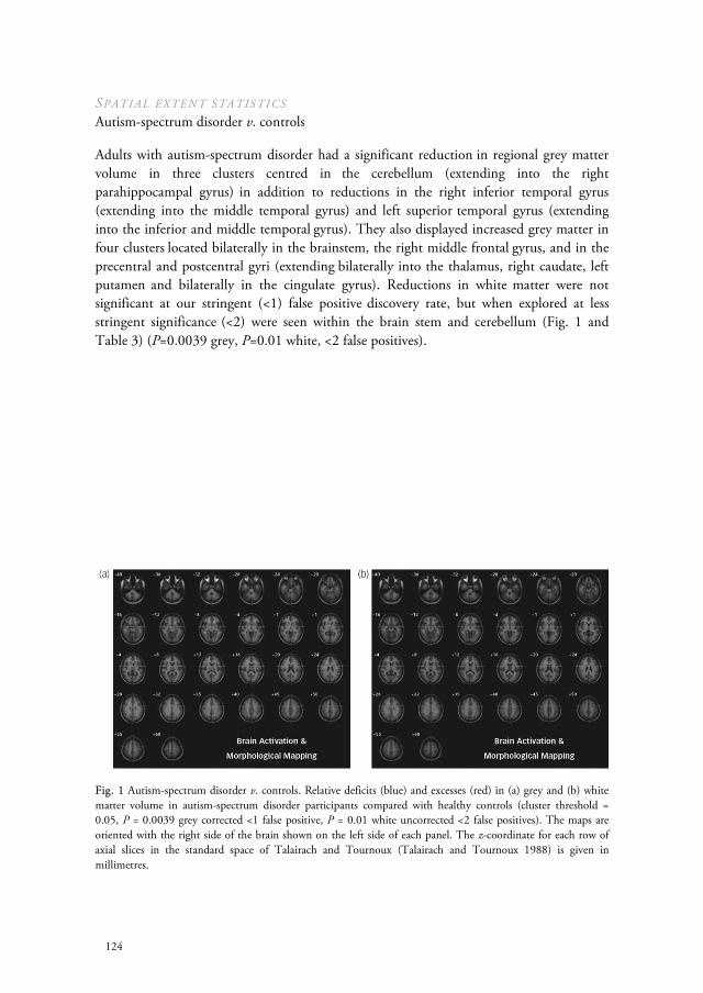
SPATI AL EXTEN T S TA TIS TI CS Autism-spectrum disorder v. controls
Adults with autism-spectrum disorder had a significant reduction in regional grey matter volume in three clusters centred in the cerebellum (extending into the right parahippocampal gyrus) in addition to reductions in the right inferior temporal gyrus
(extending into the middle temporal gyrus) and left superior temporal gyrus (extending into the inferior and middle temporal gyrus). They also displayed increased grey matter in four clusters located bilaterally in the brainstem, the right middle frontal gyrus, and in the precentral and postcentral gyri (extending bilaterally into the thalamus, right caudate, left putamen and bilaterally in the cingulate gyrus). Reductions in white matter were not significant at our stringent (<1) false positive discovery rate, but when explored at less stringent significance (<2) were seen within the brain stem and cerebellum (Fig. 1 and Table 3) (P=0.0039 grey, P=0.01 white, <2 false positives).
Fig. 1 Autism-spectrum disorder v. controls. Relative deficits (blue) and excesses (red) in (a) grey and (b) white matter volume in autism-spectrum disorder participants compared with healthy controls (cluster threshold = 0.05, P = 0.0039 grey corrected <1 false positive, P = 0.01 white uncorrected <2 false positives). The maps are oriented with the right side of the brain shown on the left side of each panel. The z-coordinate for each row of axial slices in the standard space of Talairach and Tournoux (Talairach and Tournoux 1988) is given in millimetres.
124

Table 3 Autism-spectrum disorder v. controls
Brodmann area
Cluster centre in Talairach coordinates
Voxels in
each cluster, n
Cluster locationa x y z Side
Grey matter
Decreases
Cerebellum (extending into the parahippocampal gyrus)
38 -43 -66 -33 189 Left
Inferior temporal gyrus (extending into the parahippocampal gyrus middle and superior temporal gyrus)
20 37 -4 -35 371 Right
Superior temporal gyrus (extending into the inferior and middle temporal gyrus)
38 -30 -1 -35 313 Left
Increases
Brain stem (bilaterally extending into the parahippocampal gyrus, thalamus, caudate, cingulate gyrus)
0 -19 2 1193 Right
Middle frontal gyrus (extending into the superior temporal gyrus, hippocampus, caudate, thalamus)
47 47 44 -5 251 Right
Precentral gyrus (extending into the cingulate gyrus, putamen, thalamus)
6 -40 -15 10 505 Left
Postcentral gyrus 1 47 -18 55 170 Right
White matterb
Decreases
Brain stem (extending into the cerebellum) -46 -34.1 -36.0 127 Left
a. Putative anatomical labels are suggested.
b. <2 false positives.
125

AU TIS M-S PECT RUM D IS OR D ER WITH PS YCHOS IS V . C ON TROL S Adults with autism-spectrum disorder and psychosis had a significant reduction in grey matter volume of two clusters centred in the cerebellum (extending bilaterally into the fusiform gyrus, occipital lobe, inferior and superior temporal lobe). In addition, they displayed two clusters of decreased grey matter within the frontal lobe (right precentral and left middle frontal gyrus). They also demonstrated increased grey matter in the thalamus,
caudate nuclei and brainstem (extending into the parahippocampal gyrus). Moreover, people with autism-spectrum disorder and psychosis had a significant reduction in white matter volume bilaterally in the cerebellum but bilateral increases in white matter of striatal regions (Fig. 2 and Table 4) (P=0.003 grey, P=0.007 white).
Fig. 2 Autism-spectrum disorder with psychosis v. controls. Relative deficits (blue) and excesses (red) in grey matter volume in (a) autism-spectrum disorder with comorbid psychosis participants compared with healthy controls (cluster threshold = 0.05, P = 0.003) and (b) white matter deficits (blue) (cluster threshold = 0.05, P = 0.007).
126

Table 4 Autism-spectrum disorder with psychosis v. controls
Brodmann area
Cluster centre in Talairach coordinates
Voxels in
each cluster, n
Cluster locationa x y z Side
Grey matter
Decreases
Cerebellum (extending into the right fusiform gyrus, right occipital lobe, right inferior temporal gyrus, right superior temporal gyrus)
20, 18 8 -66 -22 6659 Right
Cerebellum (extending into left fusiform, uncus, left occipital lobe, inferior temporal gyrus, middle temporal gyrus, superior temporal gyrus)
38, 20, 37 -34 2 -36 423 Left
Frontal lobe (precentral gyrus) 6, 38, 21 41 1 -25 1163 Right
Middle frontal gyrus (extending from right inferior frontal gyrus, into right middle frontal gyrus and superior frontal gyrus)
11, 10, 9 38 41 15 791 Right
Increases
Brain stem (extending into parahippocampal gyrus thalamus and caudate)
0
-24
2
1129
Right
White matter
Decreases
Cerebellum (extending into left cerebellum) 4 -49 -31 1687 Right
Increase
Right cerebrum (sub-lobar, extra-nuclear white matter)
35 -2 -7 243 Right
Left cerebrum (sub-lobar, extra-nuclear white matter)
-30
-5
-7
292
Left
a. Putative anatomical labels are suggested.
127

AU TIS M-S PECT RUM D IS OR D ER WITH PS YCHOS IS V . A UTIS M-S PECT RUM
D IS ORD ER WITHOU T PS YC HOS IS Adults with autism-spectrum disorder with and without psychosis differed mainly in the anatomy of the insular cortex and cerebellum extending into the fusiform gyrus. Those with psychosis had a significant reduction of grey matter centred in the right insular cortex extending into the right inferior and superior temporal gyrus, the right inferior frontal gyrus and cingulate gyrus. In addition, they demonstrated a significant reduction in regional grey matter volume in one large cluster centred bilaterally in the cerebellum and extending into the right and left fusiform gyrus, and the right and left inferior occipital
gyrus (including the lingual gyrus). Finally, they also had a significant reduction in white matter volume of the cerebellum bilaterally (Fig. 3 and Table 5) (P=0.003 grey, P=0.007 white).
The exclusion of the extra participant with a past history of substance misuse did not significantly change the results.
Fig. 3 Autism-spectrum disorder with psychosis v. autism-spectrum disorder without psychosis. Relative deficits (blue) in (a) grey matter volume in autism-spectrum disorder participants with comorbid psychosis compared with autism-spectrum disorder without psychosis (cluster threshold = 0.05, P = 0.003) and (b) white matter (cluster threshold = 0.05, P = 0.007).
128
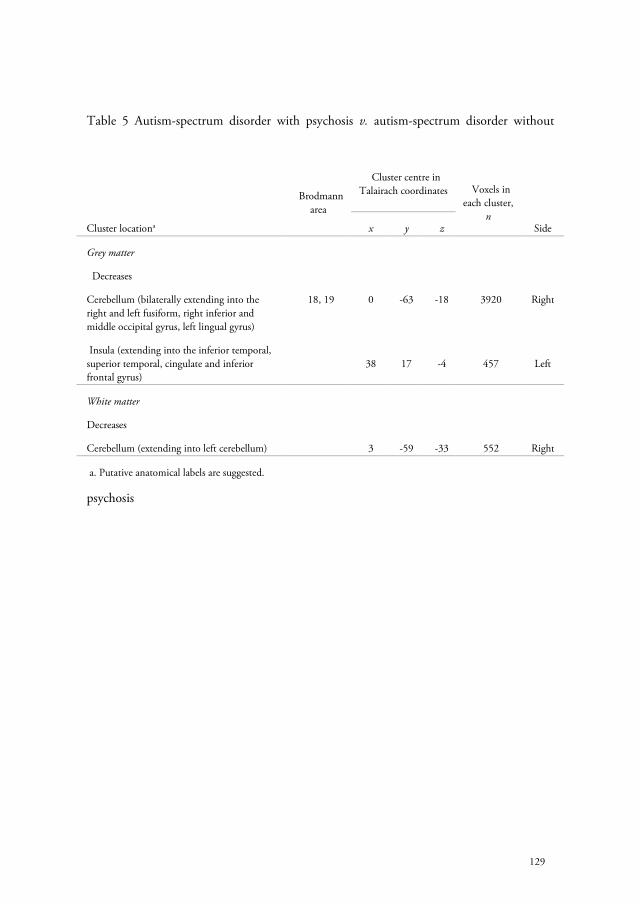
Table 5 Autism-spectrum disorder with psychosis v. autism-spectrum disorder without
psychosis
Brodmann area
Cluster centre in Talairach coordinates
Voxels in
each cluster, n
Cluster locationa x y z Side
Grey matter
Decreases
Cerebellum (bilaterally extending into the right and left fusiform, right inferior and middle occipital gyrus, left lingual gyrus)
18, 19 0 -63 -18 3920 Right
Insula (extending into the inferior temporal, superior temporal, cingulate and inferior frontal gyrus)
38
17
-4
457
Left
White matter
Decreases
Cerebellum (extending into left cerebellum) 3 -59 -33 552 Right
a. Putative anatomical labels are suggested.
129

D i s c u s s i o n We investigated, for the first time, the neuroanatomical associates of psychosis in people with autism-spectrum disorder. We found that both groups of participants with autism-spectrum disorder (i.e. those with and without psychosis) differed from healthy controls in brain regions that are typically reported as abnormal in the disorder (i.e. both groups had significant differences from controls in the anatomy of cerebellar, temporal lobe and striatal regions) (see Toal et al (Toal et al 2005) for a review).
In addition, those with psychosis, compared with healthy controls, demonstrated additional reductions in grey matter volume of the frontal and occipital lobes. Within autism-spectrum disorder, however, the diagnosis of psychosis was associated with a significant reduction in grey matter volume of the right insular cortex and bilaterally in the cerebellum extending into the fusiform gyrus, occipital lobe and lingual gyrus. Reduction in white matter volume was observed within the cerebellum and left lingual gyrus.
Thus, people with autism-spectrum disorder and psychosis have significant differences compared with those without psychosis. However, our data suggests that the neurobiological associates of psychosis in autism-spectrum disorder may differ in some
regards from those with psychosis in the non-autism-spectrum disorder population.
In the latter group, commonly reported anatomical differences in controls include increased volume of cerebral ventricles, together with a reduction in the total brain and grey matter volume of about 3% (for a review see Shenton et al (Shenton et al 2001)). Also,
localised volume and grey matter reductions mostly implicate medial temporal lobe structures (including the amygdale and hippocampus), the orbitofrontal and insular cortices and the basal ganglia. There have been fewer studies of white matter. However, diffusion tensor imaging (Foong et al 2000b;Foong et al 2002;Lim et al 1999;Shergill et al 2007) and magnetic transfer imaging studies (Foong et al 2000a) and microarray data (Hakak et al 2001) suggest that white matter integrity is also compromised in schizophrenia. Studies of those with an affective psychosis (such as bipolar disorder) have generally reported similar findings. At present, the only disease-specific finding is that people with bipolar disorder may not demonstrate the reduction in total brain volume or the amygdale (Gur et al 2007;McDonald et al 2004) that are found in schizophrenia.
Two studies have investigated changes in brain anatomy in those at high risk of developing psychosis as they transition into the disorder. Pantelis et al reported a significant reduction
in grey matter volume of the left parahippocampal and fusiform gyri, as well as other regions including the orbitofrontal, cingulate and cerebellar cortex as people developed psychosis. Job et al (Job et al 2005) reported reductions in grey matter density in right cerebellar cortex and left parahippocampal, uncus and fusiform gyrus. Thus, there is emerging evidence that people with psychosis have differences in brain development prior to the onset of psychosis, and it appears that as a psychotic illness progresses the anatomy of
130
some brain regions continue to change. Moreover, some of these regions are also implicated in autism-spectrum disorder and (in the case of the fusiform gyrus) are essential to processing facial emotion.
Within our sample of people with autism-spectrum disorder, those with and without psychosis differed mainly in the anatomy of the right insular cortex and the cerebellum extending into the fusiform and lingual gyri. Therefore, the clinical diagnosis of psychosis within people with autism-spectrum disorder is not associated with many of the anatomical differences normally reported as abnormal in cross-sectional studies of psychosis in the general population; but it is associated with differences in brain regions tentatively implicated by studies of young people at increased risk of developing psychosis.
The reason for this is unknown, but potential explanations include the following.
OVERL APPIN G D EVEL OPM EN TAL BRAIN ABN ORM AL ITI ES People with autism-spectrum disorder already have significant developmental abnormalities in brain regions typically affected in psychosis (e.g temporal and frontal lobes), therefore it may be more difficult to detect subtle additional differences associated with further neuropsychiatric disorder.
AL TE RN ATIVE ‘EN T RY-POI N T’ IN TO S CHI ZOPH REN IA On the other hand, the presence of autism-spectrum disorder-associated
neurodevelopmental abnormalities might represent an alternative ‘entry-point’ into a final common pathway of psychosis. That is, as people with autism-spectrum disorder already have differences in the development of brain regions that are also implicated in schizophrenia (frontostriatal and temporal lobe structures), they may only require relatively subtle additional abnormalities in insular cortex (implicated in sensory and emotional response), the fusiform gyrus and cerebellum (known to be responsible for both inhibitory control and face processing) to develop positive symptoms of psychosis such as delusions
and hallucinations. For example, it has been consistently demonstrated that people with autism and those with schizophrenia demonstrate similar deficits in social functioning and social cognition including overlapping deficits in specific theory of mind tasks. Recent work has demonstrated similar reduced neural activation in both autism and schizophrenia in a neural network involved in social cognition (Pinkham et al 2008). Thus, baseline deficits in understanding the mental states of others and processing internal stimuli may render those with autism more susceptible to psychosis in the face of additional insults to specific structures proposed to be specifically related to delusions and hallucinations.
For instance, the insular cortex has been repeatedly implicated in schizophrenia both in structural(Makris et al 2006;Saze et al 2007) and functional studies (Crespo-Facorro et al 2001). Grey matter volume has been demonstrated to be reduced within the insular cortex in those experiencing chronic auditory hallucinations (O'Daly et al 2007), and activation of the insular cortex has been demonstrated in individuals experiencing auditory hallucinations (Shergill et al 2000) (along with activation of the bilateral inferior frontal,
131

anterior cingulate and temporal cortices bilaterally). The insular cortex provides connections between limbic memory regions as well as the auditory and visual association areas of the sensory system. Abnormal activation of the insula has been hypothesised to result in unbalanced sensory–memory integration contributing to auditory and visual hallucinations.
In addition, the cerebellum has been tentatively proposed to be involved in the development of delusions. Functional MRI studies have demonstrated a potential correlation between cerebellar activation and delusions and suspiciousness/persecution scores. Also, neurobiological theories have directly implicated the cerebellum regarding deficits of ‘internal monitoring’ that are suggested, for example, to underlie the formation of delusions of alien control where the person attributes their own actions to an external agent (Blakemore 2003;Blakemore et al 2003;Whalley et al 2007).
Finally, the fusiform gyrus is known to be a key brain region supporting social perception, including facial identity and emotion recognition (Deeley et al 2007;Schultz 2005). Anatomical abnormalities in fusiform gyrus may be associated with difficulties in accurate social perception, increasing the likelihood that the benign or neutral actions of others could be construed as hostile – particularly in the presence of impaired mentalising abilities and prior experiences of bullying and social rejection which are common in people with autism-spectrum disorders (Blackshaw et al 2001). In other words, it may be hypothesised that fusiform gyrus abnormalities may lower the threshold for the formation of paranoid delusions. Future studies could test this hypothesis by testing for greater facial identity and emotion perception deficits in people with autism-spectrum disorder and psychosis compared with those without psychosis. Alternatively, fusiform gyrus abnormalities could lower the threshold for experiencing visual and auditory hallucinations (although the mechanisms remain unclear). For example, a recent study of people with Parkinson’s disease who experienced visual hallucinations demonstrated reduced cerebral blood flow within the fusiform gyrus (Oishi et al 2005). In addition, reductions in grey matter volume of the fusiform gyrus have been demonstrated to correlate with severity of auditory hallucinations (O'Daly et al 2007) in those with schizophrenia. Hence, future studies in larger samples should determine whether frequency and modality of hallucinations in people with autism-spectrum disorder correlates with anatomical abnormalities in the fusiform gyrus.
ABN ORM AL ITY OF N EU ROT RAN S M ITTE RS We and others have reported that some people with autism-spectrum disorder also have significant differences from controls in neurochemical pathways (the serotonergic, glutamatergic and gaba-ergic systems)(Murphy et al 2006;Page et al 2006;Yip et al 2007) that are also implicated in psychosis. For example, some people with autism-spectrum disorder have platelet hyperserotonaemia (Hranilovic et al 2007;Schain and Freedman 1961), abnormal brain serotonin synthesis capacity (Chugani et al 1999), serotonin receptor dysfunction (Hollander et al 2000), and reduced 5-HT2A (5-hydroxytryptamine)
132

receptors (Murphy et al 2006). We have previously reported that people with autism-spectrum disorder have significant differences from controls in the glutamate/glutamine
concentration of the amygdala–hipocampal complex (Page et al 2006). Thus, the combination of pre-existing anatomical, and neurochemical, differences in brain systems also implicated in psychosis may reduce the additional disease burden required to develop comorbid psychotic symptoms. If this were the case, however, we would anticipate a very significantly increased risk of developing psychosis in autism-spectrum disorder, and the evidence to substantiate that is (at best) preliminary (Mouridsen et al 2008;Stahlberg et al 2004).
LIM ITA TION S This is the first study of its kind. Nevertheless, it was a relatively small observational study of adults. Moreover, we included a person with a prior history of substance misuse. We suggest, however, that the differences we found cannot be fully accounted for by substance misuse as our results were largely unchanged when we excluded this individual. All individuals with psychosis had been treated with antipsychotics at some time in the past and the majority were on antipsychotic medication at the time of scanning (which may affect brain anatomy). However, it is unlikely that this can fully explain our results as most
MRI studies of medication effects on brain anatomy in psychosis found differences mainly in the globus pallidus (Gur et al 2007) and we found no abnormalities in this region.
It may be asserted by some that the differentiation of psychosis in autism-spectrum disorder is inappropriate and/or impossible. However, our diagnosis of autism-spectrum disorder without psychosis was based on evidence of early childhood onset, collateral
history from family/friends/teachers and use of reliable diagnostic instruments. In addition, these symptoms present in early childhood were distinct from the frank psychotic symptoms of auditory hallucinations and bizarre delusions that developed in later life. Further, inclusion in the psychosis group did not rely on features such as thought disorder or negative symptoms that (respectively) may be difficult to differentiate within autism-spectrum disorder from pre-existing speech and language abnormalities, and impassivity and lack of emotional awareness.
Another potential limitation of our study is that the psychosis group consisted of a mixed group of people; most fulfilled the diagnostic criteria for schizophrenia, but three had schizoaffective disorder and two had a psychotic bipolar affective disorder. However, increasingly genetic, neuropharmacological and neurophysiological studies are highlighting the overlap between these groups (Berrettini 2003;Gur et al 2007;Owen et al 2007;Tamminga and Davis 2007;Tsuang et al 2000).
Finally, neuroimaging techniques such as voxel-based morphometry are still evolving and inherent limitations include difficulties with registration of the images and limitations in detecting differences in areas with high interindividual variance. Therefore, we await replication of these findings in larger groups before definitive conclusions can be drawn.
133

CON CL US ION The diagnosis of autism-spectrum disorder is being made much more frequently, and so we are likely to encounter an increased number of people with autism-spectrum disorder diagnosed with psychosis. Our preliminary study suggests that the presence of neurodevelopmental abnormalities normally associated with autism-spectrum disorder might represent an alternative ‘entry-point’ into a final common pathway of psychosis. Future longitudinal studies may further help to elucidate the changes associated with the development of psychosis within this disorder.
134

R E F E R E N C E S American Psychiatric Association (1987): Diagnostic and Statistical Manual of Mental Disorders (3rd edition, revised) (DSM-II-R) : APA.
Amiet C, Gourfinkel-An I, Bouzamondo A, Tordjman S, Baulac M, Lechat P et al (2008): Epilepsy in autism is associated with intellectual disability and gender: evidence from a meta-analysis. Biol Psychiatry 64: 577-582.
Ashburner J, Friston KJ (2005): Unified segmentation. Neuroimage 26: 839-851.
Baird G, Simonoff E, Pickles A, Chandler S, Loucas T, Meldrum D et al (2006): Prevalence of disorders of the autism spectrum in a population cohort of children in South Thames: the Special Needs and Autism Project (SNAP). Lancet 368: 210-215.
Berrettini W (2003): Bipolar disorder and schizophrenia: not so distant relatives? World Psychiatry 2: 68-72.
Blackshaw AJ, Kinderman P, Hare DJ, Hatton C (2001): Theory of mind, causal attribution and paranoia in Asperger syndrome. Autism 5: 147-163.
Blakemore SJ (2003): Deluding the motor system. Conscious Cogn 12: 647-655.
Blakemore SJ, Oakley DA, Frith CD (2003): Delusions of alien control in the normal brain. Neuropsychologia 41: 1058-1067.
Bolton PF, Pickles A, Murphy M, Rutter M (1998): Autism, affective and other psychiatric disorders: patterns of familial aggregation. Psychol Med 28: 385-395.
Bullmore ET, Suckling J, Overmeyer S, Rabe-Hesketh S, Taylor E, Brammer MJ (1999): Global, voxel, and cluster tests, by theory and permutation, for a difference between two groups of structural MR images of the brain. IEEE Trans Med Imaging 18: 32-42.
Chugani DC, Muzik O, Behen M, Rothermel R, Janisse JJ, Lee J et al (1999): Developmental changes in brain serotonin synthesis capacity in autistic and nonautistic children. Ann Neurol 45: 287-295.
Crespo-Facorro B, Paradiso S, Andreasen NC, O'Leary DS, Watkins GL, Ponto LL et al (2001): Neural mechanisms of anhedonia in schizophrenia: a PET study of response to unpleasant and pleasant odors. JAMA 286: 427-435.
Deeley Q, Daly EM, Surguladze S, Page L, Toal F, Robertson D et al (2007): An event related functional magnetic resonance imaging study of facial emotion processing in Asperger syndrome. Biol Psychiatry 62: 207-217.
Foong J, Maier M, Barker GJ, Brocklehurst S, Miller DH, Ron MA (2000a): In vivo investigation of white matter pathology in schizophrenia with magnetisation transfer imaging. J Neurol Neurosurg Psychiatry 68: 70-74.
Foong J, Maier M, Clark CA, Barker GJ, Miller DH, Ron MA (2000b): Neuropathological abnormalities of the corpus callosum in schizophrenia: a diffusion tensor imaging study. J Neurol Neurosurg Psychiatry 68: 242-244.
Foong J, Symms MR, Barker GJ, Maier M, Miller DH, Ron MA (2002): Investigating regional white matter in schizophrenia using diffusion tensor imaging. Neuroreport 13: 333-336.
Good CD, Johnsrude IS, Ashburner J, Henson RN, Friston KJ, Frackowiak RS (2001): A voxel-based morphometric study of ageing in 465 normal adult human brains. Neuroimage 14: 21-36.
Gur RE, Keshavan MS, Lawrie SM (2007): Deconstructing psychosis with human brain imaging. Schizophr Bull 33: 921-931.
Hakak Y, Walker JR, Li C, Wong WH, Davis KL, Buxbaum JD et al (2001): Genome-wide expression analysis reveals dysregulation of myelination-related genes in chronic schizophrenia. Proc Natl Acad Sci U S A 98: 4746-4751.
135

Hollander E, Novotny S, Allen A, Aronowitz B, Cartwright C, DeCaria C (2000): The relationship between repetitive behaviors and growth hormone response to sumatriptan challenge in adult autistic disorder. Neuropsychopharmacology 22: 163-167.
Howlin P (2000): Autism and intellectual disability: diagnostic and treatment issues. J R Soc Med 93: 351-355.
Hranilovic D, Bujas-Petkovic Z, Vragovic R, Vuk T, Hock K, Jernej B (2007): Hyperserotonemia in adults with autistic disorder. J Autism Dev Disord 37: 1934-1940.
Job DE, Whalley HC, Johnstone EC, Lawrie SM (2005): Grey matter changes over time in high risk subjects developing schizophrenia. Neuroimage 25: 1023-1030.
Kolvin I (1971): Studies in the childhood psychoses. I. Diagnostic criteria and classification. Br J Psychiatry 118: 381-384.
Konstantareas MM, Hewitt T (2001): Autistic disorder and schizophrenia: diagnostic overlaps. J Autism Dev Disord 31: 19-28.
Lim KO, Rosenbloom MJ, Faustman WO, Sullivan EV, Pfefferbaum A (1999): Cortical gray matter deficit in patients with bipolar disorder. Schizophr Res 40: 219-227.
Lord C, Rutter M, Goode S, Heemsbergen J, Jordan H, Mawhood L et al (1989): Autism diagnostic observation schedule: a standardized observation of communicative and social behavior. J Autism Dev Disord 19: 185-212.
Lord C, Rutter M, Le CA (1994): Autism Diagnostic Interview-Revised: a revised version of a diagnostic interview for caregivers of individuals with possible pervasive developmental disorders. J Autism Dev Disord 24: 659-685.
Makris N, Goldstein JM, Kennedy D, Hodge SM, Caviness VS, Faraone SV et al (2006): Decreased volume of left and total anterior insular lobule in schizophrenia. Schizophr Res 83: 155-171.
McDonald C, Zanelli J, Rabe-Hesketh S, Ellison-Wright I, Sham P, Kalidindi S et al (2004): Meta-analysis of magnetic resonance imaging brain morphometry studies in bipolar disorder. Biol Psychiatry 56: 411-417.
Mouridsen SE, Rich B, Isager T (2008): Psychiatric disorders in adults diagnosed as children with atypical autism. A case control study. J Neural Transm 115: 135-138.
Murphy DG, Daly E, Schmitz N, Toal F, Murphy K, Curran S et al (2006): Cortical serotonin 5-HT2A receptor binding and social communication in adults with Asperger's syndrome: an in vivo SPECT study. Am J Psychiatry 163: 934-936.
O'Daly OG, Frangou S, Chitnis X, Shergill SS (2007): Brain structural changes in schizophrenia patients with persistent hallucinations. Psychiatry Res 156: 15-21.
Oishi N, Udaka F, Kameyama M, Sawamoto N, Hashikawa K, Fukuyama H (2005): Regional cerebral blood flow in Parkinson disease with nonpsychotic visual hallucinations. Neurology 65: 1708-1715.
Owen MJ, Craddock N, Jablensky A (2007): The genetic deconstruction of psychosis. Schizophr Bull 33: 905-911.
Page LA, Daly E, Schmitz N, Simmons A, Toal F, Deeley Q et al (2006): In vivo 1H-magnetic resonance spectroscopy study of amygdala-hippocampal and parietal regions in autism. Am J Psychiatry 163: 2189-2192.
Petty LK, Ornitz EM, Michelman JD, Zimmerman EG (1984): Autistic children who become schizophrenic. Arch Gen Psychiatry 41: 129-135.
Pinkham AE, Hopfinger JB, Pelphrey KA, Piven J, Penn DL (2008): Neural bases for impaired social cognition in schizophrenia and autism spectrum disorders. Schizophr Res 99: 164-175.
Russell AJ, Mataix-Cols D, Anson M, Murphy DG (2005): Obsessions and compulsions in Asperger syndrome and high-functioning autism. Br J Psychiatry 186: 525-528.
136
Saze T, Hirao K, Namiki C, Fukuyama H, Hayashi T, Murai T (2007): Insular volume reduction in schizophrenia. Eur Arch Psychiatry Clin Neurosci 257: 473-479.
Schain R, Freedman D (1961): Studies on 5-hydroxyindole metabolism in autistic and other mentally retarded children. J Pediatr 58: 315-320.
Schultz RT (2005): Developmental deficits in social perception in autism: the role of the amygdala and fusiform face area. Int J Dev Neurosci 23: 125-141.
Shenton ME, Dickey CC, Frumin M, McCarley RW (2001): A review of MRI findings in schizophrenia. Schizophrenia Research 49: 1-52.
Shergill SS, Brammer MJ, Williams SC, Murray RM, McGuire PK (2000): Mapping auditory hallucinations in schizophrenia using functional magnetic resonance imaging. Arch Gen Psychiatry 57: 1033-1038.
Shergill SS, Kanaan RA, Chitnis XA, O'Daly O, Jones DK, Frangou S et al (2007): A diffusion tensor imaging study of fasciculi in schizophrenia. Am J Psychiatry 164: 467-473.
Stahlberg O, Soderstrom H, Rastam M, Gillberg C (2004): Bipolar disorder, schizophrenia, and other psychotic disorders in adults with childhood onset AD/HD and/or autism spectrum disorders. J Neural Transm 111: 891-902.
Talairach,J, Tournoux,P (1988): Co-planar stereotaxic atlas of the human brain . New York: Thieme.
Tamminga CA, Davis JM (2007): The neuropharmacology of psychosis. Schizophr Bull 33: 937-946.
Toal F, Murphy DG, Murphy KC (2005): Autistic-spectrum disorders: lessons from neuroimaging. Br J Psychiatry 187: 395-397.
Tsakanikos E, Sturmey P, Costello H, Holt G, Bouras N (2007): Referral trends in mental health services for adults with intellectual disability and autism spectrum disorders. Autism 11: 9-17.
Tsuang MT, Stone WS, Faraone SV (2000): Toward reformulating the diagnosis of schizophrenia. Am J Psychiatry 157: 1041-1050.
Volkmar FR, Cohen DJ (1991): Comorbid association of autism and schizophrenia. Am J Psychiatry 148: 1705-1707.
Wechsler,D (1981): The Wechsler Adult Intelligence Scale-Revised : The Psychological Corporation.
Whalley HC, Gountouna VE, Hall J, McIntosh A, Whyte MC, Simonotto E et al (2007): Correlations between fMRI activation and individual psychotic symptoms in un-medicated subjects at high genetic risk of schizophrenia. BMC Psychiatry 7: 61.
Wing L (1981): Asperger's syndrome: a clinical account. Psychol Med 11: 115-129.
World Health Organization (1992): The ICD-10 Classification of Mental and Behavioural Disroders: Clinical Descriptions and Diagnostic Guidelines : WHO.
Yip J, Soghomonian JJ, Blatt GJ (2007): Decreased GAD67 mRNA levels in cerebellar Purkinje cells in autism: pathophysiological implications. Acta Neuropathol 113: 559-568.
137


C h a p t e r 8
White matter integrity in Asperger syndrome: a preliminary diffusion tensor magnetic resonance imaging study in adults.
O.J.N. Bloemen, Q. Deeley, F. Sundram, E.M. Daly, G.J. Barker, D.K. Jones, T.A.M.J. van Amelsvoort, N. Schmitz, D. Robertson, K.C. Murphy, D.G.M. Murphy: Autism Research (2010), Oct;3(5):203-13.

A b s t r a c t BACKG ROUN D. Autistic Spectrum Disorder (ASD), including Asperger syndrome and autism, is a highly genetic neurodevelopmental disorder. There is a consensus that ASD has a biological basis, and it has been proposed that it is a ‘connectivity’ disorder. Diffusion Tensor Magnetic Resonance Imaging (DT-MRI) allows measurement of the microstructural integrity of white matter (a proxy measure of ‘connectivity’). However nobody has investigated the microstructural integrity of whole brain white matter in people with Asperger syndrome.
METHOD S. We measured the fractional anisotropy (FA), mean diffusivity (MD) and radial diffusivity (RD) of white matter, using DT-MRI, in 13 adults with Asperger syndrome and 13 controls. The groups did not differ significantly in overall intelligence and age. FA, MD and RD were assessed using whole brain voxel based techniques.
RES UL TS. Adults with Asperger syndrome had a significantly lower FA than controls in thirteen clusters. These were largely bilateral and included white matter in the internal capsule, frontal, temporal, parietal and occipital lobes, cingulum and corpus callosum.
CON CL US ION S. Adults with Asperger syndrome have widespread significant differences from controls in white matter microstructural integrity.
140
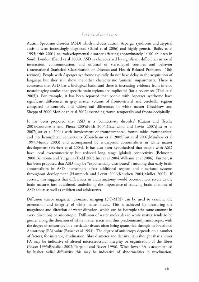
I n t r o d u c t i o n Autism Spectrum disorder (ASD) which includes autism, Asperger syndrome and atypical autism, is an increasingly diagnosed (Baird et al 2006) and highly genetic (Bailey et al 1995;Frith 2001) neurodevelopmental disorder affecting approximately 1:100 children in South London (Baird et al 2006). ASD is characterized by significant difficulties in social interaction, communication, and unusual or stereotyped routines and behavior (International Statistical Classification of Diseases and Health Related Problems—10th revision). People with Asperger syndrome typically do not have delay in the acquisition of language but they still show the other characteristic ‘autistic’ impairments. There is consensus that ASD has a biological basis, and there is increasing evidence from in-vivo neuroimaging studies that specific brain regions are implicated (for a review see (Toal et al 2005)). For example, it has been reported that people with Asperger syndrome have significant differences in grey matter volume of fronto-striatal and cerebellar regions compared to controls, and widespread differences in white matter (Bradshaw and Sheppard 2000;McAlonan et al 2002) extending fronto-temporally and fronto-occipitally.
It has been proposed that ASD is a ‘connectivity disorder’ (Catani and ffytche 2005;Courchesne and Pierce 2005;Frith 2004;Geschwind and Levitt 2007;Just et al 2007;Just et al 2004) with involvement of frontotemporal, frontolimbic, frontoparietal and interhemispheric connections (Courchesne et al 2005;Just et al 2007;Minshew et al 1997;Mundy 2003) and accompanied by widespread abnormalities in white matter development (Herbert et al 2004). It has also been hypothesised that people with ASD have local overconnectivity but reduced long range (global) connectivity (Belmonte 2000;Belmonte and Yurgelun-Todd 2003;Just et al 2004;Williams et al 2006). Further, it has been proposed that ASD may be “exponentially distributed”; meaning that early brain abnormalities in ASD increasingly affect additional regions and functional systems throughout development (Hammock and Levitt 2006;Knudsen 2004;Muller 2007). If correct, this suggests that differences in brain anatomy would become more severe as the brain matures into adulthood, underlining the importance of studying brain anatomy of ASD adults as well as children and adolescents.
Diffusion tensor magnetic resonance imaging (DT-MRI) can be used to examine the orientation and integrity of white matter tracts. This is achieved by measuring the magnitude and direction of water diffusion, which can be isotropic (the same amount in every direction) or anisotropic. Diffusion of water molecules in white matter tends to be greater along the direction of white matter tracts and thus predominantly anisotropic, with the degree of anisotropy in a particular tissues often being quantified through its Fractional Anisotropy (FA) value (Basser et al 1994). The degree of anisotropy depends on a number of factors; for instance, myelination, fibre diameter and density. It is thought that a lower FA may be indicative of altered microstructural integrity or organisation of the fibres (Basser 1995;Beaulieu 2002;Pierpaoli and Basser 1996). When lower FA is accompanied by higher radial diffusivity this may be indicative of abnormalities in myelination,
141

although evidence for this comes largely from studies of normal and abnormal development, and animal investigation (Song et al 2002;Song et al 2005) .
A prior whole brain DT-MRI study included 7 children with autism and 9 controls (Barnea-Goraly et al 2004). They reported that the children with autism had a significant reduction in FA of white matter adjacent to the ventromedial prefrontal cortices, anterior cingulate gyri, temporoparietal junctions, and in the corpus callosum. Subsequently, others investigated mixed samples of children and adults with autism (Alexander et al 2007;Keller et al 2007;Lee et al 2007). They reported that people with autism have a significant reduction in FA of areas within (or near) the corpus callosum (Alexander et al 2007;Keller et al 2007), right external capsule (Keller et al 2007) and the superior temporal gyrus and temporal stem (Lee et al 2007). These studies were valuable first steps. However, they included heterogeneous clinical populations (in terms of mixed ages) or only studied children; and some did not correct for multiple comparisons.
In summary, it has been proposed that ASD is underpinned by cortical ‘underconnectivity’, and with abnormal brain development continuing into adulthood. Further, ASD is a developmental disorder, possibly characterised by grey and white matter overgrowth in very young children and reduced volumes in later childhood (Ben et al 2007;Courchesne et al 2001). Hence differences in white matter most likely continue to develop as the brain matures into adulthood due to abnormal neuronal loss and synaptic pruning (Muller 2007). Also ASD is a heterogenous clinical disorder. However nobody has yet reported on white matter integrity across the whole brain in adults with ASD, and in a relatively homogeneous population.
Thus we compared white matter micro integrity (using DT-MRI) in adults with Asperger syndrome and healthy adult controls, using a voxel based analysis (VBM) to measure FA throughout the whole brain. We hypothesised that people with Asperger syndrome have widespread differences from controls, but that this would mainly affect white matter in brain regions and systems most implicated in the disorder (i.e. frontal, temporal, limbic, basal ganglia and cerebellar regions).
142

M e t h o d s
SUBJEC T RE CR UITM EN T We recruited 13 male adults fulfilling ICD-10 clinical research criteria for autism – but who did not have a history of language delay. Hence we defined them as having Asperger syndrome (age≥18, mean age 39 (SD+/-9.8), mean Full Scale IQ (FSIQ) 110 (SD+/-15.7)). The age of the subjects with Asperger syndrome ranged from 23 to 54 years and FSIQ ranged from 88 to 133. People with Asperger syndrome were recruited through local support groups, and our clinical research program in autism (supported by the MRC UK A.I.M.S. network). Subjects were diagnosed based on clinical interview, collateral information from family members and review of other information available such as school reports. All assessments were blind to MRI scan data using ICD-10 (International Statistical Classification of Diseases and Health Related Problems—10th revision) clinical research criteria (World Health Organization, 1992). Diagnosis was made through consensus between three clinicians trained in the Autism Diagnostic Interview (ADI-R, (Lord et al 1994)). We were able to confirm our clinical research diagnosis using the ADI-R in 6 individuals with Asperger syndrome, when their parental informants were willing/available. Another 4 subjects were assessed on the Autism Diagnostic Observation Schedule (ADOS, (Lord et al 1989)). Three subjects did not receive additional confirmation of their diagnosis using these methods. Exclusion criteria were a history of major psychiatric disorder (e.g. psychosis), head injury, toxic exposure, diabetes, abnormalities in routine blood tests, drug or alcohol abuse or clinical abnormality on routine MRI.
In addition, 13 male adult controls were recruited locally by advertisement. They did not differ in FSIQ or age (age≥18, mean age 37 (SD+/-9.6), mean FSIQ 115 (SD+/-14.4). The age of the subjects ranged from 25 to 52 years and FSIQ ranged from 89 to 133. All subjects had a structured clinical exam and routine clinical blood tests to exclude biochemical, haematological or chromosomal abnormalities. Exclusion criteria were a history of major psychiatric disorder (e.g. psychosis), head injury, toxic exposure, diabetes, abnormalities in routine blood tests, drug or alcohol abuse, clinical abnormality on routine MRI, or a medical or genetic disorder associated with autistic symptoms (e.g. fragile X or tuberous sclerosis). Ethics approval was provided by the Joint Institute of Psychiatry and South London and Maudsley NHS Trust Research Ethics Committee. All participants gave informed consent and all experiments were conducted in accordance with the Declaration of Helsinki.
NEU ROPS YCHOL OGI CAL T ES TIN G We measured overall intelligence using the vocabulary, comprehension, similarities, block design and object assembly subtests of the Wechsler Adult Intelligence Scale-Revised (WAIS-R (Weschler 1987)).
143

MR ACQUIS I TION Data were acquired using a GE Signa LX system (General Electric, Milwaukee, WI, USA), with actively shielded magnetic field gradients (maximum amplitude 40 mT m-1). A standard quadrature birdcage head coil was used for both RF transmission and signal reception. Each volume was acquired using a multi-slice peripherally-gated EPI sequence, optimised for precise measurement of the diffusion tensor in parenchyma, from 60 contiguous near-axial slice locations. Images were acquired with isotropic (2.5x2.5x2.5mm) voxels, reconstructed to 1.875x1.875x2.5mm. Image acquisition was synchronised to the cardiac cycle using a peripheral gating device placed on the subject’s forefinger. Echo time was 107 ms while the effective repetition time was 15 RR intervals. The duration of the diffusion encoding gradients was 17.3 ms giving a maximum diffusion weighting of 1300 s mm-2. At each slice location, 7 images were acquired with no diffusion gradients applied, together with 64 diffusion-weighted images in which gradient directions were uniformly distributed in space. Full details are given elsewhere (Jones et al 2002).
DTI P ROCES S IN G Visual inspection was used to ensure al data was of suitable quality for further processing, and three scans were excluded because of data quality issues. The diffusion-weighted images were initially corrected for the effects of eddy-current induced distortion using in-house software. Group mapping techniques (derived from the VBM analysis methods developed for structural (T1 and/or T2 weighted) images), compare parametric maps of MR parameters (in this case the fractional anisotropy, mean diffusivity (MD) and radial diffusivity (RD)) between subjects, following registration into a standard space. In the current study, calculation of the initial FA, MD and RD images was performed using locally written software. (Full details are given in (Catani et al 2002), but briefly: correction for eddy current induced distortions was performed using a mutual information based registration of all images to the mean non-diffusion weighted images, using an affine transformation. The diffusion tensor was then calculated for each voxel, using multivariate linear regression after logarithmic transformation of the signal intensities, and maps of FA, MD and RD were generated from the resulting tensor images).
Normalisation of the FA, MD and RD images was performed using Statistical Parametric Mapping software (SPM2, Wellcome Department of Imaging Neurosciences, University College London, UK). A two stage registration process was performed, using both the T2 weighted (b=0) images and the FA images themselves. The mean T2-weighted (b=0) image for each subject was first registered to the EPI template provided in SPM (although the former is T2 weighted, and the latter T2* weighted, the image contrast is similar, as are the geometrical distortions inherent in any EPI based acquisition). In both cases, SPM2’s default normalisation parameters were used (medium regularisation; 16 non-linear iterations; trilinear interpolation). The derived warping parameters were then applied to corresponding FA images in order to map the latter into standard space. The normalised FA images of all subjects were then averaged and smoothed (8 mm FWHM Gaussian filter) to create a new, study specific, template to which each subject's FA images were then
144

re-registered. The registered FA images were also segmented (using SPMs default a priori tissue probability information) to give maps of the probability of a tissue being either white or grey matter, and these segmented images were thresholded at a low (10%) probability to provide a binary mask of white matter. Visual inspection of example datasets confirmed that masking at this threshold produced maps, which included all major white matter areas, and did not suffer from any unexpected ‘holes’ (eg in low FA regions caused by crossing fibres). An accurate segmentation was not essential, and a relatively liberal threshold was deliberately used, in order to create a slightly 'over inclusive' mask.
Scans need to be smoothed in order to reduce confounds due to individual variation in WM anatomy. Smoothing the data in order to constrain it into specific statistical distributions is also a prerequisite for some analysis approaches, but is not necessary for our non-parametric approach (see below). The degree of smoothing to apply is still a subject of much discussion as different smoothing levels result in varying results (Jones et al 2005); in the absence of a specific hypothesis about the spatial extent of any abnormalities, we applied a smoothing filter with an extent similar to the width of many white matter tracts (Gaussian, 5mm full-width at half maximum) to aid between-subject anatomical matching and improve the signal-to-noise ratio.
STA TIS TI CS SPSS (SPSS 12·02 for windows, SPSS Inc) was used for statistical analysis of non imaging data. Group differences in age and IQ were examined using independent samples t-tests (two tailed). Level of statistical significance was defined as p<0·05 (two tailed).
Differences in white matter FA, MD and RD between the Asperger group and the controls were estimated by fitting an analysis of covariance (ANCOVA) model at each intracerebral voxel in standard space. Age, performance IQ and verbal IQ were added as covariates. Given that brain changes are likely to extend over a number of contiguous voxels, test statistics incorporating spatial information such as 3D cluster mass (the sum of suprathreshold voxel statistics), are generally more powerful than other possible test statistics, which are informed only by data at a single voxel (Bullmore et al 1999). As no parametric distribution is known for cluster mass, permutation testing was used to assess statistical significance in the analyses described below. A non-parametric approach also overcomes the difficulty that parametric methods assume that the residuals of the model tested will follow a Gaussian distribution (which has been shown not to be true for DTI data, even after large amounts of smoothing (Jones et al 2005)) and reduces the need for smoothing to constrain the data into meeting this condition.
We first set a relatively lenient p-value (p≤0.05) to detect voxels putatively demonstrating differences between groups. At this stage, we considered only those voxels to which all subjects contributed data; this - along with the masking procedure above - restricts the analysis to core white matter regions, reducing the search volume (and thus the number of comparisons made). This requirement of good “overlap” between the subjects also minimises testing at the grey/white interfaces, where the high grey/white contrast of FA
145

images exacerbates any edge effects. We then searched for spatial clusters among the voxels highlighted, and tested the “mass” of each cluster (the sum of suprathreshold voxel statistics it comprises) for significance.
Permutation based testing, implemented in the XBAM package (developed at The Institute of Psychiatry, London, UK http://www.brainmap.co.uk), was used to assess statistical significance at both the voxel and cluster levels (Bullmore et al 1999). We performed 1000 permutations at both voxel and cluster stage of the analysis. For the initial voxel level testing, the distribution is calculated separately at each voxel; this helps minimize potential complications due to the very high dynamic range of FA data, and also makes the
analysis less sensitive to differences in (physiological) noise between brain regions. At the cluster level, rather than set a single a priori p-value below which we regard findings as significant, we calculated, for a range of p-values, the number of clusters which would be expected by chance alone. We then set the statistical threshold for cluster significance for each analysis such that the expected number of false positive clusters by chance alone would be less than one, which was p=0.0025, and the threshold for cluster size at larger than 10 voxels. It is known, however (Carew et al 2007), that the variance of FA values in the brain depends upon the FA values themselves and, while we restricted our analysis to core white matter regions where this is relatively uniform, the effects of noise may still vary slightly from region to region. We therefore also inspected the voxel level maps (which treat each voxel independently and therefore inherently allow for such local differences in statistics), to ensure that these highlight the same areas as the cluster level results. In this way we retained the additional power of the cluster statistics while controlling for the potential disadvantage of its non-local nature.
Voxels and clusters were localized in Montreal-Neurological-Institute (MNI) space and transformed into Talairach and Tournoux co-ordinates (Brett et al 2002;Talairach and Tournoux 1988). Resulting voxels and cluster maps were overlaid on the FA template for anatomical assessment. To further localize significant voxel clusters, atlases were consulted (Mori et al 2005;Talairach and Tournoux 1988).
146

R e s u l t s
FRA CTION AL AN IS OT ROPY Adults with Asperger syndrome had a significantly lower FA than controls over large areas of the brain. These changes covered 13, largely bilateral, clusters (Table 1 and Figure 1).
Frontal
Two clusters located in the frontal lobe correspond to parts of the inferior fronto-occipital fasciculus (IFO), the forceps minor and anterior corona radiata. On the right the cluster extended into the anterior thalamic radiation (ATR) and the uncinate fasciculus (UNC). On the left a more posterior third cluster extended into this same region, also including parts of the ATR and UNC.
Medial
Two of the frontal clusters described above extend dorsally just into the genu of the corpus callosum (CC) in both hemispheres. Four more posterior clusters (partly) located in the posterior corona radiata and posterior limb of the internal capsule extend into the CC corresponding to large parts of the splenium and small parts of the dorsal body of the CC, and possibly the corticospinal tracts.
Temporal/occipital
On the left 3 clusters together extended from the retrolenticular part of the internal capsule via the sagittal stratum and forceps major corresponding to the inferior longitudinal fasciculus (ILF) and/or inferior fronto-occipital fasciculus (IFO). These tracts are also implicated contralaterally in a smaller, more dorsally located, cluster.
Superior frontal/parietal
We found bilateral FA decreases in the superior frontal and parietal regions corresponding to the corona radiata, superior longitudinal fascicule (SLF) and cingulum.
There were 4 very small clusters of higher FA in the Asperger group, but all were smaller that 10 voxels.
147

Table 1 - DTI group mapping analysis of fractional anisotropy of Asperger syndrome vs. controls.
Legend: (ATR) anterior thalamic radiation, (CC) corpus callosum, (CG) cingulum, (CST) corticospinal tract, (IFO) fronto-occipital fasciculus, (ILF) inferior longitudinal fasciculus, (SLF) superior longitudinal fasciculus, (T&T) Talairach & Tournoux coordinates,(UNC) uncinate fasciculus.
Lower FA in Asperger T&T Mean FA (s.d.) Possibly involved tracts Location1 Side x y z Asperger Controls p Size
CC/IFO Frontal lobe
Left -14 35 -12 0.387 (0.032) 0.414 (0.024) 0.000374 79
CC/IFO/ATR/UNC Medial frontal gyrus
Right 14 32 -7 0.387 (0.039) 0.417 (0.023) 0.000019 296
IFO Temporal lobe Left -39 -38 0 0.459 (0.063) 0.496 (0.030) 0.000248 83
IFO/ILF Temporal lobe Left -32 -32 2 0.459 (0.057) 0.497 (0.023) 0.000177 82
IFO/ILF Cuneus Right 25 -70 7 0.375 (0.046) 0.406 (0.028) 0.000231 93
IFO/ILF Middle occipital gyrus
Left -25 -73 7 0.393 (0.046) 0.437 (0.026) 0.000039 178
CC Corpus callosum
0 -32 7 0.529 (0.072) 0.594 (0.027) 0.000485 38
SLF Frontal lobe Right 30 24 14 0.393 (0.034) 0.423 (0.023) 0.000139 124
CST/CC Parietal lobe
Right 32 -44 22 0.392 (0.049) 0.432 (0.026) 0.000329 61
CC Corpus callosum Right 7 -26 22 0.422 (0.061) 0.468 (0.024) 0.000019 214
CC/SLF/CST/ATR/UNC Corpus callosum
Left -9 -26 23 0.382 (0.037) 0.416 (0.024) 0.000001 1149
SLF/CG Cingulate gyrus Right 16 -13 34 0.405 (0.034) 0.444 (0.021) 0.000009 310
SLFCG Cingulate gyrus
Left -14 0 42 0.454 (0.045) 0.505 (0.031) 0.000289 54
1) Location of most activated voxel.
148
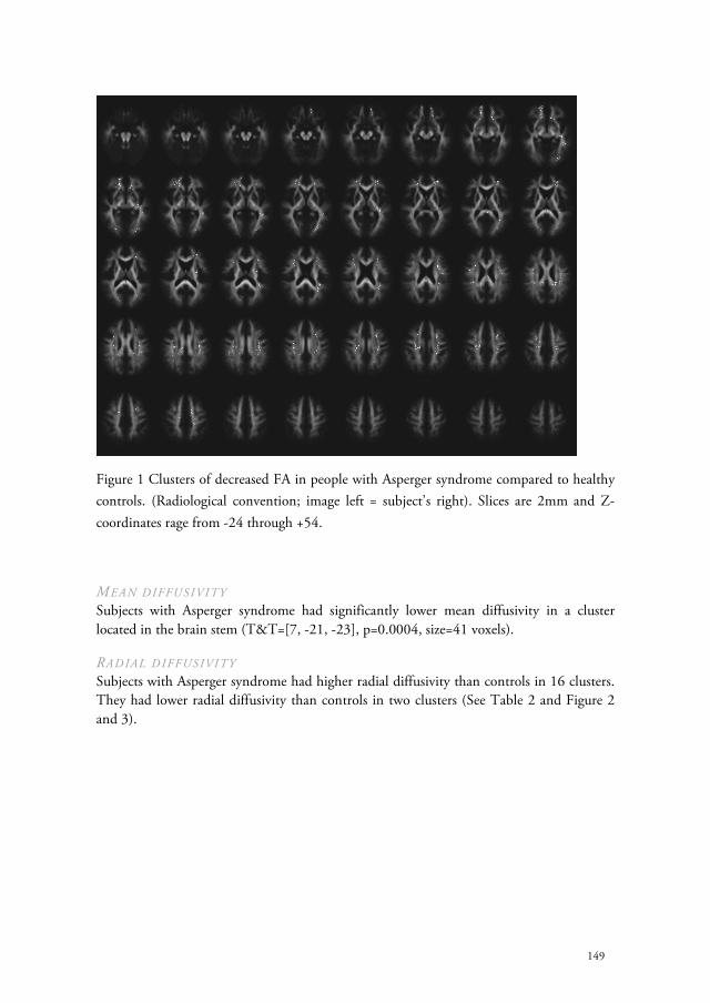
Figure 1 Clusters of decreased FA in people with Asperger syndrome compared to healthy controls. (Radiological convention; image left = subject’s right). Slices are 2mm and Z-coordinates rage from -24 through +54.
MEAN D IFFUS IVITY Subjects with Asperger syndrome had significantly lower mean diffusivity in a cluster located in the brain stem (T&T=[7, -21, -23], p=0.0004, size=41 voxels).
RAD IAL D IFFUS IVITY Subjects with Asperger syndrome had higher radial diffusivity than controls in 16 clusters. They had lower radial diffusivity than controls in two clusters (See Table 2 and Figure 2 and 3).
149

Table 2 - DTI group mapping analysis of radial diffusivity of Asperger syndrome vs. controls.
Legend: (CC) corpus callosum, (CG) cingulum, (CPT) corticopontine tract, (CST) corticospinal tract, (ICP) inferior cerebellar peduncle, (IFO) fronto-occipital fasciculus, (ILF) inferior longitudinal fasciculus, (MCP) middle cerebellar peduncle, (PTR) posterior thalamic radiation, (SCP) superior cerebellar peducle, (SLF) superior longitudinal fasciculus, (STR) superior thalamic radiation,(T&T) Talairach & Tournoux coordinates,(UNC) uncinate fasciculus.
1) Location of most activated voxel.
T&T Possibly involved tracts Location1 Side x y z p Size Higher radial diffusivity in Asperger subjects (Figure 2) UNC Inferior Frontal Gyrus Right 14 32 -15 0.000940 25 IFO/ILF Temporal Lobe Left -36 -40 -6 0.000204 40 IFO/ILF Temporal Lobe Right 38 -43 2 0.000163 54 CG/IFO/ILF Cuneus Left -16 -75 7 0.000163 28 CC Corpus callosum Right 4 -30 7 0.000061 23 CPT/CST/STR/PTR Capsula interna Right 25 -16 12 0.000797 26 SLF Insula Left -29 -6 19 0.000797 30 SLF Parietal Lobe Left -29 -51 26 0.000797 23 CG Cingulate Gyrus Left -13 -35 28 0.000307 22 SLF Inferior Parietal Lobule Right 43 -33 26 0.000960 17 SLF/CPT/CST/STR Frontal Lobe Left -30 -22 27 0.000163 53 CG Cingulate Gyrus Right 18 -9 26 0.000797 23 SLF/CPT/CST/STR
Frontal Lobe Right 25 -14 28 0.000061 122 CC/CPT/PTR Precuneus Left -16 -45 35 0.000327 26 CC/CPT/CST/STR Cingulate Gyrus Left -13 -14 41 0.000368 30 CC/CPT/CST/STR Frontal Lobe Right 20 -30 48 0.000184 21
Lower radial diffusivity in Asperger subjects (Figure 3) MCP/ICP/SCP Cerebellum Left -4 -43 -23 0.000471 38 CC Corpus callosum Left -13 -33 18 0.000264 45
150
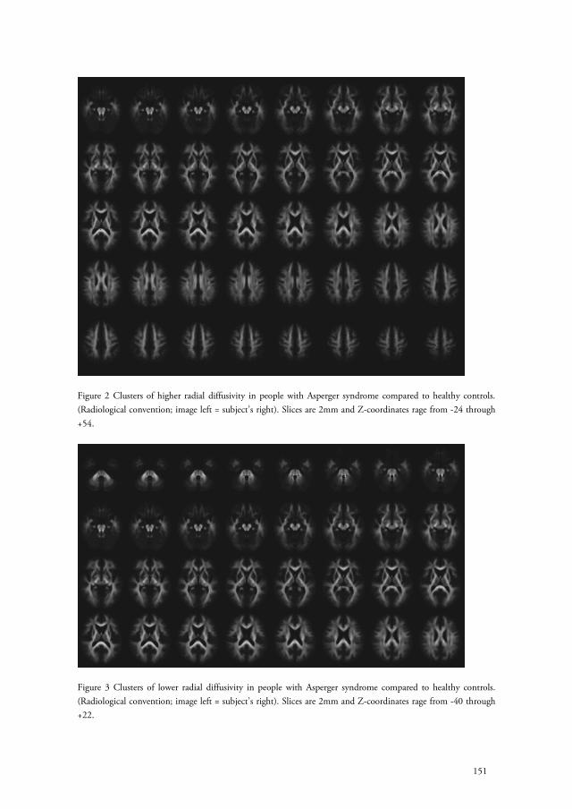
Figure 2 Clusters of higher radial diffusivity in people with Asperger syndrome compared to healthy controls. (Radiological convention; image left = subject’s right). Slices are 2mm and Z-coordinates rage from -24 through +54.
Figure 3 Clusters of lower radial diffusivity in people with Asperger syndrome compared to healthy controls. (Radiological convention; image left = subject’s right). Slices are 2mm and Z-coordinates rage from -40 through +22.
151
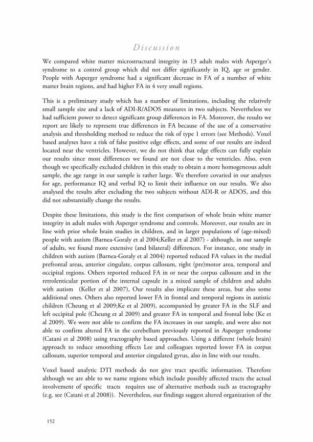
D i s c u s s i o n We compared white matter microstructural integrity in 13 adult males with Asperger’s syndrome to a control group which did not differ significantly in IQ, age or gender. People with Asperger syndrome had a significant decrease in FA of a number of white matter brain regions, and had higher FA in 4 very small regions.
This is a preliminary study which has a number of limitations, including the relatively small sample size and a lack of ADI-R/ADOS measures in two subjects. Nevertheless we had sufficient power to detect significant group differences in FA. Moreover, the results we report are likely to represent true differences in FA because of the use of a conservative analysis and thresholding method to reduce the risk of type 1 errors (see Methods). Voxel based analyses have a risk of false positive edge effects, and some of our results are indeed located near the ventricles. However, we do not think that edge effects can fully explain our results since most differences we found are not close to the ventricles. Also, even though we specifically excluded children in this study to obtain a more homogeneous adult sample, the age range in our sample is rather large. We therefore covaried in our analyses for age, performance IQ and verbal IQ to limit their influence on our results. We also analysed the results after excluding the two subjects without ADI-R or ADOS, and this did not substantially change the results.
Despite these limitations, this study is the first comparison of whole brain white matter integrity in adult males with Asperger syndrome and controls. Moreover, our results are in line with prior whole brain studies in children, and in larger populations of (age-mixed) people with autism (Barnea-Goraly et al 2004;Keller et al 2007) - although, in our sample of adults, we found more extensive (and bilateral) differences. For instance, one study in children with autism (Barnea-Goraly et al 2004) reported reduced FA values in the medial prefrontal areas, anterior cingulate, corpus callosum, right (pre)motor area, temporal and occipital regions. Others reported reduced FA in or near the corpus callosum and in the retrolenticular portion of the internal capsule in a mixed sample of children and adults with autism (Keller et al 2007), Our results also implicate these areas, but also some additional ones. Others also reported lower FA in frontal and temporal regions in autistic children (Cheung et al 2009;Ke et al 2009), accompanied by greater FA in the SLF and left occipital pole (Cheung et al 2009) and greater FA in temporal and frontal lobe (Ke et al 2009). We were not able to confirm the FA increases in our sample, and were also not able to confirm altered FA in the cerebellum previously reported in Asperger syndrome (Catani et al 2008) using tractography based approaches. Using a different (whole brain) approach to reduce smoothing effects Lee and colleagues reported lower FA in corpus callosum, superior temporal and anterior cingulated gyrus, also in line with our results.
Voxel based analytic DTI methods do not give tract specific information. Therefore although we are able to we name regions which include possibly affected tracts the actual involvement of specific tracts requires use of alternative methods such as tractography (e.g. see (Catani et al 2008)). Nevertheless, our findings suggest altered organization of the
152

corpus callosum. This adds to evidence from other structural (Barnea-Goraly et al 2004;Chung et al 2004;Egaas et al 1995;Hardan et al 2000;Keller et al 2007;Lee et al 2007;Manes et al 1999;Piven et al 1997;Vidal et al 2006;Waiter et al 2005) and functional studies (Just et al 2007;Mason et al 2008) that the CC is abnormal in ASD, and may partially explain some clinical features of the disorder. For example, recent fMRI studies reported that people with ASD have significantly lower interhemispheric connectivity during various language, working memory and visuospatial imagery tasks (Just et al 2004;Kana et al 2006;Koshino et al 2005).
We found that the Asperger group had significantly reduced FA in a number of frontal regions, that contain the uncinate fasciculi, inferior fronto-occipital fasciculi and the anterior thalamic radiation. Differences in frontal lobe development, anatomy and function are frequently reported in ASD (Carper and Courchesne 2000;Carper et al 2002;Herbert et al 2004;Levitt et al 2003;Luna et al 2002;Sundaram et al 2008) and frontal lobe deficits have been linked to abnormalities in Theory of Mind (ToM) (Baron-Cohen et al 1999;Castelli et al 2002;Frith and Frith 1999;Happe et al 1996), executive functioning (Luna et al 2002) joint attention (Mundy 2003) and language (Just et al 2004;Muller et al 1998) in ASD. Further, others have reported that people with ASD have significant differences in the activation of cortical motor areas (Muller et al 2001) and motor abnormalities are one of the earliest observable behavioural features in ASD. Hence our results add support to the work of others (Just et al 2004) suggesting that people with ASD have significant differences in frontal connectivity.
In addition we found that people with Asperger syndrome have significant differences in FA of various parietal lobe regions. These results are in line with previous studies in ASD that reported abnormalities in parietal lobe anatomy (Carper et al 2002;Courchesne et al 1993;Haznedar et al 2000;Piven et al 1996;Thakkar et al 2008) and add to evidence of underconnectivity in the temporo-parietal area (Barnea-Goraly et al 2004;Cherkassky et al 2006;Just et al 2007). The areas with reduced FA have been linked to, for instance, emotion processing (Heilman and Gilmore 1998), ToM (Castelli et al 2002), and determining the relevance of stimuli to the self (Northoff and Bermpohl 2004); and deficits in these domains are core clinical features of ASD. Further, Thakkar and colleagues reported lower FA and abnormal BOLD activation during executive function tasks bilaterally in the anterior cingulate gyrus (Thakkar et al 2008).
In our investigation people with Asperger syndrome had a significant reduction of FA bilaterally in the temporal lobe and perisylvian area. Areas with significantly reduced FA bilaterally contained the ILF, SLF and the IFO. This supports suggestions from prior DTI (Barnea-Goraly et al 2004;Conturo et al 2008;Keller et al 2007;Lee et al 2007) and structural and functional studies (Boddaert et al 2004;Gendry Meresse et al 2005) that these regions are abnormal in people with ASD. The temporal and perisylvian areas are vital for language and social cognition, and abnormalities in these areas may explain some of the difficulties ASD patients have in these domains. For example, the superior temporal
153

area is important in auditory processing (Just et al 2004;Meyer et al 2005), social perception of movement (Allison et al 2000), imitation (Iacoboni 2005) and ToM (Castelli et al 2002). Additionally, damage to the temporal region is thought to be related to visually specific semantic, emotional, memory and language deficits (Catani et al 2003;Mandonnet et al 2007;Vigneau et al 2006). Thus the significant reduction in FA we found in the temporal and perisylvian areas in people with ASD may be associated with abnormal connectivity between brain regions important in the understanding of salient social information and language.
We also found reduced FA values in the occipital lobes in people with Asperger syndrome. White matter areas which differed from controls included those adjacent to the lingual gyri and the cuneus, containing the IFO and the ILF. Previous studies also implicated these regions (Barnea-Goraly et al 2004;Deeley et al 2007;Just et al 2004;Keller et al 2007). The occipital region is important in facial emotion processing, and in a prior fMRI study of adults with Asperger syndrome we demonstrated reduced visual cortical activity (including occipital regions of BA 18/19) to a range of primary facial emotions and intensities (Deeley et al 2007). In healthy individuals, visual cortical responses to emotionally salient expressions (such as fearful expressions) are ‘boosted’ by modulatory feedback by limbic regions (Vuilleumier et al 2004). Hence, impaired connectivity between occipital lobe, limbic and other brain regions may contribute to neural hyporesponsiveness to facial expressions, and associated social impairments, in people with Asperger syndrome (Deeley et al 2007).
In addition to FA changes, we also found areas with increased radial diffusivity. Although interpretation of what these differences in radial diffusivity actually mean is somewhat difficult it has been proposed that it is a proxy measure of brain myelination (Wheeler-Kingshott and Cercignani 2009), Hence those areas where we found lower FA overlapping with increased RD (e.g. in the frontal and temporal lobes) may have differences in myelination, rather than axonal degeneration. Furthermore subjects with Asperger syndrome had lower mean diffusivity than controls in the brain stem.
In conclusion our results suggest that people with Asperger syndrome have widespread abnormalities in the microstructral integrity of white matter. Further, these findings add to increasing evidence that people with ASD have differences in long-range (inter-regional) brain connectivity.
154

R e f e r e n c e s Alexander AL, Lee JE, Lazar M, Boudos R, Dubray MB, Oakes TR et al (2007): Diffusion tensor imaging of the corpus callosum in Autism. Neuroimage 34: 61-73.
Allison T, Puce A, McCarthy G (2000): Social perception from visual cues: role of the STS region. Trends Cogn Sci 4: 267-278.
Bailey A, Le Couteur A, Gottesman I, Bolton P, Simonoff E, Yuzda E et al (1995): Autism as a strongly genetic disorder: evidence from a British twin study. Psychol Med 25: 63-77.
Baird G, Simonoff E, Pickles A, Chandler S, Loucas T, Meldrum D et al (2006): Prevalence of disorders of the autism spectrum in a population cohort of children in South Thames: the Special Needs and Autism Project (SNAP). Lancet 368: 210-215.
Barnea-Goraly N, Kwon H, Menon V, Eliez S, Lotspeich L, Reiss AL (2004): White matter structure in autism: preliminary evidence from diffusion tensor imaging. Biol Psychiatry 55: 323-326.
Baron-Cohen S, Ring HA, Wheelwright S, Bullmore ET, Brammer MJ, Simmons A et al (1999): Social intelligence in the normal and autistic brain: an fMRI study. Eur J Neurosci 11: 1891-1898.
Basser PJ (1995): Inferring microstructural features and the physiological state of tissues from diffusion-weighted images. NMR Biomed 8: 333-344.
Basser PJ, Mattiello J, LeBihan D (1994): MR diffusion tensor spectroscopy and imaging. Biophys J 66: 259-267.
Beaulieu C (2002): The basis of anisotropic water diffusion in the nervous system - a technical review. NMR Biomed 15: 435-455.
Belmonte MK (2000): Abnormal attention in autism shown by steady-state visual evoked potentials. Autism 4: 269-285.
Belmonte MK, Yurgelun-Todd DA (2003): Functional anatomy of impaired selective attention and compensatory processing in autism. Brain Res Cogn Brain Res 17: 651-664.
Ben BD, Kronfeld-Duenias V, Zachor DA, Ekstein PM, Hendler T, Tarrasch R et al (2007): Accelerated maturation of white matter in young children with autism: a high b value DWI study. Neuroimage 37: 40-47.
Boddaert N, Chabane N, Gervais H, Good CD, Bourgeois M, Plumet MH et al (2004): Superior temporal sulcus anatomical abnormalities in childhood autism: a voxel-based morphometry MRI study. Neuroimage 23: 364-369.
Bradshaw JL, Sheppard DM (2000): The neurodevelopmental frontostriatal disorders: evolutionary adaptiveness and anomalous lateralization. Brain Lang 73: 297-320.
Brett M, Johnsrude IS, Owen AM (2002): The problem of functional localization in the human brain. Nat Rev Neurosci 3: 243-249.
Bullmore ET, Suckling J, Overmeyer S, Rabe-Hesketh S, Taylor E, Brammer MJ (1999): Global, voxel, and cluster tests, by theory and permutation, for a difference between two groups of structural MR images of the brain. IEEE Trans Med Imaging 18: 32-42.
Carew JD, Wahba G, Basser PJ (2007): Implications of Heterogeneous Variance of Tensor-Derived Quantities for Group Comparisons. Proc Intl Soc Mag Reson Med 15 : 1536.
Carper RA, Courchesne E (2000): Inverse correlation between frontal lobe and cerebellum sizes in children with autism. Brain 123 ( Pt 4): 836-844.
Carper RA, Moses P, Tigue ZD, Courchesne E (2002): Cerebral lobes in autism: early hyperplasia and abnormal age effects. Neuroimage 16: 1038-1051.
155

Castelli F, Frith C, Happe F, Frith U (2002): Autism, Asperger syndrome and brain mechanisms for the attribution of mental states to animated shapes. Brain 125: 1839-1849.
Catani M, Jones DK, Daly e, Embiricos N, Deeley Q, Pugliese L et al (2008): Altered cerebellar feedback projections in asperger syndrome. Neuroimage .
Catani M, ffytche DH (2005): The rises and falls of disconnection syndromes. Brain 128: 2224-2239.
Catani M, Howard RJ, Pajevic S, Jones DK (2002): Virtual in vivo interactive dissection of white matter fasciculi in the human brain. Neuroimage 17: 77-94.
Catani M, Jones DK, Donato R, ffytche DH (2003): Occipito-temporal connections in the human brain. Brain 126: 2093-2107.
Cherkassky VL, Kana RK, Keller TA, Just MA (2006): Functional connectivity in a baseline resting-state network in autism. Neuroreport 17: 1687-1690.
Cheung C, Chua SE, Cheung V, Khong PL, Tai KS, Wong TK et al (2009): White matter fractional anisotrophy differences and correlates of diagnostic symptoms in autism. J Child Psychol Psychiatry 50: 1102-1112.
Chung MK, Dalton KM, Alexander AL, Davidson RJ (2004): Less white matter concentration in autism: 2D voxel-based morphometry. Neuroimage 23: 242-251.
Conturo TE, Williams DL, Smith CD, Gultepe E, Akbudak E, Minshew NJ (2008): Neuronal fiber pathway abnormalities in autism: an initial MRI diffusion tensor tracking study of hippocampo-fusiform and amygdalo-fusiform pathways. J Int Neuropsychol Soc 14: 933-946.
Courchesne E, Karns CM, Davis HR, Ziccardi R, Carper RA, Tigue ZD et al (2001): Unusual brain growth patterns in early life in patients with autistic disorder: an MRI study. Neurology 57: 245-254.
Courchesne E, Pierce K (2005): Why the frontal cortex in autism might be talking only to itself: local over-connectivity but long-distance disconnection. Curr Opin Neurobiol 15: 225-230.
Courchesne E, Press GA, Yeung-Courchesne R (1993): Parietal lobe abnormalities detected with MR in patients with infantile autism. AJR Am J Roentgenol 160: 387-393.
Courchesne E, Redcay E, Morgan JT, Kennedy DP (2005): Autism at the beginning: microstructural and growth abnormalities underlying the cognitive and behavioral phenotype of autism. Dev Psychopathol 17: 577-597.
Deeley Q, Daly EM, Surguladze S, Page L, Toal F, Robertson D et al (2007): An event related functional magnetic resonance imaging study of facial emotion processing in Asperger syndrome. Biol Psychiatry 62: 207-217.
Egaas B, Courchesne E, Saitoh O (1995): Reduced size of corpus callosum in autism. Arch Neurol 52: 794-801.
Frith C (2004): Is autism a disconnection disorder? Lancet Neurol 3: 577.
Frith CD, Frith U (1999): Interacting minds--a biological basis. Science 286: 1692-1695.
Frith U (2001): Mind blindness and the brain in autism. Neuron 32: 969-979.
Gendry Meresse I, Zilbovicius M, Boddaert N, Robel L, Philippe A, Sfaello I et al (2005): Autism severity and temporal lobe functional abnormalities. Ann Neurol 58: 466-469.
Geschwind DH, Levitt P (2007): Autism spectrum disorders: developmental disconnection syndromes. Curr Opin Neurobiol 17: 103-111.
Hammock,EAD and Levitt,P. Sensitive periods in the development of brain and behavior. J.Cogn Neurosci. [16], 1412-1425. 2006.
Happe F, Ehlers S, Fletcher P, Frith U, Johansson M, Gillberg C et al (1996): 'Theory of mind' in the brain. Evidence from a PET scan study of Asperger syndrome. Neuroreport 8: 197-201.
156

Hardan AY, Minshew NJ, Keshavan MS (2000): Corpus callosum size in autism. Neurology 55: 1033-1036.
Haznedar MM, Buchsbaum MS, Wei TC, Hof PR, Cartwright C, Bienstock CA et al (2000): Limbic circuitry in patients with autism spectrum disorders studied with positron emission tomography and magnetic resonance imaging. Am J Psychiatry 157: 1994-2001.
Heilman KM, Gilmore RL (1998): Cortical influences in emotion. J Clin Neurophysiol 15: 409-423.
Herbert MR, Ziegler DA, Makris N, Filipek PA, Kemper TL, Normandin JJ et al (2004): Localization of white matter volume increase in autism and developmental language disorder. Ann Neurol 55: 530-540.
Iacoboni M (2005): Neural mechanisms of imitation. Curr Opin Neurobiol 15: 632-637.
Jones DK, Symms MR, Cercignani M, Howard RJ (2005): The effect of filter size on VBM analyses of DT-MRI data. Neuroimage 26: 546-554.
Jones DK, Williams SC, Gasston D, Horsfield MA, Simmons A, Howard R (2002): Isotropic resolution diffusion tensor imaging with whole brain acquisition in a clinically acceptable time. Hum Brain Mapp 15: 216-230.
Just MA, Cherkassky VL, Keller TA, Kana RK, Minshew NJ (2007): Functional and anatomical cortical underconnectivity in autism: evidence from an FMRI study of an executive function task and corpus callosum morphometry. Cereb Cortex 17: 951-961.
Just MA, Cherkassky VL, Keller TA, Minshew NJ (2004): Cortical activation and synchronization during sentence comprehension in high-functioning autism: evidence of underconnectivity. Brain 127: 1811-1821.
Kana RK, Keller TA, Cherkassky VL, Minshew NJ, Just MA (2006): Sentence comprehension in autism: thinking in pictures with decreased functional connectivity. Brain 129: 2484-2493.
Ke X, Tang T, Hong S, Hang Y, Zou B, Li H et al (2009): White matter impairments in autism, evidence from voxel-based morphometry and diffusion tensor imaging. Brain Res 1265: 171-177.
Keller TA, Kana RK, Just MA (2007): A developmental study of the structural integrity of white matter in autism. Neuroreport 18: 23-27.
Knudsen EI (2004): Sensitive periods in the development of the brain and behavior. J Cogn Neurosci 16: 1412-1425.
Koshino H, Carpenter PA, Minshew NJ, Cherkassky VL, Keller TA, Just MA (2005): Functional connectivity in an fMRI working memory task in high-functioning autism. Neuroimage 24: 810-821.
Lee JE, Bigler ED, Alexander AL, Lazar M, Dubray MB, Chung MK et al (2007): Diffusion tensor imaging of white matter in the superior temporal gyrus and temporal stem in autism. Neurosci Lett 424: 127-132.
Levitt JG, Blanton RE, Smalley S, Thompson PM, Guthrie D, McCracken JT et al (2003): Cortical sulcal maps in autism. Cereb Cortex 13: 728-735.
Lord C, Rutter M, Goode S, Heemsbergen J, Jordan H, Mawhood L et al (1989): Autism diagnostic observation schedule: a standardized observation of communicative and social behavior. J Autism Dev Disord 19: 185-212.
Lord C, Rutter M, Le CA (1994): Autism Diagnostic Interview-Revised: a revised version of a diagnostic interview for caregivers of individuals with possible pervasive developmental disorders. J Autism Dev Disord 24: 659-685.
Luna B, Minshew NJ, Garver KE, Lazar NA, Thulborn KR, Eddy WF et al (2002): Neocortical system abnormalities in autism: an fMRI study of spatial working memory. Neurology 59: 834-840.
Mandonnet E, Nouet A, Gatignol P, Capelle L, Duffau H (2007): Does the left inferior longitudinal fasciculus play a role in language? A brain stimulation study. Brain 130: 623-629.
157

Manes F, Piven J, Vrancic D, Nanclares V, Plebst C, Starkstein SE (1999): An MRI study of the corpus callosum and cerebellum in mentally retarded autistic individuals. J Neuropsychiatry Clin Neurosci 11: 470-474.
Mason RA, Williams DL, Kana RK, Minshew N, Just MA (2008): Theory of Mind disruption and recruitment of the right hemisphere during narrative comprehension in autism. Neuropsychologia 46: 269-280.
McAlonan GM, Daly E, Kumari V, Critchley HD, van Amelsvoort T, Suckling J et al (2002): Brain anatomy and sensorimotor gating in Asperger's syndrome. Brain 125: 1594-1606.
Meyer M, Zaehle T, Gountouna VE, Barron A, Jancke L, Turk A (2005): Spectro-temporal processing during speech perception involves left posterior auditory cortex. Neuroreport 16: 1985-1989.
Minshew NJ, Goldstein G, Siegel DJ (1997): Neuropsychologic functioning in autism: profile of a complex information processing disorder. J Int Neuropsychol Soc 3: 303-316.
Mori,S, Wakana,S, Nagae-Poetscher,L, van Zijl,P (2005): MRI Atlas of Human White Matter. Amsterdam: Elsevier.
Muller RA (2007): The study of autism as a distributed disorder. Ment Retard Dev Disabil Res Rev 13: 85-95.
Muller RA, Chugani DC, Behen ME, Rothermel RD, Muzik O, Chakraborty PK et al (1998): Impairment of dentato-thalamo-cortical pathway in autistic men: language activation data from positron emission tomography. Neurosci Lett 245: 1-4.
Muller RA, Pierce K, Ambrose JB, Allen G, Courchesne E (2001): Atypical patterns of cerebral motor activation in autism: a functional magnetic resonance study. Biol Psychiatry 49: 665-676.
Mundy P (2003): Annotation: the neural basis of social impairments in autism: the role of the dorsal medial-frontal cortex and anterior cingulate system. J Child Psychol Psychiatry 44: 793-809.
Northoff G, Bermpohl F (2004): Cortical midline structures and the self. Trends Cogn Sci 8: 102-107.
Pierpaoli C, Basser PJ (1996): Toward a quantitative assessment of diffusion anisotropy. Magn Reson Med 36: 893-906.
Piven J, Arndt S, Bailey J, Andreasen N (1996): Regional brain enlargement in autism: a magnetic resonance imaging study. J Am Acad Child Adolesc Psychiatry 35: 530-536.
Piven J, Bailey J, Ranson BJ, Arndt S (1997): An MRI study of the corpus callosum in autism. Am J Psychiatry 154: 1051-1056.
Song SK, Sun SW, Ramsbottom MJ, Chang C, Russell J, Cross AH (2002): Dysmyelination revealed through MRI as increased radial (but unchanged axial) diffusion of water. Neuroimage 17: 1429-1436.
Song SK, Yoshino J, Le TQ, Lin SJ, Sun SW, Cross AH et al (2005): Demyelination increases radial diffusivity in corpus callosum of mouse brain. Neuroimage 26: 132-140.
Sundaram SK, Kumar A, Makki MI, Behen ME, Chugani HT, Chugani DC (2008): Diffusion Tensor Imaging of Frontal Lobe in Autism Spectrum Disorder. Cereb Cortex .
Talairach,J, Tournoux,P (1988): Co-planar stereotaxic atlas of the human brain . New York: Thieme.
Thakkar KN, Polli FE, Joseph RM, Tuch DS, Hadjikhani N, Barton JJ et al (2008): Response monitoring, repetitive behaviour and anterior cingulate abnormalities in autism spectrum disorders (ASD). Brain 131: 2464-2478.
Toal F, Murphy DG, Murphy KC (2005): Autistic-spectrum disorders: lessons from neuroimaging. Br J Psychiatry 187: 395-397.
Vidal CN, Nicolson R, DeVito TJ, Hayashi KM, Geaga JA, Drost DJ et al (2006): Mapping corpus callosum deficits in autism: an index of aberrant cortical connectivity. Biol Psychiatry 60: 218-225.
158

Vigneau M, Beaucousin V, Herve PY, Duffau H, Crivello F, Houde O et al (2006): Meta-analyzing left hemisphere language areas: phonology, semantics, and sentence processing. Neuroimage 30: 1414-1432.
Vuilleumier P, Richardson MP, Armony JL, Driver J, Dolan RJ (2004): Distant influences of amygdala lesion on visual cortical activation during emotional face processing. Nat Neurosci 7: 1271-1278.
Waiter GD, Williams JH, Murray AD, Gilchrist A, Perrett DI, Whiten A (2005): Structural white matter deficits in high-functioning individuals with autistic spectrum disorder: a voxel-based investigation. Neuroimage 24: 455-461.
Weschler (1987): Weschler Adult Intelligence Scale Revised . New York, NY: Psychological Corporation, Harcourt Brace.
Wheeler-Kingshott CA, Cercignani M (2009): About "axial" and "radial" diffusivities. Magn Reson Med 61: 1255-1260.
Williams DL, Goldstein G, Minshew NJ (2006): Neuropsychologic functioning in children with autism: further evidence for disordered complex information-processing. Child Neuropsychol 12: 279-298.
159


C h a p t e r 9
Summary and general discussion

S u m m a r y a n d c o n c l u s i o n s
Schizophrenia and autism spectrum disorder (ASD) are severe mental disorders with specific differences. For instance, ASD is often diagnosed in the first few years of life, whereas the first symptoms of schizophrenia usually appear in adolescence. Furthermore, ASD is often accompanied by a learning disability, contrary to schizophrenia. Nevertheless these disorders also have similarities. On a genetic level they share an increased risk due to copy number variations and rare specific loci and alleles (Carroll and Owen 2009). On a intermediate phenotype level they share specific findings in volumetric brain studies (Cheung et al 2010) and impaired stimulus filtering (Perry et al 2007;McAlonan et al 2002;Kumari et al 2008). On a clinical phenotype level they share deficits in social behavior, oddness of speech, unusual responsiveness to the sensory environment, isolated skill areas, and inappropriate affect. Furthermore both disorders have a chronic course in the majority of patients.
Schizophrenia is usually preceded by a prodromal period, with mild positive psychotic and negative symptoms, non-specific symptoms and a decline in psychosocial functioning. Using specific assessment instruments it is possible to diagnose patients at Ultra High Risk (UHR) for developing psychosis, of which 10-40% will indeed eventually develop psychosis (Yung and McGorry 1996;Yung et al 2003;Klosterkotter et al 2001;Miller et al 2003). Similarly, ASD is also associated with an increased risk of 14 – 34% for developing psychosis (Stahlberg et al 2004;Tsakanikos et al 2007;Mouridsen et al 2008), with specific subtypes like multiple complex developmental disorder (McDD) having even higher transition rates (Sprong et al 2008;van Engeland and van der Gaag 1994). One could therefore tentatively state that ASD patients, as UHR patients, are also at clinically or even at ultra high risk to develop psychosis.
We used various in vivo brain imaging techniques to asses brain anatomy and function in these two disorders. With diffusion tensor magnetic resonance imaging (DT-MRI) it is possible to study white matter integrity, and previous studies show that integrity is decreased in schizophrenia subjects (Konrad and Winterer 2008;Friedman et al 2008), and there is evidence that white matter may be abnormal in high risk populations although changes have not been consistent across studies. Similar to schizophrenia, ASD has also been called a connectivity disorder, in which disrupted white matter adds to the symptomatology and pathophysiology of the disorders. DT-MRI studies in children and in mixed children/adults groups with ASD have also shown specific abnormalities in white matter (Barnea-Goraly et al 2004;Keller et al 2007;Alexander et al 2007;Lee et al 2007).
162

Nuclear imaging studies have shown that dopaminergic neurotransmission is disrupted in schizophrenia (Abi-Dargham et al 2000;Abi-Dargham et al 2009), and recent studies showed evidence for abnormal dopamine synthesis in UHR subjects (Howes et al 2009) and altered relations between hippocampal glutamate and presynaptic dopamine function (Stone et al 2010).
In this thesis we sought to further explore the above mentioned theories. The overall aim of the studies described in this thesis was to increase our knowledge on the brain structure and function of UHR subjects and people with ASD. We focused on these groups of patients as they are thought to be at increased or at ultra high risk for developing psychosis. Specifically we sought to answer the following questions; is there enough evidence for specific treatment of UHR patients? Do UHR subjects who later develop psychosis have specific white matter integrity abnormalities compared to UHR subjects who do not and to controls? What is the current literature on challenge studies using alpha-methyl-para-tyrosine (AMPT)? Do UHR subjects have abnormalities in striatal dopaminergic function? Is there a relation between hippocampal glutamatergic function and striatal dopamine in UHR patients and do glutamate levels differ from controls? Is psychosis in ASD associated with specific structural brain differences? Do people with Asperger syndrome have white matter integrity abnormalities?
Chapter 1 contained a general introduction and the outline of the thesis. In Chapter 2 the current literature on interventions in UHR patients was reviewed. The positive (acute) effect reported in three randomized clinical trials with antipsychotic medication and/or cognitive behavioral therapy disappeared at the end of treatment. This suggests that treatment does not prevent but may delay the transition to psychosis. Other naturalistic studies present a hypothesis about a possible preventive effect of antidepressants. Improved prediction of transition to psychosis and a decrease of the number of false positives are needed. At this time the data concerning the benefits and risks do not justify prodromal intervention as standard clinical practice. In Chapter 3 we described the results of a DT-MRI study, and report that UHR subjects that later develop psychosis (UHR-P) compared to UHR subjects who do not develop psychosis (UHR-NP) have reduced fractional anisotropy (FA) of white matter (WM) lateral to the right putamen and of the left superior temporal lobe, and higher FA of the left medial temporal lobe. Furthermore UHR-P subjects also have reduced FA compared to healthy controls in medial frontal lobes bilaterally. These regions have been found to be involved in psychosis and are associated with severity of psychotic symptoms in other samples. The putative changes in connectivity that these FA differences represent may play a role in the development of psychosis or its symptomatology, or conversely may be a result of this process. From our
163
data it appears that, compared to the more extensive frontal differences with healthy controls, relatively subtle differences in WM integrity in temporal and striatal brain regions may influence whether an UHR subject develops psychosis. Chapter 4 reviewed challenge and therapeutic studies using AMPT in neuropsychiatric disorders. The reviewed studies show that catecholamine depletion induced by AMPT provides an useful paradigm for elucidating the function of catecholaminergic pathways. Furthermore, there is promising evidence that AMPT has some beneficial effects in patients with dyskinesia, Huntington´s chorea, Tourette syndrome, mania, substance abuse, 22q11 deletion syndrome and psychosis. Side effects, although seldom severe, are common and it is recommended to limit the dose as much as possible. In Chapter 5 we combined SPECT imaging of dopamine D2/3 receptors with dopamine depletion in UHR subjects and age, gender and intelligence quotient (IQ) matched controls. The data suggests that there may be a subgroup of UHR patients that already has abnormalities in dopaminergic neurotransmission. This subgroup is characterized clinically by high scores on positive symptom scales, and has elevated dopamine receptor occupancy by endogenous dopamine compared to the low positive symptom UHR subgroup and compared to healthy controls. Furthermore the relatively low AMPT dose we used leads to similar dopamine depletion in controls as higher doses used by other groups, but also to significant positive symptom reduction in UHR patients, as has also been shown in schizophrenia. High occupancy of dopamine D2/3 receptors by endogenous dopamine predicted good response of positive symptoms to AMPT in UHR patients. In Chapter 6 we investigated the relation between left hippocampal glutamate levels (measured by Proton Magnetic Resonance Spectroscopy (1H-MRS)) and postsynaptic dopamine D2/3 receptor availability and receptor occupancy by endogenous dopamine in UHR subjects and controls. A recent study reported a negative relation between left hippocampal glutamate levels and presynaptic [18F]DOPA uptake in the left striatum of UHR patients, but not in healthy controls. Additionally this study reported evidence that this relationship may predict later transition to psychosis. We found no evidence that glutamate is also related to postsynaptic dopamine neurotransmission UHR subjects or controls, but found statistically significantly decreased glutamate levels in the left hippocampus of UHR subjects compared to controls. Decreased hippocampal glutamate has also been reported in schizophrenia (Lutkenhoff et al 2010), and our data suggest that altered glutamate levels may influence the development of psychosis. In Chapter 7 we studied psychosis in (non learning disabled) adult ASD patients by performing a voxel based morphometry analysis between people with autism with comorbid psychosis and people with autism without psychosis. Additionally we also compared the autism groups with age, IQ and gender matched healthy controls. Compared to controls, both ASD groups had statistically significantly less grey matter
164

bilaterally in the temporal lobes (including the fusiform gyrus) and cerebellum, and reduced white matter in the cerebellum. The ASD groups had increased grey matter in striatal regions. However ASD patients with psychosis also had a significant reduction in grey matter content of frontal and occipital regions. Within ASD comparisons revealed that psychosis was associated with a bilateral reduction in grey matter of cerebellum and the fusiform gyrus. We concluded that the pre-existing anatomical differences in brain systems also implicated in psychosis may reduce the additional disease burden required to develop co-morbid psychotic symptoms. Our study also suggests that the biology of psychosis in people with ASD may differ from that in the non-autistic population. In Chapter 8 we described the results of the first DT-MRI study in Asperger syndrome. As there are indications that brain abnormalities may progress into adulthood we compared adults with Asperger syndrome to age, gender and IQ matched healthy controls. Adults with Asperger syndrome had a significantly lower FA than controls in thirteen clusters. These were largely bilateral and included white matter in the internal capsule, frontal, temporal, parietal and occipital lobes, cingulum and corpus callosum. Our results are in line with studies in children with autism, although we found more extensive (and bilateral) differences. Our results suggest that people with Asperger syndrome have widespread abnormalities in the microstructural integrity of white matter, in regions previously associated with ASD and its symptomatology. Further, these findings add to increasing evidence that people with ASD have differences in long-range (inter-regional) brain connectivity.
CON CL US ION S The aim of the studies described in this thesis was to increase our knowledge on the brain structure and function of people with an increased risk for developing psychosis. We focused on two groups; 1) patients with an Ultra High Risk for developing psychosis according to international criteria; 2) patients with an autism spectrum disorder. The main conclusions of this thesis are:
1. Ultra High Risk subjects that later develop psychosis (UHR-P) have reduced fractional anisotropy (FA) of white matter lateral to the right putamen and of the left superior temporal lobe and higher FA of the left medial temporal lobe compared to subjects that do not develop psychosis (UHR-NP).
2. UHR-P subjects have reduced FA in white matter of medial frontal lobes bilaterally compared to healthy controls.
165

3. A subgroup of UHR subjects has elevated dopamine D2/3 receptor occupancy by endogenous dopamine. This subgroup is characterized by more severe positive symptoms.
4. Positive symptoms decrease following dopamine depletion (induced pharmacologically by AMPT) in UHR subjects, comparable to observations in schizophrenia, suggesting a similar mechanism.
5. In UHR subjects, higher dopamine D2/3 receptor occupancy is associated with more positive symptoms and with a greater decrease of positive symptoms after acute dopamine depletion.
6. Glutamate in the left hippocampus of UHR subjects is not related to postsynaptic striatal dopamine D2/3 receptor availability or occupancy by endogenous dopamine.
7. UHR subjects have decreased left hippocampal glutamate levels compared to healthy controls, comparable to observations in schizophrenia.
8. Psychosis in autism is associated with reductions of grey matter in cerebellum and fusiform gyrus compared to non psychotic people with autism.
9. Adults with Asperger syndrome have widespread reductions in FA of white matter.
D i s c u s s i o n This thesis contains various studies that study possible biomarkers for psychiatric disorders. A biomarker suggests that the disorder is an objective and value free concept. This is interesting as mental disorders are probably most value-laden of all medical conditions. To explore this paradox thoroughly is beyond the scope of this thesis, but it is interesting to take a closer look at what we are actually measuring, and at what point in the disease process.
We conducted two studies which looked at the quality or integrity of WM tracts. WM bundles that are used frequently are restructured to work more efficiently, and for instance become more myelinated, which leads to lower FA values in DT-MRI imaging (Basser 1995). WM bundles that are not much used are less myelinated and even get pruned with time (Giorgio et al 2010). It seems thus that WM structure follows its usage (excluding certain disorders that have WM degeneration as core feature, which does not apply to schizophrenia or autism). This implies that structural changes in WM can inherently only
166
be of limited value in prediction of, for instance, transition to psychosis as these changes are occurring in response to altered patterns of usage. So when we look at WM integrity changes, we are looking at something that is the result of the disease, much like the symptoms a patient is experiencing. So what makes WM interesting then? It may very well be that WM changes occur before symptoms actually increase to psychotic intensity in patients, and thus WM abnormalities may still predict who will develop frank psychosis. Further, although we are not likely looking at the core of the disease, what we are looking at is a roadmap of the brain and its usage. Therefore the changes in WM connections tell us that something is happening differently or abnormally in the grey matter regions they are connecting and hence may direct us to the cause of the WM abnormalities.
Striatal dopamine dysfunction also seems to occur at the etiological end of the symptoms of schizophrenia, and is thought to be the final common pathway. It may however be one step closer than WM changes, as abnormal striatal dopamine function may cause symptoms but may also cause WM integrity changes close to the striatum as we reported in Chapter 4. For dopamine it has already been more defined what it is we are measuring, given the dopamine hypothesis and Kapur’s interpretation of its importance in salience (Kapur 2003). As we reported in Chapter 5 positive symptoms are also related to dopamine receptor occupancy by its natural neurotransmitter. Nevertheless there are remarkable things about dopamine. Increasing dopamine does not cause positive symptoms in most people, a view that is strengthened by the amount of recreational use of dopamine boosters like cocaine, amphetamines and cannabis, although increased salience is likely to be what drives (initial) use of these drugs. A regular dopamine challenge with amphetamine increases extracellular dopamine in the striatum by around 400% (Breier et al 1997) and does not necessarily cause psychotic symptoms. So if increased dopamine is not enough in most people to cause psychosis, something else must be mediating its effect. This is in line with the fact that we find only modest increases in dopaminergic transmission in schizophrenia, which often partly overlap with control groups (Abi-Dargham et al 2000;Abi-Dargham et al 2009;Laruelle et al 1996), although our methods to measure these differences are still quite crude. Neuroanatomically, positive symptoms like hallucinations and delusions are probably not formed in the striatum but in the cortex, so hypothetically the cortex could differ in dopaminergic sensitivity, either inherently or mediated by another agent. This hypothesis is supported by recent evidence for separate regulation of striatal and extrastriatal dopaminergic systems (Cervenka et al 2010). For instance there is increased scientific interest in the role of dopamine in cognition. Perhaps difficulties in performing cognitive tasks that occur in schizophrenia, which are cortical processes, could influence or moderate cortical dopaminergic sensitivity, or influence this indirectly via the thalamus (de Manzano et al 2010). It is postulated that dopamine
167

influence on cognition follows an inverted U-curve, and thus dopamine has an optimum level for successful cognitive functioning (Williams and Goldman-Rakic 1995). However, one could conversely hypothesize that cognitive problems, as for instance formal thought disorders, could disinhibit delusional logic and thought in susceptible patients.
Our research has focused to some extent on the dimensional model of mental disease, and has looked at for instance psychosis (or positive symptoms) within UHR and ASD subjects. As already mentioned in the introduction, one could therefore tentatively state that ASD patients, as UHR patients, are at increased risk to develop psychosis. Nevertheless the mechanism of psychosis in autism may be different from schizophrenia, as results from Chapter 6 also suggest; psychosis in autism is associated with other grey matter abnormalities than are usually associated with psychosis in schizophrenia. However, it is interesting to see that both UHR and ASD subjects appear to have only subtle brain differences associated with the occurrence of psychosis. What may be trivial for UHR subjects, might be interesting (but tentative) for autism; they are associated with increased risk for psychosis and are neuroanatomically quite similar to people with the same diagnosis who also developed psychosis.
LIM ITA TION S This thesis and the experiments in it have certain limitations. The specific limitations have been discussed in the relevant chapters, nevertheless a few that may have general implications are summarized here. As a whole, imaging studies and the recruitment of specific complex patient groups as UHR patients and patients with Asperger syndrome is very time consuming. Consequently some of the experiments in this thesis have a limited sample size, which could lead to type 2 errors. Nevertheless we were able to detect statistically significant differences with large effect sizes. It is possible that future larger studies may find additional subtle differences. Also, in some experiments only males were included, which limits the generalizability, but at the same time this increases specificity as results are not confounded by gender differences. We used SPECT imaging which has a limited spatial and temporal resolution. Future studies could use PET as this technique has a higher spatial resolution and sensitivity than SPECT. This offers the opportunity to study the different subregions of the striatum, as it is mostly the ventral part that is hypothesized to be important for psychosis and schizophrenia. Studying schizophrenia is inherently difficult as the disorder is very heterogeneous and probably encompasses multiple different diseases. The UHR concept does not unambiguously lead to schizophrenia and thus this increases heterogeneity of the sample even more. This is an inherent problem of the classification systems, but this makes the findings in subgroups of the sample interesting and scientifically plausible, as we see in Chapter 5.
168
ST REN GTHS This thesis also has specific strengths. In this thesis highly original studies were reported. For instance the first DT-MRI study of psychosis in UHR was conducted, as well as the first imaging study of psychosis in autism, both with interesting results. Also the first DT-MRI study in Asperger was reported, a relatively under researched patient group. Furthermore we conducted the first dopamine depletion SPECT study in UHR patients. All but one (chapter 7) of the clinical studies were conducted in patients who did not take anti-psychotics and lastly the control groups were always matched for IQ, age and gender.
FU TUR E D IREC TION S We plan to complete follow-up of the SPECT study described in chapter 5 to elucidate which UHR patients will develop psychosis and what the relation is to pre-psychosis striatal dopaminergic neurotransmission. However, due to the relatively low sample size, it is unlikely that we will come up with definitive conclusions about changes in receptor occupancy after transition to psychosis.
Seeing the ongoing discussion about dopamine and its relation to schizophrenia, but probably more specifically to positive symptoms, it would be very interesting to design a study which could provide more clarity in this relationship. For instance a longitudinal study in schizophrenia where a depletion PET (which has a higher spatial resolution that SPECT and can therefore identify subregions of the striatum adequately) will be performed at the time of active non-medicated first episode psychosis, followed by a retest when positive symptoms are normalized and medication has been stopped, and in turn followed by a second retest when symptoms have increased again but medication has not yet commenced. Notwithstanding the ethical difficulties in this design (which could be overcome) this design could answer interesting questions, for instance; is increase in symptoms individually accompanied by increase in dopamine? How much increase in dopamine causes how much increase in symptoms? What is the variance? Can symptoms increase while dopamine does not or decreases? In general; how closely is dopamine related to symptoms? This is interesting as these studies have until now always been done non-longitudinally, which limits what can be concluded.
Another field which has received little attention is dopaminergic neurotransmission in ASD and more attention has gone towards serotonin abnormalities and function (Posey et al 2008). It is hypothesized that a hyperdopaminergic functioning of the brain might explain overactivity and stereotyped movements in autism, and it is known that dopamine agonists worsen these symptoms. Also it is known that blockage of dopaminergic receptors
169

decrease stereotyped and hyperactive behavior in ASD. Moreover, some dopamine receptor antagonists like the antipsychotic drug risperidone are quite commonly used to treat these symptoms, and the few clinical trials that have been done in these subjects showed at least temporary effect on behavior. Although dopaminergic neurotransmission has not been thoroughly researched in ASD, there is preliminary evidence of abnormalities. One [18F]-DOPA PET study was done which showed a decrease by 39% of regional [18F]-DOPA accumulation in the anterior medial prefrontal cortex (Ernst et al 1997). Another showed increased dopamine D2/3 receptor binding in the caudate nucleus and putamen as a whole (Fernell et al 1997). Both these studies were done in children en there is no literature on dopaminergic neurotransmission in adults with ASD. Therefore it would be very interesting to conduct a dopamine depletion study with PET or SPECT to look at striatal dopamine D2/3 receptor occupancy in ASD patients, as this is the target for antipsychotic drugs used to treat some of the symptoms in ASD. The striatal dopaminergic neurotransmission could be related to autistic symptomatology (especially hyperactive and stereotyped behavior) but also to measures of psychotic symptoms in these patients. This could give much needed understanding of why (often off-label prescribed) use of antipsychotics decrease some of the symptoms associated with ASD.
Overall the combination of techniques appears to be most promising in imaging research, as for instance combining (depletion) SPECT/PET and 1H-MRS, PET and functional MRI (fMRI), SPECT/PET and DT-MRI, or combining multiple tracers in nuclear imaging, but always combined with clinical measures of symptom severity. This enables us to bridge the islands of knowledge already obtained in the separate imaging fields, and to discover what the relationship between them is. This is crucial as each provides information on different aspects of the total function, as the instruments do in music. White and grey matter integrity and density (measured by DT-MRI and MRI) acting as rhythm section providing the basic structure; glutamate and GABA (1H-MRS) as the bass section providing more tonal boundaries for the other instruments by inhibiting and disinhibiting; dopamine receptor availability and occupancy (SPECT and PET) as chords section played by piano which colors the timbre and loudness of the music; and cortical activation (fMRI) acting as the solo trumpet, playing the actual melody and improvising within (and around) the boundaries set by the other instruments. The need to study their interrelationships is the way of the future. As in music, the importance of their acting in concert is greater than the sum of their individual contributions. Nevertheless, in this age of biological psychiatry it must not be forgotten that time, place and audience are the final referees that decide if the music is either brilliant or crazy.
170

R e f e r e n c e s Abi-Dargham A, Rodenhiser J, Printz D, Zea-Ponce Y, Gil R, Kegeles LS et al (2000): Increased baseline occupancy of D2 receptors by dopamine in schizophrenia. Proc Natl Acad Sci U S A 97: 8104-8109.
Abi-Dargham A, van de Giessen E, Slifstein M, Kegeles LS, Laruelle M (2009): Baseline and amphetamine-stimulated dopamine activity are related in drug-naive schizophrenic subjects. Biol Psychiatry 65: 1091-1093.
Alexander AL, Lee JE, Lazar M, Boudos R, Dubray MB, Oakes TR et al (2007): Diffusion tensor imaging of the corpus callosum in Autism. Neuroimage 34: 61-73.
Barnea-Goraly N, Kwon H, Menon V, Eliez S, Lotspeich L, Reiss AL (2004): White matter structure in autism: preliminary evidence from diffusion tensor imaging. Biol Psychiatry 55: 323-326.
Basser PJ (1995): Inferring microstructural features and the physiological state of tissues from diffusion-weighted images. NMR Biomed 8: 333-344.
Breier A, Su TP, Saunders R, Carson RE, Kolachana BS, de Bartolomeis A et al (1997): Schizophrenia is associated with elevated amphetamine-induced synaptic dopamine concentrations: evidence from a novel positron emission tomography method. Proc Natl Acad Sci U S A 94: 2569-2574.
Carroll LS, Owen MJ (2009): Genetic overlap between autism, schizophrenia and bipolar disorder. Genome Med 1: 102.
Cervenka S, Varrone A, Fransen E, Halldin C, Farde L (2010): PET studies of D2-receptor binding in striatal and extrastriatal brain regions: Biochemical support in vivo for separate dopaminergic systems in humans. Synapse 64: 478-485.
Cheung C, Yu K, Fung G, Leung M, Wong C, Li Q et al (2010): Autistic disorders and schizophrenia: related or remote? An anatomical likelihood estimation. PLoS One 5: e12233.
de Manzano O, Cervenka S, Karabanov A, Farde L, Ullen F (2010): Thinking outside a less intact box: thalamic dopamine D2 receptor densities are negatively related to psychometric creativity in healthy individuals. PLoS One 5: e10670.
Ernst M, Zametkin AJ, Matochik JA, Pascualvaca D, Cohen RM (1997): Low medial prefrontal dopaminergic activity in autistic children. Lancet 350: 638.
Fernell E, Watanabe Y, Adolfsson I, Tani Y, Bergstrom M, Hartvig P et al (1997): Possible effects of tetrahydrobiopterin treatment in six children with autism--clinical and positron emission tomography data: a pilot study. Dev Med Child Neurol 39: 313-318.
Friedman JI, Tang C, Carpenter D, Buchsbaum M, Schmeidler J, Flanagan L et al (2008): Diffusion tensor imaging findings in first-episode and chronic schizophrenia patients. Am J Psychiatry 165: 1024-1032.
Giorgio A, Watkins KE, Chadwick M, James S, Winmill L, Douaud G et al (2010): Longitudinal changes in grey and white matter during adolescence. Neuroimage 49: 94-103.
Howes OD, Montgomery AJ, Asselin MC, Murray RM, Valli I, Tabraham P et al (2009): Elevated striatal dopamine function linked to prodromal signs of schizophrenia. Archives of General Psychiatry 66: 13-20.
Kapur S (2003): Psychosis as a state of aberrant salience: a framework linking biology, phenomenology, and pharmacology in schizophrenia. Am J Psychiatry 160: 13-23.
Keller TA, Kana RK, Just MA (2007): A developmental study of the structural integrity of white matter in autism. Neuroreport 18: 23-27.
Klosterkotter J, Hellmich M, Steinmeyer EM, Schultze-Lutter F (2001): Diagnosing schizophrenia in the initial prodromal phase. Arch Gen Psychiatry 58: 158-164.
171

Konrad A, Winterer G (2008): Disturbed structural connectivity in schizophrenia primary factor in pathology or epiphenomenon? Schizophr Bull 34: 72-92.
Kumari V, Fannon D, Geyer MA, Premkumar P, Antonova E, Simmons A et al (2008): Cortical grey matter volume and sensorimotor gating in schizophrenia. Cortex 44: 1206-1214.
Laruelle M, Abi-Dargham A, van Dyck CH, Gil R, D'Souza CD, Erdos J et al (1996): Single photon emission computerized tomography imaging of amphetamine-induced dopamine release in drug-free schizophrenic subjects. Proc Natl Acad Sci U S A 93: 9235-9240.
Lee JE, Bigler ED, Alexander AL, Lazar M, Dubray MB, Chung MK et al (2007): Diffusion tensor imaging of white matter in the superior temporal gyrus and temporal stem in autism. Neurosci Lett 424: 127-132.
Lutkenhoff ES, van Erp TG, Thomas MA, Therman S, Manninen M, Huttunen MO et al (2010): Proton MRS in twin pairs discordant for schizophrenia. Mol Psychiatry 15: 308-318.
McAlonan GM, Daly E, Kumari V, Critchley HD, van Amelsvoort T, Suckling J et al (2002): Brain anatomy and sensorimotor gating in Asperger's syndrome. Brain 125: 1594-1606.
Miller TJ, McGlashan TH, Rosen JL, Cadenhead K, Cannon T, Ventura J et al (2003): Prodromal assessment with the structured interview for prodromal syndromes and the scale of prodromal symptoms: predictive validity, interrater reliability, and training to reliability. Schizophr Bull 29: 703-715.
Mouridsen SE, Rich B, Isager T (2008): Psychiatric disorders in adults diagnosed as children with atypical autism. A case control study. J Neural Transm 115: 135-138.
Perry W, Minassian A, Lopez B, Maron L, Lincoln A (2007): Sensorimotor gating deficits in adults with autism. Biol Psychiatry 61: 482-486.
Posey DJ, Stigler KA, Erickson CA, McDougle CJ (2008): Antipsychotics in the treatment of autism. J Clin Invest 118: 6-14.
Sprong M, Becker HE, Schothorst PF, Swaab H, Ziermans TB, Dingemans PM et al (2008): Pathways to psychosis: a comparison of the pervasive developmental disorder subtype Multiple Complex Developmental Disorder and the "At Risk Mental State". Schizophr Res 99: 38-47.
Stahlberg O, Soderstrom H, Rastam M, Gillberg C (2004): Bipolar disorder, schizophrenia, and other psychotic disorders in adults with childhood onset AD/HD and/or autism spectrum disorders. J Neural Transm 111: 891-902.
Stone JM, Howes OD, Egerton A, Kambeitz J, Allen P, Lythgoe DJ et al (2010): Altered Relationship Between Hippocampal Glutamate Levels and Striatal Dopamine Function in Subjects at Ultra High Risk of Psychosis. Biol Psychiatry 68: 599-602.
Tsakanikos E, Sturmey P, Costello H, Holt G, Bouras N (2007): Referral trends in mental health services for adults with intellectual disability and autism spectrum disorders. Autism 11: 9-17.
van Engeland H, van der Gaag RJ (1994): MCDD in childhood: a precursor of schizophrenic spectrum disorders. Schizophr Res 11: 197.
Williams GV, Goldman-Rakic PS (1995): Modulation of memory fields by dopamine D1 receptors in prefrontal cortex. Nature 376: 572-575.
Yung AR, McGorry PD (1996): The prodromal phase of first-episode psychosis: past and current conceptualizations. Schizophr Bull 22: 353-370.
Yung AR, Phillips LJ, Yuen HP, Francey SM, McFarlane CA, Hallgren M et al (2003): Psychosis prediction: 12-month follow up of a high-risk ("prodromal") group. Schizophr Res 60: 21-32.
172

C h a p t e r 10
Summary in Dutch

S a m e n v a t t i n g e n c o n c l u s i e s
Schizofrenie en autisme spectrum stoornissen (ASS) zijn in aanleg ernstige psychiatrische stoornissen met specifieke verschillen en overeenkomsten. Zo wordt ASS bijvoorbeeld meestal gediagnosticeerd in de eerste levensjaren, terwijl de eerste symptomen van schizofrenie doorgaans verschijnen in de adolescentie en op jong volwassen leeftijd. Verder gaat ASS, in tegenstelling tot schizofrenie, vaak gepaard met een verstandelijke beperking. Er zijn echter ook overeenkomsten. Op moleculair genetisch niveau is het risico op beide stoornissen verhoogd door Single Nucleotide Polymorphisms (SNP’s), copy number variations (CNV’s) en door specifieke loci en allelen (Carroll and Owen 2009). Op endofenotype niveau zijn er overeenkomstige bevindingen in volumetrische MRI studies (Cheung et al 2010) en wordt er een gestoorde stimulus filtering gerapporteerd (Perry et al 2007;McAlonan et al 2002;Kumari et al 2008). Op het niveau van klinisch fenotype delen ASS en schizofrenie afwijkingen in sociaal gedrag, spraak, ongewone responsiviteit op prikkels en ongepast affect. Verder is het beloop van beide stoornissen bij de meerderheid van de patiënten chronisch.
Schizofrenie wordt doorgaans voorafgegaan door een prodromale periode, met milde positieve psychotische en negatieve symptomen, aspecifieke symptomen en een daling in psychosociaal functioneren. Met diagnostische instrumenten is het mogelijk om patiënten te diagnosticeren met een Ultra Hoog Risico (UHR) om een psychose te ontwikkelen, waarvan 10-40% daadwerkelijk ook psychotisch worden (Yung and McGorry 1996;Yung et al 2003;Miller et al 2003;Klosterkotter et al 2001). ASS wordt eveneens geassocieerd met een verhoogd risico van 14-34% om psychotisch te worden (Stahlberg et al 2004;Tsakanikos et al 2007;Mouridsen et al 2008). Daarnaast is van een specifiek subtype van ASS, multiple complex developmental disorder (McDD) is in Nederland een nog hogere transitie ratio aangetoond (Sprong et al 2008;van Engeland and van der Gaag 1994). Men zou daarom voorzichtig kunnen stellen dat mensen met ASS, net als UHR patiënten, een verhoogd of ook zelfs ultra hoog risico hebben op het ontwikkelen van een psychose.
Wij hebben met verschillende in vivo imaging technieken de anatomie en functie van de hersenen bij deze twee stoornissen zichtbaar gemaakt. Met diffusion tensor magnetic resonance imaging (DT-MRI) is het mogelijk om de integriteit van witte stof te bestuderen. Eerdere studies hebben laten zien dat patiënten met schizofrenie een gereduceerde integriteit van de witte stof van de hersenen hebben (Konrad and Winterer 2008;Friedman et al 2008). Verder zijn er aanwijzingen dat witte stof afwijkingen ook voorkomen bij hoog
174

risico populaties, maar resultaten van deze studies zijn niet consistent. Net als schizofrenie wordt ook ASS een connectiviteitsstoornis genoemd, waarbij afwijkingen in de witte stof bijdraagt aan de symptomen en de pathofysiologie van de stoornissen. DT-MTI studies bij kinderen en bij gemengde groepen met kinderen/volwassenen met ASS laten specifieke afwijkingen zien in de witte stof (Barnea-Goraly et al 2004;Keller et al 2007;Alexander et al 2007;Lee et al 2009). Nucleaire beeldvormende studies hebben laten zien dat de dopaminerge neurotransmissie van mensen met schizofrenie verschilt van die van gezonde controles (Abi-Dargham et al 2000;Abi-Dargham et al 2009); recente studies tonen aanwijzingen voor abnormale synthese van dopamine in UHR patiënten (Howes et al 2009) en verstoorde relaties tussen glutamaat in de hippocampus en presynaptische dopamine functie (Stone et al 2010).
In deze dissertatie zetten we deze onderzoeken voort. Het doel van de in dit proefschrift beschreven studies was het vergroten van de kennis van de hersenstructuur en functie bij UHR patiënten en bij patiënten met ASS. Wij hebben voor deze groepen gekozen omdat zij verondersteld worden een hoog of ultra hoog risico te hebben voor het ontwikkelen van een psychose. Meer in het bijzonder hebben we de volgende vragen proberen te beantwoorden: Is er voldoende bewijs voor specifieke behandeling van UHR patiënten? Hebben UHR patiënten die later een psychose ontwikkelen specifieke witte stof afwijkingen vergeleken met UHR patiënten die geen psychose ontwikkelen? Wat leert de huidige literatuur ons over challenge studies met alpha-methyl-para-tyrosine (AMPT)? Hebben UHR patiënten een afwijkende striatale dopaminerge neurotransmissie? Is er een relatie tussen de hippocampale glutamaterge en de striatale dopaminerge neurotransmissie in UHR patiënten en hebben UHR patiënten verschillen in glutamaat activiteit vergeleken met een gezonde controle groep? Is psychose bij ASS geassocieerd met specifieke structurele afwijkingen in de hersenen? Hebben patiënten met het syndroom van Asperger afwijkingen in de integriteit van de witte stof?
Hoofdstuk 1 bevat de algemene introductie en beschrijft de structuur van de dissertatie. In Hoofdstuk 2 wordt de huidige literatuur over interventies bij UHR patiënten beschreven. De positieve (acute) effecten uit drie gerandomiseerde klinische onderzoeken met antipsychotische medicatie en/of cognitieve gedragstherapie verdwenen aan het eind van de behandeling. Dit suggereert dat behandeling een transitie naar psychose niet voorkomt maar mogelijk wel uitstelt. Andere naturalistische studies presenteren een hypothese voor een mogelijk positief preventief effect van SSRI’s, maar zijn niet specifiek. Twee problemen bij het UHR onderzoek blijven onopgelost: een meerderheid van patiënten in de prodromale fase (’true positives’) zoekt geen hulp; het merendeel van de hulpzoekende UHR patiënten ontwikkelt binnen afzienbare tijd geen psychose (‘false
175

positives’). Deze twee problemen staan optimale voorspelling van transitie naar psychose in de weg. Op dit moment kan er op grond van de beschikbare literatuur geen sprake zijn van prodromale interventie in de klinische praktijk. In Hoofdstuk 3 hebben we de resultaten van een DT-MRI studie beschreven, en rapporteren we dat UHR patiënten die later een psychose ontwikkelen (UHR-P) vergeleken met UHR patiënten die later geen psychose ontwikkelen (UHR-NP) een lagere fractionele anisotropie (FA) hebben van witte stof lateraal van het rechter putamen en van de linker lobus temporalis superior en een hogere FA van de lobus temporalis medialis. Verder hebben UHR-P patiënten ook lagere FA dan gezonde controles bilateraal in de lobi frontalis medialis. Van deze gebieden is bekend dat zij afwijkingen vertonen bij mensen met psychose; in andere studies worden zij geassocieerd met de ernst van psychotische symptomen. De veranderingen in connectiviteit waar deze FA verschillen op duiden spelen mogelijk een rol in de ontwikkeling van psychose of haar symptomatologie, maar zouden daar ook een gevolg van kunnen zijn. Onze data suggereren dat juist relatief subtiele veranderingen in witte stof integriteit in temporale en striatale regionen een rol spelen bij de ontwikkeling van psychose bij een UHR patiënt. Hoofdstuk 4 beschrijft een review van challenge en therapeutische studies die gebruik maken van AMPT in neuro-psychiatrische stoornissen. De beschreven studies laten zien dat catecholamine depletie door AMPT een nuttig paradigma is voor het verhelderen van de functie van catecholaminerge banen en netwerken. Er zijn daarnaast aanwijzingen dat AMPT positieve effecten heeft op dyskinesie, Huntington’s chorea, Tourette syndroom, manie, middelen misbruik, 22q11 deletie syndroom en psychose. Bijwerkingen zijn zelden ernstig, en tijdelijk van aard maar komen bij hogere doseringen veel voor; aanbevolen wordt om de toe te dienen dosis zo veel als mogelijk te verlagen. In Hoofdstuk 5 combineren we SPECT imaging van dopamine D2/3 receptoren met dopamine depletie in UHR patiënten en in gezonde in leeftijd, geslacht en IQ vergelijkbare controles. De resultaten suggereren dat er mogelijk een subgroep van UHR patiënten is met reeds bestaande afwijkingen in de dopaminerge neurotransmissie. Deze subgroep wordt gekarakteriseerd door hoge scores op schalen voor positieve symptomen, en heeft een verhoogde bezetting van dopamine receptoren door endogeen dopamine vergeleken met de subgroep met lage scores voor positieve symptomen en vergeleken met gezonde controles. De relatief lage AMPT dosis die wij gebruikten leidt tot vergelijkbare dopamine depletie in gezonde controles als de hogere doses die door andere groepen gebruikt worden, maar induceert wel een significante reductie van positieve symptomen in UHR patiënten zoals dat ook bij schizofrenie gerapporteerd wordt. Hoge bezetting van dopamine D2/3 receptoren door endogeen dopamine voorspelde een goede respons op AMPT van positieve symptomen in de UHR groep. In Hoofdstuk 6 hebben wij de relatie onderzocht tussen de hoeveelheid glutamaat in de linker
176

hippocampus (gemeten met Proton Magnetic Resonance Spectroscopy (1H-MRS)) en post-synaptische dopamine D2/3 receptor beschikbaarheid en bezetting door endogeen dopamine in UHR patiënten en gezonde controles. Een recente studie bij UHR patiënten rapporteerde een negatieve relatie tussen de hoeveelheid glutamaat in de linker hippocampus en pre-synaptische [18F]DOPA opname in het linker striatum, die ontbrak bij gezonde controles. Verder waren er aanwijzingen dat deze relatie de latere transitie naar psychose kan voorspellen. In ons onderzoek vonden we geen aanwijzingen voor een significante relatie bestaat tussen glutamaat en post-synaptische D2/3 receptor beschikbaarheid en bezetting door endogeen dopamine, maar vonden wel dat UHR patiënten, vergeleken met controles, een significant lagere glutamaat concentratie in de hippocampus hebben, Een verlaagd glutamaat is eerder gerapporteerd in schizofrenie patiënten (Lutkenhoff et al 2010), en onze data suggereren dat veranderde glutamaterge neurotransmissie de ontwikkeling van psychose zou kunnen beïnvloeden. In Hoofdstuk 7 hebben we de structuur van de hersenen onderzocht door middel van structureel MRI onderzoek gebruikmakend van een voxel based morphometry analyse tussen mensen met autisme en een co-morbide psychose en mensen met autisme zonder psychose. Deze groepen hebben we verder vergeleken met een in leeftijd, IQ en geslacht vergelijkbare gezonde controle groep. Vergeleken met controles hadden beide ASS groepen significant bilateraal minder grijze stof in de lobi temporali (inclusief de gyrus fusiformus) en het cerebellum, en minder witte stof in het cerebellum. De ASS groepen hadden meer grijze stof in de striatale gebieden. Echter, ASS patiënten met psychose hadden ook significant minder grijze stof in frontale en occipitale gebieden. De analyse tussen de twee ASS groepen liet zien dat psychose geassocieerd is met bilateraal minder grijze stof in het cerebellum en de gyrus fusiformus. Wij concludeerden dat de pre-existente anatomische verschillen, in hersengebieden die ook met psychose geassocieerd zijn, mogelijk de het risico verhogen om co-morbide psychotische symptomen te ontwikkelen. Onze resultaten suggereren ook dat de biologie van psychose bij mensen met ASS mogelijk anders is dan die van mensen met psychose zonder ASS. In Hoofdstuk 8 beschrijven we de resultaten van de eerste DT-MRI studie bij het syndroom van Asperger. Omdat er aanwijzingen zijn dat hersenafwijkingen toenemen tot op volwassen leeftijd, hebben we volwassenen met het syndroom van Asperger vergeleken met in leeftijd, geslacht en IQ vergelijkbare gezonde controles. Volwassenen met het syndroom van Asperger hadden significant lagere FA dan controles in dertien clusters. Deze clusters waren grotendeels bilateraal en bevonden zich in de capsula interna, de lobi frontalis, temporalis, parietalis en occipitalis, het cingulum en het corpus callosum. Deze resultaten komen overeen met studies in kinderen met autisme. Wij vonden bovendien uitgebreidere alsook bilaterale verschillen. Onze resultaten suggereren dat mensen met het syndroom van Asperger uitgebreide afwijkingen hebben in
177

de integriteit van de witte stof, in gebieden die ook eerder geassocieerd zijn met ASS en de symptomen daarvan. Verder leveren deze resultaten extra aanwijzingen voor de hypothese dat mensen met ASS verschillen hebben in lange afstand (inter-regionale) connectiviteit van de hersenen.
CON CL US IES
Het doel van dit proefschrift was het vergroten van de kennis van de hersenstructuur en functie bij mensen die een verhoogd risico op psychose hebben. We hebben ons gericht op twee groepen; 1) patiënten met een volgens internationale criteria vastgelegd Ultra High Risk voor psychose; 2) patiënten met een autisme spectrumstoornis. De algemene conclusies van dit proefschrift zijn;
1. UHR patiënten die later een psychose ontwikkelen (UHR-P) hebben een afname van FA in witte stof lateraal van het rechter putamen en van de lobus temporalis superior, en een hogere FA in de linker lobus temporalis medialis vergeleken met UHR patiënten later geen psychose ontwikkelen (UHR-NP).
2. UHR-P patiënten hebben een lagere FA van witte stof in de lobi frontalis medialis vergeleken met gezonde controles.
3. Een subgroep van UHR patiënten heeft een verhoogde dopamine D2/3 receptor bezetting door endogeen dopamine. Deze subgroep onderscheidt zich door meer uitgesproken positieve symptomen.
4. Bij UHR patiënten reageren positieve symptomen gunstig op dopamine depletie (farmacologisch geïnduceerd door AMPT), vergelijkbaar met observaties in schizofrenie, hetgeen een soortgelijk mechanisme suggereert.
5. In UHR patiënten is een hogere dopamine D2/3 receptor bezetting met endogeen dopamine geassocieerd met meer positieve symptomen en een duidelijke verbetering van positieve symptomen na acute dopamine depletie.
6. Glutamaat in de linker hippocampus van UHR patiënten is niet gerelateerd aan post-synaptische striatale dopamine D2/3 receptor beschikbaarheid of bezetting door endogeen dopamine.
178

7. UHR patiënten hebben lagere glutamaat activiteit in de linker hippocampus vergeleken met gezonde controles, hetgeen vergelijkbaar is met observaties in schizofrenie.
8. Co-morbide psychose bij autisme is geassocieerd met minder grijze stof in het cerebellum en de gyrus fusiformus vergeleken met niet psychotische patiënten met autisme.
9. Volwassenen met het syndroom van Asperger hebben uitgebreide reducties van FA in witte stof vergeleken met gezonde controles.
179

R e f e r e n t i e s
Abi-Dargham A, Rodenhiser J, Printz D, Zea-Ponce Y, Gil R, Kegeles LS et al (2000): Increased baseline occupancy of D2 receptors by dopamine in schizophrenia. Proc Natl Acad Sci U S A 97: 8104-8109.
Abi-Dargham A, van de Giessen E, Slifstein M, Kegeles LS, Laruelle M (2009): Baseline and amphetamine-stimulated dopamine activity are related in drug-naive schizophrenic subjects. Biol Psychiatry 65: 1091-1093.
Alexander AL, Lee JE, Lazar M, Boudos R, Dubray MB, Oakes TR et al (2007): Diffusion tensor imaging of the corpus callosum in Autism. Neuroimage 34: 61-73.
Barnea-Goraly N, Kwon H, Menon V, Eliez S, Lotspeich L, Reiss AL (2004): White matter structure in autism: preliminary evidence from diffusion tensor imaging. Biol Psychiatry 55: 323-326.
Carroll LS, Owen MJ (2009): Genetic overlap between autism, schizophrenia and bipolar disorder. Genome Med 1: 102.
Cheung C, Yu K, Fung G, Leung M, Wong C, Li Q et al (2010): Autistic disorders and schizophrenia: related or remote? An anatomical likelihood estimation. PLoS One 5: e12233.
Friedman JI, Tang C, Carpenter D, Buchsbaum M, Schmeidler J, Flanagan L et al (2008): Diffusion tensor imaging findings in first-episode and chronic schizophrenia patients. Am J Psychiatry 165: 1024-1032.
Howes OD, Montgomery AJ, Asselin MC, Murray RM, Valli I, Tabraham P et al (2009): Elevated striatal dopamine function linked to prodromal signs of schizophrenia. Archives of General Psychiatry 66: 13-20.
Keller TA, Kana RK, Just MA (2007): A developmental study of the structural integrity of white matter in autism. Neuroreport 18: 23-27.
Klosterkotter J, Hellmich M, Steinmeyer EM, Schultze-Lutter F (2001): Diagnosing schizophrenia in the initial prodromal phase. Arch Gen Psychiatry 58: 158-164.
Konrad A, Winterer G (2008): Disturbed structural connectivity in schizophrenia primary factor in pathology or epiphenomenon? Schizophr Bull 34: 72-92.
Kumari V, Fannon D, Geyer MA, Premkumar P, Antonova E, Simmons A et al (2008): Cortical grey matter volume and sensorimotor gating in schizophrenia. Cortex 44: 1206-1214.
Lee JE, Chung MK, Lazar M, Dubray MB, Kim J, Bigler ED et al (2009): A study of diffusion tensor imaging by tissue-specific, smoothing-compensated voxel-based analysis. Neuroimage 44: 870-883.
Lutkenhoff ES, van Erp TG, Thomas MA, Therman S, Manninen M, Huttunen MO et al (2010): Proton MRS in twin pairs discordant for schizophrenia. Mol Psychiatry 15: 308-318.
McAlonan GM, Daly E, Kumari V, Critchley HD, van Amelsvoort T, Suckling J et al (2002): Brain anatomy and sensorimotor gating in Asperger's syndrome. Brain 125: 1594-1606.
Miller TJ, McGlashan TH, Rosen JL, Cadenhead K, Cannon T, Ventura J et al (2003): Prodromal assessment with the structured interview for prodromal syndromes and the scale of prodromal symptoms: predictive validity, interrater reliability, and training to reliability. Schizophr Bull 29: 703-715.
Mouridsen SE, Rich B, Isager T (2008): Psychiatric disorders in adults diagnosed as children with atypical autism. A case control study. J Neural Transm 115: 135-138.
Perry W, Minassian A, Lopez B, Maron L, Lincoln A (2007): Sensorimotor gating deficits in adults with autism. Biol Psychiatry 61: 482-486.
Sprong M, Becker HE, Schothorst PF, Swaab H, Ziermans TB, Dingemans PM et al (2008): Pathways to psychosis: a comparison of the pervasive developmental disorder subtype Multiple Complex Developmental Disorder and the "At Risk Mental State". Schizophr Res 99: 38-47.
180

Stahlberg O, Soderstrom H, Rastam M, Gillberg C (2004): Bipolar disorder, schizophrenia, and other psychotic disorders in adults with childhood onset AD/HD and/or autism spectrum disorders. J Neural Transm 111: 891-902.
Stone JM, Howes OD, Egerton A, Kambeitz J, Allen P, Lythgoe DJ et al (2010): Altered Relationship Between Hippocampal Glutamate Levels and Striatal Dopamine Function in Subjects at Ultra High Risk of Psychosis. Biol Psychiatry 68: 599-602.
Tsakanikos E, Sturmey P, Costello H, Holt G, Bouras N (2007): Referral trends in mental health services for adults with intellectual disability and autism spectrum disorders. Autism 11: 9-17.
van Engeland H, van der Gaag RJ (1994): MCDD in childhood: a precursor of schizophrenic spectrum disorders. Schizophr Res 11: 197.
Yung AR, McGorry PD (1996): The prodromal phase of first-episode psychosis: past and current conceptualizations. Schizophr Bull 22: 353-370.
Yung AR, Phillips LJ, Yuen HP, Francey SM, McFarlane CA, Hallgren M et al (2003): Psychosis prediction: 12-month follow up of a high-risk ("prodromal") group. Schizophr Res 60: 21-32.
181


C h a p t e r 11
Acknowledgments
List of publication
Curriculum Vitae

L i s t o f p u b l i c a t i o n s
• Bloemen OJN, de Koning MB, Gleich T, Meijer J, de Haan L, Linszen DH, Booij J, van Amelsvoort TAMJ, “Striatal dopamine D2/3 receptor binding following dopamine depletion in subjects at Ultra High Risk for psychosis”, submitted for publication.
• Bloemen OJN, Gleich T, de Koning MB, da Silva Alvis F, de Haan L, Linszen DH, Booij J, van Amelsvoort TAMJ, “Hippocampal Glutamate Levels and Striatal Dopamine D2/3 receptor occupancy in subjects at Ultra High Risk of Psychosis”, submitted for publication.
• Bloemen OJN, Deeley Q, Sundram F, Daly E, Barker GJ, van Amelsvoort TAMJ, Schmitz N, Robertson D, Murphy K, Murphy DG, "White matter integrity in Asperger’s syndrome: a preliminary diffusion tensor imaging study", Autism Res, 2010 Jul 12.
• Sundram F, Campbell LE, Azuma R, Daly E, Bloemen OJN, Barker GJ, Chitnis X, Jones DK, van Amelsvoort TAMJ, Murphy K, Murphy DGM, "White matter microstructure in children with Velocardiofacial Syndrome: A Diffusion Tensor Imaging and Voxel Based Morphometry study", Journal of Neurodevelopmental Disorders, 2010 2:77-92.
• Bloemen OJN, de Koning MB, Schmitz N, Nieman DH, Becker HE, de Haan L, Dingemans P, Linszen DH, van Amelsvoort TAMJ, “White matter markers for psychosis in a prospective ultra high risk cohort”, Psychological Medicine, 2010 Aug;40(8):1297-304.
• Toal F, Daly EM, Page L, Deeley Q, Hallahan B, Bloemen OJN, Cutter WJ, Brammer MJ, Curran S, Robertson D, Murphy C, Murphy KC, Murphy DG, “Clinical and anatomical heterogeneity in autistic spectrum disorder: a structural MRI study”, Psychological Medicine, 2010 Jul;40(7):117-81.
• de Koning MB, Bloemen OJN, van Amelsvoort TAMS, Becker HE, Linszen DH, “Early intervention in patients at ultra high risk of psychosis: benefits and risks”, Acta Psychiatrica, 2009 Jun;119(6):426-42.
• Toal F, Bloemen OJ, Deeley Q, Tunstall N, Daly EM, Page L, Brammer MJ, Murphy KC, Murphy DG, "Psychosis and Autism. An in vivo magnetic resonance
189

imaging study of brain anatomy", British Journal of Psychiatry, 2009 May;194(5):418-25.
• Bloemen OJN, de Koning MB, Boot E, Booij J, van Amelsvoort TAMJ, “Challenge and therapeutic studies using alpha-methyl-para-tyrosine (AMPT) in neuropsychiatric disorders: a review”, Central Nervous System Agents in Current Medicinal Chemistry, 2008, 8, 249-256.
190