Brain - Glendale Community Collegeweb.gccaz.edu/~phipd16661/Chap14_Brain.pdfThe Brain Chapter 14...
110
The Brain Chapter 14 • Meninges, ventricles, cerebrospinal fluid • Nervous System Development • Brain Anatomy • Higher brain functions • The cranial nerves
Transcript of Brain - Glendale Community Collegeweb.gccaz.edu/~phipd16661/Chap14_Brain.pdfThe Brain Chapter 14...

The Brain Chapter 14
• Meninges, ventricles,
cerebrospinal fluid
• Nervous System
Development
• Brain Anatomy
• Higher brain functions
• The cranial nerves

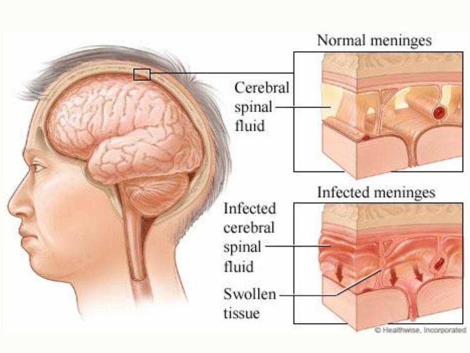
Cranial Meninges (meninx singular)


Meningitis
• Bacterial or viral invasion of the CNS by way of
the nose and throat may lead to inflammation of
the meninges.
• Pia Mater and Arachnoid are usually affected.
• Signs include high fever, stiff neck, drowsiness
and intense headache and may progress to
coma.
• Can be diagnosed by examining the CSF.

Meningitis Autopsy

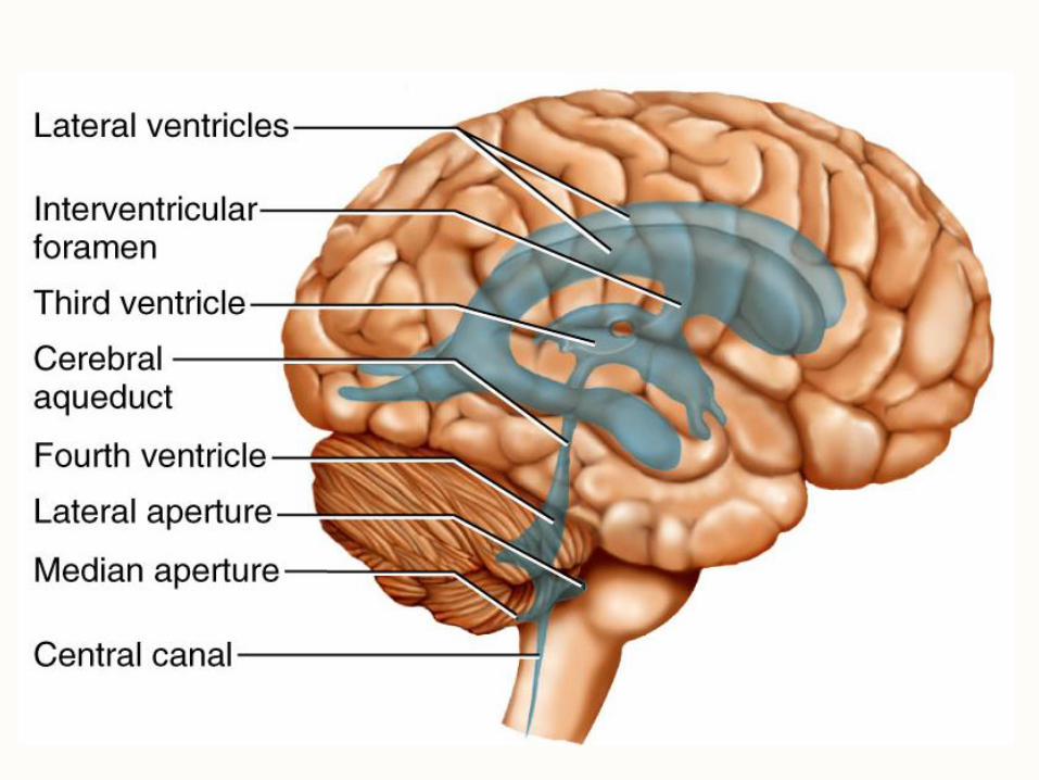
Ventricles and Cerebrospinal Fluid (CSF)
• Ventricles are internal chambers within the CNS – lateral ventricles found inside cerebral hemispheres
– third ventricle is single vertical space under corpus callosum
– cerebral aqueduct runs through midbrain and connects third ventricle and fourth ventricle
– fourth ventricle is small chamber between pons and cerebellum
– central canal runs down through spinal cord
• Choroid plexus of capillaries in the ventricles produces the CSF.
• Ventricles are lined with ciliated ependymal cells that circulate the CSF.
• Apertures in fourth ventricle let CSF out of ventricles into subarachnoid space.


Ventricles of the Brain

Cerebrospinal Fluid (CSF) • CSF is a clear liquid that fills ventricles and also
bathes the external surface of the brain in the subarachnoid space.
• Brain produces and absorbs about 500 ml/day – Produced from filtration of blood through choroid plexus
– Similar to plasma, but contains no blood cells and less protein than plasma
• Functions – buoyancy: brain is neutrally buoyant in CSF so it floats
– protection: cushions brain from inside of skull
– chemical stability: provides some ions for the brain and dilutes and washes away metabolic wastes of brain tissue
• CSF is reabsorbed by arachnoid villi into a venous sinus called the superior sagittal sinus which forms from a split in the dura mater

Flow of Cerebrospinal Fluid
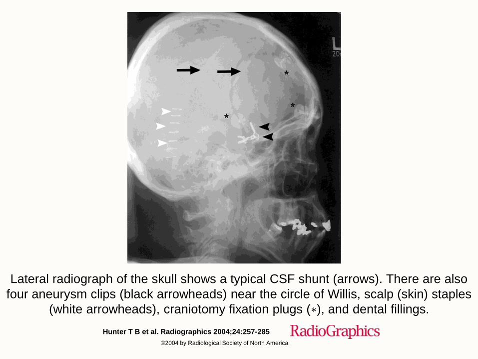
Hunter T B et al. Radiographics 2004;24:257-285
©2004 by Radiological Society of North America
Lateral radiograph of the skull shows a typical CSF shunt (arrows). There are also
four aneurysm clips (black arrowheads) near the circle of Willis, scalp (skin) staples
(white arrowheads), craniotomy fixation plugs (∗), and dental fillings.

Embryonic Development of Nervous System
• Human Nervous System develops from ectoderm into a dorsal hollow nerve cord that runs the length of the body.
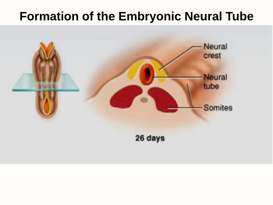
• In the first 3-4 weeks of development, the neural plate on the back of the embryo becomes a neural groove with neural folds along each side. The neural folds grow together to form the neural tube.
• Neurons develop from the cells of the neural tube.
• Lumen of the neural tube develops into the central canal of spinal cord and the ventricles of the brain.
• Cells along the margin of the neural groove are called the neural crest and they develop into sensory neurons, sympathetic neurons, Schwann cells and pigment cells.
• By the 4th week, the neural tube develops 3 anterior dilations that will become major brain regions.

Formation of the Embryonic Neural Tube

Formation of the Embryonic Neural Tube

Formation of the Embryonic Neural Tube
Formation of the Embryonic Neural Tube


Brain Regions • Forebrain develops into four specialized regions:
– Cerebrum
– Thalamus
– Hypothalamus (part of the pituitary gland is an outgrowth of it)
– Epithalamus (pineal gland)
• Midbrain
– superior and inferior colliculi
• Hindbrain
– Pons
– Medulla Oblongata
– Cerebellum
Notes: The diencephalon is composed of the thalamus and hypothalamus
The brainstem consists of the midbrain, medulla and pons

Regions of Embryonic Nervous System Forebrain
Midbrain
Hindbrain

(Forebrain)
(Midbrain)
(Hindbrain)
Forebrain
Midbrain
Hindbrain


Brain Regions
Thalamus
Pineal
Gland
Pons
Medulla
Oblongata
Pituitary Gland
Colliculi
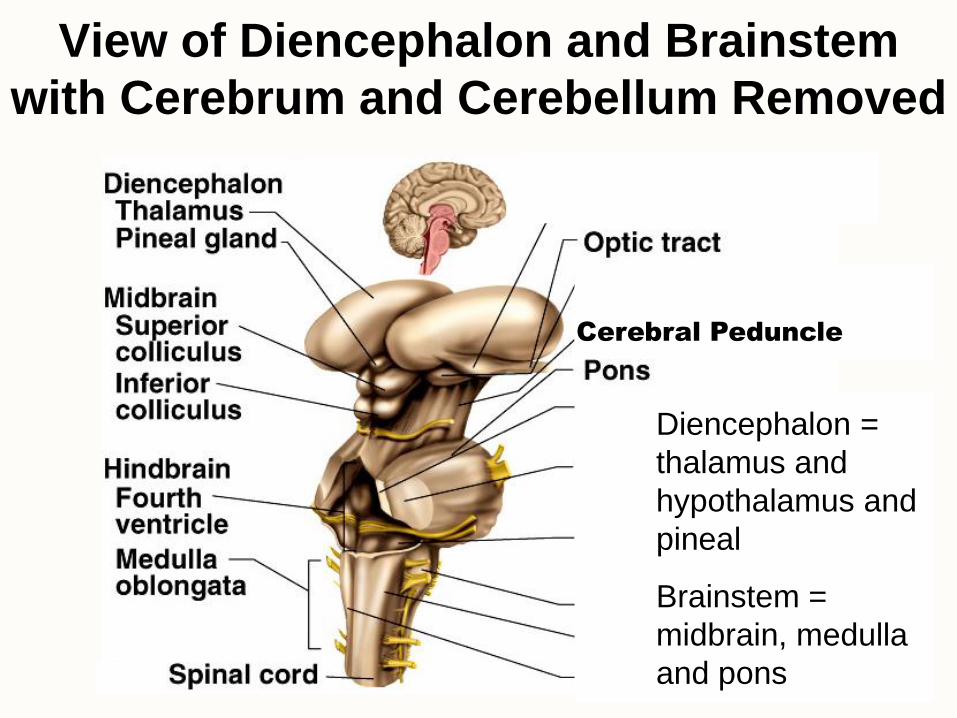
View of Diencephalon and Brainstem
with Cerebrum and Cerebellum Removed
Cerebral Peduncle
Diencephalon =
thalamus and
hypothalamus and
pineal
Brainstem =
midbrain, medulla
and pons

• Pons, medulla and cerebellum
make up the “hindbrain”
• The hindbrain is located between
the spinal cord and cerebral
hemispheres
• Nuclei (clusters of neurons) in
these regions control many
involuntary, life sustaining
processes including respiration,
circulation and digestion.
Hindbrain: Pons, Medulla and Cerebellum

• Connects cerebrum and
cerebellum through the tracts of
axons called the cerebellar
peduncles
• Nuclei (clusters of neurons)
coordinate auditory and
vestibular (balance) sensory
information with some motor
responses.
• Several cranial nerves branch off
the pons (V, VI, VII, and VIII)
Pons

Medulla • Formally called the Medulla
Oblongata
• Connects the spinal cord to pons
• Contains nuclei that control:
– Cardiac Center adjusts rate and force of heart beat
– Vasomotor Center adjusts blood vessel diameter
– Respiratory Centers control rate and depth of breathing
• Reflex centers for coughing, sneezing, gagging, vomiting, sweating
• Last 4 cranial nerves branch off the medulla oblongata (IX, X,XI, XII)

Cerebellum
• Primary role in skeletal muscle coordination and motor learning
• Also functions in awareness, memory and emotion.
• Contains about 100 billion neurons including highly dendritic Purkinje cells that integrate information and send information out into a complex network of white matter called the arbor vitae.

Midbrain
• The midbrain is the upper part of the brainstem that
connects the forebrain and the hindbrain.
• Important parts of the midbrain include the:
• superior colliculus
• inferior colliculus
• substantia nigra
• reticular formation

Corpora Quadragemina: Superior and
Inferior Colliculi
• The corpora quadrigemina
are 4 nuclei in the midbrain:
– top two nuclei form the
superior colliculus which
control visual reflexes like
visually track moving
objects
– bottom two nuclei form
the inferior colliculus
which controls the
auditory reflex of turning
of the head towards
sound
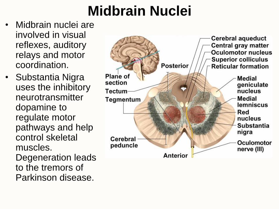
• Midbrain nuclei are involved in visual reflexes, auditory relays and motor coordination.
• Substantia Nigra uses the inhibitory neurotransmitter dopamine to regulate motor pathways and help control skeletal muscles. Degeneration leads to the tremors of Parkinson disease.
Midbrain Nuclei

Reticular Formation • The reticular formation is
composed of about 100 nuclei in the brainstem that relay information to the cerebral cortex.
• Some tracts regulate balance and posture.
• Gaze centers allow you to fixate on objects.
• Initiates habituation to repetitive, unimportant stimuli (background noise).
• Regulates sleep and conscious attention.
• Can block pain signals to the brain.
• Damage leads to coma (loss of consciousness).
•

Thalamus
• Egg-shaped masses of gray matter composed of several nuclei.
• Receives nearly all sensory information on its way to the cerebral cortex then integrates and directs the information to an appropriate area of the brain.
• Interconnected to limbic system so it is also involved in emotion and memory.

• Nuclei in walls and floor of third ventricle
• Homeostatic Functions – pituitary gland control
– autonomic NS control
– thermoregulation
– food and water intake (hunger and satiety)
– sleep/activity rhythms
– memory (mammillary bodies)
– emotional behavior (anger aggression, fear, pleasure, contentment, sexuality)
Hypothalamus
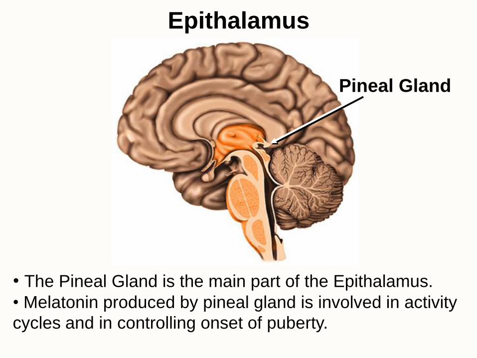
Epithalamus
Pineal Gland
• The Pineal Gland is the main part of the Epithalamus.
• Melatonin produced by pineal gland is involved in activity
cycles and in controlling onset of puberty.

• Cerebrum is divided into 5 lobes.
• The fifth lobe, the Insula, is deep to the lateral sulcus.
Cerebrum

• Frontal Lobe: voluntary motor functions and areas for motivation, planning, memory, emotion, social judgment and aggression.
• Parietal Lobe: sensory reception and integration.
• Occipital Lobe: the visual center of brain and creates a visual map of the world.
• Temporal Lobe: contains areas for hearing, smell, learning, memory, visual recognition and emotional behavior.
• Insula functions are still being studied.
Functions of the Lobes of the Cerebrum
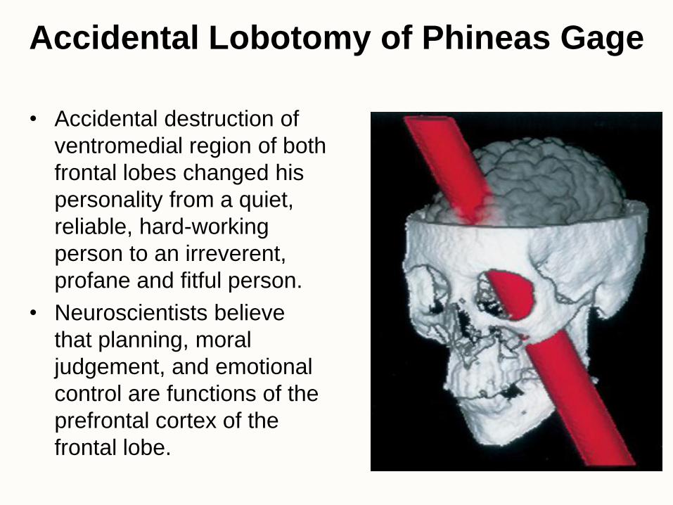
Accidental Lobotomy of Phineas Gage
• Accidental destruction of
ventromedial region of both
frontal lobes changed his
personality from a quiet,
reliable, hard-working
person to an irreverent,
profane and fitful person.
• Neuroscientists believe
that planning, moral
judgement, and emotional
control are functions of the
prefrontal cortex of the
frontal lobe.
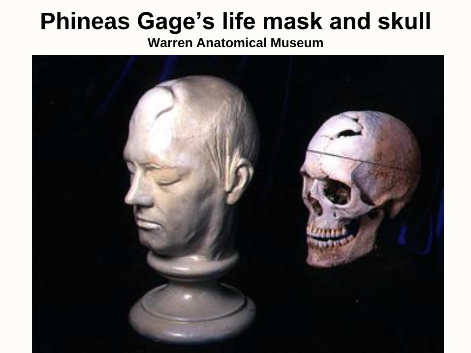
Phineas Gage’s life mask and skull Warren Anatomical Museum

Phineas Gage with the tamping rod
that was driven through his head in
an accident in 1848

Digital renderings of Gage's skull showing the trajectory of the rod and the fiber
pathways in the left hemisphere. From Van Horn, J. D., et al. (2012).

Connectogram of a healthy brain Connectogram of Phineas Gage's brain
“Connectograms are circular diagrams depicting the brain's major white
matter tracts. In these diagrams, the major brain regions - the frontal lobe,
insula, limbic system, temporal lobe, parietal lobe, occipital lobe, brain stem
and cerebellum - are color-coded and arranged on the outer ring of the
diagram, according to their position from the front. J.D. Van Horn, J. D., et al. (2012) Mapping Connectivity Damage in the Case of
Phineas Gage. PLoS ONE, 7(5): e37454. DOI: 10.1371/journal.pone.0037454

Cerebral White Matter
• White Matter of the brain consists of myelinated axons.
• Most of the volume of the cerebrum is composed of “tracts” of axons (white matter).
• Types of tracts:
– projection tracts extend vertically between brain and spinal cord.
– commissural tracts (like the corpus callosum) cross from one cerebral hemisphere to the other
– association tracts connect lobes of each hemisphere to each other

Tracts of Cerebral White Matter

Cerebral Cortex Gray Matter
• Gray Matter of cerebral cortex is a 3 mm thick layer of neurons.
• Cortex is primarily composed of 2 types of cells in 6 layers:
– stellate cells have dendrites projecting in all directions
– pyramidal cells have triangular-shaped somas (pyramidal) and have an axon that passes out of one layer into another area of the brain

basic functional areas

Functional Regions of Cerebral Cortex

Somesthetic Sensation
• Sensory signals travel up tracts in the spinal cord to
the Somatosensory Area which is in the postcentral
gyrus of the cerebral hemispheres.

Sensory Homunculus
• The area of the
postcentral
gyrus of the
cerebral cortex
dedicated to the
sensations of
various body
parts is
proportional to
how sensitive
that part of the
body is.


Sensory Association Areas
• Association areas interpret sensory information.
• Somesthetic Association Area (parietal lobe)
– position of limbs, location of touch or pain, and shape, weight and texture of an object
• Visual Association Area (occipital and temporal lobes)
– identify the things we see
– faces are recognized in temporal lobe
• Auditory Association Area (temporal lobe)
– remember the name of a song or identify a person by his/her voice

Special Senses
• Special senses are located in the head and
employ very specialized sensory receptors.
• Special Senses include:
– Vision
– Equilibrium
– Hearing (auditory)
– Taste (gustatory)
– Smell (olfactory)
• These senses are projected to specialized
regions of the brain.

Motor Control • Intention to contract a
skeletal muscle begins in the motor association area of frontal lobes and is relayed to the Primary Motor Area.
• The Primary Motor Area consists of Pyramidal Cells in the precentral gyrus.
• Signals are sent out through the spinal cord to muscles of contralateral (opposite) side.

Motor Homunculus
• The area of the
cortex dedicated
to the motor
control of
various body
parts is
proportional to
the number of
motor units in a
region.

• Aphasia is a language deficit resulting from lesions in Wernicke’s and Broca’s areas (usually in the left hemisphere)
• Lesion to Broca’s area results in nonfluent aphasia
– slow speech, difficulty choosing words, sparse vocabulary
• Lesions on other side of brain opposite of Broca’s area result in aprosody – flat, emotionless speech.
• Lesion to Wernicke’s area results in fluent aphasia
– normal speech but excessively wordy and makes little sense
• Lesion opposite Wernicke’s area causes difficulty recognizing the emotional content of another person’s speech (for example, not getting a joke)
• Other deficits: speech and understanding speech are normal but text and pictures make no sense
Language
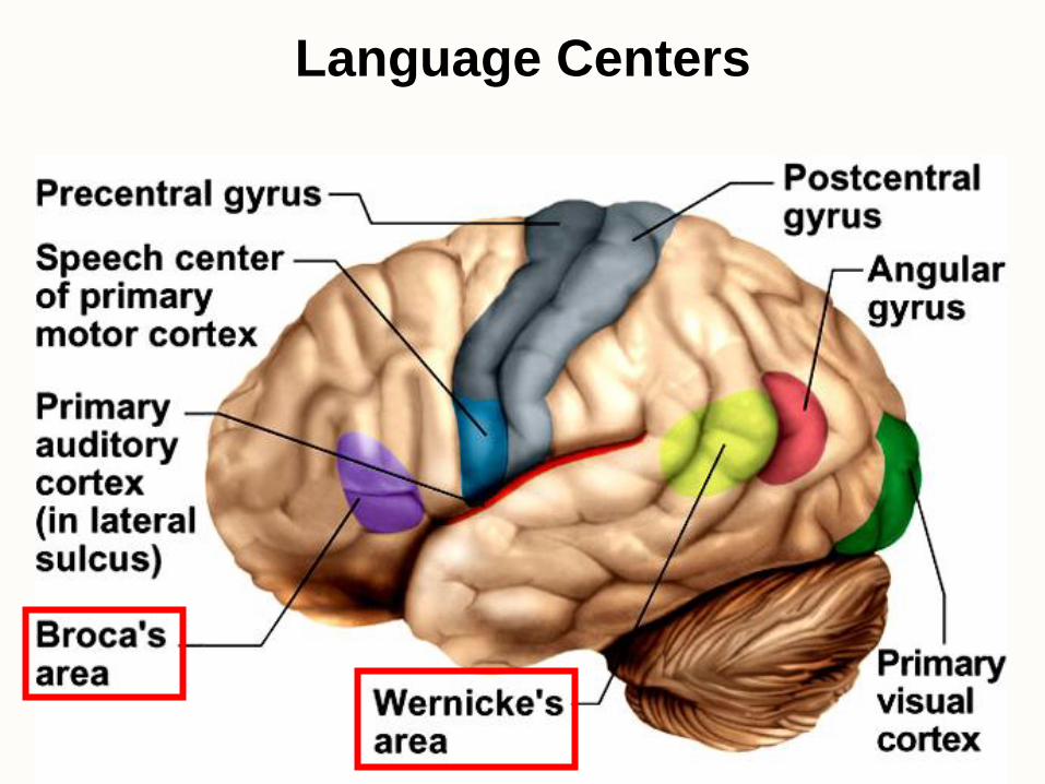
Language Centers

http://rarediseases.info.nih.gov/GARD/QnASelected.aspx?diseaseID=8
People with primary visual agnosia may have one or
several impairments in visual recognition without
impairment of intelligence, motivation, and/or
attention. Vision is almost always intact and the
mind is clear. Some affected individuals do not have
the ability to recognize familiar objects. They can
see objects, but are unable to identify them by sight.
However, objects may be identified by touch, sound,
and/or smell. For example, affected individuals may
not be able to identify a set of keys by sight, but can
identify them upon holding them in their hands.
Primary visual agnosia results from damage to the
parietal and temporal lobes of the brain. Symptoms
develop due to the inability to retrieve information
from those damaged areas that are associated with
visual memory. Lesions may occur as a result of
traumatic brain injury, stroke, tumor, or
overexposure to dangerous environmental toxins
(e.g., carbon monoxide poisoning). In some cases,
the cause of the brain damage may not be known.
Symptoms may vary, according to the area of the
brain that is affected.

Limbic System
• Loop of cortical structures composed of the
amygdala, hippocampus, fornix and cingulate gyrus
• Important in emotional processing and in memory.
Much still poorly understood.

Cerebral Lateralization
• Left hemisphere is more categorical
– specialized for spoken and written language, sequential and analytical reasoning (math and science), analyze data in linear way
• Right hemisphere is more representational
– perceives information more holistically, perception of spatial relationships, pattern, comparison of special senses, imagination and insight, music and artistic skill
• Highly correlated with handedness
– 91% of right-handed people have a categorical left hemisphere


END

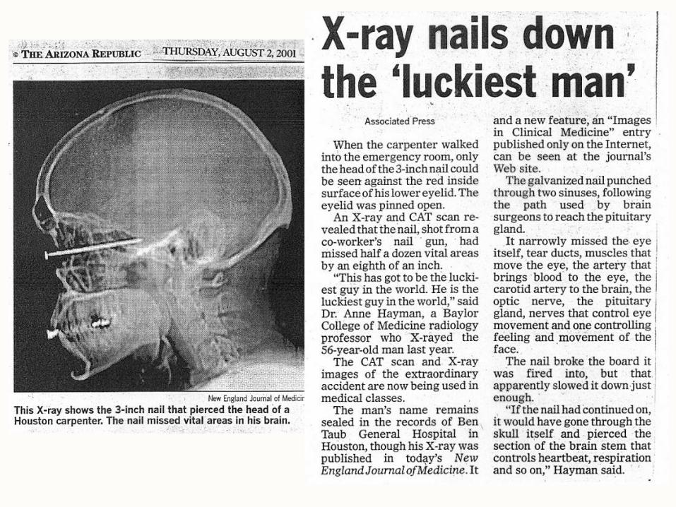

A 33-year old male who went to the hospital complaining of a
headache was found to have 12 nails embedded in his skull from a
suicide attempt with a nail gun. Some of the nails were 1.5” long and
others were 2” long and surgeons removed them with needle-nose
pliers and a drill. The man at first told doctors he had a nail gun
accident, but later admitted it was a suicide attempt while under the
influence of methamphetamine. Journal of Neurosurgery April, 2006


Cranial Nerves
• 12 pair of nerves that arise from brain and exit through cranial foramina leading to muscles, glands and sense organs in head and neck
• Learn Number, Name and Function for a matching section on the lab practical.

Cranial Nerves


Language
• Includes reading, writing, speaking and
understanding words.
• Wernicke’s Area is used to recognize
spoken and written language and creates the
plan of speech.
• Angular Gyrus allows us to read out loud
(processes text into a form we can speak).
(left hemisphere)
• Broca’s Area generates a motor program
for the muscles of the larynx, tongue, cheeks
and lips and transmits it to the speech
center of the primary motor cortex. (left

Reticular Activating System
• Nuclei scattered throughout pons, medulla and midbrain relay information from eyes and ears to cerebral cortex.
• Some tracts regulate balance and posture.
• Gaze centers allow you to track moving objects.
• Initiates habituation to repetitive, unimportant stimuli (background noise).
• Regulates sleep and conscious attention.
• Injury leads to irreversible coma.
•

Basal Nuclei
• Masses of gray matter deep to cerebral cortex that receive
input from substantia nigra and motor cortex and send
signals back to these regions.
• Important in motor control.

• Longitudinal fissure separates 2 cerebral
hemispheres.
• Surface features:
• gyri (singular = gyrus) are the raised folds
• sulci (singular = sulcus) are the grooves in between gyri

Inferior View of the Brain
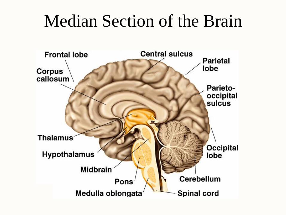
Median Section of the Brain

Photo of Sagittal Section of Brain

Brain – Directional Terms and Landmarks
• Rostral (toward the forehead) - Caudal (toward the cord)
• Major parts of the brain - cerebrum, cerebellum, brainstem
– cerebrum is 83% of brain volume; cerebellum contains 50% of the neurons
– brain weighs 3 to 3.5 pounds

Blood-Brain and Blood-CSF Barriers
• Blood-brain barrier is tightly joined endothelium
– permeable to lipid-soluble materials (alcohol, O2,
CO2, nicotine and anesthetics)
– circumventricular organs in 3rd & 4th ventricles at
breaks in the barrier where blood has direct access
• monitoring of glucose, pH, osmolarity & other variations
• allows route for HIV virus to invade the brain
• Blood-CSF barrier at choroid plexus is
ependymal cells joined by tight junctions
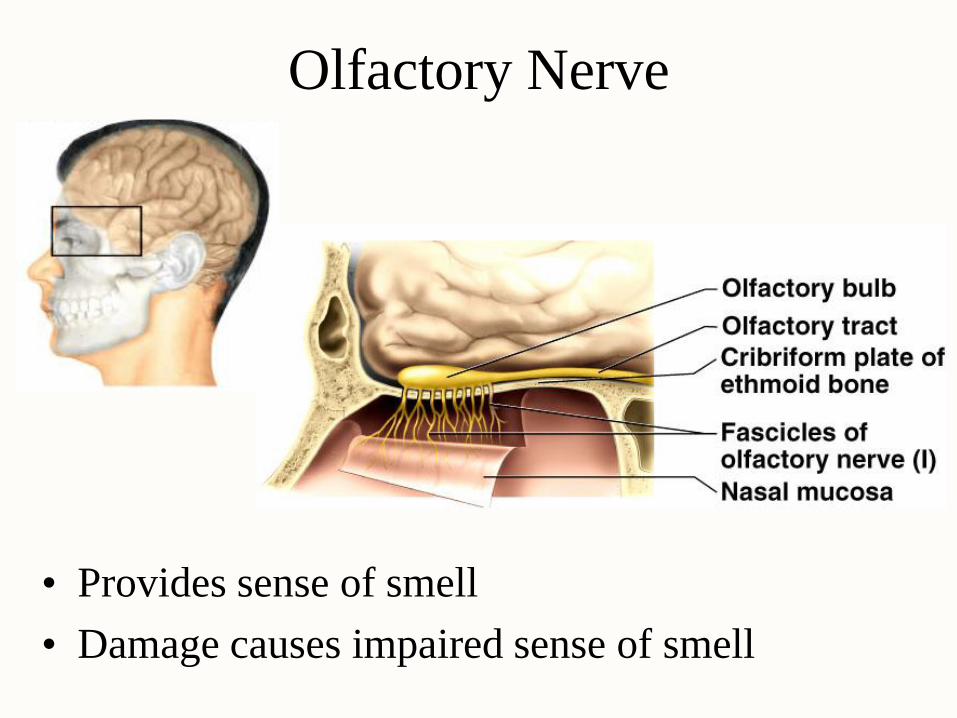
Olfactory Nerve
• Provides sense of smell
• Damage causes impaired sense of smell

Optic Nerve
• Provides vision
• Damage causes blindness in visual field

Oculomotor Nerve
• Provides some eye movement, opening of eyelid, constriction of pupil, focusing
• Damage causes drooping eyelid, dilated pupil, double vision, difficulty focusing & inability to move eye in certain directions

Trochlear Nerve
• Provides eye movement
• Damage causes double vision & inability to
rotate eye inferolaterally

Trigeminal Nerve
• Main sensory nerve to face (touch, pain and temperature) and muscles of mastication
• Damage produces loss of sensation & impaired chewing

Abducens Nerve
• Provides eye movement
• Damage results in inability to rotate eye laterally
& at rest eye rotates medially

Facial Nerve
• Provides facial expressions, sense of taste on anterior 2/3’s of tongue, salivary glands and tear, nasal & palatine glands
• Damage produces sagging facial muscles & disturbed sense of taste (missing sweet & salty)
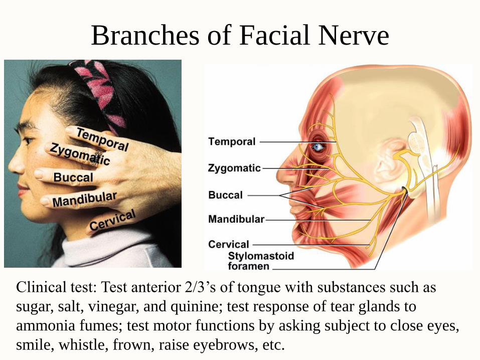
Branches of Facial Nerve
Clinical test: Test anterior 2/3’s of tongue with substances such as
sugar, salt, vinegar, and quinine; test response of tear glands to
ammonia fumes; test motor functions by asking subject to close eyes,
smile, whistle, frown, raise eyebrows, etc.

Vestibulocochlear Nerve
• Provides hearing & sense of balance
• Damage produces deafness, dizziness, nausea,
loss of balance & nystagmus

Glossopharyngeal Nerve
• Provides control over swallowing, salivation, gagging, sensations from posterior 1/3 of tongue, control of BP and respiration
• Damage results in loss of bitter & sour taste & impaired swallowing

Vagus Nerve
• Provides swallowing, speech, regulation of viscera
• Damage causes hoarseness or loss of voice, impaired swallowing & fatal if both are cut

Accessory Nerve
• Provides swallowing, head, neck & shoulder movement
• Damage causes impaired head, neck & shoulder movement, head turns towards injured side

Hypoglossal Nerve
• Provides tongue movements of speech, food
manipulation & swallowing
• Damage results in inability to protrude tongue if
both are damaged or deviation towards injured
side & ipsilateral atrophy if one side is damaged

Cranial Nerve Disorders
• Trigeminal neuralgia (tic douloureux)
– recurring episodes of intense stabbing pain in
trigeminal nerve area (near mouth or nose)
– pain triggered by touch, drinking, washing face
– treatment is cutting of nerve
• Bell palsy
– degenerative disorder of facial nerve
– paralysis of facial muscles on one side
– may appear abruptly & disappear within 3-5 weeks

Meninges
• Dura mater -- outermost, tough membrane
– outer periosteal layer against bone
– where separated from inner meningeal layer forms dural venous
sinuses draining blood from brain
– supportive structures formed by dura mater
• falx cerebri, falx cerebelli and tentorium cerebelli
– epidural space filled with fat in lower back region
• epidural anaesthesia during childbirth
• Arachnoid mater is spider web filamentous layer
• Pia mater is a thin vascular layer adherent to contours of
brain

Ventricles of the Brain
NOTE bad
lines
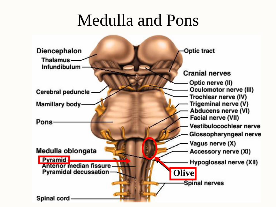
Medulla and Pons
Olive

Cerebellum

EEG and Brain Waves
• Electroencephalogram records voltage changes from
postsynaptic potentials in cerebral cortex
• Differences in amplitude & frequency distinguish 4
types of brain waves

Brain Waves & Sleep
• States of consciousness can be correlated with EEG
• 4 types of brain waves
– alpha occur when awake & resting with eyes closed
– beta occur with eyes open performing mental tasks
– theta occur during sleep or emotional stress
– delta occur during deep sleep
• Sleep is temporary state of unconsciousness
– coma is state of unconsciousness with no possible arousal
– reticular formation seems to regulate state of alertness
– suprachiasmatic nucleus acts as biological clock to set our
circadian rhythm of sleep and waking
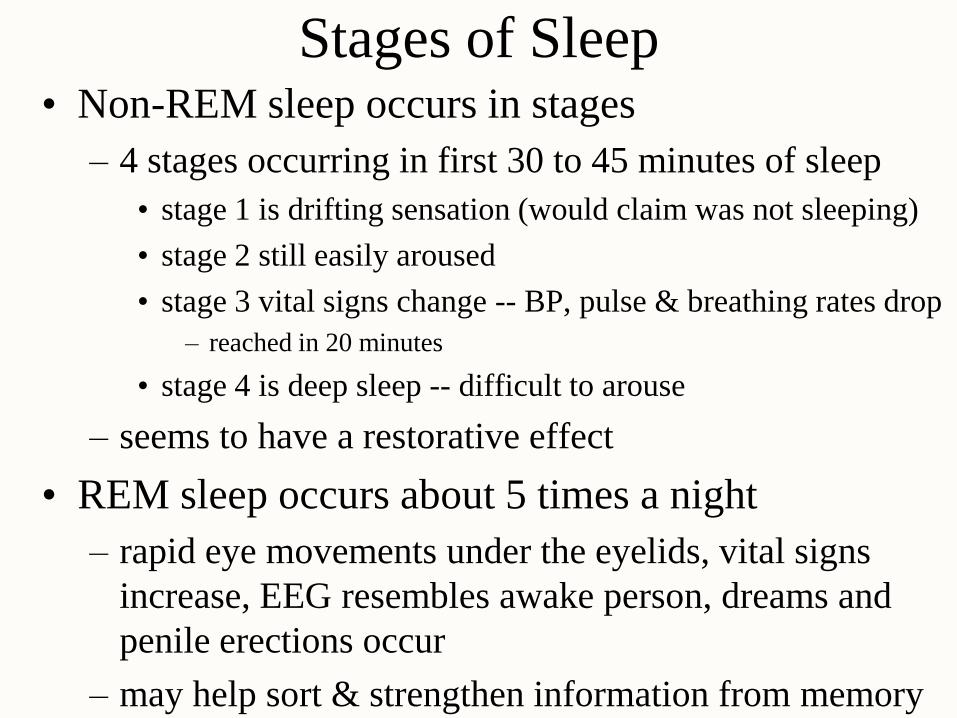
Stages of Sleep • Non-REM sleep occurs in stages
– 4 stages occurring in first 30 to 45 minutes of sleep
• stage 1 is drifting sensation (would claim was not sleeping)
• stage 2 still easily aroused
• stage 3 vital signs change -- BP, pulse & breathing rates drop
– reached in 20 minutes
• stage 4 is deep sleep -- difficult to arouse
– seems to have a restorative effect
• REM sleep occurs about 5 times a night
– rapid eye movements under the eyelids, vital signs
increase, EEG resembles awake person, dreams and
penile erections occur
– may help sort & strengthen information from memory

Sleep Stages and Brain Waves
• Brain waves change
as we pass through 4
stages of sleep
– alpha waves
– sleep spindles
– theta
– delta waves

Sleep Stages
• Notice how REM sleep periods become longer
and more frequent in the second half of the night

Cognition
• Cognition is mental processes such as awareness,
perception, thinking, knowledge & memory
– 75% of brain is association areas where integration of
sensory & motor information occurs
• Examples of effects of brain lesions
– parietal lobe -- contralateral neglect syndrome
– temporal lobe -- agnosia (inability to recognize objects)
or prosopagnosia (inability to recognize faces)
– frontal lobe -- problems with personality (inability to
plan & execute appropriate behavior)
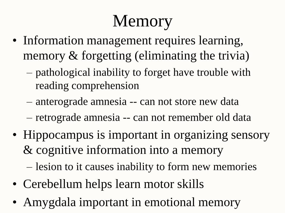
Memory • Information management requires learning,
memory & forgetting (eliminating the trivia)
– pathological inability to forget have trouble with
reading comprehension
– anterograde amnesia -- can not store new data
– retrograde amnesia -- can not remember old data
• Hippocampus is important in organizing sensory
& cognitive information into a memory
– lesion to it causes inability to form new memories
• Cerebellum helps learn motor skills
• Amygdala important in emotional memory

Emotion
• Prefrontal cortex controls how emotions are
expressed (seat of judgement)
• Emotions form in hypothalamus & amygdala
– artificial stimulation produces fear, anger, pleasure,
love, parental affection, etc.
– electrode in median forebrain bundle in rat or human
and a foot pedal
• press all day to the exclusion of food (report a quiet,
relaxed feeling)
• Much of our behavior is learned by rewards and
punishments or responses of others to them
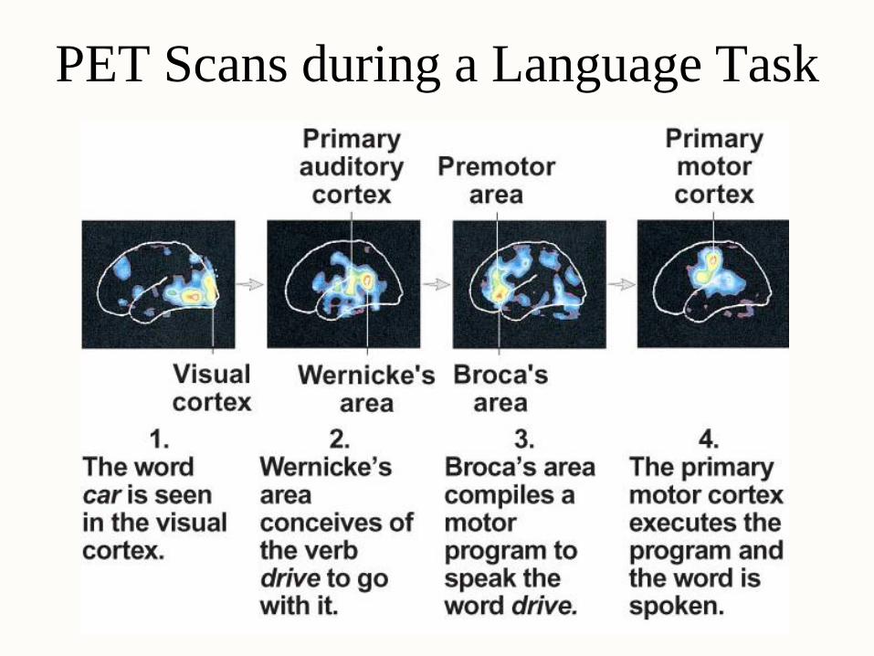
PET Scans during a Language Task
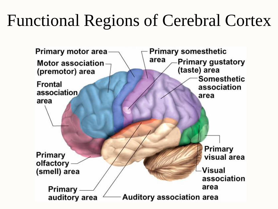
Functional Regions of Cerebral Cortex
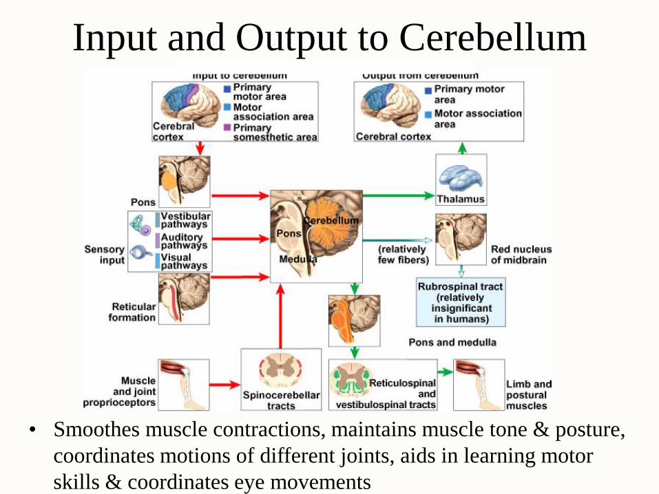
Input and Output to Cerebellum
• Smoothes muscle contractions, maintains muscle tone & posture,
coordinates motions of different joints, aids in learning motor
skills & coordinates eye movements