Brain
38
BRAIN CHAPTER 14
description
Transcript of Brain

BRAIN
CHAPTER 14

THE BRAIN
• Human brain only weighs about 3 lb.• Contains more than 100 billion neurons.• It does more than think and make decisions.• Body’s main key to homeostasis, regulating
body processes.• Nerves and specialized receptors may receive
the message, but you actually experience the sensation in your brain.
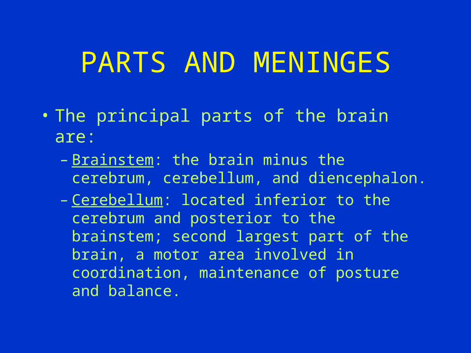
PARTS AND MENINGES
• The principal parts of the brain are:– Brainstem: the brain minus the cerebrum,
cerebellum, and diencephalon.– Cerebellum: located inferior to the cerebrum
and posterior to the brainstem; second largest part of the brain, a motor area involved in coordination, maintenance of posture and balance.

– Diencephalon: a division of the brain that includes the epithalamus, thalamus and hypothalamus
– Cerebrum: the largest portion of the brain, composed of the cerebral hemispheres, includes the cerebral cortex, the cerebral nuclei and the internal capsule.
PARTS OF BRAIN

MENINGES
• Cranial meninges: membranes surrounding the brain. This includes the tough outer dura mater, the middle web-like arachnoid layer and the thinner pia mater. Provide the necessary physical stability and shock absorption. Blood vessels branching within these layers also deliver oxygen and nutrients.
Cerebrospinal Fluid
• The four CSF-filled cavities within the brain are called the ventricles-2 lateral, third and fourth.
• It completely surrounds and bathes the exposed surfaces of the CNS.
• Cushions delicate neural structures
• supports the brain
• transports nutrients, chemical messengers and waste products.
• A spinal tap can provide useful clinical information concerning CNS injury, infection or disease.
Formation of CSF
• The site of production are the choroid plexuses which are network of capillaries in the walls of the ventricles.
• The capillaries are covered by ependymal cells that form CSF from blood plasma by filtration and secretion.
• There are tight junctions here-formation of blood-brain barrier. This allows for certain substances to enter the CSF and excludes others.
Formation of CSF
• The CSF formed in t he choroid plexuses of each lateral ventricle flows into the third ventricle.
• More CSF is added by choroid plexuses of the third ventricle.
• The fluid then flows through the cerebral aqueduct into the fourth ventricle. This contributes mors fuid.
• Then it enters the subarachnoid space.
• It then circulates in the central canal of the spinal cord.
• CSF is gradually reabsorbed into the blood through arachnoid villi. Normally CSF is reabsorbed as rapidly as it is formed. Hence the pressure of CSF is constant.

Hydrocephalous
• A condition resulting from excessive production of or inadequate drainage of CSF. The total volume of CSF and blood must remain at a stable level. If blood and CSF increases, the volume of the brain must compensate by decreasing its volume. An increase in the brain’s fluid volume compression and distortion of the brain. This in turn causes swollen facial features. Hydrocephalous is basically a condition in which intracranial pressure in altered.

Adequate Blood Supply
• Neurons have a high demand for energy. They do not have energy reserves from lipids or CHO’s or oxygen reserves. Therefore neurons depend on blood to transport oxygen to them. Hb gives red blood cells to transport oxygen in t he blood. Blood which irrigates the brain does so by means of arterial carotid arteries and vertebral arteries. The blood then leaves the brain through the internal jugular veins. Lack of oxygen has several medical complication from fainting to CVA.
Medulla Oblongata
• That part of the brain stem that connects the pons and the root of the brain to the spinal cord. All the afferent and efferent tracts from the spinal cord either pass through or terminate here. It also contains nerve centers instrumental to the regulation and control of breathing, swallowing, coughing, sneezing and vomiting. Other centers in the medulla regulate arterial blood pressure, thereby exerting control over the circulation of blood.

Pons
• The pons is a broad band of white matter located anterior to the cerebellum and between the midbrain and medulla oblongata. The pons contains fiber tracts linking the cerebellum and medulla to higher cortical areas; two-way conduction pathway between areas of the brain and other areas of the body;influences respiration.
Mid-Brain
• Located between the forebrain and hind brain. Contains a number of large afferent and efferent pathways connecting major motor areas of the fore and hind brain. Also found in the mid brain are four small masses of gray cells-corpora quadrigemina. The upper two called superior colliculi are associated with visual reflexes and tracking movements of the eyes. The lower two, inferior colliculi are involved with the sense of hearing.
Thalamus
• This is the largest of the two divisions of the diencephalon and is actually two large masses of grey cell bodies joined by a third or intermediate mass. It serves as a relay center for all sensory impulses (except olfactory) being transmitted to the sensory areas of the cortex. Besides its sensory function the thalamus also relays motor impulses from the cerebellum and basal ganglia to motor areas of the cortex. Some impulses related to emotional behavior are also passed from the hypothalamus through the thalamus to the cerebral cortex.
Hypothalamus
• This lies beneath the thalamus. Is a principal regulator of ANS activity and is associated with behavior and emotional expression. It also produces neurosecretions for the control of water balance, sugar and lipid metabolism, regulation of body temperature, sleep-cycle control, appetite and sexual arousal. It also produces hormones for the post. Pituitary gland.

Cerebrum
• Represents seven eighths of the brain’s total weight. It contains nerve centers that govern all sensory and motor activity, including sensory perception, emotions, consciousness, memory and voluntary movements. It is divide by longitudinal fissure into two cerebral hemispheres the right and left are joined by
large fiber tracts (corpus callosum)that allow for information to pass from one hemisphere to another. The surface or cortex of each hemisphere has been divided into lobes as a means of identifying certain locations. These lobes correspond to the overlying bones of the skull an are the frontal, parietal, temporal and occipital lobes.

Cerebellum
• This is the largest part of the hindbrain. It occupies a space in the back of the skull’ inferior to the cerebrum and dorsal to the pons and medulla oblongata. The cerebellum is oval in shape and is divided into deep fissures. The surface of the ceebellum has a cortex of grey cell bodies and its interior contains nerve fibers, white matter, connecting it to every part of the CNS. The cerebellum p[lays an important part in the coordination of voluntary movements.

RAS
• One of the most important brain stem components is a diffuse network in the reticular formation known as the reticular activating system (RAS). This network extends from the midbrain of the medulla oblongata. The output of the RAS projects to thalamic nuclei that influence large areas of the cerebral cortex. When the RAS is inactive, so is the cerebral cortex. When the area is active, many impulses pass upward into the thalamus and disperse to widespread areas of the cerebral cortex. The effect is a generalized increase in cortical activity making one more alert and attentive.

Pyramids
• Pyramids are thick bonds visible along the ventral surface of the medulla oblongata.
• Decussation is the movement of the axon from the left side to the right side and vice versa.
• The nucleus gracilis and nucleus cuneatus both pass sensory information to the thalamus.
Pyramids
• The wall of the diencephalon are composed of the left and right thalamus. Each thalamus contains relay and processing centers for sensory information. The brain stem includes the mesencephalon, pons and the medulla oblongata.
• The pneumotaxic centers of the pons are paired nuclei that adjust the output of the respiratory rhythmicity centers.
Blood-Brain Barrier
• Endothelial cells that line CNS capillaries create this BBB. This isolates the CNS from general circulation. The barrier exists because these cells contain tight junctions and they prevent diffusion of materials. Astrocytes maintain this barrier by providing structural support. They also regulate ion, nutrient, and dissolved gas concentrations. If the astrocytes are damaged or stop stimulating the endothelial cells, the blood brain barrier disappears.

Cerebellum and Skeletal Muscle activities
• The cerebellum is involved in synergic control of skeletal muscles and plays an important role in the coordination of voluntary muscular movements. It receives afferent impulses and discharges efferent impulses but does not serve as a reflex center in the usual sense, however it may reinforce some reflexes and inhibit others. The cerebellum does not initiate movements, it interrelates with many brain stem structures in executing a variety of movements including proper posture and balance, walking, running, fine voluntary movements as required in writing, dressing, eating and playing musical instruments. The cerebellum controls the property of movement such as speed, acceleration and trajectory.

LOBES
• Temporal lobe: lobe of the cerebrum located laterally and below the frontal and occipital lobe. Contains auditory receptive areas.
• Frontal lobe: four main convolutions in front of the central sulcus of the cerebrum.
• Parietal lobe: the division of each side of the brain lying beneath each parietal bone.
• Occipital lobe: posterior lobe of the cerebral hemisphere that is shaped like a three-sided pyramid.

Convolutions
• In anatomy one of the many folds on the surface of the cerebral hemispheres. They are separated by sulci or fissures.
• Gyri: outward folds of the cerebral cortex.
• Sulci: shallow grooves betweeb folds of the cerebral cortex.
• Fissures: deep grooves located between folds of the cerebral cortex.
Fissures
• Parieto-occipital fissure: fissure between the occipital and parietal lobes o9f the brain.
• Longitudinal fissure: two cerebral hemispheres are almost completely separated by a deep longitudinal fissure.
• Central sulcus: on each hemisphere the central sulcus a deep
groove divide the anterior frontal lobe.
• Lateral sulcus: this separates the frontal lobe from the temporal lobe.
• Postcentral: situated or happening behind a center(gyrus)• Precentral: in front of a center, as a central fissure of the brain
(gyrus).

Central White Matter
• Lies beneath the neural cortex and around the cerebral nuclei and contains 3 major groups of axons. Whole collection of fibers is called internal capsule.– 1. Association fibers: interconnects areas of neural
cortex within a single cerebral hemisphere . The short association fibers are called arcuate fibers, are curved and pass from one gyrus to another. The longer fibers are the longitudinal fasciculi, connect the frontal lo0be to the rest of the lobes in the same hemisphere.
Central White Matter
– 2. Commisural fibers: interconnect and permit communication between the two cerebral hemispheres. These fibers are the corpus callosum, carry about 4 billion impulses per second and contain 200 million axons and anterior commisure.
– 3. Projection fibers: link the cerebral cortex to the diencephalon, brain stem, cerebellum and spinal cord.

Cerebrum
• The cerebrum is the largest region of the brain and is involved in the processing of somatic sensory and motor information. Conscious thoughts and all intellectual functions originate in its right and left hemispheres.

Left Hemisphere
• The categorical hemisphere or left hemisphere contains the general interpretive and speech centers.
• It is responsible for language based skills.• It performs analytical tasks.• The premotor cortex involved with the control
of hand movements is larger on the left side for right-handed than for left-handed individuals.

Right Hemisphere
• The representational hemisphere or right hemisphere deals with spatial relationships, analyzes sensory information and releases the body to the sensory environment.
• Permits identification of objects by touch, sight, smell, feel or taste.( recognizing faces and understanding 3-D)
• It analyzes the emotional context of a conversation. (“get lost” is it a question or threat)
• individuals with a damaged right hemisphere may be unable to add emotional inflections to their own words.
Basal Ganglia
• Basal ganglia are sections of gray matter largely composed of cell bodies within each cerebral hemisphere. They lie between the diencephalon and the white matter of the hemisphere. They may perform functions important in the maintenance of focused attention. They are involved in cognitive and motor processes. They may coordinate the automated processing of sensory inputs nd motor outputs required for concentration and attention. They mey also synchronize sequential activities required to analyze and respond in an integrated way.
Limbic System
• The limbic system is composed of– amygdaloid body-acts as an interface between the limbic
system, the cerebrum and various sensory systems. It plays a role in the regulation of the heart rate and in the fight or flight response.
– cingulate gyrus-sits superior to the corpus callosum
– parahippocampal gyrus- and dentate gyrus conceal the hippocampus, a folded area of the cortex that lies inferior to the floor of the lateral ventricle. It appears to be important in learning, especially in the storage and retrieval of new long term memories.
Limbic System
– Hippocampus: – mamillary bodies: process sensory information
including olfactory sensations. They lso contain motor nuclei that control reflexes associated with eating, chewing, licking and swallowing.
– Fornix: is a tract of white matter that connects the hippocampus with the hypothalamus.

Limbic System Functions
• Establishing emotional states and related behavioral drives.
• Linking the conscious, intellectual functions of the cerebral cortex with the unconscious and autonomic functions of the brain stem.
• Facilitating memory storage and retrieval.
Functional areas in the cerebral cortex
• This map is very important as it reveals which part of the brain performs a specific function.
• An injury to a specific part of the cerebrum will be the cause of a specific neural dysfunction.
• Neurological and psychological disorders have been studied due to its construction.
Thalamus and Cortex
• It is known that the cortex performs specific functions. The thalamus is the final relay point for ascending sensory information and it coordinates the pyramidal and extrapyramidal systems. (fig. 4-12 and tab 14-4).
Waves
• Alpha waves occur in healthy people who are awake and disappear when they fall asleep or concentrate. Beta waves are of a higher frequency and are typical when a person is concentrating, under stress or in psychological tension. Delta waves are large in amplitude, low frequency. They are normal in sleeping people of all ages and in infants brain. They are found in adult awake people when tumors, vascular blockage or inflammation has damaged portions of the brain. Theta waves appear sometimes in sleeping adults but mostly in children and intensely frustrated adults. In other circumstances they may indicate the presence of a brain disorder such as tumor.
Neurological Disorders
• Ataxia: caused by problems affecting cerebellum. Disturbance in balance that can leave the person unable to stand without assistance.
• Bell’s palsy: inflammation of facial nerve. There is paralysis of facial muscles on affected side and loss of taste from anterior 2/3 of tongue.
• CVA or stroke: blood supply to a potion of the brain is cut off.
• Concussion: temporary loss of consciousness and variable period of amnesia resulting from a head injury.
• Dyslexia: disorder affecting comprehension and use of words.