Bradycardia and Cardiac Pacing - his-files.comhis-files.com/pdf/M74.pdf · Bradycardia and Cardiac...
76
Bradycardia and Cardiac Pacing overview David Luria, MD Sheba Medical Center
Transcript of Bradycardia and Cardiac Pacing - his-files.comhis-files.com/pdf/M74.pdf · Bradycardia and Cardiac...

Bradycardia and Cardiac Pacing
overview
David Luria, MDSheba Medical Center


Bradycardia
Sinus node dysfunction
AV node dysfunction(“block”)
Neuro-cardiogenic reaction (VVS, CSH)

Sick Sinus Syndrome (SSS)
Sinus bradycardia
Sino-atrial block
Sinus pauses
Chronothropic incompetence

2nd degree AV block
Mobitz II 2nd degree AV block

MOBITZ I (AH block) MOBITZ II
(HV block)

2:1 infrahisian block
Atrial Pacing
His to V block
A AH H V

Prognostic implications
MOBITZ I• Usually did not progress to Complete AVB• CAVB with nodal escape rhythm
MOBITZ II• Progress to CAVB is a rule • CAVB with ventricular (very slow) escape:
syncope and SCD can occur

Is It Mobitz I or Mobitz II?

Differential diagnosis of 2:1 AV block
• Rest ECG• QRS width• PR interval
• Stress ECG• Exersise/atropine• Carotid sinus massage
• EPS • HV interval• Atrial pacing
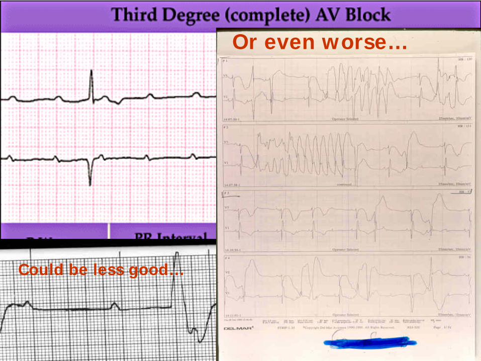
Could be less good…
Or even worse…

AHA/ACC Guidelines for PM implantation in patients with SA disease (2008)
Class I Indications
Sinus node dysfunction with documented symptomatic sinus bradycardia & sinus pauses
Symptomatic chronotropic incompetence
Class II Indications
Class IIa: Symptomatic patients with SND and HR<40 but with no clear association between symptoms and bradycardia
Class IIb: Chronic heart rate < 40 bpm while awake, in minimally symptomatic patients
Class III Indications
Asymptomatic sinus node dysfunction

AHA/ACC Guidelines for PM implantation in patients with AV node disease
Class I Indications
3rddegree and advanced 2nd degree AV block associated with:
– Symptomatic bradycardia
– Documented periods of asystole > 3 seconds in SR or > 5 sec in AF
– Escape rate < 40 bpm in SR in awake, symptom free patients
– Escape rate >40 with LV dilatation/dysfunction or escape below his
– Post AV junction ablation & Post-operative AV block
Second degree AV block with associated symptomatic bradycardia
Second degree AV block exercise induced
Asymptomatic Type II 2nd degree AV block with wide QRS

Class II Indications
Class IIa:
– Asymptomatic CHB with a ventricular rate > 40 bpm and normal LV
– Asymptomatic Type II 2nd degree AV block with narrow QRS.
– Asymptomatic Type I 2nd degree AV block within the His-Purkinje system found incidentally at EP study
– First or second degree AV block with symptoms similar to those of pacemaker syndrome or hemodynamic compromise
AHA/ACC Guidelines for PM implantation in patientswith AV node disease

AHA/ACC Guidelines for PM implantation in patients with neuro-cardiogenic syncope
Class I Indications
Recurrent syncope caused by spontaneousecarotid sinus stimulation and carotid sinus pressure induces a period of asystole> 3 seconds

AHA/ACC Guidelines for PM implantation in patients with neuro-cardiogenic syncopeClass II Indications
Class IIa:
– Syncope without clear, provocative events and with a hypersensitive cardioinhibitory response (> 3 sec)
Class IIb:
– Significant symptomatic neurocardiogenic syncope associated with bradycardia documented spontaneously or at the time of tilt-table testing

Pacing Technology “Secret”
Pacemakers do only 2 things:
Pace
Sense

Pacing Rate/Low Rate Limit
The rate at which the pacemaker will pace if the patient does not have their own rhythm

Capture
The depolarization of the Atria or Ventricles in response to a pacemaker stimulus

Sensing
The ability of the pacemaker to sense an intrinsic electrical signal

Inhibition
Suppression of the pacemaker stimulus by spontaneous (sensed) intrinsic event

Pacemaker malfuncture
Oversensing
Undersensing
Loss of capture

Oversensing
Definition: The sensing of events other than P or R-waves by the pacemaker circuitry
Oversensing leads to Underpacing

Pacing interval
Ventricular Oversensing
Pacinginterval
Pacinginterval
Pacing interval
Pacing interval

Oversensing Causes
Normal signals oversensing- repolarization (“T wave”)- opposite chamber activity or pacing stimulus (cross-talk)
Lead failure- Insulation Break- Conductor Fracture
External noise- Myopotentials- EMI
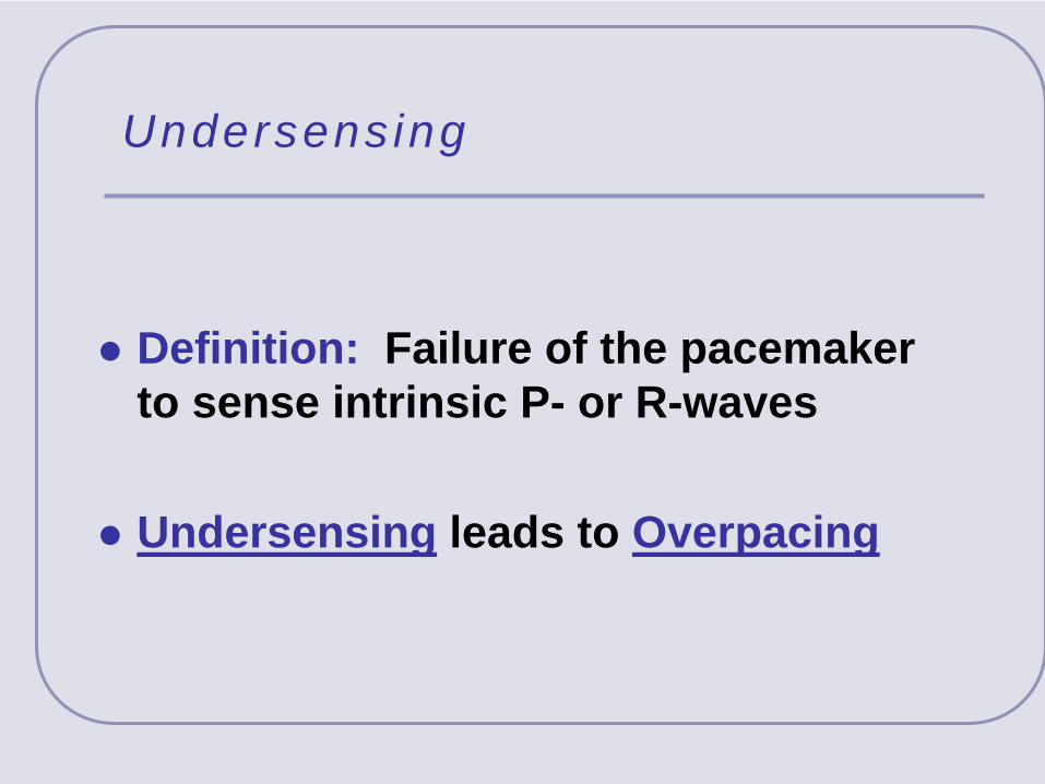
Undersensing
Definition: Failure of the pacemaker to sense intrinsic P- or R-waves
Undersensing leads to Overpacing

Ventricular Undersensing

Undersensing Causes
Inadequate cardiac signal or programmed sensitivity
Lead failure: - Insulation break - conductor fracture
Lead dislodgment

Loss of Capture
Definition: The emitted pacemaker stimulus does not cause depolarization
Loss of capture occurs when the pacemaker’s programmed energy is less than the stimulation threshold

Loss of Capture
No evidence of depolarization after pacing artifact

Loss of Capture Causes
Increased pacing threshold- Hyperkalemia- Antiarrhythmic drugs- Exit block
Decreased effective output- Dislodged lead (Perforation)- Lead failure (Insulation break, fracture)- PM end of life

ECG # 11
???

ECG # 14
???
???
מציג
הערות מצגת
An intrinsic depolarization occurs in the atrium, but this depolarization is not sensed by the pacemaker. Therefore, the pacemaker sends an inappropriate pacing pulse to that chamber. Undersensing can be thought of as “overpacing.” In this example, an AAI pacemaker is programmed to inhibit the atrial pacing pulse when a P-wave is sensed. Because the P-wave was not sensed, the pacemaker delivered an atrial pulse. If a pacemaker is undersensing, you will not see appropriate atrial sense markers on the marker channel.

PACING KODPACING MODE
response to “sensed event "
SENSING CHAMBER
PACING CHAMBER
O = NoneO = NoneO = None
I= inhibitedA = AtriumA = Atrium
T=triggeredV = VentricleV = Ventricle
D = DualD = DualD = Dual

Asynchronous Pacing – VOO/AOO/DOO
Pacing without sensing
Oldest mode of pacing
Magnet mode for most pacemakers
PACING MODE response to
“sensed event "
SENSING CHAMBER
PACING CHAMBER
O = NoneO = NoneO = None
I= inhibitedA = AtriumA = Atrium
T=triggeredV = VentricleV = Ventricle
D = DualD = DualD = Dual

VOO
Non Sensed R-wave

Demand Pacing (VVI/AAI)
Pacing with sensing
Pacing pulse is inhibited by intrinsic “P” or “R-waves”
Sensed events reset the pacing interval
PACING MODE response to
“sensed event "
SENSING CHAMBER
PACING CHAMBER
O = NoneO = NoneO = None
I= inhibitedA = AtriumA = Atrium
T=triggeredV = VentricleV = Ventricle
D = DualD = DualD = Dual

Ventricular Sensing / Inhibition
Sensed R-waves re-start the pacing interval
pacing interval
pacing interval
pacing interval

Atrial Sensing / Inhibition
Sensed P-waves re-start the pacing interval
pacing interval
pacing interval
pacing interval

ApVp
ApVs
AsVp
AsVp
Four States Of Dual Chamber Pacing (DDD)
Atrial event HAVE TO followed by ventricular event
PACING MODE response to
“sensed event "
SENSING CHAMBER
PACING CHAMBER
O = NoneO = NoneO = None
I= inhibitedA = AtriumA = Atrium
T=triggeredV = VentricleV = Ventricle
D = DualD = DualD = Dual

Maximum Tracking Rate (Upper Rate Limit)
The fastest rate the Ventricularchannel can track intrinsic P-waves
Goal:Prevent of tracking inappropriate fast
atrial rhythms (AT/ AFL)

Upper Rate BehaviorsUpper Rate Behaviors
Fixed-Ratio block (2:1, 3:1, etc)
Wenckebach behavior
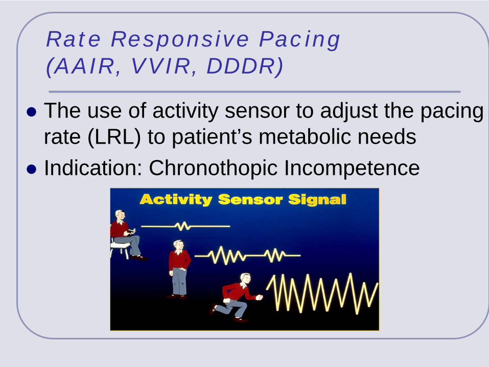
Rate Responsive Pacing (AAIR, VVIR, DDDR)
The use of activity sensor to adjust the pacing rate (LRL) to patient’s metabolic needs
Indication: Chronothopic Incompetence

Mode Selection Decision Tree
Sinus node disease
Atrial electrode
Rate responsive
pacing
מציג
הערות מצגת
This is the decision tree that we will be using to (practice) determine the optimal pacing mode for five example patients. When evaluating which pacing mode would provide optimal pacing therapy for each patient, we must ask ourselves three questions: Are atrial tachyarrhythmias present? (Can the atrium be paced and �sensed reliably?) Is AV conduction intact? Is SA node function presently adequate?

Mode Selection Decision Tree
AV node & Purkinje disease
Ventricular electrode
Ventricular pacing
Sinus RhythmChronic AF
Atrio-ventricular pacing
מציג
הערות מצגת
This is the decision tree that we will be using to (practice) determine the optimal pacing mode for five example patients. When evaluating which pacing mode would provide optimal pacing therapy for each patient, we must ask ourselves three questions: Are atrial tachyarrhythmias present? (Can the atrium be paced and �sensed reliably?) Is AV conduction intact? Is SA node function presently adequate?

Tachycardia cycle time =VA conduction time +functional PV-Delay
(usually PMT rate close to URL)
Pacemaker Mediated Tachycardia

Advanced pacing features
Mode Switch (DDD to VVI; DDD to AAI)
PMT termination
Autocapture
Rate Drop Response
Left ventricular pacing
ECG # 21
???
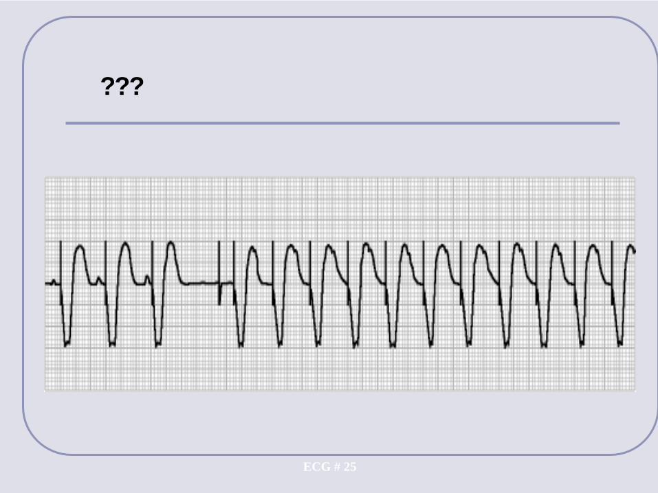
ECG # 25
???

Paced Tachycardia - DD
PMT
Tracking of atrial tachyarrhythmia
Sensor mediated tachycardia
Atrial lead oversensing
Component failure/ runaway tachycardia

A patient with severe CHF and a new DDD pacemaker developed bradycardia
1. Ventricular noncapture , atrium OK2. Crosstalk3. A+V lead dislodgment 4. Hyperkalemia
מציג
הערות מצגת
4 correct

ll succeed’and you–e methodical B
ears neckfeet

BACK UP SLIDES

Fixed-Ratio BlockFixed-Ratio Block
2:1 Block (one v-paced event per two p-waves)
P-V PVARP
TARP
PVARP PVARP PVARP PVARP
PP PP P P P PPP

Post Ventricular Atrial Refractory Period (PVARP)
PVARP
Atrial channel refractoriness after ventricular paced/sensed event

Automatic Mode Switching DDD to VVI
A A A A A A A A A A A A AS S S S R R S S S S R R S

Reversion to tracking mode
Automatic Mode Switching (AMS)

Uses of PVARP
Prevent the Atrial channel from sensing:• Far-field Ventricular signals
• Atrial Premature Contractions
• Retrograde P-waves

Pacemaker Mediated Tachycardia
Initiated by a loss of AV synchrony• PVC most common cause• Atrial loss of capture and undersensing, APC,
magnet removal
Pacemaker acts like a re-entrant pathway
Pacemaker Mediated Tachycardia
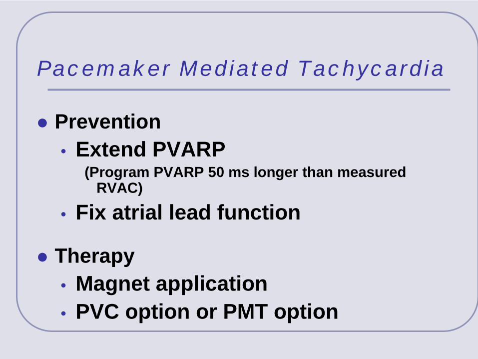
Prevention• Extend PVARP
(Program PVARP 50 ms longer than measured RVAC)
• Fix atrial lead function
Therapy• Magnet application• PVC option or PMT option

PMT Termination by pacemaker

ECG # 13
???

ECG # 14
???

ECG # 23
???

ECG # 24
???
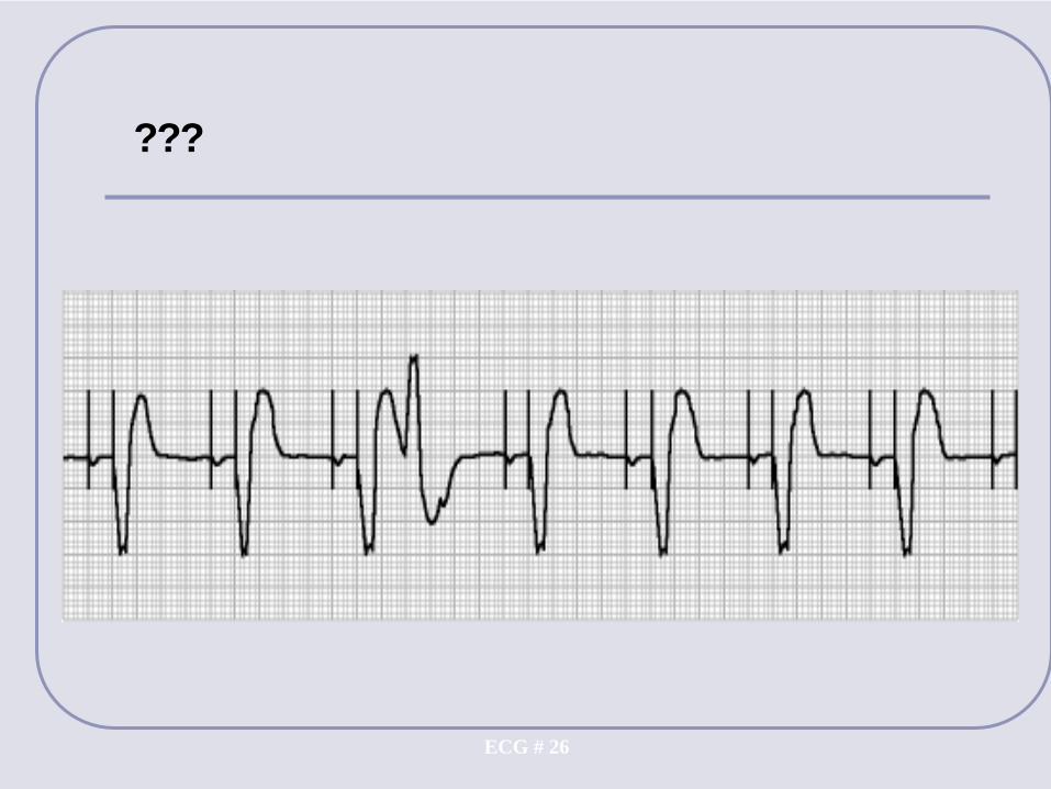
ECG # 26
???

???
מציג
הערות מצגת
At the onset of an atrial arrhythmia, the pacemaker compares a mean atrial interval (which is a running index of the atrial rate) to the current A-A interval. If the A-A interval is shorter than the MAI, the MAI is shortened by 24 ms. If the A-A interval is longer than the MAI, the MAI is lengthened by 8 ms. When the MAI reaches the interval corresponding to the mode switch detection rate interval, the pacemaker switches from the DDDR mode to the DDIR mode.

1. Ventricular noncapture 2. Normal pacemaker function 3. Ventricular undersensing 4. Multiple VPCS
מציג
הערות מצגת
Correct no 3

DDD mode
Base Rate60 ppm
MTR120 ppm
AVD150 ms
PVARP350 ms
ECG # 20
1. Atrial undersensing 2. Normal upper rate response then 2:1 tracking 3. Tracking of short atrial tachycardia4. PMT
מציג
הערות מצגת
2 correct

1. Ventricular noncapture 2. Normal pacemaker function 3. Ventricular undersensing 4. Multiple VPCS
מציג
הערות מצגת
Correct no 3

1. Atrial undersensing 2. Ventricular noncapture 3. Atrial fibrillation 4. PMT
מציג
הערות מצגת
3 correct

Myopotential inhibition

Determining the Optimal Pacing Mode -1
Patient information:
• Pt has chronic atrial fibrillation with an irregular ventricular rate• Pt’s heart rate does not reach 100 bpm in response to an
exercise stress test
VVIR
מציג
הערות מצגת
Patient information: Mrs. White has chronic atrial fibrillation with an irregular ventricular rate. Mrs. White’s heart rate does not reach 100 bpm in response to an exercise �stress test.

Determining the Optimal Pacing Mode 1
Patient information:
• Documented symptomatic sinus bradycardia
• When exercise tested, rate does not increase appropriately with increasing work loads
• At present, AV conduction is intact
AAIR
מציג
הערות מצגת
Patient information: Mrs. Peacock has documented symptomatic sinus bradycardia (rates <50 bpm) When exercise tested, her rate does not increase appropriately with increasing work loads At present, AV conduction is intact

Determining the Optimal Pacing Mode - 2
Patient information:
• Pt has intermittent 2nd degree Type II AV block with symptoms• Pt’s atrial rate responded appropriately (or not) to an exercise
test
P P QRS
DDD(R) & VDD (R)
מציג
הערות מצגת
Patient information: Professor Plum has intermittent 2nd degree Type II AV block with symptoms Professor Plum’s atrial rate responded appropriately to an exercise test