Bowel: A Case Report and Literature Review - cureus.com · There was no hematochezia, melena,...
6
Received 07/27/2018 Review began 08/02/2018 Review ended 08/04/2018 Published 08/06/2018 © Copyright 2018 Durer et al. This is an open access article distributed under the terms of the Creative Commons Attribution License CC-BY 3.0., which permits unrestricted use, distribution, and reproduction in any medium, provided the original author and source are credited. Cavernous Hemangioma of the Small Bowel: A Case Report and Literature Review Ceren Durer , Seren Durer , Mohamad Sharbatji , Isin Y. Comba , Ilan Aharoni , Umair Majeed 1. Internal Medicine, Florida Hospital-Orlando, Orlando, USA 2. Internal Medicine, UCF Internal Medicine Residency Program, Orlando, USA 3. Gastroenterology, Florida Hospital, Orlando, USA 4. Internal Medicine Residency, Florida Hospital-Orlando, Casselberry, USA Corresponding author: Mohamad Sharbatji, [email protected] Disclosures can be found in Additional Information at the end of the article Abstract Hemangiomas of the small intestine are rare and very difficult to diagnose preoperatively. Clinical presentations may include occult or massive gastrointestinal (GI) bleeding, obstruction, intussusception, and perforation. We report a 66-year-old Caucasian male patient with severe anemia secondary to occult GI bleeding from a cavernous hemangioma in the jejunum. A double balloon enteroscopy following capsule endoscopy was performed to obtain biopsy samples, which established the final diagnosis. Categories: Internal Medicine, Gastroenterology Keywords: cavernous hemangioma, capsule endoscopy, double balloon enteroscopy, chronic anemia Introduction Hemangioma of the small bowel is an uncommon condition, accounting for 5 to 10% of all benign lesions of the small bowel [1]. It usually presents in young people with no sex predilection. Intestinal hemangiomas may cause occult or massive gastrointestinal bleeding, obstruction, intussusception, and small bowel perforation [2]. According to the size of affected vessels, hemangiomas are histologically classified into cavernous, capillary or mixed-type, with the cavernous type being the most common. In the gastrointestinal tract, the most commonly involved site is the jejunum. We report a case of cavernous hemangioma of the jejunum presenting with severe anemia. No remarkable findings were detected in upper and lower endoscopies. Correspondingly, the patient underwent wireless capsule endoscopy and double- balloon enteroscopy, which demonstrated a 25-mm submucosal mass with mild superficial ulceration/erosions of the surface mucosa. The pathology demonstrated that the lesion was consistent with cavernous hemangioma without evidence of malignancy. Case Presentation A 66-year-old male presented to the emergency room due to worsening leg pain. His past medical history was significant for peripheral artery disease and iron deficiency anemia. Initial laboratory tests revealed an unexpectedly low hemoglobin level of 5.4 g/dl. He received three units of packed red blood cells and subsequently the hemoglobin level increased to 6.9 g/dl. Our gastroenterology department was consulted for evaluation of occult gastrointestinal bleeding. There was no hematochezia, melena, hematemesis, fatigue, or abdominal pain. The patient had been taking oral iron supplementation for the last five years for iron deficiency anemia. Previous upper and lower endoscopies were negative. On physical examination, the patient had pale conjunctivae. The abdomen was noted to be soft and non-tender. No masses, 1 1 1 2 3 4 Open Access Case Report DOI: 10.7759/cureus.3113 How to cite this article Durer C, Durer S, Sharbatji M, et al. (August 06, 2018) Cavernous Hemangioma of the Small Bowel: A Case Report and Literature Review. Cureus 10(8): e3113. DOI 10.7759/cureus.3113
Transcript of Bowel: A Case Report and Literature Review - cureus.com · There was no hematochezia, melena,...

Received 07/27/2018 Review began 08/02/2018 Review ended 08/04/2018 Published 08/06/2018
© Copyright 2018Durer et al. This is an open accessarticle distributed under the terms ofthe Creative Commons AttributionLicense CC-BY 3.0., which permitsunrestricted use, distribution, andreproduction in any medium, providedthe original author and source arecredited.
Cavernous Hemangioma of the SmallBowel: A Case Report and Literature ReviewCeren Durer , Seren Durer , Mohamad Sharbatji , Isin Y. Comba , Ilan Aharoni , UmairMajeed
1. Internal Medicine, Florida Hospital-Orlando, Orlando, USA 2. Internal Medicine, UCF InternalMedicine Residency Program, Orlando, USA 3. Gastroenterology, Florida Hospital, Orlando, USA 4.Internal Medicine Residency, Florida Hospital-Orlando, Casselberry, USA
Corresponding author: Mohamad Sharbatji, [email protected] Disclosures can be found in Additional Information at the end of the article
AbstractHemangiomas of the small intestine are rare and very difficult to diagnose preoperatively.Clinical presentations may include occult or massive gastrointestinal (GI) bleeding,obstruction, intussusception, and perforation. We report a 66-year-old Caucasian male patientwith severe anemia secondary to occult GI bleeding from a cavernous hemangioma inthe jejunum. A double balloon enteroscopy following capsule endoscopy was performed toobtain biopsy samples, which established the final diagnosis.
Categories: Internal Medicine, GastroenterologyKeywords: cavernous hemangioma, capsule endoscopy, double balloon enteroscopy, chronic anemia
IntroductionHemangioma of the small bowel is an uncommon condition, accounting for 5 to 10% of allbenign lesions of the small bowel [1]. It usually presents in young people with no sexpredilection. Intestinal hemangiomas may cause occult or massive gastrointestinal bleeding,obstruction, intussusception, and small bowel perforation [2]. According to the size of affectedvessels, hemangiomas are histologically classified into cavernous, capillary or mixed-type, withthe cavernous type being the most common. In the gastrointestinal tract, the most commonlyinvolved site is the jejunum. We report a case of cavernous hemangioma of the jejunumpresenting with severe anemia. No remarkable findings were detected in upper and lowerendoscopies. Correspondingly, the patient underwent wireless capsule endoscopy and double-balloon enteroscopy, which demonstrated a 25-mm submucosal mass with mild superficialulceration/erosions of the surface mucosa. The pathology demonstrated that the lesion wasconsistent with cavernous hemangioma without evidence of malignancy.
Case PresentationA 66-year-old male presented to the emergency room due to worsening leg pain. His pastmedical history was significant for peripheral artery disease and iron deficiency anemia. Initiallaboratory tests revealed an unexpectedly low hemoglobin level of 5.4 g/dl. He received threeunits of packed red blood cells and subsequently the hemoglobin level increased to 6.9 g/dl. Ourgastroenterology department was consulted for evaluation of occult gastrointestinal bleeding.There was no hematochezia, melena, hematemesis, fatigue, or abdominal pain. The patient hadbeen taking oral iron supplementation for the last five years for iron deficiency anemia.Previous upper and lower endoscopies were negative. On physical examination, the patient hadpale conjunctivae. The abdomen was noted to be soft and non-tender. No masses,
1 1 1 2 3
4
Open Access CaseReport DOI: 10.7759/cureus.3113
How to cite this articleDurer C, Durer S, Sharbatji M, et al. (August 06, 2018) Cavernous Hemangioma of the Small Bowel: ACase Report and Literature Review. Cureus 10(8): e3113. DOI 10.7759/cureus.3113
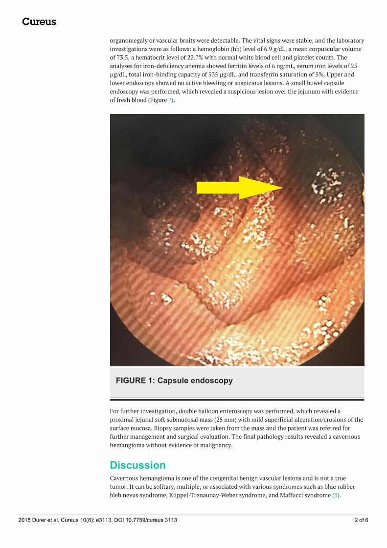
organomegaly or vascular bruits were detectable. The vital signs were stable, and the laboratoryinvestigations were as follows: a hemoglobin (hb) level of 6.9 g/dL, a mean corpuscular volumeof 73.5, a hematocrit level of 22.7% with normal white blood cell and platelet counts. Theanalyses for iron-deficiency anemia showed ferritin levels of 6 ng/mL, serum iron levels of 25μg/dL, total iron-binding capacity of 535 μg/dL, and transferrin saturation of 5%. Upper andlower endoscopy showed no active bleeding or suspicious lesions. A small bowel capsuleendoscopy was performed, which revealed a suspicious lesion over the jejunum with evidenceof fresh blood (Figure 1).
FIGURE 1: Capsule endoscopy
For further investigation, double balloon enteroscopy was performed, which revealed aproximal jejunal soft submucosal mass (25 mm) with mild superficial ulceration/erosions of thesurface mucosa. Biopsy samples were taken from the mass and the patient was referred forfurther management and surgical evaluation. The final pathology results revealed a cavernoushemangioma without evidence of malignancy.
DiscussionCavernous hemangioma is one of the congenital benign vascular lesions and is not a truetumor. It can be solitary, multiple, or associated with various syndromes such as blue rubberbleb nevus syndrome, Klippel-Trenaunay-Weber syndrome, and Maffucci syndrome [3].
2018 Durer et al. Cureus 10(8): e3113. DOI 10.7759/cureus.3113 2 of 6

Cavernous hemangioma is a soft, compressible, bluish purple lesion and consists of blood-filledspaces (cavern) between dilated vessels within the mucosa and submucosa. Its size may rangefrom a few millimeters to several centimeters. Hemangioma, telangiectasia, angiodysplasia,and phlebectasia are typical forms of small bowel vascular lesions.
Preoperative diagnosis is extremely difficult because small hemangiomas are rarelydemonstrable with traditional techniques such as upper and lower endoscopies. However,several imaging modalities including wireless capsule endoscopy, double balloon enteroscopy,multiphase computed tomography enterography (CTE), magnetic resonance enterography(MRE) are currently available options to investigate small bowel lesions [4]. Small bowel capsuleendoscopy is a noninvasive imaging test and can be recommended when the source of bleedingremains unidentified after upper and lower endoscopy. On the other hand, double balloonenteroscopy is an invasive and highly sensitive diagnostic tool providing both therapeutic anddiagnostic interventions. In the current case, the patient had severe chronic anemia withoutevidence of gross GI bleeding. Small bowel capsule endoscopy demonstrated suspiciousfindings in the jejunum and double-balloon enteroscopy helped diagnose the bluishsubmucosal mass in the small bowel.
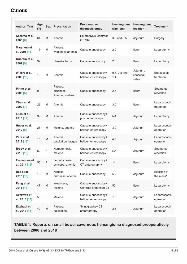
A comprehensive search of electronic databases (PubMed, Embase, and the Web of Sciencedatabases) was performed to identify studies published from January 1, 2000 until June 30, 2018using MeSH terms and keywords such as ‘cavernous hemangioma’, ‘wireless capsule endoscopy’,‘double balloon enteroscopy’, ‘anemia’ and ‘lower gastrointestinal bleeding’. Reference listswere screened to identify additional relevant studies. A standardized form was used for dataextraction. There have been about 15 cases of uncomplicated cavernous hemangioma of smallintestine diagnosed preoperatively [1, 5-18]. Patient information is summarized in Table 1.Most patients were male with age ranging from 9 to 74 years. The jejunum was the mostcommon site of small intestinal hemangiomas, and the initial clinical presentation was anemiain the majority of the cases. The diameter of the lesions varied from 0.8 cm to 50 cm. Moststudies showed suspicious findings on capsule endoscopy and the lesions were resectedsurgically or endoscopically [1, 5-7, 9-14, 16-18]. Ejtehadi et al. demonstrated a hemangioma onscintigraphy and CT enterography while no positive finding was found on capsule endoscopy[15]; another reported case was diagnosed with contrast-enhanced CT and MRI preoperatively[8]. Cavernous hemangioma can also present with other clinical symptoms and complications.Abdul Aziz et al. reported a six-year-old patient with abdominal pain and distention whodeveloped subacute intestinal obstruction secondary to cavernous hemangioma (15 cm)after blunt trauma to the abdomen [2]. Open surgical excision of the hemangioma along withaffected small bowel was performed. In a guideline by the American GastroenterologicalAssociation (AGA) in 2007 about the algorithms for the diagnosis and management of obscuregastrointestinal bleeding, capsule endoscopy is considered as the first-line procedure for theinitial examination of small bowel after normal upper and lower endoscopies [19]. Whenpositive findings are obtained on capsule endoscopy, double balloon enteroscopy or pushenteroscopy is recommended for further management when there is no evidence of obstruction[19]. The American College of Gastroenterology (ACG) clinical guideline in 2015 alsorecommends the use of capsule endoscopy followed by deep enteroscopy based on findings oncapsule endoscopy [20]. In the presented case, capsule endoscopy and balloon enteroscopy wereperformed according to these guidelines.
2018 Durer et al. Cureus 10(8): e3113. DOI 10.7759/cureus.3113 3 of 6
Author, YearAge(Y)
Sex PresentationPreoperativediagnosis study
Hemangiomasize (cm)
Hemangiomalocation
Treatment
Kazama et al.2000 [8]
64 M AnemiaEnteroclysis, contrast CT MRI
3.0 and 0.5 Jejunum Surgery
Magnano etal. 2005 [1]
13 MFatigue, weakness anemia
Capsule endoscopy 2.0 Ileum Laparotomy
Quentin et al.2007 [9]
32 F Hematochezia Capsule endoscopy 2.0 Ileum Laparotomy
Willert et al.2008 [10]
19 M AnemiaCapsule endoscopy+balloon enteroscopy
0.8, 0.8 and1.4
Jejunum,ileocecalvalve
Endoscopictreatment
Pinho et al.2008 [5]
9 FFatigue,dizziness,Anemia, melena
Capsule endoscopy 2.5 IleumSegmentalresection
Chen et al.2009 [7]
23 M Anemia Capsule endoscopy 3.0 IleumLaparoscopictreatment
Elias et al.2010 [14]
39 M AnemiaCapsule endoscopy+push enteroscopy
NA Jejunum Laparotomy
Huber et al.2012 [6]
23 M Melena, anemiaCapsule endoscopy+balloon enteroscopy
3.0 JejunumLaparoscopicoperation
Pera et al.2012 [18]
16 MAnemia,palpitation, fatigue
Capsule endoscopy+balloon enteroscopy
4.3 JejunumLaparoscopicoperation
Ersoy et al.2013 [13]
50 FHematemesis,melena
Capsule endoscopy+balloon enteroscopy
NA JejunumSegmentalresection
Fernandes etal. 2014 [12]
56 Fhematocheziasyncope, anemia
Capsule endoscopy+CT enterography
14 Ileum Laparotomy
Bae et al.2015 [16]
13 MNausea,dizziness, anemia
Capsule endoscopy 5.2 JejunumExcision ofthe mass*
Peng et al.2016 [11]
47 MWeakness,melena
Capsule endoscopy+Contrast-enhanced CT
50 Ileum Laparotomy
Akazawa etal. 2016 [17]
56 F MelenaCapsule endoscopy+balloon enteroscopy
1.3 JejunumLaparoscopicoperation
Ejtehadi etal. 2017 [15]
40 MFatigue,palpitation
Scintigraphy+ CTenterography
2.6 JejunumLaparoscopicoperation
TABLE 1: Reports on small bowel cavernous hemangioma diagnosed preoperativelybetween 2000 and 2018
2018 Durer et al. Cureus 10(8): e3113. DOI 10.7759/cureus.3113 4 of 6

ConclusionsCavernous hemangioma of the small bowel is a benign vascular lesion that can cause chronic GIbleeding presenting as chronic severe anemia. Clinical suspicion of bleeding originating fromthe small bowel is essential in the diagnosis of these lesions as they warrant a comprehensiveworkup. Based on the literature and guideline review, we recommend that capsule endoscopyfollowed by therapeutic double balloon enteroscopy be considered in those patients.
Additional InformationDisclosuresHuman subjects: Consent was obtained by all participants in this study. Conflicts of interest:In compliance with the ICMJE uniform disclosure form, all authors declare the following:Payment/services info: All authors have declared that no financial support was received fromany organization for the submitted work. Financial relationships: All authors have declaredthat they have no financial relationships at present or within the previous three years with anyorganizations that might have an interest in the submitted work. Other relationships: Allauthors have declared that there are no other relationships or activities that could appear tohave influenced the submitted work.
References1. Magnano A, Privitera A, Calogero G, Nanfito L, Basile G, Sanfilippo G: Solitary hemangioma of
the small intestine: an unusual cause of bleeding diagnosed at capsule endoscopy. J PediatrSurg. 2005, 40:25-27. 10.1016/j.jpedsurg.2005.06.014
2. Abdul Aziz DA, Khandasamy Y, Tamba RP, Zaki FM: Bleeding small bowel cavernoushaemangioma following blunt trauma to the abdomen presenting as subacute intestinalobstruction in a child. BMJ Case Rep. 2011, 2011, 10.1136/bcr.08.2011.4672
3. Wilson CL, Song LM, Chua H, Ferrara M, Devine RM, Dozois RR, Nehra V: Bleeding fromcavernous angiomatosis of the rectum in Klippel-Trenaunay syndrome: report of three casesand literature review. Am J Gastroenterol. 2001, 96:2783-2788. 10.1111/j.1572-0241.2001.04110.x
4. Watari I, Oka S, Tanaka S, Nakano M, Aoyama T, Yoshida S, Chayama K: Is occult obscuregastrointestinal bleeding a definite indication for capsule endoscopy? A retrospective analysisof diagnostic yield in patients with occult versus overt bleeding. Gastroenterol Res Pract.2013, 2013:915463. 10.1155/2013/915463
5. Pinho R, Rodrigues A, Proenca L, et al.: Solitary hemangioma of the small bowel disclosed bywireless capsule endoscopy. Gastroenterol Clin Biol. 2008, 32:15-18.10.1016/j.gcb.2007.11.004
6. Huber A, Abdel Samie A, Kychenko D, Theilmann L: A rare cause of recurrent iron-deficiencyanemia: cavernous hemangioma of the small intestine. J Gastrointestin Liver Dis. 2012,21:343.
7. Chen CH, Jones J, McGowan P: Profound iron deficiency anemia caused by a small-intestinalcavernous hemangioma. Gastrointest Endosc. 2009, 69:1392-1393. 10.1016/j.gie.2009.01.049
8. Kazama T, Kurihara Y, Tani I, Takahara T, Nakajima Y, Atari E: MR appearance of the smallintestinal cavernous hemangioma. J Comput Assist Tomogr. 2000, 24:655-656.
9. Quentin V, Lermite E, Lebigot J, Marinnes MZ, Arnaud JP, Boyer J: Small bowel cavernoushemangioma: wireless capsule endoscopy diagnosis of a surgical case. Gastrointest Endosc.2007, 65:550-552. 10.1016/j.gie.2006.12.024
10. Willert RP, Chong AK: Multiple cavernous hemangiomas with iron deficiency anemiasuccessfully treated with double-balloon enteroscopy. Gastrointest Endosc. 2008, 67:765-767.10.1016/j.gie.2007.07.044
11. Peng C, Chen H, Li W, Xu R, Zhuang W: A rare cause of recurrent gastrointestinal bleeding:giant diffuse and cavernous intestinal mesentery hemangioma in an adult. Dig Dis Sci. 2016,61:3363-3365. 10.1007/s10620-016-4259-2
12. Fernandes D, Dionisio I, Neves S, Duarte P: Cavernous hemangioma of small bowel: a rare
2018 Durer et al. Cureus 10(8): e3113. DOI 10.7759/cureus.3113 5 of 6

cause of digestive hemorrhage. Rev Esp Enferm Dig. 2014, 106:214-215.13. Ersoy O, Akin E, Demirezer A, Koseoglu H, Balci S, Kiyak G: Cavernous haemangioma of small
intestine mimicking gastrointestinal stromal tumour. Arab J Gastroenterol. 2013, 14:139-140.10.1016/j.ajg.2013.08.008
14. Elias G, Toubia N: Hemangioma of the small intestine presenting with recurrent overt,obscure gastrointestinal bleeding. Clin Gastroenterol Hepatol. 2010, 8:18-11.10.1016/j.cgh.2009.03.036
15. Ejtehadi F, Fattahi MR, Safaei A, Safarpour AR, Bananzadeh A: Practical lessons from thesmall bowel bleeding lesions: a case report on small bowel cavernous hemangioma. Iran J MedSci. 2017, 42:108-110.
16. Bae SJ, Hwang G, Kang HS, Song HJ, Chang WY, Maeng YH, Kang KS: Single cavernoushemangioma of the small bowel diagnosed by using capsule endoscopy in a child with chroniciron-deficiency anemia. Clin Endosc. 2015, 48:340-344. 10.5946/ce.2015.48.4.340
17. Akazawa Y, Hiramatsu K, Nosaka T, et al.: Preoperative diagnosis of cavernous hemangiomapresenting with melena using wireless capsule endoscopy of the small intestine. Endosc IntOpen. 2016, 4:249-251. 10.1055/s-0041-111321
18. Pera M, Marquez L, Dedeu JM, Sanchez J, Garcia M, Ramon JM, Puigvehi M: Solitary cavernoushemangioma of the small intestine as the cause of long-standing iron deficiency anemia. JGastrointest Surg. 2012, 16:2288-2290. 10.1007/s11605-012-1991-6
19. Raju GS, Gerson L, Das A, Lewis B: American Gastroenterological Association (AGA) Institutetechnical review on obscure gastrointestinal bleeding. Gastroenterology. 2007, 133:1697-1717. 10.1053/j.gastro.2007.06.007
20. Gerson LB, Fidler JL, Cave DR, Leighton JA: ACG clinical guideline: diagnosis andmanagement of small bowel bleeding. Am J Gastroenterol. 2015, 110:1265-1287.10.1038/ajg.2015.246
2018 Durer et al. Cureus 10(8): e3113. DOI 10.7759/cureus.3113 6 of 6
![Hemorrhagic Stroke Size by Optimization of - cureus.com · hemorrhagic stroke, and are associated with a higher mortality risk than ischemic strokes [3-4]. An IPH can lead to secondary](https://static.fdocuments.in/doc/165x107/5e0958916e06c4432d031ac7/hemorrhagic-stroke-size-by-optimization-of-hemorrhagic-stroke-and-are-associated.jpg)