Bovine Viral Diarrhea Virus Quasispecies during Persistent Infection · 2015-03-30 · Bovine Viral...
14
Bovine Viral Diarrhea Virus Quasispecies during Persistent Infection Margaret E. Collins, 1 Moira Desport, and Joe Brownlie Department of Pathology and Infectious Diseases, The Royal Veterinary College, Hawkshead Lane, North Mymms, Hatfield, Hertfordshire, AL9 7TA, United Kingdom Received November 9, 1998; returned to author for revision December 16, 1998; accepted March 10, 1999 Analysis of viral genome sequences from two calves persistently infected with bovine viral diarrhea virus revealed a quasispecies distribution. The sequences encoding the glycoprotein E2 were variable, translating to a number of changes in predicted amino acid sequences. The NS3 region was found to be highly conserved in both animals. The number of E2 clones showing variant amino acids increased with the age of the animal and comparison of the consensus sequences at the different time points confirmed differences in the predicted E2 sequences over time. The immune tolerance that allows the lifelong persistence of this viral infection is highly specific. It is likely that some of the variant viruses generated within these animals will differ antigenically from the persisting virus and be recognized by the immune system. Evidence of an immune response to persisting virus infection was gathered from a larger sample of cattle. Serum neutralizing antibodies were found in 4 of 21 persistently infected animals. Accumulations of viral RNA in the lymph nodes of all animals examined, particularly in the germinal center light zone, may represent antigenic variants held in the form of immune complexes on the processes of follicular dendritic cells. © 1999 Academic Press INTRODUCTION A fundamental aspect of the pathogenesis of any viral infection is the degree to which different strains can vary in their ability to cause disease. RNA viruses, in partic- ular, can mutate rapidly due to errors introduced during successive rounds of genome replication. Variant viruses or “quasispecies” will be occurring spontaneously within infected cells and the accumulated mutations can influ- ence the growth characteristics of the progeny viruses (Ahmed and Oldstone, 1988; Banner and Lai, 1991; Dock- ter et al., 1996; Domingo and Holland, 1997; Leister et al., 1993). Where mutations enhance the survival of a partic- ular genome sequence, rapid expansion of clones de- rived from this source may subsequently come to dom- inate the resulting viral population (Domingo and Hol- land, 1997; Enomoto and Sato, 1995). That this population dynamic is operating is well documented for many per- sistent or chronic viral infections, including human im- munodeficiency virus (HIV) and hepatitis C virus (HCV), where the selection pressures driving viral evolution may include the host immune response to infection or antivi- ral therapy (Enomoto and Sato, 1995). One exception to this scenario was thought to be the persistent infection of cattle with bovine viral diarrhea virus (BVDV), where the unique immunotolerance of the host for the persist- ing viral infection was considered to favor the mainte- nance rather than the antigenic evolution of the viral population (Hertig et al., 1995; Paton et al., 1994). BVDV is a positive sense, single-stranded RNA virus, classified within the Pestivirus genus in the family Flavi- viridae (Westaway et al., 1985). There are two biotypes of the virus which are distinguished by their ability to cause cytopathology in cell culture (Baker et al., 1954; Brownlie et al., 1984; Gillespie et al., 1960; Underdahl et al., 1957). The predominant, non-cytopathogenic biotype (BVDVnc) is found in both acute and persistent infections in cattle and is associated with a wide range of clinical signs. Persistent infections arise following fetal infection in utero during the first trimester; as the fetus develops the virus is not recognized by the immune system, with a consequent lack of immune response to the persisting virus (Bolin et al., 1985a; Brownlie et al., 1984). These persistently infected (p.i.) animals are, however, capable of responding normally to other antigens and to acute infections with different BVDV isolates producing a nor- mal neutralizing antibody response (Bolin et al., 1985b; McClurkin et al., 1984; Steck et al., 1980; Westenbrink et al., 1989). The BVDVnc genome is approximately 12.5 kb in size (Deng and Brock, 1992) and, in common with many other RNA viruses, readily demonstrates recombination as a result of template switching during replication (Desport et al., 1998; Meyers et al., 1992; Qi et al., 1992). This process can give rise to the second, cytopathogenic biotype of the virus (BVDVc) which only occurs in p.i. cattle and precipitates the death of the animal from Sequence data from this article have been deposited with the EMBL/ GenBank Data Libraries under Accession Nos. AJ241224 to AJ241252 inclusive. 1 To whom correspondence and reprint requests should be ad- dressed. Fax: 01707 661464. E-mail [email protected]. Virology 259, 85–98 (1999) Article ID viro.1999.9697, available online at http://www.idealibrary.com on 0042-6822/99 $30.00 Copyright © 1999 by Academic Press All rights of reproduction in any form reserved. 85
Transcript of Bovine Viral Diarrhea Virus Quasispecies during Persistent Infection · 2015-03-30 · Bovine Viral...

iiusoie(t1urildsmwirtot
Gi
d
Virology 259, 85–98 (1999)Article ID viro.1999.9697, available online at http://www.idealibrary.com on
Bovine Viral Diarrhea Virus Quasispecies during Persistent Infection
Margaret E. Collins,1 Moira Desport, and Joe Brownlie
Department of Pathology and Infectious Diseases, The Royal Veterinary College, Hawkshead Lane,North Mymms, Hatfield, Hertfordshire, AL9 7TA, United Kingdom
Received November 9, 1998; returned to author for revision December 16, 1998; accepted March 10, 1999
Analysis of viral genome sequences from two calves persistently infected with bovine viral diarrhea virus revealed aquasispecies distribution. The sequences encoding the glycoprotein E2 were variable, translating to a number of changes inpredicted amino acid sequences. The NS3 region was found to be highly conserved in both animals. The number of E2 clonesshowing variant amino acids increased with the age of the animal and comparison of the consensus sequences at thedifferent time points confirmed differences in the predicted E2 sequences over time. The immune tolerance that allows thelifelong persistence of this viral infection is highly specific. It is likely that some of the variant viruses generated within theseanimals will differ antigenically from the persisting virus and be recognized by the immune system. Evidence of an immuneresponse to persisting virus infection was gathered from a larger sample of cattle. Serum neutralizing antibodies were foundin 4 of 21 persistently infected animals. Accumulations of viral RNA in the lymph nodes of all animals examined, particularlyin the germinal center light zone, may represent antigenic variants held in the form of immune complexes on the processes
of follicular dendritic cells. © 1999 Academic Pressinp
cvtceTiaPuvcvpoimMa
(Rrepb
INTRODUCTION
A fundamental aspect of the pathogenesis of any viralnfection is the degree to which different strains can varyn their ability to cause disease. RNA viruses, in partic-lar, can mutate rapidly due to errors introduced duringuccessive rounds of genome replication. Variant virusesr “quasispecies” will be occurring spontaneously within
nfected cells and the accumulated mutations can influ-nce the growth characteristics of the progeny viruses
Ahmed and Oldstone, 1988; Banner and Lai, 1991; Dock-er et al., 1996; Domingo and Holland, 1997; Leister et al.,993). Where mutations enhance the survival of a partic-lar genome sequence, rapid expansion of clones de-
ived from this source may subsequently come to dom-nate the resulting viral population (Domingo and Hol-and, 1997; Enomoto and Sato, 1995). That this populationynamic is operating is well documented for many per-istent or chronic viral infections, including human im-unodeficiency virus (HIV) and hepatitis C virus (HCV),here the selection pressures driving viral evolution may
nclude the host immune response to infection or antivi-al therapy (Enomoto and Sato, 1995). One exception tohis scenario was thought to be the persistent infectionf cattle with bovine viral diarrhea virus (BVDV), where
he unique immunotolerance of the host for the persist-
Sequence data from this article have been deposited with the EMBL/enBank Data Libraries under Accession Nos. AJ241224 to AJ241252
nclusive.1 To whom correspondence and reprint requests should be ad-
cressed. Fax: 01707 661464. E-mail [email protected].
85
ng viral infection was considered to favor the mainte-ance rather than the antigenic evolution of the viralopulation (Hertig et al., 1995; Paton et al., 1994).
BVDV is a positive sense, single-stranded RNA virus,lassified within the Pestivirus genus in the family Flavi-iridae (Westaway et al., 1985). There are two biotypes ofhe virus which are distinguished by their ability to causeytopathology in cell culture (Baker et al., 1954; Brownliet al., 1984; Gillespie et al., 1960; Underdahl et al., 1957).he predominant, non-cytopathogenic biotype (BVDVnc)
s found in both acute and persistent infections in cattlend is associated with a wide range of clinical signs.ersistent infections arise following fetal infection intero during the first trimester; as the fetus develops theirus is not recognized by the immune system, with aonsequent lack of immune response to the persistingirus (Bolin et al., 1985a; Brownlie et al., 1984). Theseersistently infected (p.i.) animals are, however, capablef responding normally to other antigens and to acute
nfections with different BVDV isolates producing a nor-al neutralizing antibody response (Bolin et al., 1985b;cClurkin et al., 1984; Steck et al., 1980; Westenbrink et
l., 1989).The BVDVnc genome is approximately 12.5 kb in size
Deng and Brock, 1992) and, in common with many otherNA viruses, readily demonstrates recombination as a
esult of template switching during replication (Desportt al., 1998; Meyers et al., 1992; Qi et al., 1992). Thisrocess can give rise to the second, cytopathogeniciotype of the virus (BVDVc) which only occurs in p.i.
attle and precipitates the death of the animal from0042-6822/99 $30.00Copyright © 1999 by Academic PressAll rights of reproduction in any form reserved.
mpogdpmBavHfio
BspBaaceiagtopqpo1ol
atvp
Rissuqtewcav
A1
e113euqrpds
sAa yed ab
86 COLLINS, DESPORT, AND BROWNLIE
ucosal disease (MD). BVDVc genomes have been re-orted to vary in size from a defective interfering particlef 7 kb (Kupfermann et al., 1996; Tautz et al., 1994) toenomes of over 16 kb (Meyers et al., 1992), arising byeletion or duplication of portions of the genome, byoint mutations, or by recombination with host cellularRNAs (reviewed in Meyers and Thiel, 1996). As theVDVc arises by mutation of the BVDVnc within a p.i.nimal, the antigenic homology between the pairs ofiruses that cause MD is high (Corapi et al., 1988;oward et al., 1987). Thus the complex pathogenesis of
atal MD in cattle is intimately related to the specificity ofmmunological tolerance engendered as a consequencef early fetal infection.
The highest degree of variability between differentVDVnc strains occurs in the major glycoprotein (E2)equences and neutralizing antibodies to this proteinrovide complete protection from infection (Deng androck, 1992; Howard et al., 1989). In a p.i. animal it wasssumed that the specificity of the immunological toler-nce of the host favored maintenance of an antigenicallyonserved population of the persisting virus over themergence of quasispecies which stimulate an effective
mmune response. Thus although RNA sequence vari-nts may arise, those which were recognized as anti-enically heterologous by the immune system were
hought to be rapidly cleared. Analysis of E2 sequencesbtained from the serum of p.i. animals at different timeoints apparently confirmed that the persisting virus se-uence was maintained in stasis, with two groups inde-endently showing no variation in the consensus nucle-tide sequence of E2 within a p.i. animal (Hertig et al.,995; Paton et al., 1994). Although the stability of theverall virus population was not unexpected, the abso-
FIG. 1. Aligned translated protein sequences from the BVDV E2 amplimequences obtained by direct PCR sequencing of amplified viral RNAlignment of sequences derived from individual cloned PCR products ind changes from the consensus (in individual PCR clones) are displa
ute conservation of RNA sequence over time, without
ny silent nucleotide substitutions, was surprising givenhe heterogeneity of E2 sequences between differentirus strains and the known properties of viral RNAolymerases.
The aims of our study were (i) to determine whetherNA sequence variants arise during persistent BVDV
nfections and (ii) to seek evidence of an immune re-ponse to the persisting virus. In a retrospective analy-is, samples from persistently infected animals weresed as a source of viral RNA to identify potential se-uence variations and tested for the presence of neu-
ralizing antibody. Lymphoid tissues from a total of 21xperimental or field cases of persistent BVDV infectionere examined by in situ hybridization and by immuno-
ytochemistry using monoclonal antibodies to differenti-te between actively replicating virus and assembledirions.
RESULTS
nalysis of virus sequence variation from animals263 and 1279
The E2 region of BVDVnc was amplified from total RNAxtracted from two persistently infected calves (1263 and279) and two different time points (taken at 11- and8-month intervals) for each animal (giving sample data02928 5 1263 early, 309938 5 1263 late, 904928 5 1279arly, 903938 5 1279 late). The PCR products (generatedsing the enzyme Pfu polymerase to minimise the fre-uency of in vitro induced errors) were sequenced di-
ectly to determine the consensus sequence of the viralopulation present in the sample. A small number ofifferences are apparent in the alignment of the consen-us protein sequences (Fig. 1a).
rived from persistently infected animals 1263 and 1279. The consensusmples for each animal at two different time points) are aligned in (a).n in (b) to (e). The consensus sequence is displayed as the lower lineove this sequence. Agreement with the consensus is not displayed.
ers de(two sas show
The clonal composition of the virus populations at

eppuTowoaaqwq
qpscqtttasatt
—Cont
87BVDV QUASISPECIES DURING PERSISTENT INFECTION
ach time point was investigated by cloning the PCRroducts in Escherichia coli. Individual recombinantlasmids were sequenced in both directions and annambiguous sequence was obtained for each clone.hese sequences were aligned using the GCG packagef programs to highlight differences in the E2 sequencesithin an animal. A summary of the sequence changesccurring is shown in Table 1, while protein translationlignments are shown in Figs. 1b and 1c (animal 1263)nd Figs. 1d and 1e (animal 1279). The individual se-uences from each of the E2 clones showed variationith respect to other clones and to the consensus se-uence for that sample.
FIG. 1
To exclude the possibility that some of these se- 7
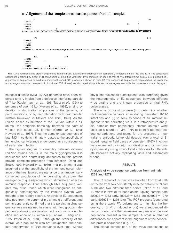
uence changes occurred in vitro during the RT/PCRrocess, clones were generated from the highly con-erved NS3 region of the viral genome. A total of 10lones were analyzed from the two animals and se-uence alignment revealed a much lower variability be-
ween both clones and animals. The consensus nucleo-ide sequences for both 1263 and 1279 were identical. Ofhe 4 nucleotide changes observed, only 1 resulted in anmino acid substitution (Fig. 2). The NS3 amplimers werehorter than the E2 amplimers examined so, to preventny bias, an analysis of the percentage of variation from
he consensus per nucleotide sequenced was under-aken. The variation of 0.49% (37 nucleotide changes/
inued
557 bases sequenced) for the E2 sequences was more

tcq
tspqttsmT
wsidr
scntoc
—Cont
88 COLLINS, DESPORT, AND BROWNLIE
han five times greater than the 0.09% (4 nucleotidehanges/4230 bases sequenced) for the NS3 se-uences.
Further analysis of the E2 nucleotide sequences fromhe earliest samples for both animals, 1263 and 1279,hows variation between individual clones within a sam-le. Clonal differences from the sample consensus se-uence were observed at between 9 and 13 positions,
ranslating to amino acid variation at 4 and 7 positions inhe E2 protein sequences. The observed variation con-tituted either a single difference in a single clone orore equal mixtures of two or more virus populations.
FIG. 1
he original virus inoculum used to infect both animals p
as not available for examination but it is possible thatuch mixed populations reflect sequence variation in the
nitial challenge dose. Comparison of these sequenceata with samples taken some months later is more
evealing (Figs. 1b to 1e).The E2 clones from the later time points sampled
how, for calf 1263, nucleotide changes resulting inlonal variation occurring at 11 positions over the 687ucleotides sequenced (4 amino acid differences), with
he result that none of the clones showed 100% homol-gy with respect to each other. The sequence variation inlones from 1279 was even greater, with variation at 18
inued
ositions (resulting in 15 amino acid differences). Align-
mppnHqasva
apqglpdt
S
(eprtssano
I
spMtwtsWbtltf(claop
EPP
3399
9
a
89BVDV QUASISPECIES DURING PERSISTENT INFECTION
ent of the clonal sequences suggests an homogenousopulation change within samples at a limited number ofoints but this must always be qualified by the limitedumber of individual clones analyzed from that sample.owever, alignment of the sample consensus se-uences (Fig. 1a) confirms changes to the nucleic acidnd also the consensus protein sequences in differentamples at different times. The consequence of thisariation is a small change in the consensus translatedmino acid sequence for E2 within an animal over time.
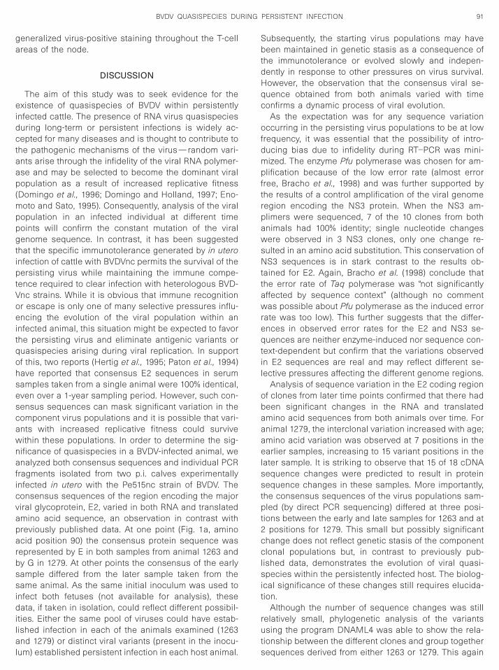
Further analysis of the variant E2 sequences from bothnimals 1263 and 1279 using the PHYLIP package ofrograms estimated the phylogeny of the clonal se-uences using the maximum likelihood analysis pro-ram, DNAML4. Figure 3 illustrates the evolutionary re-
ationships of these virus sequences as displayed by therogram, DRAWGRAM and confirms, with .97% confi-ence, the clustering of sequences derived from each of
he two different animal sources.
TABLE 1
Summary Analysis of the Consensus Sequences Derived from the2 Region of BVDVnc Amplified (687 Bases) from Total RNA of Twoersistently Infected Calves (1263 and 1279) and Two Different Timeoints
No. ofpositions
of variation Transitions Transversions
No. ofamino acid
changes
02928 9 7 2 409938 11 9 2 404928 13 9 4 703938 18 11 7 15
Note. Sample data: 302928 5 1263 early, 309938 5 1263 late,04928 5 1279 early, 903938 5 1279 late.
FIG. 2. Aligned translated protein sequences from the NS3-derived a
s the lower line and changes from the consensus are displayed above thiserum neutralization tests
Serum samples from 21 persistently infected animalsincluding 1263 and 1279) were examined for the pres-nce of virus neutralizing antibodies. Four samples wereositive in this assay when tested against the NADL
eference strain (Table 2). To try to establish whetherhese antibodies were induced by the endogenous, per-isting virus or as a result of exposure to a heterologoustrain of BVDV, virus purified from serum samples ofnimals 1263 and 1279 was used in an homologouseutralization assay. The neutralization titer to the homol-gous virus was higher than that to the NADL strain.
mmunocytochemistry
The distribution of BVDV-specific antigen during per-istent infections was investigated using cryopreservedrescapular lymph nodes from calves 1263 and 1279.onoclonal antibodies specific for the NS2/3 nonstruc-
ural protein and the E2 and Erns envelope glycoproteinsere used to discriminate between intracellular and po-
entially extracellular accumulations of the virus. Cellsupporting replication of BVDVnc, staining with antibodyB103 (specific for NS2/3), showed a widespread distri-
ution throughout the lymph node (Fig. 4c). The distribu-ion of the E2 glycoprotein (antibody WB214) was simi-arly widespread (Fig. 4a). In contrast, the distribution ofhe Erns glycoprotein (antibody WB210) was mainly con-ined to the germinal centers of the lymphoid folliclesFig. 4b). Some germinal centers (GC) had a distinctrescent-shaped area of staining corresponding to the
ight zone. The Erns protein is known to be both virionssociated and secreted from infected cells; the stainingbserved here could be attributable to either form of thisrotein.
rs from animals 1263 and 1279. The consensus sequence is displayed
mplime sequence. Agreement with the consensus is not displayed.
I
aRsFofd
Tgips(Rantd
aiomcsos(
tRpc
smyc
osi
90 COLLINS, DESPORT, AND BROWNLIE
n situ hybridization
Lymph node sections from 21 persistently infectednimals were examined for the distribution of BVDVNA using in situ hybridization (ISH) with a riboprobepecific for the NS2/3 region of the virus genome (seeig. 4e and Table 2). Viral RNA was detected through-ut the sections but additionally, intense GC staining
or the BVDV positive strand RNA was found to varyingegrees in all of the cases examined by ISH (Fig. 4e).
FIG. 3. Estimation of the phylogeny of the individual clonal E2equences amplified from the sera of animals 1263 and 1279 using theaximum likelihood method of the program DNAML4. Bootstrap anal-
sis with 100 repetitions confirmed the significance (.97%) of thelustering shown.
T
Twenty-One Persistently Infected Cattle Were Screened for the Pfor the Presence of Virus in Prescapular Lymph Nodes Us
Animalidentity no. Age
1 0232 5 months2 1216 4 months3 3382 2 months4 3452 2 months5 1263 7 months6 1279 8 months7 1280 2 months8 2342 7 months9 2350 7 months
10 2397 8 months11 2401 5 months12 DP197 N/A13 Shirley 2HS N/A14 VF 2315 N/A15 0825 33 months16 1035 24 months17 1409 16–20 months18 1448 16–20 months19 59B 2 months20 62B 2 months21 63B 2 months
Note. The level of germinal center (GC) staining by in situ hybridizatir 1/low (less than 5% positive GCs). The ISH staining pattern was vertaining reflects the distribution of NS3 antigen detected by antibody W
nhibition of virus production.
here seemed to be a reciprocal balance between theeneralized staining of T-cell areas and intense stain-
ng of GCs; animals with the highest levels of RNA-ositive GCs had the lowest levels of perifolliculartaining and vice versa. As reported in an earlier study
Desport et al., 1994), these accumulations of BVDVNA are found in many of the germinal centers that arelso positive by immunocytochemistry for Erns but doot reflect increased viral replication, as staining for
he NS2/3 protein never shows similar variation inensity.
The pattern of ISH staining could be used to group thenimals into three categories with respect to GC stain-
ng: high (111), BVDVnc RNA was detected in 35–60%f GCs with little or no staining in the T-cell areas;edium (11), BVDVnc RNA in 5–35% of the germinal
enters, with a moderate degree of RNA-positive cellstaining in the T-cell areas of the node and in the ducts;r low (1), less than 5% BVDV RNA-positive GCs butignificant staining in the T-cell areas of the node
Table 2).The four animals identified as having neutralizing an-
ibody in their sera had high levels of germinal centerNA staining. The animals with fewer than 5% RNA-ositive germinal centers were mainly the youngestases and the majority of these had a high level of
e of Serum Neutralizing Antibodies to BVDV (Strain NADL) andth Monoclonal Antibodies and a BVDV-Specific Riboprobe
evel of GCstaining
T cell areastaining
Neutralisingantibody titre
111 2 11/161 2 Not detected1 1 Not detected1 1 Not detected
111 2 11/16111 2 11/32
1 11 Not detected1 111 Not detected
111 2 Not detected111 2 11/128
1 111 Not detected11 2 Not detected
111 2 Not detected11 1 toxic , 1/24
111 2 Not detected11 2 Not detected11 1 Not detected1 11 Not detected
11 1 Not detected11 111 Not detected1 111 Not detected
scored as 111/high (35–60% of GCs), 11/medium (5–35% of GCs),r to the staining pattern of antibody WB210. The degree of T-cell area
he neutralizing antibody titer is the dilution of serum which gave a 50%
ABLE 2
resencing Bo
L
on wasy simila
B103. T
ga
eidctaap(mppgtiptVoeitqohsescawnaficvaparbssidilal
SbtdHqc
ofdmpftrpawsNttawreqtil
obaaaelsstpt2cclsit
rut
91BVDV QUASISPECIES DURING PERSISTENT INFECTION
eneralized virus-positive staining throughout the T-cellreas of the node.
DISCUSSION
The aim of this study was to seek evidence for thexistence of quasispecies of BVDV within persistently
nfected cattle. The presence of RNA virus quasispeciesuring long-term or persistent infections is widely ac-epted for many diseases and is thought to contribute to
he pathogenic mechanisms of the virus—random vari-nts arise through the infidelity of the viral RNA polymer-se and may be selected to become the dominant viralopulation as a result of increased replicative fitness
Domingo et al., 1996; Domingo and Holland, 1997; Eno-oto and Sato, 1995). Consequently, analysis of the viral
opulation in an infected individual at different timeoints will confirm the constant mutation of the viralenome sequence. In contrast, it has been suggested
hat the specific immunotolerance generated by in uteronfection of cattle with BVDVnc permits the survival of theersisting virus while maintaining the immune compe-
ence required to clear infection with heterologous BVD-nc strains. While it is obvious that immune recognitionr escape is only one of many selective pressures influ-ncing the evolution of the viral population within an
nfected animal, this situation might be expected to favorhe persisting virus and eliminate antigenic variants oruasispecies arising during viral replication. In supportf this, two reports (Hertig et al., 1995; Paton et al., 1994)ave reported that consensus E2 sequences in serumamples taken from a single animal were 100% identical,ven over a 1-year sampling period. However, such con-ensus sequences can mask significant variation in theomponent virus populations and it is possible that vari-nts with increased replicative fitness could surviveithin these populations. In order to determine the sig-ificance of quasispecies in a BVDV-infected animal, wenalyzed both consensus sequences and individual PCR
ragments isolated from two p.i. calves experimentallynfected in utero with the Pe515nc strain of BVDV. Theonsensus sequences of the region encoding the majoriral glycoprotein, E2, varied in both RNA and translatedmino acid sequence, an observation in contrast withreviously published data. At one point (Fig. 1a, aminocid position 90) the consensus protein sequence was
epresented by E in both samples from animal 1263 andy G in 1279. At other points the consensus of the earlyample differed from the later sample taken from theame animal. As the same initial inoculum was used to
nfect both fetuses (not available for analysis), theseata, if taken in isolation, could reflect different possibil-
ties. Either the same pool of viruses could have estab-ished infection in each of the animals examined (1263nd 1279) or distinct viral variants (present in the inocu-
um) established persistent infection in each host animal. s
ubsequently, the starting virus populations may haveeen maintained in genetic stasis as a consequence of
he immunotolerance or evolved slowly and indepen-ently in response to other pressures on virus survival.owever, the observation that the consensus viral se-uence obtained from both animals varied with timeonfirms a dynamic process of viral evolution.
As the expectation was for any sequence variationccurring in the persisting virus populations to be at low
requency, it was essential that the possibility of intro-ucing bias due to infidelity during RT–PCR was mini-ized. The enzyme Pfu polymerase was chosen for am-
lification because of the low error rate (almost errorree, Bracho et al., 1998) and was further supported byhe results of a control amplification of the viral genomeegion encoding the NS3 protein. When the NS3 am-limers were sequenced, 7 of the 10 clones from bothnimals had 100% identity; single nucleotide changesere observed in 3 NS3 clones, only one change re-
ulted in an amino acid substitution. This conservation ofS3 sequences is in stark contrast to the results ob-
ained for E2. Again, Bracho et al. (1998) conclude thathe error rate of Taq polymerase was “not significantlyffected by sequence context” (although no commentas possible about Pfu polymerase as the induced error
ate was too low). This further suggests that the differ-nces in observed error rates for the E2 and NS3 se-uences are neither enzyme-induced nor sequence con-
ext-dependent but confirm that the variations observedn E2 sequences are real and may reflect different se-ective pressures affecting the different genome regions.
Analysis of sequence variation in the E2 coding regionf clones from later time points confirmed that there hadeen significant changes in the RNA and translatedmino acid sequences from both animals over time. Fornimal 1279, the interclonal variation increased with age;mino acid variation was observed at 7 positions in thearlier samples, increasing to 15 variant positions in the
ater sample. It is striking to observe that 15 of 18 cDNAequence changes were predicted to result in proteinequence changes in these samples. More importantly,
he consensus sequences of the virus populations sam-led (by direct PCR sequencing) differed at three posi-
ions between the early and late samples for 1263 and atpositions for 1279. This small but possibly significant
hange does not reflect genetic stasis of the componentlonal populations but, in contrast to previously pub-
ished data, demonstrates the evolution of viral quasi-pecies within the persistently infected host. The biolog-
cal significance of these changes still requires elucida-ion.
Although the number of sequence changes was stillelatively small, phylogenetic analysis of the variantssing the program DNAML4 was able to show the rela-
ionship between the different clones and group together
equences derived from either 1263 or 1279. This again
saiics
vtvhaRiiipm9mpgafwep
r
dfalnctsmbHbBtasettoswlcwsft
ans( ar dend
92 COLLINS, DESPORT, AND BROWNLIE
uggests the evolution of related virus sequences withinn animal rather than the maintenance in stasis of an
nitial population of viruses selected from a common pooln the inoculum. Bootstrap analysis with 100 repetitionsonfirmed the significance (.97%) of the clusteringhown.
The data indicate an increase in amino acid sequenceariation of the component virus populations over timehat has the potential to generate antigenically variantiruses. The epitopes involved in the antigenicity of E2ave not yet been precisely defined for BVDV, althoughntigenic regions have been determined for CSFV (Vanijn et al., 1994). The amino acid changes observed were
n the E2 regions equivalent to domains B and C but notn A or D (Van Rijn et al., 1994). Regions of hypervariabil-ty have been outlined for BVDV by both sequence com-arisons and by the generation of neutralization escapeutants. The change from I to T (at position 182) in clone
03938c (this study) corresponds exactly with an escapeutant generated by Paton (Paton et al., 1992) and thus
otentially represents a neutralization escape mutantenerated in vivo. In the context of an immunotolerant p.i.nimal, this may represent the mutation of an epitope
rom one which is not recognized by the host to onehich becomes the target for immune clearance. Furthervidence for the presence of an antibody response to theersisting virus was then sought.
Data were collected from 21 p.i. cattle covering a
FIG. 4. Serial sections (a–d) from the prescapular lymph node of pntigens: (a) WB214 to detect distribution of structural glycoprotein Eonstructural protein NS2-3. (d) TRT 1 negative control and (e) in situupporting virus replication is indicated in (c). Differences in the detec
in b and e), may reflect the trapping of immune complexes on follicul
ange of ages and histories (including gnotobiotically c
erived calves, animals resulting from experimental in-ection of the dam but subsequently housed in isolation,nd also a number of persistently infected animals col-
ected from farm studies). The presence of low levels ofeutralizing antibody was confirmed in 4 animals. In allases these animals had been held in isolation condi-
ions, some had been housed with animals from theame cohort, p.i. with the same virus, but other cohortembers remained antibody negative. Neutralizing anti-
odies have been described in p.i. animals (Duffell andarkness, 1985; Steck et al., 1980) and this has largelyeen assumed to be due to infection with heterologousVDV isolates. Further analysis of these sera confirmed
hat, for two isolates in the current study, the neutralizingntibody titer to the homologous virus (isolated fromerum) was higher than that to the standard NADL ref-rence strain normally used for these assays. As more
han a single serum sample was available for each ofhese animals, the levels of antibody could be followedver time and strongly suggested that the antibody re-ponse reflects seroconversion to antigenic variantsithin the persisting virus population rather than to chal-
enge with a heterologous virus isolate. This surprisingonclusion is not unique. Observations made by Ed-ards et al. (1991) and Brock et al. (1998) confirm that
ome cattle maintain a state of persistent viremia in theace of a cycling neutralizing antibody directed towardhe homologous virus infection. These observations are
ntly infected calf 1279 using monoclonal antibodies to detect BVDVB210 to detect structural glycoprotein Erns, and (c) WB103 to detect
ization to detect BVDV positive sense RNA. The distribution of cellsother viral components, particularly the Erns protein and genomic RNAritic cells.
ersiste2, (b) W
hybridtion of
onsistent with the endogenous recognition of antigeni-

cboiroop
sb
gifcscpr1t
—Cont
93BVDV QUASISPECIES DURING PERSISTENT INFECTION
ally variant viruses and either partial clearance followedy recrudescence or serial emergence and recognitionf several independent variants. The alternative that an-
mals held in isolation in three different laboratories hadepeated accidental challenge with exogenous heterol-gous viruses seems less likely. It is possible that somether defect in the immune competence of these animalsrevents clearance of the infection.
The presence of a low-level neutralizing antibody re-ponse to components of a persisting virus infection may
FIG. 4
e expected to reflect some aspects of autoimmunity. In a
eneral, small immune complexes are formed when anndividual makes antibodies to self-antigens, as only aew epitopes are recognized and the formation of aross-linking lattice is restricted (Roitt et al., 1993). Theize of the aggregate influences the site of immuneomplex deposition—relatively large complexes are de-osited in kidneys (such immune complex disease is
arely associated with BVDV infection (Hewicker et al.,987)), whereas smaller complexes may pass throughhe glomerular basement membrane. The coexistence of
inued
ntiviral antibody and antigen in a p.i. animal is likely to

ftcn(pshto
imselvamf(
—Cont
94 COLLINS, DESPORT, AND BROWNLIE
orm small circulating immune complexes that would berapped and held on the processes of follicular dendriticells within the germinal centers of peripheral lymphodes, where they would persist for long periods of time
Roitt et al., 1993). The presence of small immune com-lexes in serum is difficult to detect and, in this retro-pective analysis, the freeze–thawing of samples wouldave disrupted such complexes. Enduring evidence for
heir existence in p.i. animals was sought in examinationf fixed lymph node sections.
FIG. 4
During persistent infection BVDVnc has a wide-rang- d
ng tropism, with many different cell types, includingost of the peripheral blood mononuclear cells, showing
ome degree of infection (Bielefeldt-Ohmann, 1995; Soppt al., 1994). Particularly high levels of virus occur in
ymphoid tissues, where a widespread distribution ofirus, in both T- and B-cell areas, is observed (Figs. 4and 4c, Lopez et al., 1993; Sopp et al., 1994). The use ofonoclonal antibodies to the viral NS2/3 protein con-
irmed the active replication of virus within these cellsFig. 4c and Sopp et al., 1994). In contrast to these data,
inued
etection of viral RNA by in situ hybridization consistently

iemtivetr
ts1mm1basLpptsstmdtwlp
tcscdqetaecstat
ipci(or
tri
V
nwcecpoftaasnt(m1f
oTibh
R
pBtmwgowGRapcc7t
R
95BVDV QUASISPECIES DURING PERSISTENT INFECTION
ndicated intense accumulations of BVDV in the GC ar-as of the lymphoid follicles (Fig. 4e) in addition to aore even background distribution. There is no evidence
o suggest that germinal center B-cells are preferentiallynfected with BVDVnc; the generalized distribution of theirus NS2/3 protein (Fig. 4c) indicates the contrary. How-ver, the contrasting data generated by different detec-
ion reagents (compare Figs. 4a and 4c with 4b and 4e)equires explanation.
Several viruses have been described within GCs ei-her because the cells that populate these areas arepecific targets for virus replication (Bachmann et al.,996; Hufert et al., 1997) or because virus-specific im-une complexes are held as iccosomes on the cytoplas-ic processes of the follicular dendritic cells (Tew et al.,
997). It is well established that immune complexes cane held in this location as dense aggregates of antigennd RNA, in an undegraded and potentially infectioustate, for several months (Bachmann et al., 1996; Mac-ennan, 1994; Tacchetti et al., 1997). The concentration ofositive strand viral RNA (Fig. 4e) and the viral Erns
rotein (Fig. 4b) at the GC sites would be consistent withhe trapping of virus particles from the circulation inmall immune complexes. The absence of a similartaining pattern with a monoclonal antibody specific for
he E2 protein could be explained if the epitopes on theore variable E2 protein were already blocked by en-
ogenous bovine antibody. This would also be consis-ent with the absence of an intense GC staining signal
ith the NS2/3-specific monoclonal, as there would beittle or no replicating virus (and therefore no NS2/3rotein) in the trapped immune complexes.
The observations described in this report, includinghe detection of viral sequence variants, changes in theonsensus sequence over time, antibodies to the per-isting virus, germinal centers staining strongly for virionomponents and for complement C3c (unpublishedata), are all consistent with the hypothesis that RNAuasispecies arising within a p.i. animal stimulate anndogenous antiviral immune response in the immuno-
olerant animal. The number of viral variants within annimal will change over time and will certainly be differ-nt between animals. The sequestration of the immuneomplexes generated, into the secondary lymphoid tis-ues, may contribute to the perception that lymphoid
issues are a preferred site of virus replication and maylso exacerbate perturbations of the immune compe-
ence of these animals.Relatively little is known about the effects of variation
n the E2 sequence of different BVDV isolates on viralathogenesis. In other viral infections, such as lympho-ytic choriomeningitis virus, a single amino acid change
s sufficient to dramatically alter the tropism of the virusTeng et al., 1996). Current understanding of the structuref the BVDV E2 protein and its interaction with cellular
eceptor(s) and mechanisms of virulence are too limited s
o permit speculation about the molecular events occur-ing during infection, but variation in E2 sequences couldnfluence the virulence of different strains.
MATERIALS AND METHODS
irus and animals
The Pe515nc strain of BVDV was a virologically clonedon-cytopathogenic virus isolated from a cow diagnosedith mucosal disease. Virus isolation in calf testis (CTe)
ells was followed by expansion of the virus stock. Allxperimental inocula were prepared from infected CTeells, three passes from the initial virus cloning. Twoersistently infected calves, 1279 and 1263, were theffspring of two seronegative dams that had been in-
ected intranasally with Pe51nc before 90 days of gesta-ion. The p.i. calves were held in a secure unit with othernimals infected with the same virus. Number 1154 was
normal uninfected calf of a similar age but housedeparately from the p.i. animals. Prescapular lymphodes and serum samples were taken at a number of
ime points, the first at approximately 7 months of agesamples 1263 early and 1279 early), the latest at 18
onths (sample 1279 late) or 21 months of age (sample263 late). Samples were cryopreserved at 270°C or
ixed in neutral buffered formalin until required.Lymphoid tissues from an additional 19 experimental
r field cases of persistent BVDV infection (detailed inable 2) were also examined. Tissues, fixed for 24–72 h
n neutral buffered formalin, were processed to paraffinlocks prior to use in immunocytochemistry and in situybridisation.
NA extraction
RNA was isolated from serum or tissue samples ofersistently infected animals using RNA-Stat 60 (AMSiotechnology) according to the manufacturer’s instruc-
ions. Briefly, 1.5-ml aliquots of serum were mixed with 15l of RNA-Stat 60 and 3 ml of chloroform. The total RNAas separated from the DNA and proteins by centrifu-ation at 12,000 g for 10 min. The colorless upper aque-us phase was removed to a fresh tube and the RNAas precipitated with a total of 7.5 ml of isopropanol.lycogen was added to assist with the recovery of theNA pellet. The pellet was obtained after centrifugations before and initially resuspended in 300 ml diethylyrocarbonate (DEPC)-treated H2O. The RNA was repre-ipitated with 12 ml 5 M NaCl and 600 ml ethanol andentrifuged as before. The pellet was washed with 1 ml5% ethanol before finally resuspending in 50 ml DEPC-
reated H2O.
T/PCR, cloning, and sequencing
A 5-ml sample of the total RNA was reverse tran-
cribed using random hexamers and 200 units of Super-
S4fusAsf58acbpqaTEufstoCqlt
I
utbb1wdpniiPnpcMNtsa
V
bw
ccvcCdBpuwdpwmo
I
atDuag(li1ia
tsn3stcm
O
cdPmdONsc
S
S
96 COLLINS, DESPORT, AND BROWNLIE
cript II Reverse Transcriptase (Gibco/BRL) for 50 min at2°C in a total volume of 20 ml according to the manu-
acturer’s instructions. Cloned Pfu DNA polymerase wassed for the PCR as it has a higher fidelity for DNAynthesis than Taq polymerase (Bracho et al., 1998).mplification of 2 ml of the RT reaction was according totandard procedures, with 25 pmol of each primer in a
inal volume of 50 ml using 35 cycles of 94°C for 1 min,5°C for 1 min, 72°C for 2 min, and a final extension ofmin at 72°C. Amplification products were examined by
garose gel electrophoresis. The PCR products wereloned directly into pGEM 3Zf(2) (Promega) which hadeen linearized with SmaI. Recombinant plasmids wereurified using Qiaprep Spin minprep kits (Qiagen). Se-uencing was performed on a Pharmacia ALF Expressutomated sequencer using Cy-5-labeled primers and ahermosequenase cycle sequencing kit (Amersham).ach individual clone was sequenced until an unambig-ous sequence was obtained so that clones derived
rom variant viruses could be identified. A consensusequence for the viral population present in the serum at
he time of sampling was obtained by direct sequencingf 100 ng of the purified, uncloned PCR product usingy-5-labeled primers and a Thermosequenase cycle se-uencing kit as before. DNA sequence data was ana-
yzed using the GCG package and programs available onhe SEQNET facility (Devereux et al., 1984).
n situ hybridisation
BVDV-specific digoxigenin-labeled riboprobes weresed to show the location of the viral RNA in the fixed
issues as previously described (Desport et al., 1994). Inrief, the sections were dewaxed and rehydrated to H2Oefore treating with 100 mg ml21 proteinase K at 37°C for5 min to assist with the probe penetration. The sectionsere then hybridized in 50 ml of 50% formamide, 5%extran sulfate, 23 SSC, 0.1 mM EDTA, 1 mM Tris–HCl,H 7.5, 4 mg ml21 denatured salmon sperm DNA, and 25g of riboprobe. The sections were coverslipped and
nitially denatured at 80°C for 10 min followed by hybrid-zation for 2 h at 55°C using a Hybaid Omnislide block.osthybridization washes were performed to remove anyonspecific binding and the probes were detected witholyclonal sheep anti-digoxigenin Fab fragments directlyonjugated with alkaline phosphatase (Boehringerannheim). The enzyme activity was detected using theBT/BCIP substrate and sections were left overnight for
he blue signal to develop. The sections were counter-tained with 1% neutral red, briefly cleared in acetonend ethanol, and mounted.
irus neutralization assay
The presence of BVDV-specific virus neutralizing anti-odies in the serum of the animals used in this study
as determined by assessing the inhibition of growth ofytopathogenic BVDV strain NADL using a modified mi-rotiter assay (Frey and Liess, 1971). Briefly, 100 TCID50 ofirus was added to doubling dilutions of antisera inulture media and incubated for 1 h at room temperature.alf testis cells were then added and incubated for 5ays. Viral replication was detected using a swine anti-VDV serum followed by a goat anti-swine serum cou-led to horseradish peroxidase. The substrate TMB wassed and the OD405 was read after stopping the reactionith 2 M H2SO4. The neutralizing antibody titer is theilution of serum which gave a 50% inhibition of virusroduction. In some cases where neutralizing antibodyas detected, a further assay was performed using ho-ologous virus purified from the serum sample in place
f the NADL strain.
mmunocytochemistry
Cryosections were cut into superfrost slides at 4–6 mmnd allowed to air dry. The sections were fixed in ace-
one at 4°C for 10 min. The reagents were obtained fromAKO and used as directed. The sections were stainedsing an APAAP method with the following monoclonalntibodies: WB214 (specificity for E2, the major envelopelycoprotein, and used diluted in Tris-buffered saline
TBS) at 1:200), WB210 (specificity for the second enve-ope glycoprotein, Erns, and used at 1:50), WB103 (spec-ficity for the NS2/3 nonstructural protein, and used at:1000), and as a negative control antibody TRT 1 (spec-
ficity for an unrelated turkey rhinotracheitis virus proteinnd used at 1:20).
Briefly, sections were incubated with the primary an-ibodies at 37°C for 1 h. After washing in TBS, theections were incubated with rabbit anti-mouse immu-oglobulins followed by the mouse APAAP for a further0 min each. After a final wash, the BCIP/NBT/INT sub-trate plus levamisole was allowed to develop the sec-
ions for 10 min. The slides were washed in water,ounterstained in hematoxylin, and mounted in Fara-ount.
ligonucleotides
The primer sequences used were obtained from BVDVonsensus sequences from the EMBL and GenBankatabases and, where available, known sequences ofe515nc. The SP6 1 SP3 pair specifically amplify theajority of the E2 sequence (lacking the transmembrane
omain), which is known to contain antigenic domains.ligo9 1 oligo2 amplify the first 460 nucleotides of theS3 region. The NS3 region is one of the most con-
erved between viral isolates and was chosen as aontrol for the experiment.
P6, 59 AGGGGCCAGATGGTACAGGGC 39 (SD1 2405-2425);
P3, 59 GTCTACTAATCTGTAGCCAGTCTCATT 39 (SD1
3175-3149);
o
o
tdHfmtatw
A
B
B
B
B
B
B
B
B
B
C
D
D
D
D
D
D
D
D
E
E
F
G
H
H
H
H
H
K
L
L
L
M
M
M
M
P
P
97BVDV QUASISPECIES DURING PERSISTENT INFECTION
ligo2, 59 GACCATCCTTTCAAGTTTTTT 39 (SD1 5620-5640); and
ligo9, 59 GAGCACGAAAAAATGCCAC 39 (SD1 5197-5180) were used.
ACKNOWLEDGMENTS
We are grateful to Dr. D. J. Paton (VLA, Weybridge) for the supply ofhe monoclonal antibodies WB103, WB210, and WB214 and for valuableiscussion of this work. We also thank staff at the Institute for Animalealth at Compton for their contributions, particularly Mr. M. C. Clarke
or the virus neutralization data, Mrs. B. V. Jones for the production ofonoclonal antibody TRT1, Mrs. S. M. Hacker and Mrs H. I. Cook for
heir excellent assistance in the histological preparation of samples,nd the compound staff (at IAH) for the care of the animals. The
echnical support provided by Ms. N. J. Archer throughout this projectas invaluable.
REFERENCES
hmed, R., and Oldstone, M. B. A. (1988). Organ-specific selection ofviral variants during chronic infection. J. Exp. Med. 167, 1719–1724.
achmann, M. F., Odermatt, B., Hengartner, H., and Zinkernagl, R. M.(1996). Induction of long-lived germinal centers associated with per-sisting antigen after viral infection. J. Exp. Med. 183, 2259–2269.
aker, J. A., York, C. J., Gillespie, J. H., and Mitchell, G. B. (1954). Virusdiarrhea in cattle. Vet. Res. 15, 525–531.
anner, L. R., and Lai, M. M. C. (1991). Random nature of CoronavirusRNA recombination in the absence of selection pressure. Virology185, 441–445.
ielefeldt-Ohmann, H. (1995). The pathologies of bovine virus diarrheavirus infection, In “The Veterinary Clinics of North America: FoodAnimal Practice” (J. C. Baker and H. Houe, Eds.), p. 447–476. Saun-ders, Philadelphia.
olin, S. R., McClurkin, A. W., Cutlip, R. C., and Coria, M. F. (1985a).Severe clinical disease induced in cattle persistently infected withnoncytopathic bovine viral diarrhea virus by superinfection with cy-topathic bovine viral diarrhea virus. Am. J. Vet. Res. 46, 573–576.
olin, S. R., McClurkin, A. W., Cutlip, R. C., and Coria, M. F. (1985b).Response of cattle persistently infected with noncytopathic bovineviral diarrhea virus to vaccination for bovine viral diarrhea and tosubsequent challenge exposure with cytopathic bovine viral diarrheavirus. Am. J. Vet. Res. 46, 2467–2470.
racho, M. A., Moya, A., and Barrio, E. (1998). Contribution of Taqpolymerase-induced errors to the estimation of RNA virus diversity.J. Gen. Virol. 79, 2912–2928.
rock, K. V., Grooms, D. L., Ridpath, J., and Bolin, S. (1998). Changes inlevels of viraemia in cattle persistently infected with bovine viraldiarrhea virus. J. Vet. Diagn. Invest. 10, 22–26.
rownlie, J., Clarke, M. C., and Howard, C. J. (1984). Experimentalproduction of fatal mucosal disease in cattle. Vet. Rec. 114, 535–536.
orapi, W. V., Donis, R. O., and Dubovi, E. J. (1988). Monoclonal antibodyanalyses of cytopathic and noncytopathic viruses from fatal bovineviral diarrhea virus infections. J. Virol. 62, 2823–2827.
eng, R., and Brock, K. V. (1992). Molecular cloning and nucleotidesequence of a pestivirus genome, noncytopathic bovine viral diar-rhea virus strain SD-1. Virology 191, 867–879.
esport, M., Collins, M. E., and Brownlie, J. (1994). Detection of BovineVirus Diarrhoea Virus RNA by in situ hybridisation with digoxigenin-labelled riboprobes. Intervirology 37, 269–276.
esport, M., Collins, M. E., and Brownlie, J. (1998). Genome instabilityin BVDV: An examination of the sequence and structural influenceson RNA recombination. Virology 246, 352–361.
evereux, J., Heaberli, P., and Smithies, O. (1984). A comprehensive setof sequence analysis programs for the VAX. Nucleic Acids Res. 12,
387–395.ockter, J., Evans, C. F., Tishon, A., and Oldstone, M. B. A. (1996).Competitive selection in vivo by a cell for one variant over another—Implications for RNA virus quasi-species in vivo. J. Virol. 70, 1799–1803.
omingo, E., Menendez Arias, L., and Holland, J. J. (1996). RNA virusfitness. Rev. Med. Virol. 7, 87–96.
omingo, E., and Holland, J. J. (1997). RNA virus mutations and fitnessfor survival. Annu. Rev. Microbiol. 51, 151–178.
uffell, S. J., and Harkness, J. W. (1985). Bovine viral diarrhoea–mucosaldisease infection in cattle. Vet. Rec. 117, 240–245.
dwards, S., Wood, L., Brockman, S., and Ibata, G. (1991). Clinical andvirological observations of a mucosal disease outbreak with persis-tently infected seropositive survivors. Arch. Virol. (Suppl. 3), 125–132.
nomoto, N., and Sato, C. (1995). Clinical relevance of Hepatitis Cquasispecies. J. Viral Hepatitis 2, 267–272.
rey, H.-R., and Liess, B. (1971). Growth curve studies and applicabilityof a highly cytopathogenic BVD-MD-virus strain for diagnostic pur-poses using the micro-titre method. Zentralbl. Vet. Med. B 18, 61–71.
illespie, J. H., Baker, J. A., and McEntee, K. (1960). A cytopathogenicstrain of virus diarrhea virus. Cornell Vet. 50, 73–79.
ertig, C., Stalder, H., and Peterhans, E. (1995). Genetic heterogeneitywithin the coding regions of E2 and NS3 in strains of bovine viraldiarrhea virus. Gene. 153, 191–195.
ewicker, M., Trautwein, G., Stahl, C., and Liess, B. (1987). Kidneylesions in cattle persistently infected with bovine viral diarrhea virus.J. Vet. Med. Ser. B 34, 1–12. [Infectious Diseases Immunology FoodHygiene Veterinary Public Health-Zentralblatt Fur VeterinarmedizinReihe B]
oward, C. J., Brownlie, J., and Clarke, M. C. (1987). Comparison by theneutralisation assay of pairs of non-cytopathogenic and cytopatho-genic strains of Bovine Virus Diarrhoea Virus isolated from cases ofMucosal Disease. Vet. Microbiol. 13, 361–369.
oward, C. J., Clarke, M. C., and Brownlie, J. (1989). Protection againstrespiratory infection with Bovine Virus Diarrhoea Virus by passivelyacquired antibody. Vet. Microbiol. 19, 195–203.
ufert, F. T., van Lunzen, J., Janossy, Bertram, G. S., Schmitz, J., Haller,O., Racz, P., and Von Laer, D. (1997). Germinal centre CD41 T cellsare an important site of HIV replication in vivo. AIDS 11, 849–857.
upfermann, H., Thiel, H.-J., Dubovi, E. J., and Meyers, G. (1996). Bovineviral diarrhea virus: Characterisation of a cytopathogenic defectiveinterfering particle with two internal deletions. J. Virol. 70, 8175–8181.
eister, D., Adam, K.-H., and Marquardt, O. (1993). Co-replication ofseveral isotypes of foot-and-mouth disease virus. J. Gen. Virol. 74,2753–2757.
iebler-Tenorio, E. M., Greiser-Wilke, I., and Pohlenz, J. F. (1997). Organand tissue distribution of the antigen of the cytopathogenic bovinevirus diarrhea virus in the early and advanced phase of experimentalmucosal disease. Arch. Virol. 142, 1613–1634.
opez, O. J., Osorio, F. A., Kelling, C. L., and Donis, R. O. (1993).Presence of bovine viral diarrhoea virus in lymphoid cell populationsof persistently infected cattle. J. Gen. Virol. 74, 925–929.acLennan, I. C. M. (1994). Germinal centers. Annu. Rev. Immunol. 12,117–139.cClurkin, A. W., Littledike, E. T., Cutlip, R. C., Frank, G. H., Coria, M. F.,and Bolin, S. R. (1984). Production of cattle immunotolerant to bovineviral diarrhea virus. Can. J. Comp. Med. 48, 156–161.eyers, G., and Thiel, H.-J. (1996). Molecular characterisation of pes-tiviruses. Adv. Virus. Res. 47, 53–118.eyers, G., Tautz, N., Stark, R., Brownlie, J., Dubovi, E. J., Collett, M. S.,and Thiel, H.-J. (1992). Rearrangement of viral sequences in cyto-pathogenic pestiviruses. Virology 191, 368–386.
ato, D. J., Lowings, J. P., and Barrett, A. D. T. (1992). Epitope mappingof the gp53 envelope protein of bovine viral diarrhea virus. Virology190, 763–772.
aton, D. J., Lowings, J. P., and Ramirez, G. C. (1994). Stability of thegp53 gene of a bovine viral diarrhoea virus isolated at different times
from a persistently infected steer. Br. Vet. J. 150, 603–607.
Q
R
S
S
T
T
T
T
U
V
W
W
98 COLLINS, DESPORT, AND BROWNLIE
i, F., Ridpath, J. F., Lewis, T., Bolin, S. R., and Berry, E. S. (1992).Analysis of the bovine viral diarrhea virus genome for possiblecellular insertions. Virology. 189, 285–292.
oitt, I., Brostoff, J., and Male, D. (1993). “Immunology,” 3rd ed. GowerMedical Publishing, London.
opp, P., Hooper, L. B., Clarke, M. C., Howard, C. J., and Brownlie, J.(1994). Detection of bovine viral diarrhoea virus p80 protein in sub-populations of bovine leukocytes. J. Gen. Virol. 75, 1189–1194.
teck, R., Lazary, S., Fey, H., Wanderler, A., Huggler, C., Opplinger, G.,Baumberger, H., Kaderli, R., and Martig, J. (1980). Immune respon-siveness in cattle fatally affected by bovine virus diarrhoea-mucosaldisease. Zentralbl. Vet. Med. B 27, 419–445.
acchetti, C., Favre, A., Moresco, L., Mesaros, P., Luzzi, P., Truini, M.,Rizzo, F., Grossi, C. E., and Ciccone, E. (1997). HIV is trapped andmasked in the cytoplasm of lymph node follicular dendritic cells.Am. J. Pathol. 150, 533–542.
autz, N., Thiel, H.-J., Dubovi, E. J., and Meyers, G. (1994). Pathogenesisof mucosal disease: A cytopathogenic pestivirus generated by aninternal deletion. J. Virol. 68, 3289–3297.
eng, M. N., Borrow, P., Oldstone, M. B. A., and Delatorre, J. C. (1996). A
single amino acid change in the glycoprotein of lymphocytic chorio-meningitis virus is associated with the ability to cause growth-hormone deficiency syndrome. J. Virol. 70, 8438–8443.
ew, J. G., Wu, J. H., Qin, D. H., Helm, S., and Szakal, A. K. (1997).Follicular dendritic cells and presentation of antigen and costimula-tory signals to B cells. Immunol. Rev. 156, 39–52.
nderdahl, N. R., Grace, O. D., and Hoerlein, A. B. (1957). Cultivation intissue-culture of cytopathogenic agent from bovine mucosal disease.Proc. Soc. Exp. Biol. Med. 94, 795–797.
an Rijn, P. A., Miedema, G. K. W., Wensvoort, G., Van Gennip, H. G. P.,and Moormann, R. J. M. (1994). Antigenic structure of envelopeglycoprotein E1 of hog-cholera virus. J. Virol. 68, 3934–3942.estaway, E. G., Brinton, M. A., Gaidamovich, S. Ya., Horzinek, M. C.,Igarishi, A., Kaariainen, L., Lvov, D. K., Porterfield, J. S., Russell,P. K., and Trent, D. W. (1985). Togaviridae. Intervirology 24, 125–139.
estenbrink, F., Straver, P. J., Kimman, T. G., and de Leeuw, P. W. (1989).Development of a neutralising antibody response to an inoculatedcytopathic strain of bovine virus diarrhoea virus. Vet. Rec. 125,
262–265.