Bovine myoblast cell production in a microcarriers-based ...Keywords Cellculture Bioreactor...
10
Bovine myoblast cell production in a microcarriers-based system Sanne Verbruggen . Daan Luining . Anon van Essen . Mark J. Post Received: 29 July 2016 / Accepted: 28 March 2017 / Published online: 3 May 2017 Ó The Author(s) 2017. This article is an open access publication Abstract For several tissue engineering applica- tions, in particular food products, scaling up culture of mammalian cells is a necessary task. The prevailing method for large scale cell culture is the stirred tank bioreactor where anchor dependent cells are grown on microcarriers suspended in medium. We use a spinner flask system with cells grown on microcarriers to optimize the growth of bovine myoblasts. Freshly isolated primary cells were seeded on microcarriers (Synthemax Ò , CellBIND Ò and Cytodex Ò 1 MCs). In this study, we provide proof of principle that bovine myoblasts can be cultured on microcarriers. No major differences were observed between the three tested microcarriers, except that sparsely populated beads were more common with CellBIND Ò and Synthe- max Ò II beads suggesting a slower initiation of exponential growth than on Cytodex Ò . We also provide direct evidence that bovine myoblasts display bead-to-bead transfer. A remarkable pick up of growth was observed by adding new MCs. Bovine myoblasts seem to behave like human mesenchymal stem cells. Thus, our results provide valuable data to further develop and scale-up the production of bovine myoblasts as a prerequisite for efficient and cost- effective development of cultured meat. Applicability to other anchorage dependent cells can extend the importance of these results to cell culture for medical tissue engineering or cell therapy. Keywords Cell culture Á Bioreactor Á Microcarriers Á Myoblast Introduction Large scale culture of mammalian, anchor dependent cells is a necessary condition for cell therapy and tissue engineering. We recently started to use cell and tissue culture for food application where the required scale is even orders of magnitudes higher. Large scale cell production is not only necessary to achieve large numbers of cells but also to reach an efficiency in number of cells grown per unit of medium, leading to resource efficiency. Resource efficiency and cost- effectiveness of cell culture is much more important in food production than in the medical industry (Van Der Weele and Tramper 2014; Post and Van Der Weele 2014). Of the scalable cell culture systems, the stirred tank bioreactor with cells grown on microcarriers is most commonly used (Nienow 2006). One of the advantages is that microcarriers can provide a larger surface area per unit volume of medium compared to a tissue culture flask (Nienow 2006). The smaller S. Verbruggen Á D. Luining Á A. van Essen Á M. J. Post (&) Department of Physiology, Maastricht University, Universiteitssingel 50, 6229 ER Maastricht, The Netherlands e-mail: [email protected] 123 Cytotechnology (2018) 70:503–512 https://doi.org/10.1007/s10616-017-0101-8
Transcript of Bovine myoblast cell production in a microcarriers-based ...Keywords Cellculture Bioreactor...

Bovine myoblast cell production in a microcarriers-basedsystem
Sanne Verbruggen . Daan Luining . Anon van Essen . Mark J. Post
Received: 29 July 2016 / Accepted: 28 March 2017 / Published online: 3 May 2017
� The Author(s) 2017. This article is an open access publication
Abstract For several tissue engineering applica-
tions, in particular food products, scaling up culture of
mammalian cells is a necessary task. The prevailing
method for large scale cell culture is the stirred tank
bioreactor where anchor dependent cells are grown on
microcarriers suspended in medium. We use a spinner
flask system with cells grown on microcarriers to
optimize the growth of bovine myoblasts. Freshly
isolated primary cells were seeded on microcarriers
(Synthemax�, CellBIND� and Cytodex� 1 MCs). In
this study, we provide proof of principle that bovine
myoblasts can be cultured on microcarriers. No major
differences were observed between the three tested
microcarriers, except that sparsely populated beads
were more common with CellBIND� and Synthe-
max� II beads suggesting a slower initiation of
exponential growth than on Cytodex�. We also
provide direct evidence that bovine myoblasts display
bead-to-bead transfer. A remarkable pick up of growth
was observed by adding new MCs. Bovine myoblasts
seem to behave like human mesenchymal stem cells.
Thus, our results provide valuable data to further
develop and scale-up the production of bovine
myoblasts as a prerequisite for efficient and cost-
effective development of cultured meat. Applicability
to other anchorage dependent cells can extend the
importance of these results to cell culture for medical
tissue engineering or cell therapy.
Keywords Cell culture � Bioreactor �Microcarriers �Myoblast
Introduction
Large scale culture of mammalian, anchor dependent
cells is a necessary condition for cell therapy and
tissue engineering. We recently started to use cell and
tissue culture for food application where the required
scale is even orders of magnitudes higher. Large scale
cell production is not only necessary to achieve large
numbers of cells but also to reach an efficiency in
number of cells grown per unit of medium, leading to
resource efficiency. Resource efficiency and cost-
effectiveness of cell culture is much more important in
food production than in the medical industry (Van Der
Weele and Tramper 2014; Post and Van Der Weele
2014). Of the scalable cell culture systems, the stirred
tank bioreactor with cells grown on microcarriers is
most commonly used (Nienow 2006). One of the
advantages is that microcarriers can provide a larger
surface area per unit volume of medium compared to a
tissue culture flask (Nienow 2006). The smaller
S. Verbruggen � D. Luining � A. van Essen �M. J. Post (&)
Department of Physiology, Maastricht University,
Universiteitssingel 50, 6229 ER Maastricht,
The Netherlands
e-mail: [email protected]
123
Cytotechnology (2018) 70:503–512
https://doi.org/10.1007/s10616-017-0101-8

laboratory scale stirrer flask serves as a model for the
stirred tank bioreactor.
One of the variables that needs to be optimized in
stirred flask cell culture is the choice of microcarrier as
mammalian cells are typically anchor-dependent. Four
groups of microcarriers are being distinguished based
on charge, coating, surface, and size.
Cytodex� 1 is a positively charged, non-porous
polystyrene microcarrier that is frequently used to
culture mesenchymal stem cells in stirred tank biore-
actors (Chen et al. 2013; Frauenschuh et al. 2007;
Schop et al. 2008). Others are negatively charged and
resemble the properties of 2D cell culture plates. The
second group of microcarriers is coated with collagen
(Cytodex� 3) or synthetic ECM components (Synthe-
max� II) designated for cells with low adhesive
capacity and for ease of cell harvesting. Myoblasts do
not adhere well to uncoated plastic and are therefore
commonly cultured on flasks or dishes coated with a
mixture of collagen and MatrigelTM (Macfelda et al.
2007; Stern et al. 2009). We hypothesized that
microcarrier-based culture methods must be opti-
mized for myoblasts separately from the already
established methods for mesenchymal stem cells and
iPS cells.
Cytodex� 3 has been used to culture mesenchymal
stem cells (Hewitt et al. 2011) and induced pluripotent
(iPS) (Gupta et al. 2016) cells. To increase the
adhesive surface and to offer better protection against
shear stress in the stirred tank bioreactor, microcarriers
have been made macro porous with pore sizes ranging
from 10 to 70 lm. The Cultispher� is most commonly
used for mesenchymal stem cell expansion (Eibes
et al. 2010; Ferrari et al. 2014; Wu et al. 2003).
Agitation and the resultant shear stress is particularly
important as too much agitation and resultant shear
stress may lead to loss of cells (Croughan and Wang
1989; Stathopoulos and Hellums 1985). However,
even during the attachment phase, a certain level of
agitation is required to mix the cells, to prevent cell
aggregation and to create a homogeneous culture
(Nienow 2014). Mesenchymal cells on microcarriers
subjected to shear stress in a spinner flask were shown
to survive and remain undamaged up to 7 days
(Ikonomou et al. 2004).
A recurrent issue in microcarrier-based culture
technology is the harvesting of cells from the beads.
Conflicting reports on the efficiency of trypsinization
exist (Goh et al. 2013) and additional sieving
procedures have been used to optimize cell retrieval
from the microcarriers (Caruso et al. 2014; Goh et al.
2013). None of these studies pertain to myoblasts.
In this report, we describe our experience with
Cytodex� 1, CellBIND� and Synthemax� in a stirrer
flask culture system. Other aspects such as optimal
seeding density, bead-to-bead transfer and harvesting
efficiency were investigated, with the goal to develop
an optimized system for bovine myoblast culture as a
source to create meat.
Materials and methods
Cell isolation
Bovine myoblasts were isolated from fresh beef from a
local slaughter-house (Kusters, Margraten, The
Netherlands). In this article we used 3 different
donors. Briefly, a piece of muscle was cut in small
pieces, and suspended in DMEM with 1% penicilline/
streptomycin/amphotericin (P/S/A) and 400 unit/ml
collagenase 2 (Worthington, Lakewood, NJ, USA).
The tissue was further dissociated with the Gentlemac
Dissociater (Miltenyi Biotec, Leiden, Netherlands) at
the ‘‘Heart_01’’ program and incubated for 45 min at
37 �C. The suspension was spun down for 10 min at
300 g and the supernatant cultured in a MatrigelTM-
coated (1:200, BD Bioscience, Breda, Netherlands)
cell culture flask.
Growth medium consisted of advanced Dulbecco’s
modified Eagle’s medium (adv DMEM, Gibco/
Thermo Fisher Scientific), fetal bovine serum (20%,
Gibco, Thermo Fisher Scientific), horse serum (10%,
Gibco, Thermo Fisher Scientific), penicilline/strepto-
mycin/amphotericin (1%, Gibco, Thermo Fisher Sci-
entific) and 4 mM L-glutamine (Lonza, Basel,
Switzerland). Medium was changed twice a week.
The myoblasts between passage 2 and 5 were trans-
ferred to spinner flasks.
Cells were passaged by rinsing with phosphate-
buffered saline (PBS; Mg2? and Ca2? free, Gibco,
Thermo Fisher Scientific). Preheated 0,05% Trypsin/
EDTA (Gibco, Thermo Fisher Scientific) was added to
the cells for 7 min at 37 �C. For the differentiation
experiments, the cells were incubated with differen-
tiation medium consisting of adv DMEM, horse serum
(2%) and penicillin/streptomycin/amphotericin (1%)
and 4 mM L-glutamine.
504 Cytotechnology (2018) 70:503–512
123

Preparation of microcarriers
Microcarriers (Table 2) were prepared according
to the manufacturers’ instructions. Briefly, Synthe-
max� and CellBIND� MCs (sterile MCs) were
washed twice with sterile water and Cytodex� 1 was
washed with Ca2?/Mg2?-free phosphate-buffered
saline. The Cytodex�1 beads were autoclaved at
121 �C for 20 min. Before use, MCs were rinsed in
growth medium.
Cultivation of myoblasts in spinner flask
Microcarriers (5 ml, 100 mg/ml) were added to a
250 ml spinner flask (Corning, Wiesbaden Germany)
containing 45 ml growth medium. Cells were added at
the indicated seeding densities. The first 24 h, the cells
underwent an intermittent stirring (30 min rest, 3 min
stirring) regime at 37 �C and 5% CO2. Thereafter,
50 ml growth medium was added and the agitation
rate was 50 rpm.
Proliferation assay 2D
Cultured cells were washed two times with PBS and
then a small amount of the cellTiter96 AQueous reagent
(Promega, Leiden, Netherlands) was added to the
wells. The reagent was allowed to incubate for 2 h
before recording the absorbance at 490 nm with a
96-wells plate reader (Perkin Elmer Victor 3,
Waltham, MA, USA).
Proliferation assay 3D
In the microcarrier experiments, cell number was
measured by the Qubit dsDNA BR assay (Invitrogen,
Thermo Fisher Scientific) according to the manufac-
turer’s instructions. Briefly, 1 ml of microcarrier/cell
suspension sample was washed with phosphate-
buffered saline (PBS; Mg2? and Ca2? free, Gibco,
Thermo Fisher Scientific) and dissolved in RLT lysis
buffer (Qiagen GmbH, Venlo, Netherlands). The
samples were incubated for 15 min at 55 �C in an
orbital shaker at 100 rpm. The samples were spun
down and the supernatant was added to the Qubit
working solution, incubated for 2 min and measured
with the Qubit 2.0 Fluorometer (Gibco/Thermo Fisher
Scientific). The number of cells measured by Qubit
equates to 1.5 9 105 9 QF cells/ml, where QF is the
fluorescence value supplied by the Qubit fluorometer
and assuming 6.6 pg DNA/cell.
Imaging the cells and microcarriers
The cells were stained with Hoechst 33342 (1 mg/ml,
Thermo Fisher Scientific) and incubated for 5 min.
The microcarrier/cell suspension was washed twice
with phosphate-buffered saline and then dissolved in
Optimem I (Thermo Fisher Scientific) The pictures
were taken from a plate using fluorescence micro-
scopy (Nikon, Amsterdam, Netherlands). For the
bead-to-bead transfer experiments, the freshly added
Synthemax� beads were stained with Rhodamine
(500 lM, Sigma Aldrich, Zwijndrecht, Netherlands)
and incubated for 25 min at RT. The beads were
washed three times with phosphate-buffered saline.
The labeled beads were then added to the existing
culture and the entire culture was subjected to
intermittent stirring (30 min rest, 3 min stirring) at
37 �C, 5%CO2 overnight.
Gene expression
Expression analysis was performed on samples taken
from the microcarrier/cell suspension; RNA was
collected using the RNeasy micro-kit Qiagen with
DNAse treatment (Qiagen GmbH). RNA concentra-
tion was determined with the NanoDrop microspec-
trophotometer (Thermo Fisher Scientific). A total of
100 ng RNA per sample was subjected to reverse
transcription with IScript cDNA synthese kit (Biorad,
Veenendaal, Netherlands). Quantitative polymerase
chain reaction (QPCR) was performed by iQ SYBR
Green supermix (Biorad) and a primer concentration
of 10 mM. Quantitative PCR reactions were run on the
CFX Real-timePCR detection system (Biorad). Pri-
mers were designed with Mfold (www.idtdna.com/
scitools/Application/mfold/) and were synthesized by
Eurogentec (Liege, Belgium). Primer sequences are
provided in Table 1. Samples were normalized for
input based on both b-actin and GAPDH values.
Statistical analysis
All data are presented as average and standard error,
typically with n = 3 (3 stirrer flasks) per experimental
setting. Statistical analyses on cell numbers were
Cytotechnology (2018) 70:503–512 505
123
performed by ANOVAwith a Tukey posthoc analysis,
based on the assumption of a normal distribution. Cell
numbers in the bead-to-bead transfer experiment were
analyzed with a two-way ANOVA with time and
protocol as variables and a Tukey posthoc analysis.
P values smaller than 0.05 were accepted as indication
of significant difference. The analyses were performed
with GraphPad Prism 7, (GraphPad Software Inc, La
Jolla, CA, USA).
Results
From the wide variety of available microcarriers we
made the following selection: Cytodex� 1 (positive
charge), Synthemax� II (ECM Coated MC) and
CellBIND� (negatively charged MC) (see Table 2),
based on binding principles and the desire to avoid
additional coating.
To test myoblast growth on microcarriers, spinner
flasks were seeded at a cell density of 1 9 106 cells/ml.
The first 24 hwe used an intermittent stirring regime in
order to allow efficient cell distribution and attach-
ment. The growth kinetics for all microcarrier-based
cultures exhibited a lag phase of 2 days before an
exponential phase was reached (Fig. 1a). There was no
difference between the growth curves for the micro-
carriers (Fig. 1a, b). The percent cells that attached to
beads were also comparable between the beads
(Fig. 1d). The distribution of cells across the micro-
carriers was different, however. Cytodex� beads
typically had more cells per bead then CellBIND�
and Synthemax� II (Fig. 1e). Because of the better
distribution of cells per bead for Cytodex� we
optimized the seeding density using these
microcarriers.
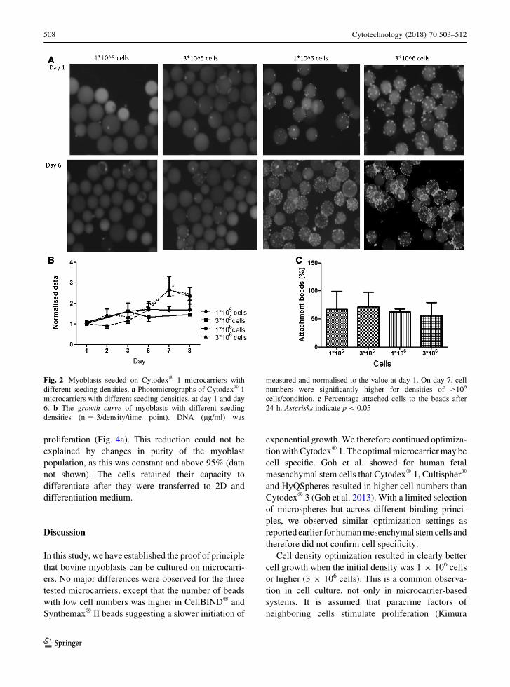
To determine the optimal seeding density, we made
a concentration curve with 105, 3 9 105, 106 and
3 9 106 cells/ml on Cytodex� 1 microcarriers in a
spinner flask. Clearly, seeding densities below 106
resulted in stationary cell numbers during the 6-day
measurement (Fig. 2a, b). In contrast, the higher
seeding densities showed proper exponential growth.
At day 6, the highest seeding density of 3 9 106 cells
displayed aggregation of cells/microcarriers. Cell
attachment to the Cytodex� microcarriers was inde-
pendent of seeding density (Fig. 2c).
Bead-to-bead transfer
Transfer of cells from populated beads to empty beads
would significantly reduce handling during expansion
of the culture by just adding new beads to increase
surface (Ferrari et al. 2014). Agitated stirring brings
fresh beads in close contact with near confluent beads
thus allowing cell transfer (Wang and Ouyang 1999).
Bead-to-bead transfer was tested using Synthe-
max� II microcarriers as these can be stained with
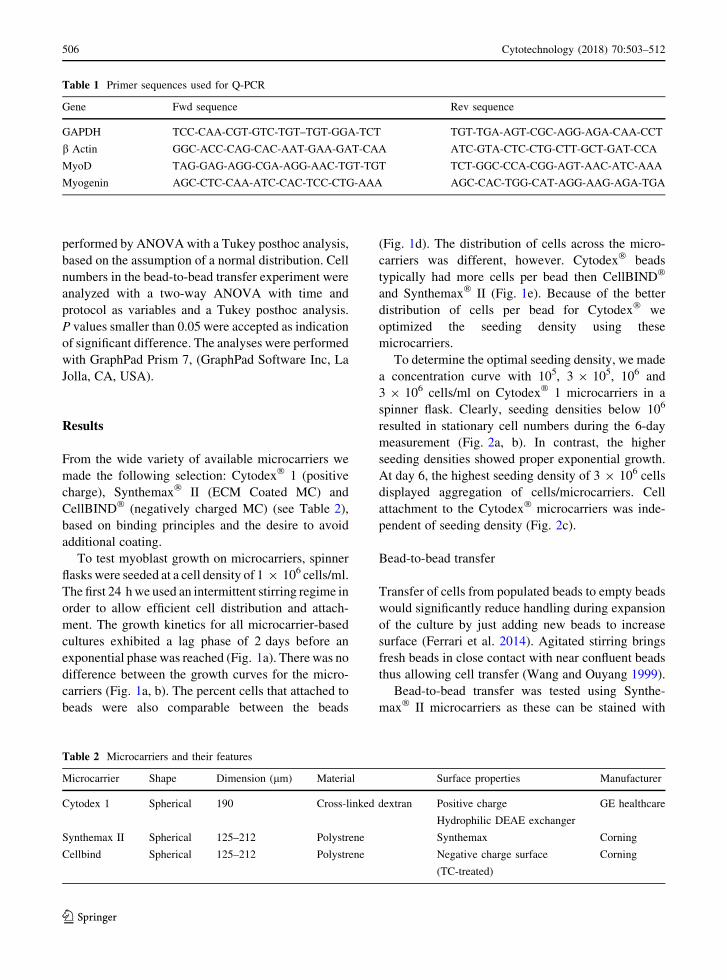
Table 1 Primer sequences used for Q-PCR
Gene Fwd sequence Rev sequence
GAPDH TCC-CAA-CGT-GTC-TGT–TGT-GGA-TCT TGT-TGA-AGT-CGC-AGG-AGA-CAA-CCT
b Actin GGC-ACC-CAG-CAC-AAT-GAA-GAT-CAA ATC-GTA-CTC-CTG-CTT-GCT-GAT-CCA
MyoD TAG-GAG-AGG-CGA-AGG-AAC-TGT-TGT TCT-GGC-CCA-CGG-AGT-AAC-ATC-AAA
Myogenin AGC-CTC-CAA-ATC-CAC-TCC-CTG-AAA AGC-CAC-TGG-CAT-AGG-AAG-AGA-TGA
Table 2 Microcarriers and their features
Microcarrier Shape Dimension (lm) Material Surface properties Manufacturer
Cytodex 1 Spherical 190 Cross-linked dextran Positive charge
Hydrophilic DEAE exchanger
GE healthcare
Synthemax II Spherical 125–212 Polystrene Synthemax Corning
Cellbind Spherical 125–212 Polystrene Negative charge surface
(TC-treated)
Corning
506 Cytotechnology (2018) 70:503–512
123

rhodamine. To an existing culture in stirrer flasks, new
beads (5 ml 100 mg/ml beads) were added on day 3
and 7. Each bead addition was followed by 24 h of
intermittent agitation. Growth was boosted by each
bead addition (Fig. 3a) and transfer to newly added,
rhodamine labelled beads was apparent (Fig. 3b). The
addition of empty beads and subsequent bead-to-bead
transfer also reduced the number of aggregates in
comparison to cultures with no extra added beads
(Fig. 3c, day 8).
Myoblast differentiation
In 2D cultures reaching confluence, myoblasts show a
tendency to differentiate into myotubes. As a measure
of differentiation and capacity to differentiate after
microcarrier-based cell expansion, expression of early
differentiation markers MyoD and Myogenin was
studied (Fig. 4a) as well as the appearance of
myotubes (Fig. 4b). As expected, the expression of
MyoD and Myogenin decreased over time during
Fig. 1 Myoblasts seeded on Cytodex� 1, Synthemax� II and
CellBIND� microcarriers using growth medium; seeded at a
density of 1 9 106 cells/ml. a The growth curve of cells for thethree microcarriers. DNA (lg/ml) was measured and normalised
to the value at day 1 (n = 3 for each day). b Photomicrographs
of the cell-laden microcarriers at day 1 and day 6. The cells were
stained with Hoechst and appear as fluorescent dots (white in
B&W). c 2D proliferation of myoblasts. dAttachment of cells to
the microcarriers at 24 h expressed as percentage of the total
amount of cells added (n = 3 for each type of microcarrier).
e The amount of cells per bead after 24 h (n = 3)
Cytotechnology (2018) 70:503–512 507
123
proliferation (Fig. 4a). This reduction could not be
explained by changes in purity of the myoblast
population, as this was constant and above 95% (data
not shown). The cells retained their capacity to
differentiate after they were transferred to 2D and
differentiation medium.
Discussion
In this study, we have established the proof of principle
that bovine myoblasts can be cultured on microcarri-
ers. No major differences were observed for the three
tested microcarriers, except that the number of beads
with low cell numbers was higher in CellBIND� and
Synthemax� II beads suggesting a slower initiation of
exponential growth.We therefore continued optimiza-
tionwithCytodex� 1. The optimalmicrocarriermay be
cell specific. Goh et al. showed for human fetal
mesenchymal stem cells that Cytodex� 1, Cultispher�
and HyQSpheres resulted in higher cell numbers than
Cytodex� 3 (Goh et al. 2013). With a limited selection
of microspheres but across different binding princi-
ples, we observed similar optimization settings as
reported earlier for humanmesenchymal stem cells and
therefore did not confirm cell specificity.
Cell density optimization resulted in clearly better
cell growth when the initial density was 1 9 106 cells
or higher (3 9 106 cells). This is a common observa-
tion in cell culture, not only in microcarrier-based
systems. It is assumed that paracrine factors of
neighboring cells stimulate proliferation (Kimura
Fig. 2 Myoblasts seeded on Cytodex� 1 microcarriers with
different seeding densities. a Photomicrographs of Cytodex� 1
microcarriers with different seeding densities, at day 1 and day
6. b The growth curve of myoblasts with different seeding
densities (n = 3/density/time point). DNA (lg/ml) was
measured and normalised to the value at day 1. On day 7, cell
numbers were significantly higher for densities of C106
cells/condition. c Percentage attached cells to the beads after
24 h. Asterisks indicate p\ 0.05
508 Cytotechnology (2018) 70:503–512
123

et al. 1991). The optimal number of cells was 106 cells/
spinner flask (5500 cells/cm2 of bead surface). Higher
densities resulted in early aggregate formation of cells
and microcarriers. Aggregate formation depends on
cell type i.e. the ability of cells to grow in multiple
layers, and on the dynamic conditions of the culture
(Muhitch et al. 2000). Early observations suggest that
cell-microcarrier aggregate formation negatively
affects growth of human mesenchymal stem cells
(Chen et al. 2013; Caruso et al. 2014; Goh et al. 2013)
and we assume that myoblasts behave similarly.
Myoblasts seem to quickly participate in aggregate
formation of microcarriers, suggesting that a tight
schedule of adding new beads or starting a new
passage will need to be followed for optimal cell
growth.
The optimal initial cell density of 5500 cell/cm2 is
comparable to earlier reports on mesenchymal stem
cells in a microcarrier-based culture system (Hewitt
et al. 2011; Rafiq et al. 2013). In 2D cultures, the
optimal seeding density is typically lower (Coles et al.
2015), which is most likely illustrative of higher
attachment to the static flat surfaces than to the
spherical surface of highly dynamic microcarriers.
We provided direct evidence that bovine myoblasts
display bead-to-bead transfer. Most experience on
bead-to-bead transfer has been accumulated with
mesenchymal stem cells (Ferrari et al. 2014).
Fig. 3 Bead-to-bead transfer of Myoblasts seeded on Synthe-
max� II microcarriers. Synthemax� II microcarriers were
chosen for these experiments because they can be labeled with
rhodamine. a The growth curve of the cells expressed as DNA
concentration (lg/ml) where empty beads were added at day 3 or
day 7. b Bead-to-bead transfer of myoblasts onto rhodamine
(red) labeled Synthemax� II beads. c Photomicrographs of the
cells on beads at day 3, 7 and 8. Asterisk indicate significant
difference for the growth when beads were added at day 7
compared to no extra beads added. (Color figure online)
Cytotechnology (2018) 70:503–512 509
123

Myoblasts, similar to mesenchymal cells, reach con-
fluence at day 3 or 4 after seeding, which is the optimal
time to add new microcarriers. The two proposed
mechanisms for bead-to-bead transfer are the
exchange of cells during bead-to-bead contact or the
pick-up of floating cells that have detached from
confluent beads and are adopted by newly added, still
barren, beads (Ferrari et al. 2014). The surprisingly
sudden and profound increase in cell number that we
observed after adding new beads can hardly be
attributed to cell proliferation but could be compatible
with the pickup of floating cells by the new beads.
These detached cells would otherwise rapidly undergo
anoikis as a result of inadequate cell–matrix interac-
tion (Frisch and Screaton 2001), thus explaining the
failure to display exponential growth in the absence of
newly added beads.
Subculturing of cells is one of the critical risks to be
considered in the scaling up of microcarrier-based cell
culture as any manipulation can lead to contamination.
Complete detachment of cells by way of trypsinization
is challenging and incomplete detachment results in
cell loss and production inefficiency (Caruso et al.
2014). Although bead-to-bead transfer may eliminate
the need for subculturing, cells eventually need to be
harvested while retaining their viability and
Fig. 4 Myoblasts seeded on Cytodex� 1 microcarriers, with
new beads added on day 3 or day 7. aMyoD andMyogenin RNA
expression by RT-QPCR. b The cells are trypsinized and then
seeded on a plate in differentiation medium for 4 days to check
myotube formation. Note the elongated structures that represent
merged myoblasts. Scale bar = 1000 lm. ‘‘prol. Ctrl’’ is a 2D
culture condition optimized for cell proliferation and ‘‘diff Ctrl’’
is optimized for myocyte differentiation. The arrows point to the
myotubes
510 Cytotechnology (2018) 70:503–512
123

propensity to differentiate. We have not quantitatively
analyzed harvesting efficiency but we show here that
the harvested cells are viable and capable of differen-
tiation into myotubes.
As experiments were performed in spinner flasks
with limited control over temperature, oxygen supply
and nutrient availability and usage, the culture system
may not be fully optimized for a large scale stirred
tank bioreactor (Nienow 2006). Future studies in fully
controlled stirred tank bioreactors need to result in
further optimization.
The overall conclusion is that it is possible to
culture bovine myoblasts on MC (Cytodex� 1 or
Synthemax� MC) and that they exhibit bead-to-bead
transfer. Bovine myoblasts seem to behave very
similar to human mesenchymal stem cells. Thus, our
results provide valuable data to further develop and
scale up the production of bovine myoblasts as a
prerequisite for efficient and cost-effective develop-
ment of cultured meat. The similarity with microcar-
rier based culture of human mesenchymal stem cells,
suggests that these results are also applicable to culture
of anchorage dependent cells in medical tissue engi-
neering and cell therapy.
Open Access This article is distributed under the terms of the
Creative Commons Attribution 4.0 International License (http://
creativecommons.org/licenses/by/4.0/), which permits unre-
stricted use, distribution, and reproduction in any medium,
provided you give appropriate credit to the original
author(s) and the source, provide a link to the Creative Com-
mons license, and indicate if changes were made.
References
Caruso SR, Orellana MD, Mizukami A, Fernandes TR, Fontes
AM, Suazo CA, Oliveira VC, Covas DT, Swiech K (2014)
Growth and functional harvesting of human mesenchymal
stromal cells cultured on a microcarrier-based system.
Biotechnol Prog 30:889–895
Chen AK, Reuveny S, Oh SK (2013) Application of human
mesenchymal and pluripotent stem cell microcarrier cul-
tures in cellular therapy: achievements and future direc-
tion. Biotechnol Adv 31:1032–1046
Coles CA, Wadeson J, Leyton CP, Siddell JP, Greenwood PL,
White JD, McDonagh MB (2015) Proliferation rates of
bovine primary muscle cells relate to liveweight and car-
case weight in cattle. PLoS ONE 10:e0124468
Croughan MS, Wang DI (1989) Growth and death in overagi-
tated microcarrier cell cultures. Biotechnol Bioeng
33:731–744
Eibes G, dos Santos F, Andrade PZ, Boura JS, Abecasis MM, da
Silva CL, Cabral JM (2010) Maximizing the ex vivo
expansion of human mesenchymal stem cells using a
microcarrier-based stirred culture system. J Biotechnol
146:194–197
Ferrari C, Olmos E, Balandras F, Tran N, Chevalot I, Guedon E,
Marc A (2014) Investigation of growth conditions for the
expansion of porcine mesenchymal stem cells on micro-
carriers in stirred cultures. Appl Biochem Biotechnol
172:1004–1017
Frauenschuh S, Reichmann E, Ibold Y, Goetz PM, Sittinger M,
Ringe J (2007) A microcarrier-based cultivation system for
expansion of primary mesenchymal stem cells. Biotechnol
Prog 23:187–193
Frisch SM, Screaton RA (2001) Anoikis mechanisms. Curr Opin
Cell Biol 13:555–562
Goh TKP, Zhang ZY, ChenAKL, Reuveny S, ChoolaniM, Chan
JKY, Oh SKW (2013) Microcarrier culture for efficient
expansion and osteogenic differentiation of human fetal
mesenchymal stem cells. Biores Open Access 2:84–97
Gupta P, Ismadi MZ, Verma PJ, Fouras A, Jadhav S, Bellare J,
Hourigan K (2016) Optimization of agitation speed in
spinner flask for microcarrier structural integrity and
expansion of induced pluripotent stem cells. Cytotechnol-
ogy 68:45–59
Hewitt CJ, Lee K, Nienow AW, Thomas RJ, Smith M,
Thomas CR (2011) Expansion of human mesenchymal
stem cells on microcarriers. Biotechnol Lett
33:2325–2335
Ikonomou L, Bastin G, Schneider YJ, Agathos SN (2004) Effect
of partial medium replacement on cell growth and protein
production for the high-five trade mark insect cell line.
Cytotechnology 44:67–76
Kimura A, Katoh O, Hyodo H, Kusumi S, Kuramoto A (1991)
Autocrine and/or paracrine mechanism operate during the
growth of human bone marrow fibroblasts. Br J Haematol
78:469–473
Macfelda K, Kapeller B, Wilbacher I, Losert UM (2007)
Behavior of cardiomyocytes and skeletal muscle cells on
different extracellular matrix components–relevance for
cardiac tissue engineering. Artif Organs 31:4–12
Muhitch JW, O’Connor KC, Blake DA, Lacks DJ, Rosenzweig
N, Spaulding GF (2000) Characterization of aggregation
and protein expression of bovine corneal endothelial cells
as microcarrier cultures in a rotating-wall vessel.
Cytotechnology 32:253–263
Nienow AW (2006) Reactor engineering in large scale animal
cell culture. Cytotechnology 50:9–33
Nienow AW, Sieck, JB, Cordes T, Budach WE, Rhiel, MH,
Suemeghy Z, Leist C, Villiger TK, Morbidelli M, Soos M
(2014). Development of a scale-down model of hydrody-
namic stress to study the performance of an industrial CHO
cell line under simulated production scale bioreactor con-
ditions [2013. Journal of Biotechnology, 164, 41–49].
J Biotechnol 171:82–84
Post MJ, Van Der Weele C (2014) Principles of tissue engi-
neering for food. In: Lanza R, Langer R, Vacanti JP (eds)
Principles of tissue engineering, 4th edn. Elsevier,
Amsterdam
Rafiq QA, Brosnan KM, Coopman K, Nienow AW, Hewitt CJ
(2013) Culture of human mesenchymal stem cells on
microcarriers in a 5 l stirred-tank bioreactor. Biotechnol
Lett 35:1233–1245
Cytotechnology (2018) 70:503–512 511
123

Schop D, Janssen FW, Borgart E, de Bruijn JD, Van Dijkhuizen-
Radersma R (2008) Expansion of mesenchymal stem cells
using a microcarrier-based cultivation system: growth and
metabolism. J Tissue Eng Regen Med 2:126–135
Stathopoulos NA, Hellums JD (1985) Shear stress effects on
human embryonic kidney cells in Vitro. Biotechnol Bioeng
27:1021–1026
Stern MM, Myers RL, Hammam N, Stern KA, Eberli D,
Kritchevsky SB, Soker S, van Dyke M (2009) The influ-
ence of extracellular matrix derived from skeletal muscle
tissue on the proliferation and differentiation of myogenic
progenitor cells ex vivo. Biomaterials 30:2393–2399
Van Der Weele C, Tramper J (2014) Cultured meat: every vil-
lage its own factory? Trends Biotechnol 32:294–296
Wang Y, Ouyang F (1999) Bead-to-bead transfer of Vero cells
in microcarrier culture. Cytotechnology 31:221–224
WuQF,WuCT, Dong B,Wang LS (2003) Cultivation of human
mesenchymal stem cells on macroporous CultiSpher G
microcarriers. Zhongguo Shi Yan Xue Ye Xue Za Zhi
11:15–21
512 Cytotechnology (2018) 70:503–512
123