Boundary detection in Medical Images using Edge Field Vector based on Law’s Texture and Canny...
6
I nter national J our nal of E ngin e e r in g Trends and Te chnology (I JE TT ) - Volume4I ss ue5- M ay 2013 ISSN: 2231-5381 http://www.ijettjournal.org Page 1912 Boundary detection in Medical Images using Edge Field Vector based on Law’s Texture and Canny Method Swetha.M 1 Jyohsna.C 2 Department of E& C, KLS VDRIT , Haliyal Karnataka, india Abstract − detecting the correct boundary in noisy images is a difficult task. Images are used in many fields,including surveillance, medical diagnostics and non-destrucive testing. Edge detection and boundary detection plays a fundamental role in image analysis. Boundaries are mainly used to detect the outline or shape of the object.image segmentation is used to locate objects and boundaries in images and it assigns a lable in every pixel in an image such that pixels with the same level share have certain virtual characteristics. The proposed edge detection technique for detecting the boundaries of object using the information from intensity gradient using the vector model and texture gradient using the edge map modle.the results show that the technique performs very well and yields better performance than the classical contour models. The proposed method is robust and applicable on various kind of noisy images without prior knowledge of noise properties. Keywords− Boundary extraction, vector field model, edge mapping model, edge following technique, boundary detection. I. INTRODUCTION Boundary detection is mainly used to detect the outline or shape of the object, so we can easily identify objects based upon the outline or shape. Segmentation is the process in which an image is divided into its constitutent objects or parts. The main goal of segmentation is to simplify and/or change of an image representation into something is an initial step before performing high-level tasks such as object recognition and understanding. Image segmentation is typically used to locate objects and boundaries in images. In medical imaging,segmentation is important for feature extraction, image measurements, and image display. In some applications it may be useful to extract boundaries of objects of interest from ultrasound images[1],[2] , microsco pic images[3]-[5]. In recent years,there have been several new methods to solve the problem of boundary detection, e.g., active contour model(ACM), geodestic active contour(GAC) model, active contours without edges(ACWE), gradient vector flow(GVF) snake model, etc. the snake models have become popular especially in boundary detection where the problem is more challenging due to the poor quality of the images. To overcome from this problem, we proposed a new technique for boundary detection for ill-defined edges in noisy images using a novel edge following. The proposed edge following technique is based on the vector image model and the edge map. The vector image model provides a more complete description of an image by considering both directions and magnitudes of image edges. The proposed edge vector field is generated by averaging magnitudes and directions in the vector image. The edge map is derived from Law’s texture feature and the Canny edge detection. The vector image model and the edge map are applied to select the best edges. II. PROPOSED SYSTEM In proposed boundary detection algorithm is used to detect the boundary of object in an image. Boundary extraction algorithm consists of following three phases. 1. Edge vector gradient 2. Edge mapping model 3. Edge detection technique
-
Upload
seventhsensegroup -
Category
Documents
-
view
216 -
download
0
Transcript of Boundary detection in Medical Images using Edge Field Vector based on Law’s Texture and Canny...

7/27/2019 Boundary detection in Medical Images using Edge Field Vector based on Law’s Texture and Canny Method
http://slidepdf.com/reader/full/boundary-detection-in-medical-images-using-edge-field-vector-based-on-laws 1/6
I nternational Journal of Engineer ing Trends and Technology (I JETT) - Volume4I ssue5- May 2013
ISSN: 2231-5381 http://www.ijettjournal.org Page 1912
Boundary detection in Medical Images using Edge
Field Vector based on Law’s Texture and Canny
MethodSwetha.M
1Jyohsna.C
2
Department of E&C, KLS VDRIT, Haliyal Karnataka, india
Abstract− detecting the correct boundary in noisy images is a
difficult task. Images are used in many fields,including
surveillance, medical diagnostics and non-destrucive testing.
Edge detection and boundary detection plays a fundamental role
in image analysis. Boundaries are mainly used to detect the
outline or shape of the object.image segmentation is used to
locate objects and boundaries in images and it assigns a lable in
every pixel in an image such that pixels with the same level share
have certain virtual characteristics. The proposed edge detection
technique for detecting the boundaries of object using the
information from intensity gradient using the vector model and
texture gradient using the edge map modle.the results show that
the technique performs very well and yields better performance
than the classical contour models. The proposed method is robust
and applicable on various kind of noisy images without prior
knowledge of noise properties.
Keywords−
Boundary extraction, vector field model, edgemapping model, edge following technique, boundary detection.
I. INTRODUCTION
Boundary detection is mainly used to detect the
outline or shape of the object, so we can easily
identify objects based upon the outline or shape.
Segmentation is the process in which an image is
divided into its constitutent objects or parts. The
main goal of segmentation is to simplify and/or
change of an image representation into something is
an initial step before performing high-level tasks
such as object recognition and understanding. Imagesegmentation is typically used to locate objects and
boundaries in images. In medical
imaging,segmentation is important for feature
extraction, image measurements, and image display.
In some applications it may be useful to extract
boundaries of objects of interest from ultrasound
images[1],[2], microscopic images[3]-[5].
In recent years,there have been several new methods
to solve the problem of boundary detection, e.g.,
active contour model(ACM), geodestic active
contour(GAC) model, active contours without
edges(ACWE), gradient vector flow(GVF) snake
model, etc. the snake models have become popular
especially in boundary detection where the problemis more challenging due to the poor quality of the
images.
To overcome from this problem, we proposed a new
technique for boundary detection for ill-defined
edges in noisy images using a novel edge following.
The proposed edge following technique is based on
the vector image model and the edge map. The vector
image model provides a more complete description
of an image by considering both directions and
magnitudes of image edges. The proposed edge
vector field is generated by averaging magnitudes
and directions in the vector image. The edge map is
derived from Law’s texture feature and the Canny
edge detection. The vector image model and the edge
map are applied to select the best edges.
II. PROPOSED SYSTEM
In proposed boundary detection algorithm is used to
detect the boundary of object in an image. Boundary
extraction algorithm consists of following three phases.
1. Edge vector gradient
2. Edge mapping model
3. Edge detection technique

7/27/2019 Boundary detection in Medical Images using Edge Field Vector based on Law’s Texture and Canny Method
http://slidepdf.com/reader/full/boundary-detection-in-medical-images-using-edge-field-vector-based-on-laws 2/6
I nternational Journal of Engineer ing Trends and Technology (I JETT) - Volume4I ssue5- May 2013
ISSN: 2231-5381 http://www.ijettjournal.org Page 1913
III. BLOCK DIAGRAM
A. Average edge vector field model
We exploit the edge vector field to devise a new
boundary extraction algorithm[29]. Give an
image f(x,y), the edge vector field is calculated
according to the following equations:
(i, j) = (Mx(i, j) + My(i, j) )……....(1)
(i, j)
– …….(2)
K= ⏟
( ….(3)
Fig.1. (a) original unclear image.(b) result from the edge vector field and
zoomed-in image.(c) result from the proposed average edge vector field and
zoomed-in image.
Each component is the convolution between the image and the
corresponding difference mask, i.e.,
Mx (i, j) = −Gy × f ( x, y) ≈
...............(4)
My (i, j) = Gx × f ( x, y) ≈ −
…………(5)
Where Gx and Gy are the difference masks of the Gaussian
weighted image moment vector operator in the x and ydirections, respectively,[29]
Gx ( x, y) =
√ ( )exp
…………(6)
Gy ( x, y) =
√ ( )
……….(7)
Edge vectors of an image indicate the magnitudes and
directions of edges which form a vector stream flowing
around an object. However, in an unclear image, the vectors
derived from the edge vector field may distribute randomly in
magnitude and direction. Therefore, we extend the capability
of the previous edge vector field by applying a local averaging
operation where the value of each vector is replaced by the
average of all the values in the local neighborhood, i.e.,
M (i, j) =
∑ ….(8)
D(i, j) = ∑ …………….(9)
Where Mr is the total number of pixels in the neighborhood N.
We apply a 3×3 window as the neighborhood N throughout
our research.
B. Edge Map
Edge map is edges of objects in an image derived
from Law’s texture and Canny edge detection.
1) Law’s Texture: The texture feature images of Law’s
texture are computed by convolving an input image
with each of the masks. Given a column vector L=(1,
4, 6, 4, 1)T, the 2-D mask l(i, j) used for texture
discrimination in this research is generated by L×LT
.
The output image is obtained by convolving input
image with texture mask.
2) Canny Edge detection: The canny approach to edge
detection is optimal for step edges corrupted by
white Gaussian noise. This edge detector is assumed
to be the output of a filter that reduces the noise and
locates the edges. There are four steps to detect
edges, the first step is to convolve the output image
obtained from the aforementioned Law’s texture t(i,
j) with a Gaussian filter. The second step is to
calculate the magnitude and direction of the gradient.
The third step is nonmaximal suppression to identify
edges. The last step is the thresholding algorithm to
detect and link edges. The double threshold
algorithm is used to detect and link edges.
Edge map shows some important information of edge. This
idea is exploited for extracting objects boundaries in unclear
images. Examples of the edge maps are shown in fig.2.
Fig.2.(a) Synthetic noisy image.(b) Left ventricle in the MR image.
(c) Prostate ultrasound image. (d)-(f) Corresponding edge maps derived from
Law’s texture and canny edge detection.
Input
image
Average
edge vector
field
Boundary
detected
Edge map
Edge
following
technique
Initial
position

7/27/2019 Boundary detection in Medical Images using Edge Field Vector based on Law’s Texture and Canny Method
http://slidepdf.com/reader/full/boundary-detection-in-medical-images-using-edge-field-vector-based-on-laws 3/6
I nternational Journal of Engineer ing Trends and Technology (I JETT) - Volume4I ssue5- May 2013
ISSN: 2231-5381 http://www.ijettjournal.org Page 1914
C. Edge Following Technique
The edge following technique is performed to find
the boundary of an object. Most edge following
algorithms take into account the magnitude as
primary information for edge following. However,
the edge magnitude information is not efficient
enough for searching the correct boundary of objects
in noisy images because it can be very weak in some
contour areas.
Fig.3. Edge masks used for detecting of image
edges(normal direction constraint)
The magnitude and direction of the average edge vector field
give information of the boundary which flows around an
object. In addition, the edge map gives information of edge
which may be a part of object boundary. Hence, both average
edge vector filed and edge map are exploited in the decision
of the edge following technique. At the position (i, j) of animage, the successive positions of the edges are then
calculated by a 3×3 matrix.
Lij(r, c) = αMij(r, c)+ βDij(r, c)+εEij(r,c)
0 ≤ r ≤ 2, 0≤ c ≤ 2…….(10)
Where α, β and ε are the weight parameters that control the
edge to flow around an object. The larger value of an element
in Lij indicates the stronger edge in the corresponding
direction. The 3×3 matrices Mij, Dij and Eij are calculated as
follows:
Mij (r, c) =
…….(11)
Dij (r, c) = 1-|| ……(12)
Eij (r, c) = E (i + r − 1 , j + c − 1) , 0 ≤ r ≤ 2 ,
0 ≤ c ≤ 2….. (13)
Where M(i, j) and D(i, j) are the proposed average magnitude
and direction of edge vector fields as shown in (8) and (9).
E(i, j) is the edge map from Law’s texture and canny edge
detection. It should be noted that the value of each element in
the matrices M ij , Dij and E ij are
Ranged between 0 and 1. The direction can be calculated by
= ∑ ∑ ….(14)
Where k=1, 2,…….,8 denoted the eight directions as indicated
by the arrows at the center of the masks shown in fig.3.
The edge following is started from the initial position to end
position.
D. Initial positionIn this section, we present a technique for
determining a good initial position of edge following
that can be used for the boundary detection. The
initial position problem is very important in the
classical contour models. Snake models can converge
to a wrong boundary if the initial position is not close
enough to the desired boundary. Finding the initial
position of the classical contour models is still
difficult and time consuming[32], [33]. In this
proposed technique,the initial position of edge
following is determined by the following steps. Thefirst step is to calculate the average magnitude [ M(i,
j)] using (8). the second step is to calculate the
density of edge length for each pixel from an edge
map. An edge map [ E(i, j)], as a binary image, is
obtained by Law’s texture and Canny edge detection.
The idea of using density is to obtain measurement of
the edge length. The density of edge length [ L(i, j)] in
each pixel can be calculated from
L(i, j) =
.........................(15)
Where C(i,j) is the number of connected pixels at each
position of pixel. An example of counting the number of
connected pixels is shown in fig.4(a) and (b). The density of
edge length from the example is shown in fig.4(c). The third
step is to calculate the initial position map P(i, j) from
summation of average magnitude and density of edge length,
i.e.,
P (i, j) = …………….(16)
The last step is the thresholding of the initial position map.
We have to threshold the map in order to detect the initial
position of edge following. If P(i,j)>T max , then P(i, j) is the
initial position of egde following. We obtained the initial
position by setting Tmax to 95% of the maximum value. The
initial positions from our method are positions that are close to
the edges of interested areas.

7/27/2019 Boundary detection in Medical Images using Edge Field Vector based on Law’s Texture and Canny Method
http://slidepdf.com/reader/full/boundary-detection-in-medical-images-using-edge-field-vector-based-on-laws 4/6
I nternational Journal of Engineer ing Trends and Technology (I JETT) - Volume4I ssue5- May 2013
ISSN: 2231-5381 http://www.ijettjournal.org Page 1915
Fig.5. (a)Aorta in cardiovascular MR image. (b) Averaged magnitude
[M(i, j)]. (c) Density of length edge [L(i, j)]. (d) Initial position map [P(i, j)]
and initial position of edge following derived by thresholding Tmax=0.95.
An example of the initial position derived from our method is
shown in fig.5 to illustrate that method can be applied to ill-
defined edges in medical images.
We can see that any one of the white circle points in the initial
position map is a good candidate to be the initial position for
our edge following technique. However, the maximum valueof the white circle points is used in this research. After
determining the suitable initial position, the proposed
technique will follow edges along the object boundary until
the closed loop contour is achieved. This causes a limitation
of the technique, i.e., the boundary must be closed loop.
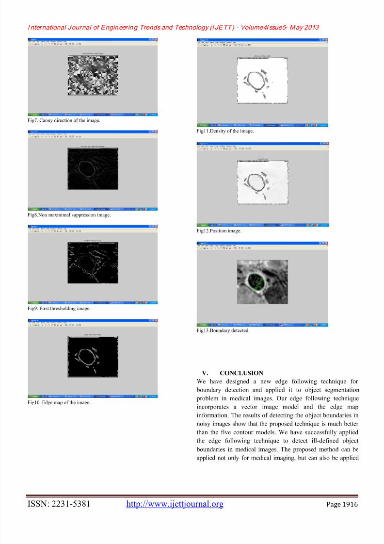
IV. RESULTS
Fig1. Original image.
Fig2.Preprocessed image.
Fig3. Magnitude of the image.
Fig4. Direction of the image.
Fig5.Law’s texture output image.
Fig6.Canny magnitude of the image.
7/27/2019 Boundary detection in Medical Images using Edge Field Vector based on Law’s Texture and Canny Method
http://slidepdf.com/reader/full/boundary-detection-in-medical-images-using-edge-field-vector-based-on-laws 5/6
I nternational Journal of Engineer ing Trends and Technology (I JETT) - Volume4I ssue5- May 2013
ISSN: 2231-5381 http://www.ijettjournal.org Page 1916
Fig7. Canny direction of the image.
Fig8.Non maxmimal suppression image.
Fig9. First thresholding image.
Fig10. Edge map of the image.
Fig11.Density of the image.
Fig12.Position image.
Fig13.Boundary detected.
V. CONCLUSION
We have designed a new edge following technique for boundary detection and applied it to object segmentation
problem in medical images. Our edge following technique
incorporates a vector image model and the edge map
information. The results of detecting the object boundaries in
noisy images show that the proposed technique is much better
than the five contour models. We have successfully applied
the edge following technique to detect ill-defined object
boundaries in medical images. The proposed method can be
applied not only for medical imaging, but can also be applied
7/27/2019 Boundary detection in Medical Images using Edge Field Vector based on Law’s Texture and Canny Method
http://slidepdf.com/reader/full/boundary-detection-in-medical-images-using-edge-field-vector-based-on-laws 6/6
I nternational Journal of Engineer ing Trends and Technology (I JETT) - Volume4I ssue5- May 2013
ISSN: 2231-5381 http://www.ijettjournal.org Page 1917
to any image processing problems in which ill-defined edge
detection is encountered.
ACKNOWLEDGEMENT
We sincerely thank our college, KLS VDRIT college for
humble facilities and necessary infrastructure made available
during the course of our work. We wish to express our thanksand sincere gratitude to our Principal, Head of the Department
and guide for their guidance to complete this work
successfully and enthusiastic encouragement.
REFERENCES[1] J. Guerrero, S.E. Salcudean, J.A.McEwen, B.A. Masri, and S. Nicolaou,
“Real-time vessel segmentation and tracking for ultrasound imaging
applications,” IEEE Trans, Med.Imag., vol.26,no.8,pp.1079-1090, Aug.2007.
[2] F.Destrempes, J. Meunier, M.-F. Giroux, G.Soulez, and G. Cloutier,
“segmentation in ultrasounic B-mode images of Nakagami distributions and
stochastic optimization,” IEEE Trans. Med.Imag., vol.28, no. 2, pp.215-229,
Feb.2009.
[3] N. Theera-Umpon and P.D. Gader, “System level training of neural
networks for counting white blood cells,” IEEE Trans. Syst., Man, Cybern. C,
App. Rev., vol.32,no.1,pp.48-53,Feb.2002.
[4] N. Threera-Umpon, “White blood cell segmentation and classification in
microscopic bone marrow images ,” Lecture Notes Comput. Sci., vol. 3614,
pp. 787-796, 2005.
[5] N. Theera-Umpon and S. Dhompongsa, “Morphological granulometric
features of nucleus in automatic bone marrow white blood cell classification,”
IEEE Trans. Inf. Technol. Biomed., vol.11, no.3, pp. 353-359, May 2007.
[6] J. Carballido-Gamio, S. J. Belongie, and S. Majumdar, “Normalized cuts
in 3-D for spinal MRI segmentation,” IEEE Trans. Med. Imag., vol.23, no.1,
pp.36-44, jan. 2004.
[7] H. Greenspan, A. Ruf, and J. Goldbeger, “Constrainted Gaussian mixture
model framework for automatic segmentation of MR brain images.” IEEE
Trans. Med. Imag., vol. 25, no. 9, pp. 1233-1245, Sep. 2006.
[8] J. – D. Lee, H.-R. su, P. E. Cheng, M. Liou, J. Aston, A. C. Tsai, and C.-Y.
Chen, “MR image segmentation using a power transformation approach .”
IEEE Trans, Med. Image., vol . 28, no. 6, pp. 894-905. Jun, 2009.