Botulinum Neurotoxins: Biology, Pharmacology, and...
36
1521-0081/69/2/200–235$25.00 https://doi.org/10.1124/pr.116.012658 PHARMACOLOGICAL REVIEWS Pharmacol Rev 69:200–235, April 2017 Copyright © 2017 by The Author(s) This is an open access article distributed under the CC BY-NC Attribution 4.0 International license. ASSOCIATE EDITOR: JEFFREY M. WITKIN Botulinum Neurotoxins: Biology, Pharmacology, and Toxicology Marco Pirazzini, Ornella Rossetto, Roberto Eleopra, and Cesare Montecucco Department of Biomedical Sciences, University of Padova, Italy (M.P., O.R., C.M.); Neurologic Department, University-Hospital S. Maria della Misericordia, Udine, Italy (R.E.); and Consiglio Nazionale delle Ricerche, Institute of Neuroscience, University of Padova, Italy (C.M.) Abstract..................................................................................... 201 I. Introduction ................................................................................. 201 A. Genetics and Structure of Botulinum Neurotoxins and Their Progenitor Toxin Complexes............................................................................... 202 B. Molecular Architecture of Botulinum Neurotoxins ........................................ 203 C. Metalloproteolytic Activity ............................................................... 204 II. Biology ...................................................................................... 204 A. Molecular Mechanism of Nerve Terminal Paralysis ....................................... 204 1. Binding and Specificity. .............................................................. 204 2. Internalization into Nerve Terminals.................................................. 211 a. Long distance effects of botulinum neurotoxins..................................... 211 3. Membrane Translocation.............................................................. 212 4. Interchain Disulfide Reduction........................................................ 213 5. SNARE Protein Cleavage............................................................. 214 B. Duration of the Neuroparalysis Induced by Botulinum Neurotoxins ....................... 215 1. Reversibility of the Neuroparalysis Induced by Botulinum Neurotoxins. ............... 215 2. Reversibility of Botulinum Neurotoxin Action in Vitro and in Vivo..................... 216 III. Pharmacology ............................................................................... 216 A. Introduction ............................................................................. 216 B. Present Botulinum Neurotoxin Formulations ............................................. 217 C. Immunogenicity of Botulinum Neurotoxin Formulations .................................. 219 D. Clinical Applications of Botulinum Neurotoxins .......................................... 219 1. Dystonias............................................................................. 220 2. Spasticity............................................................................. 221 3. Autonomic Disorders.................................................................. 221 4. Urologic Pathologic Conditions........................................................ 222 5. Pain.................................................................................. 222 a. Neuropathic pain.................................................................. 223 b. Primary headaches................................................................ 223 6. Other Applications.................................................................... 224 a. Gastroenterology and proctologic disorders......................................... 224 b. Depression........................................................................ 224 7. Cosmetic Uses........................................................................ 225 E. Adverse Effects .......................................................................... 225 F. Contraindications and Drug Interactions ................................................. 225 G. Botulinum Neurotoxin in Pregnancy ..................................................... 225 IV. Toxicity of Botulinum Neurotoxins ........................................................... 226 The authors’ research was supported by Fondazione Cariparo, the Axonomics project of Provincia di Trento, the Ministery of Defence, the University of Padova and the University of Udine. Address correspondence to: Cesare Montecucco, Department of Biomedical Sciences, University of Padova, via Ugo Bassi 58/B, 35131 Padova, Italy. E-mail: [email protected] https://doi.org/10.1124/pr.116.012658. 200 by guest on May 6, 2018 Downloaded from
Transcript of Botulinum Neurotoxins: Biology, Pharmacology, and...

1521-0081/69/2/200–235$25.00 https://doi.org/10.1124/pr.116.012658PHARMACOLOGICAL REVIEWS Pharmacol Rev 69:200–235, April 2017Copyright © 2017 by The Author(s)This is an open access article distributed under the CC BY-NC Attribution 4.0 International license.
ASSOCIATE EDITOR: JEFFREY M. WITKIN
Botulinum Neurotoxins: Biology, Pharmacology, andToxicology
Marco Pirazzini, Ornella Rossetto, Roberto Eleopra, and Cesare Montecucco
Department of Biomedical Sciences, University of Padova, Italy (M.P., O.R., C.M.); Neurologic Department, University-HospitalS. Maria della Misericordia, Udine, Italy (R.E.); and Consiglio Nazionale delle Ricerche, Institute of Neuroscience, University
of Padova, Italy (C.M.)
Abstract. . . . . . . . . . . . . . . . . . . . . . . . . . . . . . . . . . . . . . . . . . . . . . . . . . . . . . . . . . . . . . . . . . . . . . . . . . . . . . . . . . . . . 201I. Introduction. . . . . . . . . . . . . . . . . . . . . . . . . . . . . . . . . . . . . . . . . . . . . . . . . . . . . . . . . . . . . . . . . . . . . . . . . . . . . . . . . 201
A. Genetics and Structure of Botulinum Neurotoxins and Their Progenitor ToxinComplexes. . . . . . . . . . . . . . . . . . . . . . . . . . . . . . . . . . . . . . . . . . . . . . . . . . . . . . . . . . . . . . . . . . . . . . . . . . . . . . . 202
B. Molecular Architecture of Botulinum Neurotoxins . . . . . . . . . . . . . . . . . . . . . . . . . . . . . . . . . . . . . . . . 203C. Metalloproteolytic Activity . . . . . . . . . . . . . . . . . . . . . . . . . . . . . . . . . . . . . . . . . . . . . . . . . . . . . . . . . . . . . . . 204
II. Biology. . . . . . . . . . . . . . . . . . . . . . . . . . . . . . . . . . . . . . . . . . . . . . . . . . . . . . . . . . . . . . . . . . . . . . . . . . . . . . . . . . . . . . 204A. Molecular Mechanism of Nerve Terminal Paralysis . . . . . . . . . . . . . . . . . . . . . . . . . . . . . . . . . . . . . . . 204
1. Binding and Specificity. . . . . . . . . . . . . . . . . . . . . . . . . . . . . . . . . . . . . . . . . . . . . . . . . . . . . . . . . . . . . . . 2042. Internalization into Nerve Terminals.. . . . . . . . . . . . . . . . . . . . . . . . . . . . . . . . . . . . . . . . . . . . . . . . . 211
a. Long distance effects of botulinum neurotoxins. . . . . . . . . . . . . . . . . . . . . . . . . . . . . . . . . . . . . 2113. Membrane Translocation.. . . . . . . . . . . . . . . . . . . . . . . . . . . . . . . . . . . . . . . . . . . . . . . . . . . . . . . . . . . . . 2124. Interchain Disulfide Reduction. . . . . . . . . . . . . . . . . . . . . . . . . . . . . . . . . . . . . . . . . . . . . . . . . . . . . . . . 2135. SNARE Protein Cleavage. . . . . . . . . . . . . . . . . . . . . . . . . . . . . . . . . . . . . . . . . . . . . . . . . . . . . . . . . . . . . 214
B. Duration of the Neuroparalysis Induced by Botulinum Neurotoxins . . . . . . . . . . . . . . . . . . . . . . . 2151. Reversibility of the Neuroparalysis Induced by Botulinum Neurotoxins. . . . . . . . . . . . . . . . 2152. Reversibility of Botulinum Neurotoxin Action in Vitro and in Vivo. . . . . . . . . . . . . . . . . . . . . 216
III. Pharmacology . . . . . . . . . . . . . . . . . . . . . . . . . . . . . . . . . . . . . . . . . . . . . . . . . . . . . . . . . . . . . . . . . . . . . . . . . . . . . . . 216A. Introduction . . . . . . . . . . . . . . . . . . . . . . . . . . . . . . . . . . . . . . . . . . . . . . . . . . . . . . . . . . . . . . . . . . . . . . . . . . . . . 216B. Present Botulinum Neurotoxin Formulations . . . . . . . . . . . . . . . . . . . . . . . . . . . . . . . . . . . . . . . . . . . . . 217C. Immunogenicity of Botulinum Neurotoxin Formulations . . . . . . . . . . . . . . . . . . . . . . . . . . . . . . . . . . 219D. Clinical Applications of Botulinum Neurotoxins . . . . . . . . . . . . . . . . . . . . . . . . . . . . . . . . . . . . . . . . . . 219
1. Dystonias.. . . . . . . . . . . . . . . . . . . . . . . . . . . . . . . . . . . . . . . . . . . . . . . . . . . . . . . . . . . . . . . . . . . . . . . . . . . . 2202. Spasticity.. . . . . . . . . . . . . . . . . . . . . . . . . . . . . . . . . . . . . . . . . . . . . . . . . . . . . . . . . . . . . . . . . . . . . . . . . . . . 2213. Autonomic Disorders.. . . . . . . . . . . . . . . . . . . . . . . . . . . . . . . . . . . . . . . . . . . . . . . . . . . . . . . . . . . . . . . . . 2214. Urologic Pathologic Conditions. . . . . . . . . . . . . . . . . . . . . . . . . . . . . . . . . . . . . . . . . . . . . . . . . . . . . . . . 2225. Pain. . . . . . . . . . . . . . . . . . . . . . . . . . . . . . . . . . . . . . . . . . . . . . . . . . . . . . . . . . . . . . . . . . . . . . . . . . . . . . . . . . 222
a. Neuropathic pain. . . . . . . . . . . . . . . . . . . . . . . . . . . . . . . . . . . . . . . . . . . . . . . . . . . . . . . . . . . . . . . . . . 223b. Primary headaches. . . . . . . . . . . . . . . . . . . . . . . . . . . . . . . . . . . . . . . . . . . . . . . . . . . . . . . . . . . . . . . . 223
6. Other Applications.. . . . . . . . . . . . . . . . . . . . . . . . . . . . . . . . . . . . . . . . . . . . . . . . . . . . . . . . . . . . . . . . . . . 224a. Gastroenterology and proctologic disorders. . . . . . . . . . . . . . . . . . . . . . . . . . . . . . . . . . . . . . . . . 224b. Depression. . . . . . . . . . . . . . . . . . . . . . . . . . . . . . . . . . . . . . . . . . . . . . . . . . . . . . . . . . . . . . . . . . . . . . . . 224
7. Cosmetic Uses. . . . . . . . . . . . . . . . . . . . . . . . . . . . . . . . . . . . . . . . . . . . . . . . . . . . . . . . . . . . . . . . . . . . . . . . 225E. Adverse Effects . . . . . . . . . . . . . . . . . . . . . . . . . . . . . . . . . . . . . . . . . . . . . . . . . . . . . . . . . . . . . . . . . . . . . . . . . . 225F. Contraindications and Drug Interactions . . . . . . . . . . . . . . . . . . . . . . . . . . . . . . . . . . . . . . . . . . . . . . . . . 225G. Botulinum Neurotoxin in Pregnancy . . . . . . . . . . . . . . . . . . . . . . . . . . . . . . . . . . . . . . . . . . . . . . . . . . . . . 225
IV. Toxicity of Botulinum Neurotoxins. . . . . . . . . . . . . . . . . . . . . . . . . . . . . . . . . . . . . . . . . . . . . . . . . . . . . . . . . . . 226
The authors’ research was supported by Fondazione Cariparo, the Axonomics project of Provincia di Trento, the Ministery of Defence, theUniversity of Padova and the University of Udine.
Address correspondence to: Cesare Montecucco, Department of Biomedical Sciences, University of Padova, via Ugo Bassi 58/B,35131 Padova, Italy. E-mail: [email protected]
https://doi.org/10.1124/pr.116.012658.
200
by guest on May 6, 2018
Dow
nloaded from
V. Conclusions . . . . . . . . . . . . . . . . . . . . . . . . . . . . . . . . . . . . . . . . . . . . . . . . . . . . . . . . . . . . . . . . . . . . . . . . . . . . . . . . . 228Acknowledgments . . . . . . . . . . . . . . . . . . . . . . . . . . . . . . . . . . . . . . . . . . . . . . . . . . . . . . . . . . . . . . . . . . . . . . . . . . . 228References . . . . . . . . . . . . . . . . . . . . . . . . . . . . . . . . . . . . . . . . . . . . . . . . . . . . . . . . . . . . . . . . . . . . . . . . . . . . . . . . . . 228
Abstract——The study of botulinum neurotoxins(BoNT) is rapidly progressing in many aspects. NovelBoNTs are being discovered owing to next generationsequencing, but their biologic and pharmacologicalproperties remain largely unknown. The molecularstructure of the large protein complexes that the toxinforms with accessory proteins, which are included insome BoNT type A1 and B1 pharmacological prepara-tions, have been determined. By far the largest effort hasbeen dedicated to the testing and validation of BoNTs astherapeutic agents in an ever increasing number ofapplications, including pain therapy. BoNT type A1 hasbeen also exploited in a variety of cosmetic treatments,alone or in combination with other agents, and this
specificmarket has reached the size of the onededicatedto the treatment of medical syndromes. The pharmaco-logical properties and mode of action of BoNTs haveshed light on general principles of neuronal transportand protein-protein interactions and are stimulat-ing basic science studies. Moreover, the wide arrayof BoNTs discovered and to be discovered andthe production of recombinant BoNTs endowed withspecific properties suggest novel uses in therapeuticswith increasing disease/symptom specifity. These recentdevelopments are reviewed here to provide an updatedpicture of the biologic mechanism of action of BoNTs, oftheir increasing use in pharmacology and in cosmetics,and of their toxicology.
I. Introduction
Botulinum neurotoxins (BoNTs) are protein neuro-toxins produced by neurotoxigenic strains of anaerobicand spore forming bacteria of the genus Clostridium(Clostridium botulinum, Clostridium butyrricum, Clos-tridium barati, and Clostridium argentinensis) (Smithet al., 2015). However, an open reading frame similar tothe bont genes was identified within the genome ofWeissella oryzae, a bacterium that shares some biologicniches with Clostridia (Mansfield et al., 2015). ThisBoNT-like is indeed a metalloprotease that cleavesvesicle-associatedmembraneprotein (VAMP) like tetanusneurotoxin (TeNT) and several BoNTs do, but is serolog-ically different (Zornetta et al., 2016). The BoNTs causethe flaccid paralysis of botulism by inhibiting neurotrans-mitter release mainly at peripheral cholinergic nerveterminals of the skeletal and autonomic nervous system(Burgen et al., 1949; Van der Kloot and Molgo, 1994;Poulain et al., 1995; Rossetto et al., 2014). Botulism is adisease of vertebrate animals, including humans, wherepresently, it is relatively rare owing to the improvedtechniques of food preparation that prevent the growth ofanaerobes (Peck, 2006; Peck et al., 2011). BoNTsbindwithhigh affinity to peripheral cholinergic nerve terminals andenter into their cytosolwhere they cleaveSNAREproteinsthus blocking the release of neurotransmitters (Rossettoet al., 2014; Rummel, 2015).There are different forms of botulism related to the
route of entry of the toxin into the body (intestine,anaerobic wounds, respiratory tract, intramuscular in-jection of excessive doses) but, in any case, the keypathologic symptom is a generalized peripheral neuro-paralysis of variable extent that include both the skeletal
and autonomic nervous systems. Such paralysis becomesevident first at the level of ocular muscles and thenextends to the facial ones to reach respiratory muscles,causing respiratory failure. However, if the patient ismechanically ventilated and appropriately supported inan emergency room, usually recovery is complete,although it may take several months (Cherington,1998; Johnson and Montecucco, 2008).
The BoNTs have been traditionally classified intoseven serotypes distinguishable with animal antiseraand designated with alphabetical letters from A to G(Smith et al., 2015). However, more recent moleculargenetic analysis, including the use of next generationsequencing techniques, have led to the discovery ofgenes encoding for many novel BoNTs. They can begrouped within an existing serotype but are character-ized by different amino acid sequences (Gene Bank andUniprot databases). Although most, but not all, theknown antigenic properties of these variants are con-served, they have been dubbed as subtypes and in-dicated with the letter of the serotype followed by anumber (Rossetto et al., 2014; Montecucco and Rasotto,2015; Smith et al., 2015). For example, for serotype A:BoNT/A1, BoNT/A2…BoNT/An; for serotype B: BoNT/B1,BoNT/B2…BoNT/Bn,etc. Inaddition, somechimericBoNTswere identified and labeled accordingly: BoNT/DC,BoNT/CD, BoNT/FA. These chimeric neurotoxins are theresult of past recombination eventswithin the bont genes.The biologic significance of such a large and growingnumber of BoNTs has not been explained, butmost likely,it is related to the different modalities of growth, trans-mission, and toxin production of neurotoxigenicClostridia
ABBREVIATIONS: BoNT, botulinum neurotoxins; CNS, central nervous system; FDA, Food and Drug Administration; H, heavy chain;HSA, human serum albumin; HC, carboxy-terminal part of heavy chain; HC-C, C-terminal part of the HC domain; HC-N, N-terminal part ofHC; HN, amino-terminal part of heavy chain; L, light chain; LES, lower esophageal sphincter; NMJ, neuromuscular junction; NTNHA,nontoxic nonhemagglutinin; PSG, polysialoganglioside; PTC, progenitor toxin complexes; SV, synaptic vesicle; TeNT, tetanus neurotoxin; Trx,thioredoxin; TrxR, thioredoxin reductase; VAMP, vesicle-associated membrane protein.
Biological Actions of Botulinum Neurotoxins 201

causing animal botulism (Eklund and Dowell, 1987; Smithand Sugiyama, 1988; Montecucco and Rasotto, 2015).The BoNTs combine in their molecule several favor-
able pharmacological properties that have made themunique drugs. They are very potent and neurospecific,they have a limited diffusion when locally injected, andtheir action is reversible with time. These features haverendered BoNT/A1 the safest and most efficacious thera-peutics for the treatment of a variety of human syndromescharacterized by hyperfunction of selected nerve termi-nals. Their clinical use has been continuously expandingsince their introduction in human therapy in the 1980s(Scott, 1980;Dressler, 2012;Hallett et al., 2013;Naumannet al., 2013b), and even more rapid is the growth of theiruse in a variety of cosmetic treatments (Wise and Greco,2006; Carruthers et al., 2016; Gart and Gutowski, 2016).The limited data available on the biologic properties of
the novel BoNTs indicate that even minor differences inthe amino acid sequence can significantly change theiractivity and toxicity (Wang et al., 2013; Whitemarsh et al.,2013; Kull et al., 2015). In addition, novel BoNT mutantsendowed with ad hoc properties can be designed andproduced via recombinant methods (Pirazzini et al., 2013b;Masuyer et al., 2014). Therefore, it is likely that newBoNTswith improved and/or different therapeutic targets/properties/indicationswill be discovered in the near future.
A. Genetics and Structure of Botulinum Neurotoxinsand Their Progenitor Toxin Complexes
The bont genes are located within genetic mobileelements of phages or plasmids or in the chromosomalDNA. They code for 150-kDa proteins that fold in athree-domain structure (Fig. 1) (Lacy et al., 1998;Swaminathan and Eswaramoorthy, 2000; Kumaranet al., 2009). The bont gene is always positioned nextto a gene termed ntnha (nontoxic nonhemagglutinin).The NTNHA protein is significantly similar to BoNT/Aand /B sequences (;20%), but lacks the HExxH zincbinding motif characteristic of Clostridial neurotoxinmetalloproteases (Rawlings and Barrett, 1991; Schiavoet al., 1992b,c). However its structural similarity withBoNT/A is impressive, and the two proteins form a hand-in-hand–shaped heterodimer (Fig. 2A) (Gu et al., 2012;Eswaramoorthy et al., 2015). Such a structure suggeststhat NTNHA shields and protects the BoNT moleculefrom proteolytic and other chemical attacks (Miyata et al.,2009; Gu et al., 2012). This is particularly significantconsidering that this heterodimer is released withindecaying biologic materials and it has to pass throughthe protease-rich gastrointestinal tract, which is the mostcommonportal of entry of theBoNTs into the animal body.
Thebontandntnha genes are in close proximity to genesthat code for neurotoxin-associated proteins endowedwithhemagglutination activity (ha) in some strains, whereas inother strains they are next to genes named orfX. Both the
Fig. 1. Structure of BoNT/A1 and BoNT/B1 molecules. Crystal structures of BoNT/A1 (PDB ID: 3BTA) (Lacy et al., 1998) (A) and BoNT/B1 (PDB ID: 1EPW)(Swaminathan and Eswaramoorthy, 2000) (B) represented as space-filling models of the two opposite surfaces of each toxin molecule showing the organizationof the three toxin domains: the neurospecific binding HC-C subdomain (green), the lectin-like HC-N subdomain (purple), the translocation HN domain (yellow),and the metalloprotease L domain (red). The pink cavity in the HC-C subdomains shown in the lower panels is the polysialoganglioside binding site. A peptidebelt (shown in blue) surrounding the L domain and the interchain disulfide bond (white in the upper panels) linking the L and HN domain, which stabilize thestructure, is also shown.
202 Pirazzini et al.
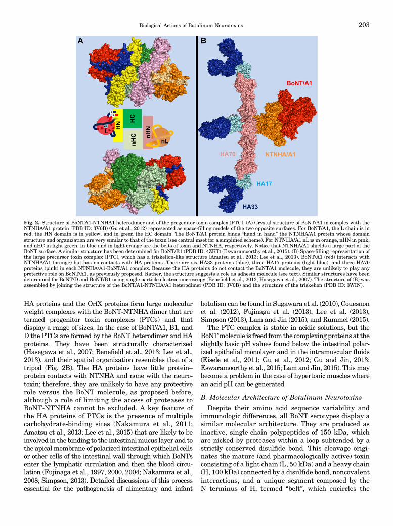
HA proteins and the OrfX proteins form large molecularweight complexes with the BoNT-NTNHA dimer that aretermed progenitor toxin complexes (PTCs) and thatdisplay a range of sizes. In the case of BoNT/A1, B1, andD the PTCs are formed by the BoNT heterodimer and HAproteins. They have been structurally characterized(Hasegawa et al., 2007; Benefield et al., 2013; Lee et al.,2013), and their spatial organization resembles that of atripod (Fig. 2B). The HA proteins have little protein–protein contacts with NTNHA and none with the neuro-toxin; therefore, they are unlikely to have any protectiverole versus the BoNT molecule, as proposed before,although a role of limiting the access of proteases toBoNT-NTNHA cannot be excluded. A key feature ofthe HA proteins of PTCs is the presence of multiplecarbohydrate-binding sites (Nakamura et al., 2011;Amatsu et al., 2013; Lee et al., 2015) that are likely to beinvolved in the binding to the intestinalmucus layer and tothe apical membrane of polarized intestinal epithelial cellsor other cells of the intestinal wall through which BoNTsenter the lymphatic circulation and then the blood circu-lation (Fujinaga et al., 1997, 2000, 2004; Nakamura et al.,2008; Simpson, 2013). Detailed discussions of this processessential for the pathogenesis of alimentary and infant
botulism can be found in Sugawara et al. (2010), Couesnonet al. (2012), Fujinaga et al. (2013), Lee et al. (2013),Simpson (2013), Lam and Jin (2015), and Rummel (2015).
The PTC complex is stable in acidic solutions, but theBoNTmolecule is freed fromthe complexingproteins at theslightly basic pH values found below the intestinal polar-ized epithelial monolayer and in the intramuscular fluids(Eisele et al., 2011; Gu et al., 2012; Gu and Jin, 2013;Eswaramoorthy et al., 2015; Lam and Jin, 2015). Thismaybecome a problem in the case of hypertonic muscles wherean acid pH can be generated.
B. Molecular Architecture of Botulinum Neurotoxins
Despite their amino acid sequence variability andimmunologic differences, all BoNT serotypes display asimilar molecular architecture. They are produced asinactive, single-chain polypeptides of 150 kDa, whichare nicked by proteases within a loop subtended by astrictly conserved disulfide bond. This cleavage origi-nates the mature (and pharmacologically active) toxinconsisting of a light chain (L, 50 kDa) and a heavy chain(H, 100 kDa) connected by a disulfide bond, noncovalentinteractions, and a unique segment composed by theN terminus of H, termed “belt”, which encircles the
Fig. 2. Structure of BoNTA1-NTNHA1 heterodimer and of the progenitor toxin complex (PTC). (A) Crystal structure of BoNT/A1 in complex with theNTNHA/A1 protein (PDB ID: 3V0B) (Gu et al., 2012) represented as space-filling models of the two opposite surfaces. For BoNT/A1, the L chain is inred, the HN domain is in yellow, and in green the HC domain. The BoNT/A1 protein binds “hand in hand” the NTNHA/A1 protein whose domainstructure and organization are very similar to that of the toxin (see central inset for a simplified scheme). For NTNHA/A1 nL is in orange, nHN in pink,and nHC in light green. In blue and in light orange are the belts of toxin and NTNHA, respectively. Notice that NTNHA/A1 shields a large part of theBoNT surface. A similar structure has been determined for BoNT/E1 (PDB ID: 4ZKT) (Eswaramoorthy et al., 2015). (B) Space-filling representation ofthe large precursor toxin complex (PTC), which has a triskelion-like structure (Amatsu et al., 2013; Lee et al., 2013). BoNT/A1 (red) interacts withNTNHA/A1 (orange) but has no contacts with HA proteins. There are six HA33 proteins (blue), three HA17 proteins (light blue), and three HA70proteins (pink) in each NTNHA/A1-BoNT/A1 complex. Because the HA proteins do not contact the BoNT/A1 molecule, they are unlikely to play anyprotective role on BoNT/A1, as previously proposed. Rather, the structure suggests a role as adhesin molecule (see text). Similar structures have beendetermined for BoNT/D and BoNT/B1 using single particle electron microscopy (Benefield et al., 2013; Hasegawa et al., 2007). The structure of (B) wasassembled by joining the structure of the BoNT/A1-NTNHA/A1 heterodimer (PDB ID: 3V0B) and the structure of the triskelion (PDB ID: 3WIN).
Biological Actions of Botulinum Neurotoxins 203
globular L domain, as shown in blue in Fig. 1 (Lacyet al., 1998; Swaminathan and Eswaramoorthy, 2000;Montal, 2010). Reduction of the single interchain S-Sbond releases the L chain metalloprotease activity(Schiavo et al., 1992b, 1993c; Simpson et al., 2004).TheH chain consists of two 50-kDa domains (the amino-
terminal part,HN, and the carboxy-terminal part,HC) andthe molecules of BoNT/A1 and /B1, viewed in a plane,resemble the shape of a butterfly (Fig. 1). The structure ofBoNT/E1 displays a different arrangement of the threedomains with HC bended over the L chain (Fischer et al.,2008; Kumaran et al., 2009). The crystal structures of theremaining serotypes are not yet available for the entiremolecule, but only for the separatedLand theHCdomains,whereas the structure of L-HN has been recently deter-mined for BoNT/D (Masuyer et al., 2015). The similartridimensional arrangement of isolated domains, togetherwith the homologous amino acid sequences, suggests thatthe overall trimodular architecture is shared by all BoNTs,withBoNT/C,BoNT/D, andBoNT/Gpredicted to be similarto typeAandBtoxins, and typeFsimilar to typeE (Rossettoet al., 2014). In the holotoxin, the active site is partiallyoccluded by the belt, thus preserving and preventing thecatalytic activity until the conserved S-S bridge is reducedand the L chain is released in the nerve terminal cytosol(Lacy et al., 1998; SwaminathanandEswaramoorthy, 2000;Brunger et al., 2007; Kumaran et al., 2009).The C-terminal part of the HC domain (subdomain
HC-C, 25 kDa, green in Fig. 1) mediates the interaction ofBoNTswithunmyelinatedareas ofmotoneurons, ensuringa rapid and strong interaction of the toxin with peripheralcholinergic nerve endings (Dolly et al., 1984; Binz andRummel, 2009; Rossetto et al., 2014). HC-C is responsiblefor the neurospecific binding to a polysialoganglioside andto the luminal domain of a synaptic vesicle protein (Binzand Rummel, 2009; Rummel, 2013). Such binding leads tothe ensuing internalization and trafficking of the toxinwithin endocytic compartments, which is initiated by theretrieval of synaptic vesicles after the release of theirneurotransmitter content (Binz and Rummel, 2009;Rummel, 2013). The N-terminal part of HC (sub-domainHC-N, 25 kDa, purple in Fig. 1) folds similarly to sugarbinding proteins of the lectin family, but its specificfunction is not known, although there is evidence indicat-ing that it may improve BoNTs adhesion to the pre-synaptic membrane by interacting with anionic lipids(Muraro et al., 2009; Montal, 2010; Zhang and Varnum,2012; Zhang et al., 2013). TheHNdomain (yellow inFig. 1)plays amajor role in the translocation of the L chain acrossthe membrane of the endocytic vesicle into the cytosol. Itconsists of two long and four shorter, parallel, a-helicesand of the belt segment (blue in Fig. 1).
C. Metalloproteolytic Activity
The L chain (red in Fig. 1) is a metalloprotease withits active site buried within the core of the structure.The discovery that tetanus neurotoxin (TeNT) and the
BoNTs aremetalloproteaseswith an atomof Zn2+ bound tothemotifHExxH (Schiavo et al., 1992b,c), rapidly led to thesubsequent identification of their unique substrates, whichare the three SNARE proteins: VAMP/synaptobrevin,SNAP-25, and syntaxin. These proteins are cleavedat single peptide bonds within their cytosolic domains(Schiavo et al., 1992a, 1993a,b,c; Blasi et al., 1993a,b).These findings provided the first strong experimentalevidence that the three SNARE proteins aremajor playersin the process of exocytosis in general. Moreover, theobservation that the cleavage of VAMP, a synaptic vesicleprotein, was sufficient to cause neuroparalysis provided afinal demonstration of the quantal hypothesis of neuro-transmitter release (Katz, 1996). Very recently, a BoNT-like metalloprotease encoded by Weissella oryzae wasfound to cleave VAMP at a unique site within itsjuxtamembrane segment, which is essential for itsfunction (Zornetta et al., 2016).
A very remarkable property of the metalloproteaseactivity of the BoNT L chains is their selectivity for thethree SNARE proteins, which are cleaved at differentpeptide bonds (Fig. 3; Tables 1, 2, and 3). The molecularbasis of such specificity has been clarified only for theBoNT/A1-SNAP-25 and BoNT/F1-VAMP complexes bysolving the structure of their cocrystals (Breidenbachand Brunger, 2004; Agarwal et al., 2009). However, theoverall picture is clear: specificity is due to multipleinteractions of the L chains with their substrates, whichinclude the cleavage site and exosites located along thesequence, both before and after the hydrolyzed peptidebond (Rossetto et al., 1994; Ahmed et al., 2001; Brungeret al., 2007). This explains why long peptide substratesare needed to test the proteolytic activity of the L chainin vitro and the current lack of specific and strong smallmolecule inhibitors of the BoNT metalloprotease activity(Rossetto et al., 2014).
II. Biology
A. Molecular Mechanism of Nerve Terminal Paralysis
BoNTs are typical examples of bacterial exotoxinstargeting intracellular substrates. These toxins haveevolved a structural organization designed to deliverthe metalloprotease domain into the host cell cytosoland this has been achieved by exploiting several phys-iologic functions of nerve terminals. The mechanism ofnerve terminal intoxication by the BoNTs is conve-niently divided into five major steps (Fig. 4): 1) bindingto nerve terminals, 2) internalization within an endo-cytic compartment, 3) low pHdriven translocation of theL chain across the vesicle membrane, 4) release of the Lchain in the cytosol by reduction of the interchaindisulfide bond, and 5) cleavage of SNAREs with ensuingblockade of neurotransmitter release and neuroparalysis.
1. Binding and Specificity. After entering the lym-phatic and blood circulations, following intestinal ab-sorption or inspiration or injection, the BoNTs rapidly
204 Pirazzini et al.

gain access to the perineuronal fluid compartment butdo not cross the blood-brain barrier (Simpson, 2013).The known BoNTs bind with high affinity to the pre-synaptic plasma membrane of skeletal and autonomiccholinergic nerve terminals in numbers estimated to be,for BoNT/A1 or /B1, in the order of hundreds ofmoleculesper square micrometers at the rat neuromuscular junc-tion (NMJ) (Dolly et al., 1984). This restricted tropism isextraordinary, particularly considering that the presyn-aptic plasma membrane of cholinergic peripheral nerveterminals represents an infinitesimal part of the totalsurface area of cells and tissues exposed to body fluids.Such neurospecificity and affinity of binding, togetherwith their catalytic activity, is at the basis of the BoNTstoxicity and, at the same time, of their pharmacologicaland therapeutic use.This unique nerve terminal binding property of the
BoNTs is due to their capacity to interact with twoindependent receptors of the presynaptic membrane: apolysialoganglioside (PSG) and the glycosylated lu-menal domain of a synaptic vesicle protein that medi-ates the following step of internalization (Montecucco,1986). Additional receptor(s) might be involved andcontribute to the preference for cholinergic terminals
(Montecucco et al., 2004; Kammerer and Benoit, 2014).It is also possible that cholinergic nerve terminalsexpress unique N-glycans attached to synaptic vesicle(SV) glycoproteins that contribute to this preferentialbinding. Fibroblast growth factor and vanilloid recep-tors were recently reported to bind BoNT/A1 (Jackyet al., 2013; Li and Coffield, 2016), but their functionalinvolvement in toxin binding has to be further validated.
The involvement of PSG in BoNT binding has beenextensively investigated and is supported by a largewealth of experimental data (Rummel, 2013), includ-ing the demonstration that mice and cell lines devoid ofcomplex PSG are largely resistant to BoNT (Kitamuraet al., 1999; Bullens et al., 2002). This fact also contrib-utes to explain why insects that are devoid of PSG areBoNT insensitive, a property that makes them reliablevectors of BoNT spread during outbreaks of animalbotulism among birds and fishes (Montecucco andRasotto, 2015).
The BoNTs-PSG interaction is rather well character-ized and reviewed (Rummel, 2013). Briefly, BoNT/A, /B,/CD, /E, /F, and /G (and TeNT) possess within the HC-Csubdomain a large cavity defined by the conservedmotifsequence E(Q in BoNT/G)…H(K in BoNT/E and G in
Fig. 3. Cleavage sites of the neuronal SNARE proteins by the different BoNT types and subtypes. The BoNT proteolytic activity is highly specific anddirected toward unique peptide bonds within the sequence of their respective SNARE protein targets. VAMP of the synaptic vesicle (in blue, isoform1 is shown here) or SNAP-25 (in green) or syntaxin (in red, isoform 1B is shown here) mainly localized on the cytosolic side of the presynapticmembrane. Available evidence indicate that all the toxin subtypes and chimeric toxins cleave the same SNARE substrate, although different subtypesmay cleave different peptide bonds. For example BoNT/F5 and BoNT/FA, a chimeric toxin derived from a genetic recombination between BoNT/F2, /F5,and A1 neurotoxin genes, cleave VAMP at a peptide bond different from the one cleaved by BoNT/F1. Notice that tetanus neurotoxin and botulinum B1neurotoxin cleave the same target at the same site proving that the different symptoms of tetanus and botulism are not due to a different targetmolecule, but to different neuronal targets: the Renshaw cells of the spinal cord for tetanus neurotoxin and peripheral nerve terminals for BoNT/B1.
Biological Actions of Botulinum Neurotoxins 205

TABLE 1Sequence alignment of mouse, rat, and human VAMP isoforms and cleavage sites of the different BoNTs
The SNARE motifs of mouse, rat, and human VAMP isoforms have been aligned using http://www.uniprot.org/align/. The conserved cleavage sites of VAMP isoformstargeted by specific BoNT types and subtypes are in the same color of the respective toxins. The cleavage site of the newly identified BoNT-like metalloprotease of Weissellaoryzae (WO) is also shown (purple). Conserved proteolytic sites whose susceptibility to cleavage is predicted, but not experimentally proven, are underlined with the color of therespective BoNT. Nonconserved cleavage sites are underlined in black as well as conserved cleavage sites, which were experimentally found to be noncleavable. When known,the recognition motif, used by the toxin to bind the substrate in addition to the cleavage sites (Rossetto et al., 1994), is underlined in red.
206 Pirazzini et al.

BoNT/G)…SXWY…G (Fig. 1, pink). The affinity of thisPSG-BoNT binding has been estimated and the Kd
values are in the 0.1–1 mM range (Rummel, 2013). Atvariance, BoNT/C, BoNT/D, and the mosaic neurotoxinBoNT/DC bind PSG at a binding site located in a similarposition but defined by a different set of amino acidslateral chains and with lower affinity (Strotmeier et al.,2010; Rummel, 2013). Interestingly, BoNT/C, BoNT/D,and BoNT/DC (and TeNT) harbor a second PSG bindingsite in their HC-C domain, defined by conserved W andY(F) residues (Strotmeier et al., 2010, 2011), whichmight bind the carbohydrate portion of a plasmamembrane glycoprotein that is endocytosed from thepresynaptic membrane. BoNT/DC was recently shownto bind a sialyl residue in a pocket of the HC domain anda cell binding mechanism involving a cooperative con-tribution of two ganglioside binding sites was proposed(Nuemket et al., 2011).Together with sphingomyelin, cholesterol, and some
proteins, PSG form anionic microdomains in the plasmamembrane (Simons and Toomre, 2000; Sonnino et al.,2007; Prinetti et al., 2009). Accordingly, BoNTs bindcomplex PSGs whose large oligosaccharide head groupprojects at a distance from the membrane surface and it
is flexible and negatively charged. There is evidencethat anionic microdomains of the presynaptic mem-brane including PSG may orient the electric dipoleassociated to the BoNT molecules whose positive endis, significantly, located around the PSG binding site.This reorienting effect of the membrane on theapproaching BoNT molecule would strongly increaseits probability of PSG binding, making it a reactioncontrolled only by diffusion (Fogolari et al., 2009).
Following membrane binding, the BoNTs are inter-nalized. The key observations that neuronal stimula-tion enhances the toxicity of BoNTs (Hughes andWhaler, 1962; Simpson, 1980, 1985; Black and Dolly,1986; Keller et al., 2004) and that BoNT/B bindssynaptotagmin in cultured cells (Nishiki et al., 1994)led to the suggestion that BoNTs endocytosis is facili-tated by a protein receptor consisting of the luminaldomain of a synaptic vesicle (SV) protein (Montecuccoand Schiavo, 1995). This second binding provides thehigh affinity necessary to bind the very low amounts ofBoNT estimated to be present in the circulation duringbotulism (10213–10214 M). During stimulation, the in-creased exoendocytosis rate causes a frequent exposureof the SV lumen, making the vesicle interior transiently
TABLE 2Sequence alignment of mouse, rat, and human SNAP25 isoforms and cleavage sites of the different BoNTs
The C-terminal SNARE motif of mouse, rat, and human SNAP isoforms have been aligned using http://www.uniprot.org/align/. The conserved cleavage sites targeted byspecific BoNT types and subtypes are colored like the respective toxin. Conserved cleavage sites whose susceptibility to cleavage is predicted, but not experimentally proven,are underlined with the color of the respective BoNT. Nonconserved cleavage sites are underlined in black as well as conserved cleavage sites that were experimentally foundto be noncleavable. Notice that mouse SNAP-23 has a cleavage site for BoNT/E different from the one present in SNAP-25, which is anyhow cleaved, whereas human SNAP-23,which has the same proteolytic site as SNAP-25, is not cleaved (Vaidyanathan et al., 1999). Similarly, mouse SNAP-23 was reported to be cleaved by BoNT/A, although thepeptide bond is not conserved. At the same time, BoNT/C, whose cleavage site is conserved (also in human SNAP-23) does not cleave mouse SNAP-23. The cleavability of ratSNAP-23 was predicted on these premises. When known, the recognition motif used by the toxin to bind the substrate in addition to the cleavage sites (Rossetto et al., 1994) isunderlined in red.
Biological Actions of Botulinum Neurotoxins 207

TABLE 3Sequence alignment of mouse, rat, and human Syntaxin isoforms and cleavage site of BoNTs
The C-terminal SNARE motifs of mouse, rat, and human syntaxin isoforms are aligned using http://www.uniprot.org/align/. BoNT/C conserved cleavage sites are shown inpurple. Conserved cleavage sites whose susceptibility to cleavage is predicted, but not experimentally proven, are underlined in purple. Nonconserved cleavage sites areunderlined in black.
208 Pirazzini et al.

available for binding. This also contributes to accountfor the efficacy of BoNT in treating human syndromescharacterized by hyperactive nerve terminals, be-cause the NMJs of these muscles have a higher rateof SVs exoendocytosis, which favors toxin uptake.Within a few years, the protein receptors of other
BoNTs were identified. BoNT/B1, BoNT/G, andBoNT/DC bind segments 39–50 of synaptotagmin Iand 47–58 of synaptotagmin II (Nishiki et al., 1994;Dong et al., 2003; Rummel et al., 2007; Peng et al., 2012;Willjes et al., 2013). BoNT/A1 and BoNT/E1 binddifferent segments of the fourth lumenal loop of SV2,
a multispanning integral protein of synaptic vesicles ofunknown function (Dong et al., 2006; Mahrhold et al.,2006; Binz and Rummel, 2009; Benoit et al., 2014).Three isoforms of SV2 (A, B, C) are expressed at motornerve terminals, but SV2C appears to be the onebinding BoNT/A1 more efficiently than SV2A or B viaits glycosylated fourth luminal domain (Mahrhold et al.,2006; Benoit et al., 2014; Mahrhold et al., 2016),whereas BoNT/E1 binds isoforms A and B, but not C(Dong et al., 2008).
The binding of BoNT/A1 to the glycosylated SV2Creceptor is mediated by protein-protein and protein-
TABLE 3—Continued
Fig. 4. The nerve terminal intoxication by botulinum neurotoxins is a multi-step process. The first step (1) is the binding of the HC domain (green) to apolysialoganglioside (PSG) receptor of the presynaptic membrane (gray and black), followed by binding to a protein receptor. The currently knownprotein receptors are i) synaptotagmin (Syt, gray) for BoNT/B1, /DC, and /G; ii) glycosylated SV2 (black with its attached N-glycan in pink) for BoNT/A1and /E1. Syt may be located either within the exocytosed synaptic vesicle or on the presynaptic membrane. The BoNT is then internalized inside SVs,which are directly recycled (2a) or inside SVs that fuse with the synaptic endosome and re-enter SV cycle by budding from this intermediatecompartment (2b). The acidification (orange) of the vesicle, operated by the v-ATPase (orange), drives the accumulation of neurotransmitter (blue dots)via the vescicular neurotransmitter transporter (light blue). The protonation of BoNT leads to the membrane translocation of the L chain into thecytosol (3), which is assisted by the HN domain (yellow). The L chain (red) is released from the HN domain by the action of the thioredoxin reductase-thioredoxin system (TrxR-Trx, blue and dark blue) and Hsp90 (mud color), which reduce the interchain disulfide bond (orange) and avoid theaggregation of the protease (4). In the cytosol, the L chain displays its metalloprotease activity: BoNT/B, /D, /F, /G cleave VAMP (blue); BoNT/A andBoNT/E cleave SNAP-25 (green); and BoNT/C cleaves both SNAP-25 and syntaxin (Stx, dark red) (5). Each of these proteolytic events is sufficient tocause a prolonged inhibition of neurotransmitter release with consequent neuroparalysis.
Biological Actions of Botulinum Neurotoxins 209

glycan interactions that do not appear to influence eachother. Of the five putative N-glycosylation sites ofSV2C, only the Asn-559 is involved; this N-glycan ishighly complex with a tetra antennary structure, whichis likely to interact with an extensive area of HC/A1(Mahrhold et al., 2016). The protein-proteinHC/A-SV2Ccontacts involvemostly the backbones of the two proteins,through the pairing of two solvent-exposed b-strands, onefrom each partner (Benoit et al., 2014). Asn559 of SV2C isat the center of the BoNT/A1-SV2 area of interaction andits N-linked glycan inserts in a crevice made by oneb-strand of the HC-N subdomain (segment 950–954) andtwo segments of the subdomain HC-C: the b-strandsegment 1064–1066 and the C-terminal loop 1289–1292(Fig. 5A) (Yao et al., 2016). As glycosylation patternsvary during development and among adult individuals(Knezevi�c et al., 2009; Pucic et al., 2010; Lauc et al.,2016), such a feature might be responsible for thedifferent onset and duration of neuroparalysis elicitedin human patients by the same dose of injected BoNT/A1as different amounts of bound toxin are likely to corre-spond to different numbers of L chains entering the nerveterminal cytosol. In addition, as invertebrate and verte-brate N-glycans are different (Moremen et al., 2012), this
may contribute to the lack of sensitivity of invertebratesto BoNTs.
The structural basis of BoNT/B1 and of BoNT/DCbinding to their synaptotagmin receptor have beendetermined and are shown in Fig. 5B (Chai et al.,2006; Jin et al., 2006; Berntsson et al., 2013a,b). Suchbinding is mediated by a pocket present in the HC-Csubdomain formed by residues of the segment Pro1114-Phe1202 (BoNT/B1 numbering), which fits the a-helicalsegment 47–58 of synaptotagmin II located next to thelumenal surface of the SV membrane. Asn24 of synap-totagmin II is predicted to be glycosylated but thepotential role of N-glycan linked to this residue on thebinding of BoNT/B1 has not yet been tested, althoughthis may be relevant with respect to the therapeutic useof BoNT/B1 in humans, because the pattern of glyco-sylation in the nervous system varies among individu-als. The presence of Leu51 in human synaptotagmin IIreduces significantly the binding affinity for BoNT/B1,/DC, and /G with respect to the corresponding Phe54of mouse synaptotagmin II. This also explains themuch higher dosage of BoNT/B1 necessary to achievethe same therapeutic effect of BoNT/A1 (Peng et al.,2012; Strotmeier et al., 2012).
Fig. 5. Close-up views of the binding interfaces between BoNT/A1 and BoNT/B1 to their synaptic vesicle protein receptors. (A) Two areas of interactionof BoNT/A1 with the synaptic vesicle protein glycosylated-SVC2 (PDB ID 5JLV). One main interaction is mediated by protein-protein contacts throughthe pairing of the backbones of two solvent-exposed b-strands (black dotted ellipsoid), one from each partner. Essential interactions are mediated byR1156 of BoNT/A1 making a cation-p stacking interaction with P563 of SV2C and also by R1294 of the toxin. The second main interaction is mediatedby N559 whose side chain carries a N-glycan modification (shown as sticks), which fits within a crevice formed at the interface between HC-N (purple)and HC-C (green). Amino acids forming the groove are colored in cyan and labeled according to their location (P953, N954, S957, S1062, H1064, andR1065 from HC-N, in purple and T1145, Y1155, D1288, D1289, and G1292 from HC-C, in green). The cartoon also shows essential water molecules(black pellets) and the H bonds (yellow dotted lines), which mediate the interaction of BoNT/A1 with the N-glycan, suggesting the possibility that theyserve to adapt the variety of N-glycans that are produced by different kind of neurons and/or by neurons of different individuals and animal species. (B)Interaction among BoNT/B1 and its synaptic vesicle protein receptors Synaptotagmin II (Syt-II) (PDB ID 4KBB). The interface of interaction is at theextreme bottom of BoNT/B and, at variance from BoNT/A1, involves exclusively HC-C (green). Syt-II is unstructured in solution but assumes an helicalconformation upon binding to the toxin, forced by the interactions occurring at the level of two hydrophobic pockets. One pocket is formed by HC-Cresidues P1117, W1178, Y1181, P1194, A1196, P1197, F1204 with Syt-II residues M46, F47, and L50. The second pocket of HC-C is lined by residuesK1113, S1116, P1117, V1118, Y1183, E1191, K1192, F1194, and F1204 with Syt-II residues F54, F55, E57, and I58. Only the most significantaminoacids involved in the interaction are shown and labeled. Note the H bond (black dotted line) formed by K1113 and E57, which may also interactelectrostatically. The binding sites for the oligosaccharide portion of polysialoganglioside receptor are not shown, but in both BoNT/A1 and /B1 arelocated within the HC-C subdomain at a distance from the protein receptor binding sites in such a position that they do not interact with them (see text).
210 Pirazzini et al.

The protein receptors of other BoNTs are not knownin such details. BoNT/C might not have a protein recep-tor, because protease pretreatment does not affectits binding and internalization into cultured cells(Tsukamoto et al., 2005). Conflicting results have beenreported for BoNT/D (Kroken et al., 2011; Peng et al.,2011; Rummel, 2013), but based on its similarity withBoNT/E, BoNT/F is expected to bind SV2, and somedata support this possibility (Rummel et al., 2009).2. Internalization into Nerve Terminals. After in-
tramuscular injection, BoNT/A1 is rapidly taken up andfound within the lumen of SV at the neuromuscularjunction (Colasante et al., 2013). In cultured primaryneurons part of such internalization is clathrin mediatedand most of the toxin is detected inside SV (Neale et al.,1999; Couesnon et al., 2009; Harper et al., 2011). Impor-tantly, the average number of BoNT/A molecules per SVwere estimated to be one to two (Harper et al., 2011;Colasante et al., 2013), a figure thatmatches the estimatedcopy number of SV2molecules per vesicle (Takamori et al.,2006). The internalization of the otherBoNTshas not beenvisualized by electron microscopy, but it is expected tooccur as well via SV because these toxins bind a SVmembrane protein.SV exocytosis is strictly coupled to endocytosis, which
can take place in different ways (Saheki and De Camilli,2012; Cousin, 2015; Jähne et al., 2015; Kononenko andHaucke, 2015; Soykan et al., 2016). Before fusion withthe presynaptic membrane at specialized release sitesof nerve terminals (active zones), SVs are morphologi-cally similar and, to be functional, they must have adefined proteic and lipidic composition (Takamori et al.,2006; Boyken et al., 2013). However, after fusionwith the presynaptic membrane, endocytosed SVs mayacquire molecular differences (Wienisch and Klingauf,2006; Soykan et al., 2016). Linked to this and to thefrequency of nerve terminal stimulation, SVs may un-dergo different forms of retrieval (Saheki and DeCamilli, 2012; Jähne et al., 2015; Kononenko andHaucke, 2015; Soykan et al., 2016). In particular afunctional SV, right after endocytosis, may be directlyrecycled and reacidified by the v-ATPase proton pump,which generates an electrochemical gradient drivingthe accumulation of neurotransmitter via a specifictransporter (Parsons, 2000; Omote et al., 2011). Alter-natively, some vesicles may fuse with endosomal com-partments, where quality-controlled SVs of appropriatecomposition reform and re-enter the synaptic cycle.Under some conditions, early endosomal compartmentscan also be generated from the infolding of a largeportion of the presynaptic membrane, and then SVs canreform by endosomal budding. Additionaly, it wasreported that SV proteins (including synaptotagmins)of exocytosed SV can freely diffuse within the pre-synaptic boutons, where they are slowly confined in aperiactive zone by endocytic adaptors and presynapticdiffusion barriers to be reinternalized by clathrin-
dependent or bulk endocytosis mechanisms (Gimberet al., 2015).
Collectively, these different possibilities of SV endo-cytosis indicate that the trafficking of BoNTs may occurvia different pathways, depending at least partially, ontheir receptors. This is supported by experimentsperformed with primary cultures of central nervoussystem neurons, showing a significant variability in theintoxication kinetics of different BoNTs (Keller et al.,2004; Sun et al., 2012). Moreover, the use of theendocytosis inhibitor 4-bromobenzaldehyde N-(2,6-dimethylphenyl)semicarbazone (EGA) (Gillespie et al.,2013) provided indirect evidence that different BoNTsmay follow different trafficking routes after the initialendocytosis also in vivo (Azarnia Tehran et al., 2015).This would result in different entry times of the L chaininto the cytosol and would explain the “rapid” entry ofBoNT/A and /E and the slower one of BoNT/B de-termined in primary cultures of neurons (Keller et al.,2004; Sun et al., 2012; Colasante et al., 2013).
Interestingly, a BoNTmay be taken up even after theinhibition of SV exocytosis by another BoNT in vitro(Keller et al., 1999, 2004; Pellett et al., 2015a) andin vivo (Eleopra et al., 1998; Adler et al., 2001; Meunieret al., 2003; Keller, 2006; Antonucci et al., 2008). Clearlythese results are highly dependent on the SNAREisoform cleaved by the BoNT used to inhibit exocytosisand on the extent of cleavage, data that were notreported. Moreover, it is well established that PSGundergoes endocytosis by the ligand-receptor pathway(Sonnino et al., 1992), and therefore it cannot be ruledout that the sole BoNT-PSG interaction is sufficient tosustain neurotoxicity, particularly for those BoNTs thathave two ganglioside binding sites. Taken togetherthese results and considerations indicate that morethan one way of uptake may be used at nerve terminalsby the different BoNTs or by the same BoNT underdifferent conditions.
a. Long distance effects of botulinum neurotoxins.Peripheral neuroparalysis is the most evident symptomof botulism and is the one at the bases of the therapeuticuse of BoNTs. However, indirect evidence that theseneurotoxins could act at a distance from the injectionsite, i.e., within spinal cord and brain neuronal circuits,were reported long ago, and in some cases it was shownto be due to retroaxonal transport of BoNTs similar tothat occurring with TeNT (Tyler, 1963; Polley et al.,1965; Garner et al., 1993; Priori et al., 1995; Santiniet al., 1999; Gilio et al., 2000; Wohlfarth et al., 2001;Marchand-Pauvert et al., 2013; Matak and Lackovic,2014; Mazzocchio and Caleo, 2015; Matak et al., 2016).Direct evidence of BoNTs retroaxonal transport afterintramuscular injection was provided by the detectionof cloramine T radioiodinated BoNT/A1 within theventral roots ganglia and other spinal cord seg-ments (Habermann, 1974; Wiegand et al., 1976). How-ever, it was not clear whether retrotransported toxins
Biological Actions of Botulinum Neurotoxins 211

remained active, because the method of radioiodinationused inactivates the BoNTs. More recently, compellingevidence of BoNT/A1 retrotransport to the centralnervous system (CNS) was provided by tracing thecleavage of the SNARE within CNS neurons afterperipheral injection of the toxin, using an antibody veryspecific for the novel epitope generated by the BoNT/A1cleavage of SNAP-25 (Antonucci et al., 2008; Mataket al., 2011, 2012; Restani et al., 2011; Matak andLackovic, 2014; Mazzocchio and Caleo, 2015). Theinjection of BoNT/A1 in the rat whisker pad led to theappearance of truncated SNAP-25 in the somatoden-dritic area of primary efferent facial motoneurons(Antonucci et al., 2008). Cleaved SNAP-25 was alsodetected in the ventral horns of the spinal cord uponinjection of BoNT/A1 in the gastrocnemius (Matak et al.,2012). Interestingly, BoNT/A1 retrograde transportcan occur also via sensory neurons, as the injection inthe whisker pad induces the appearance of truncatedSNAP-25 both in the trigeminal nucleus caudalis(Matak et al., 2011) and in the dorsal horn of the spinalcord after subcutaneous or intramuscular injection inthe hind limb (Marinelli et al., 2012; Matak et al., 2012,2014). Notably, this suggests that BoNT/A1 undergoesretrograde transport from periphery to ganglia but alsothat an anterograde movement from ganglia to afferentinnervations in the brain stem or in the spinal cord mayoccur (Restani et al., 2011). These long-distance effectsare mediated by an active retroaxonal transport ofcatalytically competent toxins inside motor axons andnot by their passive spread or to that of the cleavedproduct (Antonucci et al., 2008; Matak et al., 2011).Intriguingly, it was also found that BoNT/A1 can enterand cleave SNAP-25 in neurons which are even twosynapses away from the injection site (Restani et al.,2012b), entailing that upon retrotransport the toxin canundergo sequential cycles of transcytosis and transport,remaining catalytically active. At variance, using neu-rons isolated from superior cervical ganglia of newbornrats, it was found that BoNT/A1 migrates within nerveterminal in a nonvesicular mode without inhibitingdistal neurotransmission (Lawrence et al., 2012).The prototype of the neurotoxins binding to periph-
eral nerve terminals and reaching the CNS is tetanusneurotoxin, which has become the marker of retroaxo-nal transport inside motor axons (Schwab et al., 1979;Rossetto et al., 2013). TeNT uses the neuromuscularjunction as a portal of entry following its binding tonidogen, a protein of the basal lamina (Bercsenyi et al.,2014). Thereafter, it travels inside a compartment thatlinks the clathrin-dependent endocytosis to the sequen-tial activity of the two small GTPases Rab5 and Rab7,allowing the trafficking of the toxin from “signallingendosomes” (Zweifel et al., 2005; Schmieg et al., 2014)into the retrograde transport pathway of neurotrophins(Deinhardt et al., 2006). It is possible that BoNT/A1 isalso recruited to the same nonacidic carriers exploited
by TeNT and neurotrophins (Restani et al., 2012a). Arecent study has implicated autophagosomal structuresin the capture and transport along the axon of BoNT/A1(Wang et al., 2015), a pathway that appears to beexploited by polio virus to be delivered in the CNS(Bird et al., 2014). However, these vesicle carriers mayalso be connected to degradative pathways as suggestedby the partial costaining of BoNT/A1 with the autopha-gosome marker LC3 (Wang et al., 2015).
It should be noticed that no evident symptoms havebeen associated so far with BoNTs acting within theCNS upon peripheral injection in humans or in theanimals tested, whereas there is evidence suggestingthat they could contribute to the antinociceptive activityof BoNT/A (Matak and Lackovic, 2014; Mazzocchio andCaleo, 2015; Pellett et al., 2015d).
3. Membrane Translocation. BoNTs translocatetheir L chain into the cytosol from an acidic intracellularcompartment, and the process can be blocked by differ-ent amines and by bafilomycin A1, a specific inhibitor ofthe v-ATPase (Simpson, 1983; Williamson and Neale,1994; Keller et al., 2004). However the lumen of the SVinside the nerve terminal is not experimentally acces-sible, making the study of this process difficult. There-fore, models that bypass the intracellular delivery of thetoxin and induce the entry of the L chain of BoNTs fromthe cell surface across the plasma membrane weredevised (Pirazzini et al., 2011; Sun et al., 2011). Byusing these systems, it was found that the translocationof L takes place between pH 4.5 and 6 (Pirazzini et al.,2011) and that the entire translocation process is rapid(few minutes at 37°C) and strongly temperature de-pendent (Pirazzini et al., 2013c). Little if any trans-location occurs above pH 6, and this makes it unlikelythat the L domain translocates from early endosomes,whose internal pH is only slightly acidic. On the otherhand, BoNT/A1 has been localized within the SV lumenby immuno electron microscopy in cultured hippocam-pal neurons (Harper et al., 2011) and within the NMJ(Colasante et al., 2013). An essential component of SV isthe electrogenic v-ATPase, which injects protons intothe lumen, generating a transmembrane pH gradientDpH of 1.4 pH units and an electrical gradient DC of +39 mV (Parsons, 2000). By using fluorescent synapto-phluorins, the luminal pH of SV was estimated to be;5.8 pH units (Miesenbock et al., 1998). Accordingly,the translocation of L into the cytosol can take placefrom recycling SV or from SV budded from early endo-somes. However, it cannot be excluded that othersimilarly acidic compartments such as late endosomesand lysosomes that could be reached by a BoNT uponendosome maturation, could provide the appropriateenvironment leading to L chain translocation, althoughit should be considered that these organelles containproteases that may degrade the toxin.
Planar lipid bilayer studies have shown that at low pHseveral BoNTs and tetanus toxin form transmembrane
212 Pirazzini et al.

ion channels (reviewed in Montecucco and Schiavo,1995). Despite the intense effort of several laboratories,the molecular mechanism describing the transforma-tion of the water-soluble BoNT molecule into a trans-membrane ion channel that assists the translocation ofthe ;440 amino acids of the L metalloprotease domainacross the membrane is still missing. However, aremarkable set of patch-clamping experiments (Montal,2010; Fischer, 2013; Fischer and Montal, 2013) andrecent results obtained with the plasma membraneentry model (Pirazzini et al., 2011; Sun et al., 2011)have shed light on the initial molecular events ofthe process for BoNT/A1 and /B1. We have reviewedand modeled BoNT membrane translocation recently(Pirazzini et al., 2016), and, therefore, only the mostrelevant aspects are highlighted here.The starting point is the BoNT molecule bound to the
luminal side of the SV membrane via two interactions:with the polysialoganglioside and with the proteinreceptor. As the pH lowers, some conserved high pKacarboxylates of BoNT become protonated on the face ofthe molecule containing the interchain disulfide bond(Fig. 1, top), which acquires a net positive charge, asindicated by bioinformatics and mutagenesis analysis;remarkably, the opposite face is devoid of conservedhigh pK protonatable residues (Pirazzini et al., 2011,2013b). Consequently, the BoNT molecule, with itspositively charged surface, falls onto the anionic surfaceof the membrane generating a lipid-protein complex.The pH value close to the membrane is estimated to beat least one pH unit lower than in the lumen owing tothe Guy-Chapman effect (Nordera et al., 1997), leadingto the protonation of carboxylate residues of lower pKavalues. This triggers a process of structural changeinvolving the L and H chain together with membranelipids, leading to the formation of an ion channel.During this process, both the H and L chain enter incontact with the hydrocarbon chain of lipids as deter-mined by photoactive hydrophobic labeling (Montecuccoet al., 1989). There is a general consensus that the Hchain acts as a sort of transmembrane chaperone for thetranslocation of the L chain across the membrane(Koriazova and Montal, 2003; Montal, 2010; Fischer,2013). Different molecular roles of the H chain can beenvisaged, and two possibilities with a range of intermedi-ate cases arementionedhere:1) that of a protein conductingchannel that translocates the unfolded L chain, as it occursin the case of the protective and lethal factors of anthrax(Collier, 2009), 2) the toxin forms a “molten globule”, aprotein state that is known to be capable of interactingwiththe hydrophobic membrane interior (Ptitsyn et al., 1990;van der Goot et al., 1991). Suchmolten globulewould insertinto the membrane together with anionic lipids and woulddeliver the L chain to the other side of the membrane,whereas the H chain would assemble an ion conductingchannel. In any case, the belt has to beunfastened to permitthe passage of the L chain on the cytosolic face of the
membrane (Fischer andMontal, 2007). The understandingof the mechanism of membrane translocation is veryimportant, because it is likely to be common to all BoNTsandmay thus present a key target step for the developmentof pan-inhibitors of the entire family of BoNTs (Fischeret al., 2009; Pirazzini and Rossetto, 2017).
The number of BoNT molecules involved in mem-brane translocation is another undefined point. Thefinding that one SV contains one or two moleculesof BoNT/A1 leaves open the possibility that onetoxin molecule is sufficient to carry out the processand suggests that these toxins may have an in-builtmembrane translocating system, similarly to the bac-terial system SecY (Park and Rapoport, 2012). Diph-theria toxin, a bacterial toxin with a structuralarchitecture similar to that of BoNTs, also has a trans-locating domain that is mainly a-helical, and the avail-able evidence supports a single molecule process, withfew transmembrane helices forming an ion channel(Finkelstein et al., 2000; Gordon and Finkelstein, 2001;Leka et al., 2014). At variance, it was recently reportedthat a trimer might form the protein conducting trans-membrane channel of BoNT/B1 in PC12 cells andBoNT/E1 at physiologic pH (Sun et al., 2012). Clearly,additional experiments are needed to clarify this essentialstep of the mechanism of action of the BoNTs.
4. Interchain Disulfide Reduction. The importanceof the interchain disulfide for the toxicity of clostridialneurotoxins is demonstrated by the fact that reducedtoxins are inactive (Schiavo et al., 1990; de Paiva et al.,1993; Fischer and Montal, 2007). At the cellular level,Fischer and Montal (2007) demonstrated that the pre-mature reduction of this S-S bond, at any stage beforeits exposure to the cytosol, aborts the L chain trans-location. Also in the plasma membrane translocationmodel, the L chain has to be disulfide linked to the Hchain to cross themembrane (Pirazzini et al., 2011). Thedetachment of the L chain from the cytosolic face of themembrane by reduction releases its metalloproteaseactivity in the cytosol (Fischer and Montal, 2007).
The lumen of most intracellular organelles is oxidiz-ing and low pH prevents the reduction of disulfidebonds. At variance, the cytosol has a higher redoxpotential owing to the presence of several reducingmolecules (NADH, NADPH, reduced glutathione, cys-teine, etc.). The maintenance of an appropriate redoxbalance is particularly important for the activity of keyproteins (Arner andHolmgren, 2000; Meyer et al., 2009;Hanschmann et al., 2013). The NADPH-thioredoxinreductase (TrxR)-thioredoxin (Trx) system is a majorredox system of the cell that reduces protein disulfides.Its involvement in the BoNTs and TeNT entry intoneurons was first identified using a pharmacologicapproach (Pirazzini et al., 2013a), and then TrxR andTrx were shown to be extrinsic proteins of the cytosolicside of the SV membrane (Pirazzini et al., 2014), wheretranslocation of the L chain is expected to occur. Several
Biological Actions of Botulinum Neurotoxins 213

inhibitors of the TrxR-Trx redox couple prevent thedisplay of the SNARE specific metalloprotease activityof the L chain of all serotypes of BoNTs, from A to G incultured neurons. More importantly, these inhibitorslargely prevent the BoNT-induced paralysis in micein vivo, regardless of the serotype involved (Zanettiet al., 2015). The reduction of the single interchaindisulfide bond is therefore a general and fundamentalstep of the BoNT [and TeNT (Pirazzini et al., 2013a;Zuverink et al., 2015)] mechanism of nerve terminalintoxication. As such it has to be considered a step per sein the sequence of passages leading from BoNT nerveterminal binding to neuroparalysis. More recently itwas found that the chaperone Hsp90 is also present onthe cytosolic face of SV and that it cooperates with TrxRand Trx in the entry of a folded and active L chain in thecytosol (Azarnia Tehran et al., 2017).5. SNARE Protein Cleavage. SNARE proteins form
a large superfamily comprisingmany isoforms of VAMP(Rossi et al., 2004) and syntaxins but relatively fewerisoforms of SNAP. Specific isoforms of SNARE proteinsselectively interact and form heterotrimeric coiled-coilcomplexes (SNARE complexes), which mediate mostintracellular events of vesicle–target membrane fusion(Jahn and Scheller, 2006). The discovery that TeNT andBoNTs are metalloproteases (Schiavo et al., 1992b,c)and that TeNT and BoNT/B1 cleave the SV proteinVAMP/synaptobrevin (Schiavo et al., 1992a) opened theway to the the rapid identification of the other SNAREproteins targets of the other BoNTs (Blasi et al.,1993a,b; Schiavo et al., 1993a,b,c). The cleavage byTeNT and BoNTs of proteins that were previouslyidentified by molecular biology and biochemical meth-ods (Elferink et al., 1989; Oyler et al., 1989; Bennettet al., 1992) explained the molecular basis of tetanus andbotulism. At the same time, these findings provided thestrongest evidence of the SNARE protein involvement inneurotransmitter release and, more generally, that thethree SNAREs are the core proteins of the nanomachinethat mediates vesicle fusion to target membrane.The BoNT proteolytic activity is highly specific and
directed toward unique peptide bonds within the se-quence of the respective SNARE substrates (summa-rized in Tables 1, 2, and 3) (Binz, 2013; Blasi et al., 1993a;Pantano and Montecucco, 2014; Rossetto et al., 2014).All BoNTs, except the BoNT/As, cleave large portions ofthe cytosolic domains of their respective substrates (Fig.3), preventing the formation of a stable SNARE complexand consequently of exocytosis (Hayashi et al., 1994;Sutton et al., 1998). At variance, BoNT/As generate atruncated SNAP-25, which is still capable of forming astable SNARE complex (Hayashi et al., 1994; Otto et al.,1995) and has a life time within the nerve comparable tothat of intact SNAP-25 (Foran et al., 2003). Remarkablework showed that BoNT/A truncated SNAP-25 (SNAP-251–197) by itself inhibits exocytosis (Huang et al., 1998),and results of several experiments indicate that the
BoNT/A cleavage of a 10–15% fraction of total SNAP-25both within the NMJ (Raciborska et al., 1998) andspinal cord neurons (Keller and Neale, 2001; Kelleret al., 2004; Montecucco et al., 2005) is sufficient tocause paralysis. These results led to the suggestion thatSNAP-251–197 acts as a dominant negative factor in thefunction of a multimeric radial super-SNARE complexbecause the C-terminal segment is necessary forprotein-protein interactions underpinning the forma-tion of a radial SNARE super-complex (Montecucco et al.,2005; Pantano and Montecucco, 2014). Electron micros-copy data indicate that multimeric SNARE supercom-plexes exist in the CNS (Rickman et al., 2005), andindirect evidence for the existence of an octamericneuroexocytosis radial nanomachine have beenobtained (Megighian et al., 2013). A variety of experi-mental approaches have been used to estimate thenumber of SNARE complexes forming the nanomachinethat mediates vesicle-target membrane fusion and arange of figures have been produced (reviewed inPantano and Montecucco, 2014). Recent improvementin single particle cryo electronmicroscopy (Cheng, 2015)may soon reveal the structure of the nanomachine thatmediates neuroexocytosis, allowing one to understandthe molecular consequences of the cleavage of SNAP-25by BoNT/A and BoNT/C. However, at the present stageit cannot be excluded that SNAP-25 exists in differentpools within the nerve terminal and that a subpoolconsisting of 10–15% of total SNAP-25 is the oneinvolved in neuroexocytosis. It is also possible that theC-terminal segment of SNAP-25 inserts in the lipidbilayer playing an essential role in membrane fusion.
The specificity of L chains for the SNARE proteinsrelies on extended and multiple protein-protein inter-actions with their specific substrate, which include thecleavage site and exosites (Rossetto et al., 1994; Pellizzariet al., 1996; Breidenbach and Brunger, 2004; Jin et al.,2007; Agarwal et al., 2009; Brunger and Rummel, 2009).Such extended enzyme-substrate interaction results inhighly specific recognition and explains why no addi-tional protein substrates of the BoNTs have yet beenfound. The selectivity of BoNTs is well shown by twoexamples that are relevant to the evolutionary biologyof animal botulism. The replacement of a Gln with a Valat the P19 position of the VAMP cleavage site by TeNTmakes rats and chicken resistant to tetanus (Patarnelloet al., 1993). BoNT/D is the most potent toxin on mice(lethal dose , 0.1 ng/kg) but poorly toxic for humans(Eleopra et al., 2013) and rats because one of their VAMP1exosites is not conserved (Peng et al., 2014).
Different events of vesicle trafficking are mediated bydifferent VAMP isoforms, and this fact has been high-lighted by the recent identification of the VAMP isoformsinvolved in evoked and asynchronous release (Kavalali,2015). To favor the use of the BoNTs as a simple knockoutof specific SNARE isoforms and study their involvementin neurophysiological events, Tables 1, 2, and 3 list the
214 Pirazzini et al.
known mouse, rat, and human isoforms of the threeSNARE proteins and predict their susceptibility to the10 neurotoxins whose cleavage sites are known.
B. Duration of the Neuroparalysis Induced byBotulinum Neurotoxins
1. Reversibility of the Neuroparalysis Induced byBotulinum Neurotoxins. Compared with other bacte-rial and plant toxins, which kill cells, a very remarkableaspect of the neuroparalysis caused by the BoNTs is itscomplete reversibility, i.e., a botulism patient surviveseven a major intoxication provided that respiration ismechanically supported and supportive care is provided(Johnson and Montecucco, 2008). There are severalreports of botulism patients that have survived botu-lism after many months in an emergency room. This isdue to the fact that BoNTs are neither cytotoxic nor theycause any axonal degeneration, although toxicity invitro has been reported for BoNT/C and by the combineduse of BoNTs targeting different SNAREs (Williamsonand Neale, 1998; Berliocchi et al., 2005; Zhao et al.,2010; Peng et al., 2013; Arsenault et al., 2014). How-ever, in evaluating the latter, it should be consideredthat: 1) BoNTs were used at concentrations muchhigher than those causing botulism, 2) isolated neuronsin primary cultures are more sensitive to any form ofstress than neurons in situ where they are involved inmultiple structural and physiologic interactions withneighbor cells and extracellular matrix, 3) no neurotox-icity of any kind was detected in human volunteersinjected with a “therapeutic dose” of BoNTs (Eleopraet al., 1997, 2002, 2004). More importantly, the veryextensive and long-term experience with BoNT/A1 andBoNT/B1 as therapeutics has provided no indications ofneuronal damage after repeated treatments extendedover many years (Naumann and Jankovic, 2004;Naumann et al., 2006; Ramirez-Castaneda and Jankovic,2014). Therefore, the presently available data provideno evidence for any neurodegeneratve action of theBoNT tested so far when used at concentrations suffi-cient to paralyze the NMJ. This does not exclude thepossibility that cytotoxic toxins might be found amongthe large number of novel, yet untested, BoNTs.The duration of the paralysis varies extensively
depending on: 1) type of BoNT, 2) dose, 3) animalspecies, 4) mode of administration, and 5) type of nerveterminal. Since BoNT duration of action is of paramountimportance because it determines the duration ofhospitalization of botulism patients and the durationof the therapeutic effects, this aspect of BoNTs biologyand pharmacology will be discussed in some detailbelow.The order of duration of action inmice and humans is:
BoNT/A ; BoNT/C . BoNT/B ; BoNT/D, BoNT/F, and/G . BoNT/E (Foran et al., 2003; Eleopra et al., 2004;Keller, 2006; Morbiato et al., 2007) with the exception ofBoNT/D that is poorly active in humans but very potent
in mice (Eleopra et al., 2013). The duration of action ofBoNTs is about three times longer in humans than inmice (i.e., BoNT/A 3–4 months versus 1 month, re-spectively), and skeletal muscles recover about threetimes faster than autonomic cholinergic nerve terminals(in humans 3–4 months versus ; 1 year for BoNT/A1).
It has been estimated that as few as 1,000 moleculesof BoNT are sufficient to inactivate nerve transmissionin a muscle (Hanig and Lamanna, 1979), rendering theL chain half-life difficult to study in vivo with thecurrently available biochemical methods. As a conse-quence, this investigation has been tackled mainlyin vitro, by transfecting L chains tagged with proteinreporters (Fernandez-Salas et al., 2004a,b; Tsai et al.,2010) and mainly on BoNT/A1 and BoNT/E1 L chains,because they display respectively the longest and theshortest persistence in vivo. Within the limits of themethods used, it appears that BoNT/A1 L chain has alonger lifetime than that of BoNT/E1 because it escapesthe action of the cell degradation systems (Tsai et al.,2010). In fact, BoNT/E L chain is ubiquitinated andtargeted to the ubiquitin-proteasome system, whereasBoNT/A1 L chain appeared refractory to this degrada-tion pathway. This resistancemay come from the abilityof the L chain of BoNT/A1 to recruit deubiquitinases,specialized enzymes that remove polyubiquitin chains,thus sparing proteins from ubiquitin-proteasome sys-tem degradation (Shoemaker and Oyler, 2013). Ubiq-uitination of BoNT/B1 was also reported and associatedwith a decreased proteolysis of VAMP (Shi et al., 2009).Other factors that may come into play are the localiza-tion of the BoNT/A L chain on the cytosolic face of themembrane, the presence of a di-leucine motif at itsC terminus, and the recruitment of septins (Fernandez-Salas et al., 2004a,b; Wang et al., 2011; Vagin et al.,2014).
Recent evaluation of BoNT/As L chain persistence incultures of neuronal cells indicate that it can remainactive for over a year as detected by cleavage of SNAP-25 (Whitemarsh et al., 2014); the functionality of theubiquitin proteasome system in these neurons wasnot tested. Such a long lifetime should not surprise,because it has been reported that purified BoNT/A1remains unaltered for 4 years at room temperature(Frevert, 2009). These findings support the generallyaccepted view that duration of the paralysis induced bythe BoNTs reflects the lifetime of the L chain of a BoNTinside the neuronal cytosol (Shoemaker and Oyler,2013; Pantano and Montecucco, 2014). However, thesituation is different in the cases of the BoNT/As, theserotype predominantly used in human therapy (seenext section), and of BoNT/C. Using animal ex vivopreparations or primary cultures of spinal cord neuronsno correlationwas found among the amount of SNAP-25cleaved by BoNT/A and the extent of neuroparalysis,i.e., the cleavage of 10–15% of SNAP-25 was foundto be sufficient to cause a complete blockade of
Biological Actions of Botulinum Neurotoxins 215

neurotransmitter release (Raciborska et al., 1998;Keller et al., 2004), and 2–3% cleavage was sufficientto silence miniature postsynaptic currents (Beske et al.,2015). On the contrary, a good correlation was found inthe case of BoNT/E, which removes a large part of theC-terminal half of SNAP-25, whereas BoNT/A andBoNT/C removes only nine and eight residues fromthe C terminus, respectively (Fig. 3). As mentionedabove, the BoNT/A truncated SNAP-25 (SNAP-251–197),but not the BoNT/E truncated SNAP-25 (fragment1–180),is still capable of forming the SNARE complex. Otherseminal findings were that SNAP-251-197 inhibits insu-lin secretion from insulinoma cells as BoNT/A does(Huang et al., 1998) and that a single mutation withinthe C terminus of SNAP-25 was able to impair exocy-tosis in chromaffin cells (Criado et al., 1999). Thesimplest explanation of these results is that there aredifferent pools of SNAP-25 within nerve terminals andthat only a 10–15% of total SNAP-25 is actively involvedin neurotransmitter release and is accessible to theproteolytic action of BoNT/A and BoNT/C. However,this rationale does not explain the inhibitory activity ofSNAP-251–197, and the fact that all SNAP-25 moleculesare accessible to BoNT/E. SNAP-251–197 may persistfor long periods of times after generation by BoNT/A(Keller et al., 1999; Foran et al., 2003; Meunier et al.,2003). Taken together, these data are consistent withthe suggestion that SNAP-251–197 may contribute sig-nificantly to the long duration of action of BoNT/A byacting as a dominant negative component of a neuro-exocytosis nanomachine, consisting of several SNAREcomplexes arranged radially whose assembly/functionis critically dependent on an intact SNAP-25 C-terminus(Megighian et al., 2013; Pantano and Montecucco, 2014).Also the long duration of action of BoNT/C in human andmice is likely to be due to an inhibitory action of a long-living SNAP-251–198 fragment.2. Reversibility of Botulinum Neurotoxin Action in
Vitro and in Vivo. The comparison of the very longduration of neurotransmitter release inhibition inducedby local BoNT/A1 and BoNT/B1 injection at the humanautonomic cholinergic nerve terminals (even more thana year) (Naumann et al., 2013b), and of the similar onefound for BoNT/A in stem cell-derived neurons inculture (Pellett et al., 2015c), with the shorter durationof action of BoNT/A1 in skeletal nerve terminals(3/4 months in humans), indicates that other factorscome into play in vivo to determine duration and that atleast part of these factors are external to the nerveterminal itself. Indeed it is long known that the BoNTpoisoned NMJ undergoes a profound remodelling(Duchen, 1971). Novel nerve terminals sprout fromtheir unmyelinatedmotor axon terminal and, to a lesserextent, from the first node of Ranvier (Juzans et al.,1996; Meunier et al., 2002). These nerve sprouts followprojections that emerge from perisynaptic Schwanncells, which multiply and migrate from the original
NMJ to other sites of the sarcolemma soon after in-activation of the motor axon terminal. New contactswith muscle fibers are formed. Also the postsynapticcomponent undergoes critical changes with spreading ofACh receptors over the sarcolemma, without formationof the organized clusters characteristic of the matureNMJ (Sanes and Lichtman, 1999). The new synapses,although immature, can sustain vesicle recycling (dePaiva et al., 1999; Meunier et al., 2002), but are poorlyefficient inACh release (Rogozhin et al., 2008), providing alimited contribution to the recovery of the neurotransmis-sion from nerve to themuscle fiber. Once a certain level offunctionality is reestablished at the original site, terminaland nodal sprouts are pruned, and the newly formedsynaptic specializations are eliminated (Johnson andMontecucco, 2008). Apparently, the neuroparalysis dueto injection of BoNT/A followed by recovery of function cantake place many times, one after the other, without loss ofNMJ function, and this is at the basis of the repeatedinjections of this neurotoxins in human therapy (Borodic,2007).
The duration of BoNTs activity has a paramountsignificance with respect to their therapeutic use and tothe length of recovery of botulism patients in intensivecare units and ensuing rehabilitation. On the therapeu-tic side, long lasting BoNTs require fewer injections andlower doses, limiting the possibility of immunization.On the other hand, a potent but short lasting BoNTcould be desirable in some pathologies such as dis-jointed bone fractures, where a short duration might bemore useful in ameliorating the course and outcome ofthe illness. Accordingly, there is a growing area ofresearch that aims at changing binding specificity,affinity, and duration of BoNT action to obtain tailor-made therapeutic agents and more sophisticated toolsto be used in cell biology studies (Chen and Barbieri,2009; Ferrari et al., 2011; Masuyer et al., 2015; Sikorraet al., 2016).
III. Pharmacology
A. Introduction
The exquisite selectivity of BoNTs for nerve endings,their extreme potency in inhibiting neurotransmitterrelease, their limited diffusion from the site of injection,and their reversibility have been exploited for thera-peutic purposes with great success (Dressler, 2012). Thetherapeutic potential of BoNT/A1 was unveiled in the1970s, by pioneering studies performed by Scott andcolleagues who treated strabismus inmonkeys and thenin humans by injecting the toxin into the orbicularismuscles (Scott et al., 1973; Scott, 1980). This innovativetherapeutic indication paved the way to the therapeuticuse of BoNT/A1 for a wide range of human disorderscharacterized by hyperactivity of nerve terminals ofwhatever origin. As soon as the use of this new drugwas approved by the Food and Drug Administration
216 Pirazzini et al.

(FDA) for the treatment of blepharospam in 1989, therange of therapeutic applications expanded and theprocess is still ongoing. Meanwhile, novel BoNT/A1preparations were approved, and the use of BoNT/B1for cervical dystonia was licensed by the FDA in 2000.The use of BoNT/A1 in cosmetics was approved for thefirst time by the FDA in 2002. There is also anincreasing use of BoNT for the treatment of a varietyof animal pathologies, including the rather commonhorse laminitis and dog prostate disorders (Chuanget al., 2006; Lin et al., 2007; Carter and Ben Renfroe,2009; Hardeman et al., 2013). In addition, one canforesee that novel therapeutic BoNTs will be identifiedby analyzing the biologic properties of the large numberof known BoNTs and of those that are being discovered.BoNTs are very particular therapeutics endowed
with unique properties among pharmaceutical drugs.They are “natural products”, purified from livingbacteria of the genus Clostridium, but they are also“biopharmaceuticals”, i.e., exogenous proteins with awell-defined biologic activity. Actually, BoNTs arevery special biopharmaceuticals, because they are theonly ones exerting their pharmacological activity bymodifying a single target protein within the cytosol ofneurons. Intriguingly, this also makes them “leadcompounds” whose engineering may result in deriva-tives or biologic platforms capable of expanding theircurrent range of use (Band et al., 2010; Masuyer et al.,2014, 2015; Sikorra et al., 2016).Another important aspect of BoNT/A1 and BoNT/B1
as therapeutics is that, regrettably, no information isavailable on their pharmacokinetics at the doses usedfor therapy. In fact, even the relatively high doses usedin the treatment of hypercontracting large musclescorrespond to very low amounts of protein (few nano-grams), making pharmacokinetics studies very difficult.Moreover, BoNT/A1 and /B1 are injected locally, rapidlybind, and affect the injected muscle, but have a limitedeffect to nearby muscle around the site of injection. Theunbound toxin is expected to be washed away anddiluted in the lymphatic circulation, becoming incapa-ble of binding elsewhere because of a decreased concen-tration and consequent lack of binding. These conclusionsderive from studies in mice and rats using rather higherdosages (Carli et al., 2009; Simpson, 2013). Some furtherconsiderations are made in section IV.Another unique feature of BoNTs is the impossibility
of reversing their effects, i.e., once the toxin has reachedthe interior of neurons its activity cannot be reverseduntil it has naturally ceased. No drugs permeable to theneuronal plasma membrane and very effective in block-ing their metalloproteolytic activity are as yet available(Rossetto et al., 2014; Pirazzini and Rossetto, 2017).
B. Present Botulinum Neurotoxin Formulations
Several BoNT preparations are licensed for clinicaluse, all of them are based on serotype A1, whereas only
one is based on serotype B1 (Albanese, 2011). Threemajor branded products are commercially availableworldwide and contain BoNT/A1 deriving from Clos-tridium botulinum strain Hall: onabotulinumtoxinAmarketed as Botox/Vistabel by Allergan Inc. (Irvine,CA), abobotulinumtoxinA (ABO) marketed as Dysport/Azzalure by Ipsen (Paris, France), incobotulinumtoxinA(INCO) marketed as Xeomin/Bocouture by Merz Phar-maceutical Gmbh (Frankfurt, Germany); their character-istics are reported in Table 4. Three manufacturers arebased in Korea and in China and produce three otherBoNT/A brands marketed mainly in Asia. Medy-Tox(Korea) has the Meditoxin/Neuronox/Siax brands andHugel Inc. (Korea) has a further type A BoNT dubbedBotulax (also Zentox or Regenox in other countries).Lanzhou Institute for Biologic Products (Lanzhou,China) produces a licensed BoNT/A1 available since1997 termed BTXA, which is distributed by HughSource, in Hong Kong, and by other local/regionalcompanies under different trade names. One BoNT/B1(rimabotulinumtoxinB) product has been made avail-able for more than a decade with the trade nameMyobloc/Neurobloc. The product is presently marketedbyUSWorldMeds inUSAandbyEisaiEuropeLimited inEurope (Pickett, 2014).
Botox and Dysport are purified Precursor ToxinComplexes (PTCs), i.e., neurotoxin complexes includingthe BoNT/A1 molecule (referred to as active pharma-ceutical ingredient by regulatory agencies), NTNHA,and the HA proteins (Fig. 2B); at variance, Xeomincontains only the purified BoNT/A1 (Fig. 1A) (Frevert,2009, 2015; Pickett, 2014). All these commercial prod-ucts contain human serum albumin (HSA) as anexcipient to diminish toxin loss during lyophylization,to prevent protein aggregation and surface adsorption,to improve toxin stability and extend shelf life (Pickett,2014). The toxin preparations are freeze dried (Dysportand Xeomin) or vacuum dried (Botox) and sold aslyophilized products to be reconstituted with sterilesaline buffer, yielding a solution of slightly acidic, butvariable, pH (Frevert, 2010; Dressler et al., 2016).Despite the different formulations, these three toxinproducts display the same limited diffusion from thesite of injection (Carli et al., 2009), which is anywaydependent on the injected volume and influences thedegree of paralysis in mice (Kutschenko et al., 2016).This indicates that the best pharmacological result isachieved when BoNTs reach all the nerve terminalsinnervating the target muscle(s) and that the amount oftoxin needed for a certain application should be dilutedaccording to the size of the muscle/area to be injected.
Table 4 also reports the characteristics of Myobloc/-Neurobloc, which contains PTCs of BoNT/B1 (Callaway,2004) produced by the C. botulinum strain Bean. Itis marketed as a ready to inject solution of pH 5.6,containing 0.05% HSA. Myobloc/Neurobloc was ap-proved by the FDA in 2000 for the treatment of patients
Biological Actions of Botulinum Neurotoxins 217
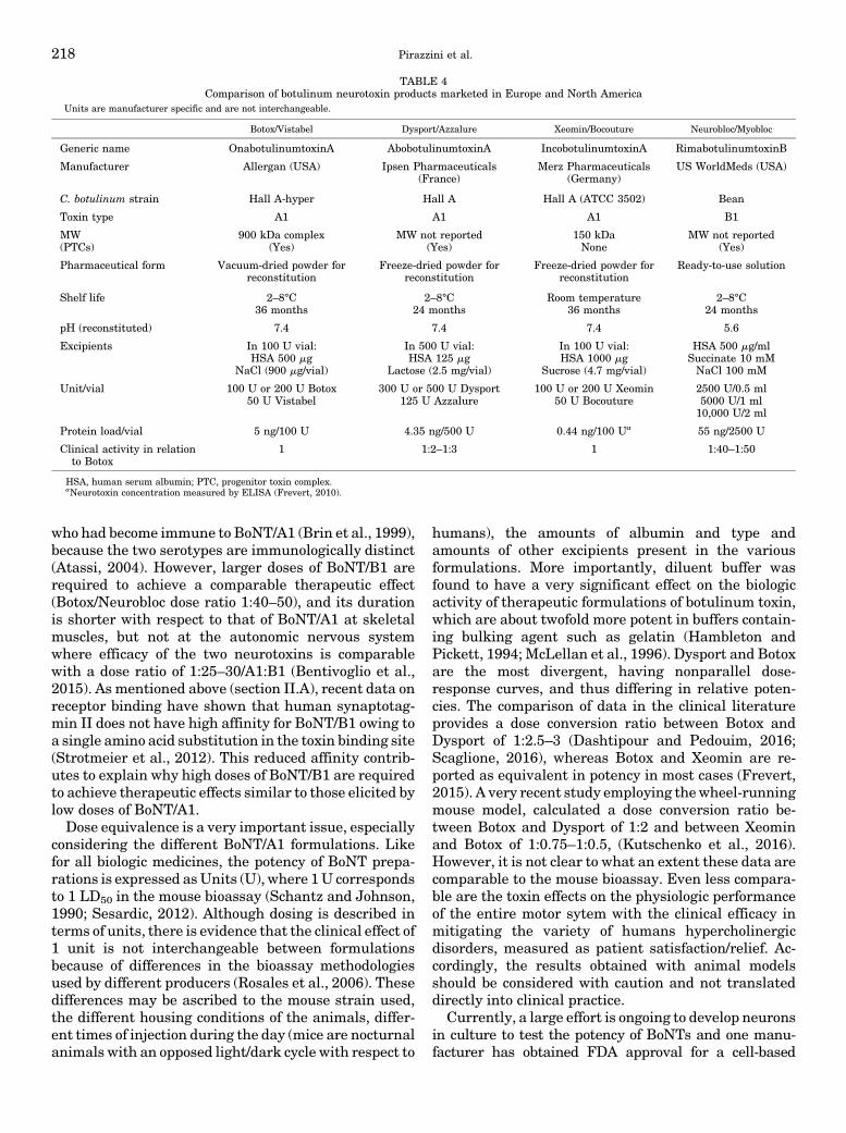
who had become immune to BoNT/A1 (Brin et al., 1999),because the two serotypes are immunologically distinct(Atassi, 2004). However, larger doses of BoNT/B1 arerequired to achieve a comparable therapeutic effect(Botox/Neurobloc dose ratio 1:40–50), and its durationis shorter with respect to that of BoNT/A1 at skeletalmuscles, but not at the autonomic nervous systemwhere efficacy of the two neurotoxins is comparablewith a dose ratio of 1:25–30/A1:B1 (Bentivoglio et al.,2015). As mentioned above (section II.A), recent data onreceptor binding have shown that human synaptotag-min II does not have high affinity for BoNT/B1 owing toa single amino acid substitution in the toxin binding site(Strotmeier et al., 2012). This reduced affinity contrib-utes to explain why high doses of BoNT/B1 are requiredto achieve therapeutic effects similar to those elicited bylow doses of BoNT/A1.Dose equivalence is a very important issue, especially
considering the different BoNT/A1 formulations. Likefor all biologic medicines, the potency of BoNT prepa-rations is expressed asUnits (U), where 1U correspondsto 1 LD50 in the mouse bioassay (Schantz and Johnson,1990; Sesardic, 2012). Although dosing is described interms of units, there is evidence that the clinical effect of1 unit is not interchangeable between formulationsbecause of differences in the bioassay methodologiesused by different producers (Rosales et al., 2006). Thesedifferences may be ascribed to the mouse strain used,the different housing conditions of the animals, differ-ent times of injection during the day (mice are nocturnalanimals with an opposed light/dark cycle with respect to
humans), the amounts of albumin and type andamounts of other excipients present in the variousformulations. More importantly, diluent buffer wasfound to have a very significant effect on the biologicactivity of therapeutic formulations of botulinum toxin,which are about twofold more potent in buffers contain-ing bulking agent such as gelatin (Hambleton andPickett, 1994; McLellan et al., 1996). Dysport and Botoxare the most divergent, having nonparallel dose-response curves, and thus differing in relative poten-cies. The comparison of data in the clinical literatureprovides a dose conversion ratio between Botox andDysport of 1:2.5–3 (Dashtipour and Pedouim, 2016;Scaglione, 2016), whereas Botox and Xeomin are re-ported as equivalent in potency in most cases (Frevert,2015). A very recent study employing thewheel-runningmouse model, calculated a dose conversion ratio be-tween Botox and Dysport of 1:2 and between Xeominand Botox of 1:0.75–1:0.5, (Kutschenko et al., 2016).However, it is not clear to what an extent these data arecomparable to the mouse bioassay. Even less compara-ble are the toxin effects on the physiologic performanceof the entire motor sytem with the clinical efficacy inmitigating the variety of humans hypercholinergicdisorders, measured as patient satisfaction/relief. Ac-cordingly, the results obtained with animal modelsshould be considered with caution and not translateddirectly into clinical practice.
Currently, a large effort is ongoing to develop neuronsin culture to test the potency of BoNTs and one manu-facturer has obtained FDA approval for a cell-based
TABLE 4Comparison of botulinum neurotoxin products marketed in Europe and North America
Units are manufacturer specific and are not interchangeable.
Botox/Vistabel Dysport/Azzalure Xeomin/Bocouture Neurobloc/Myobloc
Generic name OnabotulinumtoxinA AbobotulinumtoxinA IncobotulinumtoxinA RimabotulinumtoxinB
Manufacturer Allergan (USA) Ipsen Pharmaceuticals(France)
Merz Pharmaceuticals(Germany)
US WorldMeds (USA)
C. botulinum strain Hall A-hyper Hall A Hall A (ATCC 3502) Bean
Toxin type A1 A1 A1 B1
MW 900 kDa complex MW not reported 150 kDa MW not reported(PTCs) (Yes) (Yes) None (Yes)
Pharmaceutical form Vacuum-dried powder forreconstitution
Freeze-dried powder forreconstitution
Freeze-dried powder forreconstitution
Ready-to-use solution
Shelf life 2–8°C 2–8°C Room temperature 2–8°C36 months 24 months 36 months 24 months
pH (reconstituted) 7.4 7.4 7.4 5.6
Excipients In 100 U vial: In 500 U vial: In 100 U vial: HSA 500 mg/mlHSA 500 mg HSA 125 mg HSA 1000 mg Succinate 10 mM
NaCl (900 mg/vial) Lactose (2.5 mg/vial) Sucrose (4.7 mg/vial) NaCl 100 mM
Unit/vial 100 U or 200 U Botox 300 U or 500 U Dysport 100 U or 200 U Xeomin 2500 U/0.5 ml50 U Vistabel 125 U Azzalure 50 U Bocouture 5000 U/1 ml
10,000 U/2 ml
Protein load/vial 5 ng/100 U 4.35 ng/500 U 0.44 ng/100 Ua 55 ng/2500 U
Clinical activity in relationto Botox
1 1:2–1:3 1 1:40–1:50
HSA, human serum albumin; PTC, progenitor toxin complex.aNeurotoxin concentration measured by ELISA (Frevert, 2010).
218 Pirazzini et al.
potency assay as a substitute of the animal-based assayin the manufacturing of BOTOX (http://www.allergan.com/responsibility/animal-testing). Clearly, cultured neu-rons do not replicate the complexity of the intoxicationpathway in vivo, but one may assume that their useprovides information about the neuronal toxicity andallows one to determine the reproducibility of the BoNTproduction in terms of lot-to-lot manufacturing consis-tency. This reduces the number of animals to be usedprovided that one is able to define exactly the reproduc-ibility over time of its cultured neuron test system.Clearly, using data generated by different laboratoriesusing different products and different cell-based assayconditions (reagents, cells, media, etc.) will render itdifficult to make meaningful comparison across products.Although only BoNT serotypes A1 and B1 have
reached clinical use, other serotypes have been testedin humans. BoNT/C was demonstrated to have a neuro-paralytic profile very similar to that of BoNT/A1, with asimilar duration of action on human skeletal muscles.BoNT/C represents a valid therapeutic alternative inpatients treated with commercially available productswho have developed an immune response with conse-quent secondary treatment failure (Eleopra et al., 1997,2002, 2004, 2006).A Japanese pilot study has compared the effect of a
BoNT/A2 preparation with that of BoNT/A1 (onabotu-linumtoxinA) on human volunteers by measuring thecompound muscle action potentials of injected extensordigitorum brevis muscles. A comparable onset and dura-tion of action among them was found without significantadverse effects (Mukai et al., 2014). Currently, a Phase2 clinical trial performed in Japan is evaluating efficacyand safety of the use of BoNT/A2 for the treatment ofpoststroke lower limb spasticity (https://clinicaltrials.gov/show/NCT01910363) (Kaji et al., 2015).
C. Immunogenicity of BotulinumNeurotoxin Formulations
Botulinum neurotoxins and associated proteins pre-sent in commercial preparations may elicit antibodyformation when injected into patients. Antibodiesformed against the accessory proteins do not interferewith the biologic activity of the toxin, and they aretherefore nonneutralizing antibodies, whereas anti-bodies formed against the neurotoxin (primarily againstthe heavy chain) may prevent or not prevent its biologicactivity. Those antibodies that prevent it are neutraliz-ing antibodies and are expected to interfere with theclinical efficacy of the product (Dolimbek et al., 2007;Atassi et al., 2012). However, all BoNT serotypes can beconsidered poor antigens, particularly with respect tothe cousin molecule tetanus neurotoxin. Indeed, veryfew patients develop neutralizing antibodies, althoughBoNT therapy is often continued over many years. Theimmunologic human response to BoNT/A1 is very low,whereas it is higher for BoNT/B1 most likely because it
is injected in much larger amounts. The reportedfrequency of anti-BoNT antibodies detection in injectedpatients ranged from0 to 3% for BoNT/A1, depending onthe formulation used, and from 10 to 44% for BoNT/B1(Dressler and Rothwell, 2000; Dressler and Bigalke,2005; Naumann et al., 2013a,2013b). Various factorscan influence the immunogenicity of the botulinumneurotoxin therapeutics, including product-related fac-tors like the manufacturing process, the toxin source,the antigenic protein load, and, perhaps more impor-tant, the presence of inactive or denatured toxin actingas a toxoid. Moreover, treatment-related factors such asthe toxin dose, frequency of injections, and prior exposurevia other routes can impact on the immunogenic response.Another important factor is the site of injection asanatomic regions rich in lymph nodes, such as the neck,aremore likely to produce an immune response. In view ofthe potential risk for secondary treatment failure it isadvisable, in clinical practice, to follow the general rulesforminimizing the risk of developing an adaptive immuneresponse, including the use of the lowest effective dosesand employing the longest clinically acceptable interin-jection interval (Naumann et al., 2013b). Although therecommended minimum intervals between BoNT/A1treatment is 12 weeks, injection intervals as short as6 weeks with Xeomin have been recently shown to be welltolerated andnot associatedwith increased safety concern(Jost et al., 2015).
D. Clinical Applications of Botulinum Neurotoxins
After Scott’s seminal studies in ophtalmology, botu-linum toxins entered the therapeutic arsenal of theneurologist, first with the purpose of relieving hyper-contraction of small muscles of eyelid and of the face(blepharospasm and hemifacial spasm) and largerdystonic muscles of the head and neck (cervical andoromandibular dystonia) and later to treat limb move-ment disorders, including use-dependent cramps (occu-pational dystonia) and limb spasticity (Table 5).Because BoNTs not only block the skeletal cholinergicneuromuscular transmission but also the cholinergicautonomic innervation controlling sweat, tear, salivaryglands, and smooth muscles and sphincters, hyper-hidrotic disorders and urologic or gastrointestinal dis-orders also benefit from the use of botulinum toxins(Comella and Pullman, 2004; Dressler, 2012; Hallettet al., 2013; Naumann et al., 2013b; Ramirez-Castanedaand Jankovic, 2014; Simpson et al., 2016). The myor-elaxant and atrophic effect associated with an ill-knownanalgesic effect of BoNT brings relief from pain andBoNT/A1 has been tentatively used in different painsyndromes (Table 5).
BoNT/A1-induced paralysis can be detected after2–3 days from injection and reaches its maximum valuewithin 1–2 weeks; it maintains this level for a variableperiod of time depending on the anatomic site and thengradually declines to return to the original value within
Biological Actions of Botulinum Neurotoxins 219

3–4 months for skeletal nerve terminals and about1 year for autonomic ones.The assessment of BoNT efficacy as therapeutics for a
given pathologic condition is not an easy task andrequires the comparison of many different clinicalstudies, often performed by different research teamsin different clinical centers. Differences among individ-ual patients and variables such as dosing, dilutions,number of injections per site, targeting [visual, electro-myography (EMG), or ultrasound-guided] influence theoutcome and decrease comparability of data amongdifferent centers. Consequently, many meta-analyses
and quantitative study designs used to systematicallyassess previous research studies were recently per-formed on BoNT clinical applications to have a consol-idated and quantitative review of a large and oftencomplex, sometimes apparently conflicting, body of lit-erature. Accordingly, we have reviewed rigorous meta-analyses on different human pathologic conditionstreated with commercial BoNT preparations, and theresults are reported here for the current therapeuticapplications of BoNT/A1 and BoNT/B1.
1. Dystonias. Dystonias are a heterogeneous groupof disorders characterized by sustained involuntarymuscle contractions, frequently causing repetitive twist-ing movements, abnormal postures, and pain (Albaneseand Lalli, 2009, 2012). Localized injections of BoNTprovide a transient symptomatic relief in primary andnonprimary focal dystonia syndromes, as demonstratedby several randomized controlled studies and by a largenumber of uncontrolled studies (Hallett et al., 2013;Guidubaldi et al., 2014).
In general, the therapeutic benefits of BoNT indystonias derive from a decreased acetylcholine releasefrom alpha motoneurons at the NMJ and the ensuingrelaxation of the affected skeletal muscle. However,substantial evidence suggests that BoNT injected pe-ripherally may also influence indirectly CNS function(Gracies, 2004). In fact, the BoNT inhibition of intra-fusal muscle fibers by blocking also the gamma moto-neurons, reduces muscle spindle afferent input to theCNS and may contribute to the therapeutic effects ofBoNT in focal dystonias (Giladi, 1997; Hallett, 2000;Rosales andDressler, 2010).Moreover there is consider-able evidence that BoNTs, particularly at high doses,can reach higher structures in the brain. This is an areathat deserves further investigation as a detailed un-derstanding of direct central effects of BoNTs willprovide valuable information for present and futuretherapeutic uses of BoNTs, particularly in the case oflower limb spasticity, which requires the use of highdoses (Caleo and Schiavo, 2009; Mazzocchio and Caleo,2015).
BoNT injection is considered the treatment of choicefor most focal and segmental dystonias and in hemi-facial spasm. The efficacy of BoNT for the treatment ofthese movement disorders, which include blepharo-spasm, cervical dystonia, hemifacial spasm, oroman-dibular dystonia, focal limb dystonias, and laryngealdystonia has been recently reassessed by a thoroughmeta-analysis study,which included a total of 42 clinicaltrials (Hallett et al., 2013; Del Sorbo and Albanese,2015).
Blepharospasm is a form of dystonia of the periocularmuscles that produces forced eyelid closure, some-times leading to functional blindness. FDA approvedBoNT/A1 for the treatment of blepharospasm in 1989.Typically, 30–60 U of Botox or Xeomin, 60–180 U ofDysport or 1200–3600 U of Myobloc/Neurobloc are
TABLE 5Therapeutic uses for botulinum neurotoxin
OphtalmologyStrabismusa,b,c
NistagmusNeurology
Focal DystoniasBlepharospasma,b,c
Cervical dystoniaa,b,c (Torticollis, anterocollis, laterocollis)Occupational dystonias (writer’s crampb, musician’s cramps)Laryngeal Dysphoniac
Oromandibular dystoniaLingual dystonia
Nondystonic disordersHemifacial spasma,b,c
Tremor (essential, parkinsonism)TicsBruxism
Spasticity (poststroke, multiple sclerosis, brain or spinal cord injury)Focal spasticitya,b,c: Upper and lower limb spasticityNonfocal: hemispasticity, paraspasticity, tetraspasticityCerebral palsyab
Hyperhidrosisa,b,c
Focal: axillary, palmar, plantarDiffuse
HypersalivationSialorrheab (motoneuron diseases/amyotrophic lateral sclerosis)Droolingb (Parkinsonian syndromes)Frey’s syndrome/gustatory sweating
Aesthetic (muscle)Glabellar rythidesa,b,c
PainMuscular
DystoniaSpasticityChronic myofascial painTemporomandibular disordersLow back pain
NonmuscularMigraine (chronica and tension type migraine)Neuropathic painTrigeminal painPelvic pain
UrologyDetrusor sphincter dyssynergiaOveractive bladdera,b,c (Idiopathic or neurogenic detrusor
overactivity)Urinary retentionBladder pain syndromePelvic floor spasmsBenign prostate hyperplasia
GastroenterologyAchalasiaChronic anal fissures
PsychiatryDepressiond
aUSA approved indication.bEU approved indication.cEvidence-based therapeutic indication.dTo be evaluated.
220 Pirazzini et al.
applied to the orbicularis oculi, procerus and corugatorsupercilii muscles bilaterally. Several studies demon-strated the efficacy and safety of BoNT therapy for thisindication (Hallett et al., 2013). Response rates ofaround 90% make blepharospasm one of BoNT’s mostsuccessful indications. Adverse effects are usually mildand always transient and include local hematoma,ptosis, and diplopia (Dressler, 2012).In cervical dystonia, also referred to as “spasmodic
torticollis”, where dystonia of the neck and shouldergirdlemuscles produces impairment of head control andpain, there is adequate evidence of BoNT efficacy, basedon data from eight randomized, controlled, Class Iclinical trials (Hallett et al., 2013). In clinical practice,the average total dose injected in patients with cervicaldystonia is 100–300 U Botox or Xeomin, 400–800 UDysport, or 10,000–20,000 UMyobloc/Neurobloc. Thesedoses can vary considerably as the recommended rangehas to be adjusted depending on the individual patient’sfeatures and sensitivity to the toxin. BoNT/A1 is thepreferred treatment, whereas BoNT/B1 is recom-mended for A-resistant patients, because injectionsare generally more painful, possibly owing to the acidicpH of its solution, efficacy is shorter and immunogenic-ity higher (Bentivoglio et al., 2015).Focal limb dystonia refers to dystonia affecting one
arm or leg. Primary dystonia of the upper extremitycommonly begins during selective, usually highlyskilled, and repetitive motor tasks and are also referredas occupational or attitudinal dystonia. Typical upperlimb dystonias include musician’s cramps and writer’scramps where BoNT/A1 has been reported to be effec-tive (Karp, 2004). However, due to the functionalcomplexity, the usually high performance levels re-quired and the narrow therapeutic window in theforearm muscles, functional outcome of treatments issometime disappointing compared with those of bleph-arospasm or cervical dystonia. This also derives fromthe difficulty in obtaining the requested quality ofvoluntary movement without some weakness. BoNTapplication under electromyographic or ultrasoundguidance is recommended as it improves the outcome.The lower extremity is commonly affected in subjects
with genetic dystonia (e.g., DYT-1 Dystonia), whereasupper limb and foot dystonia can be either idiopathic, inthe context of a generalized dystonia, or symptomatic,as in Parkinson’s disease or in juvenile cerebral palsy.Successful treatments with BoNT have been reported butno controlled trials are available (Schneider et al., 2006).Higher doses of BoNT may need to be injected in the limbthan in hand dystonia becausemotor control is less refined.Hemifacial spasm describes synchronous unilateral
contractions presumably caused by vascular compres-sion of the facial nerve and is generally treated withlower doses than blepharospasm (Bentivoglio et al.,2009). Although experience with BoNT in hemifacialspasm treatment mainly originates from open-label
trials, there is no doubt on its efficacy and safety(Guidubaldi et al., 2014). Notwithstanding the limitednumber of studies available, it appears that othernondystonic disorders including tics and tremors alsobenefit from BoNT therapy (Lotia and Jankovic, 2016).
2. Spasticity. Spasticity describes the combinationof a central paresis together with various forms ofmuscle hyperactivity, including dystonia, rigidity, andspasms often associated with pain. Most frequentetiologies include cerebral stroke, multiple sclerosis,traumatic brain injury, spinal cord injury, and infantilecerebral palsy. The goal of spasticity treatment is toreduce motor overactivity to improve movement with-out worsening weakness. BoNT treatment of spasticitycan reduce muscle tone, improve function, facilitatenursing, and prevent contractures and decubitus. How-ever, successful spasticity management requires amultiprofessional task force where all medical andsurgical treatments need to be combined with physicalinterventions. Rehabilitation treatment of poststrokepatients (i.e., muscle stretching exercises, eventuallycombined with neuromuscular electrical stimulation),when begun immediately after injection enhances theefficacy of BoNT therapy (Ward, 2002). However, recentsystematic reviews have concluded that BoNT/A1 in-jections have to be considered as the pharmacologicaltreatment of choice in focal spasticity to improvelimb position, functional ability and to reduce pain(Esquenazi et al., 2013; Simpson et al., 2016), whereaslimited data are available on the efficacy of BoNT/B1 inspasticity. BoNT therapeutic effects are better estab-lished for spasticity in the upper rather than lower limb(Esquenazi et al., 2013). It usually involves large BoNTdoses: typically 300–500 U Botox or Xeomin or 600–1000 U Dysport for arm or leg spasticity. Recent studiesreported treatment of poststroke patients with up to800 U of Botox, 1800 Dysport, and up to 1200 U ofXeomin without signs of systemic toxicity (Dressleret al., 2015; Santamato et al., 2015). A higher dilutionresults in larger injection volumes and higher degree ofparalysis (Kutschenko et al., 2016), probably because oflarger spreading to neuromuscular junctions remotefrom injection site (Gracies et al., 2009).
3. Autonomic Disorders. Botulinum neurotoxins in-hibit neuroexocytosis from cholinergic nerve terminalsof the sympathetic and parasympathetic autonomicnervous systems. The use of BoNT within the broadcategory of autonomic indications includes hypersecre-tory disorders, such as hyperhidrosis, sialorrhea, andchronic rhinorrhea, and smooth muscle hyperactivitiesin urologic and gastrointestinal disorders. Owing to anapparently higher affinity for cholinergic autonomicnerve endings, BoNT/B1 is as efficacious as BoNT/A1in the therapy of secretory disorders both in terms ofextent of inhibition and duration of the beneficial effectsand at lower relative doses than those required inskeletal muscles (Bentivoglio et al., 2015).
Biological Actions of Botulinum Neurotoxins 221

Hyperhidrosis is medically benign but may be asocially devastating condition characterized as it is byexcessive sweating, which may be occurring focallywithin the axillary region or it may extend to palmsand soles. A formalized assessment of the AmericanAcademy of Neurology confirmed that BoNT therapy issafe and effective for axillary hyperhidrosis (Naumannet al., 2013b); total doses per axilla of 50–100 U Botox orXeomin, 100–200UDysport or 2500UNeuroBloc/Myoblocmay be used (Bentivoglio et al., 2015).Hypersalivation (or sialorrhea) refers to the presence
of excessive saliva in the mouth, which may causedrooling with consequent severe embarrassment forthe affected people. In several cases hypersalivation issecondary to other pathologic conditions includingParkinsonian syndromes, motoneuron diseases (amyo-trophic lateral sclerosis) and cerebral palsy and iscaused by impaired swallowing of saliva. The totaltherapeutic dose injected into the parotid glands forsialorrhea is tipically 2500 U for NeuroBloc/Myobloc,whereas BoNT/A1 ranges from 50U for Botox to 75–450U for Dysport (Naumann et al., 2013b).Allergic rhinitis is a common disorder, one that is not
disabling by itself, but it imposes a substantial burdenin medical costs and indirect costs due to loss ofproductivity (Simoens and Laekeman, 2009). BoNT/A1may be a good alternative for the treatment of rhinitispatients who are unresponsive to other treatmentmethods. Some restrictions such as painful injectionsand needing of specialized experience for adminitrationprevent its wide use (Ozcan and Ismi 2016). BoNT forthe treatment of allergic rhinitis is supported by a levelB recommendation (probably effective) (Naumannet al., 2013b).4. Urologic Pathologic Conditions. The therapeutic
applications of BoNT in urology include detrusorsphincter dyssynergia, lower urinary tract symptomsdue to benign prostatic hyperplasia, and detrusor over-activity (both neurogenic and idiopathic). These condi-tions are common among patients with spinal cordlesions of different types (multiple sclerosis, spinal cordtumors, other spinal cord diseases, traumatic spinalcord injuries). The mechanism(s) underlying the thera-peutic actions of BoNT in urologic disorders is not fullyunderstood but a complex inhibitory action on recep-tors, neuropeptides, and neurotransmitters involved inthe pathophysiology of the overactive bladder has beenproposed (Chancellor et al., 2008). In the bladder, BoNTis thought to act primarily by inhibiting acetylcholinerelease from parasympathetic nerve endings inducingdetrusormuscle relaxation (Schurch et al., 2000; Fowleret al., 2008). In addition, basic and clinical evidencesuggests that BoNTmay have sensory inhibitory effectsunrelated to its actions on acetylcholine release(Apostolidis et al., 2005). Indeed, BoNT/A1 reduces therelease of glutamate, of substance P, and of calcitoningene-related peptide from the peripheral terminals of
afferent bladder neurons (Duggan et al., 2002; Rappet al., 2006) and of neurotrophins from the urothelium(Chancellor et al., 2008). The BoNT inhibition ofsecretion of these neuropeptides and ATP as well ascyclooxygenase-2 products, which are inflammatoryresponse mediators released from nociceptive sensoryendings in response to noxious stimuli (Khera et al.,2004; Chuang et al., 2008), would explain the therapeu-tic benefit of BoNT injection in painful bladder condi-tions (Smith et al., 2004; Wang et al., 2016).
BoNT/A1 injection for overactive bladder treatment isalso associated with a significant decrease of purinergicreceptors P2X3 and capsaicin receptors TRPV1, tworeceptors involved in nociception in the suburothelialnerve fibers (Apostolidis et al., 2005; Liu et al., 2014).These changes may reflect a direct activity of BoNT onthe afferent innervation of the bladder and/or asecondary effect on the efferent innervation of thedetrusor. Moreover, evidence of BoNT/A retrogradetransport to the CNS after bladder injection in ratshas been recently provided (Papagiannopoulou et al.,2016). Further studies are needed to elucidate thistransport and its significance with respect to possiblechanges induced by BoNTA1 in the CNS after bladderinjection.
Most studies have reported injection doses of 100–300 U of Botox, with 100 U being a typical starting dosefor idiopathic detrusor overactivity and higher doses forthose patients with insufficient response to an initialinjection or those with neurogenic detrusor overactivity(Gomelsky and Dmochowski, 2014; Cheng et al., 2016).The injected volume is a key factor in the spread andaction of BoNT/A in the bladder, with a wider diffusionof the toxin when higher volumes are injected (Coelhoet al., 2012). In an experimental animal model of severebladder pain, intrathecal injection of Botox has a stronganalgesic effect, and this route of administration shouldbe further explored for “intractable” forms of pain(Coelho et al., 2014).
5. Pain. Despite current availability of a largenumber of analgesic drugs, management of chronic painis still a challenge for clinicians. Over the past decade,BoNT treatment has shown efficacy in a large spectrumof human pain disorders, and the potentially relevantuse of BoNT/A1 has been reported as efficacious in someform of chronic pain refractory to other treatments. Theestablished ability to produce neurotoxins by recombi-nant protein expression (Bade et al., 2004; Band et al.,2010; Masuyer et al., 2014, 2015) and to modify them tocreate engineered neurotoxins with enhanced specific-ity for nociceptive nerve terminals will expand thetherapeutic utility of BoNT for pain treatment. In thiscontext, an innovative class of biopharmaceuticalsobtained by replacing the native binding domain withanother protein to redirect the light chain to a differentnerve or cell has been recently proposed (Chaddocket al., 2004; Pickett, 2010; Masuyer et al., 2014).
222 Pirazzini et al.
The analgesic effects of BoNT/A1 were first reportedin 1985 in a pilot study of BoNT/A treatment of cervicaldystonia, characterized by abnormal, involuntary neckand shoulder muscle contractions and often resulting insignificant, disabling musculoskeletal pain (Tsui et al.,1985). Early in its use as a therapeutic agent, BoNT/A1was observed to provide pain relief in spastic andnonspastic muscle conditions in humans such as myo-facial pain syndromes, temporomandibular disorderand bruxism, low back pain, prostatic pain, tensiontype and migraine headache, different types of neuro-phatic pain syndromes and many others (Wheeler andSmith, 2013). However, no effect was reported in amodel of pain perception in human skin (Schulte-Mattler et al., 2007).The association betweenBoNT/A1 and pain relief was
originally thought to correlate only to its effect onmuscle overcontraction or contractures. However, it isparticularly interesting that the analgesia provided byBoNT injection occurs before muscle paralysis andoutlasts any muscle weakness. It is now well docu-mented that the analgesic effects of BoNT/A1 arerelated not only to its paralytic effect, but also toan effect on the nociceptor system (Wheeler andSmith, 2013). The widely reported antinociceptiveeffect of BoNT/A1 would be primarily mediated by theblockade of neuropeptides and inflammatory mediatorsrelease, and by the inhibition of plasma membraneexposure of pain sensors at peripheral level (asdescribed previously for bladder pain). Indeed, incultured sensory neurons, the TNF-a induced surfacetrafficking of TRPV1 and TRPA1 channels is mediatedby SNAP25, VAMP-1 and syntaxin-1 and is inhibited bythe serotypes of BoNTs that selectively cleave theirrespective SNAREs (Meng et al., 2014, 2016). ThisBoNT/A1 inhibitory effect would reduce peripheralsensitization and afferent input to the spinal cord,thereby dampening indirectly central sensitization.However, a growing number of experimental andclinical data on BoNT/A1 antinociceptive action cannotbe explained adequately only by the inhibition of therelease of peripheral neurotransmitter/inflammatorymediators. It is envisaged that the central effect maybe directly involved, via retro and anterograde axonaltransport along the branches of nociceptive neurons(Matak and Lackovic, 2014; Luvisetto et al., 2015;Mazzocchio and Caleo, 2015; Pellett et al., 2015d).Several studies reported toxin bilateral actions indistant regions after unilateral injection of BoNT/A1,suggesting that peripherally administered BoNT/A1reaches the CNS region via axonal transport totarget neurotransmission of pain sensory circuits.Enzymatic activity of BoNT/A1 has been immunohis-tochemically detected in spinal cord or brain stemareas, to which sensory and motoneurons in BoNT/A1-injected peripheral regions are projected (Matak andLackovic, 2014; Luvisetto et al., 2015). Based on these
and other evidence it was suggested that the BoNT/Aaction on pain is dominantly a central effect (Matakand Lackovic, 2014). Moreover, recently, in a rat modelof neuropathic pain, BoNT/A was shown to influencemicroglial activation and to restore neuroimmune bal-ance by reducing the levels of pronociceptive factors(IL-1 b and IL-18). In addition, it appears to increasethe level of antinociceptive factors (IL-10 and IL-1RA)in the spinal cord and in the dorsal root ganglia(Zychowska et al., 2016). These findings suggest thatneuroimmunological changes are also involved inBoNT/A-mediated analgesia.
Within the pain medicine field, only chronic migraineis an approved FDA indication for BoNT/A1. All otherareas are currently considered off label, although forseveral of them, the available literature alreadystrongly suggests efficacy. Concerning the evidence-based efficacy, the lack of standardized guidelines forBoNT application and dosage for pain management,and the lack of appropriate definition of study primaryoutcomes, in addition to the small sample size andlimited number of randomized controlled clinical trials,have led to contradictory or negative findings in someforms of pain (Jabbari and Machado, 2011). Only someexamples of chronic pain conditions for which prospec-tive and controlled data are available on BoNT efficacyare presented in this paragraph.
a. Neuropathic pain. This is one of themost commonforms of human pain, which derives from damage ordysfunction of the peripheral or central nervous sys-tems; in some cases it may result from nociceptiveinjury (Argoff, 2002). Failure of response to currentanalgesic medications is not uncommon. The data ontype A1 toxin (mostly Botox has been used in thesestudies) are encouraging and indicate efficacy or prob-able efficacy in three major and common forms ofneuropathic pain, namely, postherpetic neuralgia, post-traumatic neuralgia, and painful diabetic neuropathy.For example, in one study, to reduce the tension of thetendons passing through the tarsal tunnel that werecausing pain, 120 U permuscle (dilution 200U/ml) wereinjected under electromyographic guidance into all themuscles involved. The treatment was repeated after3 months and resulted in a defined reduction of thenumber of dysethesias with a clear gait improvementassociated to the BoNT/A injections (Buonocore et al.,2017). In addition, a recent systematic review of thetherapeutic value of BoNT/A subcutaneous or intrader-mal injection for the therapy of trigeminal neuralgiaconcluded that this treatment is to be considered as analternative option to the surgery (Morra et al., 2016).Much remains to be learned about the most effectivetechnique of injection, most effective dose, optimaldilutions, and differences among different BoNTs inthe treatment of neuropathic pain.
b. Primary headaches. These diseases are catego-rized in migraine or tension headaches. Migraine is a
Biological Actions of Botulinum Neurotoxins 223
disabling disease for the affected patients and it isdifferentiated in episodic (,15 headaches per month) orchronic ($15 headaches per month). Chronic migraineis the only pain disorder with approved BoNT/A1 use(Chen, 2012), notwithstanding the fact that the mech-anism of action is still not completely elucidated(Luvisetto et al., 2015). Migraine is thought to be causedby brain stem neuronal hyperexcitability, corticalspreading depression, abnormal release of neurotrans-mitters/neuropeptides, and trigeminal system activa-tion (Pietrobon and Moskowitz, 2013). Other datasupport the role of peripheral activation via the sensorynerves surrounding blood vessels of the head and neck(Olesen et al., 2009) and the role of vasodilatation of theextracranial arteries (Shevel, 2011).An extensive meta-analysis involving 23 studies and
more than 5000 patients has analyzed the BoNT/A1efficacy in episodic migraine, chronic migraine, chronicdaily headache and tension type headache (Jacksonet al., 2012). It was concluded that BoNT/A1may reducethe mean number of headache days per month inchronic migraine and chronic daily headache, in com-parison with placebo. BoNT/A1 was not beneficial in thetreatment of episodic migraine or tension-type head-ache (Simpson et al., 2016).6. Other Applications. The therapeutic possibilities
of BoNTs are manifold and certainly not yet exhausted.Controlled studies for many indications are frequentlylacking because of the limited incidence of some disor-ders and symptoms.a. Gastroenterology and proctologic disorders.
BoNT in the treatment of spastic gastrointestinaldisorders has gained widespread acceptance over thelast 15 years, especially for achalasia and chronic analfissures. Its administration is generally safe and rela-tively noninvasive compared with alternative therapies(Brisinda et al., 2015).Achalasia is an idiopathic esophageal motility disor-
der characterized by incomplete relaxation of thesmooth muscles of the lower esophageal sphincter(LES) with swallowing and aperistalsis in the esopha-geal body. Because the pathologic hallmark of achalasiais a loss of nitric oxide-releasing inhibitory neurons inthe esophageal myenteric plexus resulting in unop-posed excitatory activity, BoNT injection counterbal-ances the increased muscle contraction by decreasingLES pressure, and hence enables passive esophagealemptying and improve the symptoms (dysphagia, re-gurgitation, and chest pain) (Pasricha et al., 1995; Lakeand Wong, 2006). BoNT injection into all four LESquadrant (80–100 U Botox or 240–400 U Dysport intotal) is the most common initial endoscopic therapy forachalasia in the United States, owing to its excellentsafety profile, high initial response rate, and ease ofadministration. However, BoNT injection does notcompare favorably in terms of duration of action withinvasive treatments such as pneumatic dilation or
laparoscopic Hellers myotomy, which are successful in80% of patients; moreover, repeated treatments wereshown to make subsequent Heller myotomy morechallenging (Zaninotto et al., 2004; Smith et al., 2006).Thus, BoNT/A1 injection has not been recommended asa first-line therapy for achalasia, although it has animportant role to play in elderly patients and in patientswith comorbidities who are not candidates for definitivetherapy (Ramzan and Nassri, 2013; Pandolfino andGawron, 2015).
Chronic anal fissure is an ischemic ulceration relatedto sphincterhypertonia. Lateral internal sphincterotomy,the most common treatment of chronic anal fissure,may cause permanent injury to anal sphincter, lead-ing to fecal incontinence. Extensive studies begun longago (Gui et al., 1994; Maria et al., 1998) have led toBoNT injection becoming the standard treatment.BoNT is more than a symptomatic treatment of analfissure as it produces a reversible sphincterotomy re-ducing the sphincter pressure and, in addition, itinfluences the nitric oxide synthase and other agents,thus interfering with the pathogenic mechanisms ofthe chronic fissure. The exact site and dose of injec-tion remains somewhat unsettled with most studiesinjecting the internal anal sphincter and others injectingthe external anal sphincter laterally to the fissure with2.5–10 U Botox or 10–40 U Dysport (Jost, 2006; Brisindaet al., 2015).
b. Depression. Depression is a psychiatric conditionaffecting millions of people worldwide and it can betreated with many psychotropic substances, whichcause side effects, are difficult to be standardized andwhose therapeutic outcomes are often unsatisfactory(Gilmer et al., 2005; Rush et al., 2006; Helton andLohoff, 2015). A single injection of BoNT/A1 in theglabellar frown lineswas reported to improve symptomsin ten patients suffering of depressive disturbs (Finziand Wasserman, 2006). Thereafter, three main ran-domized control trials were reported; the toxin wasadministered as monotherapy or as adjunctive treat-ment and patients were evaluated after 3–6 weeks(Wollmer et al., 2012; Finzi and Rosenthal, 2014;Magid et al., 2014). Results corroborated the initialevidence of a beneficial effect of BoNT/A1 for thetreatment of depression after injection in the glabellarfrownmuscles of the forehead (Magid et al., 2015).Moodimprovement in BoNT/A1-treated patients is explainedin the facial feedback hypothesis as linked to theemotional perception to facial expression (e.g., smilingled to happiness and frowning led to sadness). In otherwords, the neuroparalytic effect of the toxin mayimprove the proprioceptive perception, neutralizingthe sadness by neutralizing frowning (Magid et al.,2014, 2015). On the other hand, reduction of depressivesymptoms was reported to continue after BoNT/A1cosmetic effect was ceased, entailing that the antide-pressive effect may not be totally dependent on toxin
224 Pirazzini et al.

paralytic action (Magid et al., 2014). It is thereforetempting to speculate a possible direct effect of the toxinon central circuits. Intriguingly, functional magneticresonance imaging showed that BoNT/A1 injection infrown muscles leads to a functional uncoupling of brainstem centers with the amygdala, resulting in a re-duction of neuronal activity within the central circuit-ries of emotion (Hennenlotter et al., 2009; Kim et al.,2014). Although this application field is only at thebeginning, and further studies are necessary to validateBoNTs efficacy in the treatment of depression, theseinitial results are encouraging, particularly consideringthat they are based on the readaptation of the well-established and safe procedure of BoNT injection forcosmetic purposes.7. Cosmetic Uses. The present major use of
BoNT/A1 in humans is related to a variety of cosmeticindications (Carruthers et al., 2016). Recently pub-lished statistics from the International Society ofAesthetic Plastic Surgery show that BoNT/A1 injectionis the most popular of all cosmetic procedures world-wide (Frevert, 2015). Current estimates suggest thatapproximately half of themedical production ofBoNT/A1is used in aesthetic medicine. The serendipitous obser-vation that BoNT/A1 smoothed facial lines when usedtherapeutically (Carruthers andCarruthers, 1992) led tostudy the toxin effect on glabellar lines, which areperceived as a sign of aging and of negative emotions.The repetitive contraction and activity of the procerusand the corrugator supercilii muscles involved in facialexpression is mainly responsible for glabellar lines andcan be attenuated by BoNT/A1 injection into three to fivesites of these muscles (Gendler and Nagler, 2015). Arecent meta-analysis showed that a 20 U total dose ofBoNT/A1 is remarkably effective and safe for the treat-ment of glabellar lines (Guo et al., 2015). BoNT/A1formulations have been FDA approved for glabellar lines(Vistabel, Allergan; Azzalure, Dysport, and Bocouture,Merz), and very recently Bocouture in Europe has beenapproved also for the treatment of horizontal frown linesand lateral periorbital lines. However, BoNT/A1 can beused to reduce all wrinkles in all skin areas of the faceand the neck as long as they are based on increasedmuscle tone. Best results are obtained when BoNT iscombined with filler substances to correct nondynamicaspects of the skin lines (Dressler, 2012). Repeated injec-tions have not been reported to cause permanent damagesto the superficial muscles involved and this is in line withthe findings obtained in the treatment of dystonic muscles(please see next paragraph).
E. Adverse Effects
A key feature of BoNT/A1 is a minimal effect even atshort distances from the site of injection, a property thatensures optimal results in most of the clinical applica-tions. However, the possibility of local diffusion orpotential leakage into the systemic circulation exists
and depends on a number of factors including volumeand speed of injection, dose, and site of injection. Theparalytic effect outside the target muscle is due to localdiffusion and is generally limited to adjacent muscles,which, for example, could cause ptosis, heavy brow, or afrozen face in facial aesthetics (de Almeida and DeBoulle, 2007). However, it should be considered that thediffusion of BoNT from the site of injection is accom-pained by its progressive dilution in larger and largervolumes of extracellular fluids. Accordingly, the in-jected toxin becomes so diluted as to be no longercapable of binding to the presynaptic membrane.
A systematic review andmeta-analysis of randomizedclinical trials did not find reports of serious side effectsdue to BoNT treatment and indicated that the overallrate of adverse events, from mild to moderate, was 25%in BoNT/A-treated subjects, compared with 15% in theplacebo group (Naumann and Jankovic, 2004). Auto-nomic side effects are rare and occur farmore often afterBoNT/B1 than after BoNT/A1 treatment, probablyowing to a higher affinity of BoNT/B1 for cholinergicautonomic nerve endings (Dressler and Benecke, 2004).Indeed the mutation on human synaptotagmin II thatdecreases its affinity for BoNT/B is not present onhuman synaptotagmin I (Strotmeier et al., 2012), whichis the isoform mainly expressed in peripheral auto-nomic and sensory innervations but not in motorendplates (Li et al., 1994).
Importantly, the long and very extensive use of BoNTtherapy indicates that it is remarkably safe whenadministered by authorized medical personell, usinglicensed toxin preparations. In contrast, the handling ofBoNTs by unskilled/nonauthorized persons or the use ofcounterfeit or unapproved agents that have permeated themarket worldwide, or their combination, may place pa-tients at risk for potentially devastating consequences. Infact, four cases of iatrogenic botulism have been describedfollowing cosmetic injectionwith high dose of an unlicensedBoNT/A1 preparation (Chertow et al., 2006).
F. Contraindications and Drug Interactions
Contraindications to BoNT therapy are limited to ahypersensitivity to the toxin or an infection at the site ofinjection, but adequate hypersensitivity tests havenot been yet developed. An increased monitoring isrecommended in patients treated simultaneously withaminoglycosides, anticholinergics, or other neuromuscular-blocking agents. Due to higher affinity for autonomicnerve terminals, BoNT-B should be applied carefully inpatients with pre-existent autonomic dysfunction, ad-ditional anticholinergic treatment, and in conditionswhere anticholinergics are contraindicated (Dresslerand Benecke, 2004).
G. Botulinum Neurotoxin in Pregnancy
There is no definitive evidence that BoNT-A treat-ment during pregnancy carries a risk. There are
Biological Actions of Botulinum Neurotoxins 225
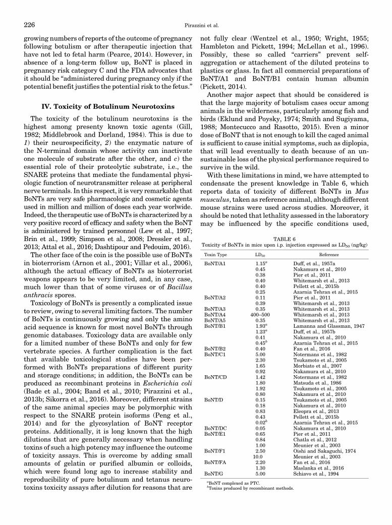
growing numbers of reports of the outcome of pregnancyfollowing botulism or after therapeutic injection thathave not led to fetal harm (Pearce, 2014). However, inabsence of a long-term follow up, BoNT is placed inpregnancy risk category C and the FDA advocates thatit should be “administered during pregnancy only if thepotential benefit justifies the potential risk to the fetus.”
IV. Toxicity of Botulinum Neurotoxins
The toxicity of the botulinum neurotoxins is thehighest among presenty known toxic agents (Gill,1982; Middlebrook and Dorland, 1984). This is due to1) their neurospecificity, 2) the enzymatic nature ofthe N-terminal domain whose activity can inactivateone molecule of substrate after the other, and c) theessential role of their proteolytic substrate, i.e., theSNARE proteins that mediate the fundamental physi-ologic function of neurotransmitter release at peripheralnerve terminals. In this respect, it is very remarkable thatBoNTs are very safe pharmacologic and cosmetic agentsused in million and million of doses each year worlwide.Indeed, the therapeutic use of BoNTs is characterized by avery positive record of efficacy and safety when the BoNTis administered by trained personnel (Lew et al., 1997;Brin et al., 1999; Simpson et al., 2008; Dressler et al.,2013; Attal et al., 2016; Dashtipour and Pedouim, 2016).The other face of the coin is the possible use of BoNTs
in bioterrorism (Arnon et al., 2001; Villar et al., 2006),although the actual efficacy of BoNTs as bioterroristweapons appears to be very limited, and, in any case,much lower than that of some viruses or of Bacillusanthracis spores.Toxicology of BoNTs is presently a complicated issue
to review, owing to several limiting factors. The numberof BoNTs is continuously growing and only the aminoacid sequence is known for most novel BoNTs throughgenomic databases. Toxicology data are available onlyfor a limited number of these BoNTs and only for fewvertebrate species. A further complication is the factthat available toxicological studies have been per-formed with BoNTs preparations of different purityand storage conditions; in addition, the BoNTs can beproduced as recombinant proteins in Escherichia coli(Bade et al., 2004; Band et al., 2010; Pirazzini et al.,2013b; Sikorra et al., 2016). Moreover, different strainsof the same animal species may be polymorphic withrespect to the SNARE protein isoforms (Peng et al.,2014) and for the glycosylation of BoNT receptorproteins. Additionally, it is long known that the highdilutions that are generally necessary when handlingtoxins of such a high potency may influence the outcomeof toxicity assays. This is overcome by adding smallamounts of gelatin or purified albumin or colloids,which were found long ago to increase stability andreproducibility of pure botulinum and tetanus neuro-toxins toxicity assays after dilution for reasons that are
not fully clear (Wentzel et al., 1950; Wright, 1955;Hambleton and Pickett, 1994; McLellan et al., 1996).Possibly, these so called “carriers” prevent self-aggregation or attachement of the diluted proteins toplastics or glass. In fact all commercial preparations ofBoNT/A1 and BoNT/B1 contain human albumin(Pickett, 2014).
Another major aspect that should be considered isthat the large majority of botulism cases occur amonganimals in the wilderness, particularly among fish andbirds (Eklund and Poysky, 1974; Smith and Sugiyama,1988; Montecucco and Rasotto, 2015). Even a minordose of BoNT that is not enough to kill the caged animalis sufficient to cause initial symptoms, such as diplopia,that will lead eventually to death because of an un-sustainable loss of the physical performance required tosurvive in the wild.
With these limitations in mind, we have attempted tocondensate the present knowledge in Table 6, whichreports data of toxicity of different BoNTs in Musmusculus, taken as reference animal, although differentmouse strains were used across studies. Moreover, itshould be noted that lethality assessed in the laboratorymay be influenced by the specific conditions used,
TABLE 6Toxicity of BoNTs in mice upon i.p. injection expressed as LD50 (ng/kg)
Toxin Type LD50 Reference
BoNT/A1 1.15a Duff, et al., 1957a0.45 Nakamura et al., 20100.38 Pier et al., 20110.40 Whitemarsh et al., 20130.40 Pellett et al., 2015b0.25 Azarnia Tehran et al., 2015
BoNT/A2 0.11 Pier et al., 20110.39 Whitemarsh et al., 2013
BoNT/A3 0.35 Whitemarsh et al., 2013BoNT/A4 400–500 Whitemarsh et al., 2013BoNT/A5 0.35 Whitemarsh et al., 2013BoNT/B1 1.93a Lamanna and Glassman, 1947
1.23a Duff, et al., 1957b0.41 Nakamura et al., 20100.45b Azarnia Tehran et al., 2015
BoNT/B2 0.40 Fan et al., 2016BoNT/C1 5.00 Notermans et al., 1982
2.30 Tsukamoto et al., 20051.65 Morbiato et al., 20070.92 Nakamura et al., 2010
BoNT/CD 1.42 Notermans et al., 19821.80 Matsuda et al., 19861.92 Tsukamoto et al., 20050.80 Nakamura et al., 2010
BoNT/D 0.15 Tsukamoto et al., 20050.18 Nakamura et al., 20100.83 Eleopra et al., 20130.43 Pellett et al., 2015b0.02b Azarnia Tehran et al., 2015
BoNT/DC 0.05 Nakamura et al., 2010BoNT/E1 0.65 Pier et al., 2011
0.84 Chatla et al., 20121.00 Meunier et al., 2003
BoNT/F1 2.50 Oishi and Sakaguchi, 197410.0 Meunier et al., 2003
BoNT/FA 2.20 Fan et al., 20161.30 Maslanka et al., 2016
BoNT/G 5.00 Schiavo et al., 1994aBoNT complexed as PTC.bToxins produced by recombinant methods.
226 Pirazzini et al.

including cage density, time of injection during the day,diet, that are not usually reported. Table 6 gives themouse lethal dose 50 values (MLD50), i.e., the dose thatcauses 50% death in caged mice within 4 days, asdetermined by intraperitoneal injection. The toxicityof BoNT/A1 in humans is not known but an estimationcan be made by extrapolating the toxicity data obtainedinmonkeys, whose intramuscular LD50 is reported to be39 U/kg (Scott and Suzuki, 1988) and the intravenousLD50 is 40 U/kg (Herrero et al., 1967), with U corre-sponding to one mouse LD50. Considering 6 kg themedian weight of a Rhesus monkey and 70 kg that of ahuman, the intramuscular LD50 would be 234 U and2730 U in Rhesus monkeys and in humans, respec-tively. In the case of BoNT/F1, subcutaneous lethal dosein Rhesus monkey was estimated to be 25 U/kg and 4 �106 U/kg for the oral route of administration (Dolmanand Murakami, 1961).The available data on the intravenous and intraper-
itoneal routes of BoNTs injection are comparable interms of general toxicity, whereas the intramuscularroute of injection in mice is less effective (Aoki, 2001). Itwas estimated that in less than 20 minutes, part of theinjected toxin has already bound and internalizedwithin local neurons in such a way that it cannotbe neutralized any longer by antitoxin antibodies(Simpson, 2013). However, very few pharmacokineticstudies on distribution, metabolism, and excretion ofBoNTs have been performed and all of them employedmultilethal doses of toxin (Simpson, 2013). It is there-fore not clear to what an extent they can be valid for thecase of local injection of minimal doses of toxin as itoccurs when BoNT is used as a drug. On the other hand,as mentioned above (section III.A), no information isavailable on the pharmacokinetic of BoNT productsused clinically, for reasons mostly related to the verylow therapeutic doses that are below detection incirculating fluids.BoNTs toxicity is lower via the oral route of delivery.
In fact, BoNTs have to pass through acidic and protease-rich anatomic regions before being absorbed in the firstportion of the intestinal tract; this lowers considerablythe number of intact BoNT molecules that reach thelymphatic and blood circulations. Consequently, theoral/intraperitoneal toxicity ratio varies considerablyfor the different toxin serotypes and for the animalspecies taken into account. As an example, humansseem to be refractory to BoNT/C envenomation throughthe oral route, because basically no records but one(Oguma et al., 1990) of BoNT/C-botulism are reported,whereas they are higly sensitive to the toxin wheninjected intramuscularly (Eleopra et al., 1997, 2002,2006).Moreover, the type of toxin preparation, i.e., toxinalone, or in complex with NTNHA or as PTC, stronglyinfluences the toxicity of the oral route. In fact, thestructure of the BoNT/NTNHA dimer, shown in Fig. 2A,clearly indicates that NTNHA protects a large part of
the BoNT surface from proteases and the attackof chemically reactive molecules (Gu et al., 2012;Eswaramoorthy et al., 2015; Lam and Jin, 2015).Accessory proteins are commonly believed to protectBoNTs, but their most important role in toxicityappears to be that of mediating the binding of PTC tothe intestinal mucus and to the polarized epithelialmonolayer (Ohishi et al., 1977; Ohishi and Sakaguchi,1980; Couesnon et al., 2012; Amatsu et al., 2013;Fujinaga et al., 2013; Lee et al., 2013; Lam and Jin,2015; Lee et al., 2015; Rossetto et al., 2014; Rummel,2015). The accessory proteins do not influence BoNTtoxicity via the intraperitoneal, intravenous, and in-tramuscular routes, because PTC rapidly dissociates atthe neutral pH of circulating fluids (Eisele et al., 2011;Frevert, 2015); therefore, they do not contribute to thetherapeutic/cosmetic effect of the different BoNT prep-arations commercially available.
The inhalational route has not caused botulism,except from one laboratory case (Holzer, 1962; Parkand Simpson, 2003), and is considered only with respectto a possible use of BoNT in bioterrorist attack. InRhesus macaques, the ratio of toxicity intraperitoneal/inhalation was estimated to be around 11 for BoNT/A1(Sanford et al., 2010). The situation appears to be similarin humans, where the lethal dose was estimated to bebetween 10 and 13 ng/kg for inhaled BoNT/A1 (Arnonet al., 2001).
A frequently used toxicity assay is the one introducedby Boroff and Fleck (1966), which measures the time todeath of mice injected in the tail vein with a very highdose of BoNT (103–105 MLD50). The animal dies withindozens ofminutes from injection. It should be noted thatno post mortem pathologic examinations were reportedby Boroff and Fleck (1966). This assay is very pratical,because it diminishes the experimental time, but itprovides figures that are only partially comparable withthe mouse lethal dose values. In fact, at the very highdoses of toxin used, BoNTs are likely to bind to non-cholinergic nerve terminals and to other cells, i.e., thechromaffin cells of the surrenal glands or neurons of thedorsal ganglia. These cells are very sensitive in vitro tothe low nanomolar concentrations of BoNTs commonlyused. Apparently, these cells exhibit lower affinity forBoNT receptors with respect to cholinergic nerve ter-minals implicated in botulism and therefore do notcontribute to death in botulism. However, additionalbindings and interactions of BoNTs with chromaffincells, ganglion cells, and others cells such as heartsympathetic and parasympathetic neurons (Lamannaet al., 1988) are likely to contribute to the very earlydeath in the Boroff and Fleck assay because of theextremely high does used.
In recent years, assays in vitro using primary neuro-nal cultures or neurons induced from pluripotent stemcells or cell lines derived from tumors have been used totest the activity of BoNTs (Pellett et al., 2007; McNutt
Biological Actions of Botulinum Neurotoxins 227

et al., 2011; Whitemarsh et al., 2012; Beske et al., 2015).Clearly the complexity of the in vivo situation is notrecapitulated by in vitro systems. For example, thetoxin is not diluted by the circulating fluids afteraddition to a neuronal culture. More importantly,in vitro, the architecture and some cell components ofthe peripheral synapses, such as muscle, Schwann, orgland cells are lacking. These “accessory cells” are maindeterminants of the duration of action of a BoNT at theneuromuscular junction (section III.B). A striking ex-ample is given by the comparison of the 3 monthduration of the BoNT/A1-induced paralysis at theNMJ synapse, whereas cleavage of SNAP-25 wasreported to last over 1 year in some neurons in culture(Whitemarsh et al., 2014). In addition cultured neuronsmay lack some receptor components present in vivo; forexample, Neuro2A or PC12 cells have different gangli-oside profiles. Apart from these limitations, neurons inculture are very useful to test many aspects of thebiochemical and cellular action of BoNTs from bindingto internalization and from membrane translocation toSNARE cleavage. They provide some comparativetoxicity data, which, however, cannot be extendedsimply to dose values to be used in human therapy orin the evaluation of cases of botulism.The pharmacological aspects of a product for human
therapy such as botulinum toxin are very relevant andmust be determined. Although greatly needed, classicpharmacokinetic studies on adsorption, distribution,metabolism, and excretion for BoNT products usedclinically, have not been performed for reasons mostlyrelated to the very low therapeutic doses and thereforeto limitations of detection.
V. Conclusions
The botulinumneurotoxins represent a unique case ofmolecular Janus, the Greek mythological creature withtwo faces. The first face became apparent with thefinding that these proteins are the most poisonouspoisons for humans and other high vertebrates. As suchthe botulinum neurotoxins have been studied in labo-ratories involved in research on biologic warfare, withwork primarily aimed at the neutralization of thepotential bioterrorist use of these neurotoxins usingnovel immunotherapies, vaccines, and drug inhibitors.Their very high toxicity is no longer surprising now
that we know the major aspects of their molecular andcellular mode of action. The complementary efforts ofbasic scientists and clinicians turned light on the otherJanus face, i.e., the therapeutic potential. The effort ofmany laboratories and pharmaceutical companies to-gether with the wisdom of ruling agencies has madebotulinum neurotoxins a therapy of choice for manyhuman diseases, and the list of syndromes that aresuccessfully treated with botulinum neurotoxins isstill increasing. The ever-expanding therapeutic and
cosmetic use of the botulinum neurotoxins is accompa-nied by a remarkably high record of safety as comparedwith the other pharmaceuticals, provided that they areused by trained medical personnel.
It is easy to anticipate that additional pathologicconditionsmight be treatedwith botulinumneurotoxinsfollowing the development of three lines of research: 1)definition of the molecular, cellular, tissue, and phar-macological properties of the many novel botulinumtoxins that are being discovered; 2) molecular designand biotechnology production of botulinum neurotoxinsendowed with novel biologic properties; 3) understand-ing the mechanisms that regulate intraneuronal local-ization and duration of action of botulinum neurotoxinsin vivo.
Acknowledgments
We thank the reviewers and the editor for insightful comments,criticisms, and suggestions. We thank Dr. J. E. Keller for a veryrelevant comment.
Authorship Contributions:
Wrote or contributed to the writing of the manuscript: Pirazzini,Rossetto, Eleopra, and Montecucco.
ReferencesAdler M, Keller JE, Sheridan RE, and Deshpande SS (2001) Persistence of botulinumneurotoxin A demonstrated by sequential administration of serotypes A and E inrat EDL muscle. Toxicon 39:233–243.
Agarwal R, Schmidt JJ, Stafford RG, and Swaminathan S (2009) Mode of VAMPsubstrate recognition and inhibition of Clostridium botulinum neurotoxin F. NatStruct Mol Biol 16:789–794.
Ahmed SA, Byrne MP, Jensen M, Hines HB, Brueggemann E, and Smith LA (2001)Enzymatic autocatalysis of botulinum A neurotoxin light chain. J Protein Chem 20:221–231.
Albanese A (2011) Terminology for preparations of botulinum neurotoxins: what adifference a name makes. JAMA 305:89–90.
Albanese A and Lalli S (2009) Is this dystonia? Mov Disord 24:1725–1731.Albanese A and Lalli S (2012) Update on dystonia. Curr Opin Neurol 25:483–490.Amatsu S, Sugawara Y, Matsumura T, Kitadokoro K, and Fujinaga Y (2013) Crystalstructure of Clostridium botulinum whole hemagglutinin reveals a huge triskelion-shaped molecular complex. J Biol Chem 288:35617–35625.
Antonucci F, Rossi C, Gianfranceschi L, Rossetto O, and Caleo M (2008) Long-distance retrograde effects of botulinum neurotoxin A. J Neurosci 28:3689–3696.
Aoki KR (2001) A comparison of the safety margins of botulinum neurotoxin sero-types A, B, and F in mice. Toxicon 39:1815–1820.
Apostolidis A, Popat R, Yiangou Y, Cockayne D, Ford AP, Davis JB, Dasgupta P,Fowler CJ, and Anand P (2005) Decreased sensory receptors P2X3 and TRPV1 insuburothelial nerve fibers following intradetrusor injections of botulinum toxin forhuman detrusor overactivity. J Urol 174:977–982, discussion 982–983.
Argoff CE (2002) A focused review on the use of botulinum toxins for neuropathicpain. Clin J Pain 18(Suppl 6)S177–S181.
Arnér ES and Holmgren A (2000) Physiological functions of thioredoxin and thio-redoxin reductase. Eur J Biochem 267:6102–6109.
Arnon SS, Schechter R, Inglesby TV, Henderson DA, Bartlett JG, Ascher MS, EitzenE, Fine AD, Hauer J, Layton M, et al.; Working Group on Civilian Biodefense(2001) Botulinum toxin as a biological weapon: medical and public health man-agement. JAMA 285:1059–1070.
Arsenault J, Cuijpers SAG, Ferrari E, Niranjan D, Rust A, Leese C, O’Brien JA, BinzT, and Davletov B (2014) Botulinum protease-cleaved SNARE fragments inducecytotoxicity in neuroblastoma cells. J Neurochem 129:781–791.
Atassi MZ (2004) Basic immunological aspects of botulinum toxin therapy. MovDisord 19 (Suppl 8):S68–S84.
Atassi MZ, Jankovic J, Steward LE, Aoki KR, and Dolimbek BZ (2012) Molecularimmune recognition of botulinum neurotoxin B. The light chain regions that bindhuman blocking antibodies from toxin-treated cervical dystonia patients. Antigenicstructure of the entire BoNT/B molecule. Immunobiology 217:17–27.
Attal N, de Andrade DC, Adam F, Ranoux D, Teixeira MJ, Galhardoni R, Raicher I,Üçeyler N, Sommer C, and Bouhassira D (2016) Safety and efficacy of repeatedinjections of botulinum toxin A in peripheral neuropathic pain (BOTNEP): arandomised, double-blind, placebo-controlled trial. Lancet Neurol 15:555–565.
Azarnia Tehran D, Pirazzini M, Leka O, Mattarei A, Lista F, Binz T, Rossetto O,and Montecucco C (2017) Hsp90 is involved in the entry of clostridial neurotoxinsinto the cytosol of nerve terminals. Cell Microbiol 19:e12647.
Azarnia Tehran D, Zanetti G, Leka O, Lista F, Fillo S, Binz T, Shone CC, Rossetto O,Montecucco C, Paradisi C, et al. (2015) A novel inhibitor prevents the peripheralneuroparalysis of botulinum neurotoxins. Sci Rep 5:17513.
228 Pirazzini et al.

Bade S, Rummel A, Reisinger C, Karnath T, Ahnert-Hilger G, Bigalke H, and Binz T(2004) Botulinum neurotoxin type D enables cytosolic delivery of enzymaticallyactive cargo proteins to neurones via unfolded translocation intermediates.J Neurochem 91:1461–1472.
Band PA, Blais S, Neubert TA, Cardozo TJ, and Ichtchenko K (2010) Recombinantderivatives of botulinum neurotoxin A engineered for trafficking studies andneuronal delivery. Protein Expr Purif 71:62–73.
Benefield DA, Dessain SK, Shine N, Ohi MD, and Lacy DB (2013) Molecular as-sembly of botulinum neurotoxin progenitor complexes. Proc Natl Acad Sci USA110:5630–5635.
Bennett MK, Calakos N, and Scheller RH (1992) Syntaxin: a synaptic protein implicatedin docking of synaptic vesicles at presynaptic active zones. Science 257:255–259.
Benoit RM, Frey D, Hilbert M, Kevenaar JT, Wieser MM, Stirnimann CU, McMillanD, Ceska T, Lebon F, Jaussi R, et al. (2014) Structural basis for recognition ofsynaptic vesicle protein 2C by botulinum neurotoxin A. Nature 505:108–111.
Bentivoglio AR, Del Grande A, Petracca M, Ialongo T, and Ricciardi L (2015) Clinicaldifferences between botulinum neurotoxin type A and B. Toxicon 107 (Pt A):77–84.
Bentivoglio AR, Fasano A, Ialongo T, Soleti F, Lo Fermo S, and Albanese A (2009)Outcome predictors, efficacy and safety of Botox and Dysport in the long-termtreatment of hemifacial spasm. Eur J Neurol 16:392–398.
Bercsenyi K, Schmieg N, Bryson JB, Wallace M, Caccin P, Golding M, Zanotti G,Greensmith L, Nischt R, and Schiavo G (2014) Tetanus toxin entry. Nidogens aretherapeutic targets for the prevention of tetanus. Science 346:1118–1123.
Berliocchi L, Fava E, Leist M, Horvat V, Dinsdale D, Read D, and Nicotera P (2005)Botulinum neurotoxin C initiates two different programs for neurite degenerationand neuronal apoptosis. J Cell Biol 168:607–618.
Berntsson RP, Peng L, Dong M, and Stenmark P (2013a) Structure of dual receptorbinding to botulinum neurotoxin B. Nat Commun 4:2058.
Berntsson RP, Peng L, Svensson LM, Dong M, and Stenmark P (2013b) Crystalstructures of botulinum neurotoxin DC in complex with its protein receptors syn-aptotagmin I and II. Structure 21:1602–1611.
Beske PH, Scheeler SM, Adler M, and McNutt PM (2015) Accelerated intoxication ofGABAergic synapses by botulinum neurotoxin A disinhibits stem cell-derivedneuron networks prior to network silencing. Front Cell Neurosci 9:159.
Binz T (2013) Clostridial neurotoxin light chains: devices for SNARE cleavage me-diated blockade of neurotransmission. Curr Top Microbiol Immunol 364:139–157.
Binz T and Rummel A (2009) Cell entry strategy of clostridial neurotoxins. J Neu-rochem 109:1584–1595.
Bird SW, Maynard ND, Covert MW, and Kirkegaard K (2014) Nonlytic viral spreadenhanced by autophagy components. Proc Natl Acad Sci USA 111:13081–13086.
Black JD and Dolly JO (1986) Interaction of 125I-labeled botulinum neurotoxins withnerve terminals. II. Autoradiographic evidence for its uptake into motor nerves byacceptor-mediated endocytosis. J Cell Biol 103:535–544.
Blasi J, Chapman ER, Link E, Binz T, Yamasaki S, De Camilli P, Südhof TC, NiemannH, and Jahn R (1993a) Botulinum neurotoxin A selectively cleaves the synapticprotein SNAP-25. Nature 365:160–163.
Blasi J, Chapman ER, Yamasaki S, Binz T, Niemann H, and Jahn R (1993b) Botu-linum neurotoxin C1 blocks neurotransmitter release by means of cleaving HPC-1/syntaxin. EMBO J 12:4821–4828.
Borodic G (2007) Botulinum toxin, immunologic considerations with long-term re-peated use, with emphasis on cosmetic applications. Facial Plast Surg Clin NorthAm 15:11–16, v.
Boroff DA and Fleck U (1966) Statistical analysis of a rapid in vivo method for thetitration of the toxin of Clostridium botulinum. J Bacteriol 92:1580–1581.
Boyken J, Grønborg M, Riedel D, Urlaub H, Jahn R, and Chua JJ (2013) Molecularprofiling of synaptic vesicle docking sites reveals novel proteins but few differencesbetween glutamatergic and GABAergic synapses. Neuron 78:285–297.
Breidenbach MA and Brunger AT (2004) Substrate recognition strategy for botuli-num neurotoxin serotype A. Nature 432:925–929.
Brin MF, LewMF, Adler CH, Comella CL, Factor SA, Jankovic J, O’Brien C, Murray JJ,Wallace JD, Willmer-Hulme A, et al. (1999) Safety and efficacy of NeuroBloc (botu-linum toxin type B) in type A-resistant cervical dystonia. Neurology 53:1431–1438.
Brisinda G, Sivestrini N, Bianco G, and Maria G (2015) Treatment of gastrointestinalsphincters spasms with botulinum toxin A. Toxins (Basel) 7:1882–1916.
Brunger AT, Breidenbach MA, Jin R, Fischer A, Santos JS, and Montal M (2007)Botulinum neurotoxin heavy chain belt as an intramolecular chaperone for thelight chain. PLoS Pathog 3:1191–1194.
Brunger AT and Rummel A (2009) Receptor and substrate interactions of clostridialneurotoxins. Toxicon 54:550–560.
Bullens RW, O’Hanlon GM, Wagner E, Molenaar PC, Furukawa K, Furukawa K,Plomp JJ, and Willison HJ (2002) Complex gangliosides at the neuromuscularjunction are membrane receptors for autoantibodies and botulinum neurotoxin butredundant for normal synaptic function. J Neurosci 22:6876–6884.
Buonocore M, Demartini L, Mandrini S, Dall’Angelo A, and Dalla Toffola E (2017)Effect of Botulinum toxin on disabling neuropathic pain: A case presentationsuggesting a new therapeutic strategy. PM R 9:200–203.
Burgen AS, Dickens F, and Zatman LJ (1949) The action of botulinum toxin on theneuro-muscular junction. J Physiol 109:10–24.
Caleo M and Schiavo G (2009) Central effects of tetanus and botulinum neurotoxins.Toxicon 54:593–599.
Callaway JE (2004) Botulinum toxin type B (Myobloc): pharmacology and bio-chemistry. Clin Dermatol 22:23–28.
Carli L, Montecucco C, and Rossetto O (2009) Assay of diffusion of different botulinumneurotoxin type a formulations injected in the mouse leg. Muscle Nerve 40:374–380.
Carruthers JD and Carruthers JA (1992) Treatment of glabellar frown lines with C.botulinum-A exotoxin. J Dermatol Surg Oncol 18:17–21.
Carruthers J, Burgess C, Day D, Fabi SG, Goldie K, Kerscher M, Nikolis A, Pavicic T,Rho NK, Rzany B, et al. (2016) Consensus recommendations for combined aestheticinterventions in the face using botulinum toxin, fillers, and energy-based devices.Dermatol Surg 42:586–597.
Carter DW and Ben Renfroe J (2009) A novel approach to the treatment and pre-vention of laminitis: botulinum toxin type A for the treatment of laminitis. JEquine Vet Sci 29:595–600.
Chaddock JA, Purkiss JR, Alexander FC, Doward S, Fooks SJ, Friis LM, Hall YH,Kirby ER, Leeds N, Moulsdale HJ, et al. (2004) Retargeted clostridial endopepti-dases: inhibition of nociceptive neurotransmitter release in vitro, and anti-nociceptive activity in in vivo models of pain. Mov Disord 19 (Suppl 8):S42–S47.
Chai Q, Arndt JW, Dong M, Tepp WH, Johnson EA, Chapman ER, and Stevens RC(2006) Structural basis of cell surface receptor recognition by botulinum neurotoxinB. Nature 444:1096–1100.
Chancellor MB, Fowler CJ, Apostolidis A, de Groat WC, Smith CP, Somogyi GT,and Aoki KR (2008) Drug Insight: biological effects of botulinum toxin A in thelower urinary tract. Nat Clin Pract Urol 5:319–328.
Chatla K, Gaunt PS, Hanson L, Gao DX, and Wills R (2012) Determination of themedian lethal dose of botulinum serotype E in channel catfish fingerlings. J AquatAnim Health 24:105–109.
Chen S (2012) Clinical uses of botulinum neurotoxins: current indications, limita-tions and future developments. Toxins (Basel) 4:913–939.
Chen S and Barbieri JT (2009) Engineering botulinum neurotoxin to extend thera-peutic intervention. Proc Natl Acad Sci USA 106:9180–9184.
Cheng Y (2015) Single-particle cryo-em at crystallographic resolution. Cell 161:450–457.Cheng T, Shuang WB, Jia DD, Zhang M, Tong XN, Yang WD, Jia XM, and Li S (2016)Efficacy and safety of onabotulinumtoxina in patients with neurogenic detrusoroveractivity: A systematic review and meta-analysis of randomized controlled tri-als. PLoS One 11:e0159307.
Cherington M (1998) Clinical spectrum of botulism. Muscle Nerve 21:701–710.Chertow DS, Tan ET, Maslanka SE, Schulte J, Bresnitz EA, Weisman RS, BernsteinJ, Marcus SM, Kumar S, Malecki J, et al. (2006) Botulism in 4 adults followingcosmetic injections with an unlicensed, highly concentrated botulinum prepara-tion. JAMA 296:2476–2479.
Chuang YC, Yoshimura N, Huang CC, Wu M, Chiang PH, and Chancellor MB (2008)Intraprostatic botulinum toxin a injection inhibits cyclooxygenase-2 expression andsuppresses prostatic pain on capsaicin induced prostatitis model in rat. J Urol 180:742–748.
Chuang YC, Tu CH, Huang CC, Lin HJ, Chiang PH, Yoshimura N, and ChancellorMB (2006) Intraprostatic injection of botulinum toxin type-A relieves bladder outletobstruction in human and induces prostate apoptosis in dogs. BMC Urol 6:12.
Coelho A, Cruz F, Cruz CD, and Avelino A (2012) Spread of onabotulinumtoxinAafter bladder injection. Experimental study using the distribution of cleavedSNAP-25 as the marker of the toxin action. Eur Urol 61:1178–1184.
Coelho A, Oliveira R, Rossetto O, Cruz CD, Cruz F, and Avelino A (2014) Intrathecaladministration of botulinum toxin type A improves urinary bladder function andreduces pain in rats with cystitis. Eur J Pain 18:1480–1489.
Colasante C, Rossetto O, Morbiato L, Pirazzini M, Molgó J, and Montecucco C (2013)Botulinum neurotoxin type A is internalized and translocated from small synapticvesicles at the neuromuscular junction. Mol Neurobiol 48:120–127.
Collier RJ (2009) Membrane translocation by anthrax toxin. Mol Aspects Med 30:413–422.
Comella CL and Pullman SL (2004) Botulinum toxins in neurological disease.MuscleNerve 29:628–644.
Couesnon A, Molgó J, Connan C, and Popoff MR (2012) Preferential entry of botu-linum neurotoxin A Hc domain through intestinal crypt cells and targeting tocholinergic neurons of the mouse intestine. PLoS Pathog 8:e1002583.
Couesnon A, Shimizu T, and Popoff MR (2009) Differential entry of botulinum neu-rotoxin A into neuronal and intestinal cells. Cell Microbiol 11:289–308.
Cousin MA (2015) Synaptic vesicle endocytosis and endosomal recycling in centralnerve terminals: discrete trafficking routes? Neuroscientist 21:413–423.
Criado M, Gil A, Viniegra S, and Gutiérrez LM (1999) A single amino acid near the Cterminus of the synaptosomeassociated protein of 25 kDa (SNAP-25) is essentialfor exocytosis in chromaffin cells. Proc Natl Acad Sci USA 96:7256–7261.
Dashtipour K and Pedouim F (2016) Botulinum toxin: Preparations for clinical use,immunogenicity, side effects, and safety profile. Semin Neurol 36:29–33.
de Almeida AT and De Boulle K (2007) Diffusion characteristics of botulinum neu-rotoxin products and their clinical significance in cosmetic applications. J CosmetLaser Ther 9 (Suppl 1):17–22.
de Paiva A, Meunier FA, Molgó J, Aoki KR, and Dolly JO (1999) Functional repair ofmotor endplates after botulinum neurotoxin type A poisoning: biphasic switch ofsynaptic activity between nerve sprouts and their parent terminals. Proc Natl AcadSci USA 96:3200–3205.
de Paiva A, Poulain B, Lawrence GW, Shone CC, Tauc L, and Dolly JO (1993) A rolefor the interchain disulfide or its participating thiols in the internalization ofbotulinum neurotoxin A revealed by a toxin derivative that binds to ecto-acceptorsand inhibits transmitter release intracellularly. J Biol Chem 268:20838–20844.
Deinhardt K, Salinas S, Verastegui C, Watson R, Worth D, Hanrahan S, Bucci C,and Schiavo G (2006) Rab5 and Rab7 control endocytic sorting along the axonalretrograde transport pathway. Neuron 52:293–305.
Del Sorbo F and Albanese A (2015) Botulinum neurotoxins for the treatment of focaldystonias: Review of rating tools used in clinical trials. Toxicon 107 (Pt A):89–97.
Dolimbek BZ, Aoki KR, Steward LE, Jankovic J, and Atassi MZ (2007) Mapping ofthe regions on the heavy chain of botulinum neurotoxin A (BoNT/A) recognized byantibodies of cervical dystonia patients with immunoresistance to BoNT/A. MolImmunol 44:1029–1041.
Dolly JO, Black J, Williams RS, and Melling J (1984) Acceptors for botulinum neu-rotoxin reside on motor nerve terminals and mediate its internalization. Nature307:457–460.
Dolman CE and Murakami L (1961) Clostridium botulinum type F with recent ob-servation on other types. J Infect Dis 109:107.
Dong M, Liu H, Tepp WH, Johnson EA, Janz R, and Chapman ER (2008) Glycosy-lated SV2A and SV2B mediate the entry of botulinum neurotoxin E into neurons.Mol Biol Cell 19:5226–5237.
Biological Actions of Botulinum Neurotoxins 229

Dong M, Richards DA, Goodnough MC, Tepp WH, Johnson EA, and Chapman ER(2003) Synaptotagmins I and II mediate entry of botulinum neurotoxin B into cells.J Cell Biol 162:1293–1303.
Dong M, Yeh F, Tepp WH, Dean C, Johnson EA, Janz R, and Chapman ER (2006)SV2 is the protein receptor for botulinum neurotoxin A. Science 312:592–596.
Dressler D (2012) Clinical applications of botulinum toxin. Curr Opin Microbiol 15:325–336.
Dressler D, Adib Saberi F, and Bigalke H (2016) Botulinum toxin therapy: reductionof injection site pain by pH normalisation. J Neural Transm (Vienna) 123:527–531.
Dressler D and Benecke R (2004) Autonomic side effects of botulinum toxin type Btherapy. Adv Neurol 94:315–320.
Dressler D and Bigalke H (2005) Botulinum toxin type B de novo therapy of cervicaldystonia: frequency of antibody induced therapy failure. J Neurol 252:904–907.
Dressler D, Paus S, Seitzinger A, Gebhardt B, and Kupsch A (2013) Long-term effi-cacy and safety of incobotulinumtoxinA injections in patients with cervical dysto-nia. J Neurol Neurosurg Psychiatry 84:1014–1019.
Dressler D and Rothwell JC (2000) Electromyographic quantification of the para-lysing effect of botulinum toxin in the sternocleidomastoid muscle. Eur Neurol 43:13–16.
Dressler D, Saberi FA, Kollewe K, and Schrader C (2015) Safety aspects of incobo-tulinumtoxinA high-dose therapy. J Neural Transm (Vienna) 122:327–333.
Duchen LW (1971) An electron microscopic study of the changes induced by botuli-num toxin in the motor end-plates of slow and fast skeletal muscle fibres of themouse. J Neurol Sci 14:47–60.
Duff JT, Wright GG, Klerer J, Moore DE, and Bibler RH (1957a) Studies on immu-nity to toxins of Clostridium botulinum. I. A simplified procedure for isolation oftype A toxin. J Bacteriol 73:42–47.
Duff JT, Klerer J, Bibler RH, Moore DE, Gottfried C, and Wright GG (1957b) Studieson immunity to toxins of Clostridium botulinum. II. Production and purification oftype B toxin for toxoid. J Bacteriol 73:597–601.
Duggan MJ, Quinn CP, Chaddock JA, Purkiss JR, Alexander FC, Doward S, FooksSJ, Friis LM, Hall YH, Kirby ER, et al. (2002) Inhibition of release of neuro-transmitters from rat dorsal root ganglia by a novel conjugate of a Clostridiumbotulinum toxin A endopeptidase fragment and Erythrina cristagalli lectin. J BiolChem 277:34846–34852.
Eisele KH, Fink K, Vey M, and Taylor HV (2011) Studies on the dissociation ofbotulinum neurotoxin type A complexes. Toxicon 57:555–565.
Eklund MW and Dowell VR (1987) Avian Botulism, Charles C Thomas, Springfield, IL.Eklund MW and Poysky FT (1974) Interconversion of type C and D strains of Clos-tridium botulinum by specific bacteriophages. Appl Microbiol 27:251–258.
Eleopra R, Montecucco C, Devigili G, Lettieri C, Rinaldo S, Verriello L, Pirazzini M,Caccin P, and Rossetto O (2013) Botulinum neurotoxin serotype D is poorly effective inhumans: an in vivo electrophysiological study. Clin Neurophysiol 124:999–1004.
Eleopra R, Tugnoli V, Quatrale R, Gastaldo E, Rossetto O, De Grandis D,and Montecucco C (2002) Botulinum neurotoxin serotypes A and C do not affectmotor units survival in humans: an electrophysiological study by motor unitscounting. Clin Neurophysiol 113:1258–1264.
Eleopra R, Tugnoli V, Quatrale R, Rossetto O, and Montecucco C (2004) Differenttypes of botulinum toxin in humans. Mov Disord 19 (Suppl 8):S53–S59.
Eleopra R, Tugnoli V, Quatrale R, Rossetto O, Montecucco C, and Dressler D (2006)Clinical use of non-A botulinum toxins: botulinum toxin type C and botulinumtoxin type F. Neurotox Res 9:127–131.
Eleopra R, Tugnoli V, Rossetto O, De Grandis D, and Montecucco C (1998) Differenttime courses of recovery after poisoning with botulinum neurotoxin serotypes Aand E in humans. Neurosci Lett 256:135–138.
Eleopra R, Tugnoli V, Rossetto O, Montecucco C, and De Grandis D (1997) Botulinumneurotoxin serotype C: a novel effective botulinum toxin therapy in human. Neu-rosci Lett 224:91–94.
Elferink LA, Trimble WS, and Scheller RH (1989) Two vesicle-associated membraneprotein genes are differentially expressed in the rat central nervous system. J BiolChem 264:11061–11064.
Esquenazi A, Albanese A, Chancellor MB, Elovic E, Segal KR, Simpson DM, SmithCP, and Ward AB (2013) Evidence-based review and assessment of botulinumneurotoxin for the treatment of adult spasticity in the upper motor neuron syn-drome. Toxicon 67:115–128.
Eswaramoorthy S, Sun J, Li H, Singh BR, and Swaminathan S (2015) MolecularAssembly of Clostridium botulinum progenitor M complex of type E. Sci Rep 5:17795.
Fan Y, Barash JR, Lou J, Conrad F, Marks JD, and Arnon SS (2016) ImmunologicalCharacterization and Neutralizing Ability of Monoclonal Antibodies DirectedAgainst Botulinum Neurotoxin Type H. J Infect Dis 213:1606–1614.
Fernández-Salas E, Ho H, Garay P, Steward LE, and Aoki KR (2004a) Is the lightchain subcellular localization an important factor in botulinum toxin duration ofaction? Mov Disord 19 (Suppl 8):S23–S34.
Fernández-Salas E, Steward LE, Ho H, Garay PE, Sun SW, Gilmore MA, Ordas JV,Wang J, Francis J, and Aoki KR (2004b) Plasma membrane localization signals inthe light chain of botulinum neurotoxin. Proc Natl Acad Sci USA 101:3208–3213.
Ferrari E, Maywood ES, Restani L, Caleo M, Pirazzini M, Rossetto O, Hastings MH,Niranjan D, Schiavo G, and Davletov B (2011) Re-assembled botulinum neurotoxininhibits CNS functions without systemic toxicity. Toxins (Basel) 3:345–355.
Finkelstein A, Oh KJ, Senzel L, Gordon M, Blaustein RO, and Collier RJ (2000) Thediphtheria toxin channel-forming T-domain translocates its own NH2-terminalregion and the catalytic domain across planar phospholipid bilayers. Int J MedMicrobiol 290:435–440.
Finzi E and Rosenthal NE (2014) Treatment of depression with onabotulinumtoxinA:a randomized, double-blind, placebo controlled trial. J Psychiatr Res 52:1–6.
Finzi E and Wasserman E (2006) Treatment of depression with botulinum toxin A: acase series. Dermatol Surg 32:645–649, discussion 649–650.
Fischer A (2013) Synchronized chaperone function of botulinum neurotoxin domainsmediates light chain translocation into neurons. Curr Top Microbiol Immunol 364:115–137.
Fischer A, Garcia-Rodriguez C, Geren I, Lou J, Marks JD, Nakagawa T, and MontalM (2008) Molecular architecture of botulinum neurotoxin E revealed by singleparticle electron microscopy. J Biol Chem 283:3997–4003.
Fischer A and Montal M (2007) Crucial role of the disulfide bridge between botuli-num neurotoxin light and heavy chains in protease translocation across mem-branes. J Biol Chem 282:29604–29611.
Fischer A and Montal M (2013) Molecular dissection of botulinum neurotoxin revealsinterdomain chaperone function. Toxicon 75:101–107.
Fischer A, Nakai Y, Eubanks LM, Clancy CM, Tepp WH, Pellett S, Dickerson TJ,Johnson EA, Janda KD, and Montal M (2009) Bimodal modulation of the botuli-num neurotoxin protein-conducting channel. Proc Natl Acad Sci USA 106:1330–1335.
Fogolari F, Tosatto SC, Muraro L, and Montecucco C (2009) Electric dipole reor-ientation in the interaction of botulinum neurotoxins with neuronal membranes.FEBS Lett 583:2321–2325.
Foran PG, Mohammed N, Lisk GO, Nagwaney S, Lawrence GW, Johnson E, Smith L,Aoki KR, and Dolly JO (2003) Evaluation of the therapeutic usefulness of botuli-num neurotoxin B, C1, E, and F compared with the long lasting type A. Basis fordistinct durations of inhibition of exocytosis in central neurons. J Biol Chem 278:1363–1371.
Fowler CJ, Griffiths D, and de Groat WC (2008) The neural control of micturition.Nat Rev Neurosci 9:453–466.
Frevert J (2009) Xeomin: an innovative new botulinum toxin type A. Eur J Neurol 16(Suppl 2):11–13.
Frevert J (2010) Content of botulinum neurotoxin in Botox®/Vistabel®, Dysport®/Azzalure®, and Xeomin®/Bocouture®. Drugs R D 10:67–73.
Frevert J (2015) Pharmaceutical, biological, and clinical properties of botulinumneurotoxin type A products. Drugs R D 15:1–9.
Fujinaga Y, Inoue K, Nomura T, Sasaki J, Marvaud JC, Popoff MR, Kozaki S,and Oguma K (2000) Identification and characterization of functional subunits ofClostridium botulinum type A progenitor toxin involved in binding to intestinalmicrovilli and erythrocytes. FEBS Lett 467:179–183.
Fujinaga Y, Inoue K, Watanabe S, Yokota K, Hirai Y, Nagamachi E, and Oguma K(1997) The haemagglutinin of Clostridium botulinum type C progenitor toxin playsan essential role in binding of toxin to the epithelial cells of guinea pig smallintestine, leading to the efficient absorption of the toxin. Microbiology 143:3841–3847.
Fujinaga Y, Inoue K, Watarai S, Sakaguchi Y, Arimitsu H, Lee J-C, Jin Y, Matsu-mura T, Kabumoto Y, Watanabe T, et al. (2004) Molecular characterization ofbinding subcomponents of Clostridium botulinum type C progenitor toxin for in-testinal epithelial cells and erythrocytes. Microbiology 150:1529–1538.
Fujinaga Y, Sugawara Y, and Matsumura T (2013) Uptake of botulinum neurotoxinin the intestine. Curr Top Microbiol Immunol 364:45–59.
Garner CG, Straube A, Witt TN, Gasser T, and Oertel WH (1993) Time course ofdistant effects of local injections of botulinum toxin. Mov Disord 8:33–37.
Gart MS and Gutowski KA (2016) Overview of botulinum toxins for aesthetic uses.Clin Plast Surg 43:459–471.
Gendler E and Nagler A (2015) Aesthetic use of BoNT: Options and outcomes. Tox-icon 107 (Pt A):120–128.
Giladi N (1997) The mechanism of action of botulinum toxin type A in focal dystoniais most probably through its dual effect on efferent (motor) and afferent pathwaysat the injected site. J Neurol Sci 152:132–135.
Gill DM (1982) Bacterial toxins: a table of lethal amounts. Microbiol Rev 46:86–94.Gillespie EJ, Ho C-LC, Balaji K, Clemens DL, Deng G, Wang YE, Elsaesser HJ,Tamilselvam B, Gargi A, Dixon SD, et al. (2013) Selective inhibitor of endosomaltrafficking pathways exploited by multiple toxins and viruses. Proc Natl Acad SciUSA 110:E4904–E4912.
Gilio F, Currà A, Lorenzano C, Modugno N, Manfredi M, and Berardelli A (2000)Effects of botulinum toxin type A on intracortical inhibition in patients with dys-tonia. Ann Neurol 48:20–26.
Gilmer WS, Trivedi MH, Rush AJ, Wisniewski SR, Luther J, Howland RH, YohannaD, Khan A, and Alpert J (2005) Factors associated with chronic depressive epi-sodes: a preliminary report from the STAR-D project. Acta Psychiatr Scand 112:425–433.
Gimber N, Tadeus G, Maritzen T, Schmoranzer J, and Haucke V (2015) Diffusionalspread and confinement of newly exocytosed synaptic vesicle proteins. Nat Com-mun 6:8392.
Gomelsky A and Dmochowski RR (2014) Clinical Use of Botulinum Neurotoxin:Urogenital Disorders Including Overactive Bladder, in Clinical Applications ofBotulinum Neurotoxin (Foster AK, ed) pp 123–152, Springer, NY.
Gordon M and Finkelstein A (2001) The number of subunits comprising the channelformed by the T domain of diphtheria toxin. J Gen Physiol 118:471–480.
Gracies JM (2004) Physiological effects of botulinum toxin in spasticity. Mov Disord19 (Suppl 8):S120–S128.
Gracies JM, Lugassy M, Weisz DJ, Vecchio M, Flanagan S, and Simpson DM (2009)Botulinum toxin dilution and endplate targeting in spasticity: a double-blindcontrolled study. Arch Phys Med Rehabil 90:9–16.e2.
Gu S and Jin R (2013) Assembly and function of the botulinum neurotoxin progenitorcomplex. Curr Top Microbiol Immunol 364:21–44.
Gu S, Rumpel S, Zhou J, Strotmeier J, Bigalke H, Perry K, Shoemaker CB, RummelA, and Jin R (2012) Botulinum neurotoxin is shielded by NTNHA in an interlockedcomplex. Science 335:977–981.
Gui D, Cassetta E, Anastasio G, Bentivoglio AR, Maria G, and Albanese A (1994)Botulinum toxin for chronic anal fissure. Lancet 344:1127–1128.
Guidubaldi A, Bentivoglio RA, and Albanese A (2014) Clinical Use of BotulinumNeurotoxin: Neuromuscular Disorders, in Clinical Applications of BotulinumNeurotoxin (Foster AK, ed) pp 51–93, Springer, NY.
Guo Y, Lu Y, Liu T, Zhou Y, Yang P, Zhu J, Chen L, and Yang Q (2015) Efficacy andsafety of botulinum toxin type a in the treatment of glabellar lines: A meta-analysisof randomized, placebo-controlled, double-blind trials. Plast Reconstr Surg 136:310e–318e.
230 Pirazzini et al.

Habermann E (1974) 125I-labeled neurotoxin from Clostridium botulinum A: prep-aration, binding to synaptosomes and ascent to the spinal cord. Naunyn Schmie-debergs Arch Pharmacol 281:47–56.
Hallett M (2000) How does botulinum toxin work? Ann Neurol 48:7–8.Hallett M, Albanese A, Dressler D, Segal KR, Simpson DM, Truong D, and Jankovic J(2013) Evidence-based review and assessment of botulinum neurotoxin for thetreatment of movement disorders. Toxicon 67:94–114.
Hambleton P and Pickett AM (1994) Potency equivalence of botulinum toxin prep-arations. J R Soc Med 87:719.
Hanig JP and Lamanna C (1979) Toxicity of botulinum toxin: a stoichiometric modelfor the locus of its extraordinary potency and persistence at the neuromuscularjunction. J Theor Biol 77:107–113.
Hanschmann EM, Godoy JR, Berndt C, Hudemann C, and Lillig CH (2013) Thio-redoxins, glutaredoxins, and peroxiredoxins–molecular mechanisms and healthsignificance: from cofactors to antioxidants to redox signaling. Antioxid RedoxSignal 19:1539–1605.
Hardeman LC, van der Meij BR, Oosterlinck M, Veraa S, van der Kolk JH, WijnbergID, and Back W (2013) Effect of Clostridium botulinum toxin type A injections intothe deep digital flexor muscle on the range of motion of the metacarpus and carpus,and the force distribution underneath the hooves, of sound horses at the walk. VetJ 198 (Suppl 1):e152–e156.
Harper CB, Martin S, Nguyen TH, Daniels SJ, Lavidis NA, Popoff MR, Hadzic G,Mariana A, Chau N, McCluskey A, et al. (2011) Dynamin inhibition blocks botu-linum neurotoxin type A endocytosis in neurons and delays botulism. J Biol Chem286:35966–35976.
Hasegawa K, Watanabe T, Suzuki T, Yamano A, Oikawa T, Sato Y, Kouguchi H,Yoneyama T, Niwa K, Ikeda T, et al. (2007) A novel subunit structure of Clos-tridium botulinum serotype D toxin complex with three extended arms. J BiolChem 282:24777–24783.
Hayashi T, McMahon H, Yamasaki S, Binz T, Hata Y, Südhof TC, and Niemann H(1994) Synaptic vesicle membrane fusion complex: action of clostridial neurotoxinson assembly. EMBO J 13:5051–5061.
Helton SG and Lohoff FW (2015) Serotonin pathway polymorphisms and the treat-ment of major depressive disorder and anxiety disorders. Pharmacogenomics 16:541–553.
Hennenlotter A, Dresel C, Castrop F, Ceballos-Baumann AO, Wohlschläger AM,and Haslinger B (2009) The link between facial feedback and neural activity withincentral circuitries of emotion–new insights from botulinum toxin-induced de-nervation of frown muscles. Cereb Cortex 19:537–542.
Herrero BA, Ecklung AE, Streett CS, Ford DF, and King JK (1967) Experimentalbotulism in monkeys–a clinical pathological study. Exp Mol Pathol 6:84–95.
Holzer E (1962) Botulism caused by inhalation [in German]. Med Klin 57:1735–1738.Huang X, Wheeler MB, Kang YH, Sheu L, Lukacs GL, Trimble WS, and Gaisano HY(1998) Truncated SNAP-25 (1-197), like botulinum neurotoxin A, can inhibit in-sulin secretion from HIT-T15 insulinoma cells. Mol Endocrinol 12:1060–1070.
Hughes R and Whaler BC (1962) Influence of nerve-ending activity and of drugs onthe rate of paralysis of rat diaphragm preparations by Cl. botulinum type A toxin. JPhysiol 160:221–233.
Jabbari B and Machado D (2011) Treatment of refractory pain with botulinum toxins—an evidence-based review. Pain Med 12:1594–1606.
Jackson JL, Kuriyama A, and Hayashino Y (2012) Botulinum toxin A for prophylactictreatment of migraine and tension headaches in adults: a meta-analysis. JAMA307:1736–1745.
Jacky BPS, Garay PE, Dupuy J, Nelson JB, Cai B, Molina Y, Wang J, Steward LE,Broide RS, Francis J, et al. (2013) Identification of fibroblast growth factor receptor3 (FGFR3) as a protein receptor for botulinum neurotoxin serotype A (BoNT/A).PLoS Pathog 9:e1003369.
Jahn R and Scheller RH (2006) SNAREs—engines for membrane fusion.Nat Rev MolCell Biol 7:631–643.
Jin R, Rummel A, Binz T, and Brunger AT (2006) Botulinum neurotoxin B recognizesits protein receptor with high affinity and specificity. Nature 444:1092–1095.
Jin R, Sikorra S, Stegmann CM, Pich A, Binz T, and Brunger AT (2007) Structuraland biochemical studies of botulinum neurotoxin serotype C1 light chain protease:implications for dual substrate specificity. Biochemistry 46:10685–10693.
Johnson EA and Montecucco C (2008) Botulism. Handb Clin Neurol 91:333–368.Jost WH (2006) Other indications of botulinum toxin therapy. Eur J Neurol 13 (Suppl1):65–69.
Jost WH, Benecke R, Hauschke D, Jankovic J, Ka�novský P, Roggenkämper P,Simpson DM, and Comella CL (2015) Clinical and pharmacological properties ofincobotulinumtoxinA and its use in neurological disorders. Drug Des Devel Ther 9:1913–1926.
Juzans P, Comella JX, Molgo J, Faille L, and Angaut-Petit D (1996) Nerve terminalsprouting in botulinum type-A treated mouse levator auris longus muscle. Neu-romuscul Disord 6:177–185.
Jähne S, Rizzoli SO, and Helm MS (2015) The structure and function of presynapticendosomes. Exp Cell Res 335:172–179.
Kaji R, Miyashiro A, Sato N, Takeuchi T, Kaji S, and Furumoto T (2015) Randomiseddouble-blind clinical trial of botulinum toxin subtype A2 (A2NTX) in comparisonwith subtype A1 (onabotulinumtoxinA). Neurology 84:14 (Suppl P4.333) (Clin-icalTrials.gov Identifier: NCT01910363)
Kammerer RA and Benoit RM (2014) Botulinum neurotoxins: new questions arisingfrom structural biology. Trends Biochem Sci 39:517–526.
Karp BI (2004) Botulinum toxin treatment of occupational and focal hand dystonia.Mov Disord 19 (Suppl 8):S116–S119.
Katz B (1996) Neural transmitter release: from quantal secretion to exocytosis andbeyond. The Fenn Lecture. J Neurocytol 25:677–686.
Kavalali ET (2015) The mechanisms and functions of spontaneous neurotransmitterrelease. Nat Rev Neurosci 16:5–16.
Keller JE (2006) Recovery from botulinum neurotoxin poisoning in vivo.Neuroscience139:629–637.
Keller JE, Cai F, and Neale EA (2004) Uptake of botulinum neurotoxin into culturedneurons. Biochemistry 43:526–532.
Keller JE and Neale EA (2001) The role of the synaptic protein snap-25 in the po-tency of botulinum neurotoxin type A. J Biol Chem 276:13476–13482.
Keller JE, Neale EA, Oyler G, and Adler M (1999) Persistence of botulinum neuro-toxin action in cultured spinal cord cells. FEBS Lett 456:137–142.
Khera M, Somogyi GT, Kiss S, Boone TB, and Smith CP (2004) Botulinum toxin Ainhibits ATP release from bladder urothelium after chronic spinal cord injury.Neurochem Int 45:987–993.
Kim MJ, Neta M, Davis FC, Ruberry EJ, Dinescu D, Heatherton TF, Stotland MA,and Whalen PJ (2014) Botulinum toxin-induced facial muscle paralysis affectsamygdala responses to the perception of emotional expressions: preliminary find-ings from an A-B-A design. Biol Mood Anxiety Disord 4:11.
Kitamura M, Takamiya K, Aizawa S, Furukawa K, and Furukawa K (1999) Gan-gliosides are the binding substances in neural cells for tetanus and botulinumtoxins in mice. Biochim Biophys Acta 1441:1–3.
Knezevi�c A, Polasek O, Gornik O, Rudan I, Campbell H, Hayward C, Wright A, KolcicI, O’Donoghue N, Bones J, et al. (2009) Variability, heritability and environmentaldeterminants of human plasma N-glycome. J Proteome Res 8:694–701.
Kononenko NL and Haucke V (2015) Molecular mechanisms of presynaptic mem-brane retrieval and synaptic vesicle reformation. Neuron 85:484–496.
Koriazova LK and Montal M (2003) Translocation of botulinum neurotoxin lightchain protease through the heavy chain channel. Nat Struct Biol 10:13–18.
Kroken AR, Karalewitz AP, Fu Z, Kim JJ, and Barbieri JT (2011) Novel ganglioside-mediated entry of botulinum neurotoxin serotype D into neurons. J Biol Chem 286:26828–26837.
Kull S, Schulz KM, Weisemann J, Kirchner S, Schreiber T, Bollenbach A, DabrowskiPW, Nitsche A, Kalb SR, Dorner MB, et al. (2015) Isolation and functional char-acterization of the novel Clostridium botulinum neurotoxin A8 subtype. PLoS One10:e0116381.
Kumaran D, Eswaramoorthy S, Furey W, Navaza J, Sax M, and Swaminathan S(2009) Domain organization in Clostridium botulinum neurotoxin type E is unique:its implication in faster translocation. J Mol Biol 386:233–245.
Kutschenko A, Manig A, Reinert MC, Mönnich A, and Liebetanz D (2016) In-vivocomparison of the neurotoxic potencies of incobotulinumtoxinA, onabotuli-numtoxinA, and abobotulinumtoxinA. Neurosci Lett 627:216–221.
Lacy DB, TeppW, Cohen AC, DasGupta BR, and Stevens RC (1998) Crystal structureof botulinum neurotoxin type A and implications for toxicity. Nat Struct Biol 5:898–902.
Lake, JM, and Wong RK (2006) Review article: the management of achalasia - acomparison of different treatment modalities. Aliment Pharmacol Ther 24:909–918.
Lam KH and Jin R (2015) Architecture of the botulinum neurotoxin complex: amolecular machine for protection and delivery. Curr Opin Struct Biol 31:89–95.
Lamanna C and Glassman HN (1947) The isolation of type B botulinum toxin. JBacteriol 54:575–584.
Lamanna C, el-Hage AN, and Vick JA (1988) Cardiac effects of botulinal toxin. ArchInt Pharmacodyn Ther 293:69–83.
Lauc G, Pezer M, Rudan I, and Campbell H (2016) Mechanisms of disease: Thehuman N-glycome. Biochim Biophys Acta 1860:1574–1582.
Lawrence GW, Ovsepian SV, Wang, J, Aoki KR, and Dolly JO (2012) Extravesicularintraneuronal migration of internalized botulinum neurotoxins without detectableinhibition of distal neurotransmission. Biochem J 441:443–452.
Lee K, Gu S, Jin L, Le TT, Cheng LW, Strotmeier J, Kruel AM, Yao G, Perry K,Rummel A, et al. (2013) Structure of a bimodular botulinum neurotoxin complexprovides insights into its oral toxicity. PLoS Pathog 9:e1003690.
Lee K, Lam KH, Kruel AM, Mahrhold S, Perry K, Cheng LW, Rummel A, and Jin R(2015) Inhibiting oral intoxication of botulinum neurotoxin A complex by carbo-hydrate receptor mimics. Toxicon 107 (Pt A):43–49.
Leka O, Vallese F, Pirazzini M, Berto P, Montecucco C, and Zanotti G (2014) Diph-theria toxin conformational switching at acidic pH. FEBS J 281:2115–2122.
LewMF, Adornato BT, Duane DD, Dykstra DD, Factor SA, Massey JM, BrinMF, JankovicJ, Rodnitzky RL, Singer C, et al. (1997) Botulinum toxin type B: a double-blind, placebo-controlled, safety and efficacy study in cervical dystonia. Neurology 49:701–707.
Li JY, Jahn R, and Dahlström A (1994) Synaptotagmin I is present mainly in auto-nomic and sensory neurons of the rat peripheral nervous system. Neuroscience 63:837–850.
Li X and Coffield JA (2016) Structural and functional interactions between transientreceptor potential vanilloid subfamily 1 and botulinum neurotoxin serotype A.PLoS One 11:e0143024.
Lin AT, Yang AH, and Chen KK (2007) Effects of botulinum toxin A on the contractilefunction of dog prostate. Eur Urol 52:582–589.
Liu BL, Yang F, Zhan HL, Feng ZY, Zhang ZG, Li WB, and Zhou XF (2014) Increasedseverity of inflammation correlates with elevated expression of TRPV1 nerve fibersand nerve growth factor on interstitial cystitis/bladder pain syndrome. Urol Int 92:202–208.
Lotia M and Jankovic J (2016) Botulinum toxin for the treatment of tremor and tics.Semin Neurol 36:54–63.
Luvisetto S, Gazerani P, Cianchetti C, and Pavone F (2015) Botulinum toxin type aas a therapeutic agent against headache and related disorders. Toxins (Basel) 7:3818–3844.
Magid M, Finzi E, Kruger TH, Robertson HT, Keeling BH, Jung S, Reichenberg JS,Rosenthal NE, and Wollmer MA (2015) Treating depression with botulinum toxin:a pooled analysis of randomized controlled trials. Pharmacopsychiatry 48:205–210.
Magid M, Reichenberg JS, Poth PE, Robertson HT, LaViolette AK, Kruger TH,and Wollmer MA (2014) Treatment of major depressive disorder using botulinumtoxin A: a 24-week randomized, double-blind, placebo-controlled study. J ClinPsychiatry 75:837–844.
Mahrhold S, Rummel A, Bigalke H, Davletov B, and Binz T (2006) The synapticvesicle protein 2C mediates the uptake of botulinum neurotoxin A into phrenicnerves. FEBS Lett 580:2011–2014.
Biological Actions of Botulinum Neurotoxins 231
Mahrhold S, Bergström T, Stern D, Dorner BG, Åstot C, and Rummel A (2016) Onlythe complex N559-glycan in the synaptic vesicle glycoprotein 2C mediates highaffinity binding to botulinum neurotoxin serotype A1. Biochem J 473:2645–2654.
Mansfield MJ, Adams JB, and Doxey AC (2015) Botulinum neurotoxin homologs innon-Clostridium species. FEBS Lett 589:342–348.
Marchand-Pauvert V, Aymard C, Giboin L-S, Dominici F, Rossi A, and Mazzocchio R(2013) Beyond muscular effects: depression of spinal recurrent inhibition afterbotulinum neurotoxin A. J Physiol 591:1017–1029.
Maria G, Brisinda G, Bentivoglio AR, Cassetta E, Gui D, and Albanese A (1998)Botulinum toxin injections in the internal anal sphincter for the treatment ofchronic anal fissure: long-term results after two different dosage regimens. AnnSurg 228:664–669.
Marinelli S, Vacca V, Ricordy R, Uggenti C, Tata AM, Luvisetto S, and Pavone F(2012) The analgesic effect on neuropathic pain of retrogradely transported botu-linum neurotoxin A involves Schwann cells and astrocytes. PLoS One 7:e47977.
Maslanka SE, Lúquez C, Dykes JK, Tepp WH, Pier CL, Pellett S, Raphael BH, KalbSR, Barr JR, Rao A, et al. (2016) A novel botulinum neurotoxin, previously reportedas serotype H, has a hybrid-like structure with regions of similarity to the struc-tures of serotypes a and f and is neutralized with serotype a antitoxin. J Infect Dis213:379–385.
Masuyer G, Chaddock JA, Foster KA, and Acharya KR (2014) Engineered botulinumneurotoxins as new therapeutics. Annu Rev Pharmacol Toxicol 54:27–51.
Masuyer G, Davies JR, Moore K, Chaddock JA, and Ravi Acharya K (2015) Struc-tural analysis of Clostridium botulinum neurotoxin type D as a platform for thedevelopment of targeted secretion inhibitors. Sci Rep 5:13397.
Matak I, Bach-Rojecky L, Filipovi�c B, and Lackovi�c Z (2011) Behavioral and immu-nohistochemical evidence for central antinociceptive activity of botulinum toxin A.Neuroscience 186:201–207.
Matak I and Lackovi�c Z (2014) Botulinum toxin A, brain and pain. Prog Neurobiol119-120:39–59.
Matak I, Lackovi�c Z, and Relja M (2016) Botulinum toxin type A in motor nervoussystem: unexplained observations and new challenges. J Neural Transm (Vienna)123:1415–1421.
Matak I, Riederer P, and Lackovi�c Z (2012) Botulinum toxin’s axonal transport fromperiphery to the spinal cord. Neurochem Int 61:236–239.
Matak I, Rossetto O, and Lackovi�c Z (2014) Botulinum toxin type A selectivity forcertain types of pain is associated with capsaicin-sensitive neurons. Pain 155:1516–1526.
Matsuda M, Ozutsumi K, Du PY, and Sugimoto N (1986) Rapid method for purifi-cation of Clostridium botulinum type C neurotoxin by high performance liquidchromatography (HPLC). Eur J Epidemiol 2:265–271.
Mazzocchio R and Caleo M (2015) More than at the neuromuscular synapse: actionsof botulinum neurotoxin A in the central nervous system. Neuroscientist 21:44–61.
Mclellan K, Das RE, Ekong TA, and Sesardic D (1996) Therapeutic botulinum type Atoxin: factors affecting potency. Toxicon 34:975–985.
McNutt P, Celver J, Hamilton T, and Mesngon M (2011) Embryonic stem cell-derivedneurons are a novel, highly sensitive tissue culture platform for botulinum re-search. Biochem Biophys Res Commun 405:85–90.
Megighian A, Zordan M, Pantano S, Scorzeto M, Rigoni M, Zanini D, Rossetto O,and Montecucco C (2013) Evidence for a radial SNARE super-complex mediatingneurotransmitter release at the Drosophila neuromuscular junction. J Cell Sci 126:3134–3140.
Meng J, Dolly JO, and Wang J (2014) Selective cleavage of SNAREs in sensoryneurons unveils protein complexes mediating peptide exocytosis triggered by dif-ferent stimuli. Mol Neurobiol 50:574–588.
Meng J, Wang J, Steinhoff M, and Dolly JO (2016) TNFa induces co-trafficking ofTRPV1/TRPA1 in VAMP1-containing vesicles to the plasmalemma via Munc18-1/syntaxin1/SNAP-25 mediated fusion. Sci Rep 6:21226.
Meunier FA, Schiavo G, and Molgó J (2002) Botulinum neurotoxins: from paralysis torecovery of functional neuromuscular transmission. J Physiol Paris 96:105–113.
Meunier FA, Lisk G, Sesardic D, and Dolly JO (2003) Dynamics of motor nerveterminal remodeling unveiled using SNARE-cleaving botulinum toxins: the extent andduration are dictated by the sites of SNAP-25 truncation.Mol Cell Neurosci 22:454–466.
Meyer Y, Buchanan BB, Vignols F, and Reichheld JP (2009) Thioredoxins and glu-taredoxins: unifying elements in redox biology. Annu Rev Genet 43:335–367.
Middlebrook JL and Dorland RB (1984) Bacterial toxins: cellular mechanisms ofaction. Microbiol Rev 48:199–221.
Miesenböck G, De Angelis DA, and Rothman JE (1998) Visualizing secretion andsynaptic transmission with pH-sensitive green fluorescent proteins. Nature 394:192–195.
Miyata K, Yoneyama T, Suzuki T, Kouguchi H, Inui K, Niwa K, Watanabe T,and Ohyama T (2009) Expression and stability of the nontoxic component of thebotulinum toxin complex. Biochem Biophys Res Commun 384:126–130.
Montal M (2010) Botulinum neurotoxin: a marvel of protein design. Annu Rev Bio-chem 79:591–617.
Montecucco C (1986) How do tetanus and botulinum toxins bind to neuronal mem-branes? Trends Biochem Sci 11:314–317.
Montecucco C and Rasotto MB (2015) On botulinum neurotoxin variability. MBio 6:e02131–14.
Montecucco C, Rossetto O, and Schiavo G (2004) Presynaptic receptor arrays forclostridial neurotoxins. Trends Microbiol 12:442–446.
Montecucco C and Schiavo G (1995) Structure and function of tetanus and botulinumneurotoxins. Q Rev Biophys 28:423–472.
Montecucco C, Schiavo G, and Dasgupta BR (1989) Effect of pH on the interaction ofbotulinum neurotoxins A, B and E with liposomes. Biochem J 259:47–53.
Montecucco C, Schiavo G, and Pantano S (2005) SNARE complexes and neuro-exocytosis: how many, how close? Trends Biochem Sci 30:367–372.
Morbiato L, Carli L, Johnson EA, Montecucco C, Molgó J, and Rossetto O (2007)Neuromuscular paralysis and recovery in mice injected with botulinum neuro-toxins A and C. Eur J Neurosci 25:2697–2704.
Moremen KW, Tiemeyer M, and Nairn AV (2012) Vertebrate protein glycosylation:diversity, synthesis and function. Nat Rev Mol Cell Biol 13:448–462.
Morra ME, Elgebaly A, Elmaraezy A, Khalil AM, Altibi AM, Vu TL, Mostafa MR,Huy NT, and Hirayama K (2016) Therapeutic efficacy and safety of BotulinumToxin A Therapy in Trigeminal Neuralgia: a systematic review and meta-analysisof randomized controlled trials. J Headache Pain 17:63 10.1186/s10194-016-0651-8.
Mukai Y, Shimatani Y, Sako W, Asanuma K, Nodera H, Sakamoto T, Izumi Y, KohdaT, Kozaki S, and Kaji R (2014) Comparison between botulinum neurotoxin type A2and type A1 by electrophysiological study in healthy individuals. Toxicon 81:32–36.
Muraro L, Tosatto S, Motterlini L, Rossetto O, and Montecucco C (2009) TheN-terminal half of the receptor domain of botulinum neurotoxin A binds tomicrodomains of the plasma membrane. Biochem Biophys Res Commun 380:76–80.
Nakamura K, Kohda T, Umeda K, Yamamoto H, Mukamoto M, and Kozaki S (2010)Characterization of the D/C mosaic neurotoxin produced by Clostridium botulinumassociated with bovine botulism in Japan. Vet Microbiol 140:147–154.
Nakamura T, Tonozuka T, Ide A, Yuzawa T, Oguma K, and Nishikawa A (2008)Sugar-binding sites of the HA1 subcomponent of Clostridium botulinum type Cprogenitor toxin. J Mol Biol 376:854–867.
Nakamura T, Tonozuka T, Ito S, Takeda Y, Sato R, Matsuo I, Ito Y, Oguma K,and Nishikawa A (2011) Molecular diversity of the two sugar-binding sites of theb-trefoil lectin HA33/C (HA1) from Clostridium botulinum type C neurotoxin. ArchBiochem Biophys 512:69–77.
Naumann M, Albanese A, Heinen F, Molenaers G, and Relja M (2006) Safety andefficacy of botulinum toxin type A following long-term use. Eur J Neurol 13 (Suppl4):35–40.
Naumann M, Boo LM, Ackerman AH, and Gallagher CJ (2013a) Immunogenicity ofbotulinum toxins. J Neural Transm (Vienna) 120:275–290.
Naumann M, Dressler D, Hallett M, Jankovic J, Schiavo G, Segal KR, and Truong D(2013b) Evidence-based review and assessment of botulinum neurotoxin for thetreatment of secretory disorders. Toxicon 67:141–152.
Naumann M and Jankovic J (2004) Safety of botulinum toxin type A: a systematicreview and meta-analysis. Curr Med Res Opin 20:981–990.
Neale EA, Bowers LM, Jia M, Bateman KE, and Williamson LC (1999) Botulinumneurotoxin A blocks synaptic vesicle exocytosis but not endocytosis at the nerveterminal. J Cell Biol 147:1249–1260.
Nishiki T, Kamata Y, Nemoto Y, Omori A, Ito T, Takahashi M, and Kozaki S (1994)Identification of protein receptor for Clostridium botulinum type B neurotoxin inrat brain synaptosomes. J Biol Chem 269:10498–10503.
Nordera P, Serra MD, and Menestrina G (1997) The adsorption of Pseudomonasaeruginosa exotoxin A to phospholipid monolayers is controlled by pH and surfacepotential. Biophys J 73:1468–1478.
Notermans S, Dufrenne J, and Kozaki S (1982) The relation between toxicity andtoxin-related-antigen contents of Clostridium botulinum types C and D cultures asdetermined by mouse bioassay and ELISA. Jpn J Med Sci Biol 35:203–211.
Nuemket N, Tanaka Y, Tsukamoto K, Tsuji T, Nakamura K, Kozaki S, Yao M,and Tanaka I (2011) Structural and mutational analyses of the receptor bindingdomain of botulinum D/C mosaic neurotoxin: insight into the ganglioside bindingmechanism. Biochem Biophys Res Commun 411:433–439.
Ozcan C and Ismi O (2016) Botulinum toxin for rhinitis. Curr Allergy Asthma Rep 16:58.Oguma K, Yokota K, Hayashi S, Takeshi K, Kumagai M, Itoh N, Tachi N, and ChibaS (1990) Infant botulism due to Clostridium botulinum type C toxin. Lancet 336:1449–1450.
Ohishi I and Sakaguchi G (1980) Oral toxicities of Clostridium botulinum type C andD toxins of different molecular sizes. Infect Immun 28:303–309.
Ohishi I, Sugii S, and Sakaguchi G (1977) Oral toxicities of Clostridium botulinumtoxins in response to molecular size. Infect Immun 16:107–109.
Oishi I and Sakaguchi G (1974) Purification of Clostridium botuliunum type F pro-genitor toxin. Appl Microbiol 28:923–928.
Olesen J, Burstein R, Ashina M, and Tfelt-Hansen P (2009) Origin of pain in mi-graine: evidence for peripheral sensitisation. Lancet Neurol 8:679–690.
Omote H, Miyaji T, Juge N, and Moriyama Y (2011) Vesicular neurotransmittertransporter: bioenergetics and regulation of glutamate transport. Biochemistry 50:5558–5565.
Otto H, Hanson PI, Chapman ER, Blasi J, and Jahn R (1995) Poisoning by botulinumneurotoxin A does not inhibit formation or disassembly of the synaptosomal fusioncomplex. Biochem Biophys Res Commun 212:945–952.
Oyler GA, Higgins GA, Hart RA, Battenberg E, Billingsley M, Bloom FE, and WilsonMC (1989) The identification of a novel synaptosomal-associated protein, SNAP-25,differentially expressed by neuronal subpopulations. J Cell Biol 109:3039–3052.
Pandolfino JE and Gawron AJ (2015) Achalasia: a systematic review. JAMA 313:1841–1852.
Pantano S and Montecucco C (2014) The blockade of the neurotransmitter releaseapparatus by botulinum neurotoxins. Cell Mol Life Sci 71:793–811.
Papagiannopoulou D, Vardouli L, Dimitriadis F, and Apostolidis A (2016) Retrogradetransport of radiolabelled botulinum neurotoxin type A to the CNS after intra-detrusor injection in rats. BJU Int 117:697–704.
Park E and Rapoport TA (2012) Bacterial protein translocation requires only onecopy of the SecY complex in vivo. J Cell Biol 198:881–893.
Park JB and Simpson LL (2003) Inhalational poisoning by botulinum toxin andinhalation vaccination with its heavy-chain component. Infect Immun 71:1147–1154.
Parsons SM (2000) Transport mechanisms in acetylcholine and monoamine storage.FASEB J 14:2423–2434.
Pasricha PJ, Ravich WJ, Hendrix TR, Sostre S, Jones B, and Kalloo AN (1995)Intrasphincteric botulinum toxin for the treatment of achalasia.N Engl J Med 332:774–778.
Patarnello T, Bargelloni L, Rossetto O, Schiavo G, and Montecucco C (1993) Neu-rotransmission and secretion. Nature 364:581–582.
Pearce CS (2014) Can I treat this pregnant patient with botulinum toxin? PractNeurol 14:32–33.
232 Pirazzini et al.

Peck MW (2006) Clostridium botulinum and the safety of minimally heated, chilledfoods: an emerging issue? J Appl Microbiol 101:556–570.
Peck MW, Stringer SC, and Carter AT (2011) Clostridium botulinum in the post-genomic era. Food Microbiol 28:183–191.
Pellett S, Tepp WH, Clancy CM, Borodic GE, and Johnson EA (2007) A neuronal cell-based botulinum neurotoxin assay for highly sensitive and specific detection ofneutralizing serum antibodies. FEBS Lett 581:4803–4808.
Pellett S, Tepp WH, Scherf JM, and Johnson EA (2015a) Botulinum neurotoxins canenter cultured neurons independent of synaptic vesicle recycling. PLoS One 10:e0133737.
Pellett S, Tepp WH, Scherf JM, Pier CL, and Johnson EA (2015b) Activity of botu-linum neurotoxin type D (strain 1873) in human neurons. Toxicon 101:63–69.
Pellett S, Tepp WH, Whitemarsh RC, Bradshaw M, and Johnson EA (2015c) In vivoonset and duration of action varies for botulinum neurotoxin A subtypes 1-5.Toxicon 107 (Pt A):37–42.
Pellett S, Yaksh TL, and Ramachandran R (2015d) Current status and future di-rections of botulinum neurotoxins for targeting pain processing. Toxins (Basel) 7:4519–4563.
Pellizzari R, Rossetto O, Lozzi L, Giovedi’ S, Johnson E, Shone CC, and Montecucco C(1996) Structural determinants of the specificity for synaptic vesicle-associatedmembrane protein/synaptobrevin of tetanus and botulinum type B and G neuro-toxins. J Biol Chem 271:20353–20358.
Peng L, Adler M, Demogines A, Borrell A, Liu H, Tao L, Tepp WH, Zhang S-C,Johnson EA, Sawyer SL, et al. (2014) Widespread sequence variations in VAMP1across vertebrates suggest a potential selective pressure from botulinum neuro-toxins. PLoS Pathog 10:e1004177.
Peng L, Berntsson RP, Tepp WH, Pitkin RM, Johnson EA, Stenmark P, and Dong M(2012) Botulinum neurotoxin D-C uses synaptotagmin I and II as receptors, andhuman synaptotagmin II is not an effective receptor for type B, D-C and G toxins. JCell Sci 125:3233–3242.
Peng L, Liu H, Ruan H, Tepp WH, Stoothoff WH, Brown RH, Johnson EA, Yao WD,Zhang SC, and Dong M (2013) Cytotoxicity of botulinum neurotoxins reveals adirect role of syntaxin 1 and SNAP-25 in neuron survival. Nat Commun 4:1472.
Peng L, Tepp WH, Johnson EA, and Dong M (2011) Botulinum neurotoxin D usessynaptic vesicle protein SV2 and gangliosides as receptors. PLoS Pathog 7:e1002008.
Pickett A (2010) Re-engineering clostridial neurotoxins for the treatment of chronicpain: current status and future prospects. BioDrugs 24:173–182.
Pickett A (2014) Botulinum toxin as a clinical product: manufacture and pharma-cology, in Clinical Applications of Botulinum Neurotoxin (Foster AK, ed) pp 7–49,Springer, NY.
Pier CL, Chen C, Tepp WH, Lin G, Janda KD, Barbieri JT, Pellett S, and Johnson EA(2011) Botulinum neurotoxin subtype A2 enters neuronal cells faster than subtypeA1. FEBS Lett 585:199–206.
Pietrobon D and Moskowitz MA (2013) Pathophysiology of migraine. Annu RevPhysiol 75:365–391.
Pirazzini M, Azarnia Tehran D, Zanetti G, Megighian A, Scorzeto M, Fillo S, ShoneCC, Binz T, Rossetto O, Lista F, et al. (2014) Thioredoxin and its reductase arepresent on synaptic vesicles, and their inhibition prevents the paralysis induced bybotulinum neurotoxins. Cell Reports 8:1870–1878.
Pirazzini M, Bordin F, Rossetto O, Shone CC, Binz T, and Montecucco C (2013a)The thioredoxin reductase-thioredoxin system is involved in the entry of tetanus andbotulinum neurotoxins in the cytosol of nerve terminals. FEBS Lett 587:150–155.
Pirazzini M, Henke T, Rossetto O, Mahrhold S, Krez N, Rummel A, Montecucco C,and Binz T (2013b) Neutralisation of specific surface carboxylates speeds uptranslocation of botulinum neurotoxin type B enzymatic domain. FEBS Lett 587:3831–3836.
Pirazzini M and Rossetto O (2017) Challenges in searching for therapeutics againstBotulinum Neurotoxins, Exp Op Drug Discov DOI: 10.1080/17460441.2017.1303476[published ahead of print].
Pirazzini M, Rossetto O, Bertasio C, Bordin F, Shone CC, Binz T, and Montecucco C(2013c) Time course and temperature dependence of the membrane translocationof tetanus and botulinum neurotoxins C and D in neurons. Biochem Biophys ResCommun 430:38–42.
Pirazzini M, Rossetto O, Bolognese P, Shone CC, and Montecucco C (2011) Doubleanchorage to the membrane and intact inter-chain disulfide bond are required forthe low pH induced entry of tetanus and botulinum neurotoxins into neurons. CellMicrobiol 13:1731–1743.
Pirazzini M, Azarnia Tehran D, Leka O, Zanetti G, Rossetto O, andMontecucco C (2016)On the translocation of botulinum and tetanus neurotoxins across the membrane ofacidic intracellular compartments. Biochim Biophys Acta 1858:467–474.
Polley EH, Vick JA, Ciuchta HP, Fischetti DA, MacChitelli FJ, and Montanarelli N(1965) Botulinum toxin, type A: effects on central nervous system. Science 147:1036–1037.
Poulain B, Molgó J, and Thesleff S (1995) Quantal neurotransmitter release and theclostridial neurotoxins’ targets. Curr Top Microbiol Immunol 195:243–255.
Prinetti A, Loberto N, Chigorno V, and Sonnino S (2009) Glycosphingolipid behaviourin complex membranes. Biochim Biophys Acta 1788:184–193.
Priori A, Berardelli A, Mercuri B, and Manfredi M (1995) Physiological effects pro-duced by botulinum toxin treatment of upper limb dystonia. Changes in reciprocalinhibition between forearm muscles. Brain 118:801–807.
Ptitsyn OB, Pain RH, Semisotnov GV, Zerovnik E, and Razgulyaev OI (1990) Evi-dence for a molten globule state as a general intermediate in protein folding. FEBSLett 262:20–24.
Pucic M, Pinto S, Novokmet M, Knezevic A, Gornik O, Polasek O, Vlahovicek K,Wang W, Rudd PM, Wright AF, et al. (2010) Common aberrations from the normalhuman plasma N-glycan profile. Glycobiology 20:970–975.
Raciborska DA, Trimble WS, and Charlton MP (1998) Presynaptic protein interac-tions in vivo: evidence from botulinum A, C, D and E action at frog neuromuscularjunction. Eur J Neurosci 10:2617–2628.
Ramirez-Castaneda J and Jankovic J (2014) Long-term efficacy, safety, and side effectprofile of botulinum toxin in dystonia: a 20-year follow-up. Toxicon 90:344–348.
Ramzan Z and Nassri AB (2013) The role of Botulinum toxin injection in the man-agement of achalasia. Curr Opin Gastroenterol 29:468–473.
Rapp DE, Turk KW, Bales GT, and Cook SP (2006) Botulinum toxin type a inhibitscalcitonin gene-related peptide release from isolated rat bladder. J Urol 175:1138–1142.
Rawlings ND and Barrett AJ (1991) Homologues of insulinase, a new superfamily ofmetalloendopeptidases. Biochem J 275:389–391.
Restani L, Antonucci F, Gianfranceschi L, Rossi C, Rossetto O, and Caleo M (2011)Evidence for anterograde transport and transcytosis of botulinum neurotoxin A(BoNT/A). J Neurosci 31:15650–15659.
Restani L, Giribaldi F, Manich M, Bercsenyi K, Menendez G, Rossetto O, Caleo M,and Schiavo G (2012a) Botulinum neurotoxins A and E undergo retrograde axonaltransport in primary motor neurons. PLoS Pathog 8:e1003087.
Restani L, Novelli E, Bottari D, Leone P, Barone I, Galli-Resta L, Strettoi E,and Caleo M (2012b) Botulinum neurotoxin A impairs neurotransmission followingretrograde transynaptic transport. Traffic 13:1083–1089.
Rickman C, Hu K, Carroll J, and Davletov B (2005) Self-assembly of SNARE fusionproteins into star-shaped oligomers. Biochem J 388:75–79.
Rogozhin AA, Pang KK, Bukharaeva E, Young C, and Slater CR (2008) Recovery ofmouse neuromuscular junctions from single and repeated injections of botulinumneurotoxin A. J Physiol 586:3163–3182.
Rosales RL, Bigalke H, and Dressler D (2006) Pharmacology of botulinum toxin:differences between type A preparations. Eur J Neurol 13 (Suppl 1):2–10.
Rosales RL and Dressler D (2010) On muscle spindles, dystonia and botulinum toxin.Eur J Neurol 17 (Suppl 1):71–80.
Rossetto O, Pirazzini M, and Montecucco C (2014) Botulinum neurotoxins: genetic,structural and mechanistic insights. Nat Rev Microbiol 12:535–549.
Rossetto O, Schiavo G, Montecucco C, Poulain B, Deloye F, Lozzi L, and Shone CC(1994) SNARE motif and neurotoxins. Nature 372:415–416.
Rossetto O, Scorzeto M, Megighian A, and Montecucco C (2013) Tetanus neurotoxin.Toxicon 66:59–63.
Rossi V, Banfield DK, Vacca M, Dietrich LE, Ungermann C, D’Esposito M, Galli T,and Filippini F (2004) Longins and their longin domains: regulated SNAREs andmultifunctional SNARE regulators. Trends Biochem Sci 29:682–688.
Rummel A (2013) Double receptor anchorage of botulinum neurotoxins accounts fortheir exquisite neurospecificity. Curr Top Microbiol Immunol 364:61–90.
Rummel A (2015) The long journey of botulinum neurotoxins into the synapse.Toxicon 107 (Pt A):9–24.
Rummel A, Eichner T, Weil T, Karnath T, Gutcaits A, Mahrhold S, Sandhoff K, ProiaRL, Acharya KR, Bigalke H, et al. (2007) Identification of the protein receptorbinding site of botulinum neurotoxins B and G proves the double-receptor concept.Proc Natl Acad Sci USA 104:359–364.
Rummel A, Häfner K, Mahrhold S, Darashchonak N, Holt M, Jahn R, Beermann S,Karnath T, Bigalke H, and Binz T (2009) Botulinum neurotoxins C, E and F bindgangliosides via a conserved binding site prior to stimulation-dependent uptakewith botulinum neurotoxin F utilising the three isoforms of SV2 as second receptor.J Neurochem 110:1942–1954.
Rush AJ, Trivedi MH, Wisniewski SR, Nierenberg AA, Stewart JW, Warden D,Niederehe G, Thase ME, Lavori PW, Lebowitz BD, et al. (2006) Acute and longer-term outcomes in depressed outpatients requiring one or several treatment steps: aSTAR*D report. Am J Psychiatry 163:1905–1917.
Saheki Y and De Camilli P (2012) Synaptic vesicle endocytosis. Cold Spring HarbPerspect Biol 4:a005645.
Sanes JR and Lichtman JW (1999) Development of the vertebrate neuromuscularjunction. Annu Rev Neurosci 22:389–442.
Sanford DC, Barnewall RE, Vassar ML, Niemuth N, Metcalfe K, House RV, Hen-derson I, and Shearer JD (2010) Inhalational botulism in rhesus macaques exposed tobotulinum neurotoxin complex serotypes A1 and B1. Clin Vaccine Immunol 17:1293–1304.
Santamato A, Micello MF, Ranieri M, Valeno G, Albano A, Baricich A, Cisari C, IntisoD, Pilotto A, Logroscino G, et al. (2015) Employment of higher doses of botulinumtoxin type A to reduce spasticity after stroke. J Neurol Sci 350:1–6.
Santini M, Fabri S, Sagnelli P, Manfredi M, and Francia A (1999) Botulism: a caseassociated with pyramidal signs. Eur J Neurol 6:91–93.
Scaglione F (2016) Conversion Ratio between Botox®, Dysport®, and Xeomin® inClinical Practice. Toxins (Basel) 8:E65.
Schantz EJ and Johnson EA (1990) Dose standardisation of botulinum toxin. Lancet335:421.
Schiavo G, Benfenati F, Poulain B, Rossetto O, Polverino de Laureto P, DasGupta BR,and Montecucco C (1992a) Tetanus and botulinum-B neurotoxins block neuro-transmitter release by proteolytic cleavage of synaptobrevin. Nature 359:832–835.
Schiavo G, Malizio C, Trimble WS, Polverino de Laureto P, Milan G, Sugiyama H,Johnson EA, and Montecucco C (1994) Botulinum G neurotoxin cleaves VAMP/synaptobrevin at a single Ala-Ala peptide bond. J Biol Chem 269:20213–20216.
Schiavo G, Papini E, Genna G, and Montecucco C (1990) An intact interchaindisulfide bond is required for the neurotoxicity of tetanus toxin. Infect Immun 58:4136–4141.
Schiavo G, Poulain B, Rossetto O, Benfenati F, Tauc L, and Montecucco C (1992b)Tetanus toxin is a zinc protein and its inhibition of neurotransmitter release andprotease activity depend on zinc. EMBO J 11:3577–3583.
Schiavo G, Rossetto O, Catsicas S, Polverino de Laureto P, DasGupta BR, BenfenatiF, and Montecucco C (1993a) Identification of the nerve terminal targets of botu-linum neurotoxin serotypes A, D, and E. J Biol Chem 268:23784–23787.
Schiavo G, Rossetto O, Santucci A, DasGupta BR, and Montecucco C (1992c) Botu-linum neurotoxins are zinc proteins. J Biol Chem 267:23479–23483.
Schiavo G, Santucci A, Dasgupta BR, Mehta PP, Jontes J, Benfenati F, Wilson MC,and Montecucco C (1993b) Botulinum neurotoxins serotypes A and E cleave SNAP-25 at distinct COOH-terminal peptide bonds. FEBS Lett 335:99–103.
Biological Actions of Botulinum Neurotoxins 233

Schiavo G, Shone CC, Rossetto O, Alexander FC, and Montecucco C (1993c) Botuli-num neurotoxin serotype F is a zinc endopeptidase specific for VAMP/synapto-brevin. J Biol Chem 268:11516–11519.
Schmieg N, Menendez G, Schiavo G, and Terenzio M (2014) Signalling endosomes inaxonal transport: travel updates on the molecular highway. Semin Cell Dev Biol27:32–43.
Schneider SA, Edwards MJ, Cordivari C, Macleod WN, and Bhatia KP (2006) Bot-ulinum toxin A may be efficacious as treatment for jaw tremor in Parkinson’sdisease. Mov Disord 21:1722–1724.
Schulte-Mattler WJ, Opatz O, Blersch W, May A, Bigalke H, and Wohlfahrt K (2007)Botulinum toxin A does not alter capsaicin-induced pain perception in human skin.J Neurol Sci 260:38–42.
Schurch B, Stöhrer M, Kramer G, Schmid DM, Gaul G, and Hauri D (2000) Botulinum-A toxin for treating detrusor hyperreflexia in spinal cord injured patients: a newalternative to anticholinergic drugs? Preliminary results. J Urol 164:692–697.
Schwab ME, Suda K, and Thoenen H (1979) Selective retrograde transsynaptictransfer of a protein, tetanus toxin, subsequent to its retrograde axonal transport.J Cell Biol 82:798–810.
Scott AB (1980) Botulinum toxin injection into extraocular muscles as an alternativeto strabismus surgery. Ophthalmology 87:1044–1049.
Scott AB, Rosenbaum A, and Collins CC (1973) Pharmacologic weakening of extra-ocular muscles. Invest Ophthalmol 12:924–927.
Scott AB and Suzuki D (1988) Systemic toxicity of botulinum toxin by intramuscularinjection in the monkey. Mov Disord 3:333–335.
Sesardic T (2012) Bioassays for evaluation of medical products derived from bacterialtoxins. Curr Opin Microbiol 15:310–316.
Shevel E (2011) The extracranial vascular theory of migraine–a great story confirmedby the facts. Headache 51:409–417.
Shi X, Curran JE, Liao Z, and Gordon RK (2009) The biological activity of ubiquiti-nated BoNT/B light chain in vitro and in human SHSY-5Y neuronal cells. J CellBiochem 108:660–667.
Shoemaker CB and Oyler GA (2013) Persistence of Botulinum neurotoxin in-activation of nerve function. Curr Top Microbiol Immunol 364:179–196.
Sikorra S, Litschko C, Müller C, Thiel N, Galli T, Eichner T, and Binz T (2016) Iden-tification and Characterization of Botulinum Neurotoxin A Substrate Binding Pocketsand Their Re-Engineering for Human SNAP-23. J Mol Biol 428 (2 Pt A):372–384.
Simoens S and Laekeman G (2009) Pharmacotherapy of allergic rhinitis: a pharmaco-economic approach. Allergy 64:85–95.
Simons K and Toomre D (2000) Lipid rafts and signal transduction. Nat Rev Mol CellBiol 1:31–39.
Simpson DM, Blitzer A, Brashear A, Comella C, Dubinsky R, Hallett M, Jankovic J,Karp B, Ludlow CL, Miyasaki JM, et al.; Therapeutics and Technology AssessmentSubcommittee of the American Academy of Neurology (2008) Assessment: Botuli-num neurotoxin for the treatment of movement disorders (an evidence-based re-view): report of the Therapeutics and Technology Assessment Subcommittee of theAmerican Academy of Neurology. Neurology 70:1699–1706.
Simpson DM, Hallett M, Ashman EJ, Comella CL, Green MW, Gronseth GS, Arm-strong MJ, Gloss D, Potrebic S, Jankovic J, et al. (2016) Practice guideline updatesummary: Botulinum neurotoxin for the treatment of blepharospasm, cervicaldystonia, adult spasticity, and headache: Report of the Guideline DevelopmentSubcommittee of the American Academy of Neurology. Neurology 86:1818–1826.
Simpson L (2013) The life history of a botulinum toxin molecule. Toxicon 68:40–59.Simpson LL (1980) Kinetic studies on the interaction between botulinum toxin type Aand the cholinergic neuromuscular junction. J Pharmacol Exp Ther 212:16–21.
Simpson LL (1983) Ammonium chloride and methylamine hydrochloride antagonizeclostridial neurotoxins. J Pharmacol Exp Ther 225:546–552.
Simpson LL (1985) Pharmacological experiments on the binding and internalizationof the 50,000 dalton carboxyterminus of tetanus toxin at the cholinergic neuro-muscular junction. J Pharmacol Exp Ther 234:100–105.
Simpson LL, Maksymowych AB, Park JB, and Bora RS (2004) The role of the in-terchain disulfide bond in governing the pharmacological actions of botulinumtoxin. J Pharmacol Exp Ther 308:857–864.
Smith CD, Stival A, Howell DL, and Swafford V (2006) Endoscopic therapy forachalasia before Heller myotomy results in worse outcomes than heller myotomyalone. Ann Surg 243:579–584, discussion 584–586.
Smith CP, Radziszewski P, Borkowski A, Somogyi GT, Boone TB, and Chancellor MB(2004) Botulinum toxin a has antinociceptive effects in treating interstitial cystitis.Urology 64:871–875, discussion 875.
Smith LDS and Sugiyama H (1988) Botulism: the organism, its toxins, the disease,2nd ed. Charles C Thomas, Springfield, IL.
Smith TJ, Hill KK, and Raphael BH (2015) Historical and current perspectives onClostridium botulinum diversity. Res Microbiol 166:290–302.
Sonnino S, Chigorno V, Valsecchi M, Pitto M, and Tettamanti G (1992) Specificganglioside-cell protein interactions: a study performed with GM1 gangliosidederivative containing photoactivable azide and rat cerebellar granule cells in cul-ture. Neurochem Int 20:315–321.
Sonnino S, Mauri L, Chigorno V, and Prinetti A (2007) Gangliosides as components oflipid membrane domains. Glycobiology 17:1R–13R.
Soykan T, Maritzen T, and Haucke V (2016) Modes and mechanisms of synapticvesicle recycling. Curr Opin Neurobiol 39:17–23.
Strotmeier J, Gu S, Jutzi S, Mahrhold S, Zhou J, Pich A, Eichner T, Bigalke H, RummelA, Jin R, et al. (2011) The biological activity of botulinum neurotoxin type C is de-pendent upon novel types of ganglioside binding sites. Mol Microbiol 81:143–156.
Strotmeier J, Lee K, Völker AK, Mahrhold S, Zong Y, Zeiser J, Zhou J, Pich A,Bigalke H, Binz T, et al. (2010) Botulinum neurotoxin serotype D attacks neuronsvia two carbohydrate-binding sites in a ganglioside-dependent manner. Biochem J431:207–216.
Strotmeier J, Willjes G, Binz T, and Rummel A (2012) Human synaptotagmin-II isnot a high affinity receptor for botulinum neurotoxin B and G: increased thera-peutic dosage and immunogenicity. FEBS Lett 586:310–313.
Sugawara Y, Matsumura T, Takegahara Y, Jin Y, Tsukasaki Y, Takeichi M,and Fujinaga Y (2010) Botulinum hemagglutinin disrupts the intercellular epi-thelial barrier by directly binding E-cadherin. J Cell Biol 189:691–700.
Sun S, Suresh S, Liu H, Tepp WH, Johnson EA, Edwardson JM, and Chapman ER(2011) Receptor binding enables botulinum neurotoxin B to sense low pH fortranslocation channel assembly. Cell Host Microbe 10:237–247.
Sun S, Tepp WH, Johnson EA, and Chapman ER (2012) Botulinum neurotoxins Band E translocate at different rates and exhibit divergent responses to GT1b andlow pH. Biochemistry 51:5655–5662.
Sutton RB, Fasshauer D, Jahn R, and Brunger AT (1998) Crystal structure of aSNARE complex involved in synaptic exocytosis at 2.4 A resolution. Nature 395:347–353.
Swaminathan S and Eswaramoorthy S (2000) Structural analysis of the catalytic andbinding sites of Clostridium botulinum neurotoxin B. Nat Struct Biol 7:693–699.
Takamori S, Holt M, Stenius K, Lemke EA, Grønborg M, Riedel D, Urlaub H,Schenck S, Brügger B, Ringler P, et al. (2006) Molecular anatomy of a traffickingorganelle. Cell 127:831–846.
Tsai YC, Maditz R, Kuo CL, Fishman PS, Shoemaker CB, Oyler GA, and WeissmanAM (2010) Targeting botulinum neurotoxin persistence by the ubiquitin-proteasome system. Proc Natl Acad Sci USA 107:16554–16559.
Tsui JK, Eisen A, Mak E, Carruthers J, Scott A, and Calne DB (1985) A pilot study onthe use of botulinum toxin in spasmodic torticollis. Can J Neurol Sci 12:314–316.
Tsukamoto K, Kohda T, Mukamoto M, Takeuchi K, Ihara H, Saito M, and Kozaki S(2005) Binding of Clostridium botulinum type C and D neurotoxins to gangliosideand phospholipid. Novel insights into the receptor for clostridial neurotoxins. JBiol Chem 280:35164–35171.
Tyler HR (1963) Botulinus toxin: effect on the central nervous system of man. Science139:847–848.
Vagin O, Tokhtaeva E, Garay PE, Souda P, Bassilian S, Whitelegge JP, Lewis R,Sachs G, Wheeler L, Aoki R, et al. (2014) Recruitment of septin cytoskeletal pro-teins by botulinum toxin A protease determines its remarkable stability. J Cell Sci127:3294–3308.
Vaidyanathan VV, Yoshino K, Jahnz M, Dörries C, Bade S, Nauenburg S, NiemannH, and Binz T (1999) Proteolysis of SNAP-25 isoforms by botulinum neurotoxintypes A, C, and E: domains and amino acid residues controlling the formation ofenzyme-substrate complexes and cleavage. J Neurochem 72:327–337.
van der Goot FG, González-Mañas JM, Lakey JH, and Pattus F (1991) A ‘molten-globule’ membrane-insertion intermediate of the pore-forming domain of colicin A.Nature 354:408–410.
Van der Kloot W and Molgó J (1994) Quantal acetylcholine release at the vertebrateneuromuscular junction. Physiol Rev 74:899–991.
Villar RG, Elliott SP, and Davenport KM (2006) Botulism: the many faces of botu-linum toxin and its potential for bioterrorism. Infect Dis Clin North Am 20:313–327, ix.
Wang D, Krilich J, Pellett S, Baudys J, Tepp WH, Barr JR, Johnson EA, and Kalb SR(2013) Comparison of the catalytic properties of the botulinum neurotoxin subtypesA1 and A5. Biochim Biophys Acta 1834:2722–2728.
Wang J, Zurawski TH, Meng J, Lawrence G, Olango WM, Finn DP, Wheeler L,and Dolly JO (2011) A dileucine in the protease of botulinum toxin A underlies itslong-lived neuroparalysis: transfer of longevity to a novel potential therapeutic. JBiol Chem 286:6375–6385.
Wang T, Martin S, Papadopulos A, Harper CB, Mavlyutov TA, Niranjan D, Glass NR,Cooper-White JJ, Sibarita JB, Choquet D, et al. (2015) Control of autophagosomeaxonal retrograde flux by presynaptic activity unveiled using botulinum neuro-toxin type a. J Neurosci 35:6179–6194.
Wang J, Wang Q, Wu Q, Chen Y, and Wu P (2016) Intravesical botulinum toxin ainjections for bladder pain syndrome/interstitial cystitis: a systematic review andmeta-analysis of controlled studies. Med Sci Monit 22:3257–3267.
Ward AB (2002) A summary of spasticity management–a treatment algorithm. Eur JNeurol 9 (Suppl 1):48–52, 53–61.
Wentzel LM, Sterne M, and Polson A (1950) High toxicity of pure botulinum type Dtoxin. Nature 166:739–740.
Wheeler A and Smith HS (2013) Botulinum toxins: mechanisms of action, anti-nociception and clinical applications. Toxicology 306:124–146.
Whitemarsh RC, Strathman MJ, Chase LG, Stankewicz C, Tepp WH, Johnson EA,and Pellett S (2012) Novel application of human neurons derived from inducedpluripotent stem cells for highly sensitive botulinum neurotoxin detection. ToxicolSci 126:426–435.
Whitemarsh RC, Tepp WH, Bradshaw M, Lin G, Pier CL, Scherf JM, Johnson EA,and Pellett S (2013) Characterization of botulinum neurotoxin A subtypes1 through 5 by investigation of activities in mice, in neuronal cell cultures, andin vitro. Infect Immun 81:3894–3902.
Whitemarsh RC, Tepp WH, Johnson EA, and Pellett S (2014) Persistence of botuli-num neurotoxin a subtypes 1-5 in primary rat spinal cord cells. PLoS One 9:e90252.
Wiegand H, Erdmann G, and Wellhöner HH (1976) 125I-labelled botulinum A neu-rotoxin: pharmacokinetics in cats after intramuscular injection. Naunyn Schmie-debergs Arch Pharmacol 292:161–165.
Wienisch M and Klingauf J (2006) Vesicular proteins exocytosed and subsequentlyretrieved by compensatory endocytosis are nonidentical. Nat Neurosci 9:1019–1027.
Williamson LC and Neale EA (1994) Bafilomycin A1 inhibits the action of tetanustoxin in spinal cord neurons in cell culture. J Neurochem 63:2342–2345.
Williamson LC and Neale EA (1998) Syntaxin and 25-kDa synaptosomal-associatedprotein: differential effects of botulinum neurotoxins C1 and A on neuronal sur-vival. J Neurosci Res 52:569–583.
Willjes G, Mahrhold S, Strotmeier J, Eichner T, Rummel A, and Binz T (2013) Bot-ulinum neurotoxin G binds synaptotagmin-II in a mode similar to that of serotypeB: tyrosine 1186 and lysine 1191 cause its lower affinity. Biochemistry 52:3930–3938.
234 Pirazzini et al.

Wise JB and Greco T (2006) Injectable treatments for the aging face. Facial PlastSurg 22:140–146.
Wohlfarth K, Schubert M, Rothe B, Elek J, and Dengler R (2001) Remote F-wavechanges after local botulinum toxin application. Clin Neurophysiol 112:636–640.
Wollmer MA, de Boer C, Kalak N, Beck J, Götz T, Schmidt T, Hodzic M, Bayer U,Kollmann T, Kollewe K, et al. (2012) Facing depression with botulinum toxin: arandomized controlled trial. J Psychiatr Res 46:574–581.
Wright GP (1955) The neurotoxins of Clostridium botulinum and Clostridium tetani.Pharmacol Rev 7:413–465.
Yao G, Zhang S, Mahrhold S, Lam KH, Stern D, Bagramyan K, Perry K, KalkumM, Rummel A, Dong M, et al. (2016) N-linked glycosylation of SV2 is required forbinding and uptake of botulinum neurotoxin A. Nat Struct Mol Biol 23:656–662.
Zanetti G, Azarnia Tehran D, Pirazzini M, Binz T, Shone CC, Fillo S, Lista F, RossettoO, and Montecucco C (2015) Inhibition of botulinum neurotoxins interchain disulfidebond reduction prevents the peripheral neuroparalysis of botulism. Biochem Pharmacol98:522–530.
Zaninotto G, Annese V, Costantini M, Del Genio A, Costantino M, Epifani M, GattoG, D’onofrio V, Benini L, Contini S, et al. (2004) Randomized controlled trial ofbotulinum toxin versus laparoscopic heller myotomy for esophageal achalasia. AnnSurg 239:364–370.
Zhang Y, Gardberg AS, Edwards TE, Sankaran B, Robinson H, Varnum SM,and Buchko GW (2013) Structural insights into the functional role of the Hcn sub-domain of the receptor-binding domain of the botulinum neurotoxin mosaic sero-type C/D. Biochimie 95:1379–1385.
Zhang Y and Varnum SM (2012) The receptor binding domain of botulinum neuro-toxin serotype C binds phosphoinositides. Biochimie 94:920–923.
Zhao LC, Yang B, Wang R, Lipton SA, and Zhang D (2010) Type C botulinum toxincauses degeneration of motoneurons in vivo. Neuroreport 21:14–18.
Zychowska M, Rojewska E, Makuch W, Luvisetto S, Pavone F, Marinelli S, PrzewlockaB, and Mika J (2016) Participation of pro- and anti-nociceptive interleukins inbotulinum toxin A-induced analgesia in a rat model of neuropathic pain. Eur JPharmacol 791:377–388.
Zornetta I, Azarnia Tehran D, Arrigoni G, Anniballi F, Bano L, Leka O, Zanotti G,Binz T, and Montecucco C (2016) The first non Clostridial botulinum-like toxincleaves VAMP within the juxtamembrane domain. Sci Rep 6:30257.
Zuverink M, Chen C, Przedpelski A, Blum FC, and Barbieri JT (2015) A heterologousreporter defines the role of the tetanus toxin interchain disulfide in light-chaintranslocation. Infect Immun 83:2714–2724.
Zweifel LS, Kuruvilla R, and Ginty DD (2005) Functions and mechanisms of retro-grade neurotrophin signalling. Nat Rev Neurosci 6:615–625.
Biological Actions of Botulinum Neurotoxins 235

![INDEX [pharmrev.aspetjournals.org]pharmrev.aspetjournals.org/content/pharmrev/5/4/local/back-matter.pdf · effectonmyasthenia gravis, 64 ... Anemia, effects ofantibiotics on, 406](https://static.fdocuments.in/doc/165x107/5d45af6088c993f1188d864c/index-effectonmyasthenia-gravis-64-anemia-effects-ofantibiotics-on.jpg)

![Index [pharmrev.aspetjournals.org]pharmrev.aspetjournals.org/content/pharmrev/43/4/local/back-matter.pdf · primary structure, kidney (fig.), 497 ... pathophysiology, ... schematic](https://static.fdocuments.in/doc/165x107/5b82cde67f8b9a315b8bcf1a/index-primary-structure-kidney-fig-497-pathophysiology-schematic.jpg)
![INDEX [pharmrev.aspetjournals.org]pharmrev.aspetjournals.org/content/pharmrev/7/4/local/... · 2005-07-23 · INDEX 499 Receptor hypothesis, development, 197 theory fornon-competitive](https://static.fdocuments.in/doc/165x107/5f792a265f98132bd25e45a7/index-2005-07-23-index-499-receptor-hypothesis-development-197-theory-fornon-competitive.jpg)


![INDEX [pharmrev.aspetjournals.org]pharmrev.aspetjournals.org/content/pharmrev/6/4/local/back-matter.… · 546 INDEX Drugallergy-Continued dosage ofdrugaffecting, 395 duration of,408](https://static.fdocuments.in/doc/165x107/5e9d1621ae88997f3e7425b1/index-546-index-drugallergy-continued-dosage-ofdrugaffecting-395-duration-of408.jpg)