Bone Tissue - HCC Learning Web
44
How to identify pseudostratified columnar epithelium?
Transcript of Bone Tissue - HCC Learning Web

How to identify pseudostratified columnar epithelium?

Bone Tissue
Chapter 6

Skeletal systemWhat is skeleton made of?.....Bones??Just bones??-cartilage, ligaments and bone marrow
Embryo:--bony skeleton begins to form 6 weeks after fertilization-skeleton is made up of cartilage
Adult skeleton:- Cartilage and membranes gradually become solid
(ossification/calcification/ mineralization) process completed around 40 years age.
- Bones will grow in size and proportion until around age 25.
Part of skeleton remain cartilaginous:
Osteology: study of bones structure and its disorders.Orthopedics: branch of medicine that deals with functions of skeletal system and its disorders.
Costal cartilage attach ribs to breast bone (sternum).
Discs between the vertebrae.Articular cartilage at the end of long bones

A. Cartilage: has collagenous and elastic fibers with jelly-like ground substance called chondroitin sulfate.Chondrocytes – cartilage cells are located in spaces called lacuna.Has no blood vessels or nerves….cells are nourished by diffusion….slow growth and repair.
Depending on the type and ratio of fibers and matrix, cartilage can be:Hyaline cartilage – sternum, part of ribcage, covers ends of long bones, in tracheal wall.Fibrocartilage – makes up vertebral discs, pads in knee joint.Elastic cartilage – found in external earlobe.
Classification of connective tissue:III. Supporting connective tissue- has fewer types of cells with matrix that has denser
network of fibers with gelatinous/solid ground substance.Provides protection and supports softer tissues and organs.
ChondrocyteLacuna
Matrix
Connective Tissue
Hyaline Fibro Elastic

Skeleton - Functions
1. Support: forms the framework of the body and allows muscles to attach to support the body.
2. Protection: protects internal organs…brain, heart, lungs, urinary and reproductive organs.
3. Movement: serves as a lever for skeletal muscles involved in movement.
4. Mineral homeostasis: stores Ca and PO4 needed for many body functions.
5. Hemopoiesis: RBC, WBC and platelet formation in red bone marrow.
6. Energy storage: fat storage in yellow bone marrow - adipose tissue.

Bone Classification-Shapes

Bone Shapes – Long Bones
- Long bones are long and slender.- They are located in arms, legs, hands, feet, fingers and toes.- Support weight and facilitate movement

Bone Shapes – Short Bones
Short bones are small, cube-like.They are located in wrist and ankle.
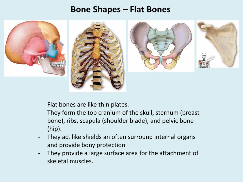
Bone Shapes – Flat Bones
- Flat bones are like thin plates.- They form the top cranium of the skull, sternum (breast
bone), ribs, scapula (shoulder blade), and pelvic bone (hip).
- They act like shields an often surround internal organs and provide bony protection
- They provide a large surface area for the attachment of skeletal muscles.

Bone Shapes – Sutural Bones
- Small and flat bones - Found between the flat bones of the skull.- They vary in size and number.
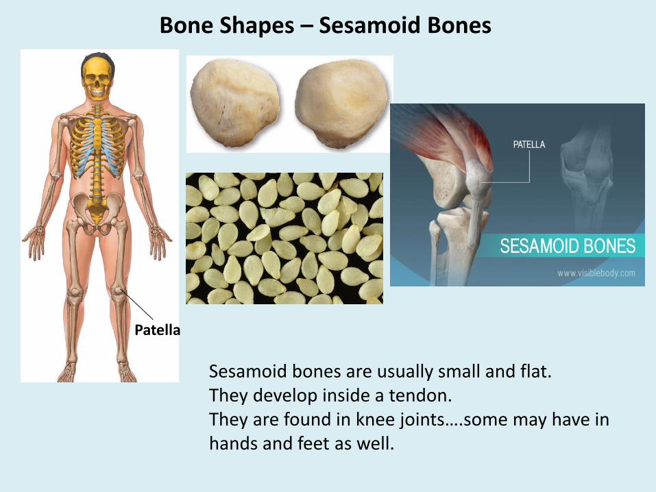
Bone Shapes – Sesamoid Bones
Sesamoid bones are usually small and flat.They develop inside a tendon.They are found in knee joints….some may have in hands and feet as well.
Patella

Bone Shapes – Irregular Bones
Irregular bones have complex shapes with projections.They form the mandible (lower jaw), vertebrae and sacral bone.They often have projections for bone/muscle/ligament attachments.
Mandible
Sacrum
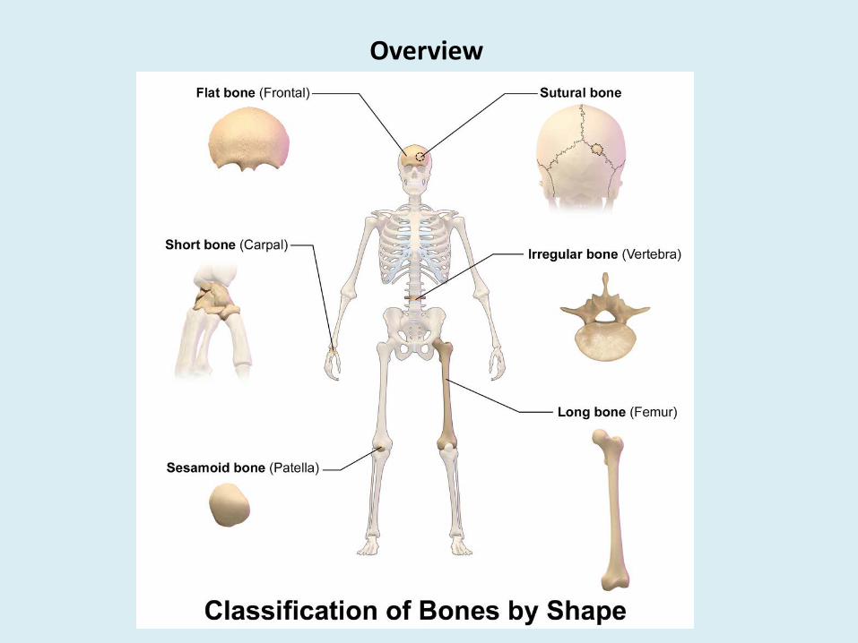
Overview

Bone Markings
Bones have specific markings/landmarks:To allow one bone to fit into another.To form surfaces for muscle tendons or bone ligaments to attach.To form passageways needed for blood vessels and nerves to pass.
A set of terminology used to define specific markings.
Memorize…….it will help to learn bone names and bone parts.

Bone Markings
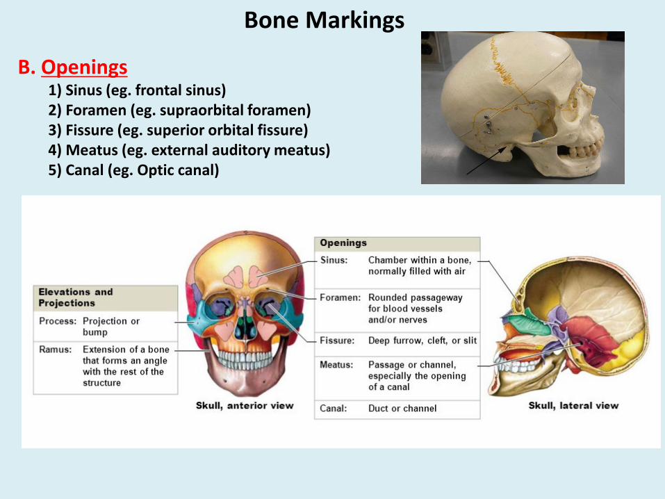
A. Elevations & Projections
1) Process- Projection or bump(e.g.- zygomatic bone-cheek bone)
2) Ramus- Extension of a bone thatforms an angle with the rest ofthe structure
(e.g.- vertical portion of mandible(lower jaw)
Bone Markings
B. Openings1) Sinus (eg. frontal sinus)2) Foramen (eg. supraorbital foramen)3) Fissure (eg. superior orbital fissure)4) Meatus (eg. external auditory meatus)5) Canal (eg. Optic canal)

Bone MarkingsC. Depressions
1) Sulcus-narrow groove (eg. sulcus intertubercular)2) Fossa- shallow depression (eg. coronoid fossa)
Anterior viewPosterior view
Humerus

D. Processes where tendons and ligaments attach (tubercle, tuberosity, trochanter, crest, spine)Tubercle-small rounded projectionTuberosity-Rough projectionTrochanter-Large rough projectionCrest-prominent ridgeSpine-Pointed process
Bone Markings
Humerus
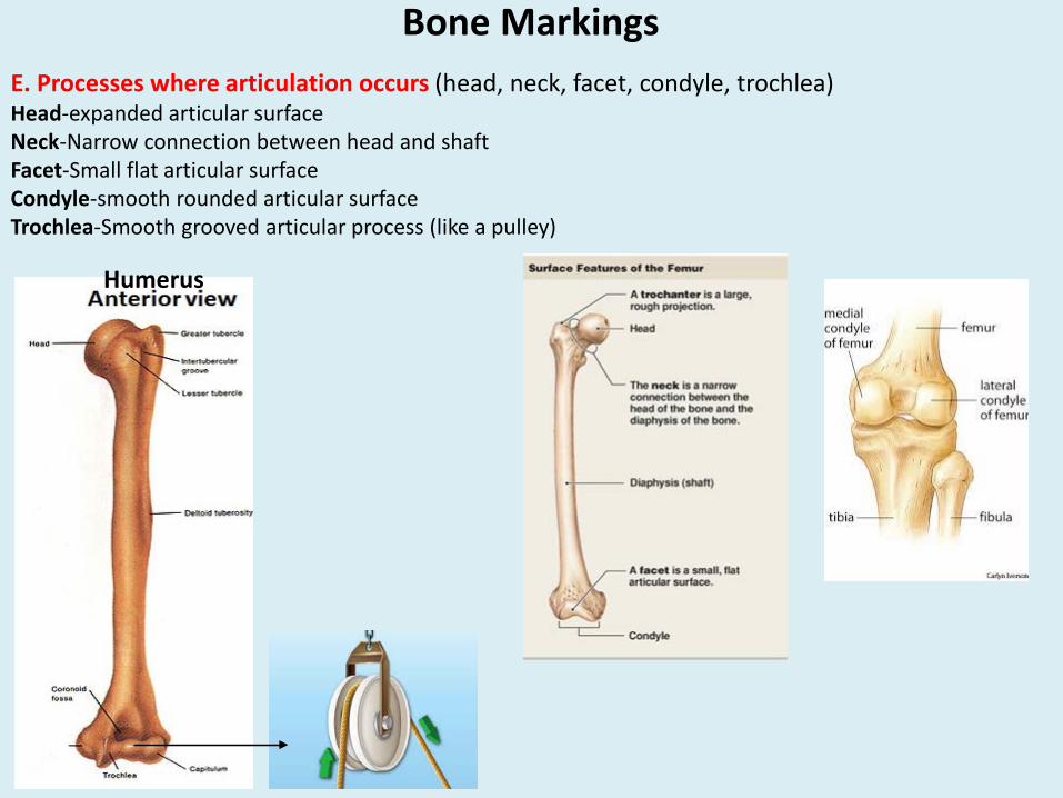
Bone MarkingsE. Processes where articulation occurs (head, neck, facet, condyle, trochlea)Head-expanded articular surfaceNeck-Narrow connection between head and shaftFacet-Small flat articular surfaceCondyle-smooth rounded articular surfaceTrochlea-Smooth grooved articular process (like a pulley)
Humerus

Structure of a Long Bone
1) Diaphysis: the shaft or the length between the 2 ends.- External wall made of solid/compact bone tissue.- Medullary cavity: central space in diaphysis- contains adipose tissue – yellow bone marrow energy storage.
2) Epiphysis: the wide area of bones at two ends.Made of mostly spongy bone tissue.Spaces in the sponge filled with red bone marrow site for hemopoiesis.
Articularcartilage

3) Metaphysis: area between diaphysis and epiphysis.Includes epiphyseal plate: In a growing child- epiphyseal plate is made of cartilage grows increase in length of the bone.
During puberty- epiphyseal plate becomes ossified bone stops growing epiphyseal lineappears indicates sealing of the bone.
Articular cartilage: cartilage caps over the 2 ends/ epiphyses protection in joints.
Articular cartilage
Structure of a Long BoneArticularcartilage

Structure of a Long Bone
Membrane coverings: dense connective tissue covers outer and inner surface of the bone.Contains blood vessels, nerves, bone cells.
Periosteum: membrane covering outer surface of the bone.
- Outer fibrous layer (collagen fibers)- cellular layer (osteoblast)
Functions: helps in attachment of tendons and ligaments.Has cells for bone growth and repair.Provide blood vessels and nerves to the bone tissue.
Endosteum: membrane lining the medullary cavity and trabeculae of spongy bone.
Function: has cells for bone growth and repair.
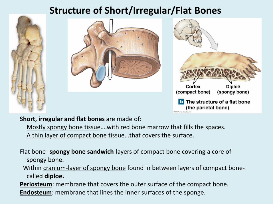
Structure of Short/Irregular/Flat Bones
Short, irregular and flat bones are made of:Mostly spongy bone tissue….with red bone marrow that fills the spaces.A thin layer of compact bone tissue…that covers the surface.
Flat bone- spongy bone sandwich-layers of compact bone covering a core of spongy bone.
Within cranium-layer of spongy bone found in between layers of compact bone-called diploe.
Periosteum: membrane that covers the outer surface of the compact bone.Endosteum: membrane that lines the inner surfaces of the sponge.
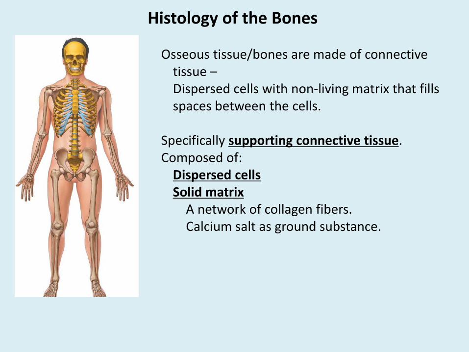
Histology of the Bones
Osseous tissue/bones are made of connective tissue –Dispersed cells with non-living matrix that fills spaces between the cells.
Specifically supporting connective tissue.Composed of:
Dispersed cellsSolid matrix
A network of collagen fibers.Calcium salt as ground substance.

Histology of the Bones – Bone Cells
There are 4 types of bone cells:Osteoprogenitor/Osteogenic cells: dividing stem cells found in periosteum and endosteum
form new cells called osteoblasts.
Osteoblasts: non-dividing bone forming cells that are also found in periosteum and endosteum.- Secrete and deposit collagen fibers and bone-forming proteins- organic matrix (osteoid)- inorganic calcium & phosphorous salts added to osteoid osteoblasts become trapped inside the matrix become osteocytes.
Osteocytes: mature, non-dividing bone cells maintain the protein & mineral content of matrix in bone tissue.
Osteoclasts: Break down and remove bone matrix (osteolysis).- Motile, multinucleated-Derived from WBCs and found in endosteum.- Helps in bone growth, repair and remodeling

Sources of Calcium and Phosphorous- From diet (fortified milk, egg yolk, fish oil)- UV exposure-steroid in skin vitamin D3 (cholecalciferol) calcitriol (kidney) important for Ca and P absorption in the intestine.
- Vitamin D deficiency low Ca weak bones.
Rickets in children:Vitamin D deficiency low calcitriol (hormone) Ca deficiency weak leg bones bowed legs, inflamed and painful joints.
Osteomalacia or adult ricket:Vitamin D deficiency low calcitriol (hormone) Ca deficiency weak bones.
Osteogenesis imperfecta or brittle bone disease:- Inborn genetic disorder- Deficiency of collagen fibers bones break easily frequent fractures.
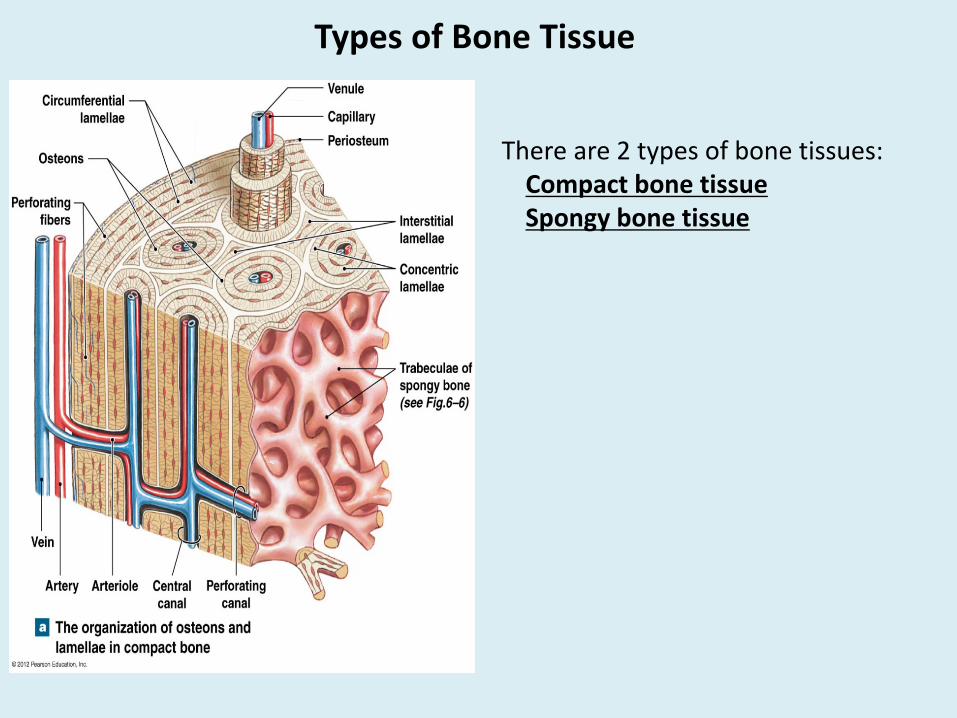
Types of Bone Tissue
There are 2 types of bone tissues:Compact bone tissueSpongy bone tissue

Types of Bone Tissue – Compact Bone Tissue
Compact bone tissue: makes up most of the diaphysis of long bones.Composed of compactly arranged microscopic tubes – osteons/Haversian systemsEach osteon/Haversian system is made of:
Central canal: that contains blood vessels and nerves.Lamellae: concentric circles of matrix – fibers and Ca salts.Osteocytes inside lacunae: dispersed between lamellae, maintain bone tissue.Canaliculi: tiny canals that connect osteocytes in lacuna with central canal.

Types of Bone Tissue – Spongy Bone Tissue
Spongy bone tissue: makes up most of the short bones, irregular bones, flat bones and epiphysis of long bones.
- Looks like a sponge- matrix of spongy bone is made of bundle fibers/thin plates called trabeculae (lamellae that are arranged as rods/plates)
- Trabeculae lacks central canal (no capillaries/veins in the matrix of spongy bone)
- Do not have typical osteon/Haversian system
- Red bone marrow found in trabeculae of spongy bone-provides nutrients to trabeculae.
- Trabeculae are surrounded by osteoblasts (for growth) and osteoclasts (for repair).

Bone Formation- Bone formation begins around 6th week of embryo development.
- Skeleton in an embryo begins as:cartilage molds
- Transition from cartilaginous template to bone is called ossification/osteogenesis.
- Clavicle is the first bone to ossify (seventh embryonic week).
- Bone formation is completed around age 40.
- Two types of ossification processes:1. Endochondral ossification- develops
from a hyaline cartilage mold.
2. Intramembranous ossification-Develops from fibrous connective tissue membrane (mesenchymal cells)
- No cartilage involved

Bone Formation – Endochondral Ossification
Endochondral ossification:Forms most of the long bones of the body.Begins as a small, avascular cartilage mold that is shaped like a bone becomes a
large, solid bone with all the structural details.
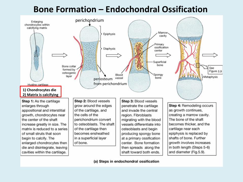
Bone Formation – Endochondral Ossification
1) Chondrocytes die2) Matrix is calcifying
perichondrium
periosteum from perichondrium
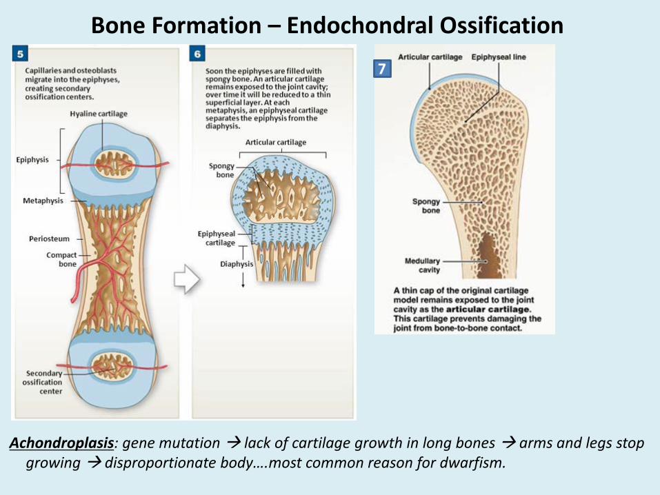
Bone Formation – Endochondral Ossification
Achondroplasis: gene mutation lack of cartilage growth in long bones arms and legs stop growing disproportionate body….most common reason for dwarfism.
7
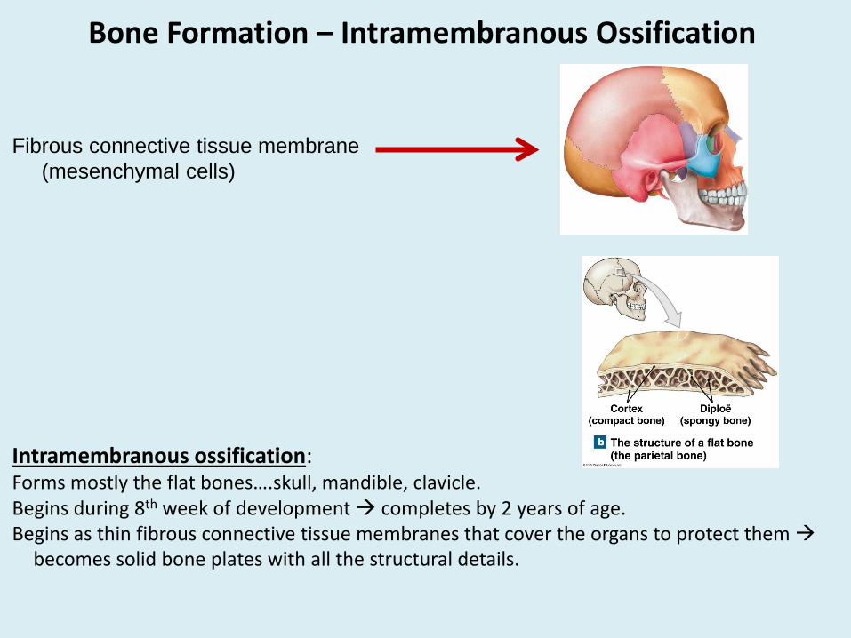
Bone Formation – Intramembranous Ossification
Intramembranous ossification:Forms mostly the flat bones….skull, mandible, clavicle.Begins during 8th week of development completes by 2 years of age.Begins as thin fibrous connective tissue membranes that cover the organs to protect them
becomes solid bone plates with all the structural details.
Fibrous connective tissue membrane(mesenchymal cells)
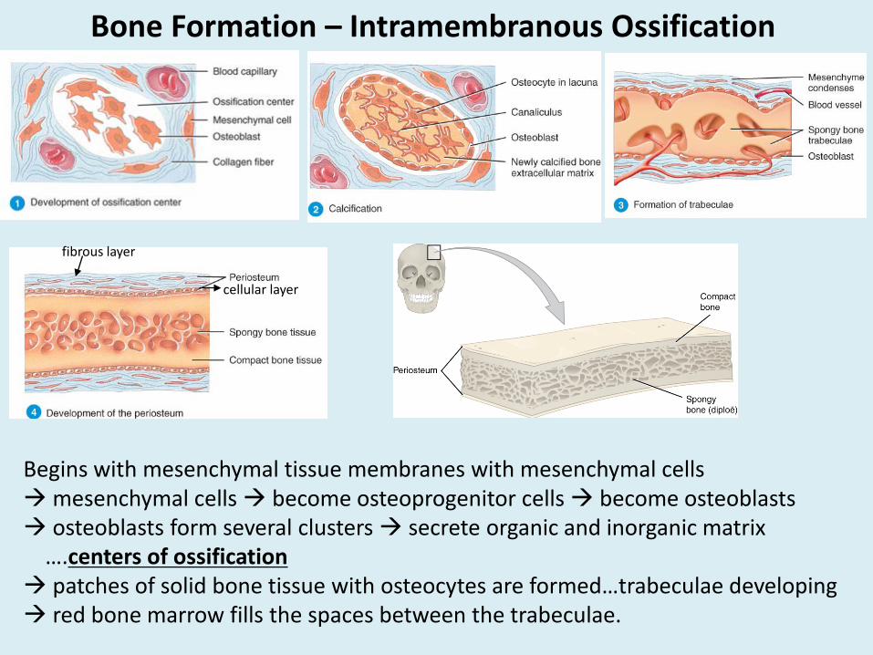
Bone Formation – Intramembranous Ossification
Begins with mesenchymal tissue membranes with mesenchymal cellsmesenchymal cells become osteoprogenitor cells become osteoblasts osteoblasts form several clusters secrete organic and inorganic matrix
….centers of ossification patches of solid bone tissue with osteocytes are formed…trabeculae developing red bone marrow fills the spaces between the trabeculae.
fibrous layer
cellular layer
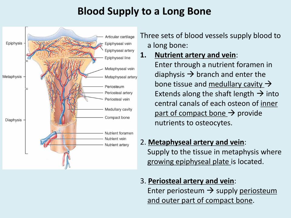
Blood Supply to a Long Bone
Three sets of blood vessels supply blood to a long bone:
1. Nutrient artery and vein: Enter through a nutrient foramen in diaphysis branch and enter the bone tissue and medullary cavity Extends along the shaft length into central canals of each osteon of inner part of compact bone provide nutrients to osteocytes.
2. Metaphyseal artery and vein: Supply to the tissue in metaphysis where growing epiphyseal plate is located.
3. Periosteal artery and vein: Enter periosteum supply periosteum and outer part of compact bone.

Bone Growth
As the body grows, bones have to grow with it.
Bones grow in:LengthThickness
Bone Growth – In Length
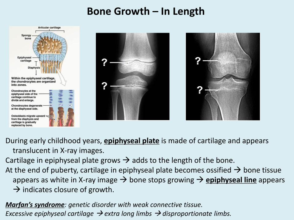
During early childhood years, epiphyseal plate is made of cartilage and appears translucent in X-ray images.
Cartilage in epiphyseal plate grows adds to the length of the bone.At the end of puberty, cartilage in epiphyseal plate becomes ossified bone tissue
appears as white in X-ray image bone stops growing epiphyseal line appears indicates closure of growth.
Marfan’s syndrome: genetic disorder with weak connective tissue.Excessive epiphyseal cartilage extra long limbs disproportionate limbs.

Bone Growth – In Thickness
As the bones become longer they also need to become thicker.For a long bone:
New tissue needs to be added on the surface of diaphysis.Tissue in the center needs to be broken down to increase the medullary cavity.
In periosteum, osteoprogenitor cells divide form new osteoblasts deposit new matrix and form new osteocytes new tissue formed on the outside.
In endosteum, osteoclasts breakdown matrix around medullary cavity larger medullary cavity.
Bone Growth & Remodeling
Growth: during fetal development until early pubertyRemodeling: during adulthood• Breakdown (Osteolysis) of older tissue by osteoclasts• Replacement with new tissue by osteoblasts• Maintenance by osteocyte (add & remove Ca from matrix and blood to maintain
several body functions).
Factors affecting growth & remodeling:
1) Minerals: Ca, P, Mg, B, Mn: via food or pills increase osteoblast activity.2) Vitamins:
• Vit. D3: essential for producing calcitriol (hormone) for Ca absorption.Deficiency of vitamin D (or Ca) causes rickets in children, osteomalacia in
adults.• C: making new collagen fibers (part of matrix).• B12: bone growth and remodeling.• A: increase osteoblast activity.

3. Hormones:• Human growth hormone (hGH): Secreted by pituitary gland.
Growth & development (fetal stage to early puberty)Declines in later part of puberty low levels in adults to maintain bones.Hypersecretion during early childhood years gigantism.Hyposecretion during early childhood years pituitary dwarfism.Hypersecretion during adulthood acromegaly (larger and thicker forehead,
larger jaws and hands).• Calcitriol:
Exposure to UV cholecalciferol/vitamin D3(skin) intermediary compound (liver) calcitrol (kidney) absorb calcium and phosphate by small intestine.
• Calcitonin: Secreted by thyroid gland.Facilitates transfer of Ca from blood to bone (lowers blood Ca and increases bone Ca).
• Parathyroid hormone (PTH): secreted by parathyroid glands.Facilitates transfer of Ca from bone to blood (increases blood Ca and lowers bone Ca).
• Sex hormones (estrogen and testosterone): secreted by ovaries and testes.Moderate increase during early puberty encourages growth.Further increase stimulates ossification of epiphyseal plate growth stops.
Factors affecting growth & remodeling:

Bone Fracture
Fracture: any break in a bone-
Stress fracture: break due to trauma…..fall, sports activity.Pathologic fracture: break due to a disease that may weaken the bones…cancer, osteoporosis, osteogenesis imperfecta.
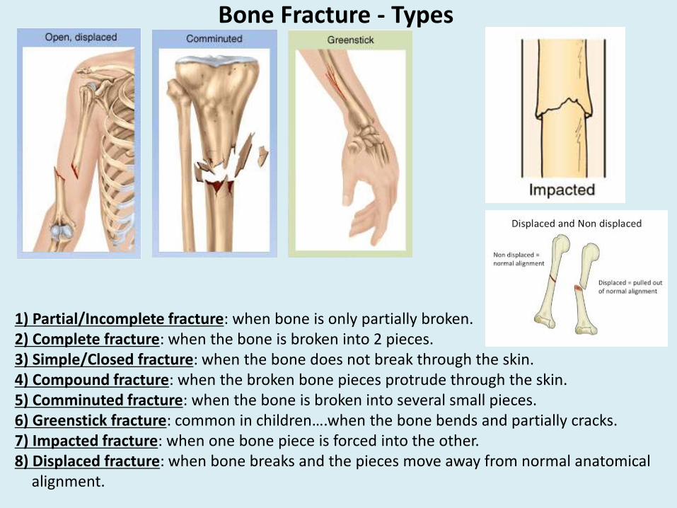
Bone Fracture - Types
1) Partial/Incomplete fracture: when bone is only partially broken.2) Complete fracture: when the bone is broken into 2 pieces.3) Simple/Closed fracture: when the bone does not break through the skin.4) Compound fracture: when the broken bone pieces protrude through the skin.5) Comminuted fracture: when the bone is broken into several small pieces.6) Greenstick fracture: common in children….when the bone bends and partially cracks.7) Impacted fracture: when one bone piece is forced into the other.8) Displaced fracture: when bone breaks and the pieces move away from normal anatomical
alignment.

Skeleton – Aging & Disorders
With age:Decrease in collagen fibers bones become brittle fracture easily.Decreased osteoblast activity slow healing.Vertebrae and intervertebral discs compress decreased flexibility.
Osteopenia: age related decreased activity of osteoblasts reduced ossification bone become thinner and weakerAffects epiphysis of long bones, jaws and vertebrae.Leads to osteoporosis.
Osteomyelitis: Inflammation of bones due to bacteria – Staphylococcus aureus severe pain in bones antibiotic treatment required.
Osteosarcoma: cancer of bone tissue.
Chondrosarcoma: cancer of cartilage.